Cell, Vol. 102, 683–694, September 1, 2000, Copyright 2000 by Cell Press A FRET-Based Sensor Reveals Large ATP Hydrolysis–Induced Conformational Changes and Three Distinct States of the Molecular Motor Myosin 1993; Schroder et al., 1993), provided strong support for a modification of this model that involves a relatively fixed catalytic domain bound to actin and swinging of the light chain binding region through a considerable angle, providing a working stroke of 5–15 nm (Cooke, William M. Shih,* Zygmunt Gryczynski, Joseph R. Lakowicz, and James A. Spudich* ‡ * Department of Biochemistry Stanford University School of Medicine Stanford, California 94305 Center for Fluorescence Spectroscopy 1986; Spudich, 1994). Thus, the light chain binding re- gion of S1 is thought to act like a lever arm to amplify Department of Biochemistry University of Maryland School of Medicine smaller conformational changes in S1 near the nucleo- tide binding site. Mg.ATP binding to S1 and its hydrolysis Baltimore, Maryland 21201 releases the myosin from the actin and regenerates the prestroke state of the myosin head for a subsequent working stroke. Actin rebinding stimulates phosphate Summary release, which triggers the power stroke. More recently, crystal structures have been solved for The molecular motor myosin is proposed to bind to actin and swing its light-chain binding region through various truncated isoforms of myosin-II head fragments complexed with nucleotides (Fisher et al., 1995; Smith a large angle to produce an z10 nm step in motion coupled to changes in the nucleotide state at the active and Rayment, 1996; Dominguez et al., 1998; Houdusse et al., 1999). Relative to the globular catalytic domain, site. To date, however, direct dynamic measurements have largely failed to show changes of that magnitude. the carboxy-terminal converter domain has been found in five angular positions whose extremes place the lever Here, we use a cysteine engineering approach to cre- ate a high resolution, FRET-based sensor that reports arm domain pointing in almost opposite directions. Fur- thermore, the light chain bound a-helical lever arm has a large, z70 degree nucleotide-dependent angle change of the light-chain binding region. The combination of been found with or without a bend immediately distal to the converter domain (Rayment et al., 1993; Houdusse steady-state and time-resolved fluorescence reso- nance energy transfer measurements unexpectedly et al., 1999). Two themes that emerge from consideration of these structures are as follows: (1) Crystal structures reveals two distinct prestroke states. The measure- ments also show that bound Mg.ADP.P i , and not bound that have been proposed as models of a prestroke state imply a step size of z10 nm in magnitude, and (2) the Mg.ATP, induces the myosin to adopt the prestroke states. lever arm may have the potential to stably adopt more than two different angles. It is critical to test the validity of these structural Introduction themes for myosin outside the context of a crystal lat- tice, both in muscle fibers and in purified protein sys- The myosin molecular motor uses the energy of Mg.ATP hydrolysis to power the vectorial transport of adjacent tems. Can the conformations seen by crystallography be detected under dynamic and more physiological con- actin filaments. A swinging crossbridge model of con- traction was proposed by H. E. Huxley (1969) based ditions? Can additional conformations not yet seen by crystallography be resolved by other methods? Cryoelec- on low-angle X-ray diffraction and electron microscope studies of muscle. A cycle of actin–myosin interactions tron microscopic studies on actin filaments decorated with certain isoforms of myosin S1 suggest a small rota- is thought to occur as follows (Lymn and Taylor, 1971; Spudich, 1994): the myosin crossbridge binds to ATP, tion of the lever arm domain upon release of ADP, thus implying two poststroke like conformations of myosin and then releases its attached actin filament. Next, the myosin crossbridge hydrolyzes the ATP and primes it- (Jontes et al., 1995; Whittaker et al., 1995). In a recent tomographic three-dimensional reconstruction study on self in preparation for a productive working stroke. Actin rebinding triggers phosphate release, which in turn quick-frozen contracting insect flight muscle, the au- prompts the myosin crossbridge to return to its starting thors interpreted tomographic images in terms of twenty conformation, in a motion termed the “powerstroke.” six rebuilt S1s, for which the angle between the lever arm The net result is that the attached actin filament gets domain and the catalytic domain varied continuously in translocated in the direction of its minus (pointed) end. a range of z508, where the rigor structure was placed The myosin crossbridge is the subfragment 1 (S1) do- near the middle of that range (Taylor et al., 1999). Thus, main, which consists of the actin binding and nucleotide none of the rebuilt S1s exhibited a lever arm to catalytic binding globular catalytic domain and the z10 nm long domain angle similar to the crystallographic prestroke light chain binding region (Figure 1). Crystal structures state models (z708–808 away from the rigor angle). of monomeric actin (Kabsch et al., 1990) and myosin S1 An important approach to measuring conformational (Rayment et al., 1993), combined with electron micro- changes in proteins is the use of reporter probes graphic reconstructions of actin filaments with and with- attached to the purified protein that allow readout of out S1 decoration (Holmes et al., 1990; Rayment et al., dynamic changes in structure in response to changing conditions of environment. Dynamic measurements of changes in the myosin head structure, for example, have ‡ To whom correspondence should be addressed (e-mail: jspudich- @cmgm.stanford.edu). involved the use of various probes that report changes

Welcome message from author
This document is posted to help you gain knowledge. Please leave a comment to let me know what you think about it! Share it to your friends and learn new things together.
Transcript

Cell, Vol. 102, 683–694, September 1, 2000, Copyright 2000 by Cell Press
A FRET-Based Sensor Reveals LargeATP Hydrolysis–Induced Conformational Changes andThree Distinct States of the Molecular Motor Myosin
1993; Schroder et al., 1993), provided strong supportfor a modification of this model that involves a relativelyfixed catalytic domain bound to actin and swinging ofthe light chain binding region through a considerableangle, providing a working stroke of 5–15 nm (Cooke,
William M. Shih,* Zygmunt Gryczynski,†Joseph R. Lakowicz,† and James A. Spudich*‡
*Department of BiochemistryStanford University School of MedicineStanford, California 94305†Center for Fluorescence Spectroscopy 1986; Spudich, 1994). Thus, the light chain binding re-
gion of S1 is thought to act like a lever arm to amplifyDepartment of BiochemistryUniversity of Maryland School of Medicine smaller conformational changes in S1 near the nucleo-
tide binding site. Mg.ATP binding to S1 and its hydrolysisBaltimore, Maryland 21201releases the myosin from the actin and regenerates theprestroke state of the myosin head for a subsequentworking stroke. Actin rebinding stimulates phosphateSummaryrelease, which triggers the power stroke.
More recently, crystal structures have been solved forThe molecular motor myosin is proposed to bind toactin and swing its light-chain binding region through various truncated isoforms of myosin-II head fragments
complexed with nucleotides (Fisher et al., 1995; Smitha large angle to produce an z10 nm step in motioncoupled to changes in the nucleotide state at the active and Rayment, 1996; Dominguez et al., 1998; Houdusse
et al., 1999). Relative to the globular catalytic domain,site. To date, however, direct dynamic measurementshave largely failed to show changes of that magnitude. the carboxy-terminal converter domain has been found
in five angular positions whose extremes place the leverHere, we use a cysteine engineering approach to cre-ate a high resolution, FRET-based sensor that reports arm domain pointing in almost opposite directions. Fur-
thermore, the light chain bound a-helical lever arm hasa large, z70 degree nucleotide-dependent angle changeof the light-chain binding region. The combination of been found with or without a bend immediately distal
to the converter domain (Rayment et al., 1993; Houdussesteady-state and time-resolved fluorescence reso-nance energy transfer measurements unexpectedly et al., 1999). Two themes that emerge from consideration
of these structures are as follows: (1) Crystal structuresreveals two distinct prestroke states. The measure-ments also show that bound Mg.ADP.Pi, and not bound that have been proposed as models of a prestroke state
imply a step size of z10 nm in magnitude, and (2) theMg.ATP, induces the myosin to adopt the prestrokestates. lever arm may have the potential to stably adopt more
than two different angles.It is critical to test the validity of these structuralIntroduction
themes for myosin outside the context of a crystal lat-tice, both in muscle fibers and in purified protein sys-The myosin molecular motor uses the energy of Mg.ATP
hydrolysis to power the vectorial transport of adjacent tems. Can the conformations seen by crystallographybe detected under dynamic and more physiological con-actin filaments. A swinging crossbridge model of con-
traction was proposed by H. E. Huxley (1969) based ditions? Can additional conformations not yet seen bycrystallography be resolved by other methods? Cryoelec-on low-angle X-ray diffraction and electron microscope
studies of muscle. A cycle of actin–myosin interactions tron microscopic studies on actin filaments decoratedwith certain isoforms of myosin S1 suggest a small rota-is thought to occur as follows (Lymn and Taylor, 1971;
Spudich, 1994): the myosin crossbridge binds to ATP, tion of the lever arm domain upon release of ADP, thusimplying two poststroke like conformations of myosinand then releases its attached actin filament. Next, the
myosin crossbridge hydrolyzes the ATP and primes it- (Jontes et al., 1995; Whittaker et al., 1995). In a recenttomographic three-dimensional reconstruction study onself in preparation for a productive working stroke. Actin
rebinding triggers phosphate release, which in turn quick-frozen contracting insect flight muscle, the au-prompts the myosin crossbridge to return to its starting thors interpreted tomographic images in terms of twentyconformation, in a motion termed the “powerstroke.” six rebuilt S1s, for which the angle between the lever armThe net result is that the attached actin filament gets domain and the catalytic domain varied continuously intranslocated in the direction of its minus (pointed) end. a range of z508, where the rigor structure was placedThe myosin crossbridge is the subfragment 1 (S1) do- near the middle of that range (Taylor et al., 1999). Thus,main, which consists of the actin binding and nucleotide none of the rebuilt S1s exhibited a lever arm to catalyticbinding globular catalytic domain and the z10 nm long domain angle similar to the crystallographic prestrokelight chain binding region (Figure 1). Crystal structures state models (z708–808 away from the rigor angle).of monomeric actin (Kabsch et al., 1990) and myosin S1 An important approach to measuring conformational(Rayment et al., 1993), combined with electron micro- changes in proteins is the use of reporter probesgraphic reconstructions of actin filaments with and with- attached to the purified protein that allow readout ofout S1 decoration (Holmes et al., 1990; Rayment et al., dynamic changes in structure in response to changing
conditions of environment. Dynamic measurements ofchanges in the myosin head structure, for example, have‡ To whom correspondence should be addressed (e-mail: jspudich-
@cmgm.stanford.edu). involved the use of various probes that report changes

Cell684
changes of the true lever arm with respect to the cata-lytic domain of the S1.
In addition to the question of the extent of a leverarm angle change, there has been considerable debateabout whether myosin S1 discriminates between Mg.ATPand Mg.ADP.Pi, in terms of lever arm conformations(Lymn and Taylor, 1971; Eisenberg and Greene, 1980;Fisher et al., 1995; Holmes, 1997; Dominguez et al., 1998;Huxley, 1998; Suzuki et al., 1998; Brust-Mascher et al.,1999; Houdusse et al., 1999). Crystal structures of myo-sin fragments complexed with the presumed Mg.ATPanalog Mg.ADP.BeFx have been found in both post-stroke and prestroke angles, depending on the context(Fisher et al., 1995; Houdusse et al., 1999). Mutants ofthe Dictyostelium myosin catalytic domain have beenfound that bind weakly to actin when complexed toMg.ATP, but which exhibit either prestroke- or post-stroke-like angles of the converter domain (Sasaki etal., 1998; Suzuki et al., 1998). Thomas and colleagueshave argued based on EPR data that S1 does not dis-
Figure 1. View of Doubly Labeled Myosin-II S1 with Three Proposed criminate between ATP and ADP.Pi (Brust-Mascher etLever Arm Anglesal., 1999). Answering this question and determining the
The heavy chain (HC) is colored white, the essential light chain (ELC)extent of lever arm angle change requires the develop-is colored light violet, and the regulatory light chain (RLC) is coloredment of a reliable assay for the lever arm angle in theblue. Tetramethylrhodamine-5-maleimide (the acceptor dye) attachedMg.ATP state and in the Mg.ADP.Pi state. Dynamic ap-to HC-Cys250 is colored red, and Oregon green 488 maleimide (the
donor dye) is colored green or yellow according to its attachment proaches to date have usually involved putting probesto the RLC at either RLC-Cys114 or RLC-Cys116 (residues 113 and on the myosin head where possible, given the structure115 in the chicken skeletal sequence), respectively. The distance and sequence of the native protein. Ideally, a myosinbetween the donor and acceptor dyes decreases dramatically in
motor domain needs to be created that allows one togoing from the poststroke to the prestroke states, leading to a largeput probes at any site one chooses, to optimally assessincrease in FRET efficiency.the conformational change that one wishes to measure.
To achieve this goal, we have used cysteine engi-neering to remove native cysteines present in Dictyostel-in S1 structure in different nucleotide states. Such mea-ium myosin-II, and have shown that this cysteine-lightsurements have provided support for structural changes,myosin-II is functional both in vitro and in vivo. We thenbut have often been hampered by restrictions as toinserted cysteine residues in specific locations of Dicty-where the probe can be placed on the molecule. Thus,ostelium myosin-II S1 (Manstein et al., 1989). Specifica wide variety of angle changes of the lever arm havelabeling of those introduced cysteines with a donor andbeen estimated by these dynamic approaches (for re-acceptor dye provided a FRET-based sensor capableview, see Goldman, 1998). Studies observing polarizedof resolving a wide range of lever arm angles. This ap-fluorescence of rabbit muscle labeled on the regulatoryproach—removal of native cysteines and introductionlight chain (Irving et al., 1995; Corrie et al., 1999) suggestof new cysteines at rationally selected positions on twoat least a modest change in lever arm angle upon bind-separate polypeptides—helps circumvent problems ofing and hydrolysis of ATP, while EPR studies on spin-labeling specificity, perturbation of activity, and/or lim-labeled myosin heads suggest dynamic disorder in theited sensitivity to conformational changes that are oftenangle between the catalytic domain and lever arm inencountered with FRET studies. To accurately resolverelaxed and active muscle (Roopnarine et al., 1998).the FRET distances, we measured a combination ofResonance energy transfer measurements between flu-steady-state and time-resolved FRET efficiencies. Theorescent or luminescent probes on the catalytic domainmeasurements reported here unexpectedly indicate twoand the regulatory light chain have also indicated nucle-prestroke conformations, one of which has not beenotide-induced rotation of the lever arm (Burmeister-Getzseen yet crystallographically. In addition, we observeet al., 1998; Xiao et al., 1998; Palm et al., 1999). A steady-that bound ADP.Pi biases S1 toward prestroke statesstate FRET study on a Dictyostelium catalytic domainmuch more than does bound ATP.in the absence of the light chain binding domain involved
engineering green fluorescent protein onto the aminoterminus and blue fluorescent protein onto the carboxy Results and Discussionterminus. This study was consistent with a large anglechange in the converter domain, from which the lever Generation of a Dictyostelium Cysteine-Light
Myosin-II: Design of the Substitutionsarm domain extends (Suzuki et al., 1998). Crystallo-graphic studies (Houdusse et al., 1999) have shown, The paramount concern for mutagenizing myosin to re-
move cysteines was to retain function of the enzyme.however, that the absence of the essential light chaincan lead to a different conformation of the converter We used the information that the cysteine residue dem-
onstrates a tolerance for substitution with small anddomain, and the location of the carboxy terminus indifferent nucleotide states may not reflect the relative medium sized hydrophobic residues such as alanine,

FRET-Based Sensor of Myosin Conformational States685
myosin-II gene were assayed for growth in suspension.The cysteine-light myosin completely rescued the cyto-kinesis defect of the myosin-II null host strain (Figure2). This is a very stringent test of myosin-II function,and we conclude that none of the cysteines that weremutagenized are essential. A cysteine-light myosin-IIharboring the mutation A250C (to facilitate acceptor la-beling for FRET) also completely rescued the cytokinesisdefect of the myosin-II null strain (data not shown).
The S1 portion of the cysteine-light myosin-II genewas subcloned into another plasmid to facilitate theexpression of an S1 that was tagged at the carboxyterminus of the heavy chain with a 63His tag. Cysteine-light S1 was expressed, purified, and assayed for basaland actin-activated ATPase activity in vitro. The actin-activated ATPase activity of the cysteine-light S1 wasvery similar to that of wild-type S1 (1.0 s21), and washigher than the respective basal rate (the rate in the ab-sence of actin). Therefore, the cysteine-light S1 retainsFigure 2. Dictyostelium Growth in Suspensionactin-activation of its ATPase. The basal rate of the
Growth is plotted as the log of the number of cells per ml versuscysteine-light S1 ATPase was about 2-fold higher thantime. The growth of cells lacking the myosin-II gene is rendered withthat of the wild-type enzyme (0.40 s21 versus 0.17 s21),open circles and a solid line, the growth of cells with a wild-typesuggesting that the rate-limiting phosphate release stepmyosin-II gene is rendered with tetrasected open squares and a
dashed line, and the growth of cells with a cysteine-light myosin-II is not as well gated as for the wild-type S1.gene is rendered with a dotted line and open triangles. In summary, the cysteine-light myosin-II is a functional
motor both in vivo and in vitro. It should serve as auseful starting point for future structure-function studies
valine, isovaline, leucine, and isoleucine, as well as with involving site specific chemical modification, such asserine, threonine, and tyrosine (Dayhoff, 1978). Sequence disulfide cross-linking and spectroscopic probe label-alignments of the myosin-IIs provided more information ing, of the protein.about which substitutions might be tolerated (Sellersand Goodson, 1995). Furthermore, the trends in the se- FRET Strategyquence alignment were compared with the environment Starting from the cysteine-light S1 gene describedof these residues in the crystal structures that have been above, we generated S1 heavy chains and regulatorysolved. For cysteine 312, for example, the sequence light chains with single reactive cysteines positionedalignment shows a strong predisposition toward tyro- such that, according to models based on crystallo-sine and phenylalanine. The structure shows that one graphic structures, the intercysteine distance would de-face of cysteine 312 is surface exposed. For cysteine crease from z75 A to z25 A during the poststroke to470, the sequence alignment shows that cysteine is fairly prestroke transition (Rayment et al., 1993; Dominguezwell conserved, with some substitutions of tryptophan, et al., 1998) (Figure 1). Recombinant E. coli expressedisoleucine, valine, methionine, and lysine. The crystal Dictyostelium regulatory light chain (RLC) was donorstructure shows that cysteine 470 appears well buried, labeled with Oregon green 488 maleimide at either resi-
due 114 or 116, and then exchanged onto a recombinantconsistent with the sequence alignment. For cysteineDictyostelium expressed S1 whose heavy chain had655, the sequence alignment shows cysteine used al-been previously acceptor labeled at residue 250 withmost exclusively. Cysteine 655 is the most conservedtetramethylrhodamine-5-maleimide. Averaging the mea-of the cysteines in Dictyostelium myosin-II. The structuresured FRET distances obtained from S1 labeled withshows that cysteine 655 is completely buried, and wasthe donor dye at two different residues decreases thethe only cysteine residue we did not change. In sum-uncertainty due to potential constraints on the relativemary, the cysteine-light myosin-II sequence was de-orientation of a given donor–acceptor dye pair (Stryer,signed with the following substitutions: C49S, C312Y,1978). The doubly labeled S1s were designated asC442S, C470I, C599L, C678Y.D114A250 and D116A250, respectively. S1s lacking either thedonor or the acceptor dye (designated as D114, D116, and
Cysteine-Light Myosin-II is Functional In Vivo A250) were generated as controls. A common concern inand In Vitro applying FRET to study protein structure involves theDictyostelium cells that are missing the myosin-II gene challenge of specifically labeling one site with a donorare unable to divide in suspension, and instead become dye, and the other site with an acceptor dye, withoutlarge and multinucleate before eventually lysing (De Lo- cross contamination of the two labeling sites. The abilityzanne and Spudich, 1987; Knecht and Loomis, 1987). to label the heavy chain and RLC separately, and thenThus, transformation of a mutant myosin-II gene into form a complex after labeling has been completed,these myosin-II null cells provides a robust assay for allows this problem to be solved. A specific concern inin vivo function—assaying for rescue of the growth in applying FRET to discriminate between prestroke andsuspension defect. Myosin-II null cells transformed with poststroke structures (as seen in crystallographic mod-
els; see prestroke state A versus poststroke state ina plasmid encoding either a wild-type or a cysteine-light
Cell686
FRET dye is placed on the so-called SH1 native cysteine(C707 in the chicken sequence) or in the nucleotidebinding site, then only a small change in FRET efficiencyis measured upon addition of nucleotides and nucleo-tide analogs, since both locations lie near the axis ofpseudosymmetry (Xiao and et al., 1998; Palm et al.,1999). We placed the acceptor dye as far as possiblefrom this axis, at an introduced cysteine on position250, in order to maximize the sensitivity of the FRETefficiency to a rotation in the lever arm angle.
Bound Mg.ADP.Pi Biases Myosin toward PrestrokeStates Much More Than Does Bound Mg.ATPIn the steady-state, Dictyostelium S1 in the presence ofsaturating amounts of Mg.ATP exists primarily as a mix-ture of S1.Mg.ATP and S1.Mg.ADP.Pi nucleotide states(Ritchie et al., 1993). If S1.Mg.ATP occupied the samedistribution of lever arm angles as S1.Mg.ADP.Pi, thenthe amount of steady-state FRET observed for D114A250
and D116A250 should be the same in the presence of Mg.ATPas in the presence of Mg.ADP.Vi, a Mg.ADP.Pi analog(Werber et al., 1992). The measured steady-state FRETefficiency for D114A250 increased from 16% in the absenceof nucleotide to 36% upon addition of Mg.ATP, andfurther increased to 61% upon addition of Mg.ADP.Vi
(Figure 3A and Table 1). For D116A250, the measured steady-state FRET efficiency increased from 11% in the absenceof nucleotide to 32% upon addition of Mg.ATP, andfurther increased to 57% upon addition of Mg.ADP.Vi
(Figure 3B and Table 1). Thus, Mg.ADP.Vi induces amuch greater amount of steady-state FRET than doesMg.ATP. A simple model to account for this data isthat S1.Mg.ATP exists primarily with a poststroke anglelever arm conformation (low FRET efficiency), whileS1.Mg.ADP.Pi exists primarily with a prestroke anglelever arm conformation (high FRET efficiency). The sim-plest interpretation is that in the presence of Mg.ATP,D114A250 and D116A250 exist as a roughly equimolar mixtureof S1.Mg.ATP and S1.Mg.ADP.Pi nucleotide states, andtherefore exhibit only half the FRET efficiency as in thepresence of ADP.Vi.Figure 3. Steady-State FRET Emission Spectra
Spectra were taken with no nucleotides, then after the addition of In the Steady-State, D114A250 Exists Primarily asMg.ATP, and finally after the addition of Mg.ADP.Vi. The addition of
a Mixture of S1.Mg.ATP and S1.Mg.ADP.PiMg.ATP induces only about half as much FRET as the additionStates, Just Like Wild-type S1of Mg.ADP.Vi.The above interpretation is only valid if D114A250 in the(A) Emission spectra of D114A250.
(B) Emission spectra of D116A250. presence of saturating Mg.ATP exists primarily as a mix-ture of S1.Mg.ATP and S1.Mg.ADP.Pi states, as in thecase of the wild-type S1, with the Pi-free S1.Mg.ADP
Figure 1) is that these two conformations, when superim- state representing an insignificant part of the total popu-posed using their catalytic domains, are pseudosym- lation. If the rate of ADP release for D114A250 were slowedmetric about an axis that bisects the angle made by the by .100-fold compared to wild-type S1, to the samelever arm in the two conformations. Placement of an level as the rate of Pi release, or if the Pi release rateacceptor dye near this axis of pseudosymmetry would were enhanced by .100-fold, then S1.Mg.ADP couldlead to only a very small change in the FRET efficiency account for nearly half of the S1 molecules in the pres-in transiting from the poststroke to the prestroke struc- ence of saturating Mg.ATP (Woodward et al., 1991; Rit-
chie et al., 1993; Woodward et al., 1995). Then the lowerture. In fact, this observation explains why when one
Table 1. Steady State FRET Efficiencies
Nucleotide added D114A250 steady-state FRET efficiency D116A250 steady state FRET efficiency
No nucleotide 16% 6 1% 11% 6 2%Mg.ATP 36% 6 3% 32% 6 3%Mg.ADP.Vi 61% 6 1% 57% 6 2%

FRET-Based Sensor of Myosin Conformational States687
the FRET distributions present in the sample. Steady-statefluorescence reports only on the mean FRET efficiencyof the dye-pairs in solution, and gives no informationregarding the distribution about that mean. We thereforemeasured time-resolved fluorescence in the frequencydomain to recover donor lifetime distributions, from whichFRET and distance distributions could be calculated(Table 2). FRET distributions are most easily extractedin the case where the donor alone control behaves asa single exponential homogeneous species. Fortunately,this was the case for both the D114 and D116 mutants(Figures 5A and 5C; Table 2). Addition of nucleotideshad insignificant effects on the fitted lifetimes for thedonor alone controls (Figures 5A and 5C). D114 phaseand modulation responses (taken in the absence of nu-cleotides, then after the addition of Mg.ATP, and finallyafter the addition of Mg.ADP.Vi) fit well in a global analy-sis to a single lifetime of 4.09 ns (x2 5 6.0), while globalanalysis of D116 responses under the same conditionsfit well to a single lifetime of 4.28 ns (x2 5 8.3). AnalysisFigure 4. MantADP Release Monitored by Stopped Flow Fluores-of D114A250 phase and modulation responses, taken undercencethe same three nucleotide conditions, yielded a goodS1 (0.1 mM) was preincubated with 2.5 mM mantADP, and the com-three lifetime fit in global analysis (t1 5 3.95 ns, t2 5plex was mixed in the stopped-flow device with 0.4 mM ATP. Wild-
type Dictyostelium S1 releases mantATP at 0.9 6 0.1 s21, while 1.00 ns, and t350.020 ns) (x2 5 3.6) (Tables 2 and 3;D114A250 releases mantADP at 1.3 6 0.1 s21. Figure 5B). The x2 for this fit remained roughly constant
for t3 between 0–0.20 ns. The implied steady-state FRETagreed with the measured steady-state FRET when the
FRET efficiency observed in the presence of Mg.ATP fit was performed with t3 fixed at 0.020 ns. Similar resultswould be reflecting the poststroke configuration of the were found for the global analysis of the D116A250 re-Pi-free S1.Mg.ADP population, and not of the poststroke sponses under the same conditions (t1 5 3.93 ns, t2 5configuration of the S1.Mg.ATP population. Convention- 0.93 ns, and t350.010 ns) (x2 5 3.9) (Tables 2 and 4;ally, measurements of ADP release rate from myosins have Figure 5D). Again, the x2 for this fit remained roughlyinvolved the use of the fluorescent analog mantADP constant for t3 between 0–0.20 ns, and the implied(Woodward et al., 1991; Ritchie et al., 1993; Woodward steady-state FRET agreed with the measured steady-et al., 1995). We found that the rate of release of state FRET when the fit was performed with t3 fixed atmantADP from D114A250 (1.3 6 0.1 s21) is not slower than 0.010 ns.that of wild-type S1 (0.9 6 0.1 s21), but is instead about A two component fit (t1 5 3.90 ns and t2 5 0.76 ns) was40% faster (Figure 4). With the assumption that the attempted for D114A250 and was found to be inappropriate,relative rates of mantADP release for the D114A250 and since the implied steady-state donor quenching waswild-type S1 reflect the relative rates of ADP release, much lower than the measured steady-state donorwe conclude that the ADP release rate is not significantly quenching, especially for the Mg.ADP.Vi data (37% im-changed. If the Pi release rate were enhanced by plied vs. 61% measured.) Furthermore, the x2 for the.100-fold, one would expect this to be reflected in a two lifetime fit was 80% higher than for the three lifetimelarge increase in the steady state basal ATPase activity fit. A two component fit was attempted for D116A250 asof the D114A250 compared to wild-type S1. We found these well, but was also found to be inappropriate, as therates to be within a factor of two (0.27 6 0.01 s21 for implied steady-state FRET was in poor agreement withD114A250 versus 0.17 6 0.01 s21 for wild-type S1). the measured steady-state FRET, especially for the
Mg.ADP.Vi data set (31% implied versus 57% mea-S1 Populates Three Lever Arm Angles, Whose sured), similar to the case with D114A250.
Distribution Is Influenced by Nucleotides The observed three lifetime components for D114A250
If S1 exists as a heterogeneous mixture of conforma- correspond to FRET efficiencies of 3.4%, 76%, andtional states, then meaningful assignment of FRET effi- 99.5%. These FRET efficiencies indicate apparent FRET
distances of 93 A, 44 A, and 22 A, respectively. Theciencies to distances can only be made after resolving
Table 2. Donor Lifetimes, FRET Efficiencies, and Distance Distributions
S1 Fitted donor lifetime FRET efficiency Distance
D114 4.09 6 0.01 nsD114A250 3.95 6 0.02 ns 3.4 6 0.8% 93 6 4 A
1.00 6 0.02 ns 76 6 1% 43.7 6 0.4 A0.020 6 0.002 ns 99.5 6 0.1% 21.9 6 0.8 A
D116 4.28 6 0.02 nsD116A250 3.93 6 0.02 ns 8.2 6 0.9% 79 6 2 A
0.93 6 0.04 ns 78 6 1% 42.9 6 0.4 A0.010 6 0.003 ns 99.8 6 0.1% 18.8 6 1.7 A

Cell688
Figure 5. Frequency Domain Time-Resolved Fluorescence Spectra
Phase and modulation responses with no nucleotides (circles), in the presence of Mg.ATP (squares), and in the presence of Mg.ADP.Vi
(diamonds) measured over a frequency range of 4 to 700 MHz.(A) Phase and modulation responses of D114.(B) Phase and modulation responses of D114A250.(C) Phase and modulation responses of D116.(D) Phase and modulation responses of D116A250.

FRET-Based Sensor of Myosin Conformational States689
Table 3. D114A250 Donor Lifetime Mole Fractions
D114A250 donor lifetime No nucleotide mole fraction Mg.ATP mole fraction Mg.ADP.Vi mole fraction
3.95 ns 0.67 0.41 0.101.00 ns 0.10 0.17 0.230.020 ns 0 0.19 0.44
0.23 mole fraction fixed at 4.09 ns, to account for 77% acceptor labeling efficiency.
analysis of the D116A250 data yielded FRET efficiencies of state directly to the prestroke angle, weak actin affinitystate. ATP hydrolysis would not be associated with any8%, 78%, and 99.8%, corresponding to distances of
79 A, 43 A, and 19 A, respectively. Shorter distances conformational changes, but rather would serve to pre-pare the g-phosphate for subsequent release.imply more prestroke-like angle of the lever arm. The
remarkable agreement between the FRET distances re- More recently, Holmes suggested that the availablecrystal structures were consistent with the simpler Eisen-ported from D114A250 and D116A250 suggests that there are
no significant errors due to unique donor-acceptor inter- berg and Greene type model (Holmes, 1997). Further-more, Smith and Rayment proposed that the poststrokedye orientational restrictions.angle crystal structure should be catalytically inactive,due to lack of positioning of the putative nucleophilicUpdated Model: Bound Mg.ADP.Pi Biases Myosinwater in that structure (Rayment, 1996; Smith and Ray-Toward Two Distinct Prestroke States Muchment, 1996). Therefore, the S1 should be able to existMore Than Does Bound Mg.ATPin the prestroke angle conformation at least transientlyThe simple model presented in the previous sectionif not primarily, in order to hydrolyze the ATP in a timelyproposes that addition of Mg.ADP.Vi induces a largemanner. Crystallization of myosin fragments in a prestrokefraction of S1 heads to adopt a prestroke angle, andangle conformation complexed with Mg.ADP.BeFx, aaddition of Mg.ATP induces only half as many heads tosupposed Mg.ATP analog, led further credence to theadopt a prestroke angle. The time-resolved data re-simple two-state model (Dominguez et al., 1998).quires this simple model to be revised, however, to in-
Suzuki and colleagues studied the conformation of theclude two prestroke angle states, as opposed to justDictyostelium myosin catalytic domain, a head fragmentone (Tables 3 and 4). Taken together, the steady-statemissing the light chain binding domain, using FRET be-and time-resolved data support the model shown intween GFP on the amino terminus and BFP on the car-Figure 6, where Mg.ATP hydrolysis is more responsibleboxy terminus (Suzuki et al., 1998). They examined thisfor population of prestroke configurations than isprotein in the presence of ATP, using two different mu-Mg.ATP binding. Thus, S1 can discriminate betweentants that arrest myosin in a weak actin affinity, ATPMg.ATP and Mg.ADP.Pi.bound state (Sasaki et al., 1998; Suzuki et al., 1998).This result is novel in showing that a functional myosinInterestingly, the G457A mutant exhibited FRET effi-motor in the S1.Mg.ATP nucleotide state exists primarilyciency consistent with a poststroke angle, while thewith a poststroke state angle of the lever arm. An advan-E459A mutant exhibited FRET efficiency consistent withtage of this study is that it did not require inferencea prestroke angle. Assuming that the intact motor con-from experiments with presumed ATP analogs (such astaining the light chain binding domain would behave inADP.BeFx) or ATPase cycle arresting mutants, but rathera similar manner, this result suggests that myosin in thereported on the conformation of working motors turningS1.Mg.ATP state can access weak actin affinity statesover ATP in solution. The question of the relative roleswith both poststroke angle and prestroke angle confor-of ATP and ADP.Pi in priming the motor has been de-mations. Thus, the two-state model is at best an over-bated for three decades. Lymn and Taylor (1971) pro-simplification, since S1 can reach a poststroke angle,posed that ATP binding drives S1 from a poststrokeweak actin affinity state. However, these mutant studiesangle, strong actin affinity state to a poststroke angle,did not reveal whether the conformational equilibriumweak actin affinity state, and that ATP hydrolysis driveslies toward the poststroke state angle, as in the Lymnthe motor to a prestroke angle, weak actin affinity state.and Taylor model, or toward the prestroke state angle,Eisenberg and Greene (1980) invoked the principle ofmore similar to the Eisenberg and Greene model, sinceparsimony in suggesting that a simpler two-state modelthe mutations are likely perturbing that equilibrium.would suffice, where S1 can exist only in a poststrokeThus, our results are a significant extension of this previ-angle, strong actin affinity state and a prestroke angle,ous work, in investigating the lever arm conformationalweak actin affinity state. ATP binding would force the
myosin from the poststroke angle, strong actin affinity equilibrium of the full length S1.Mg.ATP complex.
Table 4. D116A250 Donor Lifetime Mole Fractions
D116A250 donor lifetime No nucleotide mole fraction Mg.ATP mole fraction Mg.ADP.Vi mole fraction
3.93 ns 0.73 0.42 0.170.93 ns 0.04 0.09 0.190.010 ns 0 0.26 0.41
0.23 mole fraction fixed at 4.28 ns, to account for 77% acceptor labeling efficiency.
Cell690
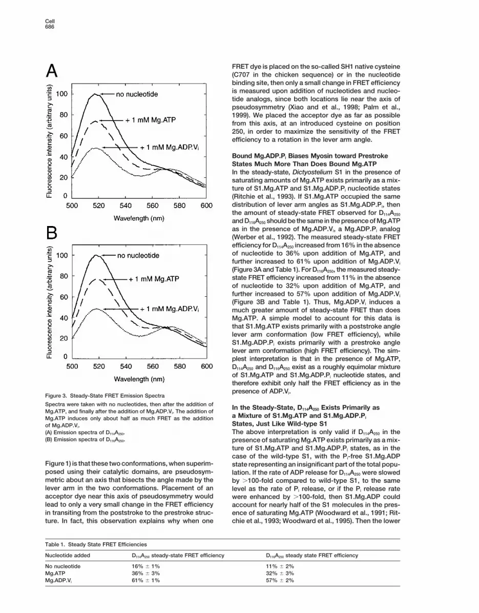
Figure 6. Conformations of S1 Bound toMg.ATP and Mg.ADP.Pi
Structures on the left represent Mg.ATPstates, and structures on the right representMg.ADP.Pi states. The two prestroke anglestates represent prestroke state A and B inFigure 1. Structures in color are the predomi-nant conformations that are populated ineach nucleotide state.
Brust-Mascher and colleagues (1999) note that EPR positions), and a distance of 28 A from RLC-Cys116 toHC-Cys250. The a carbon to b carbon vectors of the twostudies suggest dynamic disorder in the angle between
the catalytic domain and the lever arm domain in both cysteine residues generally point toward each other inthe models (for both D114A250 and D116A250), and thus theS1.Mg.ATP and S1.Mg.ADP.Pi states. They furthermore
point out that, in a muscle fiber, interactions of the lever distances between the dyes themselves might be ex-pected to be shorter than the distances between thearm domain with the myosin thick filament and interac-
tions of the catalytic domain with the actin filament could cysteines to which they are attached. The length of thedyes, measuring from the Cb to which they are attached,influence the conformational equilibrium of the lever arm
angle. Although we see three discrete lever arm angles is about 10 A. Therefore, the measured FRET distancesof 22 A and 19 A are consistent with prestroke state A.and not dynamic disorder, our results are consistent with
the proposed notion that the conformational equilibrium A model based on the chicken skeletal nucleotide freeS1 structure (poststroke state in Figure 1) predicts adoes not lie too far from unity, and that external influ-
ences can perturb that equilibrium. Even though the distance of 75 A from RLC-Cys114 to HC-Cys250, and adistance of 76 A from RLC-Cys116 to HC-Cys250 (measur-Dictyostelium S1.Mg.ATP spends most of its time in the
poststroke angle conformation, it may have to tran- ing from the b carbon positions). The average of the93 A and 79 A measured FRET distances, at 86 A, issiently visit the prestroke angle conformation in order
to catalyze ATP hydrolysis. Another important point is roughly consistent with the 75 A predicted from thechicken skeletal structure based poststroke statethat when ATP binds to a myosin head attached to an
actin filament while exerting positive tension, that ten- model. The data is more consistent with a poststrokestate whose lever arm is angled further downwards, withsion energetically favors the S1 transiting first to a
poststroke angle, weak actin affinity state, at least until respect to the orientation shown in Figure 1. Electronmicrographic reconstructions suggest that Dictyostel-the actin filament is released. Thus, this would be an
example of the outside environment perturbing the lever ium and smooth muscle myosin-II both have rigor stateswith a more downwards angle of the lever arm than thearm conformational equilibrium of a myosin.Mg.ATP nu-
cleotide state. skeletal myosin-II (Rosenfeld et al., 2000), but uncer-tainty in distance is significant for measured FRET effi-ciencies below z10%—in this case, for distancesFRET Distances Compared with Crystal
Structures of Myosin greater than z75 A—due in part to the R26 dependenceon the rate of FRET. Furthermore, calculations of lowCrystal structures of myosin head fragments containing
the ELC have been found with different lever arm angles FRET efficiencies are also particularly sensitive to errorsin the measurements of fluorescence intensity (for(Rayment et al., 1993; Dominguez et al., 1998; Houdusse
et al., 1999). A prestroke state model based on the chicken steady-state experiments) or lifetime (for time-resolvedexperiments) of the donor alone control species, as cansmooth muscle MDE.Mg.ADP.AlF4 structure (prestroke
state A in Figure 1) predicts a distance of 25 A from be seen from inspection of the equation for FRET effi-ciency: ET 5 1 2 FDA/FD 5 1 2 tDA/tD. Thus, the 86 ARLC-Cys114 to HC-Cys250 (measuring from the b carbon

FRET-Based Sensor of Myosin Conformational States691
measured FRET distance could be consistent with con- the lever arm may rest transiently at discrete intermedi-ate lever arm angles. These intermediate angles, if theyformational states whose cysteines are predicted to be
even farther apart, such as in the case with the scallop exist, might be stabilized mechanically through the influ-ence of optical tweezers. Alternatively, these intermedi-ADP bound S1 structure (Houdusse et al., 1999), where
the predicted distance is 108 A. On the other hand, if ate angles may be stabilized by mutations to the myosinmotor which effectively slow down different parts of thethe addition of Mg.ATP to nucleotide free S1 were to
cause half of the population to adopt the scallop ADP ATPase cycle.In summary, these experiments demonstrate a dra-bound S1 structure, then one would expect to see the
lifetime of a fraction of the signal to increase. However, matic change in orientation of the myosin-II lever armupon hydrolysis of ATP. These solution studies fit wellthis was not observed. Thus, our results suggest that
the scallop ADP bound S1 structure is not the major with various crystallographic states previously ob-served. In addition, we have described a high resolutionS1.Mg.ATP conformation. Our experiment is not sensi-
tive enough to rule out the possibility that a minority of sensor of lever arm angle that can be used to examinenot only the effects of binding various nucleotides tothe S1.Mg.ATP heads are in the scallop S1.ADP confor-
mation. the active site of myosin, but also how a wide varietyof myosin mutations affect the myosin-II structure.
An Unexpected, Novel Lever Arm Angle Inducedby Bound Mg.ADP.PI Experimental ProceduresThe measured 44 A and 43 A FRET distances for D114A250
Protein Engineering and Preparationand D116A250, respectively, suggest yet another lever armSubcloning procedures were carried out using standard protocolsconformation, which we have named prestroke state B(Sambrook et al., 1989). The cysteine-light Dictyostelium myosin-II(Figure 1). A tomographic three-dimensional reconstruc-S1 gene fragment, containing the mutations (C49S, C312Y, C442S,
tion of quick-frozen contracting insect flight muscle has C470I, C599L, and C678Y), was generated using splice-overlap ex-shown three angles of the lever arm domain, of 1258, tension PCR mutagenesis (Ho et al., 1989), and was spliced into a
full-length Dictyostelium myosin-II gene in the expression vector1058, and 708 (Taylor et al., 1999). Prestroke B may bepTIKL-Myo. The introduced mutations were verified by dideoxyDNAreflecting a similar conformation to the 1058 lever armsequencing. The S1 gene fragment was then subcloned intoangle of that study. On the other hand, the authors inter-pTIKLOES1, an expression vector for producing S1 with a carboxypret the difference between the 1258 and 1058 anglesterminal 63His tag on the heavy chain. The mutation A250C was
as due to a rotation of the entire S1 head as a whole, introduced into the cysteine-light S1 gene sequence via splice over-and thus conclude that the angle between the catalytic lap extension PCR mutagenesis. The newly created gene was then
subcloned back into pTIKLOES1 to facilitate S1 expression. Thedomain and the lever arm domain is constant betweenintroduced mutation was verified by dideoxyDNA sequencing.the two structures.
Dictyostelium S1 63His was expressed in Dictyostelium AX3-What is the significance of this novel structure? OneORF1 cells (grown in suspension) and purified as described (Giesepossibility is that this second prestroke state gives theand Spudich, 1997). The Dictyostelium RLC gene was synthesized
S1 more flexibility in attaching to the actin filament if it from 40-mer oligonucleotides to incorporate E. coli codon bias intoapproaches the actin out of register or at a bad angle. the DNA sequence (Stemmer et al., 1995). A 63His sequence was
inserted into the beginning of the RLC gene to facilitate purification.The sacrifice is that if the S1 attaches in prestroke stateA 63Arg sequence was appended to the end of the RLC geneB, then the resulting powerstroke will cover a shorteras well. Two RLC mutant genes were constructed, containing thedistance than had the S1 attached in prestroke statemutations (C49L and L114C) or (C49L and K116C), respectively. TheA. Thus in this model, maximum stepsize is no longerRLC was expressed in E. coli, purified by Ni-NTA chromatography
guaranteed, but the increased flexibility allows for a in the presence of 7 M GuCl, and refolded by dialysis against 25greater chance for a productive attachment to nearby mM HEPES (pH 7.0), 150 mM NaCl, 1 mM EDTA, and 0.1 mM Tris
(2-carboxyethyl) phosphine (TCEP).actin filaments. Thomas and colleagues have proposeda similar model, termed the disorder-to-order transitionmechanism for force generation (Roopnarine et al., Dictyostelium Manipulations1998). Comparison of EPR spectra from muscle fiber The full-length myosin-II gene encoding plasmids were electropor-
ated into HS1, a myosin-II null strain of Dictyostelium discoideummyosin heads labeled at SH1 versus those labeled on(Ruppel et al., 1994), while the S1 gene encoding plasmids werethe RLC has led to the interpretation that the angleelectroporated into AX3-ORF1 cells (Manstein et al., 1995), as de-between the catalytic domain and the lever arm domainscribed previously (Egelhoff et al., 1991). Transformants were se-is dynamically disordered on the microsecond timelected for and maintained in DD HL5 media containing 8 mg/ml G418
scale in the S1.Mg.ATP and S1.Mg.ADP.Pi states (Gibco, BRL), 100 units/ml penicillin, and 100 mg/ml streptomycin.(Roopnarine et al., 1998). Our FRET evidence, on the Growth curves were generated by measuring cell number of aliquots
in a hemacytometer over the course of six days, with at least oneother hand, suggests two discrete angles of the levertime point per day, of cells in a 25 ml volume in a 125 ml Erlenmeyerarm with respect to the catalytic domain that are visitedflask, shaking at 200 rpm at 228C, inoculated with a starting densityin the S1.Mg.ADP.Pi state (the presumptive prestrokeof about 104 cells/ml.nucleotide state). Both models are similar, though, in
that they suggest that there is more than one prestrokeLabeling and RLC Exchangestate angle.A suspension of 20 mM RLC, 25 mM HEPES (pH 7.0), 300 mMA further implication of this discovery of a novel myo-NaCl, 1 mM EDTA, 0.025 mM TCEP, and 25 mM Oregon green 488sin lever arm conformation is that the list of stable levermaleimide (Molecular Probes) was incubated at 228C for 1 min, and
arm angles that are being detected experimentally is the reaction was quenched by the addition of 1 mM DTT. Meanwhile,growing. It is conceivable that all myosins execute their a suspension of 1 mM S1, 25 mM HEPES (pH 7.0), 25 mM NaCl, 10
mM MgCl2, and 0.1 mM TCEP was mixed with 0.053 volume 1 Mpowerstroke through multiple steps, in the sense that

Cell692
HEPES (pH 4.2), and incubated for 1 min. Then tetramethylrhoda- and Spudich, 1982). Curves were fit to the Michaelis-Menten equa-tion using Kaleidograph (Abelbeck Software). We used a PK/LDH-mine-5-maleimide (Molecular Probes) was added to a final concen-
tration of 20 mM, and the reaction proceeded for 1 min before linked ATPase assay to measure the ATPase rate of D114A250 andwild-type S1 at 208C (Furch et al., 1998). The buffer conditions usedquenching with DTT at a final concentration of 1 mM. The S1 mixture
was neutralized with the addition of 0.0753 volume 1 M HEPES (pH were the same as for the FRET fluorescence measurements (25 mMHEPES [pH 7.0], 150 mM NaCl, 2 mM MgCl2, and 0.1 mM TCEP at7.4). The S1 was mixed with a final concentration of 3 mM F actin
for 30 min at 228C. The F actin S1 was pelleted by centrifugation at 208C). NADH oxidation was observed using fluorescence (excitationat 340 nm, emission at 450 nm). Samples contained 1 mM DTT, 0.18000 3 g and resuspended in 25 mM HEPES (pH 7.0), 300 mM
NaCl, 10 mM MgCl2, and 1 mM DTT, centrifuged at 8000 3 g, and mM NADH, 0.25 mM PEP, 10 units/ml lactate dehydrogenase, and12 units/ml pyruvate kinase.resuspended. The labeled S1, bound to adin, and RLC were mixed
together and incubated at 228C for 1 hr. The exchanged S1 F actinwas pelleted and resuspended in 25 mM HEPES (pH 7.0), 300 mM Stopped-Flow FluorometryNaCl, 10 mM MgCl2, and 1 mM DTT. The material was pelleted and Stopped-flow experiments were performed as described (Murphyresuspended in 25 mM HEPES (pH 7.0), 25 mM NaCl, 10 mM MgCl2, and Spudich, 1999) on a DX.17MV sequential stopped-flow fluorom-1mM DTT, and 2 mM ADP. This process was repeated three more eter (Applied Photophysics, Leatherhead, UK). Illumination was gen-times. The pellet was resuspended in 25 mM HEPES (pH 7.0), 150 erated with a Hamamatsu mercury-xenon lamp (Middlesex, NJ) andmM NaCl, 1 mM EDTA, 10 mM ATP, and 1 mM DTT and centrifuged. passed through an Applied Photophysics SpectraKinetic mono-The supernatant was collected, and the extraction was repeated. A chromator. The release of 29(39)-O-(N-methylanthraniloyl) ADPfinal concentration of 10 mM MgCl2 was added to the supernatant, (mantADP) was followed by measurement of fluorescence excitedincubated for 10 min at 228C, and the sample was centrifuged at at 290 nm and collected through a KV 380 nm filter. Data was10000 3 g. The resulting supernatant was dialyzed against 25 mM analyzed by a least-squares fitting procedure (Kaleidograph).HEPES (pH 7.0), 150 mM NaCl, 2 mM MgCl2, and 0.1 mM TCEP. Assays were carried out under the same buffer conditions as forThe dialyzed protein was frozen in liquid nitrogen and thawed prior the FRET fluorescence measurements (25 mM HEPES [pH 7.0], 150to use. After thawing, the protein was centrifuged at 10000 3 g just mM NaCl, 2 mM MgCl2, and 0.1 mM TCEP at 208C). To measure thebefore use. Acceptor labeling was measured on A250, the S1 control off-rate of mantADP, 0.1–0.2 mM S1 (final) was premixed with 1.3lacking the donor dye, by comparing absorbance at 280 nm with mM mantADP (final), and then the complex was rapidly mixed withabsorbance at 554 nm, using e280 5 0.8 M21cm21 for S1(Ritchie et al., 0.2 mM Mg.ATP (final), after which the mant fluorescence decrease1993), e280 5 14,000 M21cm21, and e554 5 95,000 M21cm21 (Molecular was measured over time.Probes) for the attached acceptor dye. The acceptor labeling wascalculated as 0.92 acceptor dyes per S1. Acceptor labeling for the Time-Resolved Fluorescence Measurements and Data Fittingcontrol S1 lacking the A250C mutation was measured to be 0.15 Fluorescence lifetimes were measured using a frequency-domainacceptor dyes per S1. Thus, the Cys250 specific labeling was calcu- 10 GHz fluorometer equipped with a Hamamatsu 6 min microchannellated to be 0.77 acceptor dyes per S1. plate detector (MCP-PMT) as previously described (Laczko et al.,
1990). The instrument covered a wide frequency range (4–5000MHz), which allowed detection of lifetimes ranging from severalSteady-State Fluorescence Measurementsnanoseconds to a few picoseconds. Samples were placed in a 2 mmSteady-state fluorescence was measured with an SLM-Aminco-path-length cuvette. The excitation was provided by the frequency-Bowman Series 2 luminescence spectrometer. Solutions containeddoubled output of a cavity-dumped pyridine-2 dye laser tuned at25 mM HEPES (pH 7.0), 150 mM NaCl, 2 mM MgCl2, 0.1 mM TCEP385 nm synchronously pumped by a mode-locked argon ion laser.at 208C, and 0.2 mM S1. The dyes were excited at 490 nm, andSample emission was filtered through an ORIEL interference filterdonor emission was read out at 518 nm, where no appreciablecentered at 520 nm (10 nm bandwidth) together with CORNINGacceptor fluorescence occurs. FRET efficiencies were calculated3-72 and 4-96 filters. This filter combination reinsured that excitationfrom donor quenching as 1 2 Fda/Fd, where Fda is the fluorescencelight and acceptor fluorescence would not contribute to the ob-of the donor and acceptor labeled sample and Fd is the fluorescenceserved donor fluorescence. For the reference signal, DCS in metha-of the donor only labeled sample. Fluorescence intensities werenol (463 ps fluorescence lifetime) (Laczko et al., 1990) was observednormalized according to fluorescence in the presence of 0.5% SDS,through the same filter combination. Samples under the same condi-where the RLC is dissociated from the HC (Smyczynski and Kas-tions as the steady-state fluorescence experiments outlined aboveprzak, 1997). Distance between the dyes was calculated using thewere used for the time-resolved experiments (25 mM HEPES [pHForster equation (Stryer, 1978), where R 5 R0 (1/E 21)1/6. R0 is the7.0], 150 mM NaCl, 2 mM MgCl2, and 0.1 mM TCEP at 208C). Mea-Forster distance, and was calculated as R0 5 [(8.79 3 1025)J k2 n24
surements were taken for the no nucleotide condition, then 1 mMφD], where J is the overlap integral between the donor and acceptorMg.ATP final concentration was added, and measurements takendyes attached to the S1, expressed in M21cm21nm24 (J 5 3.1 3 1015
again, then 1 mM Mg.ADP.Vi final concentration was added, thefor both D114A250 and D116A250), k2 is the orientation factor (assumedsample incubated at 208C for 10 min, and measurements were takento be 0.667), n is the refractive index (assumed to be 1.4), and φDa final time. The governing equations for the time-resolved intensityis the quantum yield measured as 0.68 for D114 and 0.73 for D116,decay data were assumed to be a sum of discrete exponentials asusing disodium fluorescein in 0.01 M NaOH as a reference (φD 5in I(t) 5 S ai exp(2t/ti), where I(t) is the intensity decay, ai is the0.91) (Heidecker et al., 1995). The calculated R0 was 53 A for bothamplitude (preexponential factor), and ti the fluorescence lifetimeD114A250 and D116A250. Measurements were taken for the no nucleotideof the i-th discrete component. Fractional intensity, amplitude, andcondition, then 1 mM Mg.ATP final concentration was added, andlifetime parameters were recovered by a nonlinear least squaresmeasurements taken again, then 1 mM Mg.ADP.Vi final concentra-procedure using the CFS software (Lakowicz et al., 1987).tion was added, the sample incubated at 208C for 10 min, and
measurements were taken a final time. The same energy transferModel Buildingefficiencies were measured for samples when the S1.Mg.ADP.Vi wasPrestroke state A in Figure 1 was built by combining the Dictyostel-assayed directly (no Mg.ATP added).ium ADP.Vi catalytic domain (Smith and Rayment, 1996), theMDE.ADP.AlF4 converter and ELC binding domain (Dominguez etal., 1998), and the RLC binding portion of the lever arm from theSteady-State ATPase Measurements
ATPase activities for the wild-type and cysteine-light myosin-II S1 skeletal S1 structure (Rayment et al., 1993), using superposition ofthe ELC between the latter two models for proper alignment. Thewere determined at 308C by measuring the rate of release of labeled
Pi using [g-32P]ATP in a TLC assay as described by Giese and Spud- dyes were modeled into the structure using InsightII (Molecular Sim-ulations, Inc.). The poststroke state in Figure 1 was built by combin-ich (1997). The buffer conditions used were 25 mM imidazole (pH
7.4), 25 mM KCl, 4 mM MgCl2, 1 mM DTT, 3 mM ATP, and 0.2–1 mM ing the same Dictyostelium catalytic domain described above withthe converter and lever arm from the skeletal S1 structure (RaymentS1. Actin-activated ATPase was measured in the presence of 50
mM F actin, which was prepared as previously described (Pardee et al., 1993). Prestroke state B in Figure 1 is a model to fit the 44 A

FRET-Based Sensor of Myosin Conformational States693
measured FRET distance by rigid body translation and rotation of Huxley, H.E. (1969). The mechanism of muscular contraction. Sci-ence 164, 1356–1365.the lever arm domain.
Huxley, A.F. (1998). Muscle: support for the lever arm. Nature 396,Acknowledgments 317–318.
St. Irving, M., Claire-Allen, T., Sabido-David, C., Craik, J.S., Brand-We thank Wen Liang for measuring Dictyostelium growth rates. This meier, B., Kendrick-Jones, J., Corrie, J.E., Trentham, D.R., and Gold-work was supported by NIH grant AR42895 to J. A. S. and NIH man, Y.E. (1995). Tilting of the light-chain region of myosin duringgrants RR-08110 and GM-35154 (to J. R. L.). W. M. S. was supported step length changes and active force generation in skeletal muscle.by a Howard Hughes Medical Institute Predoctoral Fellowship. Nature 375, 688–691.
Jontes, J.D., Wilson-Kubalek, E.M., and Milligan, R.A. (1995). A 32Received March 7, 2000; revised July 10, 2000. degree tail swing in brush border myosin I on ADP release. Nature
378, 751–753.References
Kabsch, W., Mannherz, H.G., Suck, D., Pai, E.F., and Holmes, K.C.(1990). Atomic structure of the actin: DNase I complex. Nature 347,Brust-Mascher, I., LaConte, L.E., Baker, J.E., and Thomas, D.D.37–44.(1999). Myosin light-chain domain rotates upon muscle activationKnecht, D.A., and Loomis, W.F. (1987). Antisense RNA inactivation ofbut not ATP hydrolysis. Biochemistry 38, 12607–12613.myosin heavy chain gene expression in Dictyostelium discoideum.
Burmeister-Getz, E., Cooke, R., and Selvin, P.R. (1998). Lumines-Science 236, 1081–1086.
cence resonance energy transfer measurements in myosin. Biophys.Laczko, G., Gryczynski, I., Gryczynski, Z., Wiczk, W., Malak, H.,J. 74, 2451–2458.and Lakowicz, J.R. (1990). A 10-GHz frequency-domain fluorometer.
Cooke, R. (1986). The mechanism of muscle contraction. CRC Crit.Rev. Sci. Instrum. 61, 2331–2337.
Rev. Biochem. 21, 53–118.Lakowicz, J.R., Cherek, H., Gryczynski, I., Joshi, N., and Johnson,
Corrie, J.E., Brandmeier, B.D., Ferguson, R.E., Trentham, D.R., Ken-M.L. (1987). Analysis of fluorescence decay kinetics measured in
drick-Jones, J., Hopkins, S.C., van der Heide, U.A., Goldman, Y.E.,the frequency domain using distributions of decay times. Biophys.
Sabido-David, C., Dale, R.E., et al. (1999). Dynamic measurementChem. 28, 35–50.
of myosin light-chain-domain tilt and twist in muscle contraction.Lymn, R.W., and Taylor, E.W. (1971). Mechanism of adenosine tri-Nature 400, 425–430.phosphate hydrolysis of actomyosin. Biochemistry 10, 4617–4624.
Dayhoff, M.O. (1978). Atlas of Protein Sequence and Structure, Vol-Manstein, D.J., Ruppel, K.M., and Spudich, J.A. (1989). Expressionume 5 (Washington, D.C.: National Biomedical Research Foun-and characterization of a functional myosin head fragment in Dicty-dation).ostelium discoideum. Science 246, 656–658.
De Lozanne, A., and Spudich, J.A. (1987). Disruption of the Dictyos-Murphy, C.T., and Spudich, J.A. (1999). The sequence of the myosintelium myosin heavy chain gene by homologous recombination.50K–20K loop affects myosin’s affinity for actin throughout the actin-Science 236, 1086–1091.myosin ATPase cycle and its maximum ATPase activity. Biochemis-
Dominguez, R., Freyzon, Y., Trybus, K.M., and Cohen, C. (1998).try 38, 3785–3792.
Crystal structure of a vertebrate smooth muscle myosin motor do-Palm, T., Sale, K., Brown, L., Li, H., Hambly, B., and Fajer, P.G.main and its complex with the essential light chain: visualization of(1999). Intradomain distances in the regulatory domain of the myosinthe pre-power stroke state. Cell 94, 559–571.head in prepower and postpower stroke states: fluorescence energy
Egelhoff, T.T., Titus, M.A., Manstein, D.J., Ruppel, K.M., and Spud- transfer. Biochemistry 38, 13026–13034.ich, J.A. (1991). Molecular genetic tools for study of the cytoskeleton
Pardee, J.D., and Spudich, J.A. (1982). Purification of muscle actin.in Dictyostelium. Methods Enzymol. 196, 319–334.Methods Cell Biol. 24, 271–289.
Eisenberg, E., and Greene, L.E. (1980). The relation of muscle bio-Rayment, I. (1996). The structural basis of the myosin ATPase activ-chemistry to muscle physiology. Ann. Rev. Physiol. 42, 293–309.ity. J. Biol. Chem. 271, 15850–15853.
Fisher, A.J., Smith, C.A., Thoden, J.B., Smith, R., Sutoh, K., Holden,Rayment, I., Holden, H.M., Whittaker, M., Yohn, C.B., Lorenz, M.,H.M., and Rayment, I. (1995). X-ray structures of the myosin motorHolmes, K.C., and Milligan, R.A. (1993). Structure of the actin-myosindomain of Dictyostelium discoideum complexed with MgADP.BeFxcomplex and its implications for muscle contraction. Science 261,and MgADP.AlF4. Biochemistry 34, 8960–8972.58–65.
Furch, M., Geeves, M.A., and Manstein, D.J. (1998). Modulation ofRayment, I., Rypniewski, W.R., Schmidt-Base, K., Smith, R., Tom-actin affinity and actomyosin adenosine triphosphatase by chargechick, D.R., Benning, M.M., Winkelmann, D.A., Wesenberg, G., andchanges in the myosin motor domain. Biochemistry 37, 6317–6326.Holden, H.M. (1993). Three-dimensional structure of myosin sub-
Giese, K.C., and Spudich, J.A. (1997). Phenotypically selected muta- fragment-1: a molecular motor. Science 261, 50–58.tions in myosin’s actin binding domain demonstrate intermolecular
Ritchie, M.D., Geeves, M.A., Woodward, S.K., and Manstein, D.J.contacts important for motor function. Biochemistry 36, 8465–8473.(1993). Kinetic characterization of a cytoplasmic myosin motor do-
Goldman, Y.E. (1998). Wag the tail: structural dynamics of actomyo- main expressed in Dictyostelium discoideum. Proc. Natl. Acad. Sci.sin. Cell 93, 1–4. USA 90, 8619–8623.Ho, S.N., Hunt, H.D., Horton, R.M., Pullen, J.K., and Pease, L.R. Roopnarine, O., Szent-Gyorgyi, A.G., and Thomas, D.D. (1998). Mi-(1989). Site-directed mutagenesis by overlap extension using the crosecond rotational dynamics of spin-labeled myosin regulatorypolymerase chain reaction. Gene 77, 51–59. light chain induced by relaxation and contraction of scallop muscle.Heidecker, M., Yan-Marriott, Y., and Marriott, G. (1995). Proximity Biochemistry 37, 14428–14436.relationships and structural dynamics of the phalloidin binding site Rosenfeld, S.S., Xing, J., Whitaker, M., Cheung, H.C., Brown, F.,of actin filaments in solution and on single actin filaments on heavy Wells, A., Milligan, R.A., and Sweeney, H.L. (2000). Kinetic and spec-meromyosin. Biochemistry 34, 11017–11025. troscopic evidence for three actomyosin:ADP states in smooth mus-Holmes, K.C. (1997). The swinging lever-arm hypothesis of muscle cle. J. Biol. Chem., in press.contraction. Curr. Biol. 7, R112–R118. Ruppel, K.M., Uyeda, T.Q., and Spudich, J.A. (1994). Role of highlyHolmes, K.C., Popp, D., Gebhard, W., and Kabsch, W. (1990). Atomic conserved lysine 130 of myosin motor domain. In vivo and in vitromodel of the actin filament. Nature 347, 44–49. characterization of site specifically mutated myosin. J. Biol. Chem.
269, 18773–18780.Houdusse, A., Kalabokis, V.N., Himmel, D., Szent-Gyorgyi, A.G., andCohen, C. (1999). Atomic structure of scallop myosin subfragment Sambrook, J., Fritsch, E.F., and Maniatis, T. (1989). Molecular Clon-
ing: A Laboratory Manual, 2nd Edition (Plainview, NY: Cold SpringS1 complexed with MgADP: a novel conformation of the myosinhead. Cell 97, 459–470. Harbor Laboratory Press).

Cell694
Sasaki, N., Shimada, T., and Sutoh, K. (1998). Mutational analysisof the switch II loop of Dictyostelium myosin II. J. Biol. Chem. 273,20334–20340.
Schroder, R.R., Manstein, D.J., Jahn, W., Holden, H., Rayment, I.,Holmes, K.C., and Spudich, J.A. (1993). Three-dimensional atomicmodel of F-actin decorated with Dictyostelium myosin S1. Nature364, 171–174.
Sellers, J.R., and Goodson, H.V. (1995). Motor Proteins 2: Myosin,Volume 2, P. Sheterline, ed. (London: Academic Press Limited).
Smith, C.A., and Rayment, I. (1996). X-ray structure of the magne-sium(II).ADP.vanadate complex of the Dictyostelium discoideummyosin motor domain to 1.9 A resolution. Biochemistry 35, 5404–5417.
Smyczynski, C., and Kasprzak, A.A. (1997). Effect of nucleotides onthe orientation of the light chain-binding domain in myosin subfrag-ment 1. Biochemistry 36, 13201–13207.
Spudich, J.A. (1994). How molecular motors work. Nature 372,515–518.
Stemmer, W.P., Crameri, A., Ha, K.D., Brennan, T.M., and Heyneker,H.L. (1995). Single-step assembly of a gene and entire plasmid fromlarge numbers of oligodeoxyribonucleotides. Gene 164, 49–53.
Stryer, L. (1978). Fluorescence energy transfer as a spectroscopicruler. Annu. Rev. Biochem. 47, 819–846.
Suzuki, Y., Yasunaga, T., Ohkura, R., Wakabayashi, T., and Sutoh,K. (1998). Swing of the lever arm of a myosin motor at the isomeriza-tion and phosphate-release steps. Nature 396, 380–383.
Taylor, K.A., Schmitz, H., Reedy, M.C., Goldman, Y.E., Franzini-Armstrong, C., Sasaki, H., Tregear, R.T., Poole, K., Lucaveche, C.,Edwards, R.J, et al. (1999). Tomographic 3D reconstruction of quick-frozen, Ca21-activated contracting insect flight muscle. Cell 99,421–431.
Werber, M.M., Peyser, Y.M., and Muhlrad, A. (1992). Characterizationof stable beryllium fluoride, aluminum fluoride, and vanadate con-taining myosin subfragment-1 nucleotide complexes. Biochemistry31, 7190–7197.
Whittaker, M., Wilson-Kubalek, E.M., Smith, J.E., Faust, L., Milligan,R.A., and Sweeney, H.L. (1995). A 35 A movement of smooth musclemyosin on ADP release. Nature 378, 748–751.
Woodward, S.K., Eccleston, J.F., and Geeves, M.A. (1991). Kineticsof the interaction of 2’(39)-O-(N-methylanthraniloyl)-ATP with myosinsubfragment 1 and actomyosin subfragment 1: characterization oftwo acto-S1-ADP complexes. Biochemistry 30, 422–430.
Woodward, S.K., Geeves, M.A., and Manstein, D.J. (1995). Kineticcharacterization of a cytoplasmic myosin motor domain expressedin Dictyostelium discoideum. Biochemistry 90, 8619–8623.
Xiao, M., Li, H., Snyder, G.E., Cooke, R., Yount, R.G., and Selvin,P.R. (1998). Conformational changes between the active-site andregulatory light chain of myosin as determined by luminescenceresonance energy transfer: the effect of nucleotides and actin. Proc.Natl. Acad. Sci. USA 95, 15309–15314.
Related Documents