University of Wollongong University of Wollongong Research Online Research Online Faculty of Science - Papers (Archive) Faculty of Science, Medicine and Health 2007 A fragmentation study of isoflavones in negative electrospray ionization by A fragmentation study of isoflavones in negative electrospray ionization by MSn ion trap mass spectrometry and triple quadrupole mass spectrometry MSn ion trap mass spectrometry and triple quadrupole mass spectrometry Jinguo Kang University of Wollongong, [email protected] Larry A. Hick University of Wollongong William E. Price University of Wollongong, [email protected] Follow this and additional works at: https://ro.uow.edu.au/scipapers Part of the Life Sciences Commons, Physical Sciences and Mathematics Commons, and the Social and Behavioral Sciences Commons Recommended Citation Recommended Citation Kang, Jinguo; Hick, Larry A.; and Price, William E.: A fragmentation study of isoflavones in negative electrospray ionization by MSn ion trap mass spectrometry and triple quadrupole mass spectrometry, Rapid Communications in Mass Spectrometry: 21(6) 2007, 857-868. https://ro.uow.edu.au/scipapers/1106 Research Online is the open access institutional repository for the University of Wollongong. For further information contact the UOW Library: [email protected]

Welcome message from author
This document is posted to help you gain knowledge. Please leave a comment to let me know what you think about it! Share it to your friends and learn new things together.
Transcript
University of Wollongong University of Wollongong
Research Online Research Online
Faculty of Science - Papers (Archive) Faculty of Science, Medicine and Health
2007
A fragmentation study of isoflavones in negative electrospray ionization by A fragmentation study of isoflavones in negative electrospray ionization by
MSn ion trap mass spectrometry and triple quadrupole mass spectrometry MSn ion trap mass spectrometry and triple quadrupole mass spectrometry
Jinguo Kang University of Wollongong, [email protected]
Larry A. Hick University of Wollongong
William E. Price University of Wollongong, [email protected]
Follow this and additional works at: https://ro.uow.edu.au/scipapers
Part of the Life Sciences Commons, Physical Sciences and Mathematics Commons, and the Social
and Behavioral Sciences Commons
Recommended Citation Recommended Citation Kang, Jinguo; Hick, Larry A.; and Price, William E.: A fragmentation study of isoflavones in negative electrospray ionization by MSn ion trap mass spectrometry and triple quadrupole mass spectrometry, Rapid Communications in Mass Spectrometry: 21(6) 2007, 857-868. https://ro.uow.edu.au/scipapers/1106
Research Online is the open access institutional repository for the University of Wollongong. For further information contact the UOW Library: [email protected]
A fragmentation study of isoflavones in negative electrospray ionization by MSn A fragmentation study of isoflavones in negative electrospray ionization by MSn ion trap mass spectrometry and triple quadrupole mass spectrometry ion trap mass spectrometry and triple quadrupole mass spectrometry
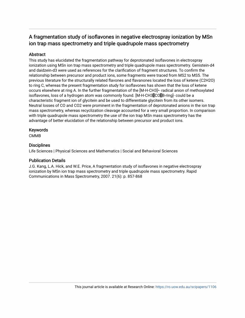
Abstract Abstract This study has elucidated the fragmentation pathway for deprotonated isoflavones in electrospray ionization using MSn ion trap mass spectrometry and triple quadrupole mass spectrometry. Genistein-d4 and daidzein-d3 were used as references for the clarification of fragment structures. To confirm the relationship between precursor and product ions, some fragments were traced from MS2 to MS5. The previous literature for the structurally related flavones and flavanones located the loss of ketene (C2H2O) to ring C, whereas the present fragmentation study for isoflavones has shown that the loss of ketene occurs elsewhere at ring A. In the further fragmentation of the [M-H-CH3]•- radical anion of methoxylated isoflavones, loss of a hydrogen atom was commonly found. [M-H-CH3COB-ring]- could be a characteristic fragment ion of glycitein and be used to differentiate glycitein from its other isomers. Neutral losses of CO and CO2 were prominent in the fragmentation of deprotonated anions in the ion trap mass spectrometry, whereas recyclization cleavage accounted for a very small proportion. In comparison with triple quadrupole mass spectrometry the use of the ion trap MSn mass spectrometry has the advantage of better elucidation of the relationship between precursor and product ions.
Keywords Keywords CMMB
Disciplines Disciplines Life Sciences | Physical Sciences and Mathematics | Social and Behavioral Sciences
Publication Details Publication Details J.G. Kang, L.A. Hick, and W.E. Price, A fragmentation study of isoflavones in negative electrospray ionization by MSn ion trap mass spectrometry and triple quadrupole mass spectrometry. Rapid Communications in Mass Spectrometry, 2007. 21(6): p. 857-868
This journal article is available at Research Online: https://ro.uow.edu.au/scipapers/1106
1
A fragmentation study of isoflavones in negative electrospray
ionization by MSn ion trap mass spectrometry and triple
quadrupole mass spectrometry
Jinguo Kang, Larry A. Hick and William E. Price∗
Department of Chemistry, University of Wollongong, Wollongong NSW, 2522, Australia
∗Correspondence to: W. E. Price,
Tel.: +61 2 42213529; fax: +61 2 42214287. E-mail: [email protected]
This study has elucidated the fragmentation pathway for deprotonated isoflavones in electrospray ionization
using MSn ion trap mass spectrometry and triple quadrupole mass spectrometry. Genistein-d4 and daidzein-d3
were used as references for the clarification of fragment structures. To confirm the relationship between
precursor and product ions, some fragments were traced from MS2 to MS
5. The previous literature for the
structurally related flavones and flavanones located the loss of ketene (C2H2O) to ring C, whereas the present
fragmentation study for isoflavones has shown that the loss of ketene occurs elsewhere at ring A. In the
further fragmentation of the [M−H−CH3]·−
radical anion of methoxylated isoflavones, loss of a hydrogen atom
was commonly found. [M−H−CH3−CO−B-ring]− could be a characteristic fragment ion of glycitein and be
used to differentiate glycitein from its other isomers. Neutral losses of CO and CO2 were prominent in the
fragmentation of deprotonated anions in the ion trap mass spectrometry, whereas recyclization cleavage
accounted for a very small proportion. In comparison with triple quadrupole mass spectrometry the use of the
ion trap MSn mass spectrometry has the advantage of better elucidation of the relationship between precursor
and product ions.
INTRODUCTION Isoflavones are the most well known class of phytoestrogens with functional estrogenic and antiestrogenic action and
structural similarity to mammalian estrogenic hormones. These compounds are primarily found in the Fabacease family,
and are distributed in edible plants and derived products.1, 2
Isoflavones belong to a subclass of the flavonoids.3 In the past
decade many analytical methods for the identification and quantitation of flavonoids in plant derived products and
biological matrices have been reported.3-5
Among them mass spectrometry coupled with high performance liquid
chromatography (HPLC) has proved to be one of the most effective techniques particularly for the analysis of complex
mixtures in biological samples.5, 6
For this reason, a number of papers dealing with the fragmentation mechanism of a range
of flavonoids, mainly flavones, flavanones and flavonols, have been published.7-21
Ma et al7, 8
analysed the fragmentation
behavior of flavones, flavonols and methoxyflavones in positive ion mode using fast-atom bombardment and collision-
induced dissociation tandem mass spectrometry. Fabre et al9 proposed a fragmentation scheme and product ion structures
for flavone, flavonol, and flavanone aglycones in negative ion mode. Kuhn et al10
found a characteristic double neutral loss
of CO at ring C for flavonoid type compounds. March et al11, 12
studied fragmentation scheme of a flavonol and an
2
isoflavone glycoside using electrospray quadrupole time-of flight mass spectrometry in both positive and negative mode,
and successfully explained a fragmentation mechanism by an intermediate structure of seven-membered ring C. Justesen13
examined the fragmentation rule of methoxylated flavones and flavonols in negative ion mode, and found that the loss of a
methyl group (−15u) was the characteristic fragmentation.
The structures of isoflavones differ from the isomeric flavones by the position of the ring B, which could lead to
significantly different fragmentation behavior to the flavones, flavanones and flavonols. In a previous paper we have
described a robust method for the simultaneous identification and quantitation of isoflavones and lignans.22
Given the
importance of isoflavones to a number of research areas and the inreasing use of MS techniques for their identification and
quantitation a clear understanding of the fragmentation behavior of isoflavones is needed. To our knowledge, no systematic
study has been reported before for the fragmentation scheme and structures on isoflavones in negative ESI mode by step
MSn fragmentation using ion trap mass spectrometry.
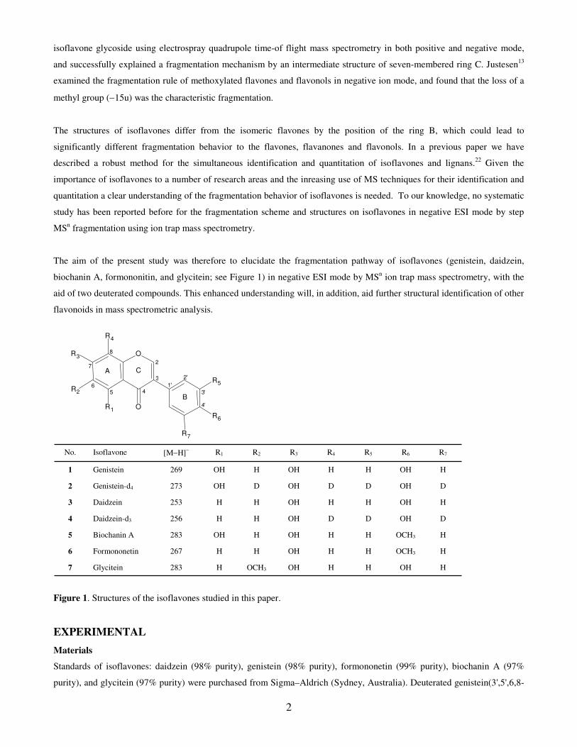
The aim of the present study was therefore to elucidate the fragmentation pathway of isoflavones (genistein, daidzein,
biochanin A, formononitin, and glycitein; see Figure 1) in negative ESI mode by MSn ion trap mass spectrometry, with the
aid of two deuterated compounds. This enhanced understanding will, in addition, aid further structural identification of other
flavonoids in mass spectrometric analysis.
Figure 1. Structures of the isoflavones studied in this paper.
EXPERIMENTAL
Materials
Standards of isoflavones: daidzein (98% purity), genistein (98% purity), formononetin (99% purity), biochanin A (97%
purity), and glycitein (97% purity) were purchased from Sigma–Aldrich (Sydney, Australia). Deuterated genistein(3',5',6,8-
O
O
R 3
R 1
R 2
R 4
R 5
R 6 R 7
A
B
C 2
3
456
7
8
1'
2'
3'
4'
H OH H H OH OCH3 H 283 Glycitein 7 H OCH3 H H OH H H 267 Formononetin 6 H OCH3 H H OH H OH 283 Biochanin A 5 D OH D D OH H H 256 Daidzein-d3 4 H OH H H OH H H 253 Daidzein 3 D OH D D OH D OH 273 Genistein-d4 2 H OH H H OH H OH 269 Genistein 1 R7 R6 R5 R4 R3 R2 R1 [M−H]
− Isoflavone No.
3
d4) (98% purity, 95% isotopic enrichment) and deuterated daidzein (3',5',8-d3) (98% purity, 97% isotopic enrichment) were
purchased from Cambridge Isotope Laboratories (Andover, MA, USA). Chemical structures and trivial names of all the
standard isoflavones are shown in Figure 1. Acetonitrile and methanol, both HPLC grade, were supplied by Crown
Scientific (Sydney, Australia). Milli-Q water (Milli-Q plus 185, Australia) was used for making up all aqueous solutions.
Standard stock solutions of each compound were prepared at a concentration of 100µg/mL in acetonitrile or acetonitrile plus
20% methanol. Working solutions were prepared in acetonitrile/water (1:3, v/v) and obtained by tenfold dilution to a
concentration of 10µg/mL. The solutions was infused to the ESI source by the syringe pump of the mass spectrometer,
using a 500-µl Unimetrics syringe at a flow rate of 10µl min−1
(for MS2 and MS
3 experiment) and 60µl min
−1 (for MS
4 and
MS5 experiment).
Mass spectrometry
The fragmentation experiments by ion trap mass spectrometry were performed using a ThermoElectron Finnigan LTQ
linear ion trap mass spectrometer (ThermoFinnigan, San Jose, CA, USA) equipped with an electrospray ionization source.
Standard solutions were directly infused into the LTQ linear ion trap mass spectrometer, via an ESI source. All the mass
spectra were acquired in negative ion mode with the spray voltage 3.61 kV, capillary voltage at -11.83V, capillary
temperature 274.8°C. Nitrogen was used as both the sheath and auxiliary gas at 29 and 3 arbitrary units respectively.
Helium was used as a damping and collision gas at a partial pressure of 0.1 Pa. The relative collision energies for each
compound were from 36% to 49%, respectively. The data range utilized was from 80 to 300u.
The fragmentation experiments by triple quadrupole mass spectrometry were performed using a Micromass Quattro Micro
triple quadrupole mass spectrometer (Micromass, Manchester, UK) with an electrospray ionization source. Data acquisition
was in a negative mode. The electrospray source parameters were fixed as follows: electrospray capillary voltage 3.0kV,
cone 40eV, source temperature 100°C, desolvation temperature 120°C, and desolvation gas N2 at 300 L/hr. The collision
energies for each compound were from 30 to 35 eV, respectively. Spectra were recorded in the range 100–300u.
RESULT AND DISCUSSION
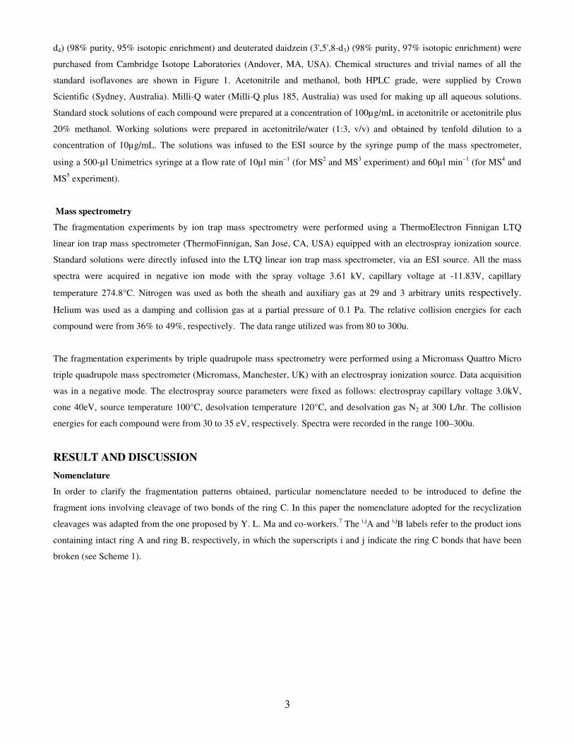
Nomenclature
In order to clarify the fragmentation patterns obtained, particular nomenclature needed to be introduced to define the
fragment ions involving cleavage of two bonds of the ring C. In this paper the nomenclature adopted for the recyclization
cleavages was adapted from the one proposed by Y. L. Ma and co-workers.7 The
i,jA and
i,jB labels refer to the product ions
containing intact ring A and ring B, respectively, in which the superscripts i and j indicate the ring C bonds that have been
broken (see Scheme 1).
4
O
O
HO
OHOH
A
B
C
2
3
45
6
7
8
1'
4'
0 1
2
34
1,3A-
0,3B-
1,3B-
0,4B-
Scheme 1. Nomenclature adopted for defining retrocyclization cleavages observed in this study.
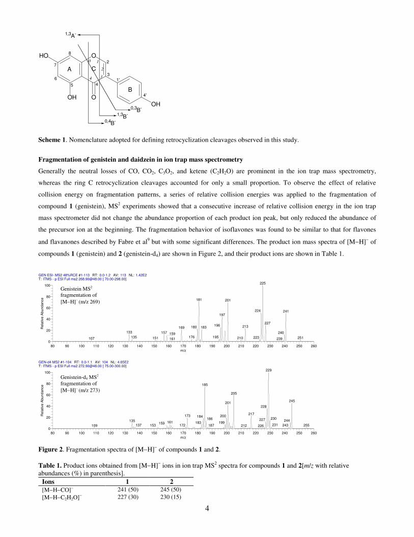
Fragmentation of genistein and daidzein in ion trap mass spectrometry
Generally the neutral losses of CO, CO2, C3O2, and ketene (C2H2O) are prominent in the ion trap mass spectrometry,
whereas the ring C retrocyclization cleavages accounted for only a small proportion. To observe the effect of relative
collision energy on fragmentation patterns, a series of relative collision energies was applied to the fragmentation of
compound 1 (genistein), MS2 experiments showed that a consecutive increase of relative collision energy in the ion trap
mass spectrometer did not change the abundance proportion of each product ion peak, but only reduced the abundance of
the precursor ion at the beginning. The fragmentation behavior of isoflavones was found to be similar to that for flavones
and flavanones described by Fabre et al9 but with some significant differences. The product ion mass spectra of [M−H]
− of
compounds 1 (genistein) and 2 (genistein-d4) are shown in Figure 2, and their product ions are shown in Table 1.
Figure 2. Fragmentation spectra of [M−H]− of compounds 1 and 2.
Table 1. Product ions obtained from [M−H]− ions in ion trap MS
2 spectra for compounds 1 and 2[m/z with relative
abundances (%) in parenthesis].
Ions 1 2
[M−H−CO]− 241 (50) 245 (50)
[M−H−C2H2O]− 227 (30) 230 (15)
GEN ESI- MS2 48%RCE #1-113 RT: 0.0-1.2 AV: 113 NL: 1.42E2T: ITMS - p ESI Full ms2 [email protected] [ 70.00-298.00]
80 90 100 110 120 130 140 150 160 170 180 190 200 210 220 230 240 250 260
m/z
0
20
40
60
80
100
Rela
tive A
bund
ance
225
181 201
224 241197
227196 213180 183169
133 157 240159176 195135 223 251210151107 239161
GEN-d4 MS2 #1-104 RT: 0.0-1.1 AV: 104 NL: 4.65E2T: ITMS - p ESI Full ms2 [email protected] [ 75.00-300.00]
80 90 100 110 120 130 140 150 160 170 180 190 200 210 220 230 240 250 260
m/z
0
20
40
60
80
100
Rela
tive A
bund
ance
229
185
205
245201
228
217173 200184
230186 227 244135 161 199183159 231137 243172 255153 187109 212 226
Genistein MS2
fragmentation of
[M−H]− (m/z 269)
Genistein-d4 MS2
fragmentation of
[M−H]− (m/z 273)
5
[M−H−CO2]− 225 (100) 229 (100)
[M−H−2CO]− 213 (25) 217 (25)
[M−H−C3O2]− 201 (70) 205 (60)
[M−H−CO2−CO]− 197 (45) 201 (40)
[M−H −C2H2O−CO2]− 183 (20) 186 (15)
[M−H−2CO2]− 181 (70) 185 (70)
[M−H−2CO−CO2]− 169 (20) 173 (20)
[M−H−C3O2−C2H2O]− 159 (10) 162 (3) 0,3B− 133 (15) 135 (10) 0,3A− 135 (5) 137 (2) 1,3A− 151 (2) 153 (1) 1,3A−−CO2
107 (1) 109 (1)
*48% of the relative collision energy was used in the fragmentation.
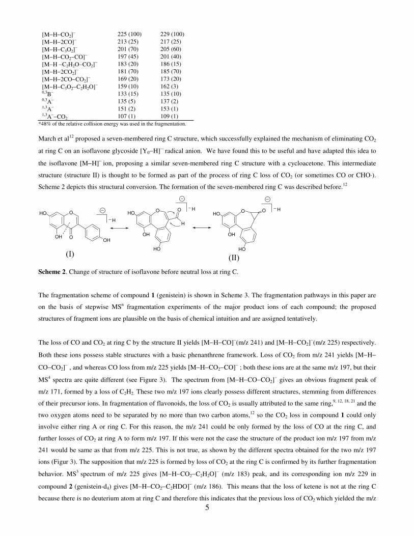
March et al12
proposed a seven-membered ring C structure, which successfully explained the mechanism of eliminating CO2
at ring C on an isoflavone glycoside [Y0−H]·−
radical anion. We have found this to be useful and have adapted this idea to
the isoflavone [M−H]−
ion, proposing a similar seven-membered ring C structure with a cycloacetone. This intermediate
structure (structure II) is thought to be formed as part of the process of ring C loss of CO2 (or sometimes CO or CHO·).
Scheme 2 depicts this structural conversion. The formation of the seven-membered ring C was described before.12
O
O
OH
HO
OH
H
OH
HOO
HO
O
H
H
OH
HOO
HO
HO
(Ι)(ΙΙ)
Scheme 2. Change of structure of isoflavone before neutral loss at ring C.
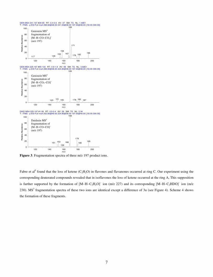
The fragmentation scheme of compound 1 (genistein) is shown in Scheme 3. The fragmentation pathways in this paper are
on the basis of stepwise MSn fragmentation experiments of the major product ions of each compound; the proposed
structures of fragment ions are plausible on the basis of chemical intuition and are assigned tentatively.
The loss of CO and CO2 at ring C by the structure II yields [M−H−CO]−(m/z 241) and [M−H−CO2]
−(m/z 225) respectively.
Both these ions possess stable structures with a basic phenanthrene framework. Loss of CO2 from m/z 241 yields [M−H−
CO−CO2]− , and whereas CO loss from m/z 225 yields [M−H−CO2−CO]
− ; both these ions are at the same m/z 197, but their
MS4 spectra are quite different (see Figure 3). The spectrum from [M−H−CO−CO2]
− gives an obvious fragment peak of
m/z 171, formed by a loss of C2H2. These two m/z 197 ions clearly possess different structures, stemming from differences
of their precursor ions. In fragmentation of flavonoids, the loss of CO2 is usually attributed to the same ring,9, 12, 18, 21
and the
two oxygen atoms need to be separated by no more than two carbon atoms,12
so the CO2 loss in compound 1 could only
involve either ring A or ring C. For this reason, the m/z 241 could be only formed by the loss of CO at the ring C, and
further losses of CO2 at ring A to form m/z 197. If this were not the case the structure of the product ion m/z 197 from m/z
241 would be same as that from m/z 225. This is not true, as shown by the different spectra obtained for the two m/z 197
ions (Figur 3). The supposition that m/z 225 is formed by loss of CO2 at the ring C is confirmed by its further fragmentation
behavior. MS3
spectrum of m/z 225 gives [M−H−CO2−C2H2O]− (m/z 183) peak, and its corresponding ion m/z 229 in
compound 2 (genistein-d4) gives [M−H−CO2−C2HDO]− (m/z 186). This means that the loss of ketene is not at the ring C
because there is no deuterium atom at ring C and therefore this indicates that the previous loss of CO2 which yielded the m/z
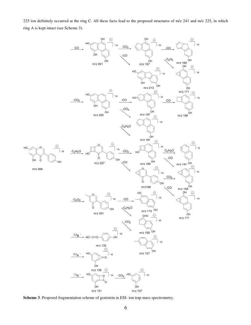
6
225 ion definitely occurred at the ring C. All these facts lead to the proposed structures of m/z 241 and m/z 225, in which
ring A is kept intact (see Scheme 3).
O
O
OH
HO
OH
m/z 269
H
-CO -COH
OH
-CO
m/z 241 m/z 169
-CO2H
OHm/z 225 m/z 169
-C3O2
O
O
OH
m/z 201
H -CO
m/z 173-C2H2O
-CO2
OHm/z 157
0,3BHO C C OH
m/z 133
0,3A
OH
HO
OH
m/z 135
-CO2
-C2H2O
OHm/z 183
O
O
OH
m/z 227
HO-CO2-C2H2O
-CO2
1,3A
OH
HO H
m/z 151
O
O
OH
HO H
m/z 107
H
-CO
-CO2
HO
m/z 155OH
H
H
-CO2
-C2H2O
OH
H
m/z 213
H
m/z 197
OH
OH
H
-CO
OH
HO H
m/z 197
-CO
OH
H
m/z 181
HO
OH
CHO
OH
OH
H
-C2H2
m/z 159
OH
H
m/z 141
m/z 171
OH
HO
OH
H
H
H
O
O
OH
m/z199
H
OH
OH
H
m/z 171
-CO
-CO
OH
HO
OH
H
OH
HO
OH
OH
H
Scheme 3. Proposed fragmentation scheme of genistein in ESI- ion trap mass spectrometry.
7
Figure 3. Fragmentation spectra of three m/z 197 product ions.
Fabre et al9 found that the loss of ketene (C2H2O) in flavones and flavanones occurred at ring C. Our experiment using the
corresponding deuterated compounds revealed that in isoflavones the loss of ketene occurred at the ring A, This supposition
is further supported by the formation of [M−H−C2H2O]− ion (m/z 227) and its corresponding [M−H−C2HDO]
− ion (m/z
230). MS3 fragmentation spectra of these two ions are identical except a difference of 3u (see Figure 4). Scheme 4 shows
the formation of these fragments.
GEN MS4 241-197 #29-65 RT: 0.4-0.9 AV: 37 SM: 7G NL: 1.49E1T: ITMS - p ESI Full ms4 [email protected] [email protected] [email protected] [ 50.00-300.00]
120 140 160 180 200
m/z
0
20
40
60
80
100
Rela
tive A
bundance
169
171
158195167155 182178146117
GEN MS4 225-197 #65-133 RT: 0.9-1.9 AV: 69 SM: 7G NL: 3.92E1T: ITMS - p ESI Full ms4 [email protected] [email protected] [email protected] [ 50.00-300.00]
120 140 160 180 200
m/z
0
20
40
60
80
100
Rela
tive A
bundance
169
151 182179155 187143
DAID MS4 225-197 #1-28 RT: 0.0-0.4 AV: 28 SM: 7G NL: 2.34T: ITMS - p ESI Full ms4 [email protected] [email protected] [email protected] [ 50.00-300.00]
120 140 160 180 200
m/z
0
20
40
60
80
100
Rela
tive A
bundance
169
179
195153 168151 180
158
Genistein MS4
fragmentation of
[M−H−CO−CO2]−
(m/z 197)
Genistein MS4
fragmentation of
[M−H−CO2−CO]−
(m/z 197)
Daidzein MS4
fragmentation of
[M−H−CO−CO]−
(m/z 197)
8
Figure 4. Fragmentation spectra of m/z 227 and its corresponding deuterated anion m/z 230.
O
O
OH
HO
OH
D
D
D
D
m/z 273m/z 269 (D=H)
HO
O
OH
D
D
H
-C2HDOHO
D
m/z 230m/z 227 (D=H)
OH
m/z 186m/z 183 (D=H)
HO
D D
D
-CO2
H
-CO2 H
OH
HO
OH
D
D
D D
m/z 229m/z 225 (D=H)
-C2HDO
Scheme 4. Formation of ion through loss of ketene in the deuterated fragment.
It is worth mentioning that the idea that the losses of ketene and C3O2 occurred at the ring A and not ring B is supported by
the fact that m/z 133 ion corresponding to a 0,3
B− cleavage is found in MS
3 fragmentation of both m/z 227 and m/z 201 ions.
This indicates that after the loss of ketene and C3O2 both ions still keep the ring B intact, for which the conventional
structures (structure I) with intact ring C and ring B are accepted for m/z 227 and m/z 210 ions as shown in their proposed
structures in Scheme 3.
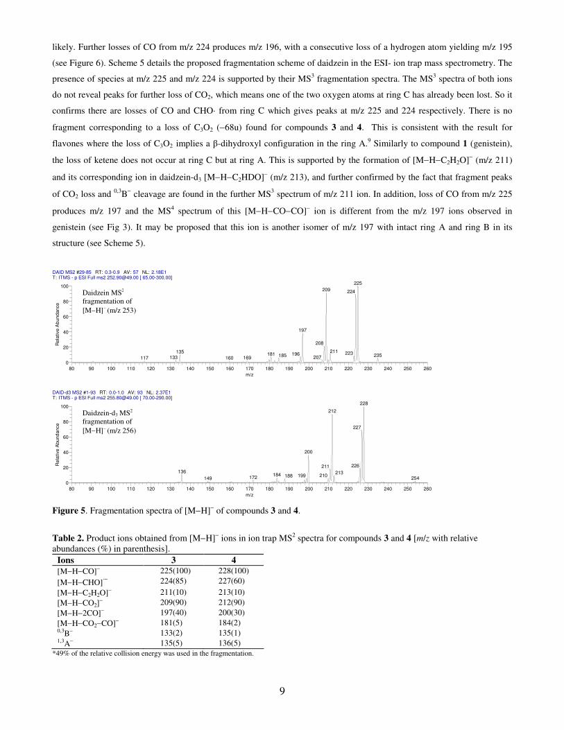
Figure 5 shows the fragmentation spectra of [M−H]− ions for compounds 3 (daidzein) and 4 (daidzein-d3), and their
proposed product ions are shown in Table 2. In the MS2 spectrum of daidzein m/z 224 peak is distinct (about 90% of that of
the base peak, see Figure 5). This radical anion could be formed by either loss of 29u (−CHO·) from m/z 253 or loss of a
hydrogen atom from m/z 225. Further MS3 fragmentation of m/z 225 does not give any obvious peak at m/z 224, which
suggests the latter is not likely. Therefore formation by loss of CHO· from its precursor [M−H]− ion (m/z 253) is more
GEN MS3 227 #28-64 RT: 0.3-0.7 AV: 37 NL: 1.77E2T: ITMS - p ESI Full ms3 [email protected] [email protected] [ 60.00-300.00]
140 160 180 200 220
m/z
0
20
40
60
80
100R
ela
tive A
bundance
183
199
155
159 171 225133 181 198
GEN-d4 MS3 272-230 #45-88 RT: 0.6-1.2 AV: 44 SM: 7G NL: 8.99E-1T: ITMS - p ESI Full ms3 [email protected] [email protected] [ 60.00-300.00]
140 160 180 200 220
m/z
0
20
40
60
80
100
Rela
tive A
bundance
186
202
158
135 201162 174 228
Genistein MS3
fragmentation of
[M−H−C2H2O]−
(m/z 227)
Genistein-d4 MS3
fragmentation of
[M−H−C2HDO]−
(m/z 230)
9
likely. Further losses of CO from m/z 224 produces m/z 196, with a consecutive loss of a hydrogen atom yielding m/z 195
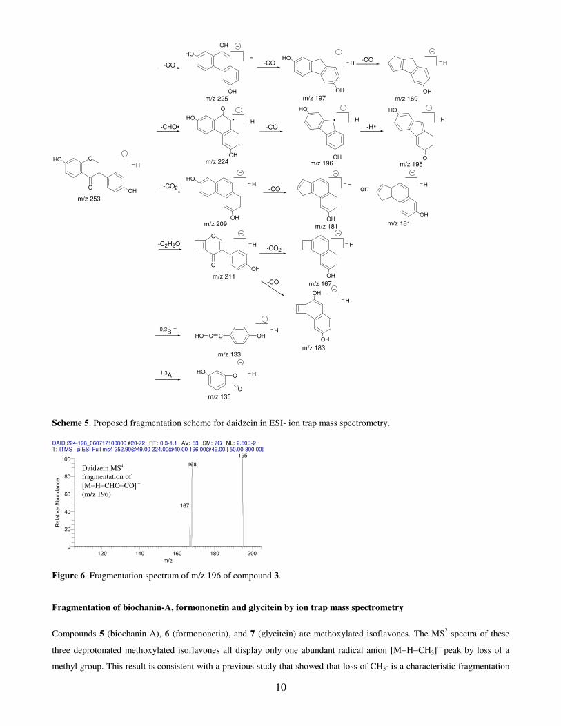
(see Figure 6). Scheme 5 details the proposed fragmentation scheme of daidzein in the ESI- ion trap mass spectrometry. The
presence of species at m/z 225 and m/z 224 is supported by their MS3 fragmentation spectra. The MS
3 spectra of both ions
do not reveal peaks for further loss of CO2, which means one of the two oxygen atoms at ring C has already been lost. So it
confirms there are losses of CO and CHO· from ring C which gives peaks at m/z 225 and 224 respectively. There is no
fragment corresponding to a loss of C3O2 (−68u) found for compounds 3 and 4. This is consistent with the result for
flavones where the loss of C3O2 implies a β-dihydroxyl configuration in the ring A.9 Similarly to compound 1 (genistein),
the loss of ketene does not occur at ring C but at ring A. This is supported by the formation of [M−H−C2H2O]− (m/z 211)
and its corresponding ion in daidzein-d3 [M−H−C2HDO]− (m/z 213), and further confirmed by the fact that fragment peaks
of CO2 loss and 0,3
B− cleavage are found in the further MS
3 spectrum of m/z 211 ion. In addition, loss of CO from m/z 225
produces m/z 197 and the MS4 spectrum of this [M−H−CO−CO]
− ion is different from the m/z 197 ions observed in
genistein (see Fig 3). It may be proposed that this ion is another isomer of m/z 197 with intact ring A and ring B in its
structure (see Scheme 5).
Figure 5. Fragmentation spectra of [M−H]− of compounds 3 and 4.
Table 2. Product ions obtained from [M−H]− ions in ion trap MS
2 spectra for compounds 3 and 4 [m/z with relative
abundances (%) in parenthesis].
Ions 3 4
[M−H−CO]− 225(100) 228(100)
[M−H−CHO]·−
224(85) 227(60)
[M−H−C2H2O]− 211(10) 213(10)
[M−H−CO2]− 209(90) 212(90)
[M−H−2CO]− 197(40) 200(30)
[M−H−CO2−CO]− 181(5) 184(2) 0,3B− 133(2) 135(1) 1,3A− 135(5) 136(5)
*49% of the relative collision energy was used in the fragmentation.
DAID MS2 #29-85 RT: 0.3-0.9 AV: 57 NL: 2.18E1T: ITMS - p ESI Full ms2 [email protected] [ 65.00-300.00]
80 90 100 110 120 130 140 150 160 170 180 190 200 210 220 230 240 250 260
m/z
0
20
40
60
80
100
Rela
tive A
bund
ance
225
209 224
197
208
211135 223196181 235185 207133 169117 160
DAID-d3 MS2 #1-93 RT: 0.0-1.0 AV: 93 NL: 2.37E1T: ITMS - p ESI Full ms2 [email protected] [ 70.00-290.00]
80 90 100 110 120 130 140 150 160 170 180 190 200 210 220 230 240 250 260
m/z
0
20
40
60
80
100
Rela
tive A
bund
ance
228
212
227
200
226211136 213184 210199188172 254149
Daidzein MS2
fragmentation of
[M−H]− (m/z 253)
Daidzein-d3 MS2
fragmentation of
[M−H]− (m/z 256)
10
O
OHO
OH
m/z 253
-CO
H
OH
m/z 181
-CO2H
OH
HO
m/z 209
0,3BHO C C OH
H
m/z 133
-CO
-CO
-C2H2O
OH
m/z 167
H
1,3AHO H
m/z 135
O
O
-CO2
m/z 181
or:
OH
-CO
m/z 169
H
OH
H
m/z 225 m/z 197
-CHO
m/z 224
H
OH
O
HO
m/z 196
H
HO
-CO
OH
-H
m/z 195
H
HO
O
OH
OH
H
O
O
OH
m/z 211
H
H
m/z 183
H
OH
OH
HO
HHO
OH
-CO
Scheme 5. Proposed fragmentation scheme for daidzein in ESI- ion trap mass spectrometry.
Figure 6. Fragmentation spectrum of m/z 196 of compound 3.
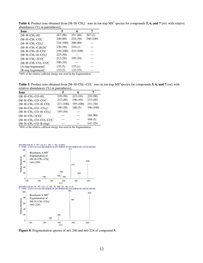
Fragmentation of biochanin-A, formononetin and glycitein by ion trap mass spectrometry
Compounds 5 (biochanin A), 6 (formononetin), and 7 (glycitein) are methoxylated isoflavones. The MS2 spectra of these
three deprotonated methoxylated isoflavones all display only one abundant radical anion [M−H−CH3]·−
peak by loss of a
methyl group. This result is consistent with a previous study that showed that loss of CH3· is a characteristic fragmentation
Daidzein MS4
fragmentation of
[M−H−CHO−CO]·−
(m/z 196)
DAID 224-196_060717100806 #20-72 RT: 0.3-1.1 AV: 53 SM: 7G NL: 2.50E-2T: ITMS - p ESI Full ms4 [email protected] [email protected] [email protected] [ 50.00-300.00]
120 140 160 180 200
m/z
0
20
40
60
80
100
Re
lative
Abu
nd
an
ce
195
168
167
11
in methoxylated flavonoids.13
In addition, loss of a hydrogen atom is easily found in these radical anions’ following
fragmentation spectra. Figure 7 shows fragmentation spectra of these three compounds, and Table 3−5 shows the proposed
product ions observed in the MSn frgmentation
spectra by ion trap mass spectrometry. The suggested fragmentation pathway
and structures for [M−H−CH3]·−
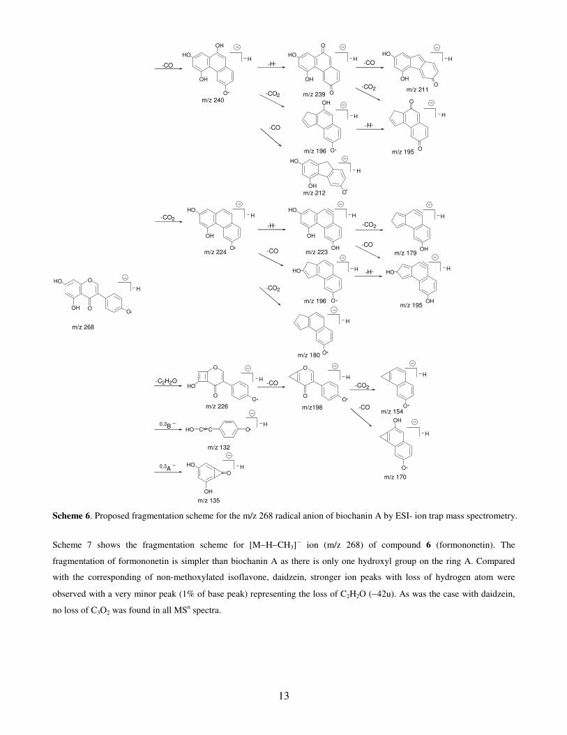
(m/z 268) of compound 5 (biochanin A) is shown in Scheme 6. The fragmentation behavior
of this compound is different to that of the corresponding non-methoxylated one (genistein). The fragmentation spectrum of
m/z 268 gives strong fragment peaks m/z 267 by losing a hydrogen atom; and also the MS4 fragmentation spectra of m/z240
and m/z 224 both give strong peaks (m/z 239 and m/z 223) by loss of a hydrogen atom (see Figure 8). This can be explained
by the possible formation of a more stable conjugated structure after loss of a hydrogen atom. The fragment ion peak (m/z
226) formed by loss of C2H2O (−42u) is observed in the MS3 fragmentation spectrum of [M−H−CH3]
·− (see Figure 7), but
no fragment peak loss of C3O2 (-68u) is found in all MSn fragmentation spectra of biochainin A, although it possesses a β-
dihydroxy moiety in ring A.
Figure 7. Fragmentation spectra of [M−H−CH3]·− of compounds 5, 6, 7; and fragmentation spectrum of [M−H−CH3−CO] ·−
of compound 7.
Table 3. Product ions obtained from [M−H]− ions in ion trap MS
2 spectra for compounds 5, 6, and 7 [m/z with relative
abundances (%) in parenthesis].
Ions 5 6 7
[M−H−CH3]·− 268 (100) 252 (100) 268 (100)
*40% of the relative collision energy was used in the fragmentation.
Glycitein MS4 #1-73 RT: 0.0-1.2 AV: 73 NL: 2.02E2T: ITMS - p ESI Full ms4 [email protected] [email protected] [email protected] [ 65.00-298.00]
100 120 140 160 180 200 220 240 260 280
m/z
0
20
40
60
80
100
Re
lative A
bu
nda
nce
196
239
184
212
147
195223168
183 238
Glycitein MS4
fragmentation of
[M−H−CH3−CO]·−
(m/z 240)
BIA ESI- MS3 #1-88 RT: 0.0-1.2 AV: 88 NL: 4.64E2T: ITMS - p ESI Full ms3 [email protected] [email protected] [ 70.00-298.00]
100 120 140 160 180 200 220 240 260 280
m/z
0
20
40
60
80
100
Re
lative A
bu
nda
nce
224 239
267
240
223
226
212
211196135132 250180 199
FORM ESI- MS3 #1-91 RT: 0.0-1.2 AV: 91 NL: 3.27E3T: ITMS - p ESI Full ms3 [email protected] [email protected] [ 90.00-298.00]
100 120 140 160 180 200 220 240 260 280
m/z
0
20
40
60
80
100
Re
lative A
bu
nda
nce
223
208
224
251
132195
135 210
Glycitein MS3 #1-59 RT: 0.0-0.8 AV: 59 NL: 3.88E3T: ITMS - p ESI Full ms3 [email protected] [email protected] [ 70.00-298.00]
100 120 140 160 180 200 220 240 260 280
m/z
0
20
40
60
80
100
Re
lative A
bu
nda
nce
240
239 267224212
Biochanin A MS3
fragmentation of
[M−H−CH3]·−
(m/z 268)
Formononetin MS3
fragmentation of
[M−H−CH3]·−
(m/z 252)
Glycitein MS3
fragmentation of
[M−H−CH3]·−
(m/z 268)
12
Table 4. Product ions obtained from [M−H−CH3]·− ions in ion trap MS
3 spectra for compounds 5, 6, and 7 [m/z with relative
abundances (%) in parenthesis].
Ions 5 6 7
[M−H−CH3−H]− 267 (90) 251 (40) 267 (2)
[M−H−CH3−CO]·− 240 (80) 224 (50) 240 (100)
[M−H−CH3−CO2]·− 224 (100) 208 (80) —
[M−H−CH3−C2H2O]·− 226 (30) 210 (1) —
[M−H−CH3−H−CO]− 239 (100) 223 (100) —
[M−H−CH3−H−CO2]− 223 (50) — —
[M−H−CH3−2CO]·− 212 (20) 195 (10) —
[M−H−CH3−CO2−CO]·− 196 (10) — —
[A-ring fragmment]− 135 (5) 135 (1) —
[B-ring fragmment]− 132 (2) 132 (15) —
*40% of the relative collision energy was used in the fragmentation.
Table 5. Product ions obtained from [M−H−CH3−CO]·− ions in ion trap MS4spectra for compounds 5, 6, and 7 [m/z with
relative abundances (%) in parenthesis].
Ions 5 6 7
[M−H−CH3−CO−H]− 239 (50) 223 (10) 239 (90)
[M−H−CH3−CO−CO]·− 212 (40) 196 (10) 212 (40)
[M−H−CH3−CO−H−CO]− 211 (100) 195 (100) 211 (30)
[M−H−CH3−CO −CO2]·− 196 (20) 180 (5) 196 (100)
[M−H−CH3−CO−H−CO2]− 195 (10) —
[M−H−CH3−3CO]·− — — 184 (80)
[M−H−CH3−CO−CO2−CO]·− — — 168 (5)
[M−H−CH3−CO−B-ring]− — — 147 (25)
*40% of the relative collision energy was used in the fragmentation.
Figure 8. Fragmentation spectra of m/z 240 and m/z 224 of compound 5.
BIA MS4 240 #1-7 RT: 0.0-0.1 AV: 7 NL: 3.05E1T: ITMS - p ESI Full ms4 [email protected] [email protected] [email protected] [ 65.00-300.00]
140 160 180 200 220 240
m/z
0
20
40
60
80
100
Rela
tive A
bundance
211
239
212
196225
195168 199
BIA MS4 224 #1-76 RT: 0.0-1.3 AV: 76 SM: 7G NL: 4.15T: ITMS - p ESI Full ms4 [email protected] [email protected] [email protected] [ 60.00-300.00]
140 160 180 200 220
m/z
0
20
40
60
80
100
Rela
tive A
bundance
195223
196
180
168
Biochanin A MS4
fragmentation of
[M−H−CH3−CO]·−
(m/z 240)
Biochanin A MS4
fragmentation of
[M−H−CH3−CO2]·−
(m/z 224)
13
O
O
OH
HO
O
m/z 268
H
-CO
-CO2m/z 240
-CO2H
O
HO
OH
m/z 224
0,3BHO C C O
H
m/z 132
0,3A
OH
HO
OH
m/z 135
-C2H2O
O
O
O
m/z 226
HO
m/z 212
OH
HO
OH
O
H
H
m/z 196
OH
O
H
O
HO H
m/z 196
O
H
OH
HO
O
-CO
m/z 239
OH
HO
O
H
O
-CO
m/z 195
O
H
O
m/z 211
H
OH
HO
-CO2O
H
OH
HO
OH
m/z 223
H
OHm/z 195
-CO2
HO
-COOH
m/z 179
H
-CO
-CO2
m/z 180
-CO
m/z 154O
H
-CO2
O
O
O
m/z198
H
-CO
OH
O
H
m/z 170
H
-H·
-H·
-H·
-H·
Scheme 6. Proposed fragmentation scheme for the m/z 268 radical anion of biochanin A by ESI- ion trap mass spectrometry.
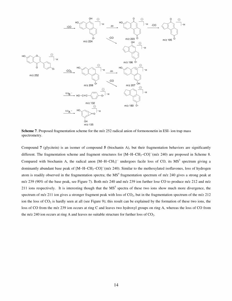
Scheme 7 shows the fragmentation scheme for [M−H−CH3]·− ion (m/z 268) of compound 6 (formononetin). The
fragmentation of formononetin is simpler than biochanin A as there is only one hydroxyl group on the ring A. Compared
with the corresponding of non-methoxylated isoflavone, daidzein, stronger ion peaks with loss of hydrogen atom were
observed with a very minor peak (1% of base peak) representing the loss of C2H2O (−42u). As was the case with daidzein,
no loss of C3O2 was found in all MSn spectra.
14
O
OHO
O
m/z 252
H
-CO
m/z 224
-CO2H
O
HO
m/z 208
0,3BHO C C O
H
m/z 132
0,3A
OH
HO
OH
m/z 135
-H
HO
OH
O
H
m/z 196
OH
O
H
O
H
m/z 180
m/z 223
HO
O
H
O
-CO
m/z 195
O
H
O
-HH
OH
HO
m/z 207
-CO
-CO
Scheme 7. Proposed fragmentation scheme for the m/z 252 radical anion of formononetin in ESI- ion trap mass
spectrometry.
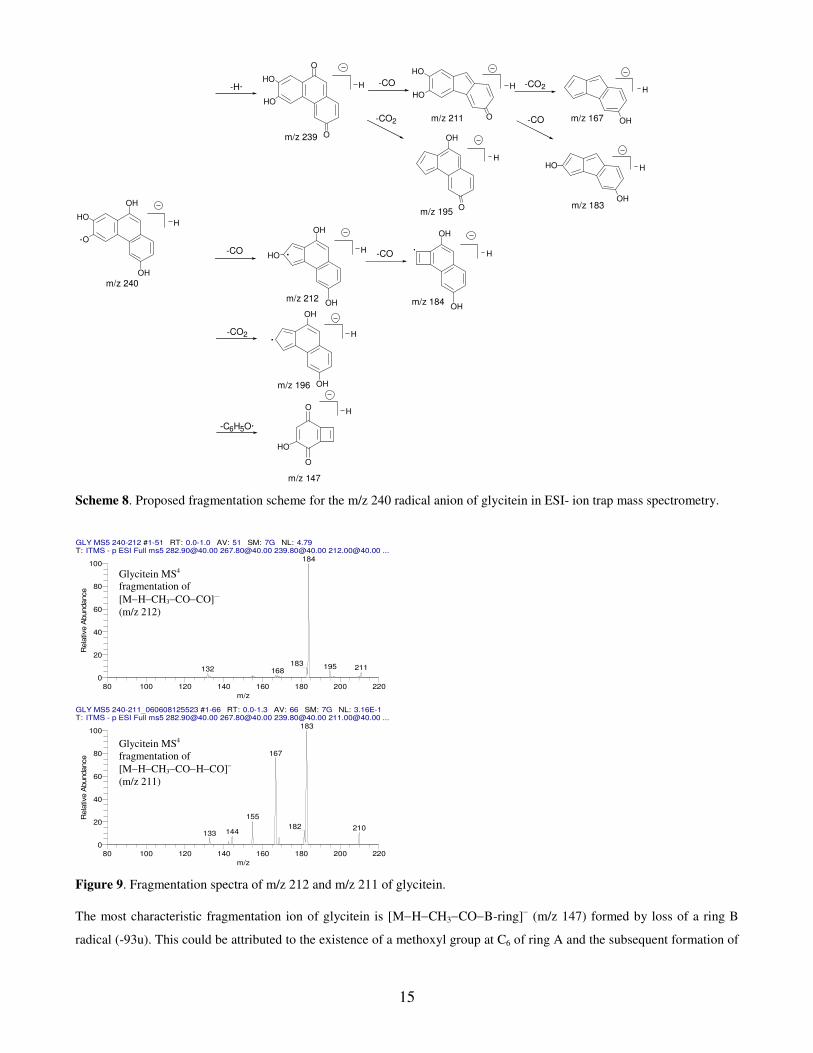
Compound 7 (glycitein) is an isomer of compound 5 (biochanin A), but their fragmentation behaviors are significantly
different. The fragmentation scheme and fragment structures for [M−H−CH3−CO]·−
(m/z 240) are proposed in Scheme 8.
Compared with biochanin A, the radical anon [M−H−CH3]·− undergoes facile loss of CO, its MS
3 spectrum giving a
dominantly abundant base peak of [M−H−CH3−CO]·−(m/z 240). Similar to the methoxylated isoflavones, loss of hydrogen
atom is readily observed in the fragmentation spectra; the MS4 fragmentation spectrum of m/z 240 gives a strong peak at
m/z 239 (90% of the base peak, see Figure 7). Both m/z 240 and m/z 239 ion further lose CO to produce m/z 212 and m/z
211 ions respectively. It is interesting though that the MS5 spectra of these two ions show much more divergence, the
spectrum of m/z 211 ion gives a stronger fragment peak with loss of CO2, but in the fragmentation spectrum of the m/z 212
ion the loss of CO2 is hardly seen at all (see Figure 9); this result can be explained by the formation of these two ions, the
loss of CO from the m/z 239 ion occurs at ring C and leaves two hydroxyl groups on ring A, whereas the loss of CO from
the m/z 240 ion occurs at ring A and leaves no suitable structure for further loss of CO2.
15
-CO2
m/z 240
HO
OH
OH
H
m/z 196
OH
OH
H
-CO
m/z 239
HO
O
H
O
-CO
m/z 195
OH
H
O
-CO2
O
HO
OH
HHO
m/z 212
m/z 211
H
HO
HO
OH
O
-CO
OH
H
m/z 184OH
-CO2
m/z 167
H
H
m/z 147
O
O
HO
OH
m/z 183
H
OH
-CO
HO
-C6H5O·
-H·
Scheme 8. Proposed fragmentation scheme for the m/z 240 radical anion of glycitein in ESI- ion trap mass spectrometry.
Figure 9. Fragmentation spectra of m/z 212 and m/z 211 of glycitein.
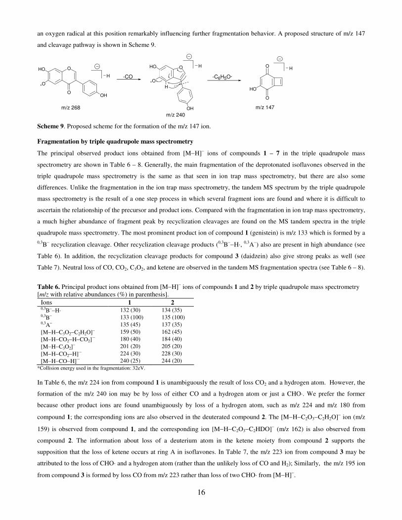
The most characteristic fragmentation ion of glycitein is [M−H−CH3−CO−B-ring]− (m/z 147) formed by loss of a ring B
radical (-93u). This could be attributed to the existence of a methoxyl group at C6 of ring A and the subsequent formation of
GLY MS5 240-211_060608125523 #1-66 RT: 0.0-1.3 AV: 66 SM: 7G NL: 3.16E-1T: ITMS - p ESI Full ms5 [email protected] [email protected] [email protected] [email protected] ...
80 100 120 140 160 180 200 220
m/z
0
20
40
60
80
100
Rela
tive A
bundance
183
167
155
182 210144133
GLY MS5 240-212 #1-51 RT: 0.0-1.0 AV: 51 SM: 7G NL: 4.79T: ITMS - p ESI Full ms5 [email protected] [email protected] [email protected] [email protected] ...
80 100 120 140 160 180 200 220
m/z
0
20
40
60
80
100
Rela
tive A
bundance
184
183 195 211132 168
Glycitein MS4
fragmentation of
[M−H−CH3−CO−CO]·−
(m/z 212)
Glycitein MS4
fragmentation of
[M−H−CH3−CO−H−CO]−
(m/z 211)
16
an oxygen radical at this position remarkably influencing further fragmentation behavior. A proposed structure of m/z 147
and cleavage pathway is shown in Scheme 9.
HO H
O
O
OH
H
H
m/z 240
m/z 147
O
O
HOO
OHO
OH
H -CO
m/z 268
O
-C6H5O·
Scheme 9. Proposed scheme for the formation of the m/z 147 ion.
Fragmentation by triple quadrupole mass spectrometry
The principal observed product ions obtained from [M−H]− ions of compounds 1 – 7 in the triple quadrupole mass
spectrometry are shown in Table 6 – 8. Generally, the main fragmentation of the deprotonated isoflavones observed in the
triple quadrupole mass spectrometry is the same as that seen in ion trap mass spectrometry, but there are also some
differences. Unlike the fragmentation in the ion trap mass spectrometry, the tandem MS spectrum by the triple quadrupole
mass spectrometry is the result of a one step process in which several fragment ions are found and where it is difficult to
ascertain the relationship of the precursor and product ions. Compared with the fragmentation in ion trap mass spectrometry,
a much higher abundance of fragment peak by recyclization cleavages are found on the MS tandem spectra in the triple
quadrupole mass spectrometry. The most prominent product ion of compound 1 (genistein) is m/z 133 which is formed by a
0,3B− recyclization cleavage. Other recyclization cleavage products (
0,3B−−H·,
0,3A−) also are present in high abundance (see
Table 6). In addition, the recyclization cleavage products for compound 3 (daidzein) also give strong peaks as well (see
Table 7). Neutral loss of CO, CO2, C3O2, and ketene are observed in the tandem MS fragmentation spectra (see Table 6 – 8).
Table 6. Principal product ions obtained from [M−H]− ions of compounds 1 and 2 by triple quadrupole mass spectrometry
[m/z with relative abundances (%) in parenthesis].
Ions 1 2 0,3B−−H· 132 (30) 134 (35) 0,3B− 133 (100) 135 (100) 0,3A− 135 (45) 137 (35)
[M−H−C3O2−C2H2O]− 159 (50) 162 (45)
[M−H−CO2−H−CO2]·− 180 (40) 184 (40)
[M−H−C3O2]− 201 (20) 205 (20)
[M−H−CO2−H]·− 224 (30) 228 (30)
[M−H−CO−H]·− 240 (25) 244 (20)
*Collision energy used in the fragmentation: 32eV.
In Table 6, the m/z 224 ion from compound 1 is unambiguously the result of loss CO2 and a hydrogen atom. However, the
formation of the m/z 240 ion may be by loss of either CO and a hydrogen atom or just a CHO·. We prefer the former
because other product ions are found unambiguously by loss of a hydrogen atom, such as m/z 224 and m/z 180 from
compound 1; the corresponding ions are also observed in the deuterated compound 2. The [M−H−C2O3−C2H2O]− ion (m/z
159) is observed from compound 1, and the corresponding ion [M−H−C2O3−C2HDO]− (m/z 162) is also observed from
compound 2. The information about loss of a deuterium atom in the ketene moiety from compound 2 supports the
supposition that the loss of ketene occurs at ring A in isoflavones. In Table 7, the m/z 223 ion from compound 3 may be
attributed to the loss of CHO· and a hydrogen atom (rather than the unlikely loss of CO and H2); Similarly, the m/z 195 ion
from compound 3 is formed by loss CO from m/z 223 rather than loss of two CHO· from [M−H]−.
17
Table 7. Principal product ions obtained from [M−H]− ions from compounds 3 and 4 in triple quadrupole mass spectrometry
[m/z with relative abundances (%) in parenthesis].
Ions 3 4 0,3B−−H· 132 (100) 134 (100) 0,3B− 133 (65) 135 (60)
[M−H−CO2−H−CO]·− 180 (50) 183 (30)
[M−H−CHO−H−CO]− 195 (60) 198 (45)
[M−H−CO2−H]·− 208 (70) 211 (60)
[M−H−CHO−H]− 223 (90) 226 (65)
*Collision energy used in the fragmentation: 35eV.
The fragmentation spectra of the deprotonated methoxylated isoflavones (compounds 5, 6, and 7) produce prominent peaks
of [M−H−CH3]−·
radical anions by loss of a methyl group (see Table 8). This is consistent with Justesen13
who observed
this process for other deprotonated methoxylated flavonoids. In addition, the compound 7 (glycitein) displays a base peak at
m/z 240 by further loss of CO from [M−H−CH3]−·
radical anion (m/z 268). This is consistent with the experiment by ion
trap mass spectrometry, where the m/z 240 ion from compound 7 is almost the only abundant product ion from its precursor
[M−H−CH3]·−. Similar to the result in ion trap mass spectrometry, all three [M−H−CH3]
·− give obvious [M−H−CH3−H]−
peaks by further loss of a hydrogen atom. This may be indicative that the loss of a hydrogen atom may be a common
fragmentation feature for radical anions of isoflavones.
Table 8. Principal product ions obtained from [M−H]− ions of compounds 5, 6 and 7 by triple quadrupole mass spectrometry
[m/z with relative abundances (%) in parenthesis].
Ions 5 6 7 0,3B−−H· 132 (10) 132 (40) — 0,3A− — 135 (20) —
[M−H−CH3−3CO]·− — — 184 (45)
[M−H−CH3−H−2CO]− — 195 (40) —
[M−H−CH3−CO−CO2]·− — — 196 (40)
[M−H−CH3−H−2CO]− 211 (10) — 211 (20)
[M−H−CH3−H−CO2]− 223 (10) — —
[M−H−CH3−H−CO]− 239 (20) 223 (90) 239 (20)
[M−H−CH3−CO]·− — — 240 (100)
[M−H−CH3−H]− 267 (30) 251 (50) 267 (10)
[M−H−CH3]·− 268 (100) 252 (100) 268 (60)
*Collision energy used in the fragmentation: 30eV.
Overall, all the principal product ions observed in the triple quadrupole mass spectrometry are matched with the schemes
and structures proposed from the ion trap mass spectrometry experiments, so it is not necessary to propose another scheme
for the fragmentation in triple quadrupole mass spectrometry.
CONCLUSIONS
In the present study, the fragmentation pathways of isoflavones (genistein, daidzein, biochanin A, formononitin, and
glycitein) have been elucidated through MSn step fragmentation experiments in the ion trap mass spectrometer. With the aid
of genistein-d4 and daidzein-d3, the location of a loss of ketene from the isoflavones is found to be at ring A. This is quite
differenct to that reported previously for flavones and flavanones9, where the loss of ketene is shown to occur at ring C.
Tentative structures for the product ions are proposed based on their fragmentation behaviors and chemical intuition.
Methoxylated isoflavones give only abundant peaks for the loss of a methyl group. Radical anions [M−H−CH3]·− easily lose
a hydrogen atom to form a more stable conjugated structure. Glycitein produces a characteristic fragment ion [M−H−
18
CH3−CO−B-ring]− (m/z 147), which may result from the existence of a methoxy group at C6. The ion trap spectrometry has
a number of advantages over the triple quadrupole tandem mass spectrometry as the ion trap permits multiple step
fragmentation experiments, which gives additional information to ascertain the relationship between precursor and product
ions.
REFERENCES
1. Cornwell T, Cohick W, Raskin I. Phytochemistry 2004; 65: 995.
2. Ososki AL, Kennelly EJ. Phytotherapy Research 2003; 17: 845.
3. de Rijke E, Out P, Niessen WMA, Ariese F, Gooijer C, Brinkman UAT. Journal of Chromatography A 2006; 1112:
31.
4. Wang C-C, Prasain JK, Barnes S. Journal of Chromatography B 2002; 777: 3.
5. Wu Q, Wang M, Simon JE. Journal of Chromatography B 2004; 812: 325.
6. Prasain JK, Wang C-C, Barnes S. Free Radical Biology and Medicine 2004; 37: 1324.
7. Ma YL, Li QM, VandenHeuvel H, Claeys M. Rapid Communications in Mass Spectrometry 1997; 11: 1357.
8. Ma YL, Van den Heuvel H, Claeys M. Rapid Communications in Mass Spectrometry 1999; 13: 1932.
9. Fabre N, Rustan I, de Hoffmann E, Quetin-Leclercq J. Journal of the American Society for Mass Spectrometry 2001;
12: 707.
10. Kuhn F, Oehme M, Romero F, Abou-Mansour E, Tabacchi R. Rapid Communications in Mass Spectrometry 2003; 17:
1941.
11. March RE, Miao X-S. International Journal of Mass Spectrometry 2004; 231: 157.
12. March RE, Miao X-S, Metcalfe CD, Stobiecki M, Marczak L. International Journal of Mass Spectrometry 2004; 232:
171.
13. Justesen U. Journal of Mass Spectrometry 2001; 36: 169.
14. Borges C, Martinho P, Martins A, Rauter AP, Ferreira MAA. Rapid Communications in Mass Spectrometry 2001; 15:
1760.
15. Hughes RJ, Croley TR, Metcalfe CD, March RE. International Journal of Mass Spectrometry 2001; 210-211: 371.
16. Antignac JP, Cariou R, Le Bizec B, Cravedi JP, Andre F. Rapid Communications in Mass Spectrometry 2003; 17:
1256.
17. de Rijke E, Zappey H, Ariese F, Gooijer C, Brinkman UAT. Journal of Chromatography A 2003; 984: 45.
18. Zhang JM, Brodbelt JS. Journal of Mass Spectrometry 2003; 38: 555.
19. Locati D, Morandi S, Cupisti A, Ghiadoni L, Arnoldi A.Rapid Communications in Mass Spectrometry 2005; 19: 3473.
20. Morandi S, Locati D, Ferrario F, Chiesa G, Arnoldi A. Rapid Communications in Mass Spectrometry 2005; 19: 153.
21. Liu RX, Ye M, Guo HZ, Bi KS, Guo DA. Rapid Communications in Mass Spectrometry 2005; 19: 1557.
22. Kang J, Price WE, Hick LA. Rapid Communications in Mass Spectrometry 2006; 20: 2411.
Related Documents