A facile method for expression and purification of the Alzheimer’s disease-associated amyloid b-peptide Dominic M. Walsh 1 , Eva Thulin 2 , Aedı´n M. Minogue 1 , Niklas Gustavsson 3 , Eric Pang 4 , David B. Teplow 4 and Sara Linse 1,2 1 Laboratory for Neurodegenerative Research, School of Biomolecular and Biomedical Science, Conway Institute, Belfield, University College Dublin, Republic of Ireland 2 Department of Biophysical Chemistry, Chemical Centre, Lund University, Sweden 3 Department of Biochemistry, Chemical Centre, Lund University, Sweden 4 Biopolymer Laboratory, David Geffen School of Medicine at UCLA, Los Angeles, CA, USA Multiple lines of evidence indicate that the amyloid b peptide (Ab) plays an important role in the patho- genesis of Alzheimer’s disease [1]. In nature, Ab does not occur as a single molecular species, and more than 20 different Ab sequences have been detected in human cerebrospinal fluid and brain. The most com- mon Ab isoform is Ab1–40, a 40-residue peptide that begins at Asp1 and terminates at Val40 (Fig. 1) [2– 11]. Increased production of Ab1–42, a peptide that differs from Ab1–40 by addition of Ile and Ala to Keywords Aß; Alzheimer’s disease; aggregation; amyloid; fibrillogenesis Correspondence D. M. Walsh, Laboratory for Neurodegenerative Research, School of Biomolecular and Biomedical Science, Conway Institute, Belfield, University College Dublin, Dublin 4, Republic of Ireland Fax: 353 1 716 6890 Tel: 353 1 716 6751 E-mail: [email protected] S. Linse, Department of Biophysical Chemistry, Chemical Centre, Lund University, PO Box 124, SE-22100 Lund, Sweden Fax: 46 46 2228246 Tel: 46 46 224543 E-mail: [email protected] Re-use of this article is permitted in accordance with the Creative Commons Deed, Attribution 2.5, which does not permit commercial exploitation (Received 18 November 2008, revised 10 December 2008, accepted 17 December 2008) doi:10.1111/j.1742-4658.2008.06862.x We report the development of a high-level bacterial expression system for the Alzheimer’s disease-associated amyloid b-peptide (Ab), together with a scaleable and inexpensive purification procedure. Ab(1–40) and Ab(1–42) coding sequences together with added ATG codons were cloned directly into a Pet vector to facilitate production of Met-Ab(1–40) and Met-Ab(1– 42), referred to as Ab(L1–40) and Ab(L1–42), respectively. The expression sequences were designed using codons preferred by Escherichia coli, and the two peptides were expressed in this host in inclusion bodies. Peptides were purified from inclusion bodies using a combination of anion-exchange chromatography and centrifugal filtration. The method described requires little specialized equipment and provides a facile and inexpensive procedure for production of large amounts of very pure Ab peptides. Recombinant peptides generated using this protocol produced amyloid fibrils that were indistinguishable from those formed by chemically synthesized Ab1–40 and Ab1–42. Formation of fibrils by all peptides was concentration-dependent, and exhibited kinetics typical of a nucleation-dependent polymerization reaction. Recombinant and synthetic peptides exhibited a similar toxic effect on hippocampal neurons, with acute treatment causing inhibition of MTT reduction, and chronic treatment resulting in neuritic degeneration and cell loss. Abbreviations Ab, amyloid b-peptide; GuHCL, guanidine hydrochlorise; MetAP-TG, methionine aminopeptidase TG; MTT, 3-(4,5-dimethylthiazol-2-yl)-2,5- diphenyltetrazolium bromide; SEC, size-exclusion chromatography; ThT, thioflavin T. 1266 FEBS Journal 276 (2009) 1266–1281 ª 2009 The Authors Journal compilation ª 2009 FEBS

Welcome message from author
This document is posted to help you gain knowledge. Please leave a comment to let me know what you think about it! Share it to your friends and learn new things together.
Transcript
A facile method for expression and purification of theAlzheimer’s disease-associated amyloid b-peptideDominic M. Walsh1, Eva Thulin2, Aedın M. Minogue1, Niklas Gustavsson3, Eric Pang4,David B. Teplow4 and Sara Linse1,2
1 Laboratory for Neurodegenerative Research, School of Biomolecular and Biomedical Science, Conway Institute, Belfield, University College
Dublin, Republic of Ireland
2 Department of Biophysical Chemistry, Chemical Centre, Lund University, Sweden
3 Department of Biochemistry, Chemical Centre, Lund University, Sweden
4 Biopolymer Laboratory, David Geffen School of Medicine at UCLA, Los Angeles, CA, USA
Multiple lines of evidence indicate that the amyloid bpeptide (Ab) plays an important role in the patho-
genesis of Alzheimer’s disease [1]. In nature, Ab does
not occur as a single molecular species, and more
than 20 different Ab sequences have been detected in
human cerebrospinal fluid and brain. The most com-
mon Ab isoform is Ab1–40, a 40-residue peptide that
begins at Asp1 and terminates at Val40 (Fig. 1) [2–
11]. Increased production of Ab1–42, a peptide that
differs from Ab1–40 by addition of Ile and Ala to
Keywords
Aß; Alzheimer’s disease; aggregation;
amyloid; fibrillogenesis
Correspondence
D. M. Walsh, Laboratory for
Neurodegenerative Research, School of
Biomolecular and Biomedical Science,
Conway Institute, Belfield, University
College Dublin, Dublin 4, Republic of Ireland
Fax: 353 1 716 6890
Tel: 353 1 716 6751
E-mail: [email protected]
S. Linse, Department of Biophysical
Chemistry, Chemical Centre, Lund
University, PO Box 124, SE-22100 Lund,
Sweden
Fax: 46 46 2228246
Tel: 46 46 224543
E-mail: [email protected]
Re-use of this article is permitted in
accordance with the Creative Commons
Deed, Attribution 2.5, which does not
permit commercial exploitation
(Received 18 November 2008, revised 10
December 2008, accepted 17 December
2008)
doi:10.1111/j.1742-4658.2008.06862.x
We report the development of a high-level bacterial expression system for
the Alzheimer’s disease-associated amyloid b-peptide (Ab), together with a
scaleable and inexpensive purification procedure. Ab(1–40) and Ab(1–42)coding sequences together with added ATG codons were cloned directly
into a Pet vector to facilitate production of Met-Ab(1–40) and Met-Ab(1–42), referred to as Ab(L1–40) and Ab(L1–42), respectively. The expression
sequences were designed using codons preferred by Escherichia coli, and
the two peptides were expressed in this host in inclusion bodies. Peptides
were purified from inclusion bodies using a combination of anion-exchange
chromatography and centrifugal filtration. The method described requires
little specialized equipment and provides a facile and inexpensive procedure
for production of large amounts of very pure Ab peptides. Recombinant
peptides generated using this protocol produced amyloid fibrils that were
indistinguishable from those formed by chemically synthesized Ab1–40 and
Ab1–42. Formation of fibrils by all peptides was concentration-dependent,
and exhibited kinetics typical of a nucleation-dependent polymerization
reaction. Recombinant and synthetic peptides exhibited a similar toxic
effect on hippocampal neurons, with acute treatment causing inhibition of
MTT reduction, and chronic treatment resulting in neuritic degeneration
and cell loss.
Abbreviations
Ab, amyloid b-peptide; GuHCL, guanidine hydrochlorise; MetAP-TG, methionine aminopeptidase TG; MTT, 3-(4,5-dimethylthiazol-2-yl)-2,5-
diphenyltetrazolium bromide; SEC, size-exclusion chromatography; ThT, thioflavin T.
1266 FEBS Journal 276 (2009) 1266–1281 ª 2009 The Authors Journal compilation ª 2009 FEBS
the C-terminus, is particularly associated with disease
[12]. Through biochemical and animal modeling stud-
ies, researchers have built up a detailed picture of the
natural economy of brain Ab. Like all proteins, the
steady-state level of Ab is controlled by its produc-
tion, degradation and clearance, and it is proposed
that a defect leading to over-production or decreased
clearance causes an accumulation of Ab and that this
triggers a pathogenic cascade culminating in the cog-
nitive deficits that characterize Alzheimer’s disease
[13–16]. The self-association constants of Ab are rela-
tively high, and a variety of assemblies are formed at
micromolar concentrations, ranging from dimers to
aggregates of amyloid fibrils [17]. However, as yet
the specific form(s) of Ab that causes injury to neu-
rons in vivo has not been identified [16]. Clearly a
detailed understanding of the structure of both the
Ab monomer and its various assemblies could help in
the design of new therapeutic strategies targeted at
preventing the formation or ameliorating the activity
of toxic Ab assemblies.
Although much progress has been made since the
sequence of Ab was first determined, high-resolution
structural analysis of Ab monomer and its assemblies
has been hampered because of the lack of an afford-
able source of Ab peptides. Chemical synthesis of
various Ab peptides is now routine [18,19], but is
time-consuming and requires access to specialized
equipment, and is relatively expensive, especially for
isotope labeling. Moreover, solid-phase synthesis of
Ab peptides containing radioisotopes such as 35S-Met
is not practical. Thus we aimed to develop a simple
inexpensive procedure for the production of recombi-
nant Ab peptides that would allow isotope labeling
and the generation of Ab peptides with design or
disease-associated amino acid substitutions. Produc-
tion and purification of recombinant Ab peptides has
been investigated previously, but most published
methods either require highly specialized equipment
and ⁄or expensive reagents [20–22], or are only suit-
able for the production of short biologically irrelevant
fragments of Ab [23]. Here we describe a rapid and
inexpensive protocol for the expression and purifica-
tion of Ab(1–40) and Ab(1–42) with exogenous initi-
ating Met residues. This procedure does not require
specialized equipment, is suitable for isotopic labeling
of peptides, and can be readily adapted for the gener-
ation of Ab peptides containing an array of sequence
variations.
Results
Expression of Ab(M1–40) and Ab(M1–42)
Sequence-verified PetSac plasmids containing either the
Ab(L1–40) or Ab(L1–42) gene (Fig. 1) were used for
expression in Escherichia coli as described in Experi-
mental procedures. For Ab(M1–40) and Ab(M1–42),
the highest yields were obtained between 3 and 4 h
after induction, with similar yields at concentrations of
isopropyl thio-b-d-galactoside ranging from 0.1–
1.2 mm and temperatures ranging from 37–41 �C (data
not shown). Under these conditions, the cells grow to
an attenuance at 600 nm (D660 nm) of 3.0–3.1.
SDS-PAGE and agarose gel electrophoresis of soni-
cates of the bacterial cell pellet and the urea extract
revealed that the first and second supernatants after
sonication contained mainly E. coli proteins, and the
majority of Ab(M1–40) and Ab(M1–42) was present in
the urea extract (Fig. 2). On agarose gels, the major
band migrated as expected according to the net charge
of the Ab peptides at pH 8.4, and on SDS-PAGE the
major band migrated between 4 and 5 kDa (Fig. 2).
These data indicate that both peptides accumulate in
inclusion bodies, and that Ab(L1–40) is the dominant
protein in the inclusion bodies. In contrast, the major
protein in the Ab(M1–42) inclusions was not Ab, butwas the small heat shock protein IbpB (accession num-
ber B1IYQ8), identified by mass spectrometry after
tryptic digestion of the gel band (data not shown).
The PCR protocol used to generate Ab(M1–40) and
Ab(M1–42) was designed to facilitate incorporation of
familial mutants by exchange of only the middle pri-
mer. We produced six plasmids encoding Ab(M1–40)
that incorporate the point mutations F19P, A21G,
E22G, E22K, E22Q and D23N, and another six
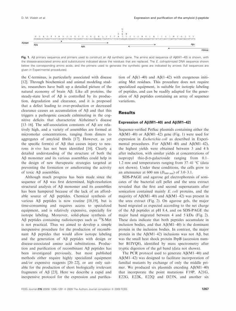
Fig. 1. Ab primary sequence and primers used to construct an Ab synthetic gene. The amino acid sequence of Ab(M1–40) is shown, with
the disease-associated amino acid substitutions indicated above the residues that are replaced. The E. coli-optimized DNA sequence shown
below the corresponding amino acids, and the primers used to generate the synthetic gene are indicated by arrows (full sequences are
given in Experimental procedures).
D. M. Walsh et al. Expression and purification of the amyloid b-peptide
FEBS Journal 276 (2009) 1266–1281 ª 2009 The Authors Journal compilation ª 2009 FEBS 1267

plasmids encoding Ab(M1–42) with the point muta-
tions F19P, A21G, E22G, E22K, E22Q and D23N.
These mutated versions can be expressed and purified
using the procedure described here, although the
higher aggregation tendency of some of these mutants
leads to lower yields. On agarose gel electrophoresis,
the peptides were found to migrate according to their
respective net charge relative to wild-type (Fig. 2E).
Purification of Ab(M1–40) and Ab(M1–42)
The present work describes a rapid and inexpensive
purification scheme to produce high-purity Ab(M1–40)
and Ab(M1–42) in 24 h. The purification scheme, as
described in detail in Experimental procedures,
involves ion-exchange chromatography in batch mode,
followed by molecular mass fractionation using centrif-
ugal devices. This simple two-step purification results
in a highly pure product, and yields 10–20 mg of
Ab(M1–40) per liter of culture. In the example shown
in Fig. 3, 30 mg of peptide was obtained from 2.2 L of
bacterial culture. The process can easily be scaled pro-
portionally for other amounts. In the example shown
in Fig. 3, the resin was washed with low-salt buffer fol-
lowed by stepwise elution using 50, 75, 100, 125, 150,
200, 250, 300 and 500 mm NaCl, and fractions eluted
using 50–125 mm NaCl were collected for molecular
mass fractionation. In later batches, we washed the
resin with buffer containing 25 mm NaCl and then
eluted the peptide with buffer containing 125 mm
NaCl, simplifying the procedures even further.
Urea-solubilized inclusion bodies containing
Ab(M1–42) were purified by anion-exchange chroma-
tography in the same fashion as for Ab(M1–40)
(Fig. 3C,D). Fractions eluted with 75–125 mm NaCl
were passed through a 30 kDa molecular mass cut-off
filter, yielding a total of 5 mg of Ab(M1–42) in
150 mL. Another 3 mg in 100 mL was obtained in the
30 kDa filtrate from fractions eluted at 150–200 mm
NaCl. For both Ab(M1–40) and Ab(M1–42), all
manipulations were performed at slightly alkaline pH
to avoid the formation of structural contaminants pro-
duced by isoelectric precipitation. Depending on the
required use, peptides can be lyophilized, used directly
or concentrated.
Ion-exchange column chromatography
Attempts to purify Ab(M1–40) or Ab(M1–42) by ion-
exchange column chromatography (not shown) led to
much lower yields of monomeric peptide than the
batch method. When repeated using 8 m urea-contain-
A
B
C
E D
Fig. 2. Ab(M1–40) and Ab(M1–42) are
expressed in inclusion bodies. (A–D) Pellets
of bacteria expressing Ab(M1–40) (A,B) or
Ab(M1–42) (C,D) were subjected to three
rounds of sonication in buffer, and at the
end of each sonication step the suspension
was centrifuged and the supernatants
(labeled S1, S2 and S3) were stored pending
analysis. The pellet was then extracted in
8 M urea (fraction labeled U), and purified by
ion exchange (fraction labeled IE), filtration
through a 30 kDa molecular mass cut-off
filter (fraction labeled 30) and concentration
on a 3 kDa molecular mass cut-off filter
(fraction labeled 3). All fractions were elec-
trophoresed on 10–20% polyacrylamide
Tris-tricine gels (A,C) and 1% agarose gels
(B,D), and proteins were visualized by Coo-
massie stain. Lanes HS and LS are molecu-
lar mass standards, with the molecular
mass in kDa given on the left. (E) 1% aga-
rose gel electrophoresis of urea extracts of
inclusion bodies from bacteria expressing
Ab(M1–40) with wild-type (wt) sequence or
with the following point mutations: A21G,
E22G, E22K, E22Q and D23N. The net
charge of each peptide is indicated under-
neath each lane.
Expression and purification of the amyloid b-peptide D. M. Walsh et al.
1268 FEBS Journal 276 (2009) 1266–1281 ª 2009 The Authors Journal compilation ª 2009 FEBS
ing buffers, the yields of eluted peptide were as high
as or higher than with the batch mode, but the pep-
tide was eluted at very high concentration and the
majority of the material did not pass through the
30 kDa filter.
Concentration of purified Ab(M1–40) and
Ab(M1–42)
Ab(M1–40) and Ab(M1–42) each contain a single tyro-
sine residue, and absorption of tyrosine at 275 nm
(e275 = 1400 m)1Æcm)1) was used to estimate the
concentration of Ab in solution. In four separate puri-
fication experiments, the concentration of Ab(M1–40)
in the 30 kDa filtrate was determined to be between 30
and 50 lm. The average Ab(M1–40) concentration
in the 30 kDa filtrate of the peak fractions (eluting at
75–125 mm NaCl) was 40 lm, based on the absor-
bance at 275 nm. This concentration is higher than
required for thioflavin T (ThT)-based fibrillation
assays (typical concentrations used are 3–10 lm), but
is not sufficient for other biophysical studies. We
therefore examined a number of methods to further
concentrate the Ab solution. Although several different
methods proved useful (e.g. C18 SepPak reverse-phase
columns), the best yield and most rapid results were
obtained using a 3 kDa molecular mass cut-off
centrifugal filtration device. When a solution of
Ab(M1–40) of approximately 40 lm was concentrated
approximately eightfold, approximately 75% of the
peptide was recovered at a concentration of approxi-
mately 230 lm.
Amino acid analysis, mass spectrometry and
sequencing
The purified peptides were subjected to mass spectrom-
etry, amino acid analysis and N-terminal amino acid
sequencing. These methods confirm expression of the
correct peptide and that the peptide species contains
the N-terminal methionine residue. For Ab(M1–40),
the observed relative molecular mass (mono-isotopic
mass) was 4459.19 (expected 4459.21), and the isotope
distribution was as predicted from the sequence
(Fig. S1). The amino acid analysis after acid hydrolysis
(Table 1) shows a very close correspondence with the
expected composition, indicating that the peptide is of
the correct sequence and free of contaminating pro-
teins. Five cycles of N-terminal sequencing confirmed
the expected residues including the presence of methio-
nine at position 1 (not shown). MS ⁄MS fragment ion
analysis confirmed the correct sequence of Ab(M1–40)
(data not shown).
Co-expression of Ab(M1–40) with aminopeptidase
Mass spectrometric analysis of Ab(M1–40) and
Ab(M1–42) from several batches very clearly showed
A
B
C
D
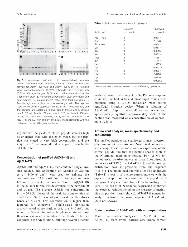
Fig. 3. Ion-exchange purification of urea-solubilized inclusion
bodies. Anion-exchange chromatography in batch mode was per-
formed for Ab(M1–40) (A,B) and Ab(M1–42) (C,D). All fractions
were electrophoresed on 10–20% polyacrylamide Tris-tricine gels
(A,C) or 1% agarose gels (B,D), and proteins were visualized by
Coomassie stain. S, combined supernatants after sonication and
centrifugation; U, urea-solubilized pellet after third sonication; F,
flow-through from application to ion-exchange resin. The peptides
were eluted using a stepwise increase in NaCl concentration, and
the fractions are labeled as follows: lane 0, 0 mM; lane 1, 50 mM;
lane 2, 75 mM; lane 3, 100 mM; lane 4, 125 mM; lane 5, 150 mM;
lane 6, 200 mM; lane 7, 250 mM; lane 8, 300 mM; lane 9, 500 mM
NaCl. HS and LS, high and low molecular mass standards with the
molecular mass in kDa given on the left.
Table 1. Amino acid analysis after acid hydrolysis.
Amino acid
Expected
composition
Observed
composition
Asp + Asn 4 3.9917
Ser 2 2.1648
Glu + Gln 4 4.0643
Gly 6 6.0117
Ala 3 3.0265
Val 5 5.0813
Met 2 1.7661
Ilea 2 1.1517
Leu 2 1.9935
Tyr 1 0.96174
Phe 3 2.9287
His 3 2.8426
Lys 2 2.0270
Arg 1 0.99652
a Ile–Ile peptide bonds are known to be inefficiently hydrolyzed.
D. M. Walsh et al. Expression and purification of the amyloid b-peptide
FEBS Journal 276 (2009) 1266–1281 ª 2009 The Authors Journal compilation ª 2009 FEBS 1269
A
B
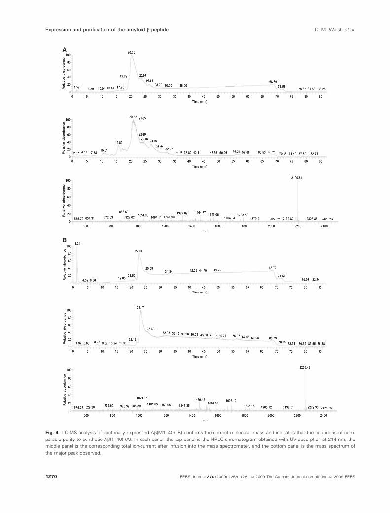
Fig. 4. LC-MS analysis of bacterially expressed Ab(M1–40) (B) confirms the correct molecular mass and indicates that the peptide is of com-
parable purity to synthetic Ab(1–40) (A). In each panel, the top panel is the HPLC chromatogram obtained with UV absorption at 214 nm, the
middle panel is the corresponding total ion-current after infusion into the mass spectrometer, and the bottom panel is the mass spectrum of
the major peak observed.
Expression and purification of the amyloid b-peptide D. M. Walsh et al.
1270 FEBS Journal 276 (2009) 1266–1281 ª 2009 The Authors Journal compilation ª 2009 FEBS
the presence of Ab(M1–40) or Ab(M1–42), with no
indication of any product resulting from spontaneous
cleavage of the N-terminal methionine in E. coli
(Figs 4, S1 and S2). Co-expression of the E. coli
aminopeptidase methionine aminopeptidase TG
(MetAP-TG) [24] and Ab(M1–40) was therefore
attempted, and was found to results in a low yield of
Ab(1–40). Ab was purified from the cell pellet as
described above, and analyzed by MALDI-TOF MS
(Fig. S1). Assuming similar ionization of Ab(M1–40)
and Ab(1–40), we found that less than 20% of
Ab(M1–40) was converted to Ab(1–40) by this method
(Fig. S1), although the expression level of aminopepti-
dase MetAP-TG was higher than that for Ab(M1–40)
as determined by SDS-PAGE (not shown). MS ⁄MS
fragment ion analysis confirmed the correct sequence
of the Ab(1–40) produced by co-expression with
aminopeptidase.
Isolation of monomeric Ab and kinetic analysis of
aggregation
As aggregation of Ab peptides is strongly influenced
by the presence of structural and chemical impurities,
all samples were denatured using 5 m guanidine
hydrochloride (GuHCl) in 50 mm Tris-HCl pH 8.0
and subjected to size-exclusion chromatography (SEC)
to isolate homogenous monomeric Ab solutions, as
described previously [25]. All four peptides produced
a large peak that eluted around 12.5 mL from a
Superdex 75 10 ⁄ 30 HR column (data not shown). Fur-
ther analysis of these peaks by reverse-phase HPLC
A
B
E
C
D
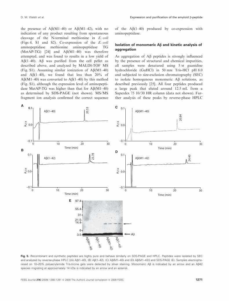
Fig. 5. Recombinant and synthetic peptides are highly pure and behave similarly on SDS-PAGE and HPLC. Peptides were isolated by SEC
and analyzed by reverse-phase HPLC [(A) Ab(1–40), (B) Ab(1–42), (C) Ab(M1–40) and (D) Ab(M1–42)] and SDS-PAGE (E). Samples electropho-
resed on 10–20% polyacrylamide Tris-tricine gels were detected by silver staining. Monomeric Ab is indicated by an arrow and an Ab42
species migrating at approximately 14 kDa is indicated by an arrow and an asterisk.
D. M. Walsh et al. Expression and purification of the amyloid b-peptide
FEBS Journal 276 (2009) 1266–1281 ª 2009 The Authors Journal compilation ª 2009 FEBS 1271
and SDS-PAGE ⁄ silver staining revealed highly pure
starting material. In each case, the peptides produced a
single peak on HPLC (Fig. 5A–D). The retention times
of Ab(1–40) and Ab(M1–40) were highly similar and
the peaks were typically symmetrical. The retention
times and peak shapes for Ab(1–42) and Ab(M1–42)
were similar to each other, but were distinct from
those of the peptides terminating at Val40. The more
hydrophobic peptides ending at Ala42 were retained
on the column for longer, and produced less symmetri-
cal peaks, as found previously for synthetic peptides
[26]. On SDS-PAGE, all four peptides produced a
band that migrated at approximately 4 kDa. Given the
small molecular mass difference between the peptides
ending at Val40 and Ala42, it is not possible to resolve
these peptides on standard SDS-PAGE [27]; however,
this system is useful to confirm the correct migration
of Ab peptides and their relative purity as assessed by
silver staining. In the examples shown, 100 ng of each
peptide were loaded per lane, and only a single band
was detected in the lanes containing Ab(1–40) and
Ab(M1–40) (Fig. 5E). In other experiments, 400 ng of
peptide were loaded in each well, and very darkly
stained broad Ab bands were detected upon silver
staining, but no additional non-Ab bands were
detected. Prior experience indicates that the silver
staining protocol used can detect as little as 10 ng of
protein [28], thus the present results suggest that SEC-
isolated Ab(1–40) and Ab(M1–40) are at least 97%
pure. In the lanes containing Ab(1–42) and Ab(M1–
42), there were prominent bands at approximately
4 kDa and faint bands at approximately 14 kDa. The
band at approximately 14 kDa is not an impurity as it
was present in both the recombinant and synthetic
peptides, but probably represents an artifact of SDS-
PAGE [29] as it was also detected by Western blotting
using anti-Ab specific antibodies (not shown). Thus, as
with the peptides terminating at Val40, Ab(1–42) and
Ab(M1–42) are also at least 97% pure. Together, these
results confirm that our recombinant Ab(M1–40) and
Ab(M1–42) are at least as pure as the synthetic
peptides purified by reverse-phase HPLC, a finding
corroborated by LC-MS analysis (Figs 4 and S2).
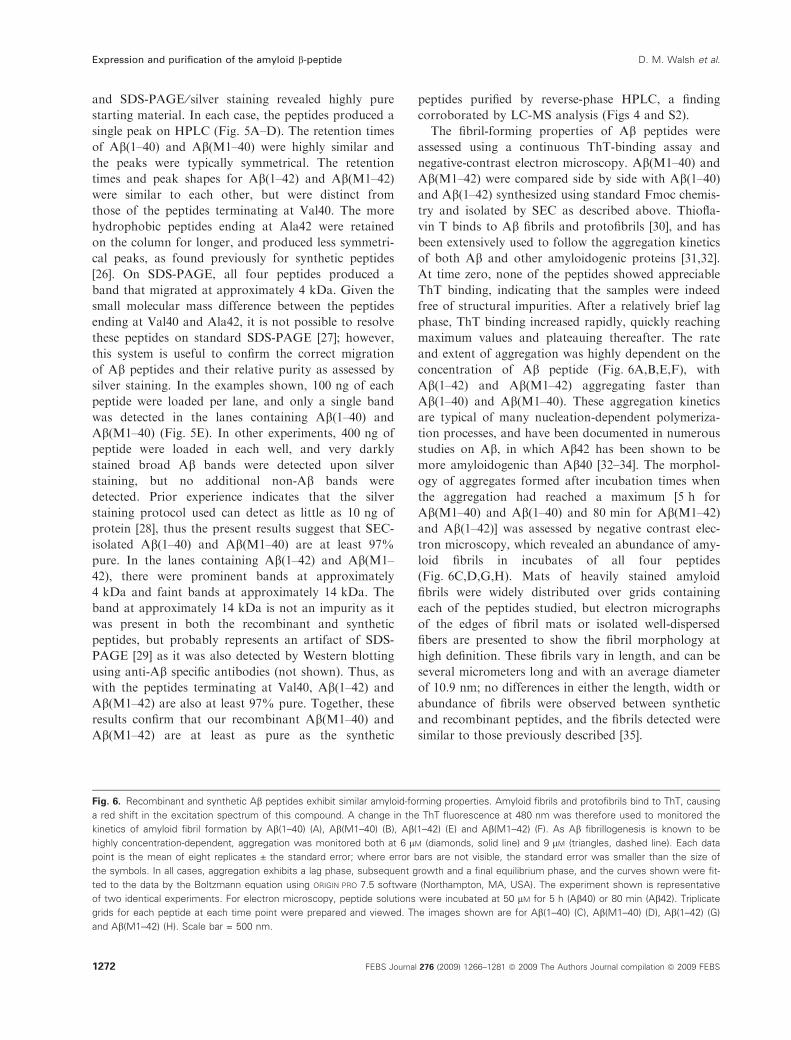
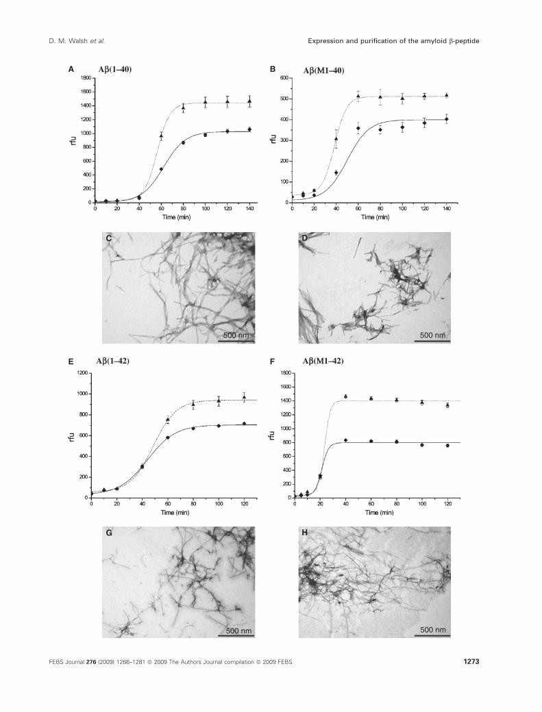
The fibril-forming properties of Ab peptides were
assessed using a continuous ThT-binding assay and
negative-contrast electron microscopy. Ab(M1–40) and
Ab(M1–42) were compared side by side with Ab(1–40)and Ab(1–42) synthesized using standard Fmoc chemis-
try and isolated by SEC as described above. Thiofla-
vin T binds to Ab fibrils and protofibrils [30], and has
been extensively used to follow the aggregation kinetics
of both Ab and other amyloidogenic proteins [31,32].
At time zero, none of the peptides showed appreciable
ThT binding, indicating that the samples were indeed
free of structural impurities. After a relatively brief lag
phase, ThT binding increased rapidly, quickly reaching
maximum values and plateauing thereafter. The rate
and extent of aggregation was highly dependent on the
concentration of Ab peptide (Fig. 6A,B,E,F), with
Ab(1–42) and Ab(M1–42) aggregating faster than
Ab(1–40) and Ab(M1–40). These aggregation kinetics
are typical of many nucleation-dependent polymeriza-
tion processes, and have been documented in numerous
studies on Ab, in which Ab42 has been shown to be
more amyloidogenic than Ab40 [32–34]. The morphol-
ogy of aggregates formed after incubation times when
the aggregation had reached a maximum [5 h for
Ab(M1–40) and Ab(1–40) and 80 min for Ab(M1–42)
and Ab(1–42)] was assessed by negative contrast elec-
tron microscopy, which revealed an abundance of amy-
loid fibrils in incubates of all four peptides
(Fig. 6C,D,G,H). Mats of heavily stained amyloid
fibrils were widely distributed over grids containing
each of the peptides studied, but electron micrographs
of the edges of fibril mats or isolated well-dispersed
fibers are presented to show the fibril morphology at
high definition. These fibrils vary in length, and can be
several micrometers long and with an average diameter
of 10.9 nm; no differences in either the length, width or
abundance of fibrils were observed between synthetic
and recombinant peptides, and the fibrils detected were
similar to those previously described [35].
Fig. 6. Recombinant and synthetic Ab peptides exhibit similar amyloid-forming properties. Amyloid fibrils and protofibrils bind to ThT, causing
a red shift in the excitation spectrum of this compound. A change in the ThT fluorescence at 480 nm was therefore used to monitored the
kinetics of amyloid fibril formation by Ab(1–40) (A), Ab(M1–40) (B), Ab(1–42) (E) and Ab(M1–42) (F). As Ab fibrillogenesis is known to be
highly concentration-dependent, aggregation was monitored both at 6 lM (diamonds, solid line) and 9 lM (triangles, dashed line). Each data
point is the mean of eight replicates ± the standard error; where error bars are not visible, the standard error was smaller than the size of
the symbols. In all cases, aggregation exhibits a lag phase, subsequent growth and a final equilibrium phase, and the curves shown were fit-
ted to the data by the Boltzmann equation using ORIGIN PRO 7.5 software (Northampton, MA, USA). The experiment shown is representative
of two identical experiments. For electron microscopy, peptide solutions were incubated at 50 lM for 5 h (Ab40) or 80 min (Ab42). Triplicate
grids for each peptide at each time point were prepared and viewed. The images shown are for Ab(1–40) (C), Ab(M1–40) (D), Ab(1–42) (G)
and Ab(M1–42) (H). Scale bar = 500 nm.
Expression and purification of the amyloid b-peptide D. M. Walsh et al.
1272 FEBS Journal 276 (2009) 1266–1281 ª 2009 The Authors Journal compilation ª 2009 FEBS
A B
E F
C D
G H
500 nm 500 nm
500 nm500 nm
D. M. Walsh et al. Expression and purification of the amyloid b-peptide
FEBS Journal 276 (2009) 1266–1281 ª 2009 The Authors Journal compilation ª 2009 FEBS 1273
Toxicity of recombinant and synthetic
Ab peptides
The precise assembly form(s) of Ab that cause neuro-
nal compromise are, as yet, ill-defined [36]; thus, rather
than attempt to prepare a single Ab assembly, we
deliberately ‘aged’ our peptide preparations until they
attained 50% of maximal thioflavin T binding. Using
these matched mixed assemblies of recombinant and
chemical synthesized peptides, we assessed the effect of
both acute and chronic exposure to neurons. For acute
experiments, we measured inhibition of MTT reduc-
tion, and compared the outcome in cultures that had
been treated with synthetic Ab(1–40) versus recombi-
nant Ab(M1–40) or synthetic Ab(1–42) versus recom-
binant Ab(M1–42). Firstly, we tested the effect of Abpeptides on MTT reduction by mature primary rat
hippocampal neurons. All four peptides caused a dose-
A B
C
30 µm
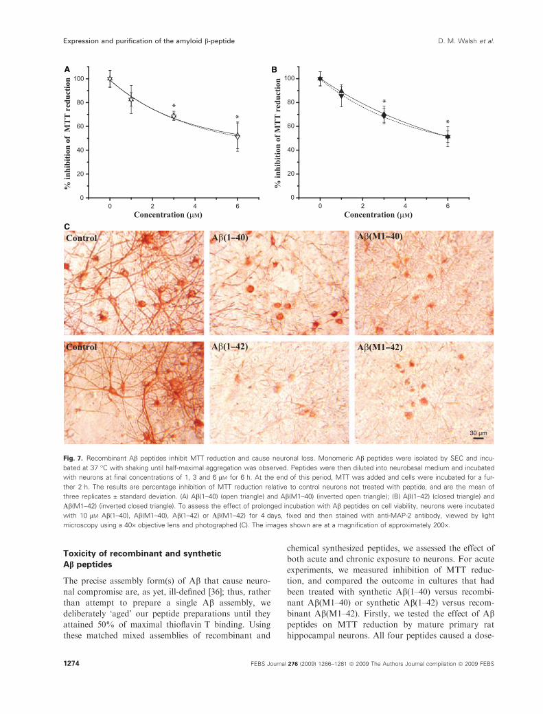
Fig. 7. Recombinant Ab peptides inhibit MTT reduction and cause neuronal loss. Monomeric Ab peptides were isolated by SEC and incu-
bated at 37 �C with shaking until half-maximal aggregation was observed. Peptides were then diluted into neurobasal medium and incubated
with neurons at final concentrations of 1, 3 and 6 lM for 6 h. At the end of this period, MTT was added and cells were incubated for a fur-
ther 2 h. The results are percentage inhibition of MTT reduction relative to control neurons not treated with peptide, and are the mean of
three replicates ± standard deviation. (A) Ab(1–40) (open triangle) and Ab(M1–40) (inverted open triangle); (B) Ab(1–42) (closed triangle) and
Ab(M1–42) (inverted closed triangle). To assess the effect of prolonged incubation with Ab peptides on cell viability, neurons were incubated
with 10 lM Ab(1–40), Ab(M1–40), Ab(1–42) or Ab(M1–42) for 4 days, fixed and then stained with anti-MAP-2 antibody, viewed by light
microscopy using a 40· objective lens and photographed (C). The images shown are at a magnification of approximately 200·.
Expression and purification of the amyloid b-peptide D. M. Walsh et al.
1274 FEBS Journal 276 (2009) 1266–1281 ª 2009 The Authors Journal compilation ª 2009 FEBS
dependent inhibition of MTT reduction that was
apparent within 6 h of treatment (Fig. 7A,B), at which
time the number and morphology of neurons did not
differ either from time zero or from vehicle-treated
controls (data not shown). At the three concentrations
tested, inhibition of MTT by Ab(1–40) and Ab(M1–
40) was essentially identical; similarly, the degrees of
inhibition caused by Ab(1–42) and Ab(M1–42) were
indistinguishable at each concentration studied. More-
over, the extent of MTT inhibition was not signifi-
cantly different for peptides ending at residues 40 and
42, with approximately 50% inhibition at 6 lm for all
four peptides. Longer-term treatment of neurons with
the same peptides caused neuritic degeneration and
loss of neurons (Fig. 7C), with a similar loss evident
for all peptides.
Discussion
Because extensive evidence supports a crucial role for
Ab in Alzheimer’s disease pathogenesis, there is huge
interest in understanding the structural and biological
properties of this molecule [13–16]. Using chemically
synthesized Ab peptides, substantial progress has been
made in understanding of the aggregation and toxic
properties of Ab assemblies [17,37]. However, given
that chemically synthesized Ab peptides are expensive
to purchase and ⁄or make, this has curtailed the extent
of experiments, and may have deterred new investiga-
tors from studying Ab, or forced others to study small
irrelevant fragments (e.g. Ab25-35) rather than the
full-length Ab sequence. Thus, we set ourselves the
goal of developing a facile inexpensive procedure for
the production of recombinant Ab peptides. In addi-
tion to being more cost-effective than production of
synthetic peptides, a bacterial expression system allows
isotope labeling, which is essential for high-resolution
structural analysis of Ab using NMR spectroscopy
and allows use of high specific activity radiotracers to
study Ab uptake, transport and clearance. Moreover,
a recombinant system should also allow the generation
of Ab peptides with design or disease-associated amino
acid substitutions, and we have produced some such
peptides in this study. Importantly, the protocol
described for expression and purification of Ab(M1–
40) and Ab(M1–42) is inexpensive, relatively rapid and
only utilizes rudimentary equipment that is available in
most biochemistry laboratories.
Recombinant expression in E. coli of human
proteins smaller than about 50 residues is often
hampered by proteolytic degradation of unstructured
proteins ⁄peptides; therefore small entities are com-
monly expressed fused to a larger protein to prevent
degradation. A common drawback of such approaches
is the cost of the affinity resins used to isolate the
fusion protein and the proteases required to liberate
the protein of interest from the fusion protein. Such
considerations lead to practical obstacles in terms of
scale-up of the purification and consequently the
amount of pure peptide that can be produced at rea-
sonable cost. Thus we decided to express the Ab(M1–
40) and Ab(M1–42) peptides without fusion to another
protein. The rationale behind this approach was sim-
ple. Ab peptides show a strong propensity to aggre-
gate, with aggregation proceeding rapidly at high
peptide concentrations [33,38,39], thus high-level
expression of Ab peptides should lead to aggregation
and formation of inclusion bodies, and that Ab would
be less susceptible to degradation in this form. More-
over, the formation of inclusion bodies enables high-
level expression because the peptide is cleared from the
bacterial cytosol and hence does not interfere with any
essential functions. In addition, proteins deposited in
inclusion bodies contain fewer E. coli proteins, thus
simplifying purification.
The purification protocol that we have developed is
quick and efficient. The peptide is produced at high
yield in E. coli as inclusion bodies, which are washed
by sonication, solubilized in urea, purified by anion-
exchange chromatography in batch format, and finally
any aggregates removed using SEC. The advantage of
this protocol is that it relies on affordable tools and
can be scaled up to any production size. The batch
mode has the additional advantage of avoiding precipi-
tation. In batch mode, the peptide is spread out over
the entire resin and is eluted from the resin into buffer
in relatively dilute form, which is controlled by the
amount of resin and buffer volume used. In column
mode, the peptide becomes more concentrated and the
yield of eluted monomer is much reduced compared to
batch mode due to its aggregation tendency. In column
mode, the peptide is bound in a concentrated manner
at the top of the column, or, if bound to the resin
prior to packing the column, the salt gradient concen-
trates the peptide on its way out of the column. The
molecular mass fractionation in centrifugal devices
leads to smaller losses than gel filtration due to more
rapid handling using the devices and loss of peptide on
the column resin. The only detriment of the peptides
produced here is the fact that they contain an exo-
genous N-terminal methionine. However, the presence
of this methionine is not insurmountable, and we have
found that co-expression of the E. coli aminopeptidase
MetAP-TG [24] and Ab(M1–40) results in a low-yield
production of Ab(1–40); however, separation of
Ab(1–40) and Ab(M1–40) requires an additional
D. M. Walsh et al. Expression and purification of the amyloid b-peptide
FEBS Journal 276 (2009) 1266–1281 ª 2009 The Authors Journal compilation ª 2009 FEBS 1275
HPLC step and substantially increases the cost and
complexity of production.
Additionally, the presence of the exogenous N-ter-
minal methionine does not affect the fibrillation
kinetics or morphology of the fibrils formed by
Ab(M1–40) or Ab(M1–42). Thus such peptides should
prove useful in high-throughput screens designed to
identify molecules or conditions that modulate Abfibrillogenesis. Moreover, these peptides have indistin-
guishable effects on hippocampal neurons, causing
inhibition of MTT reduction within 6 h of treatment
and neuritic degeneration and cell loss upon pro-
longed treatment. Importantly, these results indicated
that an N-terminal aspartate is not necessary for neuro-
toxicity. An additional advantage of the N-terminal
methionine is the fact that this residue will not be
easily seen in NMR spectra relying on amide protons
as the N-terminal amine protons are likely to
exchange rapidly with water [40]; thus the presence of
the N-terminal methionine may enable detection of
Asp1 that would otherwise be invisible in 1H15N-
HSQC spectra. Therefore, the presence of the N-termi-
nal methionine will allow a broader coverage of struc-
tural assignments to the N-terminus of Ab. Moreover,
the procedures described here are also suitable for
expression and purification of mutant versions of
Alzheimer’s disease-associated amyloid b-peptides. In
this work, we have evaluated this capacity by produc-
ing and cloning genes for familial mutants in the
19–23 region of Ab(L1–40) and Ab(L1–42), and
expressing and purifying the peptides. The procedure is
of course not limited to the peptide variants produced
in this work (F19P, A21G, E22G, E22K, E22Q and
D23N), and the availability of a rapid and simple
expression and purification protocol will facilitate
large-scale investigations of the molecular determinants
of aggregation and fibrillation. Given the intense inter-
est in Ab, significant attempts to produce pure recom-
binant Ab have also been made by several other
groups, but most of these have relied on the generation
of Ab fusions. Perhaps the best of these was reported
by Lee et al. using a system in which Ab was fused to
ubiquitin. This protocol relies on the use of Ni-NTA
affinity chromatography for purification and subse-
quent liberation of Ab by digestion using yeast ubiqu-
itin hydrolase, but the authors did not provide data on
the purity of the end product [21]. Similarly, Wieschan
et al. also employed a fusion strategy, Ni-NTA affinity
chromatography and digestion with thrombin [22].
Zhang et al. also used a fusion strategy coupled with
GSH affinity chromatography and subsequent throm-
bin cleavage [41]. The use of thrombin significantly
increases the cost of the purification, and the require-
ment for HPLC increases the length and complexity of
the purification procedure. Moreover, as with the
studies by Lee et al. [21] and Subramanian and Shree
[42], there was no rigorous assessment of the purity of
the product or the correctness of the sequence. In con-
trast, the purification protocol that we have developed
is quick and efficient, and leads to the production of
highly pure Ab peptides with the anticipated molecular
mass, amino acid composition, correct primary
sequence and appropriate biophysical and neurotoxic
characteristics. In short, the protocol described has the
potential to facilitate a massive increase in the
number and extent of studies aimed at better under-
standing the molecular details of Ab oligomerization
and aggregation.
Experimental procedures
Unless otherwise stated, all chemicals were purchased from
Sigma-Aldrich (St Louis, MO, USA) and were of the
highest purity available. Synthetic peptides Ab(1–40) and
Ab(1–42) were synthesized in the W. M. Keck Foundation
Biotechnology Resource Laboratory (Yale University, New
Haven, CT, USA), and purified using reverse-phase HPLC.
For both synthetic and recombinant Ab peptides, the
correct mass was confirmed by MALDI-TOF MS and
LC-MS.
PCR and cloning procedure
Synthetic genes for Ab(M1–40) and Ab(M1–42) were
designed using E. coli-favored codons preceded by an ATG
initiation codon (Fig. 1). The requirement for a start codon
adds a methionine residue at the N-terminus; hence, the
peptides expressed here are referred to as Ab(M1–40) and
Ab(M1–42).
The synthetic gene for Ab(M1–40) was produced by
PCR using Pfusion DNA polymerase (Finnzymes, Espoo,
Finland) according to the manufacturer’s guidelines and
using the following primers: Aba, 5¢-ATGGACGCTGAAT
TCCGTCACGACTCTGGTTACGAAGTTCACCACCAG
AAGCTGGTG-3¢; Abb, 5¢-GTTCACCACCAGAAGCT
GGTGTTCTTCGCTGAAGACGTGGGTTCTAACAAG
GGTGCT-3¢; Abc, 5¢-CACAACGCCACCAACCATCAGA
CCGATGATAGCACCCTTGTTAGAACCCAC-3¢; Ab-start, 5¢-GCGTAGGGTCGACATATGGACGCTGAATT
CCGTCACG-3¢; Abstop, 5¢-CCTGCCGAGCTCCTATTA
CACAACGCCACCAACCATCAG-3¢.The PCR solution was prepared in the buffer supplied
with the enzyme, and contained Aba, Abb and Abc at
40 nm each, and the start and stop primers Abstart and
Abstop at 600 nm each, and 200 lm each of dATP, dCTP,
dGTP and dTTP. The product was separated from prim-
ers by agarose gel electrophoresis (2% gel). The full-length
Expression and purification of the amyloid b-peptide D. M. Walsh et al.
1276 FEBS Journal 276 (2009) 1266–1281 ª 2009 The Authors Journal compilation ª 2009 FEBS
gene was cut out from the gel, purified using a GFX PCR
and gel band purification kit (GE Healthcare, Chalfont
St Giles, UK). The gene was digested with NdeI and SacI
restriction enzymes and subjected to a second agarose gel
electrophoresis (2% gel), and the cleaved product was
purified using the GFX PCR and gel band purification
kit. The purified cut gene was ligated into PetSac vector
(a modified from of Pet3a with NdeI and SacI cloning
sites [43]) that had been previously cleaved by NdeI and
SacI, and used to transformed Ca2+-competent E. coli
cells (ER2566) by heat shock. The transformed cells were
spread on LB agar plates containing ampicillin
(50 mgÆL)1), single colonies were picked for 2 mL over-
night cultures in LB medium containing ampicillin
(50 mgÆL)1), and plasmids were prepared using a GFX
plasmid purification kit (GE Healthcare) and sequenced.
The gene for Ab(L1–42) was then produced by PCR
using the primers Abstart and Ab42stop (5¢-CCTGCCGAGCTCCTATTAAGCGATCACAACGCCACCAA
CCATCAG-3¢) and a sequence-verified plasmid carrying
the Ab(L1–40) gene. This adds Ile41 and Ala42 to the pep-
tide sequence. The PCR product corresponding to the full-
length Ab(L1–42) gene was purified as above and ligated
into PetSac. In our PCR design, regions encompassing resi-
dues 1–6, 12–18, 24–30 and 34–40 were used as primer
annealing sites, and the following codons in these regions
were altered to achieve more stable duplexes and ⁄oravoid repeat of similar sequences (K16, AAA fi AAG;
V24, GTT fi GTG; K28, AAA fi AAG; G38,
GGT fi GGC; V40, GTT fi GTG). Residues 21–23 are
mutated in several Alzheimer’s-like familial disorders [44–
48]. In our design, residues 19–23 are therefore uniquely
encompassed by the middle primer, such that only one
additional primer is required for the production of syn-
thetic genes bearing Alzheimer’s disease-associated point
mutants.
Bacterial expression
Sequence-verified plasmids from wild-type and each mutant
were transformed into Ca2+-competent E. coli cells (BL21
DE3 PLysS Star) by heat shock and spread on LB agar
plates containing ampicillin (50 mgÆL)1) and chlorampheni-
col (30 mgÆL)1). Single colonies were used to inoculate
50 mL overnight cultures in LB medium with ampicillin
(50 mgÆL)1) and chloramphenicol (30 mgÆL)1). The next
morning, 5 mL of overnight culture was transferred to
500 mL day culture (LB medium with 50 mgÆL)1 ampicillin
and 30 mgÆL)1 chloramphenicol). When the density of cells
was sufficient to produce an attenuance at 600 nm (D600 nm)
of approximately 0.6, protein expression was induced by
addition of isopropyl thio-b-d-galactoside. The cells were
harvested between 3 and 4 h after induction, dispensed
in Millipore (Carrigtwohill, Cork, Republic of Ireland)
H2O (12–25 mL H2O per liter culture), and frozen.
To assay and optimize expression levels, test samples of
1 mL cultures were collected for each transformed bacterial
culture at various temperatures (30, 37 and 41 �C) and at
various times (1, 2, 3, 4, 5 or 6 h) after induction, and using
seven different isopropyl thio-b-d-galactoside concentra-
tions ranging from 0.1 to 2.0 mm for induction. The cell
suspension was centrifuged at 5400 g and 4 �C for 15 min,
the cell pellet was resuspended in H2O (100 lL) and centri-
fuged again, after which the supernatant was collected and
the pellet dissolved in 8 m urea (100 lL). Both the super-
natant and urea-solubilized pellet were then analyzed by
agarose gel electrophoresis at pH 8.4 and by SDS-PAGE.
Sonication
The frozen cell pellet from a 4.5 L culture was thawed, son-
icated in a total of 100 mL 10 mm Tris ⁄HCl pH 8.0, 1 mm
EDTA, for 2 min on ice (1 ⁄ 2 horn, 50% duty cycle), and
centrifuged for 10 min at 18 000 g. The supernatant (S1 in
Fig. 2) was removed, and the pellet was resuspended twice
in 100 mL 10 mm Tris ⁄HCl pH 8.0, 1 mm EDTA, soni-
cated and centrifuged as above. The third supernatant was
removed, and the pellet was resuspended in 50 mL 8 m
urea, 10 mm Tris ⁄HCl pH 8.0, 1 mm EDTA, and sonicated
as above, resulting in a clear solution. To minimize carbam-
ylation of Ab, fresh solutions of ice-cold, deionized ACS
grade urea were used, and the duration of exposure to urea
was limited to less than 12 h.
Purification of Ab(M1–40) and Ab(M1–42)
The procedures described here are for 50 mL of urea-solu-
bilized inclusion bodies originating from 4.5 L of culture,
but this process can be scaled proportionally for other
amounts. The urea-solubilized inclusion bodies (50 mL)
were diluted with 150 mL of 10 mm Tris ⁄HCl pH 8.0 con-
taining 1 mm EDTA (buffer A), added to 50 mL DEAE-
cellulose equilibrated in buffer A, and gently agitated for
20 min. The slurry was then applied to a Buchner funnel
with filter paper on a vacuum glass bottle [alternatively, a
Nalgene (Lima, OH, USA) 0.45 lm filter on a vacuum
bottle can be used]. Subsequently, the resin was washed
with buffer A (50 mL), followed by stepwise elution using
50 mL aliquots of buffer A with 50, 75, 100, 125, 150,
200, 250, 300 and 500 mm NaCl, respectively. Each ali-
quot was incubated with the resin for 5 min before collec-
tion under vacuum. Eluates were analyzed by SDS-PAGE
and agarose gel electrophoresis, and fractions with highly
pure Ab were pooled and fractionated by centrifugation
through a 30 kDa molecular mass cut-off filter. The wash-
ing and elution processes can also be performed as fol-
lows: the resin is washed with 50 mL buffer A, and then
with 50 mL buffer A with 25 mm NaCl followed by three
or four 50 mL aliquots of buffer A with 125 mm NaCl.
Using SDS-PAGE, the peptide is then found in the first
D. M. Walsh et al. Expression and purification of the amyloid b-peptide
FEBS Journal 276 (2009) 1266–1281 ª 2009 The Authors Journal compilation ª 2009 FEBS 1277
two (or first three) 125 mm aliquots, which are combined
and used for centrifugal filtration.
Ion-exchange chromatography in column mode
Urea-solubilized inclusion bodies (25 mL originating from
2.2 L of bacterial cell culture) were diluted with 150 mL of
buffer A and applied to a 50 mL DEAE-cellulose column
equilibrated in buffer A. The column was washed with 50 mL
buffer A, followed by elution using a linear gradient from
0–300 mm NaCl with a total gradient volume of 500 mL.
Fractions were analyzed by electrophoresis on 10–20%
polyacrylamide Tris-tricine gels and 1% agarose gels. In a
second set of experiments, the column was equilibrated
in buffer A containing 8 m urea, and the sample was eluted
with a gradient of 0–300 mm NaCl in buffer A containing
8 m urea.
Mass spectrometry, amino acid analysis and
sequencing
Amino acid analysis was performed at the Amino Acid
Analysis Center, University of Uppsala, Sweden. Sequence
analysis was performed using an Applied Biosystems
Procise 492 cLC sequenator (Applied Biosystems, Framing-
ham, MA, USA) employing standard Edman chemistry,
and MS analysis was undertaken using an LCQDECA
LC ⁄MS system (ThermoFinnigan, San Jose, CA, USA).
The MS system consisted of a Surveyor HPLC system with
a diphenyl 150 · 1.0 mm column (Grace Vydac, Palo Alto,
CA, USA) interfaced to an LCQ-DECA electrospray ioni-
zation ⁄ ion trap mass spectrometer, and eluted using an
acetonitrile ⁄ trifluoroacetic acid gradient. MALDI-TOF
mass spectrometry was performed using a 4700 proteomics
analyzer (Applied Biosystems). Samples were dispensed
onto a MALDI sample support, and allowed to air-dry
prior to addition of matrix solution (4-hydroxy a-cyanocinnamic acid in 50% acetonitrile, 0.1% trifluoroacetic
acid, 25 mm citric acid). All analyses were performed in
positive reflector mode, collecting data from approximately
3000 and 5000 single laser shots for MS and MS ⁄MS anal-
yses, respectively.
Preparation of aggregate-free monomer for
fibrillation assays
For fibrillation assays, it is essential to start with a uni-
form monomeric peptide sample. Solutions of monomeric
Ab were prepared by dissolving lyophilized peptides in
5 m GuHCl, Tris ⁄HCl pH 8.0 at a concentration of
approximately 1 mgÆmL)1, and isolating monomers using
SEC. Ab solutions were chromatographed on a Super-
dex 75 10 ⁄ 300 GL column using an AKTA purifier (GE
Healthcare), and eluted at 0.8 mLÆmin)1 using 50 mm
ammonium acetate, pH 8.5. Fractions (0.5 mL) were
collected, peak fractions pooled, and the concentration of
peptide determined by absorbance at 275 nm using e275 =1400 m
)1 cm)1.
Assessment of aggregation using thioflavin T
binding and electron microscopy
The kinetics of fibril formation was determined using a con-
tinuous ThT assay [49]. Solutions of Ab isolated by SEC
were diluted to concentrations of 36 or 24 lm using 50 mm
ammonium acetate, pH 8.5. Peptides were then incubated
in a 96-well black fluorescence plate at a final concentration
of 6 or 9 lm in the presence of 10 lm ThT at 37 �C, andshaken at 700 r.p.m. using a VorTemp 56� incuba-
tor ⁄ shaker with an orbit of 3 mm (Labnet International,
Windsor, UK). Measurements were made at regular inter-
vals using a SpectraMax M2 microplate reader (Molecular
Devices, Sunnyvale, CA, USA) with excitation and emission
at 440 and 480 nm, respectively. Each experimental point is
the mean of the fluorescence signal of at least eight wells
containing aliquots of the same solution. The morphology
of Ab aggregates formed from solutions incubated as above
but in the absence of ThT and at a concentration of 50 lm
was assessed by negative-contrast electron microscopy as
described previously [25]. Briefly, samples were applied to a
carbon-coated formvar grid, left for 1 min, fixed with glu-
taraldehye, wicked dry with filter paper, and 2% uranyl
acetate was added and the mixture was incubated for
2 min. The grid was wicked dry and allowed to air dry for
10 min. Samples were stored in a sealed container and
viewed under a Tecani G2 BIOTWIN electron transmission
microscope operated at 120 V. All reagents were supplied
by Electron Microscopy Sciences (Hatfield, PA, USA).
Assessement of SEC-isolated peptides by HPLC
and SDS-PAGE
Samples (100 lL) of peptides isolated by SEC were injected
on to a CN capcell column (4.6 mm · 25 cm) (Shiseido
Fine Chemicals, Toyko, Japan) using a Varian Pro Star 410
autosampler (Varian Inc., Palo Alto, CA, USA), and eluted
at 1.5 mLÆmin)1 with a 14–49% acetonitrile gradient using
a Varian Pro Star HPLC system fitted with a photodiode
array detector. For SDS-PAGE, samples (10 lL) were
mixed with 2· sample buffer, and immediately electropho-
resed on 10–20% polyacrylamide Tris-tricine gels. Proteins
were stained with silver as described previously [28].
Primary culture
Primary hippocampal neuronal cultures were prepared as
described previously [30] with minor modifications. Briefly,
primary hippocampal cultures were generated from embry-
Expression and purification of the amyloid b-peptide D. M. Walsh et al.
1278 FEBS Journal 276 (2009) 1266–1281 ª 2009 The Authors Journal compilation ª 2009 FEBS
onic day 18 Wistar rats. Hippocampi were dissected out in
Hanks’ balanced salt solution buffered with HEPES, and
dissociated using papain. Cells were plated at 6 · 104 cells
on 48-well dishes pre-coated with poly-d-lysine
(50 lgÆmL)1) and maintained in neurobasal medium
containing 2 mm glutamine and B27 supplement without
antioxidants. Half the medium was exchanged every
3 days. All media reagents were purchased from Invitrogen
(Dun Laoghaire, Republic of Ireland).
Preparation of peptide for cell treatment
Lyophilized peptides were resuspended and incubated for a
minimum of 2 h in 5 m GuHCl, pH 8.0. Thereafter, sam-
ples were injected onto a Superdex 75 column HR 10 ⁄ 30column (Amersham Biosciences, Amersham, UK), and
eluted with 10.9 mm HEPES pH 7.4 at a flow rate of
0.8 mLÆmin)1. Peak fractions were then examined for
absorbance at 275 nm, and the concentration of Ab was
calculated. Fractions containing monomeric peptide were
diluted such that all peptides were of equal concentration.
To induce peptide aggregation, samples were incubated at
37 �C and shaken at 700 r.p.m. using a VorTemp 56�incubator ⁄ shaker with an orbit of 3 mm (Labnet Interna-
tional) until 50% of the maximal thioflavin T fluorescence
had been achieved; maximal aggregation was taken as the
mean plateau fluorescent signal. Peptides were then diluted
with 2· neurobasal medium, and 50% of the medium of
each well was replaced with an equal volume of neurobasal
medium containing either Ab(1–40), Ab(M1–40), Ab(1–42)or Ab(M1–42) (1, 3 or 6 lm, final concentration) and incu-
bated for 6 h. Cell-mediated reduction of 3-(4,5-dim-
ethylthiazol-2-yl)-2,5-diphenyltetrazolium bromide (MTT)
was assessed as described previously [30]. Briefly, following
incubation with peptides, 2.5 mgÆmL)1 MTT (25 lL) was
added to each well, and incubation was continued for a fur-
ther 2 h. Cells were then solubilized in 250 lL of 20% w ⁄ vSDS in 50% v ⁄ v N,N¢-dimethylformamide, 25 mm HCl,
2% v ⁄ v glacial acetic acid, pH 4.7, and levels of reduced
MTT were determined by measuring the difference in
absorbance at 570 and 650 nm using a Molecular Devices
Spectramax M2 microplate reader.
In a separate series of experiments, neurons were incu-
bated for 4 days with each of the peptides (10 lm), and
cells were fixed and used for immunocytochemical analyses.
Immunocytochemistry
Neurons were fixed in 4% paraformaldehyde for 20 min
at room temperature, and cells were stained for microtu-
bule-associated protein-2 (MAP-2) using a Vectastain kit
(Vector Laboratories, Peterborough, UK). Staining was
performed according to the manufacturer’s instructions.
Briefly, endogenous peroxidases were blocked in 0.3%
H2O2, rinsed in NaCl ⁄Pi and incubated in blocking solu-
tion for 20 min (Vectastain). Neurons were then incubated
with mouse monoclonal anti-MAP-2 (Sigma, Poole, UK)
diluted 1 : 2000 in blocking solution for 30 min. Cells were
rinsed in NaCl ⁄Pi several times and incubated in blocking
serum containing anti-mouse IgG (Vectastain) for a fur-
ther 30 min. Staining was developed by incubation of cells
with Vectastain ABC reagent for 30 min, followed by
incubation with substrate solution until colour had devel-
oped. Cells were visualized by light-phase contrast micros-
copy using a 40· objective lens, and captured using an
SP-500 UZ digital compact camera (Olympus, Watford,
UK).
Co-expression with Met aminopeptidase
Plasmids encoding MetAP-TG (a mutated form of Met
aminopeptidase that can cleave N-terminal Met when the
second residues is charged [24]) and Ab were electroporated
into E. coli cells (BL21 DE3 PLysS Star) and spread on LB
plates with ampicillin, kanamycin and chloramphenicol.
Single colonies were picked for cultivation in liquid culture
as described for Ab alone, except that the medium con-
tained 50 mgÆL)1 ampicillin, 100 mgÆL)1 kanamycin and
30 mgÆL)1 chloramphenicol.
Acknowledgements
We thank Dr Celia Cabaleiro Lago for useful discus-
sion and Rocio Fedrani for assistance with electron
microscopy. We are also indebted to Dr You-Di Liao
(Academia Sinica, Taiwan) for providing the MetAP-
TG expression plasmid. This work was supported by
Wellcome Trust grant 067660 (to D.M.W.), a Science
Foundation Ireland E.T.S Walton Visitor Award (to
S.L.) and the Swedish Research Council (S.L.).
References
1 Selkoe DJ (2001) Alzheimer’s disease: genes, proteins
and therapies. Physiol Rev 81, 742–761.
2 Tabaton M, Nunzi MG, Xue R, Usiak M, Autilio-
Gambetti L & Gambetti P (1994) Soluble amyloid
b-protein is a marker of Alzheimer amyloid in brain
but not in cerebrospinal fluid. Biochem Biophys Res
Commun 200, 1598–1603.
3 Vigo-Pelfrey C, Lee D, Keim PS, Lieberburg I &
Schenk D (1993) Characterization of b-amyloid peptide
from human cerebrospinal fluid. J Neurochem 61, 1965–
1968.
4 Naslund J, Karlstrom AR, Tjernberg LO, Schierhorn
A, Terenius L & Nordstedt C (1996) High-resolution
separation of amyloid b-peptides: structural variantspresent in Alzheimer’s disease amyloid. J Neurochem
67, 294–301.
D. M. Walsh et al. Expression and purification of the amyloid b-peptide
FEBS Journal 276 (2009) 1266–1281 ª 2009 The Authors Journal compilation ª 2009 FEBS 1279
5 Maler JM, Klafki HW, Paul S, Spitzer P, Groemer
TW, Henkel AW, Esselmann H, Lewczuk P, Kornhuber
J & Wiltfang J (2007) Urea-based two-dimensional elec-
trophoresis of b-amyloid peptides in human plasma: evi-
dence for novel Abeta species. Proteomics 7, 3815–3820.
6 Bibl M, Esselmann H, Otto M, Lewczuk P, Cepek L,
Ruther E, Kornhuber J & Wiltfang J (2004) Cerebrospi-
nal fluid amyloid beta peptide patterns in Alzheimer’s
disease patients and nondemented controls depend on
sample pretreatment: indication of carrier-mediated epi-
tope masking of amyloid beta peptides. Electrophoresis
25, 2912–2918.
7 Lewczuk P, Esselmann H, Bibl M, Beck G, Maler JM,
Otto M, Kornhuber J & Wiltfang J (2004) Tau protein
phosphorylated at threonine 181 in CSF as a neuro-
chemical biomarker in Alzheimer’s disease: original data
and review of the literature. J Mol Neurosci 23, 115–
122.
8 Lewczuk P, Esselmann H, Bibl M, Paul S, Svitek J,
Miertschischk J, Meyrer R, Smirnov A, Maler JM,
Klein C et al. (2004) Electrophoretic separation of amy-
loid b peptides in plasma. Electrophoresis 25, 3336–
3343.
9 Lewczuk P, Esselmann H, Groemer TW, Bibl M, Maler
JM, Steinacker P, Otto M, Kornhuber J & Wiltfang J
(2004) Amyloid b peptides in cerebrospinal fluid as pro-
filed with surface enhanced laser desorption ⁄ ionizationtime-of-flight mass spectrometry: evidence of novel bio-
markers in Alzheimer’s disease. Biol Psychiatry 55, 524–
530.
10 Portelius E, Tran AJ, Andreasson U, Persson R, Brink-
malm G, Zetterberg H, Blennow K & Westman-Brink-
malm A (2007) Characterization of amyloid b peptides
in cerebrospinal fluid by an automated immunoprecipi-
tation procedure followed by mass spectrometry. J Pro-
teome Res 6, 4433–4439.
11 Thorsell A, Portelius E, Blennow K & Westman-Brink-
malm A (2007) Evaluation of sample fractionation
using micro-scale liquid-phase isoelectric focusing on
mass spectrometric identification and quantitation of
proteins in a SILAC experiment. Rapid Commun Mass
Spectrom 21, 771–778.
12 Bentahir M, Nyabi O, Verhamme J, Tolia A, Horre K,
Wiltfang J, Esselmann H & De Strooper B (2006) Prese-
nilin clinical mutations can affect gamma-secretase
activity by different mechanisms. J Neurochem 96, 732–
742.
13 Hardy J & Allsop D (1991) Amyloid deposition as the
central event in the aetiology of Alzheimer’s disease.
Trends Pharmacol 12, 383–388.
14 Selkoe DJ (1991) The molecular pathology of Alzhei-
mer’s disease. Neuron 6, 487–498.
15 Hardy J & Selkoe DJ (2002) The amyloid hypothesis of
Alzheimer’s disease: progress and problems on the road
to therapeutics. Science 297, 353–356.
16 Walsh DM & Selkoe DJ (2007) Ab oligomers – a
decade of discovery. J Neurochem 101, 1172–1184.
17 Walsh DM, Hartley DM & Selkoe DJ (2003) The many
faces of Ab: structures and activity. Curr Med Chem
Immunol Endocr Metab Agents 3, 277–291.
18 Zarandi M, Soos K, Fulop L, Bozso Z, Datki Z, Toth
GK & Penke B (2007) Synthesis of Ab[1–42] and its
derivatives with improved efficiency. J Pept Sci 13, 94–
99.
19 Tickler AK, Barrow CJ & Wade JD (2001) Improved
preparation of amyloid b-peptides using DBU as
Nalpha-Fmoc deprotection reagent. J Pept Sci 7, 488–
494.
20 Dobeli H, Draeger N, Huber G, Jakob P, Schmidt D,
Seilheimer B, Stuber D, Wipf B & Zulauf M (1995) A
biotechnological method provides access to aggregation-
competent monomeric Alzheimer’s 1–42 residue amyloid
peptide. Bio ⁄Technology 13, 988–993.
21 Lee EK, Hwang JH, Shin DY, Kim DI & Yoo YJ
(2005) Production of recombinant amyloid b-peptide 42
as an ubiquitin extension. Protein Expr Purif 40, 183–
189.
22 Wiesehan K, Funke SA, Fries M & Willbold D (2007)
Purification of recombinantly expressed and cytotoxic
human amyloid b-peptide 1–42. J Chromatogr B Analyt
Technol Biomed Life Sci 856, 229–233.
23 Sharpe S, Yau WM & Tycko R (2005) Expression and
purification of a recombinant peptide from the Alzhei-
mer’s b-amyloid protein for solid-state NMR. Protein
Expr Purif 42, 200–210.
24 Liao YD, Jeng JC, Wang CF, Wang SC & Chang ST
(2004) Removal of N-terminal methionine from recom-
binant proteins by engineered E. coli methionine amino-
peptidase. Protein Sci 13, 1802–1810.
25 Walsh DM, Lomakin A, Benedek GB, Condron MM &
Teplow DB (1997) Amyloid b-protein fibrillogenesis:
detection of a protofibrillar intermediate. J Biol Chem
272, 22364–22374.
26 Kametani F, Tanaka K, Tokuda T & Allsop D (1995)
The immunoreactive profile at the N-terminal region of
Ab 1–39 ⁄ 40 but not Ab 1–42 changes with transition
from monomer ⁄ dimer to further peptide aggregates.
Brain Res 703, 237–241.
27 Wiltfang J, Smirnov A, Schnierstein B, Kelemen G,
Matthies U, Klafki HW, Staufenbiel M, Huther G,
Ruther E & Kornhuber J (1997) Improved electropho-
retic separation and immunoblotting of b-amyloid (Ab)peptides 1–40, 1–42, and 1–43. Electrophoresis 18, 527–
532.
28 Shevchenko A, Wilm M, Vorm O & Mann M (1996)
Mass spectrometric sequencing of proteins silver-stained
polyacrylamide gels. Anal Chem 68, 850–858.
29 Hepler RW, Grimm KM, Nahas DD, Breese R, Dod-
son EC, Acton P, Keller PM, Yeager M, Wang H,
Shughrue P et al. (2006) Solution state characterization
Expression and purification of the amyloid b-peptide D. M. Walsh et al.
1280 FEBS Journal 276 (2009) 1266–1281 ª 2009 The Authors Journal compilation ª 2009 FEBS
of amyloid b-derived diffusible ligands. Biochemistry 45,
15157–15167.
30 Walsh DM, Hartley DM, Kusumoto Y, Fezoui Y,
Condron MM, Lomakin A, Benedek GB, Selkoe DJ &
Teplow DB (1999) Amyloid b-protein fibrillogenesis.
Structure and biological activity of protofibrillar
intermediates. J Biol Chem 274, 25945–25952.
31 Levine H (1995) Thioflavin T interaction with amyloid
beta-sheet structures. Amyloid Int J Exp Clin Invest 2,
1–6.
32 Naiki H & Nakakuki K (1996) First-order kinetic
model of Alzheimer’s beta-amyloid fibril extension in vi-
tro. Lab Invest 74, 374–383.
33 Jarrett JT, Berger EP & Lansbury PT Jr (1993) The car-
boxy terminus of the beta amyloid protein is critical for
the seeding of amyloid formation: implications for the
pathogenesis of Alzheimer’s disease. Biochemistry 32,
4693–4697.
34 Walsh DM, Hartley DM, Condron MM, Selkoe DJ &
Teplow DB (2001) In vitro studies of amyloid b-proteinfibril assembly and toxicity provide clues to the aetiol-
ogy of Flemish variant (Ala692 fi Gly) Alzheimer’s dis-
ease. Biochem J 355, 869–877.
35 Harper JD, Wong SS, Lieber CM & Lansbury PT
(1999) Assembly of Ab amyloid protofibrils: an in vitro
model for a possible early event in Alzheimer’s disease.
Biochemistry 38, 8972–8980.
36 Glabe CG (2008) Structural classification of toxic
amyloid oligomers. J Biol Chem 283, 29639–29643.
37 Klein WL, Krafft GA & Finch CE (2001) Targeting
small Abeta oligomers: the solution to an Alzheimer’s
disease conundrum? Trends Neurosci 24, 219–224.
38 Halverson K, Fraser PE, Kirschner DA & Lansbury PT
Jr (1990) Molecular determinants of amyloid deposition
in Alzheimer’s disease: conformational studies of syn-
thetic beta-protein fragments. Biochemistry 29, 2639–
2644.
39 Burdick D, Soreghan B, Kwon M, Kosmoski J, Knauer
M, Henschen A, Yates J, Cotman C & Glabe C (1992)
Assembly and aggregation properties of synthetic Alz-
heimer’s A4 ⁄ b amyloid peptide analogs. J Biol Chem
267, 546–554.
40 Wutrich K (1986) NMR of Proteins and Nucleic Acids.
Wiley, New York, NY.
41 Zhang L, Yu H, Song C, Lin X, Chen B, Tan C, Cao
G & Wang Z (2009) Expression, purification, and char-
acterization of recombinant human b-amyloid42 peptide
in Escherichia coli. Protein Expr Purif 64, 55–62.
42 Subramanian S & Shree A (2007) Expression, purifica-
tion and characterization of a synthetic gene encoding
human amyloid b (Ab1–42) in Escherichia coli. Indian J
Biochem Biophys 44, 71–75.
43 Brodin P, Grundstrom T, Hofmann T, Drakenberg T,
Thulin E & Forsen S (1986) Expression of bovine intes-
tinal calcium binding protein from a synthetic gene in
Escherichia coli and characterization of the product.
Biochemistry 25, 5371–5377.
44 Nilsberth C, Westlind-Danielsson A, Eckman CB, Con-
dron MM, Axelman K, Forsell C, Stenh C, Luthman J,
Teplow DB, Younkin SG et al. (2001) The ‘Arctic’ APP
mutation (E693G) causes Alzheimer’s disease by
enhanced Ab protofibril formation. Nat Neurosci 4,
887–893.
45 Levy E, Carman MD, Fernandez-Madrid IJ, Power
MD, Lieberburg I, van Duinen SG, Bots GTAM,
Luyendijk W & Frangione B (1990) Mutation of
the Alzheimer’s disease amyloid gene in hereditary
cerebral hemorrhage, Dutch-type. Science 248, 1124–
1126.
46 Hendriks L, van Duijn CM, Cras P, Cruts M, Van Hul
W, van Harskamp F, Warren A, McInnis MG, Anton-
arakis SE, Martin J-J et al. (1992) Presenile dementia
and cerebral haemorrhage linked to a mutation at
codon 692 of the b-amyloid precursor protein gene. Nat
Genet 1, 218–221.
47 Kamino K, Orr HT, Payami H, Wijsman EM, Alonso
E, Pulst SM, Anderson L, O’dahl S, Nemens E, White
JA et al. (1992) Linkage and mutational analysis of
familial Alzheimer disease kindreds for the APP gene
region. Am J Hum Genet 51, 998–1014.
48 Bugiani O, Padovani A, Magoni M, Andora G, Sgarzi
M, Savoiardo M, Bizzi A, Giaccone G, Rossi G &
Tagliavini F (1998) An Italian type of HCHWA.
Neurobiol Aging 19, S238.
49 Betts V, Leissring ML, Dolios G, Wang R, Selkoe DJ
& Walsh DM (2008) Aggregation and catabolism of
disease-associated intra-Ab mutations: reduced proteol-
ysis of AbA21G by neprilysin. Neurobiol Dis 31, 442–
450.
Supporting information
The following supplementary material is available:
Fig. S1. MS analysis of bacterially expressed Ab(M1–
40).
Fig. S2. LC-MS analysis of bacterially expressed
Ab(M1–42) confirms the correct molecular mass and
indicates that the peptide is of comparable purity to
synthetic Ab(1–42).This supplementary material can be found in the
online version of this article.
Please note: Wiley-Blackwell is not responsible for
the content or functionality of any supplementary
materials supplied by the authors. Any queries (other
than missing material) should be directed to the corre-
sponding author for the article.
D. M. Walsh et al. Expression and purification of the amyloid b-peptide
FEBS Journal 276 (2009) 1266–1281 ª 2009 The Authors Journal compilation ª 2009 FEBS 1281
Related Documents