10.1128/MCB.20.9.3069-3078.2000. 2000, 20(9):3069. DOI: Mol. Cell. Biol. Joyce Ng, Craig M. Hart, Kelly Morgan and Jeffrey A. Simon Modified ESC Complexes and Contains Covalently Distinct from Other Polycomb Group ESC-E(Z) Protein Complex Is Drosophila A http://mcb.asm.org/content/20/9/3069 Updated information and services can be found at: These include: REFERENCES http://mcb.asm.org/content/20/9/3069#ref-list-1 at: This article cites 68 articles, 38 of which can be accessed free CONTENT ALERTS more» articles cite this article), Receive: RSS Feeds, eTOCs, free email alerts (when new http://journals.asm.org/site/misc/reprints.xhtml Information about commercial reprint orders: http://journals.asm.org/site/subscriptions/ To subscribe to to another ASM Journal go to: on October 31, 2014 by guest http://mcb.asm.org/ Downloaded from on October 31, 2014 by guest http://mcb.asm.org/ Downloaded from

Welcome message from author
This document is posted to help you gain knowledge. Please leave a comment to let me know what you think about it! Share it to your friends and learn new things together.
Transcript
10.1128/MCB.20.9.3069-3078.2000.
2000, 20(9):3069. DOI:Mol. Cell. Biol. Joyce Ng, Craig M. Hart, Kelly Morgan and Jeffrey A. Simon Modified ESCComplexes and Contains CovalentlyDistinct from Other Polycomb Group
ESC-E(Z) Protein Complex IsDrosophilaA
http://mcb.asm.org/content/20/9/3069Updated information and services can be found at:
These include:
REFERENCEShttp://mcb.asm.org/content/20/9/3069#ref-list-1at:
This article cites 68 articles, 38 of which can be accessed free
CONTENT ALERTS more»articles cite this article),
Receive: RSS Feeds, eTOCs, free email alerts (when new
http://journals.asm.org/site/misc/reprints.xhtmlInformation about commercial reprint orders: http://journals.asm.org/site/subscriptions/To subscribe to to another ASM Journal go to:
on October 31, 2014 by guest
http://mcb.asm
.org/D
ownloaded from
on O
ctober 31, 2014 by guesthttp://m
cb.asm.org/
Dow
nloaded from

MOLECULAR AND CELLULAR BIOLOGY,0270-7306/00/$04.0010
May 2000, p. 3069–3078 Vol. 20, No. 9
Copyright © 2000, American Society for Microbiology. All Rights Reserved.
A Drosophila ESC-E(Z) Protein Complex Is Distinct fromOther Polycomb Group Complexes and Contains
Covalently Modified ESCJOYCE NG, CRAIG M. HART, KELLY MORGAN, AND JEFFREY A. SIMON*
Department of Genetics, Cell Biology and Development and Department of Biochemistry,Molecular Biology and Biophysics, University of Minnesota,
Minneapolis, Minnesota 55455
Received 23 December 1999/Accepted 2 February 2000
The extra sex combs (ESC) and Enhancer of zeste [E(Z)] proteins, members of the Polycomb group (PcG)of transcriptional repressors, interact directly and are coassociated in fly embryos. We report that these twoproteins are components of a 600-kDa complex in embryos. Using gel filtration and affinity chromatography,we show that this complex is biochemically distinct from previously described complexes containing the PcGproteins Polyhomeotic, Polycomb, and Sex comb on midleg. In addition, we present evidence that ESC is phos-phorylated in vivo and that this modified ESC is preferentially associated in the complex with E(Z). ModifiedESC accumulates between 2 and 6 h of embryogenesis, which is the developmental time when esc function isfirst required. We find that mutations in E(z) reduce the ratio of modified to unmodified ESC in vivo. We havealso generated germ line transformants that express ESC proteins bearing site-directed mutations that disruptESC-E(Z) binding in vitro. These mutant ESC proteins fail to provide esc function, show reduced levels of mod-ification in vivo, and are still assembled into complexes. Taken together, these results suggest that ESC phos-phorylation normally occurs after assembly into ESC-E(Z) complexes and that it contributes to the functionor regulation of these complexes. We discuss how biochemically separable ESC-E(Z) and PC-PH complexesmight work together to provide PcG repression.
The Drosophila homeotic proteins encoded by the Antenna-pedia and bithorax complexes are transcription factors re-quired for anterior-posterior (A-P) body patterning (30, 36).These proteins are each expressed in spatially restricted re-gions along the A-P axis that correspond to their domains ofdevelopmental function (7, 8, 29, 67). The expression of ho-meotic proteins is controlled by two sets of repressors: the gapproteins, such as hunchback and Kruppel, act early in embryo-genesis to set the limits of homeotic gene expression (46, 52,53, 68), and the Polycomb group (PcG) proteins maintainhomeotic gene repression throughout the remainder of devel-opment (for reviews, see references 45 and 54). In PcG mu-tants, homeotic genes are expressed outside of their normalA-P domains (39, 56, 59).
Approximately 15 PcG genes have been identified. Severallines of evidence indicate that this large set of repressors workstogether in multimeric protein complexes. The majority of thePcG proteins that have been cloned and characterized containconserved domains that function in protein-protein interac-tions (2, 5, 12, 22, 28, 37, 42, 48, 55). Multiple pairwise inter-actions between different PcG members have been describedfor both Drosophila and mammalian PcG proteins (1, 14, 21,23, 26, 35, 43, 50, 62, 65). Moreover, a PcG complex estimatedat 2 MDa in size (17), which contains the Polycomb (PC),polyhomeotic (PH), and Posterior sex combs (PSC) proteins,has been characterized from Drosophila (35, 51, 61). A similarcomplex has also been identified in mammals (1, 21, 23).
Although a PC-PH-PSC complex is likely to be a key com-ponent in homeotic gene repression, several lines of evidenceindicate that the PcG proteins do not function as members of
a single large complex. First, the phenotypes of different PcGmutants are distinct; for example, ph mutants show an epider-mal defect not seen with other PcG mutants (16). Second, thePcG proteins colocalize at numerous loci on polytene chromo-somes but their distributions are not identical. In particular,the PC, PH, and Polycomblike distributions completely overlapwhereas PSC and Additional sex combs are also found atdistinct chromosomal sites (12, 17, 37, 38, 47, 57). Finally,immunoprecipitation assays on in vivo cross-linked chromatinshow differential distributions of PC, PH, and PSC on regula-tory sequences of the invected gene (61). These observationssuggest that there is division of labor among the PcG proteinsand that they function in multiple, distinct complexes.
PcG repression begins at about 3 to 4 h of embryogenesisand continues throughout the subsequent embryonic, larval,and pupal stages. Consistent with this, most of the PcG pro-teins are required and expressed continuously during thesestages. The PcG member extra sex combs (esc) is distinct, how-ever, in that its function is most critical during early embryo-genesis (55, 60) and that esc mRNA is expressed primarilyduring early embryonic stages (18, 48, 55). The early require-ment for esc function has led to the hypothesis that it may playa role in the molecular transition between gap protein and PcGprotein repression (22, 48, 55).
The majority of the ESC protein is composed of seven WDrepeats, a motif involved in protein-protein interactions (22,48, 55). Homology modeling to another WD repeat protein,the G-protein b subunit, indicates that ESC folds into a circu-lar structure known as a b-propeller (40, 66). The b-propelleracts as a scaffold that displays variable loops on the proteinsurface for interactions with other proteins. The predictedESC b-propeller contains two large surface loops that arehighly conserved in evolution (40). Clustered alanine substitu-tions introduced into these loops disrupt esc function in tran-
* Corresponding author. Mailing address: 321 Church St. S.E., Min-neapolis, MN 55455. Phone: (612) 626-5097. Fax: (612) 626-7031.E-mail: [email protected].
3069
on October 31, 2014 by guest
http://mcb.asm
.org/D
ownloaded from
sient-rescue experiments (40), indicating that these loops areimportant for esc function in vivo.
The ESC protein binds directly to another PcG protein,Enhancer of zeste [E(Z)] (26, 62). The ESC interaction do-main in E(Z) has been mapped to an N-terminal 33-amino-acid region. In addition, mutations in the ESC surface loopsthat impair function in vivo also disrupt ESC-E(Z) interactionsin vitro (26). ESC-E(Z) association in vivo is demonstrated bythe coimmunoprecipitation of the proteins from embryo ex-tracts (26, 62) and their colocalization on polytene chromo-somes (62). Taken together, these results establish a molecularpartnership between ESC and E(Z) and suggest that this re-lationship is important for homeotic gene repression.
The ESC-E(Z) partnership shows striking evolutionary con-servation. Mouse homologs of ESC and E(Z), i.e., EED andEZH1 or EZH2, respectively, interact directly and coimmuno-precipitate from cell extracts (14, 26, 50, 65). In addition,Caenorhabditis elegans homologs of ESC and E(Z) have beenidentified; these homologs are encoded by the maternal effectsterile genes mes-6 and mes-2 (25, 33). The spatial distributionsof the MES-6 and MES-2 proteins are identical, and mutationsin either gene disrupt the nuclear accumulation of the oth-er protein (25, 33). These results are consistent with MES-6/MES-2 physical association. Furthermore, these MES proteinsfunction as transcriptional repressors during germ line devel-opment (31). Although this reflects a distinct developmentalrole from the somatic function of ESC and E(Z) in flies, thepartnership between the two proteins as repressors at the levelof chromatin appears to be conserved.
Intriguingly, homologs of the other PcG proteins have notbeen identified in database searches of the C. elegans genome(33). This implies that ESC and E(Z) may function together asgene repressors through a mechanism independent of otherPcG proteins. If this is the case, ESC-E(Z) complexes in Dro-sophila may be biochemically distinct from complexes contain-ing other PcG proteins. Although PC-PH-PSC complexes infly embryo extracts have been described (17, 51, 61), little isknown about the nature of ESC-E(Z) complexes. To addresspossible biochemical separability and to begin analysis of ESC-E(Z) molecular function, we have examined ESC-E(Z) com-plexes from embryonic nuclear extracts. We report that ESCand E(Z) coassociate in stable complexes of about 600 kDaand that these complexes are distinct from those containing thePcG protein PH. In addition, we found that ESC is covalentlymodified in vivo. Multiple lines of evidence from biochemical,mutational, and developmental expression studies correlateESC modification with function in vivo. We present evidencethat this posttranslational modification is phosphorylation andthat it occurs after incorporation of ESC into ESC-E(Z) pro-tein complexes.
MATERIALS AND METHODS
Developmental Western blot analysis. Staged embryos, larvae, and pupae wereflash-frozen in liquid nitrogen and then pulverized using a mortar and pestle. Anequal volume of 23 sodium dodecyl sulfate (SDS) sample buffer with 1 mMphenylmethylsulfonyl fluoride (PMSF), 1 mg of leupeptin per ml, 10 mM NaF,and 1 mM ammonium molybdate was added, and the extract was sonicated for30 s, heated at 95°C for 5 min, and centrifuged for 10 min to remove particulatematerial. Relative protein concentrations were determined by Coomassie bluestaining of proteins after SDS-polyacrylamide gel electrophoresis (PAGE). Im-munodetection of HA-ESC protein was performed with mouse monoclonalHA.11 antibody (1:1,000) (Covance) and horseradish peroxidase-conjugatedgoat anti-mouse antibody (1:20,000) (Jackson Laboratories). Immunodetectionof E(Z) protein was performed with rabbit polyclonal anti-E(Z) antibody (1:1,000) (6) and horseradish peroxidase-conjugated goat anti-rabbit antibody (1:10,000) (Bio-Rad). Signals were developed with an ECL detection kit (Amer-sham Pharmacia Biotech).
Phosphatase assays. Total embryonic extracts (see Fig. 4A) were preparedfrom 0- to 24-h HA-esc transgenic embryos as described previously (15). Nuclear
extracts (see Fig. 4B) were also prepared from 0- to 24-h HA-esc transgenicembryos as follows. Embryos were homogenized in nuclear isolation buffer (37.5mM Tris [pH 7.4], 0.05 mM spermine, 0.125 mM spermidine, 0.5 mM EDTA [pH7.4], 20 mM KCl, 0.5% thiodiglycol, 0.05% Empigen BB, 0.1 mM PMSF, 2 mg ofaprotinin per ml) using a Dounce homogenizer and A and B pestles. Nuclei werefiltered through Miracloth (Calbiochem) and pelleted by centrifugation at 5,000rpm in a JS13.1 rotor (Beckman). The nuclei were washed twice in nuclearisolation buffer, then resuspended in 1 ml of nuclear extraction buffer (10 mMHEPES [pH 7.6], 360 mM KCl, 3 mM MgCl2, 0.1 mM EDTA, 1 mM dithio-threitol, 10% glycerol, 4 mg of aprotinin per ml, 0.2 mM PMSF, 5 mg each ofleupeptin, antipain, pepstatin A, and chymostatin per ml) per 5 g of embryos, andincubated for 30 min at 4°C with gentle agitation. Extracts were centrifuged at40,000 rpm for 1 h in a Beckman SW60 rotor. The supernatant was then flash-frozen and stored at 270°C.
Samples were treated with either 1 U of calf alkaline phosphatase (Roche) perml or with 2 U of potato acid phosphatase (Sigma) per ml. Alkaline phosphataseassays were performed in 50 mM Tris-HCl (pH 8.5)–0.1 mM EDTA–1 mMPMSF–2 mg of aprotinin per ml–1 mg of leupeptin per ml; acid phosphataseassays were performed in 10 mM piperazine-N,N9-bis(2-ethanesulfonic acid)(PIPES) (pH 6.1)–2 mM MgCl2–0.05% Triton X-100–100 mM NaCl–1 mMPMSF–2 mg of aprotinin per ml–1 mg of leupeptin per ml. Samples were treatedat 37°C for 40 min in the presence or absence of the phosphatase inhibitors NaF(10 mM) and ammonium molybdate (1 mM).
Generation and purification of PH antibodies. A 0.8-kb XhoI-StuI fragmentencoding amino acids 86 to 340 of the proximal PH protein was inserted intopGEX-BgRP3i (26). The resulting glutathione S-transferase (GST)-PH fusionprotein was purified on glutathione-agarose beads, subjected to preparativeSDS-PAGE, and used as an immunogen in rabbits. Crude sera that testedpositive for immunogen reactivity were affinity purified against the GST-PHimmunogen coupled to the Actigel ALD affinity chromatography resin (Stero-gene Bioseparations). Antibodies were bound and eluted from the Actigel col-umn as specified by the manufacturer. Specificity of the antibody for PH wasdemonstrated by (i) detection of bands at the predicted molecular mass (170kDa) on Western blots of embryo extracts, (ii) depletion experiments that showloss of this immunoreactivity on Western blots after preincubation of the anti-body with the PH immunogen, and (iii) immunostaining of polytene chromo-somes at sites previously shown to accumulate PH (12).
Gel filtration analysis. Nuclear extracts were prepared from 0- to 24-h HA-esctransgenic embryos as described above, with the addition of phosphatase inhib-itors. The extracts were fractionated using a 24-ml Superose 6 gel filtrationcolumn (Amersham Pharmacia Biotech) on a BioLogic chromatography system(Bio-Rad). Molecular mass standards (thyroglobulin [670 kDa], apoferritin [450kDa], catalase [240 kDa], and bovine serum albumin [68 kDa]) were used tocalibrate the column. Fractions were eluted in column buffer (45 mM HEPES[pH 7.6], 360 mM NaCl, 10% glycerol, 0.1% Tween 20, 0.1 mM EGTA, 1 mMMgCl2, 1 mM ammonium molybdate, 10 mM sodium fluoride, 0.1 mM dithio-threitol, 1 mg of aprotinin per ml, 5 mg each of leupeptin, antipain, pepstatin A,and chymostatin per ml), and 0.5-ml fractions were collected. For the experimentin Fig. 2, top, 150 ml of each fraction was precipitated with 8 volumes of acetone,resuspended in SDS sample buffer, and separated by SDS-PAGE. For the ex-periment in Fig. 2, bottom, 20 ml of each fraction was run on an SDS-containinggel. For the experiment shown in Fig. 8, 100 ml of each fraction was precipitatedwith 8 volumes of acetone, resuspended in SDS sample buffer, and separated bySDS-PAGE. The HA-ESC and E(Z) proteins were detected on Western blots asdescribed above. PH protein was detected using rabbit polyclonal anti-PH anti-body (1:1,000) and horseradish peroxidase-conjugated goat anti-rabbit antibody(1:10,000) (Bio-Rad).
Immunoaffinity chromatography. Nuclear extracts (15 mg) prepared from 0-to 24-h HA-esc transgenic embryos were incubated with anti-HA.11 resin (100 ml;Babco) at 4°C for 16 h with rotation. The resin was then packed into a columnand washed at room temperature with 50 column volumes of nuclear extractionbuffer (described above). Bound proteins were eluted with five 100-ml aliquots ofnuclear extraction buffer plus 1 mg of HA peptide (YPYDVPDYA; Babco) perml for 45 min each. Aliquots of the nuclear extract, unbound flowthrough, finalcolumn wash, and peptide-eluted material (HA) were analyzed on immunoblots.HA-ESC, E(Z), and PH were detected as described above. Sex comb on midleg(SCM) and pleiohomeotic (PHO) were detected using rabbit affinity-purifiedpolyclonal antibodies (3, 19) at 1:2,000 and 1:750, respectively.
Generation and testing of mutant HA-esc germ line transformants. The site-directed esc mutations have been described previously (40). A 0.6-kb genomicEcoRI fragment containing each mutation was isolated and used to replace thewild-type EcoRI fragment in cep420, which is a germ line transformation con-struct that contains an influenza virus HA epitope-tagged genomic copy of esc(26). The resulting constructs were identical to cep420 except for the mutations.Germ line transformants were generated in a y Df(1)w67c23 genetic background.For each mutant construct, three independent transformants with HA-esc geneinserts on the X or third chromosome were tested for rescue of esc function asdescribed previously (55). This standard rescue assay generates embryos fromfemales bearing a single copy of the transgene to be tested. One transformantline for each construct was also tested in assays with females bearing two copiesof the transgene. None of the mutant lines tested rescued esc embryonic lethalityin either case.
3070 NG ET AL. MOL. CELL. BIOL.
on October 31, 2014 by guest
http://mcb.asm
.org/D
ownloaded from
To examine transgene expression levels, 6- to 12-h-old embryos were collectedfrom three independent lines for each of the wild-type and three mutant con-structs. The embryos were dechorionated in 50% bleach and homogenized in anequal volume of SDS sample buffer with 1 mM PMSF, 1 mg of leupeptin per ml,1 mM ammonium molybdate, and 10 mM sodium fluoride. The samples weresonicated for 30 s, heated at 95°C for 5 min, and then centrifuged for 10 min toremove particulate material. Western blots were performed as described above.Levels of wild-type and mutant proteins were quantitated using a Bio-RadGS-700 imaging densitometer and were analyzed with Molecular Analyst v.2.1software (Bio-Rad). Data were obtained from at least four independent trials foreach of the three mutant lines measured.
Analysis of HA-ESC and E(Z) levels in E(z) mutant embryos. Transformantlines homozygous for an HA-esc transgene on the X chromosome and eitherE(z)28 or E(z)61 on the third chromosome were generated. Embryos that were 12to 24 h old were collected from these HA-esc; E(z) lines reared at 20°C, andembryos that were 4 to 8 h old were collected from the lines reared at 29°C. Theembryos were aged for different times at the permissive and restrictive temper-atures to adjust for different rates of development under these conditions. Total-embryo extracts were prepared and immunoblots were performed as describedabove.
RESULTS
Expression of ESC during development. Previous experi-ments have suggested that esc function is required most criti-cally early in development (55, 60). To examine the timing ofESC protein expression during development, we used trans-formants that express an epitope-tagged version of ESC. Thesetransformants produce HA-tagged ESC from a genomic con-struct under control of the normal genomic promoter (26).This HA-ESC protein provides full esc function in vivo (26).Total-protein extracts were prepared from HA-esc transfor-mants at different developmental stages, and relative levels ofHA-ESC were assessed on immunoblots. Figure 1 shows thatHA-ESC is expressed most abundantly during embryogenesis,with peak levels at about 6 to 12 h of development (top panel).This is consistent with previous studies showing that escmRNA is most abundant during early embryogenesis (18, 48,55). The level of HA-ESC is severely reduced by the end ofembryogenesis and remains very low during early larval stages.HA-ESC is also detected in a second peak during the third-instar larval and early pupal stages, albeit at much lower levelsthan in embryos. Although esc function is not required forviability at this stage, genetic studies have shown that this lateexpression of ESC reflects function in imaginal disc develop-ment (58, 63). HA-ESC is also present at very low levels inunfertilized eggs. Since levels of esc mRNA in ovaries and earlyembryos are similar (18, 55), this protein profile indicates thatthe bulk of maternal esc product is in the form of mRNA. In
contrast to the ESC developmental profile, several other PcGproteins show more uniform expression during development(3, 11, 38).
The bottom panel of Fig. 1 shows the developmental profilefor the E(Z) protein, detected with an affinity-purified poly-clonal anti-E(Z) antibody (6). The E(Z) profile is similar tothat of HA-ESC, with the highest expression levels observedduring embryogenesis and lower levels detected later in devel-opment. These results suggest that the most critical time forESC-E(Z) functional partnership is during embryogenesis.During pupal stages, E(Z) levels rebound to a greater degreethan do HA-ESC levels. This is consistent with a role for E(Z)in cell proliferation late in development (44), which apparentlydoes not involve ESC.
Figure 1 also shows that HA-ESC is detected as a doublet atspecific developmental stages. In contrast to the lower band,which is relatively constant during the first half of embryogen-esis, the upper species increases in abundance between 2 and6 h. This corresponds to the developmental time during whichESC is first required for homeotic gene repression (55, 56, 60).The upper species is also a major component of the HA-ESCdetected in larval and pupal stages (Fig. 1). These results showthat alternative forms of ESC are present in vivo. Since thereis no evidence for alternative splicing of fly esc mRNA fromNorthern blot and cDNA analyses (18, 55), the alternativeforms most likely result from posttranslational modification.
ESC-E(Z) complexes in fly embryo extracts. To gain insightinto the nature of ESC-E(Z) protein complexes in vivo, wefractionated nuclear extracts from 0- to 24-h-old HA-esc em-bryos by size exclusion chromatography on a Superose 6 col-umn. The fractions were assayed for HA-ESC and E(Z) byimmunoblotting (Fig. 2). We found that HA-ESC and E(Z)cofractionate in complexes of about 600 kDa (fractions 28 to30). The coincidence of the HA-ESC and E(Z) peaks is con-sistent with the previously described in vivo association of thetwo proteins (26, 62) and implies a stable ESC-E(Z) associa-tion rather than a transient interaction. Furthermore, the lackof free E(Z) indicates that embryonic E(Z) exists primarily inthe complexed form.
Figure 2 also shows that there is differential fractionation ofthe multiple forms of HA-ESC. We find that the upper speciesof the HA-ESC doublet is preferentially associated in the high-molecular-weight complex with E(Z), whereas the bulk of thelower HA-ESC species elutes in fractions corresponding tofree ESC monomer (Fig. 2, compare fractions 28 and 30 to
FIG. 1. Expression of ESC and E(Z) during development. Detection of HA-ESC and E(Z) proteins by immunoblotting of wild-type HA-esc extracts from theindicated embryonic, larval, and pupal stages. Approximately equal amounts of total protein were loaded per lane. The two ESC forms are indicated by arrows.
VOL. 20, 2000 ESC-E(Z) COMPLEX 3071
on October 31, 2014 by guest
http://mcb.asm
.org/D
ownloaded from
fraction 38). Thus, ESC modification correlates with its asso-ciation in the 600-kDa complex, which is likely the molecularspecies that functions in gene repression.
The ESC-E(Z) complex is biochemically separable fromother PcG complexes. Multimeric complexes containing thePcG proteins PH and PC and 10 to 15 other protein compo-nents have been described previously (17, 51). The size of thesePH-PC complexes was estimated at about 2 MDa (17). Toinvestigate the biochemical relationship between PH-contain-ing complexes and the ESC-E(Z) complex, we determined theelution profile of PH under the same gel filtration conditionsused to identify the ESC-E(Z) complex. PH was detected onimmunoblots using an affinity-purified polyclonal anti-PH an-tibody generated against an N-terminal portion of PH (seeMaterials and Methods). Figure 2, bottom, shows that PH isdetected in a separate peak corresponding to complexes sig-nificantly larger than the ESC-E(Z) complex. This result indi-cates that the 600-kDa ESC-E(Z) complex and PH-containingcomplexes are biochemically distinct.
To further investigate the biochemical separability of PcGproteins, we used affinity chromatography to test for coenrich-ment of multiple PcG proteins with HA-ESC. Nuclear extractfrom 0- to 24-h HA-esc embryos was incubated with anti-HAantibodies covalently coupled to Sepharose beads. After bind-ing, the affinity column was washed extensively and boundproteins were eluted under native conditions with HA peptide.Figure 3 shows immunoblot detection of PcG proteins in thestarting material (nuclear extract samples) and in the peptide-eluted fractions (HA samples). HA-ESC and E(Z) are coen-riched in this affinity chromatography test, consistent with theirassociation in the 600-kDa complex. In contrast, PH is notcoenriched, which agrees with its separability from ESC andE(Z) by gel filtration (Fig. 2).
We also tested for coenrichment of two additional PcGproteins, SCM and PHO, using affinity-purified polyclonal an-tibodies against these proteins (3, 19). A fraction of the SCMpresent in fly embryos copurifies with PRC1, a PH-PC-PSCcomplex (51). Consistent with SCM association in complexesdistinct from ESC and E(Z), we found that SCM is not coen-riched (Fig. 3). PHO is the sole fly PcG protein characterizedto date that has sequence-specific DNA-binding activity (4). Ithas been hypothesized to play a role in recruiting PcG com-plexes to target DNA sites. The lack of PHO coenrichment(Fig. 3) suggests that, like PH and SCM, PHO is not a stablecomponent of ESC-E(Z) complexes. Taken together, the gel
filtration and affinity chromatography results show that mem-bers of the functionally related family of PcG repressors sortinto distinct biochemical entities.
Evidence for ESC phosphorylation. We reasoned that theposttranslational modification on ESC might be phosphoryla-tion, especially since ESC is rich in serine and threonine resi-dues. We therefore used phosphatase assays to test if HA-ESCis phosphorylated in embryo extracts. Total-protein extractswere prepared from 0- to 24-h-old HA-esc embryos, and theextracts were treated with either calf alkaline phosphatase orpotato acid phosphatase (Fig. 4A, lanes 5 and 10). These treat-ments eliminate the slower-migrating ESC species seen in theinput lanes. When the phosphatase inhibitors sodium fluorideand ammonium molybdate were included in the enzyme treat-ments, the loss of this upper species was prevented (lanes 4 and9), suggesting that ESC is phosphorylated. We found that thisband was similarly eliminated in samples incubated at 37°Cwithout the addition of exogenous enzyme (lanes 3 and 8),which implies that whole-embryo extracts contain endogenousactivities that can remove the ESC modification. Addition ofphosphatase inhibitors also prevents the loss of the upper bandunder these conditions (lanes 2 and 7). Similarly, an endoge-nous phosphatase in fly embryo extracts that removes modifi-cations from the transcription factor dorsal has been describedpreviously (20).
To substantiate the results obtained with crude extracts, we
FIG. 2. Gel filtration analysis of embryo extracts from HA-esc transformants. Nuclear extracts were fractionated by Superose 6 chromatography. Fraction numbersare indicated at the top. The elution positions of molecular mass standards are indicated by arrows. (Top) Detection of HA-ESC and E(Z) by immunoblotting. (Bottom)Detection of PH by immunoblotting.
FIG. 3. Tests for coenrichment of PcG proteins with HA-ESC by immuno-affinity chromatography. Immunoblots to detect the indicated PcG proteins areshown. NE, nuclear extract starting material; FT, flowthrough containing un-bound material; W, final wash of affinity column; HA, material eluted with HApeptide. The HA lanes on the E(Z), PH, SCM, and PHO blots contain sixfoldmore material loaded than for the corresponding lane on the HA-ESC blot.
3072 NG ET AL. MOL. CELL. BIOL.
on October 31, 2014 by guest
http://mcb.asm
.org/D
ownloaded from
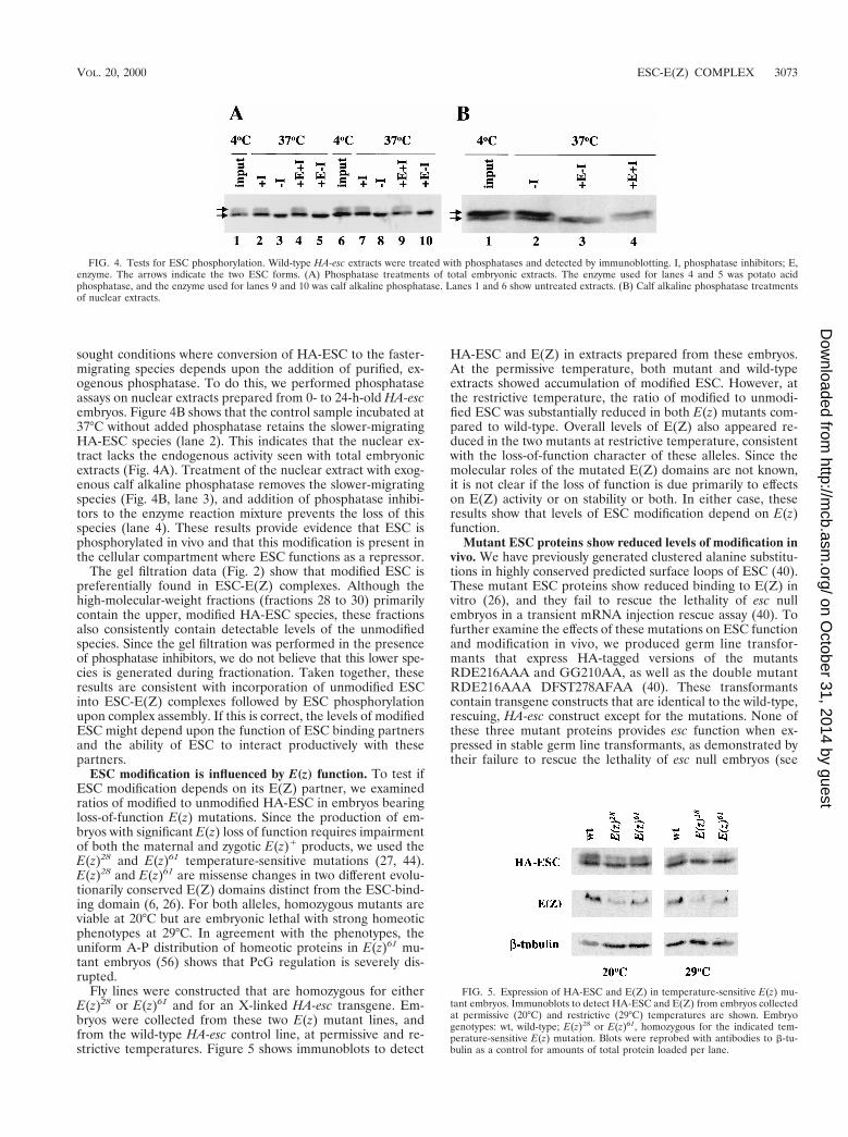
sought conditions where conversion of HA-ESC to the faster-migrating species depends upon the addition of purified, ex-ogenous phosphatase. To do this, we performed phosphataseassays on nuclear extracts prepared from 0- to 24-h-old HA-escembryos. Figure 4B shows that the control sample incubated at37°C without added phosphatase retains the slower-migratingHA-ESC species (lane 2). This indicates that the nuclear ex-tract lacks the endogenous activity seen with total embryonicextracts (Fig. 4A). Treatment of the nuclear extract with exog-enous calf alkaline phosphatase removes the slower-migratingspecies (Fig. 4B, lane 3), and addition of phosphatase inhibi-tors to the enzyme reaction mixture prevents the loss of thisspecies (lane 4). These results provide evidence that ESC isphosphorylated in vivo and that this modification is present inthe cellular compartment where ESC functions as a repressor.
The gel filtration data (Fig. 2) show that modified ESC ispreferentially found in ESC-E(Z) complexes. Although thehigh-molecular-weight fractions (fractions 28 to 30) primarilycontain the upper, modified HA-ESC species, these fractionsalso consistently contain detectable levels of the unmodifiedspecies. Since the gel filtration was performed in the presenceof phosphatase inhibitors, we do not believe that this lower spe-cies is generated during fractionation. Taken together, theseresults are consistent with incorporation of unmodified ESCinto ESC-E(Z) complexes followed by ESC phosphorylationupon complex assembly. If this is correct, the levels of modifiedESC might depend upon the function of ESC binding partnersand the ability of ESC to interact productively with thesepartners.
ESC modification is influenced by E(z) function. To test ifESC modification depends on its E(Z) partner, we examinedratios of modified to unmodified HA-ESC in embryos bearingloss-of-function E(z) mutations. Since the production of em-bryos with significant E(z) loss of function requires impairmentof both the maternal and zygotic E(z)1 products, we used theE(z)28 and E(z)61 temperature-sensitive mutations (27, 44).E(z)28 and E(z)61 are missense changes in two different evolu-tionarily conserved E(Z) domains distinct from the ESC-bind-ing domain (6, 26). For both alleles, homozygous mutants areviable at 20°C but are embryonic lethal with strong homeoticphenotypes at 29°C. In agreement with the phenotypes, theuniform A-P distribution of homeotic proteins in E(z)61 mu-tant embryos (56) shows that PcG regulation is severely dis-rupted.
Fly lines were constructed that are homozygous for eitherE(z)28 or E(z)61 and for an X-linked HA-esc transgene. Em-bryos were collected from these two E(z) mutant lines, andfrom the wild-type HA-esc control line, at permissive and re-strictive temperatures. Figure 5 shows immunoblots to detect
HA-ESC and E(Z) in extracts prepared from these embryos.At the permissive temperature, both mutant and wild-typeextracts showed accumulation of modified ESC. However, atthe restrictive temperature, the ratio of modified to unmodi-fied ESC was substantially reduced in both E(z) mutants com-pared to wild-type. Overall levels of E(Z) also appeared re-duced in the two mutants at restrictive temperature, consistentwith the loss-of-function character of these alleles. Since themolecular roles of the mutated E(Z) domains are not known,it is not clear if the loss of function is due primarily to effectson E(Z) activity or on stability or both. In either case, theseresults show that levels of ESC modification depend on E(z)function.
Mutant ESC proteins show reduced levels of modification invivo. We have previously generated clustered alanine substitu-tions in highly conserved predicted surface loops of ESC (40).These mutant ESC proteins show reduced binding to E(Z) invitro (26), and they fail to rescue the lethality of esc nullembryos in a transient mRNA injection rescue assay (40). Tofurther examine the effects of these mutations on ESC functionand modification in vivo, we produced germ line transfor-mants that express HA-tagged versions of the mutantsRDE216AAA and GG210AA, as well as the double mutantRDE216AAA DFST278AFAA (40). These transformantscontain transgene constructs that are identical to the wild-type,rescuing, HA-esc construct except for the mutations. None ofthese three mutant proteins provides esc function when ex-pressed in stable germ line transformants, as demonstrated bytheir failure to rescue the lethality of esc null embryos (see
FIG. 4. Tests for ESC phosphorylation. Wild-type HA-esc extracts were treated with phosphatases and detected by immunoblotting. I, phosphatase inhibitors; E,enzyme. The arrows indicate the two ESC forms. (A) Phosphatase treatments of total embryonic extracts. The enzyme used for lanes 4 and 5 was potato acidphosphatase, and the enzyme used for lanes 9 and 10 was calf alkaline phosphatase. Lanes 1 and 6 show untreated extracts. (B) Calf alkaline phosphatase treatmentsof nuclear extracts.
FIG. 5. Expression of HA-ESC and E(Z) in temperature-sensitive E(z) mu-tant embryos. Immunoblots to detect HA-ESC and E(Z) from embryos collectedat permissive (20°C) and restrictive (29°C) temperatures are shown. Embryogenotypes: wt, wild-type; E(z)28 or E(z)61, homozygous for the indicated tem-perature-sensitive E(z) mutation. Blots were reprobed with antibodies to b-tu-bulin as a control for amounts of total protein loaded per lane.
VOL. 20, 2000 ESC-E(Z) COMPLEX 3073
on October 31, 2014 by guest
http://mcb.asm
.org/D
ownloaded from

Materials and Methods). We compared expression levels ofthese mutant HA-ESC proteins to that of wild-type HA-ESCin crude embryonic extracts. Three independent lines weretested for each of the wild-type and mutant constructs. All lineswere homozygous for the respective transgenes and behavedgenetically as lines with single transgene insertions. Figure 6shows that the transgenic lines express different levels of HA-ESC depending on the line. The failure of these mutant pro-teins to provide esc function is not due simply to loweredoverall expression levels, since several of the nonrescuing linesaccumulate mutant HA-ESC at levels similar to those in thewild type. Mean expression levels (n $ 4) determined fromdensitometric analyses are 83, 54, and 126% of wild-type levelsfor the nonrescuing lines shown in Fig. 6, lanes 4, 9, and 10.Moreover, doubling the dosage of maternally provided mutantESC in the standard rescue test (see Materials and Methods)did not alter the lack of rescue. The failure to rescue here alsoparallels results obtained in mRNA injection rescue experi-ments (40), where the amounts of in vitro-transcribed escmRNA injected far exceed endogenous levels of the geneproduct.
Although not optimized to resolve the ESC forms, the blotin Fig. 6 suggests that levels of modified HA-ESC are spe-cifically reduced in the RDE216AAA DFST278AFAA andGG210AA mutants. Consequently, we examined this more pre-cisely by testing a single line bearing each transgenic constructunder gel conditions that improve the separation of the two ESCforms. Extracts were prepared from 6- to 12-h staged embryos,which contain peak levels of modified wild-type HA-ESC (Fig.1). Figure 7A shows that the RDE216AAA DFST278AFAAand GG210AA mutant lines accumulate the faster-migratingESC species but that the relative amount of modified ESC isdramatically reduced. Thus, ESC mutant proteins with im-paired E(Z) binding in vitro show reduced levels of modifica-tion in vivo.
In contrast to the mutant results in Fig. 7A, the RDE216AAAmutant shows a more subtle reduction in the relative levels ofmodified to unmodified ESC (Fig. 7B). Intriguingly, the twoESC mutants with severe loss of the modified species behave asnull mutants in the transient esc rescue assay whereas themutant with more subtle reduction retains some residual ac-tivity (40). This correlation provides another link between escfunction and modification in vivo.
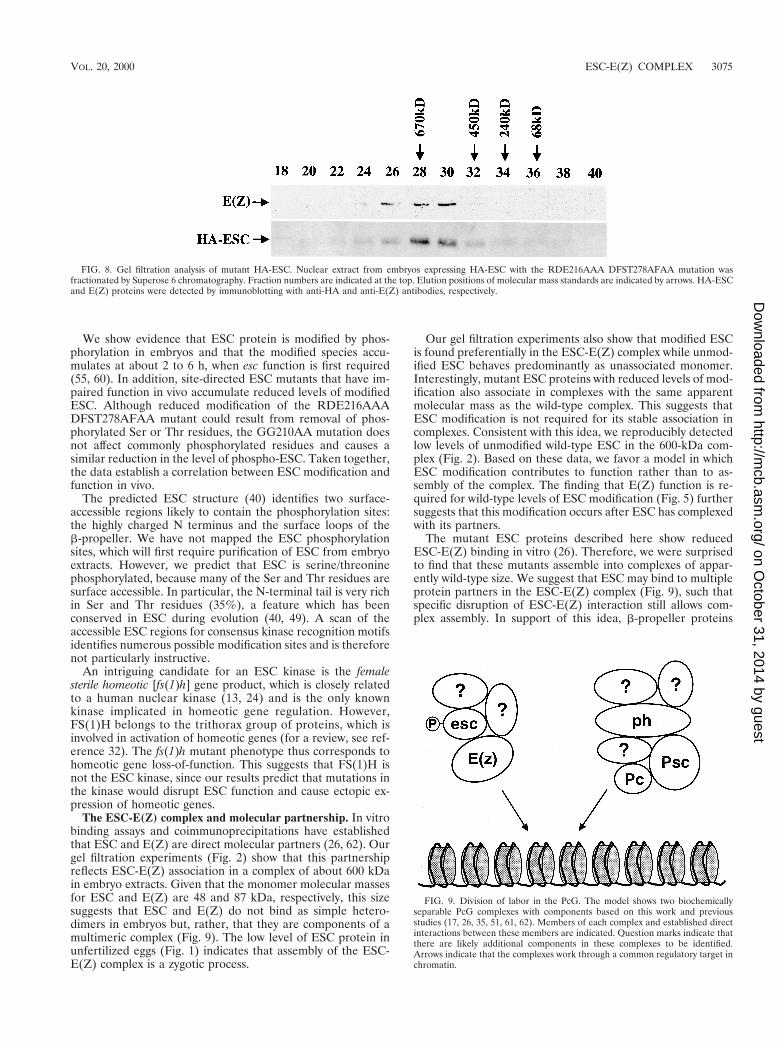
Association of mutant ESC in complexes. We wished to de-termine why the RDE216AAA DFST278AFAA and GG210AAESC mutants failed to function in vivo. We have identified twomolecular defects; their direct binding to E(Z) in vitro is dis-rupted (26), and they fail to accumulate wild-type levels of mod-ification (Fig. 7A). One explanation, given the E(Z) bindingdefect, is simply that these mutant ESC proteins are unable toassemble into the 600-kDa ESC-E(Z) complexes. To addressthis question, we prepared nuclear extracts from 0- to 24-hembryos homozygous for the RDE216AAA DFST278AFAA
mutant HA-esc transgene and fractionated the extracts on aSuperose 6 column. Figure 8 shows that the RDE216AAADFST278AFAA protein associates in complexes with an ap-parent molecular mass similar to that of the complex contain-ing wild-type ESC (compare fractions 28 and 30 in Fig. 8 to thesame fractions in Fig. 2). Similarly, analysis of the GG210AAmutant protein shows that it also is present in complexes ofabout wild-type size (data not shown). It is possible that theresolution of these gel filtration experiments is insufficient todistinguish ESC complexes that contain or lack the 87-kDaE(Z) component. However, coimmunoprecipitation experimentsperformed on RDE216AAA DFST278AFAA mutant extractdetect an association of E(Z) with the mutant ESC (data notshown). The simplest interpretation is that these mutant ESCproteins are incorporated into complexes that are renderedfunctionally defective. In addition, the combined results for invitro binding and in vivo complex assembly suggest that con-tacts between ESC and other partner proteins besides E(Z)contribute to ESC association in the 600-kDa complex.
DISCUSSION
Expression and modification of ESC protein. esc mRNA isexpressed primarily during early development, with the highestlevels being found before 4 h of embryogenesis (18, 55). Thisearly expression has prompted the hypothesis that esc func-tions in the transition between initiation of homeotic gene re-pression by gap proteins, such as hunchback, and maintenanceof this repression by PcG proteins (22, 48, 55). This transitionoccurs at about 4 h, when gap gene products decay. In thisstudy we have shown that ESC protein is expressed at peaklevels at 6 to 12 h (Fig. 1), after esc mRNA has decayed to lowlevels. In addition, ESC is detected until the end of embryo-genesis. The presence of substantial levels of ESC in mid- tolate-stage embryos suggests that ESC may play a greater rolethan simply in the transition between gap protein and PcGprotein repression. In addition, a second peak of ESC proteinis detected during larval and pupal stages, consistent with itsnonessential function in imaginal discs (58, 63).
FIG. 6. Expression of mutant HA-ESC proteins. Immunoblot detection of wild-type (lanes 1 to 3) and the indicated mutant (lanes 4 to 12) HA-ESC proteins from6- to 12-h total-embryo extracts is shown. Three independent lines were used for each transgene construct. All lines were homozygous for the transgene. Approximatelyequal amounts of total protein were loaded per lane.
FIG. 7. Effect of ESC surface loop mutations upon ESC modification. Im-munoblot detection of wild-type and mutant HA-ESC proteins from 6- to 12-hembryo extracts is shown. Arrows indicate the two ESC forms. Mutants in panelA show severe loss of esc function in vivo, and the mutant in panel B showsmoderate loss of function in vivo.
3074 NG ET AL. MOL. CELL. BIOL.
on October 31, 2014 by guest
http://mcb.asm
.org/D
ownloaded from
We show evidence that ESC protein is modified by phos-phorylation in embryos and that the modified species accu-mulates at about 2 to 6 h, when esc function is first required(55, 60). In addition, site-directed ESC mutants that have im-paired function in vivo accumulate reduced levels of modifiedESC. Although reduced modification of the RDE216AAADFST278AFAA mutant could result from removal of phos-phorylated Ser or Thr residues, the GG210AA mutation doesnot affect commonly phosphorylated residues and causes asimilar reduction in the level of phospho-ESC. Taken together,the data establish a correlation between ESC modification andfunction in vivo.
The predicted ESC structure (40) identifies two surface-accessible regions likely to contain the phosphorylation sites:the highly charged N terminus and the surface loops of theb-propeller. We have not mapped the ESC phosphorylationsites, which will first require purification of ESC from embryoextracts. However, we predict that ESC is serine/threoninephosphorylated, because many of the Ser and Thr residues aresurface accessible. In particular, the N-terminal tail is very richin Ser and Thr residues (35%), a feature which has beenconserved in ESC during evolution (40, 49). A scan of theaccessible ESC regions for consensus kinase recognition motifsidentifies numerous possible modification sites and is thereforenot particularly instructive.
An intriguing candidate for an ESC kinase is the femalesterile homeotic [fs(1)h] gene product, which is closely relatedto a human nuclear kinase (13, 24) and is the only knownkinase implicated in homeotic gene regulation. However,FS(1)H belongs to the trithorax group of proteins, which isinvolved in activation of homeotic genes (for a review, see ref-erence 32). The fs(1)h mutant phenotype thus corresponds tohomeotic gene loss-of-function. This suggests that FS(1)H isnot the ESC kinase, since our results predict that mutations inthe kinase would disrupt ESC function and cause ectopic ex-pression of homeotic genes.
The ESC-E(Z) complex and molecular partnership. In vitrobinding assays and coimmunoprecipitations have establishedthat ESC and E(Z) are direct molecular partners (26, 62). Ourgel filtration experiments (Fig. 2) show that this partnershipreflects ESC-E(Z) association in a complex of about 600 kDain embryo extracts. Given that the monomer molecular massesfor ESC and E(Z) are 48 and 87 kDa, respectively, this sizesuggests that ESC and E(Z) do not bind as simple hetero-dimers in embryos but, rather, that they are components of amultimeric complex (Fig. 9). The low level of ESC protein inunfertilized eggs (Fig. 1) indicates that assembly of the ESC-E(Z) complex is a zygotic process.
Our gel filtration experiments also show that modified ESCis found preferentially in the ESC-E(Z) complex while unmod-ified ESC behaves predominantly as unassociated monomer.Interestingly, mutant ESC proteins with reduced levels of mod-ification also associate in complexes with the same apparentmolecular mass as the wild-type complex. This suggests thatESC modification is not required for its stable association incomplexes. Consistent with this idea, we reproducibly detectedlow levels of unmodified wild-type ESC in the 600-kDa com-plex (Fig. 2). Based on these data, we favor a model in whichESC modification contributes to function rather than to as-sembly of the complex. The finding that E(Z) function is re-quired for wild-type levels of ESC modification (Fig. 5) furthersuggests that this modification occurs after ESC has complexedwith its partners.
The mutant ESC proteins described here show reducedESC-E(Z) binding in vitro (26). Therefore, we were surprisedto find that these mutants assemble into complexes of appar-ently wild-type size. We suggest that ESC may bind to multipleprotein partners in the ESC-E(Z) complex (Fig. 9), such thatspecific disruption of ESC-E(Z) interaction still allows com-plex assembly. In support of this idea, b-propeller proteins
FIG. 8. Gel filtration analysis of mutant HA-ESC. Nuclear extract from embryos expressing HA-ESC with the RDE216AAA DFST278AFAA mutation wasfractionated by Superose 6 chromatography. Fraction numbers are indicated at the top. Elution positions of molecular mass standards are indicated by arrows. HA-ESCand E(Z) proteins were detected by immunoblotting with anti-HA and anti-E(Z) antibodies, respectively.
FIG. 9. Division of labor in the PcG. The model shows two biochemicallyseparable PcG complexes with components based on this work and previousstudies (17, 26, 35, 51, 61, 62). Members of each complex and established directinteractions between these members are indicated. Question marks indicate thatthere are likely additional components in these complexes to be identified.Arrows indicate that the complexes work through a common regulatory target inchromatin.
VOL. 20, 2000 ESC-E(Z) COMPLEX 3075
on October 31, 2014 by guest
http://mcb.asm
.org/D
ownloaded from
have been shown to make simultaneous contacts with multiplepartners (66). Another possibility is that mutant ESC isbrought into complexes through homotypic interactions withendogenous wild-type ESC. This seems unlikely, however,since in vitro binding assays do not detect self-association ofESC (A. Peterson and J. Simon, unpublished data). Moreover,the majority of ESC is occupied by the b-propeller domain,and b-propellers do not typically function as homodimers.
If the ESC mutants assemble into 600-kDa complexes, whydo they fail to function in vivo? One possibility is that disrup-tion of direct ESC-E(Z) contact renders the complex unable toadopt an active conformation. Another possibility is that E(Z)is required to produce or maintain ESC phosphorylation,which could be key for function of the complex. This is anattractive model, since E(Z) contains a motif known as theSET domain (28), which has been shown to bind proteins thatact as phosphatase inhibitors (10).
Division of labor in the PcG. The PcG proteins PC and PHare associated in a complex estimated to be 2 MDa (17). Inaddition, PC and PH coimmunoprecipitate and interact withanother PcG protein, PSC (35, 61). Here, we have shown thatthe ESC-E(Z) complex is biochemically distinct from com-plexes containing PH (Fig. 2 and 3). In agreement with this, aPH-PC-PSC complex recently purified from fly embryos doesnot contain E(Z) (51). Taken together, these results support amodel (Fig. 9) in which there are at least two distinct PcGcomplexes in vivo, one containing ESC and E(Z) and the othercontaining PH, PC, and PSC. Consistent with this idea, themammalian ESC and E(Z) homologs, EED and EZH2, fail tocoimmunoprecipitate with the mammalian PH, PSC, and PChomologs (50, 64, 65). In addition, EED and EZH2 do notcolocalize with mammalian PH, PSC, and PC within nuclei ofosteosarcoma cells (50, 65). Furthermore, the patterns of pair-wise interactions among Drosophila PcG proteins are reiter-ated among their mammalian counterparts (1, 14, 21, 23, 26,35, 43, 50, 62, 65), which suggests that this division of labor inthe PcG (Fig. 9) has been conserved in evolution.
Although the existence of at least two different PcG com-plexes has been established, the complete spectrum of PcGprotein interactions has not yet been elucidated. There ap-pears to be further division among PH-PC-PSC complexes,which have different compositions at different target genes(61). In addition, multiple complexes containing the mamma-lian PH, PC, and PSC proteins have been detected (23). More-over, there are additional PcG proteins, such as ASX, PCL,and PHO, whose in vivo associations have yet to be described.Some of these proteins may correspond to as yet unidentifiedcomponents of ESC-E(Z) or PH-PC-PSC complexes (Fig. 9),or they may sort into additional distinct complexes. In partic-ular, complexes containing PHO, the only known DNA-bind-ing member of the PcG (4), may be important for targetingother PcG complexes to sites of action. We note that PHO isnot detected as a stable member of either the ESC-E(Z) (Fig.3) or PH-PC-PSC complexes (51).
Despite the presence of biochemically separable PcG com-plexes, the similar mutant phenotypes and genetic interactionsof PcG genes indicate that they work together at some level.Any model for PcG repression must therefore accommodateboth the biochemical separability and functional synergy ofPcG complexes. One possibility is that repression requires mul-tiple chromatin-modifying events by the different PcG com-plexes. This would be similar to the in vivo synergy between thechromatin-modifying SWI-SNF and SAGA complexes, whichare both required for maintenance of HO expression in yeast(9, 34). An alternative possibility is that one PcG complexdirectly modifies chromatin while the other complex counter-
acts trithorax group activation by inhibiting the chromatin-remodeling activity of the brahma complex (41, 51). Indeed,the first evidence that a PcG complex may covalently modifychromatin is provided by the recent report of histone deacety-lase activity associated with mammalian homologs of ESC andE(Z) (64).
These mechanisms are inconsistent with an esc role limitedto the transition from gap repressors to PcG repressors (22, 48,55). Instead, we suggest that ESC is more globally involved inchromatin regulation and that this involvement is most criticalearly in fly development. Consistent with a global role, EEDmRNA is expressed in many tissues during mouse develop-ment (49). Furthermore, the C. elegans homolog of ESC,MES-6, is a transcriptional repressor that functions in germline development (31, 33). MES-6 in worms therefore plays adistinct developmental role from ESC in flies. This suggeststhat ESC participates in a general repression mechanism thathas been adapted for use in different cell lineages, rather thanin the specific transition between gap protein and PcG proteinrepression.
If ESC-E(Z) complexes function as general chromatin reg-ulators, the early requirement for ESC in Drosophila must bereconciled with the need for long-term PcG repression duringdevelopment. One possibility is that another protein replacesESC in the ESC-E(Z) complex at late developmental stages,when ESC is no longer critically required. Alternatively, E(Z)may associate with a completely different set of PcG proteinsto supply the biochemical function provided by ESC-E(Z)complexes during embryogenesis. To address these possibili-ties, the nature of E(Z) complexes at postembryonic stages willhave to be investigated.
ACKNOWLEDGMENTS
We thank Ellen Miller, Doug Bornemann, and Aidan Peterson forhelping to generate and characterize the PH antibody, and we thankRick Jones for providing E(Z) antibody and Judy Kassis for providingPHO antibody. We thank Ophelia Papoulas, Mary Porter, ZhaohuiShao, Osamu Shimmi, Steve Johnson, and Natalie Coe for advice ongel filtration experiments. We are grateful to Laura Mauro for usefuldiscussions about protein phosphorylation. We also thank OpheliaPapoulas, Mike O’Connor, Tom Hays, and members of the Simonlaboratory for discussions, input and critical comments on the manu-script.
This work was supported by NIH grant GM49850 to J.S., and J.N.was supported in part by NIH training grant HD07480.
REFERENCES
1. Alkema, M. J., M. Bronk, E. Verhoeven, A. Otte, L. J. van’t Veer, A. Berns,and M. van Lohuizen. 1997. Identification of Bmi1-interacting proteins asconstituents of a multimeric mammalian Polycomb complex. Genes Dev. 11:226–240.
2. Bornemann, D., E. Miller, and J. Simon. 1996. The Drosophila Polycombgroup gene Sex comb on midleg (Scm) encodes a zinc finger protein withsimilarity to polyhomeotic protein. Development 122:1621–1630.
3. Bornemann, D., E. Miller, and J. Simon. 1998. Expression and properties ofwild-type and mutant forms of the Drosophila Sex combs on Midleg repres-sor protein. Genetics 150:675–686.
4. Brown, J. L., D. Mucci, M. Whiteley, M. L. Dirksen, and J. A. Kassis. 1998.The Drosophila Polycomb group gene pleiohomeotic encodes a DNA bindingprotein with homology to the transcription factor YY1. Mol. Cell 1:1057–1064.
5. Brunk, B. P., E. C. Martin, and P. N. Adler. 1991. Drosophila genes Posteriorsex combs and suppressor two of zeste encode proteins with homology to themurine bmi-1 oncogene. Nature 353:351–353.
6. Carrington, E. C., and R. S. Jones. 1996. The Drosophila Enhancer of zestegene encodes a chromosomal protein: examination of wild-type and mutantprotein distribution. Development 122:4073–4083.
7. Carroll, S. B., R. A. Laymon, M. A. McCutcheon, P. D. Riley, and M. P.Scott. 1986. The localization and regulation of the Antennapedia proteinexpression in Drosophila embryos. Cell 47:113–122.
8. Celniker, S. E., D. J. Keelan, and E. B. Lewis. 1989. The molecular genetics
3076 NG ET AL. MOL. CELL. BIOL.
on October 31, 2014 by guest
http://mcb.asm
.org/D
ownloaded from

of the bithorax complex of Drosophila: characterization of the product of theAbdominal-B domain. Genes Dev. 3:1424–1436.
9. Cosma, M. P., T. Tanaka, and K. Nasmyth. 1999. Ordered recruitment oftranscription and chromatin remodeling factors to a cell cycle- and develop-mentally regulated promoter. Cell 97:299–311.
10. Cui, X., I. De Vivo, R. Slany, A. Miyamoto, R. Firestein, and M. L. Cleary.1998. Association of SET domain and myotubularin-related proteins mod-ulates growth control. Nat. Genet 18:331–337.
11. DeCamillis, M., and H. W. Brock. 1994. Expression of the polyhomeotic locusin development of Drosophila melanogaster. Roux’s Arch. Dev. Biol. 203:429–438.
12. DeCamillis, M., N. Cheng, D. Pierre, and H. W. Brock. 1992. The polyho-meotic gene of Drosophila encodes a chromatin protein that shares polytenechromosome-binding sites with Polycomb. Genes Dev. 6:223–232.
13. Denis, G. V., and M. R. Green. 1996. A novel, mitogen-activated nuclearkinase is related to a Drosophila developmental regulator. Genes Dev. 10:261–271.
14. Denisenko, O., M. Shnyreva, H. Suzuki, and K. Bomsztyk. 1998. Pointmutations in the WD40 domain of Eed block its interaction with Ezh2. Mol.Cell. Biol. 18:5634–5642.
15. Dingwall, A. K., S. J. Beek, C. M. McCallum, J. W. Tamkun, G. V. Kalpana,S. P. Goff, and M. P. Scott. 1995. The Drosophila snr1 and brm proteins arerelated to yeast SWI/SNF proteins and are components of a large proteincomplex. Mol. Biol. Cell. 6:777–791.
16. Dura, J.-M., N. B. Randsholt, J. Deatrick, I. Erk, P. Santamaria, J. D.Freeman, S. J. Freeman, D. Weddell, and H. W. Brock. 1987. A complexgenetic locus, polyhomeotic, is required for segmental specification and epi-dermal development in D. melanogaster. Cell 51:829–839.
17. Franke, A., M. DeCamillis, D. Zink, N. Cheng, H. W. Brock, and R. Paro.1992. Polycomb and polyhomeotic are constituents of a multimeric proteincomplex in chromatin of Drosophila melanogaster. EMBO J. 11:2941–2950.
18. Frei, E., D. Bopp, M. Burri, S. Baumgartner, J. E. Edstrom, and M. Noll.1985. Isolation and structural analysis of the extra sex combs gene of Dro-sophila. Cold Spring Harbor Symp. Quant. Biol. 50:127–134.
19. Fritsch, C., J. L. Brown, J. A. Kassis, and J. Muller. 1999. The DNA-bindingpolycomb group protein pleiohomeotic mediates silencing of a Drosophilahomeotic gene. Development 126:3905–3913.
20. Gillespie, S. K., and S. A. Wasserman. 1994. Dorsal, a Drosophila Rel-likeprotein, is phosphorylated upon activation of the transmembrane proteinToll. Mol. Cell. Biol. 14:3559–3568.
21. Gunster, M. J., D. P. E. Satijn, K. M. Hamer, J. L. den Blaauwen, D. deBruijn, M. J. Alkema, M. van Lohuizen, R. van Driel, and A. P. Otte. 1997.Identification and characterization of interactions between the vertebratePolycomb-group protein BMI1 and human homologs of polyhomeotic. Mol.Cell. Biol. 17:2326–2335.
22. Gutjahr, T., E. Frei, C. Spicer, S. Baumgartner, R. A. White, and M. Noll.1995. The Polycomb-group gene, extra sex combs, encodes a nuclear memberof the WD-40 repeat family. EMBO J. 14:4296–4306.
23. Hashimoto, N., H. W. Brock, M. Nomura, M. Kyba, J. Hodgson, Y. Fujita, Y.Takihara, K. Shimada, and T. Higashinakagawa. 1998. RAE28, BMI1 andM33 are members of heterogeneous multimeric mammilian Polycomb groupcomplexes. Biochem. Biophys. Res. Commun. 245:356–365.
24. Haynes, S. R., B. A. Mozer, N. Bhatia-Dey, and I. B. Dawid. 1989. TheDrosophila fsh locus, a maternal effect homeotic gene, encodes apparentmembrane proteins. Dev. Biol. 134:246–257.
25. Holdemann, R., S. Nehrt, and S. Strome. 1998. MES-2, a maternal proteinessential for viability of the germline in Caenorhabditis elegans, is homolo-gous to a Drosophila Polycomb group protein. Development 125:2457–2467.
26. Jones, C. A., J. Ng, A. J. Peterson, K. Morgan, J. Simon, and R. S. Jones.1998. The Drosophila esc and E(z) proteins are direct partners in Polycombgroup-mediated repression. Mol. Cell. Biol. 18:2825–2834.
27. Jones, R. S., and W. M. Gelbart. 1990. Genetic analysis of the enhancer ofzeste locus and its role in gene regulation in Drosophila melanogaster. Ge-netics 126:185–199.
28. Jones, R. S., and W. M. Gelbart. 1993. The Drosophila Polycomb-group geneenhancer of zeste contains a region with sequence similarity to trithorax. Mol.Cell. Biol. 13:6357–6366.
29. Karch, F., W. Bender, and B. Weiffenbach. 1990. abdA expression in Dro-sophila embryos. Genes Dev. 4:1573–1587.
30. Kaufman, T. C., R. Lewis, and B. Wakimoto. 1980. Cytogenetic analysis ofchromosome 3 in Drosophila melanogaster: the homeotic gene complex inpolytene interval 84A-B. Genetics 94:115–133.
31. Kelly, W. G., and A. Fire. 1998. Chromatin silencing and the maintenance ofa functional germline in Caenorhabditis elegans. Development 125:2451–2456.
32. Kennison, J. A. 1993. Transcriptional activation of Drosophila homeoticgenes from distant regulatory elements. Trends Genet. 9:75–79.
33. Korf, I., Y. Fan, and S. Strome. 1998. The Polycomb group in Caenorhabditiselegans and maternal control of germline development. Development 125:2469–2478.
34. Krebs, J. E., M. H. Kuo, C. D. Allis, and C. L. Peterson. 1999. Cell cycle-
regulated histone acetylation required for expression of the yeast HO gene.Genes Dev. 13:1412–1421.
35. Kyba, M., and H. W. Brock. 1998. The Drosophila Polycomb group proteinPsc contacts ph and Pc through specific conserved domains. Mol. Cell. Biol.18:2712–2720.
36. Lewis, E. B. 1978. A gene complex controlling segmentation in Drosophila.Nature 276:565–570.
37. Lonie, A., R. D’Andrea, R. Paro, and R. Saint. 1994. Molecular character-ization of the Polycomblike gene of Drosophila melanogaster, a trans-actingnegative regulator of homeotic gene expression. Development 120:2629–2636.
38. Martin, E. C., and P. N. Adler. 1993. The Polycomb group gene Posterior sexcombs encodes a chromosomal protein. Development 117:641–655.
39. McKeon, J., and H. W. Brock. 1991. Interactions of the Polycomb group ofgenes with homeotic loci of Drosophila. Roux’s Arch. Dev. Biol. 199:387–396.
40. Ng, J., R. Li, K. Morgan, and J. Simon. 1997. Evolutionary conservation andpredicted structure of the Drosophila extra sex combs repressor protein. Mol.Cell. Biol. 17:6663–6672.
41. Papoulas, O., S. J. Beek, S. L. Moseley, C. M. McCallum, M. Sarte, A.Shearn, and J. W. Tamkun. 1998. The Drosophila trithorax group proteinsBRM, ASH1 and ASH2 are subunits of distinct protein complexes. Devel-opment 125:3955–3966.
42. Paro, R., and D. S. Hogness. 1991. The Polycomb protein shares a homolo-gous domain with a heterochromatin-associated protein of Drosophila. Proc.Natl. Acad. Sci. USA 88:263–267.
43. Peterson, A. J., M. Kyba, D. Bornemann, K. Morgan, H. W. Brock, and J.Simon. 1997. A domain shared by the Polycomb group proteins Scm and phmediates heterotypic and homotypic interactions. Mol. Cell. Biol. 17:6683–6692.
44. Phillips, M. D., and A. Shearn. 1990. Mutations in polycombeotic, a Dro-sophila Polycomb group gene, cause a wide range of maternal and zygoticphenotypes. Genetics 125:91–101.
45. Pirrotta, V. 1997. PcG complexes and chromatin silencing. Curr. Opin.Genet. Dev. 7:249–258.
46. Qian, S., M. Capovilla, and V. Pirrotta. 1993. Molecular mechanisms ofpattern formation by the BRE enhancer of the Ubx gene. EMBO J. 12:3865–3877.
47. Rastelli, L., C. S. Chan, and V. Pirrotta. 1993. Related chromosome bindingsites for zeste, suppressors of zeste and Polycomb group proteins in Drosophilaand their dependence on Enhancer of zeste function. EMBO J. 12:1513–1522.
48. Sathe, S. S., and P. J. Harte. 1995. The Drosophila extra sex combs proteincontains WD motifs essential for its function as a repressor of homeoticgenes. Mech. Dev. 52:77–87.
49. Schumacher, A., C. Faust, and T. Magnuson. 1996. Positional cloning of aglobal regulator of anterior-posterior patterning in mice. Nature 383:250–253.
50. Sewalt, R. G. A. B., J. van der Vlag, M. J. Gunster, K. M. Hamer, J. L. denBlaauwen, D. P. E. Satijn, T. Hendrix, R. van Driel, and A. P. Otte. 1998.Characterization of interactions between the mammalian Polycomb-groupproteins Enx1/EZH2 and EED suggests the existence of different mamma-lian Polycomb-group protein complexes. Mol. Cell. Biol. 18:3586–3595.
51. Shao, Z., F. Raible, R. Mollaaghababa, J. R. Guyon, C. T. Wu, W. Bender,and R. E. Kingston. 1999. Stabilization of chromatin structure by PRC1, aPolycomb complex. Cell 98:37–46.
52. Shimell, M. J., A. J. Peterson, J. Burr, J. A. Simon, and M. B. O’Connor.2000. Functional analysis of repressor binding sites in the iab-2 regulatoryregion of the abdominal-A homeotic gene. Dev. Biol. 218:38–52.
53. Shimell, M. J., J. Simon, W. Bender, and M. B. O’Connor. 1994. Enhancerpoint mutation results in a homeotic transformation in Drosophila. Science264:968–971.
54. Simon, J. 1995. Locking in stable states of gene expression: transcriptionalcontrol during Drosophila development. Curr. Opin. Cell Biol. 7:376–385.
55. Simon, J., D. Bornemann, K. Lunde, and C. Schwartz. 1995. The extra sexcombs product contains WD40 repeats and its time of action implies a roledistinct from other Polycomb group products. Mech. Dev. 53:197–208.
56. Simon, J., A. Chiang, and W. Bender. 1992. Ten different Polycomb groupgenes are required for spatial control of the abdA and AbdB homeoticproducts. Development 114:493–505.
57. Sinclair, D. A. R., T. A. Milne, J. W. Hodgson, J. Shellard, C. A. Salinas, M.Kyba, F. Randazzo, and H. W. Brock. 1998. The Additional sex combs geneof Drosophila encodes a chromatin protein that binds to shared and uniquePolycomb group sites on polytene chromosomes. Development 125:1207–1216.
58. Struhl, G. 1981. A gene product required for correct initiation of segmentaldetermination in Drosophila. Nature 293:36–41.
59. Struhl, G., and M. Akam. 1985. Altered distributions of Ultrabithorax tran-scripts in extra sex combs mutant embryos of Drosophila. EMBO J. 4:3259–3264.
60. Struhl, G., and D. Brower. 1982. Early role of the esc1 gene product in thedetermination of segments in Drosophila. Cell 31:285–292.
61. Strutt, H., and R. Paro. 1997. The Polycomb group protein complex of
VOL. 20, 2000 ESC-E(Z) COMPLEX 3077
on October 31, 2014 by guest
http://mcb.asm
.org/D
ownloaded from
Drosophila melanogaster has different compositions at different target genes.Mol. Cell. Biol. 17:6773–6783.
62. Tie, F., T. Furuyama, and P. J. Harte. 1998. The Drosophila Polycomb groupproteins ESC and E(Z) bind directly to each other and co-localize at mul-tiple chromosomal sites. Development 125:3483–3496.
63. Tokunaga, C., and C. Stern. 1965. The developmental autonomy of extra sexcombs in Drosophila melanogaster. Dev. Biol. 11:50–81.
64. van der Vlag, J., and A. P. Otte. 1999. Transcriptional repression mediatedby the human polycomb-group protein EED involves histone deacetylation.Nat. Genet. 23:474–478.
65. van Lohuizen, M., M. Tijms, J. W. Voncken, A. Schumacher, T. Magnuson,
and E. Wientjens. 1998. Interaction of mouse Polycomb-group (Pc-G) pro-teins Enx1 and Enx2 with Eed: indication for separate PcG complexes. Mol.Cell. Biol. 18:3572–3579.
66. Wall, M. A., D. E. Coleman, E. Lee, J. A. Iniguez-Lluhi, B. A. Posner, A. G.Gilman, and S. R. Sprang. 1995. The structure of the G protein heterotrimerGia1b1g2. Cell 83:1047–1058.
67. White, R. A. H., and M. Wilcox. 1985. Distribution of Ultrabithorax proteinsin Drosophila. EMBO J. 4:2035–2043.
68. Zhang, C.-C., and M. Bienz. 1992. Segmental determination in Drosophilaconferred by hunchback (hb), a repressor of the homeotic gene Ultrabithorax(Ubx). Proc. Natl. Acad. Sci. USA 89:7511–7515.
3078 NG ET AL. MOL. CELL. BIOL.
on October 31, 2014 by guest
http://mcb.asm
.org/D
ownloaded from
Related Documents


