A Dissertation on A CLINICO PATHOLOGICAL STUDY AND MANAGEMENT OF NECROTIZING FASCIITIS COIMBATORE MEDICAL COLLEGE HOSPITAL Dissertation submitted to THE TAMIL NADU DR.M.G.R. MEDICAL UNIVERSITY CHENNAI - 32, TAMIL NADU With partial fulfilment of the regulations For the award of the degree of M.S. DEGREE EXAMINATION BRANCH I – GENERAL SURGERY COIMBATORE MEDICAL COLLEGE HOSPITAL COIMBATORE APRIL 2016

Welcome message from author
This document is posted to help you gain knowledge. Please leave a comment to let me know what you think about it! Share it to your friends and learn new things together.
Transcript
A Dissertation on
A CLINICO PATHOLOGICAL STUDY AND MANAGEMENT OF
NECROTIZING FASCIITIS
COIMBATORE MEDICAL COLLEGE HOSPITAL
Dissertation submitted to
THE TAMIL NADU DR.M.G.R. MEDICAL UNIVERSITY
CHENNAI - 32, TAMIL NADU
With partial fulfilment of the regulations
For the award of the degree of
M.S. DEGREE EXAMINATION
BRANCH I – GENERAL SURGERY COIMBATORE MEDICAL COLLEGE HOSPITAL
COIMBATORE
APRIL 2016
DECLARATION BY THE CANDIDATE
I hereby declare that this dissertation titled “A CLINICO
PATHOLOGICAL STUDY AND MANAGEMENT OF
NECROTIZING FASCIITIS CASES IN CMCH ” is a bonafide and
genuine research work carried out by me under the guidance of Dr. V.
ELANGO, M.S, FAIS., Professor and Head of the Department
GENERAL SURGERY, Coimbatore Medical College and
Hospital, Tamil Nadu, India.
DATE: Signature of the Candidate
PLACE: Dr. G.NIRUBAN
CHAKRAVARTHY
CERTIFICATE
This is to certify that dissertation entitled, “A CLINICO
PATHOLOGICAL STUDY AND MANAGEMENT OF
NECROTIZING FASCIITIS CASES IN CMCH” Submitted by
Dr. G. NIRUBAN CHAKRAVARTHY in partial fulfilment for the
award of the degree of master of surgery in GENERAL SURGERY by
The Tamil Nadu Dr .M.G.R. Medical University, Chennai, is a
bonafide record of the work done by him in the Department of general
surgery, Coimbatore Medical College and Hospital, during the academic
year 2013-2016.
Guide & Professor Professor
& HOD
Department of general surgery Department of
general surgery
Dean
Coimbatore Medical College Hospital
ACKNOWLEDGEMENT
I am extremely thankful to DEAN, Prof. Dr. A.
EDWIN JOE M.D., Coimbatore Medical College and hospital,
for his kind permission to carry out this study.
I am immensely grateful to Professor Dr. V. ELANGO,
M.S , PROFESSOR and Head of Department of GENRAL
SURGERY, Coimbatore Medical College and Hospital for her
concern and support in conducting the study.
I am greatly indebted to my assistant professors
Dr. R. Narayanamoorthy M.S, Dr. P.Sumitra M.S., DGO.,
and Dr. B.Jayalakshmi M.S., Department of General
Surgery, Coimbatore Medical College and Hospital for his
inspiration, guidance and comments at all stages of this study.
I am thankful to all our General Surgery unit chiefs for
their support in conducting the study.

I am thankful to all assistant professors for their guidance
and help. I am thankful to all my colleagues for the help
rendered in carrying out this dissertation.
I thank all the patients for their support in this study.

S. NO TITLE PAGE
NO
1. INTRODUCTION
2. AIMS
3. OBJECTIVES
4. REVIEW OF LITERATURE
5. MATERIALS
6. METHODOLOGY
7. OBSERVATION AND RESULTS
8. DISCUSSION
9. CONCLUSION
10.
ANNEXURE
BIBILOGRAPHY
PROFORMA
PATIENT CONSENT FORM
MASTER CHART
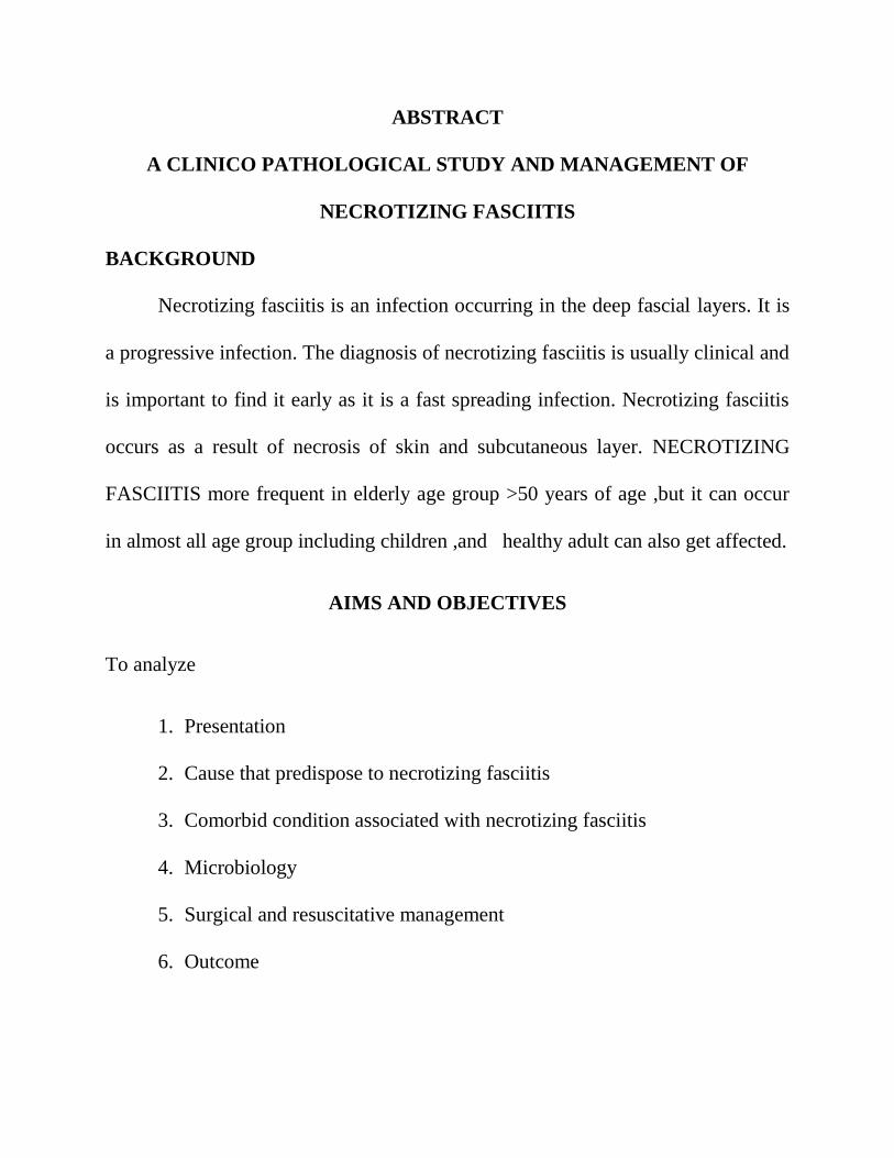
ABSTRACT
A CLINICO PATHOLOGICAL STUDY AND MANAGEMENT OF
NECROTIZING FASCIITIS
BACKGROUND
Necrotizing fasciitis is an infection occurring in the deep fascial layers. It is
a progressive infection. The diagnosis of necrotizing fasciitis is usually clinical and
is important to find it early as it is a fast spreading infection. Necrotizing fasciitis
occurs as a result of necrosis of skin and subcutaneous layer. NECROTIZING
FASCIITIS more frequent in elderly age group >50 years of age ,but it can occur
in almost all age group including children ,and healthy adult can also get affected.
AIMS AND OBJECTIVES
To analyze
1. Presentation
2. Cause that predispose to necrotizing fasciitis
3. Comorbid condition associated with necrotizing fasciitis
4. Microbiology
5. Surgical and resuscitative management
6. Outcome
MATERIALS AND METHODS
STUDY DESIGN
Descriptional study
SOURCE OF DATA
60 patients of necrotizing fasciitis getting admitted in surgical ward
PLACE OF STUDY
Coimbatore Medical College and Hospital
STUDY PERIOD
September 2014 to august 2015
INCLUSION CRITERIA
All patients presenting with features of necrotizing fasciitis to Coimbatore
medical college and hospital
EXCLUSION CRITERIA
Pregnant women
Age<13 yrs
METHODOLOGY
Patients presenting with features of necrotizing fasciitis were admitted in the
general surgery ward in Coimbatore medical college and hospital were included in
the study during the study period of September 2014-august 2015. Initial diagnosis
were made by both clinical and anatomical findings. Details of the patient were
noted. Detailed interview with the patient were made regarding history and other
comorbid conditions.
Following complete history taking physical examination for the patient were
done including blood pressure measurement and temperature and other clinical
finding related to necrotizing fasciitis. Following clinical examination, routine
investigations were investigated. Radiological investigations were done to note for
gas formation in subcutaneous layer. Treatment were started as soon as diagnosis is
suspected. It includes resuscitation of patient with intravenous fluids, antibiotics
and wound debridement. And bacteriological culture is done for both aerobic and
anaerobic bacteria.
The sample taken for culture is transported through proper transporation
technique to culture laboratory. These samples were then cultured in blood agar
and mc Conkey agar for aerobic bacteria and in Robertson’s cooked meat media
for anaerobic bacteria. The cultured organisms were tested for resistance pattern by
disc diffusion method.
Following initial debridement the wound was inspected regularly and
subsequent debridement were done periodically whenever necessary. And dressing
were done using povidone iodine and saline guaze. After the wound is fit, patient
undergone split skin graft surgery for raw area.
STATISTICAL ANALYSIS
STATISTICAL METHOD
In our study we used descriptive statistical analysis. Continuous
measurement were represented on mean with or without standard deviation.
Categorical measurementwere represented in number (%).
Confidence interval of 95% is used to find significancyofvalue. Confidence
limit >50% is associated with statistical significance.
STATISTICAL SOFTWARE
Tables and charts were completed using Microsoft word and excel
software.
RESULTS
This study was conducted during the period of September 2014- august
2015. About 60 patients were included in our study and their different aspects for
predisposing factors, age of presentation, microbiological pattern and antibiotic
pattern were analysed.
DISCUSSION
Most commonly the disease is poly microbial of about 62%. Most common
isolated organism is E.coli 35% .almost all patients treated with broad
spectrum antibiotics .most common antibiotic sensitive is ceftriaxone
followed by aminoglycosides and meteronidazole.
Almost all patients underwent surgical debridement of about 95%and some
may underwent procedures like primary suturing secondary suturing and ssg.
CONCLUSION :
NECROTIZING FASCITIS is most commonly seen in the elderly males.
source of the infection is identifiable in most of the cases. diabetic mellitus is the
most common comorbid factor . The disease is most commonly polymicrobical.
Most common bacterial includes gram positive cocci ( streptococci and
staphylococcal species ). Gram negative (E.coli, actinobacter, pseudomonas ).
Most common anaerobes is bacteroides .

INTRODUCTION
Necrotizing fasciitis is an infection occurring in the deep fascial
layers. It is a progressive infection. The diagnosis of necrotizing fasciitis
is usually clinical and is important to find it early as it is a fast spreading
infection. Necrotizing fasciitis occurs as a result of necrosis of skin and
subcutaneous layer. In some cases there will be necrosis of underlying
muscles causing necrotizing myositis. Necrotizing fasciitis often spreads
by direct spread. In severe cases, the microorganisms can spread via
blood vessel and lymphatics resulting in sepsis and shock.
INCIDENCE:
NECROTIZING FASCIITIS more frequent in elderly age group
>50 years of age ,but it can occur in almost all age group including
children ,and healthy adult can also get affected.
0.4 to 0.53 cases per 1 lakh adults was reported .necrotizing fasciitis has
higher pre ponderanceto male population .
Male to female ratio is 3:1 .
Necrotizing fasciitis mainly affects lower limb followed by perineum
(Fournier s gangrene ). Necrotizing fasciitis infection mainly seen in low
economic status .
Incidence of Fournier s gangrene is about 1.6/1 lakh population .
Male to female ratio in Fournier s gangrene is 10:1, low incidence in
female is mainly due to because of good drainage of genito urinary
secretion .
Skin and soft tissue infection (SSTIs):
Divided in to three groups byinfection disease society of america
1.superficial infection
2.uncomplicated infection
3.necrotizing infection .
There has been recent increase in the prevelance of necrotizing
fasciitis due to increased incidence of diabetes and other
immunocompromised states like HIV. Necrotizing fascittis is common in
adults, mostly in men. Its prevalence is about 0.4/1,00,000 population.
Necrotizing fasciitis occurs due to several predisposing factors.
Immunodeficiency is the important risk factor for development of
necrotizing fasciitis. The main immunodeficiency states are diabetes,
AIDS, malignancy and drugs.
In our setup the most common causing being diabetes which occurs
as result of microvascular and macrovascular complications. Along with
these factors there is presenceof immunosuppression. As result of these

factors there will be formation of ulcer and infection and finally resulting
in necrotizing fasciitis.
Diabetic neuropathy causes degeneration of nerve fibresi.e both
sensory and motor nerve fibres and it also result in loss of autonomic
function. As a result of these there will be formation of pressure ulcer,
which results in secondary infection. It finally results in spread of
uncontrolled infection and necrotizing fasciitis.
Any risk fator that results in disruption of mucosa or skin integrity
it causes necrotizing fasciitis. Risk factors like skin trauma, iv drug abuse
and needle prick/ thron prick injury results in necrotizing fasciitis.
Sometimes mucosal injury in gastrointestinal or genitourinary tract can
result in necrotizing fasciitis.
In our study, we are trying to identify the incidence of necrotizing
fasciitis in our hospital. And also to find the predisposing factors,
bacteriological profile and outcome of the patients.
AIMS AND OBJECTIVES
To analyze
1. Presentation
2. Cause that predispose to necrotizing fasciitis
3. Comorbid condition associated with necrotizing fasciitis
4. Microbiology
5. Surgical and resuscitative management
6. Outcome
In case of necrotizing fasciitis
HISTORY OF NECROTIZING FASCIITIS
History of necrotizing fasciitis goes back to 18 th century.
In the year 1871 army surgeon JOSEPH JONES during united
states civil war first described the disease. JOSEPH JONES (1833-
1896)medical professor and surgeon in us army . In ancient days it was
thought that disease was restricted to military persons. NECROTIZING
FASCIITIS rarely seen in civilian population .
In the year 1863 largest civilian out break in London almost ninety
cases has been reported .
NECROTIZING FASCIITIS prevalent in many parts of the world.
FOURNIER S GANGRENE is nothing but NECROTIZING FASCIITIS
in the perianal and genital region .FOURNIER'S GANGRENE first
described in the year 1764 by a physician named BAURIENNE. He
described as idiopathic , rapidly spreading soft tissue infection .
In 1909 british surgeon FEDDE FEDDEN described cases of
necrotizing fasciitis caused by streptococcus pyogenes.
American surgeon named FRANK MELENEY says “
streptococcus “” as a causative organism for gangrene.
In 1918 a Frenchvenerologist named FOURNIER documented
NECROTIZING FASCIITIS in the perineal and genital region .fournier s
gangrene was named after JEAN-ALFRED FOURNIER.the most
important character of the disease is
1.rapidly spreading to the neighbouring structures,
2.no definitive cause ,
3.sudden onset.
In the year 1952 necrotizing fasciitis was named by WILSON .
NECROTIZING FASCIITIS is a rare life threatening disease .it is
commonly known as
FLESH EATING BACTERIA.
FLESH EATING DISEASE OR
FLESH EATING BACTERIA SYNDROME.
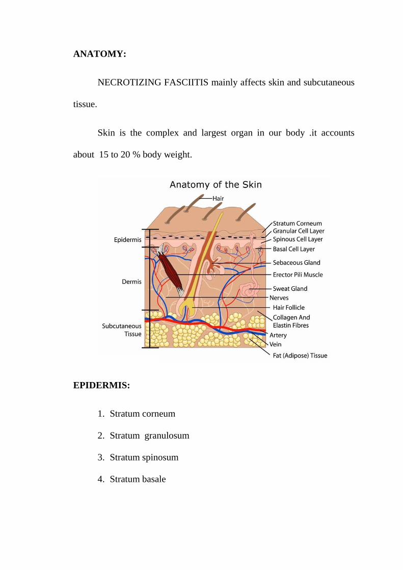
ANATOMY:
NECROTIZING FASCIITIS mainly affects skin and subcutaneous
tissue.
Skin is the complex and largest organ in our body .it accounts
about 15 to 20 % body weight.
EPIDERMIS:
1. Stratum corneum
2. Stratum granulosum
3. Stratum spinosum
4. Stratum basale
Stratum lucidum seen in betweens.corneum and s. granulosum ,this
layer is seen mainly in the palmo-plantar region. Almost 80 t0 90 percent
of epidermis is of ectodermal origin.
EPIDERMAL COMPONENTS:
1.keratinocytes - cytoskeleton
2.langerhans cells-antigenic presenting cells
3.melanocytes-melanin
4.merkel cells
5.toker cells
6.epidermal appendages
7.sweat gland
8. pilosebaceous follicles.
DERMIS:
It is of two types
1.PAPILLARY DERMIS
It helps in good adhesion between epidermis and dermis , made up
of loose collagen bundles and thin elastic fibre.
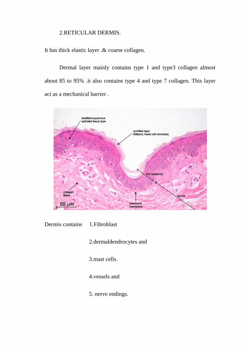
2.RETICULAR DERMIS.
It has thick elastic layer .& coarse collagen.
Dermal layer mainly contains type 1 and type3 collagen almost
about 85 to 95% .it also contains type 4 and type 7 collagen. This layer
act as a mechanical barrier .
Dermis contains 1.Fibroblast
2.dermaldendrocytes and
3.mast cells.
4.vessels and
5. nerve endings.
HYPODERMIS:
Very essential for storage of energy , thermoregulation, insulation,
and protection from external injuries. It contains mainly adipocytes.
Necrotizing fasciitis mainly affects superficial fascia ,severe
inflammation and edema of sub cutaneous tissue and dermis. In severe
disease there will be necrosis of skin also present.
ANATOMY:
NECROTIZING FASCIITIS of scrotum and penis is said to be
Fournier s gangrene.fournier s gangrene can affect superficial and deep
planes. Subcutaneous layer divided in to
Outer fatty layer –camper ‘s fascia
Depper membranous layer –scarpa’s fascia.
Fatty layer is absent in penis and scrotum ,only membranous layer
and is in direct continuous with anterior abdominal wall through camper s
fascia .it leads to rapid spread of infection from the perineal region to
anterior abdominal wall.camper s and scarpa s fascia fuse together and
attached to clavicle superiorly , so it prevents the spread of infection .
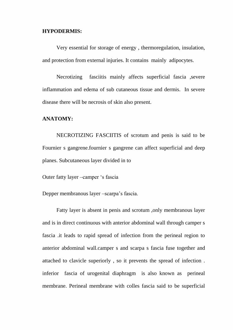
inferior fascia of urogenital diaphragm is also known as perineal
membrane. Perineal membrane with colles fascia said to be superficial
perinealspace .it includes membranous ,bulbar urethra and bulbourethral
gland .superficial perineal membrane is in nearby to ischio rectal fossa
and anterior part of anal wall.
Infection from rectum ,perineum,urethra and bulbourethral gland
directlydrains in to superficialperineal space . infection can easily spreads
in to scrotum and anterior abdominal wall.
Blood supply of anterior abdominal wall from branches of
inferiorepigastric artery and deep circumflex iliac artery.
COLLES FASCIA:
Iaterallycolles fascia attached to the conjoint rami of ischium and
pubis . anteriorly it attached to the anterior abdominal wall . posteriorly
to pubic arch .
Layers of scrotum from superficial to deep .
1.skin
2.subcutaneous fascia- dartous muscle .
Dartous muscle helps in maintaining optimum temperature .
3.external spermatic fascia – aponeurosis of external oblique
muscle .
4.cremaster
5.internal spermatic fascia – from transversalis fascia .
6.areolar tissue
7.testis.
External pudendal and internal pudendal arteries supplies blood to
the scrotal wall .internal pudendal vessels supplies posterior aspect of
scrotal wall. Because of infection and toxins released by bacterial
organisms leads to thrombosis of artery travels in campers fascia ,except
internal pudendal artery so blood supply to the posterior scrotal wall will
be intact so the skin in posterior aspect can be used for reconstruction .
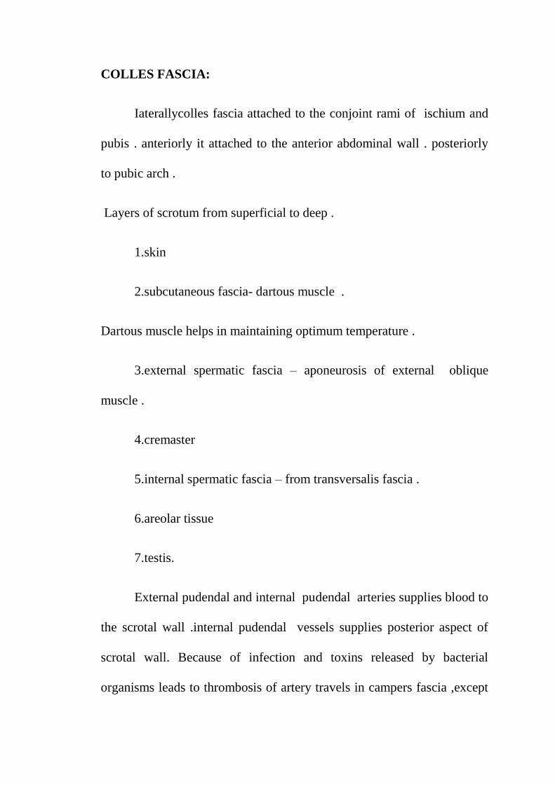
Necrotizing fasciitis infection which is present in the perineal
fascia can spread to scrotum through dartous fascia .sometimes it may
spread to the anterior abdominal wall through scarpa s fascia .structures
which is usually affected is skin, subcutaneous tissue ,and fascial layer.
In Fournier s gangrene the unaffected structures are
1.testis
2.urethra
3.cord structures
4.corporacavernosa .
REVIEW OF LITERATURE
Due to recent knowledge in the recent past and development in the
field of medicine, there has been increase intresent in the area of
necrotising fascitiis.
EPIDEMIOLOGY
Till now there is no apt definition for necrotizing fasciitis. It is also
otherwise called as progressive synergistic gangrene, suppurative
fasciitis, and acute dermal gangrene. When it occurs in genitals it is called
as fournier's gangrene. when it occurs in the postoperative wounds it is
called as progressive postoperative synergistic bacterial gangrene, in
these cases it usually spreads cutaneously sparing deep fascia.
Necrotizing fasciitis has no sex or age predilection and is common
in immunosuppressed states. But it can also occur in healthy young
individuals also without any predisposing factors, the most common
organism being group A hemolytic streptococci. And it usually present as
toxic shock syndrome.
ETIOLOGY
Necrotizing fasciitis can occur anywhere in the body. But it is most
commonly seen in lowerlimb, upperlimb, abdominal wall and genitals.
Inoculation of organisms into the disrupted mucosa, skin by trauma,
burns, or other modes of injury result in development of local infection
followed by necrotizing fasciitis. Though development of necrotizing
fasciitis is usually due to direct local site infection it also results from
infection in the distant site like pharyngitis.
Necrotizing fasciitis of abdominal wall is often fatal if not treated
adequately. It occurs as complication of abdominal surgery. It occurs
most commonly after surgery in the contaminated or clean contaminated
environment. Necrotizing fasciitis of limbs are common when compared
to abdominal wall fasciitis. In the perineal region it occurs as a result of
neglected perianal abscess or trauma. Rarely it occurs in retroperitoneal
region and it has highest mortality when compared to other necrotizing
fasciitis of other areas.
Most of the cases of necrotizing fasciitis of vulva occur in diabetic
patients. It begins as infection in the bartholin's gland which results in
abscess and necrotizing fasciitis. There are also other causes which results
in necrotizing fasciitis, these includes postoperative infection following
hysterectomy, caesarean section or episotomy.
Necrotizing infection of genitals occur commonly due to perianal
abscess or trauma. But it can also occurs following urinary tract infection
with stricture urthera with or without extravasation of urine. It can also
occur following traumatic instrumentation, uretheral calculi and prostatic
massage.
Necrotising fasciitis involving the head and neck region are
relativellyrareand is sommon after blunt trauma, eyelid infection and
pruritis.
Most of the cases of scalp were monomicrobial in origin and the
commonest organism being Streptococcus pyogenes. Most of the cases of
scalp necrotising fasciitis are benign.
Necrotizing fasciitis involving face and cervical region were
common after dental infections, tonsillar abscess and cervical
lymphadenitis. The most common complication being airway obstruction
and mediastinitis. Most cases of culture showed polymicrobial growth.
In some instances necrotizing fasciitis occur in case of
percutaneous catheter usage and following tube thoracostomy. In about
13 to 31% there has been no initiating factor for development of
necrotizing fasciitis. It has been thought in these cases there has been
undetected break in skin with inoculation of organism. Rarely the spread
may be hematological in origin.
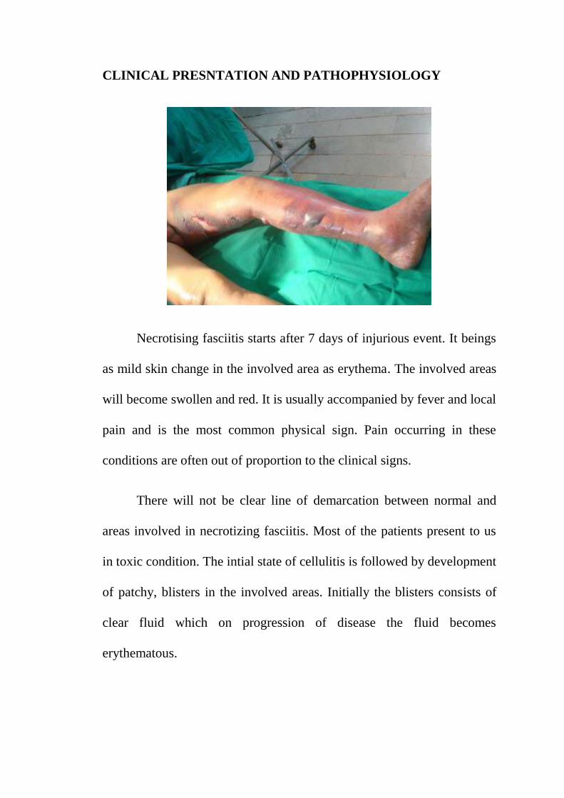
CLINICAL PRESNTATION AND PATHOPHYSIOLOGY
Necrotising fasciitis starts after 7 days of injurious event. It beings
as mild skin change in the involved area as erythema. The involved areas
will become swollen and red. It is usually accompanied by fever and local
pain and is the most common physical sign. Pain occurring in these
conditions are often out of proportion to the clinical signs.
There will not be clear line of demarcation between normal and
areas involved in necrotizing fasciitis. Most of the patients present to us
in toxic condition. The intial state of cellulitis is followed by development
of patchy, blisters in the involved areas. Initially the blisters consists of
clear fluid which on progression of disease the fluid becomes
erythematous.
This stage is followed by necrosis of superificial fascia and fat in
the underlying skin and it results in the formation foul smelling pus and
is often called dishwater pus. Necrosis occurring in these layers are
considered to be due to liquefactive necrosis or due to enzymes like
hyaluronidase secreted by the bacteria causing necrotizing fasciitis.
Involvement of fascial layer is usually more than that of the overlying
skin in the affected region. After4-5 days , the overlying skin becomes
completely gangrenous and about 2 weeks the overlying skin will
completely slough off.
If necrostising fasciitis is not treated adequately in the early
conditions it results in release of toxic contents into theblood stream and
it results in sepsis and septic shock and death. Rarely some patients
recover though untreated.
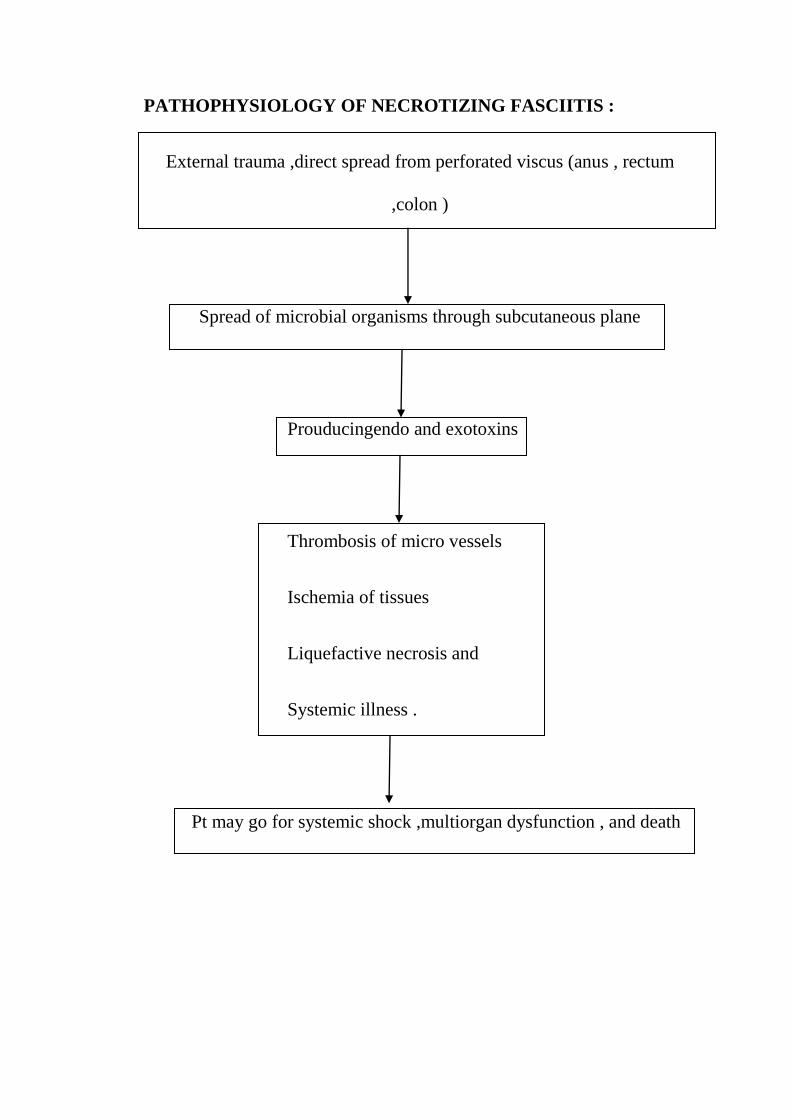
PATHOPHYSIOLOGY OF NECROTIZING FASCIITIS :
External trauma ,direct spread from perforated viscus (anus , rectum
,colon )
Spread of microbial organisms through subcutaneous plane
Prouducingendo and exotoxins
Thrombosis of micro vessels
Ischemia of tissues
Liquefactive necrosis and
Systemic illness .
Pt may go for systemic shock ,multiorgan dysfunction , and death
MICROBIOLOGY
No single organism is causative in case of necrotizing fasciitis.
Necrotizing fasciitis is usually caused by polymicrobialinfection and is
often due to both aerobic and anaerobic bacteria. Due to thecombined
action of the bacteria this condition is often fatal.
Giuliano et al categorized the bacterial pathogens involve in the
necrotizing fasciitis into three groups.
Type I – it occurs due to polymicrobial infection. It includes non
groupA streptococci and anaerobes.
Type II – is usually caused bygroupA streptococci and anaerobic
infection or along with staphylococci.
Type III- is caused by marine vibrio species and the most common
species that causes necrotizing fasciitis is vibrio vulnifacies and is due to
secretion of extracellular toxins due to these organisms. The other
organisms that cause necrotizing fasciitis are group B streptococcus and
pasturellamulticida.
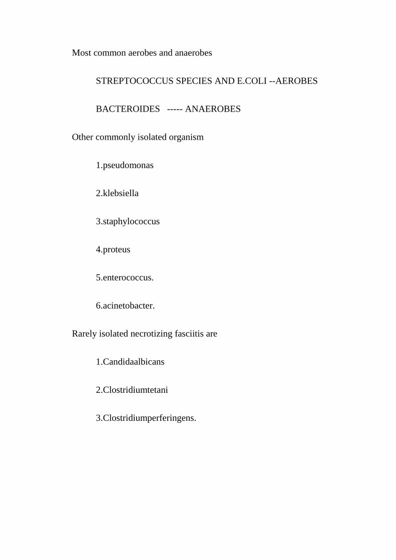
Most common aerobes and anaerobes
STREPTOCOCCUS SPECIES AND E.COLI --AEROBES
BACTEROIDES ----- ANAEROBES
Other commonly isolated organism
1.pseudomonas
2.klebsiella
3.staphylococcus
4.proteus
5.enterococcus.
6.acinetobacter.
Rarely isolated necrotizing fasciitis are
1.Candidaalbicans
2.Clostridiumtetani
3.Clostridiumperferingens.
The type organism that causes necrotizing fasciitis often depend on
the site involved. Type I is most commonly seen in case of abdominal and
perianal infection and is oftendue to polymicrobial in origin. Themost
common pathogens being in these conditions were enteric organisms and
enterococci and anaerobic bacteria.
In case of extremities, types II is most common and are usually
monomicrobial.
In case of necroting fasciitis the common area for culture is
necrotingcentre of the lesion which is in contrast to cellulitis in which the
culture is taken from the edges of the ulcer. Diabetics were the most
common individual affected and is due to involvement of small vessel
and decreased WBC function and low oxygen tension which are the
substrate for bacterial growth.
The destructive effect in case of necrotizing fasciitis is due to the
toxins secreted by the streptococcal species and also due to cytokine
release. There aretwo main toxins released by the invasive virulent group
A streptococcal bacteria, they are Exotoxin A and Exotoxin B.
Exotoxin A is seen in cases of invasive pathogen. Exotoxin B
consists of cysteine proteases and is involved in necrotizing fasciitis and
myositis by destructing the proteins. There has been recent increase in the
fatality following necrotizing fasciitis and is due to increased virulence of
the organism and the development of resistance. Some authors suggest
that increase in mortality rate may be due to increase in interest in this
field.
RISK FACTORS :
Predisposing factor are
1. Diabetic mellitus
2. Obesity
3. Alcoholism
4. Renal disease
5. Liver failure
6. Cirrhosis
7. AIDS
8. Malignancy
9. Leukaemia
10. SLE
11. Crohn s disease
12. Immunosuppersion .
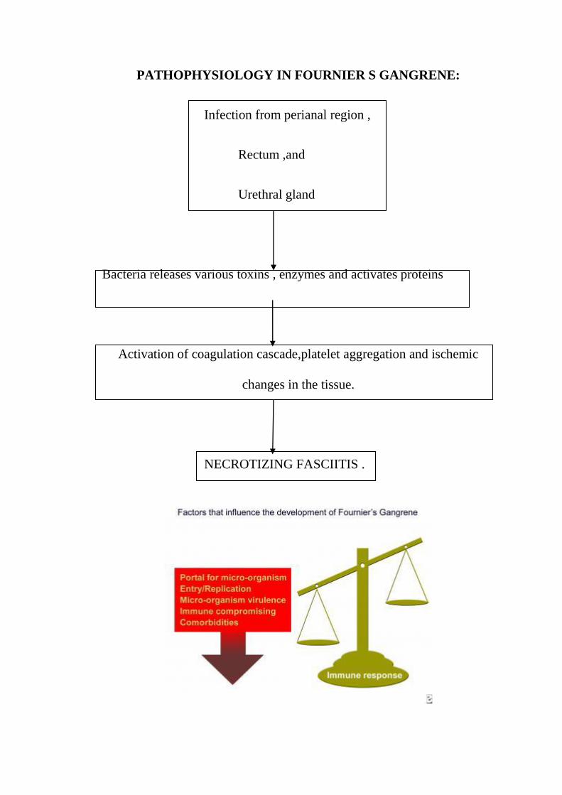
PATHOPHYSIOLOGY IN FOURNIER S GANGRENE:
Infection from perianal region ,
Rectum ,and
Urethral gland
Bacteria releases various toxins , enzymes and activates proteins
Activation of coagulation cascade,platelet aggregation and ischemic
changes in the tissue.
NECROTIZING FASCIITIS .
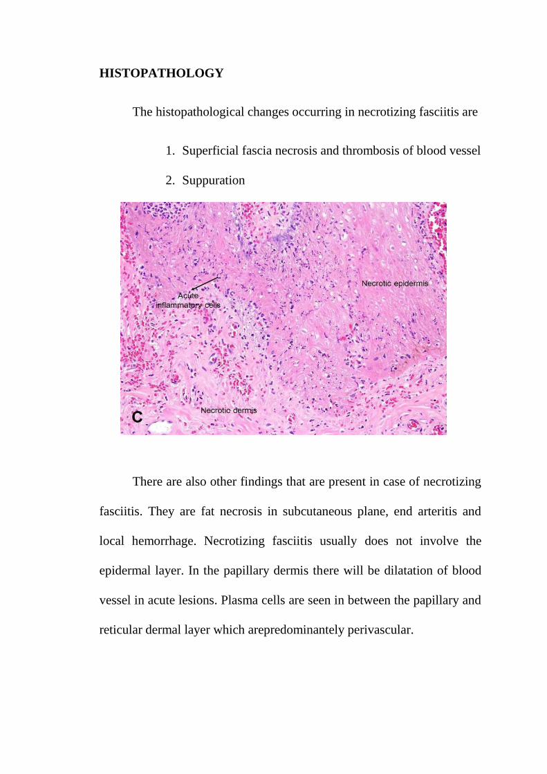
HISTOPATHOLOGY
The histopathological changes occurring in necrotizing fasciitis are
1. Superficial fascia necrosis and thrombosis of blood vessel
2. Suppuration
There are also other findings that are present in case of necrotizing
fasciitis. They are fat necrosis in subcutaneous plane, end arteritis and
local hemorrhage. Necrotizing fasciitis usually does not involve the
epidermal layer. In the papillary dermis there will be dilatation of blood
vessel in acute lesions. Plasma cells are seen in between the papillary and
reticular dermal layer which arepredominantely perivascular.
In the deeper layers of skin i.e, reticular layer, edemaand
inflammatory infiltrate are present.
In certain cases there is were necrosis of eccrine glands and ducts
and it is due to thrombosis of blood vessel resulting in the infarction of
the gland. Fascial layer may sometime be edematous and also has
inflammatory infiltrate. In advanced cases there will be thrombosis of
blood vessel.
Microorganisms are most commonly seen between the collagen
and in between fat tissues. In severe cases there will be necrosis of
underlying muscle fibres.
DIAGNOSIS
Diagnosing necrotizing fasciitis in early condition is often very
difficult. And the diagnosis of necrotizing fasciitis is often clinically
made. Important clinical features are pain, redness and toxic symptoms. It
is highly important to identify these cases early to intervene at the right
time. If diagnosis of necrotizing fasciitis is made, it is necessary to do
appropriate debridement.
Decreased resistance in the fascial layer, while performing
debridement is often a sign of necrotizing fasciitis. In some studies, it
recommends use of frozen section biopsy in case where there is doubt in
clinical diagnosis. In study conducted by Stamenkovic , he insisted the
use of full thickness biopsy for diagnosis of necrotizing fasciitis.
The radiological conditions useful in the diagnosis of necrotizing
fasciitis are
1. Plain radiograph
2. CT
3. Ultrasonography
4. MRI
The important finding in case of necrotizing fasciitis in plain
radiography is presence of soft tissue gas. It is more important than
physical finding in diagnosing this condition.
CT scan is useful in diagnosis in case of cervical necrotizing
fasciitis. It is more accurate than plain x ray. It delineates the exact
extension of the disease.
Ultrasonography is important in cases like fournier's gangrene. It is
also helpful in differentiating it from other causes of acute conditions.
MRI plays a significant role in early diagnosis of necrotizing
fasciitis. It has the ability to show the soft tissue fluid and has good tissue
contrast. It is highly sensitive in delineating the pathology. In study
conducted by Rahmouni, he used MRI in detecting the early cases of
necrotising fasciitis and also differentiate between cellulitis. Though MRI
helps to identify the early cases, due to high cost routine use of MRI in
the diagnosis of necrotizing fasciitis is not used in our setup.
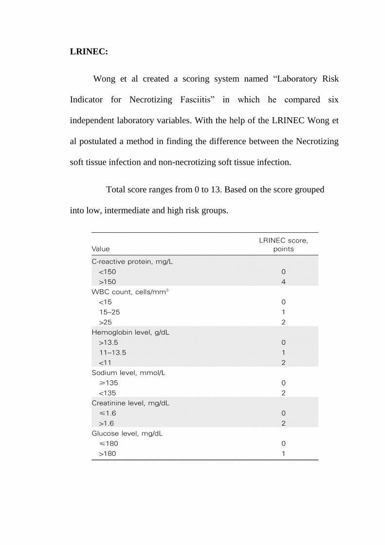
LRINEC:
Wong et al created a scoring system named “Laboratory Risk
Indicator for Necrotizing Fasciitis” in which he compared six
independent laboratory variables. With the help of the LRINEC Wong et
al postulated a method in finding the difference between the Necrotizing
soft tissue infection and non-necrotizing soft tissue infection.
Total score ranges from 0 to 13. Based on the score grouped
into low, intermediate and high risk groups.
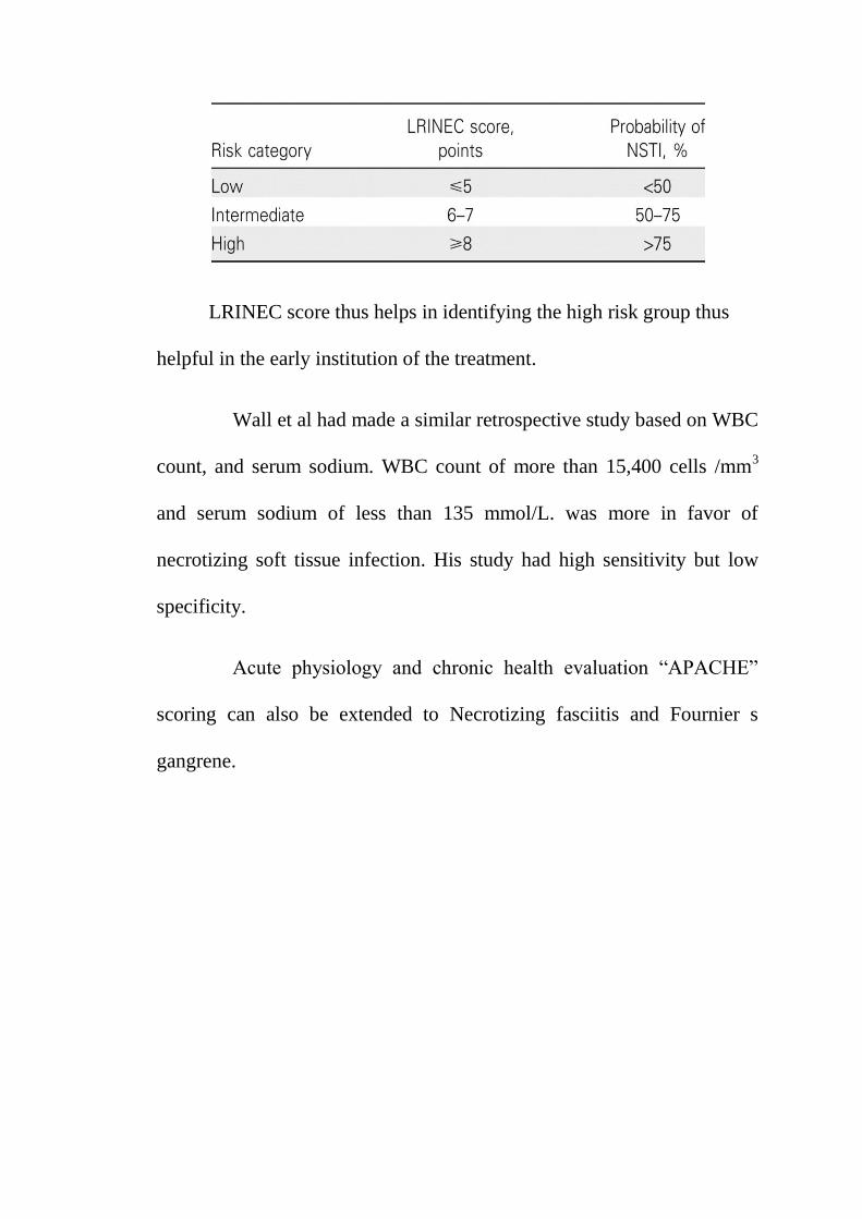
LRINEC score thus helps in identifying the high risk group thus
helpful in the early institution of the treatment.
Wall et al had made a similar retrospective study based on WBC
count, and serum sodium. WBC count of more than 15,400 cells /mm3
and serum sodium of less than 135 mmol/L. was more in favor of
necrotizing soft tissue infection. His study had high sensitivity but low
specificity.
Acute physiology and chronic health evaluation “APACHE”
scoring can also be extended to Necrotizing fasciitis and Fournier s
gangrene.
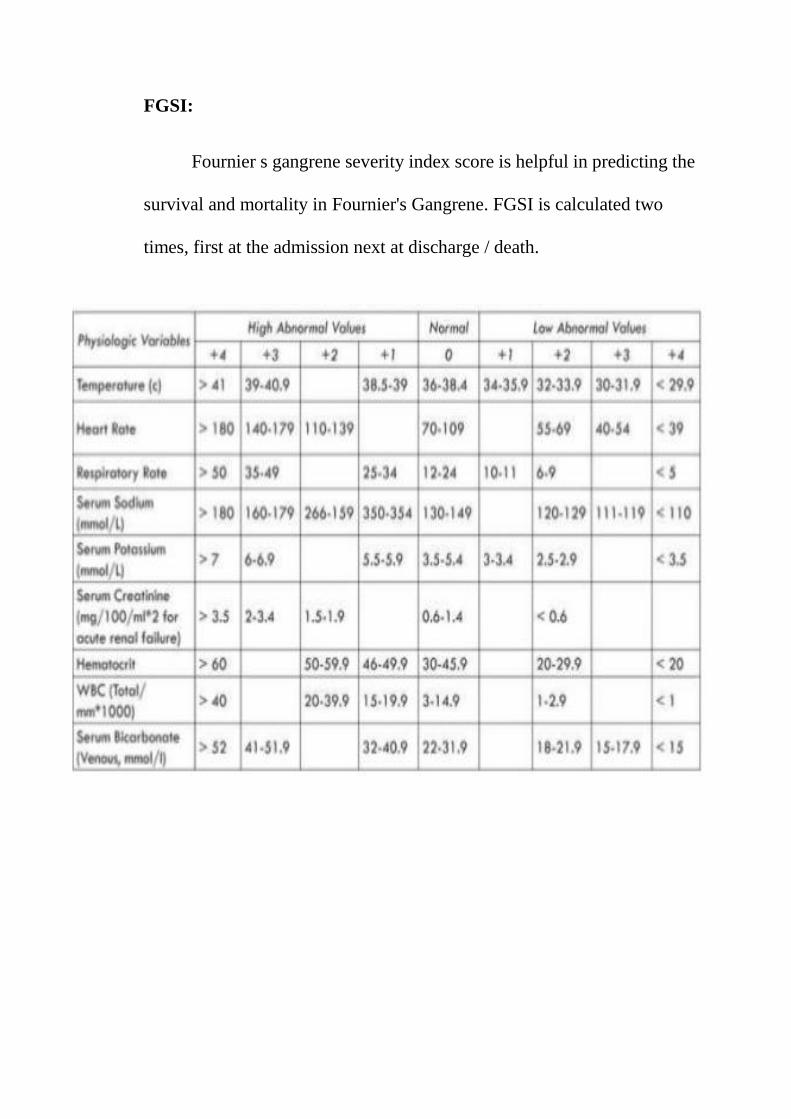
FGSI:
Fournier s gangrene severity index score is helpful in predicting the
survival and mortality in Fournier's Gangrene. FGSI is calculated two
times, first at the admission next at discharge / death.
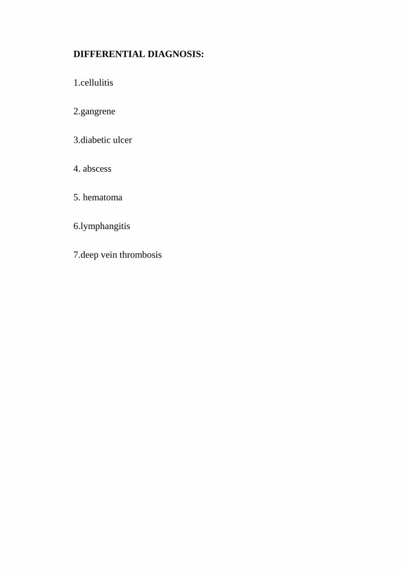
DIFFERENTIAL DIAGNOSIS:
1.cellulitis
2.gangrene
3.diabetic ulcer
4. abscess
5. hematoma
6.lymphangitis
7.deep vein thrombosis
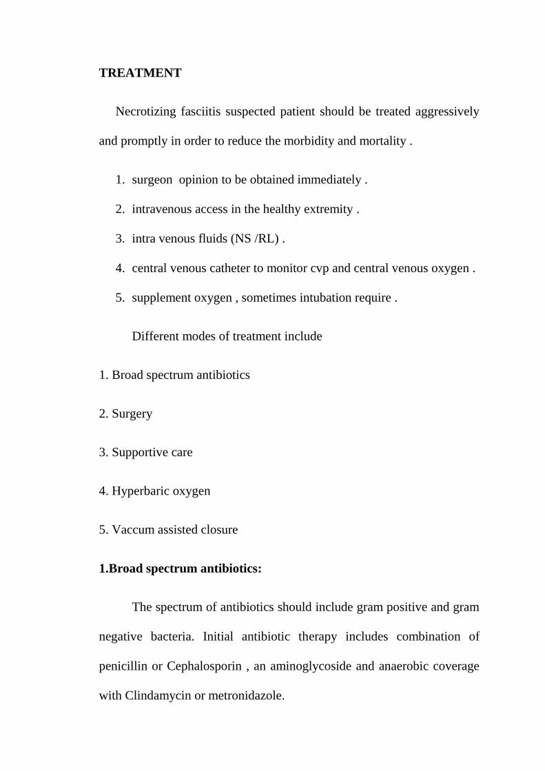
TREATMENT
Necrotizing fasciitis suspected patient should be treated aggressively
and promptly in order to reduce the morbidity and mortality .
1. surgeon opinion to be obtained immediately .
2. intravenous access in the healthy extremity .
3. intra venous fluids (NS /RL) .
4. central venous catheter to monitor cvp and central venous oxygen .
5. supplement oxygen , sometimes intubation require .
Different modes of treatment include
1. Broad spectrum antibiotics
2. Surgery
3. Supportive care
4. Hyperbaric oxygen
5. Vaccum assisted closure
1.Broad spectrum antibiotics:
The spectrum of antibiotics should include gram positive and gram
negative bacteria. Initial antibiotic therapy includes combination of
penicillin or Cephalosporin , an aminoglycoside and anaerobic coverage
with Clindamycin or metronidazole.
Antibiotic therapy should be carried on according to culture and
sensitivity. High dose Penicillin remains drug of choice for necrotizing
fascitis.
Third generation cephalosporin is drug of choice in early stage of
disease. It is active against gram negative and low efficacy to gram
positive organisms.
Vancomycin can be used in penicillin and cephalosporin resistant
individuals used to treat septicemia and skin infection.Metronidazole
active against protozoa and anaerobes.
Thrombosis of superficial veins precludes effective antibiotic
penetration into site of infection and tissue hypoxia impairs oxidative
killing mechanism of leucocytes. Accumulation of bacteria and toxins
occurs, which leads to development of sepsis. So early surgical
intervention is crucial.
NON STEROIDAL ANTIINFLAMMATORY DRUGS AND
NECROTIZING FASCIITIS
NSAID’s are the most commonly used drug as analgesic in case of
minor injury and pain. In the recent studies there has been correlation
between the use of NSAID’s and development of necrotizing fasciitis due
to group A streptococcal infection in healthy individuals.
Since NSAID’s are most common drug ingested there has been
simple correlation between development of necrotizing fasciitis, rather
than cause and effect relationship. Some advised that NSAID should be
used appropriately in case minor soft tissue inflammation, when infection
cannot be ruled out.
2.Surgery:
Treatment of necrotising include mainly surgical management.
Adequate surgical management involves removal of necrotic tissueand
drainage of fascialplanes via extensive fasciotomy till healthy fascia is
encountered.early surgical treatment is associated with improved survival
than delayed surgery.
Extension of fascial necrosis needs repeated fasciotomy and wound
debridement. If extremities are involved amputation may be needed to
control infection, particular vascular disease and /or
diabetes.Inperinealnecrotising fasciitis diversion colostomy or urinary
diversion needed to control infection.
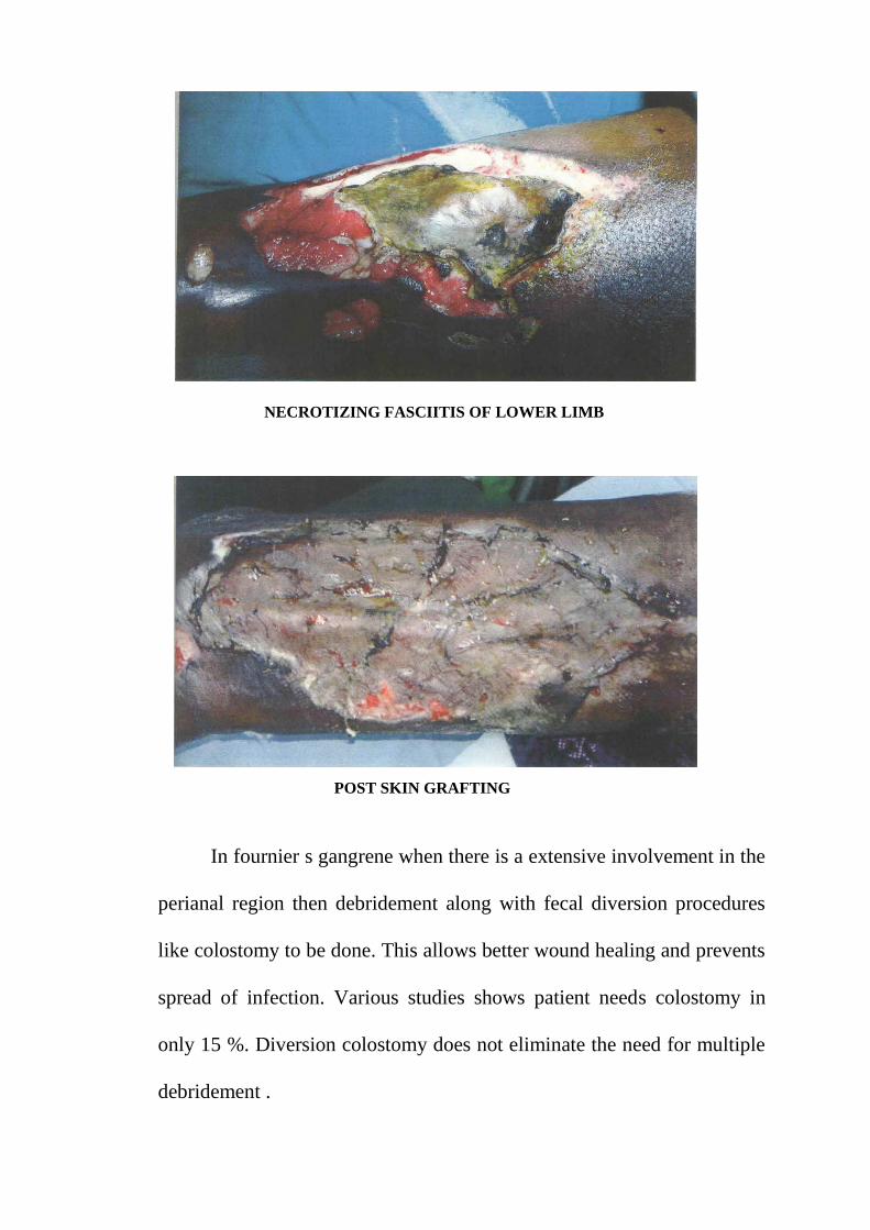
In fournier s gangrene when there is a extensive involvement in the
perianal region then debridement along with fecal diversion procedures
like colostomy to be done. This allows better wound healing and prevents
spread of infection. Various studies shows patient needs colostomy in
only 15 %. Diversion colostomy does not eliminate the need for multiple
debridement .
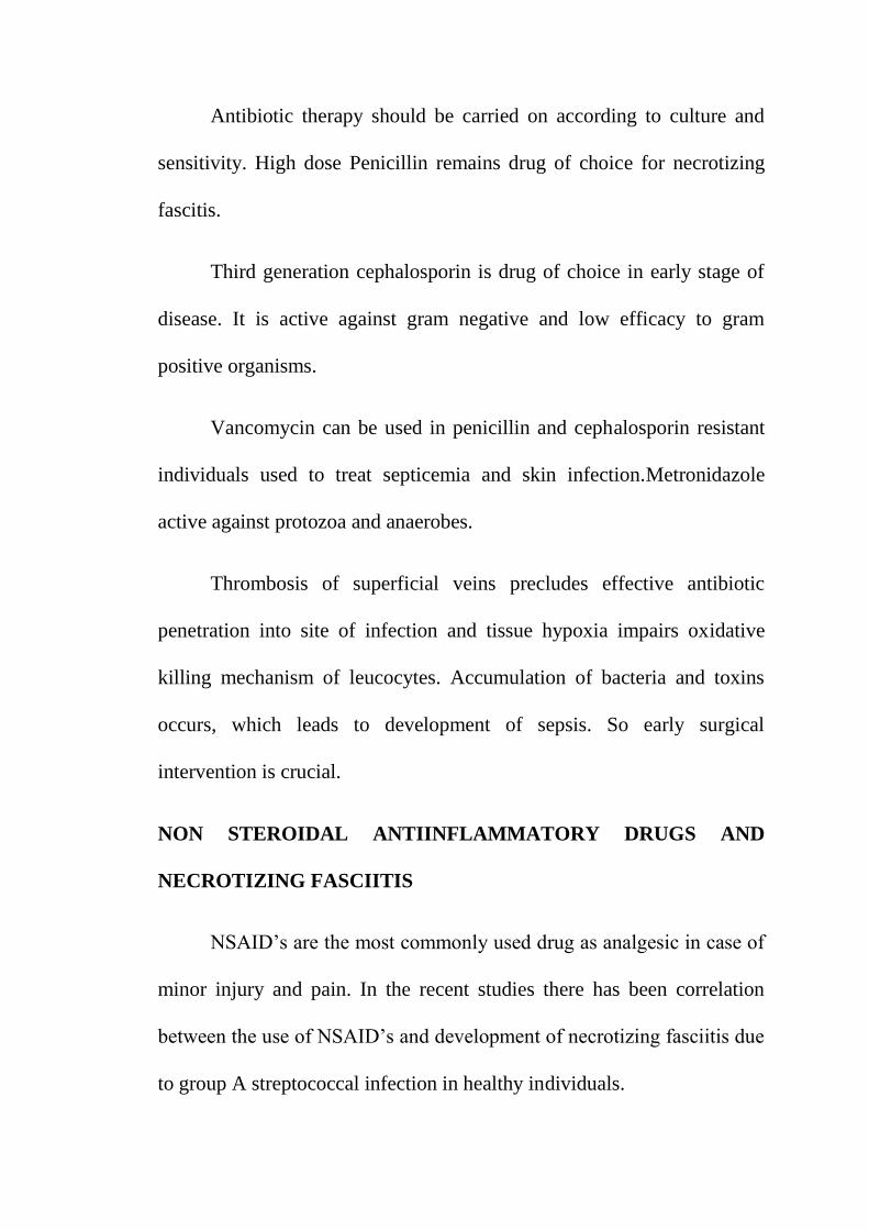
NECROTIZING FASCIITIS OF LOWER LIMB
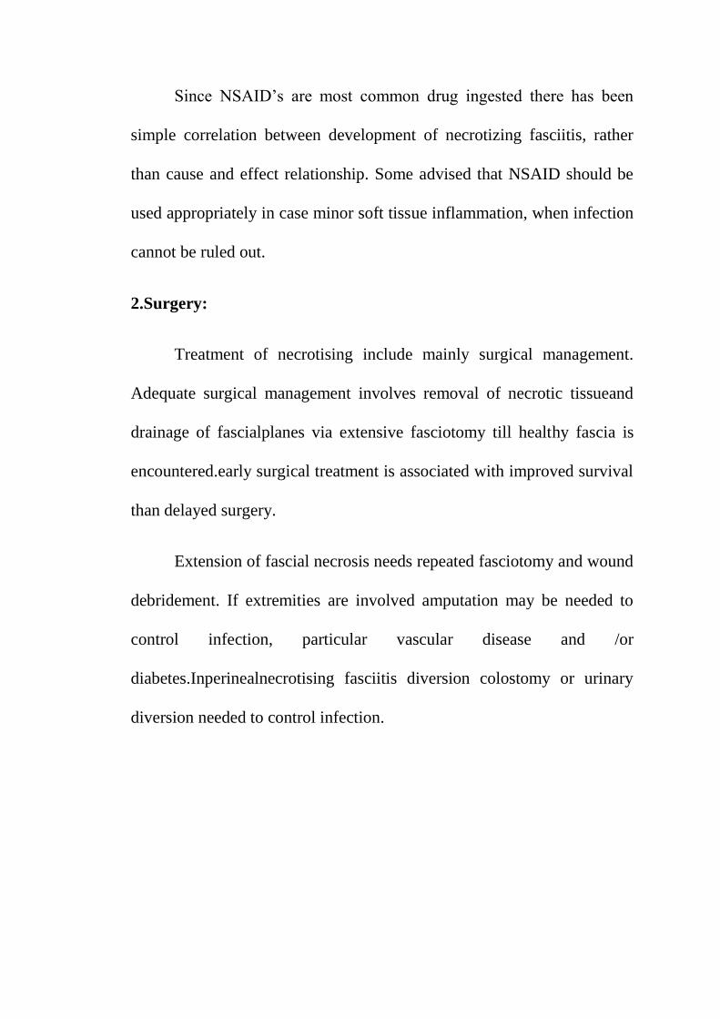
POST SKIN GRAFTING
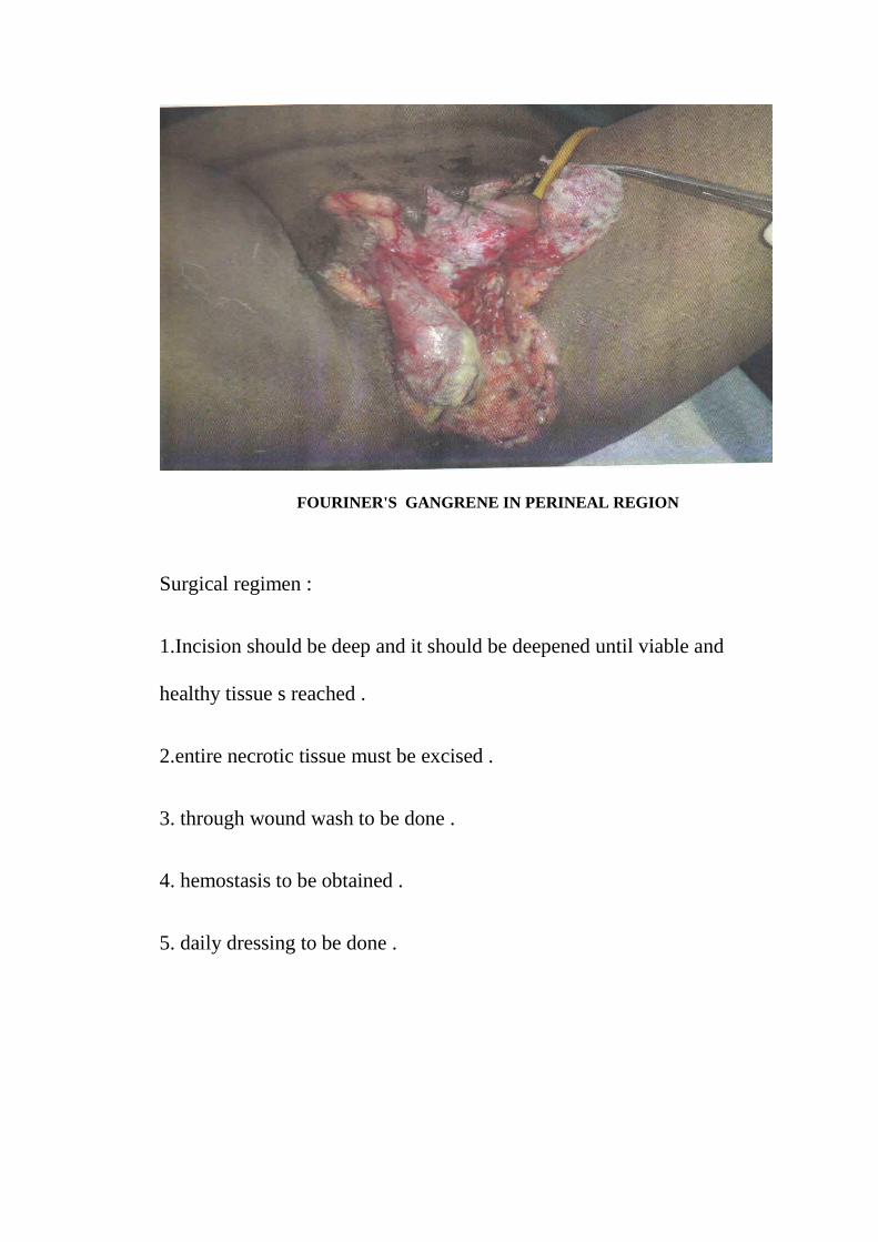
Surgical regimen :
1.Incision should be deep and it should be deepened until viable and
healthy tissue s reached .
2.entire necrotic tissue must be excised .
3. through wound wash to be done .
4. hemostasis to be obtained .
5. daily dressing to be done .
FOURINER'S GANGRENE IN PERINEAL REGION
DRESSINGS:
Dressings can be done with
1.silversulphadiazine
2.polysporin
3.bacitracin .
3.Supportive care:
It includes aggressive fluid management , analgesia and early
intensive care. After initial debridement cooperation of multiple
specialists needed for optimal patient treatment. Once patient general
condition is stabilized and patient begins to recover plastic surgery
evaluation is needed for reconstruction and skin grafting.
4. Hyperbaric oxygen:
It includes breathing oxygen at high atmospheric pressure.
Hyperoxia at tissue level includes increased leucocyte killing, killing of
anaerobes, Reduction of edema stimulation of fibroblasts and better
collagen formation. It should be started as early as possible and it should
not delay surgical treatment.
Delivering 100% oxygen through a pressure chamber ,which is
greater than atmospheric pressure .
HBOT was given at the rate of 2.5 to 3.0 atmospheres for 90
minutes twice daily, to be given after surgical debridement. It improves
the tissue oxygenation in both healthy and devitalized tissue also .
It can be used in
1. gas embolism
2.gas gangrene,
3.carbon mono oxide poisoning.
Indication of HBOT:
1.patient with necrotizing fasciitis fail to resolve inspite of adequate
medical and surgical management.
2.patient with clostridial infection .
3.gangrene of muscle and deeper tissue involvement.
Advantages of HBOT:
1.by supplying 100% oxygen to the tissue HBOT inhibits the growth
ofanaerobic organisms .
2.angiogenesis and fibroblast multiplication
3.improved phagocytic neutrophilic function
4.decreases edema by vasoconstriction andby increased delivery of
intracellular antibiotics .
5.HBOT is beneficial for drugs to act .
Eg aminoglycosides acts via oxygen dependent pump .
6.it helps in production of collagen .
Disadvantage of HBOT :
Absolute contraindication :
1.untreated tension pneumothorax .
Relative contraindication :
1.barotrauma to middle ear cavity
2. cardiac disease
3.malignancy
4.URTI
5 emphysema with carbondioxideretention .
But the use of HBOT is still controversial ,it can be used after
failure of conventional medical and surgical management.
Six studies which conducted for effectiveness of HBOT in treating
necroziting fasciitis ,out of which four reports HBOT improves survival
of the patients ,and two of them not. No studies demonstrated the efficacy
of hyperbaric oxygen therapy .
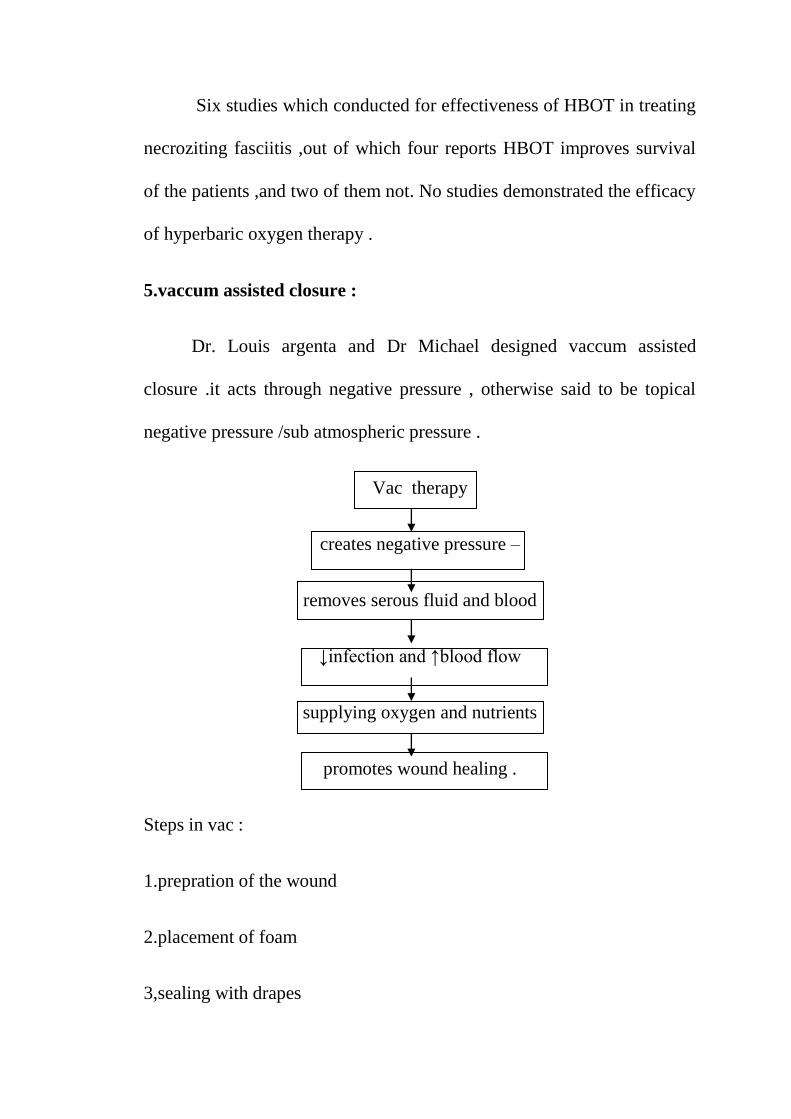
5.vaccum assisted closure :
Dr. Louis argenta and Dr Michael designed vaccum assisted
closure .it acts through negative pressure , otherwise said to be topical
negative pressure /sub atmospheric pressure .
Vac therapy
creates negative pressure –
removes serous fluid and blood
↓infection and ↑blood flow
supplying oxygen and nutrients
promotes wound healing .
Steps in vac :
1.prepration of the wound
2.placement of foam
3,sealing with drapes
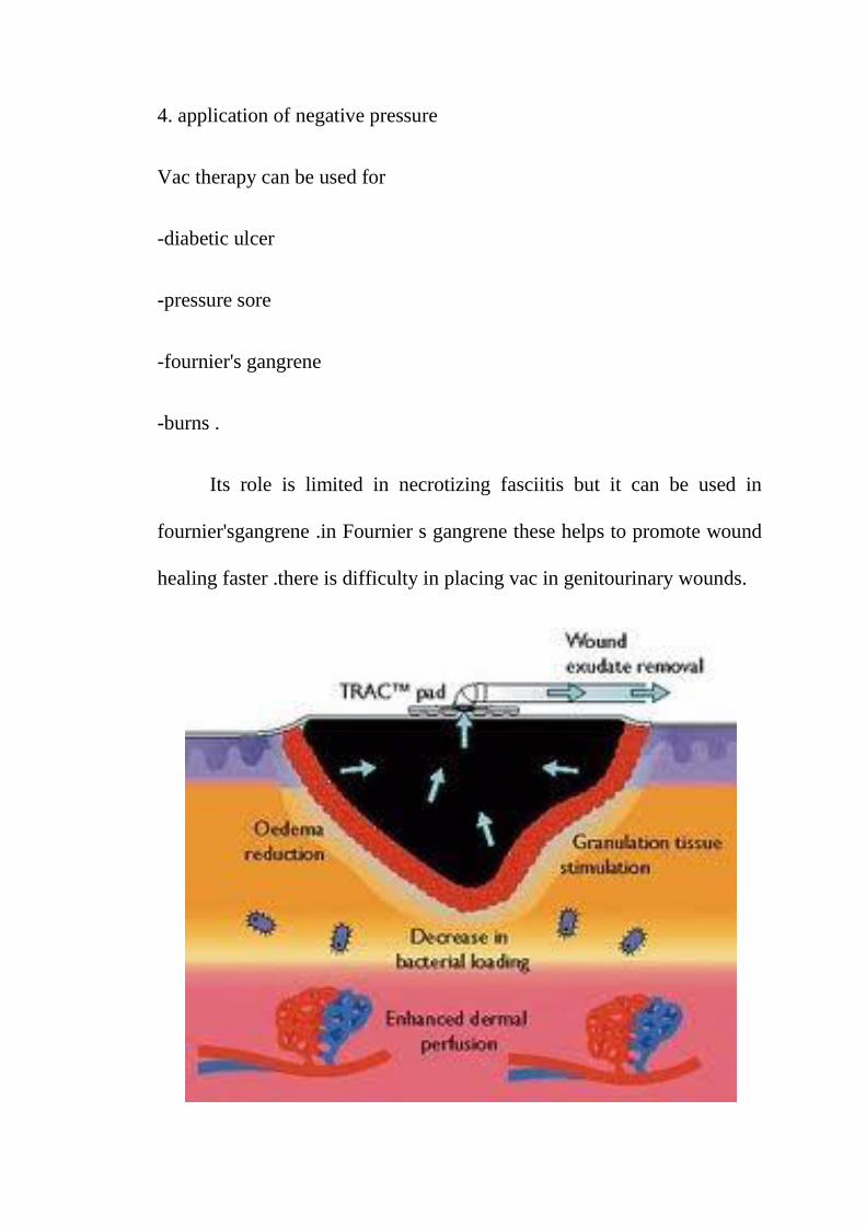
4. application of negative pressure
Vac therapy can be used for
-diabetic ulcer
-pressure sore
-fournier's gangrene
-burns .
Its role is limited in necrotizing fasciitis but it can be used in
fournier'sgangrene .in Fournier s gangrene these helps to promote wound
healing faster .there is difficulty in placing vac in genitourinary wounds.
MORTALITY
Mortality due to necrotizing fasciitis in untreated condition is very
high. Mortality rate has changed little since Meleney first recognized that
early intervention needed in case of necrotizing fasciitis. Mortality rate
ranges from 29% to 76%. There are other comorbid conditions that
increases the mortality in case of necrotizing fasciitis they are diabetes
mellitus, peripheral vascular disease and poor nutritional status.
The mortality in necrotizing fasciitis is due to sepsis or multiorgan
dysfunction. Early cause of death were due to sepsis syndrome and late
death were due to multiple organ dysfunction.
PREVENTION :
1.STREPTOCOCCUS infection can be easily prevented after good
hand washing .
2. necrotizing fasciitis can be prevented by intact skin .
3.patient with rapidly spreading wound infection with toxic features
should immediately require medical attention .
4. wound should be regularly dressed and through dressing should be
done daily ,and patient should be look for any signs of infection like any
erythema ,swelling ,tenderness and any discharge from wound .

5. patient with streptococcal throat infection should remain in home until
24 hrs after their last antibiotic dose .
MATERIALS AND METHODS
STUDY DESIGN
Descriptional study
SOURCE OF DATA
60 patients of necrotizing fasciitis getting admitted in surgical ward
PLACE OF STUDY
Coimbatore Medical College and Hospital
STUDY PERIOD
September 2014 to august 2015
INCLUSION CRITERIA
All patients presenting with features of necrotizing fasciitis to
Coimbatore medical college and hospital
EXCLUSION CRITERIA
Pregnant women
Age<13 yrs
METHODOLOGY
Patients presenting with features of necrotizing fasciitis were
admitted in the general surgery ward in Coimbatore medical college and
hospital were included in the study during the study period of September
2014-august 2015. Initial diagnosis were made by both clinical and
anatomical findings. Details of the patient were noted. Detailed interview
with the patient were made regarding history and other comorbid
conditions.
Following complete history taking physical examination for the
patient were done including blood pressure measurement and temperature
and other clinical finding related to necrotizing fasciitis. Following
clinical examination, routine investigations were investigated.
Radiological investigations were done to note for gas formation in
subcutaneous layer. Treatment were started as soon as diagnosis is
suspected. It includes resuscitation of patient with intravenous fluids,
antibiotics and wound debridement. And bacteriological culture is done
for both aerobic and anaerobic bacteria.
The sample taken for culture is transported through proper
transporation technique to culture laboratory. These samples were then
cultured in blood agar and mc Conkey agar for aerobic bacteria and in
Robertson’s cooked meat media for anaerobic bacteria. The cultured
organisms were tested for resistance pattern by disc diffusion method.
Following initial debridement the wound was inspected regularly
and subsequent debridement were done periodically whenever necessary.
And dressing were done using povidone iodine and saline guaze. After
the wound is fit, patient undergone split skin graft surgery for raw area.
STATISTICAL ANALYSIS
STATISTICAL METHOD
In our study we used descriptive statistical analysis. Continuous
measurement were represented on mean with or without standard
deviation. Categorical measurementwere represented in number (%).
Confidence interval of 95% is used to find significancyofvalue.
Confidence limit >50% is associated with statistical significance.
STATISTICAL SOFTWARE
Tables and charts were completed using Microsoft word and
excel software.
RESULTS
This study was conducted during the period of September 2014-
august 2015. About 60 patients were included in our study and their
different aspects for predisposing factors, age of presentation,
microbiological pattern and antibiotic pattern were analysed.
In this study we noted that the patients affected with necrotising
fasciitis ranges from 14-81yrs. Mean patients were in the age group of
50.42 ±17.31yrs. most of the patients wereinagegroup of above 50 yrs.
In our study most of the patients were male (49) when compared to
female (11)and in ratio of 4.45:1. The most common occupation among
the patients presenting to us were farmers (28.3%).
Among 60 patients we had studied, the most common part to be
affected was lowerlimb (53.3%), next to it was perinealregion(30%) and
involvement of trunk in 8.3% patients.
Regarding the clinical presentation, the commonest symptom being
pain and is present in about 95% cases. Next to pain is fever and
discharge in 61.7% and 58.3% respectively. Presence of swelling is seen
inabout 45% . Blisters were seen in about 18.3%.
In this study we noticed there was delay in presentation of patients
to healthcare setup in most cases. Only 55% of patients presented to us
within 1 week of clinical symptoms. 31.7% in 2nd
week and 8% in >2
weeks group. The mean duration of presentation was 10.08±10.09 days.
Oedema and ulcer were the most common clinical signs and were
seen in about 85% and 76.7% cases respectively.
We noticed in our study that the commonest cause for developing
necrotizing fasciitis is trauma which is about 30% cases, in about 6.7%
there was no defined etiological factor for development of necrotizing
fasciitis.
Regarding the comorbid conditions causing necrotizing fasciitis,
the commonestbeing diabetes mellitus in 38.3% cases. Next to it was
hypertension in 30% cases.
By blood investigations from our study we notices that, 30% of
patients were anemic and 23.3% had elevated blood sugar levels. 23.3%
had elevated serum creatinine value and 15% had
hyponatremia(<128meq/l).
The cultured organism in our study werepolymicrobial in 79.9%
and mono microbial in case of 13.3%. there were no growth in about
6.6%. most of the cultured were aerobic organisms in our study and it
constitutes around 86.7 % and anaerobic about 13.3%.
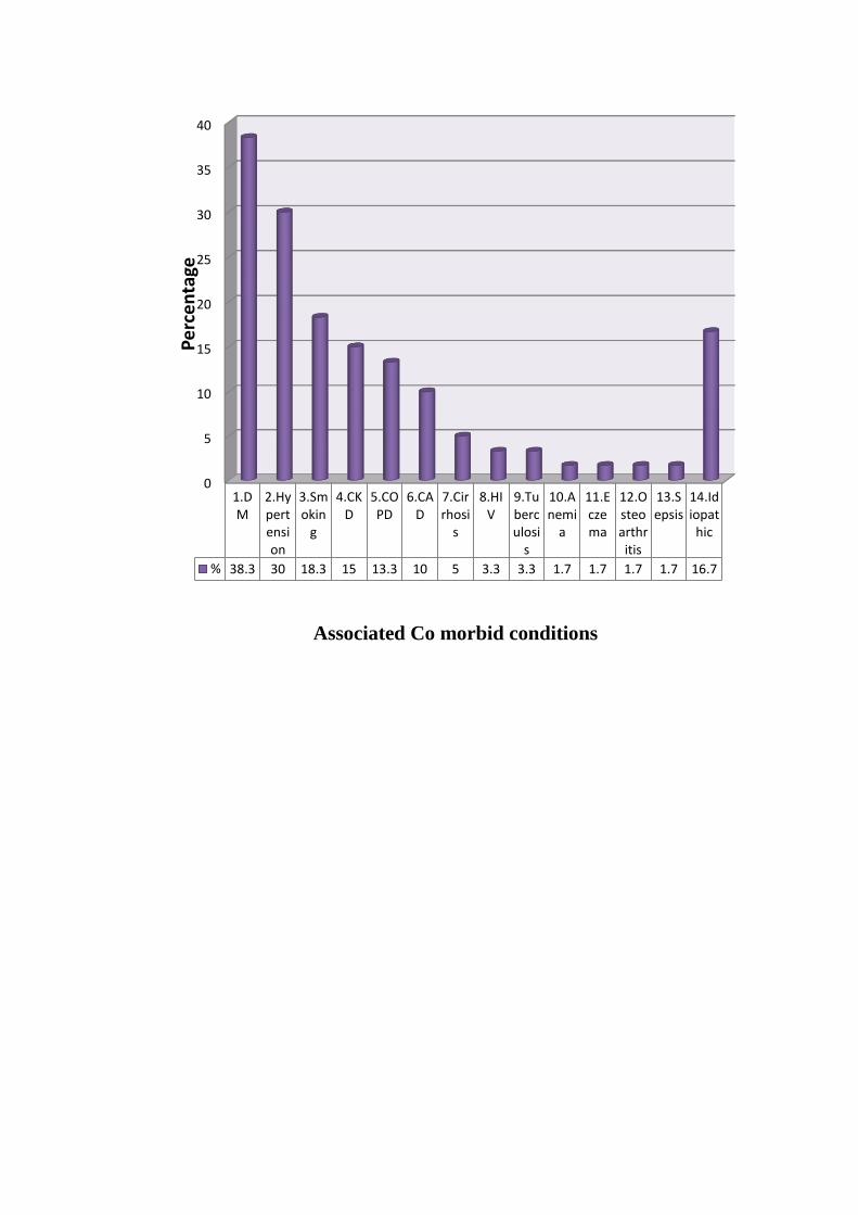
The most common organism cultured in our study were
Escherichia coli in about 46.6%. The next most common species being
streptococcus in 41.6% and klebsiella in about 36.6%. All the patients
initially received broad spectrum antibiotic and it consists of
cephalosporin with aminoglycoside with metronidazole.
In about 95% patients we performed wound debridement. In about
5% we had done fasciotomy and secondary suturing in about 28.3%.
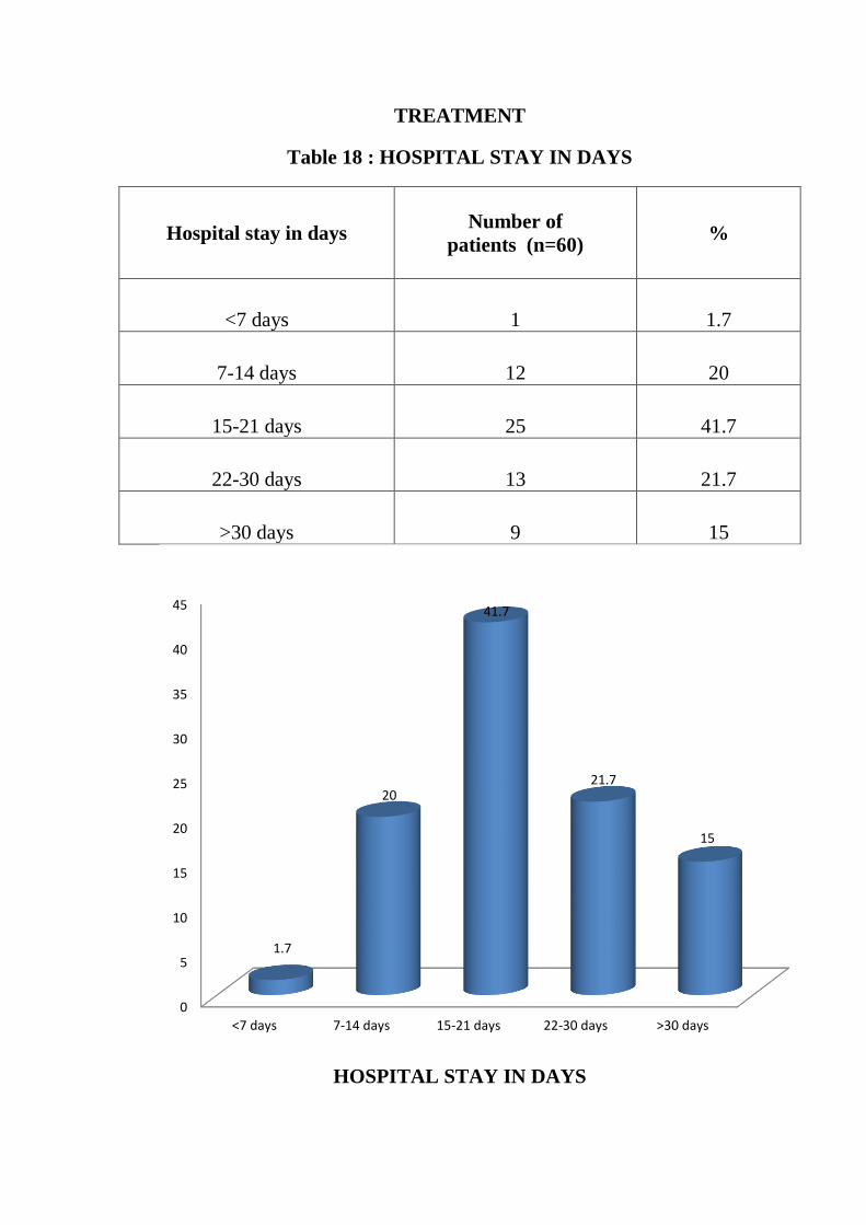
Most of the patient discharged after 25 days of hospital stay. And
there mortality in our study was 21.7%.
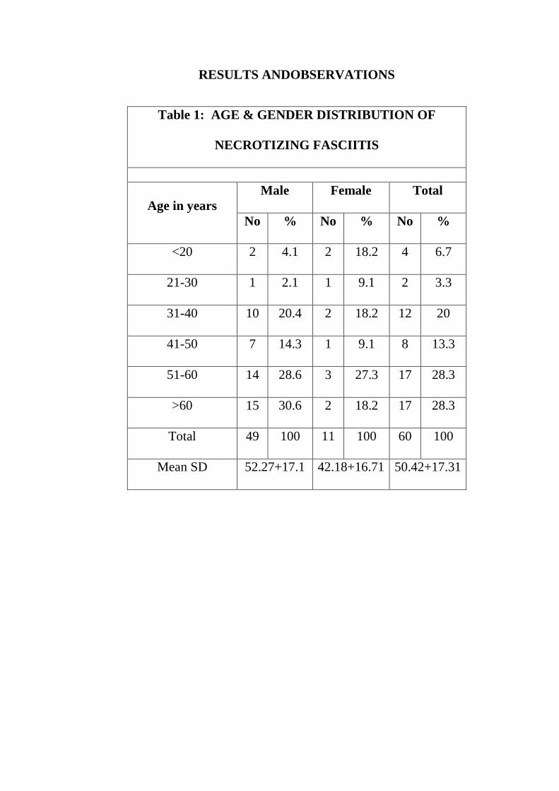
RESULTS ANDOBSERVATIONS
Table 1: AGE & GENDER DISTRIBUTION OF
NECROTIZING FASCIITIS
Age in years
Male Female Total
No % No % No %
<20 2 4.1 2 18.2 4 6.7
21-30 1 2.1 1 9.1 2 3.3
31-40 10 20.4 2 18.2 12 20
41-50 7 14.3 1 9.1 8 13.3
51-60 14 28.6 3 27.3 17 28.3
>60 15 30.6 2 18.2 17 28.3
Total 49 100 11 100 60 100
Mean SD 52.27+17.1 42.18+16.71 50.42+17.31
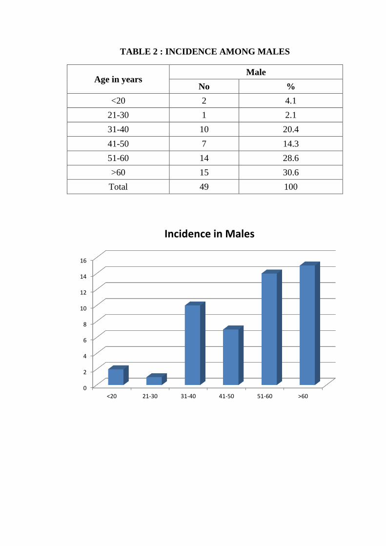
TABLE 2 : INCIDENCE AMONG MALES
Age in years Male
No %
<20 2 4.1
21-30 1 2.1
31-40 10 20.4
41-50 7 14.3
51-60 14 28.6
>60 15 30.6
Total 49 100
0
2
4
6
8
10
12
14
16
<20 21-30 31-40 41-50 51-60 >60
Incidence in Males
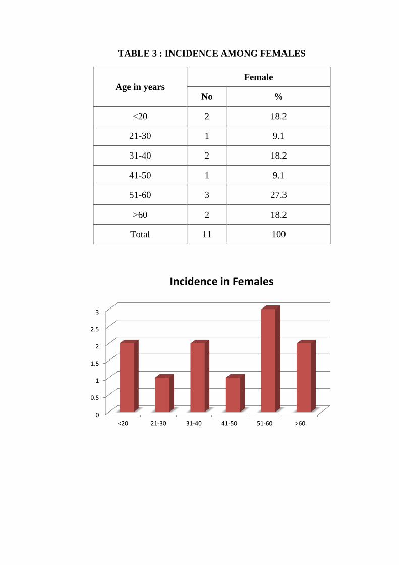
TABLE 3 : INCIDENCE AMONG FEMALES
Age in years Female
No %
<20 2 18.2
21-30 1 9.1
31-40 2 18.2
41-50 1 9.1
51-60 3 27.3
>60 2 18.2
Total 11 100
0
0.5
1
1.5
2
2.5
3
<20 21-30 31-40 41-50 51-60 >60
Incidence in Females
TABLE 4 : INCIDENCE
Age in years
Male Female Total
No % No % No %
<20 2 4.1 2 18.2 4 6.7
21-30 1 2.1 1 9.1 2 3.3
31-40 10 20.4 2 18.2 12 20
41-50 7 14.3 1 9.1 8 13.3
51-60 14 28.6 3 27.3 17 28.3
>60 15 30.6 2 18.2 17 28.3
Total 49 100 11 100 60 100
Mean SD 52.27+17.1 42.18+16.71 50.42+17.31
0
2
4
6
8
10
12
14
16
18
<20 21-30 31-40 41-50 51-60 >60
Incidence in Females
Incidence in Males
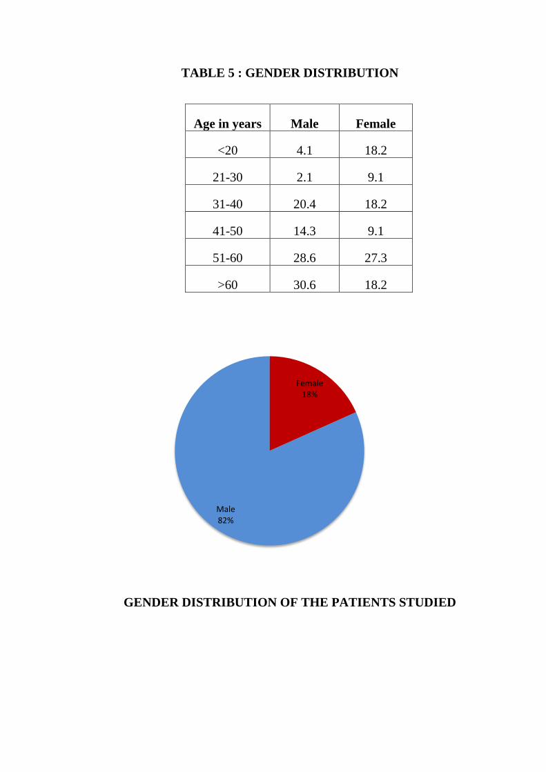
TABLE 5 : GENDER DISTRIBUTION
Age in years Male Female
<20 4.1 18.2
21-30 2.1 9.1
31-40 20.4 18.2
41-50 14.3 9.1
51-60 28.6 27.3
>60 30.6 18.2
GENDER DISTRIBUTION OF THE PATIENTS STUDIED
Female 18%
Male 82%
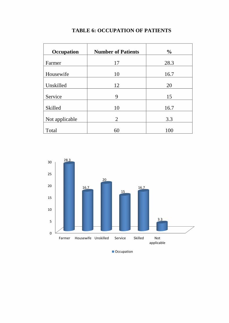
TABLE 6: OCCUPATION OF PATIENTS
Occupation Number of Patients %
Farmer 17 28.3
Housewife 10 16.7
Unskilled 12 20
Service 9 15
Skilled 10 16.7
Not applicable 2 3.3
Total 60 100
0
5
10
15
20
25
30
Farmer Housewife Unskilled Service Skilled Notapplicable
28.3
16.7
20
15 16.7
3.3
Occupation
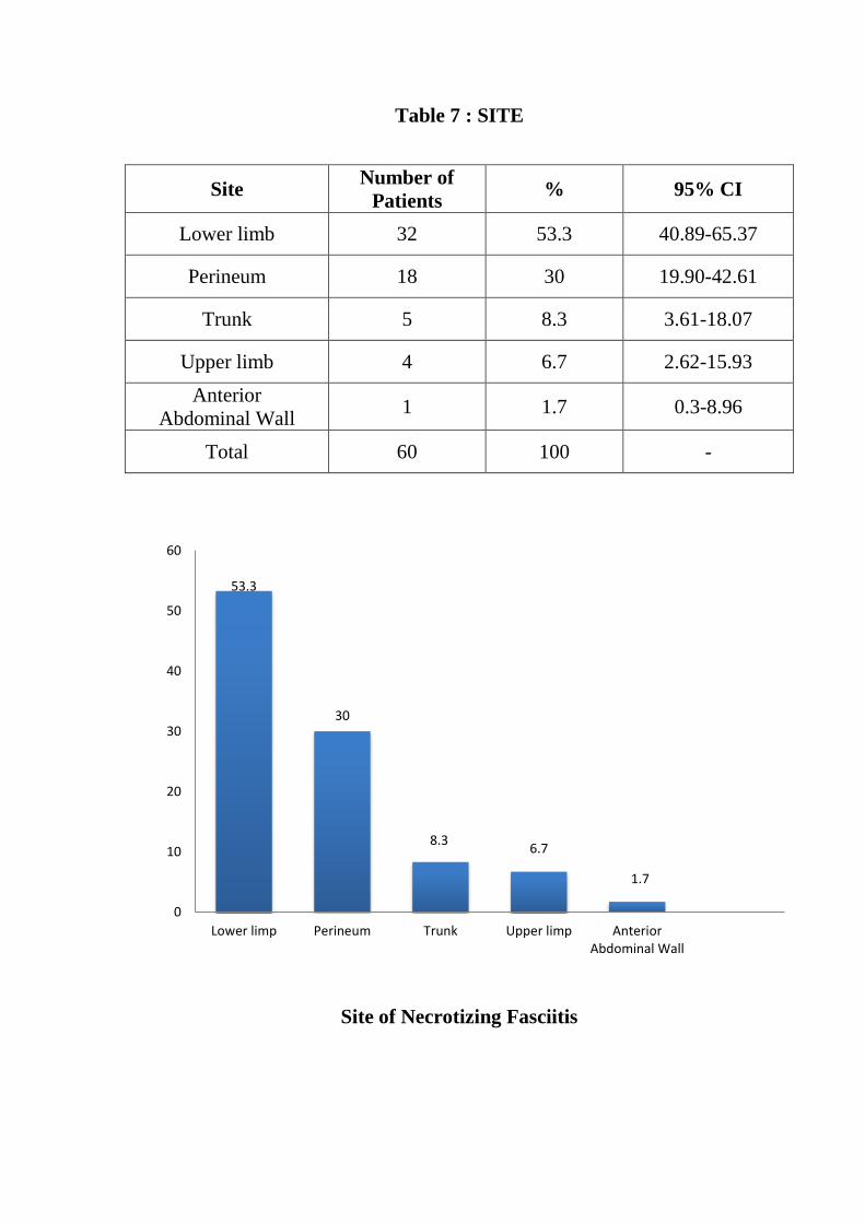
Table 7 : SITE
Site Number of
Patients % 95% CI
Lower limb 32 53.3 40.89-65.37
Perineum 18 30 19.90-42.61
Trunk 5 8.3 3.61-18.07
Upper limb 4 6.7 2.62-15.93
Anterior
Abdominal Wall 1 1.7 0.3-8.96
Total 60 100 -
Site of Necrotizing Fasciitis
53.3
30
8.3 6.7
1.7
0
10
20
30
40
50
60
Lower limp Perineum Trunk Upper limp AnteriorAbdominal Wall
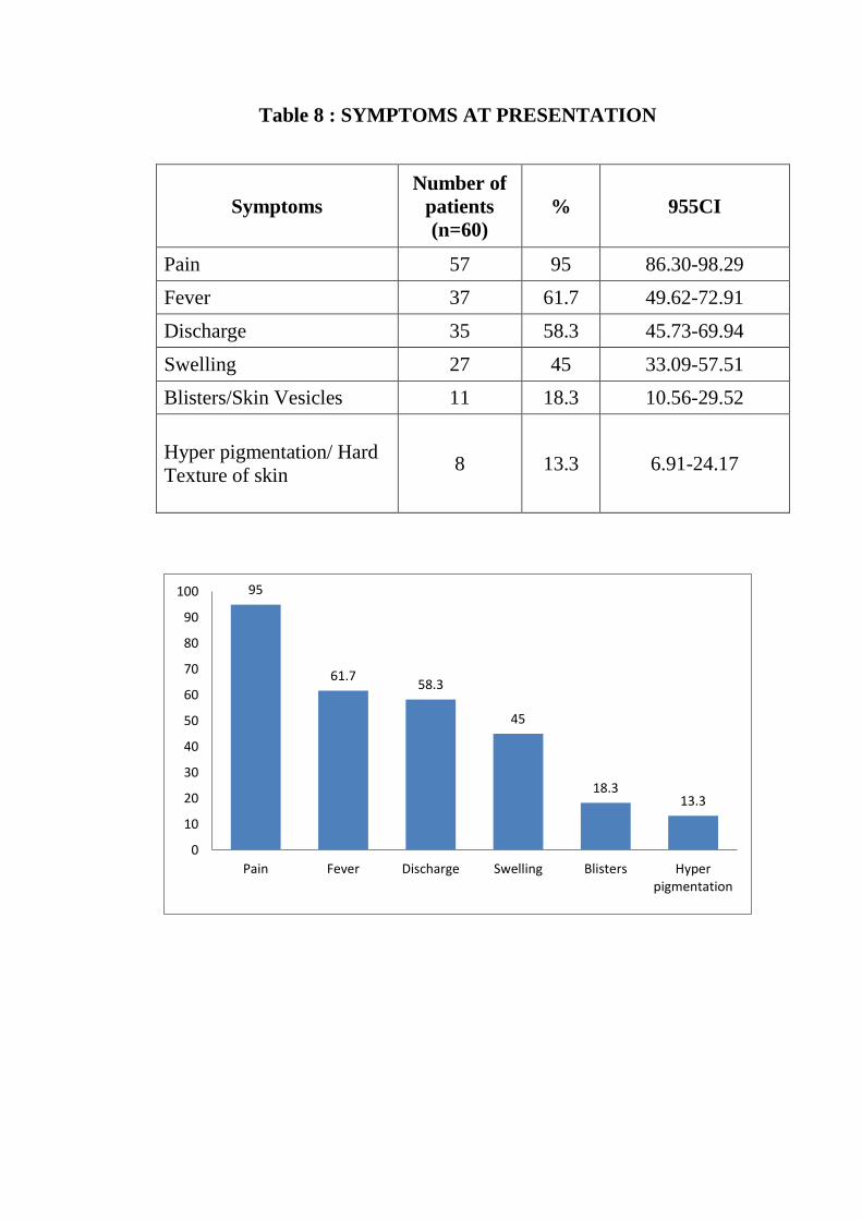
Table 8 : SYMPTOMS AT PRESENTATION
Symptoms
Number of
patients
(n=60)
% 955CI
Pain 57 95 86.30-98.29
Fever 37 61.7 49.62-72.91
Discharge 35 58.3 45.73-69.94
Swelling 27 45 33.09-57.51
Blisters/Skin Vesicles 11 18.3 10.56-29.52
Hyper pigmentation/ Hard
Texture of skin 8 13.3 6.91-24.17
95
61.7 58.3
45
18.3 13.3
0
10
20
30
40
50
60
70
80
90
100
Pain Fever Discharge Swelling Blisters Hyperpigmentation
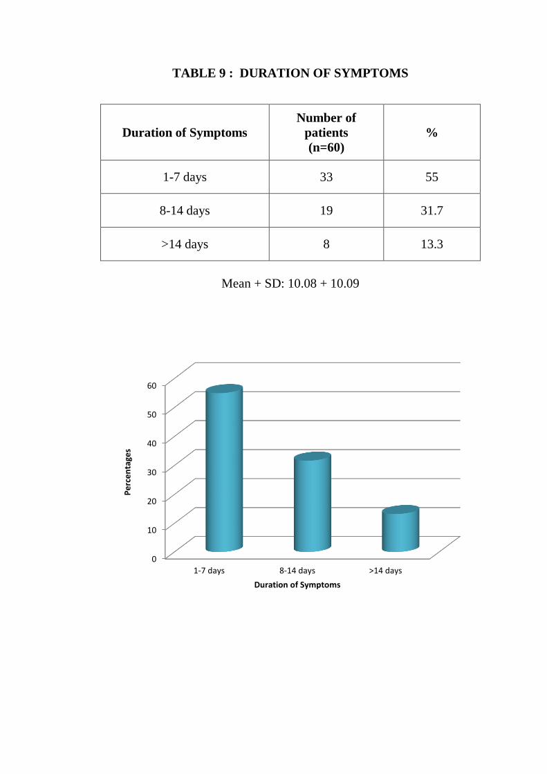
TABLE 9 : DURATION OF SYMPTOMS
Duration of Symptoms
Number of
patients
(n=60)
%
1-7 days 33 55
8-14 days 19 31.7
>14 days 8 13.3
Mean + SD: 10.08 + 10.09
0
10
20
30
40
50
60
1-7 days 8-14 days >14 days
Pe
rce
nta
ges
Duration of Symptoms
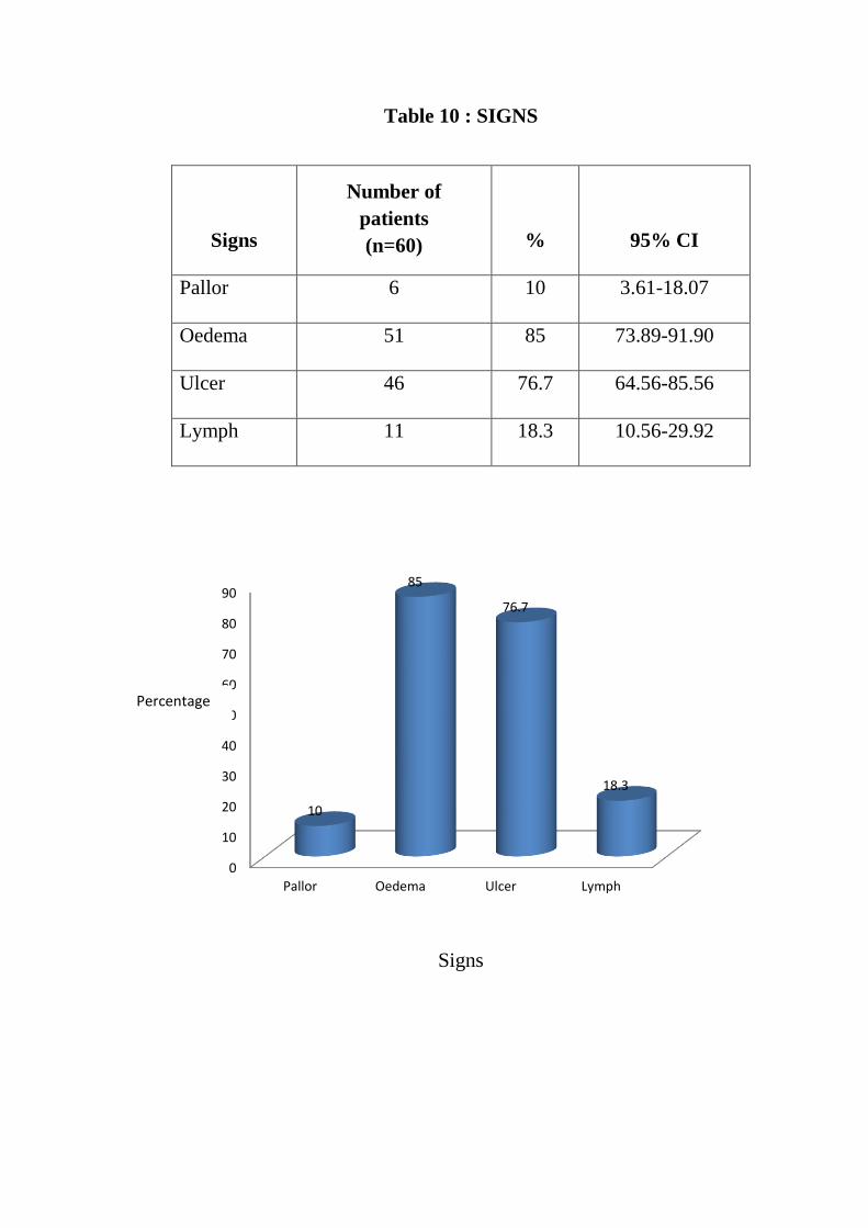
Table 10 : SIGNS
Signs
Number of
patients
(n=60) % 95% CI
Pallor 6 10 3.61-18.07
Oedema 51 85 73.89-91.90
Ulcer 46 76.7 64.56-85.56
Lymph 11 18.3 10.56-29.92
Signs
0
10
20
30
40
50
60
70
80
90
Pallor Oedema Ulcer Lymph
10
85
76.7
18.3
Percentage
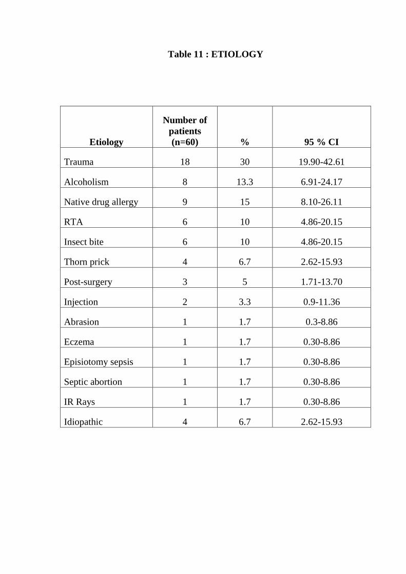
Table 11 : ETIOLOGY
Etiology
Number of
patients
(n=60) % 95 % CI
Trauma 18 30 19.90-42.61
Alcoholism 8 13.3 6.91-24.17
Native drug allergy 9 15 8.10-26.11
RTA 6 10 4.86-20.15
Insect bite 6 10 4.86-20.15
Thorn prick 4 6.7 2.62-15.93
Post-surgery 3 5 1.71-13.70
Injection 2 3.3 0.9-11.36
Abrasion 1 1.7 0.3-8.86
Eczema 1 1.7 0.30-8.86
Episiotomy sepsis 1 1.7 0.30-8.86
Septic abortion 1 1.7 0.30-8.86
IR Rays 1 1.7 0.30-8.86
Idiopathic 4 6.7 2.62-15.93
Etiology of Necrotizing Fasciitis
6.7
1.7
1.7
1.7
1.7
1.7
3.3
5
6.7
10
10
15
13.3
30
0 5 10 15 20 25 30 35
Idiopathic
IR Rays
Septic abortion
Episiotomy sepsis
Eczema
Abrasion
Injection
Post-surgery
Thorn prick
Insect bite
RTA
Native drug allergy
Alcoholism
Trauma
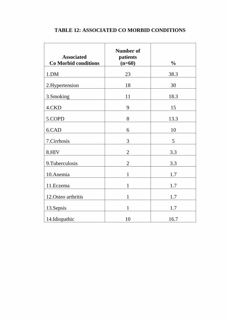
TABLE 12: ASSOCIATED CO MORBID CONDITIONS
Associated
Co Morbid conditions
Number of
patients
(n=60) %
1.DM 23 38.3
2.Hypertension 18 30
3.Smoking 11 18.3
4.CKD 9 15
5.COPD 8 13.3
6.CAD 6 10
7.Cirrhosis 3 5
8.HIV 2 3.3
9.Tuberculosis 2 3.3
10.Anemia 1 1.7
11.Eczema 1 1.7
12.Osteo arthritis 1 1.7
13.Sepsis 1 1.7
14.Idiopathic 10 16.7
Associated Co morbid conditions
0
5
10
15
20
25
30
35
40
1.DM
2.Hypertension
3.Smokin
g
4.CKD
5.COPD
6.CAD
7.Cirrhosi
s
8.HIV
9.Tuberculosi
s
10.Anemi
a
11.Eczema
12.Osteoarthritis
13.Sepsis
14.Idiopat
hic
% 38.3 30 18.3 15 13.3 10 5 3.3 3.3 1.7 1.7 1.7 1.7 16.7
Pe
rce
nta
ge
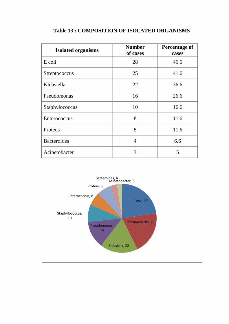
Table 13 : COMPOSITION OF ISOLATED ORGANISMS
Isolated organisms Number
of cases
Percentage of
cases
E coli 28 46.6
Streptococcus 25 41.6
Klebsiella 22 36.6
Pseudomonas 16 26.6
Staphylococcus 10 16.6
Enterococcus 8 11.6
Proteus 8 11.6
Bacteroides 4 6.6
Acinetobacter 3 5
E coli, 28
Streptococcus, 25
Klebsiella, 22
Pseudomonas, 16
Staphylococcus, 10
Enterococcus, 8
Proteus, 8
Bacteroides, 4 Acinetobacter, 3
Table 14 : MICROBES ISOLATED
Number of
microbes isolated
Number
of
cases
Percentage of cases
Nil 4 6.6
One 8 13.3
Two 35 58.3
Three 8 13.3
Four 5 8.3
0
5
10
15
20
25
30
35
40
Nil One Two Three Four
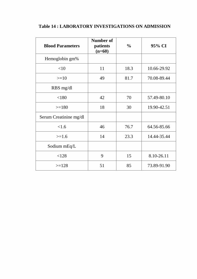
Table 14 : LABORATORY INVESTIGATIONS ON ADMISSION
Blood Parameters
Number of
patients
(n=60)
% 95% CI
Hemoglobin gm%
<10 11 18.3 10.66-29.92
>=10 49 81.7 70.08-89.44
RBS mg/dl
<180 42 70 57.49-80.10
>=180 18 30 19.90-42.51
Serum Creatinine mg/dl
<1.6 46 76.7 64.56-85.66
>=1.6 14 23.3 14.44-35.44
Sodium mEq/L
<128 9 15 8.10-26.11
>=128 51 85 73.89-91.90
Hemoglobin gm%
RBS mg/dl
0
10
20
30
40
50
60
70
80
90
<10 >=10
18.3
81.7
0
10
20
30
40
50
60
70
80
<180 >=180
Serum Creatinine mg/dl
Sodium mEq/L
0
10
20
30
40
50
60
70
80
90
<1.6 >=1.6
0
10
20
30
40
50
60
70
80
90
100
<128 >=128
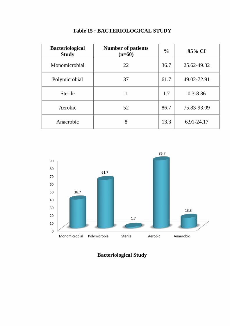
Table 15 : BACTERIOLOGICAL STUDY
Bacteriological
Study
Number of patients
(n=60) % 95% CI
Monomicrobial 22 36.7 25.62-49.32
Polymicrobial 37 61.7 49.02-72.91
Sterile 1 1.7 0.3-8.86
Aerobic 52 86.7 75.83-93.09
Anaerobic 8 13.3 6.91-24.17
Bacteriological Study
0
10
20
30
40
50
60
70
80
90
Monomicrobial Polymicrobial Sterile Aerobic Anaerobic
36.7
61.7
1.7
86.7
13.3
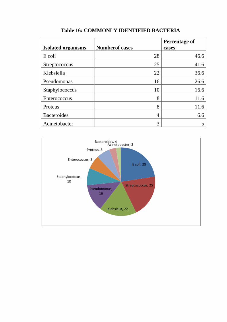
Table 16: COMMONLY IDENTIFIED BACTERIA
Isolated organisms Numberof cases
Percentage of
cases
E coli 28 46.6
Streptococcus 25 41.6
Klebsiella 22 36.6
Pseudomonas 16 26.6
Staphylococcus 10 16.6
Enterococcus 8 11.6
Proteus 8 11.6
Bacteroides 4 6.6
Acinetobacter 3 5
E coli, 28
Streptococcus, 25
Klebsiella, 22
Pseudomonas, 16
Staphylococcus, 10
Enterococcus, 8
Proteus, 8
Bacteroides, 4 Acinetobacter, 3

Table 17: TREATMENT
Treatment Number of
Patients %
Debridement 57 95
Fasciotomy 3 5
Split Skin Graft 21 35
Secondary Suturing 17 28.33
0
10
20
30
40
50
60
70
80
90
100
Debridement Fasciotomy Split Skin Graft SecondarySuturing
95
5
35
28.33
TREATMENT
Table 18 : HOSPITAL STAY IN DAYS
Hospital stay in days Number of
patients (n=60) %
<7 days 1 1.7
7-14 days 12 20
15-21 days 25 41.7
22-30 days 13 21.7
>30 days 9 15
HOSPITAL STAY IN DAYS
0
5
10
15
20
25
30
35
40
45
<7 days 7-14 days 15-21 days 22-30 days >30 days
1.7
20
41.7
21.7
15
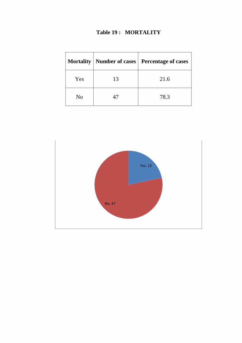
Table 19 : MORTALITY
Mortality Number of cases Percentage of cases
Yes 13 21.6
No 47 78.3
Yes, 13
No, 47
DISCUSSION
Incidence of necrotizing fasciitis was high in the males. The
incidence of necrotizing fasciitis increases with increase in age with the
highest incidence noted in above 60 yrs. age. On the contrary the
incidence of necrotizing fasciitis in women has more even distribution
with the highest in the 50-60 yrs. age group. Majority of the cases of
necrotizing fasciitis occurs in the males with more than 80% occurrence
in male. The overall incidence was higher in the older age group with the
highest in the 50 – 60 yrs.
This increase in the incidence with age might be due to higher
occurrence of the risk factors in the older age group.Male population have
higher incidence of traumatic injury, alsohigher workplace hazards and
alcoholism among male population are the predisposing factors for higher
incidence of necrotizing fasciitis in male population.
Work place forms the one of the most common place for the origin
of infection. Minor injuries are common in workplaces that requires
physical labor. Lack of proper safety precautions and bad hygiene in the
work place forms it a perfect combination for the origin of infection. The
incidence was highest among the farmers. Unskilled labor and
housewife’s form the major bulk along with the farmers. This pattern
shows that with improving the hygiene, safety and the working
environment and through proper training of manual labor the incidence of
necrotizing fasciitis can be reduced.
Majority of the cases of necrotizing fasciitis follow minor injury due to
trauma, RTA, insect bite, thorn prick, etc.,.poor care for the wound
following the trivial trauma is the major cause for necrotizing fasciitis.
The incidence of necrotizing fasciitis was highest following the trauma.
The foreign body that might get lodged or the deep inoculation that
occurs with trauma, thorn prick and other cause forms a perfect incubator
foe the organisms to flourish this complemented by lowered host defense
due to alcoholism, diabetes leads to fulminant local infection leading to
necrotizing fasciitis.
Lower limb is the most common site for necrotizing fasciitis.
This is followed by perineum and upperlimb. The least common site for
necrotizing fasciitis is the anterior abdominal wall. Similar studies
conducted in other parts of the world show the perineum as the most
common site of necrotizing fasciitis. This difference might due to
difference in work pattern, higher safety precautions in the west and
difference in the hygiene among the population.
Most of the patients came with the presenting complain of pain,
swelling and discharge of the affected area. Most of them had high grade
fever. Most common sign in necrotizing fasciitis is edema followed by
ulcer .farmer s has highest incidence of necrotizing fasciitis ,that is
because due to minor trauma duringoccupation which was unnoticed at
the earliest and rapid spread of infection .trauma is the most common
etiology in this study .
Host defense lost due to severe systemic illness. of the patient with
diabetic mellitus 20 to 40 percent were found to be diabetic followed by
hypertension .
Most commonly the disease is poly microbial of about 62%
Most common isolated organism is E.coli 35% .almost all patients
treated with broad spectrum antibiotics .most common antibiotic sensitive
is ceftriaxone followed by aminoglycosides and meteronidazole.
Almost all patients underwent surgical debridement of about
95%and some may underwent procedures like primary suturing
secondary suturing and ssg .
CONCLUSION :
NECROTIZING FASCITIS is most commonly seen in the elderly
males. source of the infection is identifiable in most of the cases.
diabetic mellitus is the most common comorbid factor . The disease is
most commonly polymicrobical.
Most common bacterial includes gram positive cocci (
streptococci and staphylococcal species ).
Gram negative (E.coli, actinobacter ,pseudomonas ).
Most common anaerobes is bacteroides .
Necrotizing fasciitis is a rare life threatening condition , early
recognition of disease , and through wound debridement and broad
spectrum antibiotics is essential extensive raw area due to debridement
can be managed by reconstructive procedures .
Inspite of early diagnosis and aggressive management of disease
also leads to significant mortality and morbidity . surgical management
must be aggressive and standard procedures to be followed . new
modalities of treatment like HBOT and VAC may be considered .
Diagnosis of necrotizing fasciitis is mainly through by means of
clinical examination ,but diagnostic adjuvant such as LRINEC scoring
system can be used for early diagnosis .
Multidisciplinary team approach is required for necrotizing
fasciitis.
BIBILOGRAPHY
1. Bailey and love’s TEXT BOOK OF SURGERY,25th
edition, pg no
29-30
2. SRB manual of surgery, 4th
edition, pg no 65-67
3. Sabiston text book of surgery, 19th edition , pg no 2011-2012
4. Schwartz principles of surgery, 10thedition,pg no 152-154
5. Learning surgery, the surgery clerkship manual, pgno 111-112
6. S das text book of clinical surgery, pg no 618, 632
7. T S Ranganathan, 9th
edition, pg no 140-143
8. Baek JH et al surgical management of Fournier’s gangrene. J
Korean Soccolproctol 19:349-53
9. Basoglu M, Ozbey et al, management of fournier’s gangrene, surg
today 37: 558-563
10. Wilson B Necrotisingfasciitis,Amsurg 1952 Apr;18(4): 416-31
11. Loudon I. necrotizing fasciitis, hospital gangrene and phagedema,
Lancet1994: 344: 1416-1419
12. Nallathambi MN, Ivatury et al craniocervicalnecrotising fasciitis
critical factor in management. Can J surg 1987; 30:61-3
13. Rea WJ , necrotizing fasciitis. Ann surg 1970; 172;957-64
PROFORMA
Name : Age: Sex:
IP.No:
Address : D.O.Admission :
D.O.Discharge/Death :
Duration of illness:
History:
1. Fever
2. Lethargy
3. Pain
4. Pruritis
5. Redness of skin
6. Other skinchanges
7. Purulent discharge
8. Obvious gangrene
9. Anorectal symptoms
10. Urogenital symptoms
11. History of skin infections
12. History of comorbid illness
13. History of recent invasive procedures
14. History of alcohol intake
GENERAL EXAMINATION
Pulse
BP
Temperature
Respiratory rate
Nutrition
Hydrational status
LOCAL EXAMINATION
Fulctuations
Localizing tenderness
Crepitations
Occult wounds
Skin changes
Foul smelling odour
Subcutaneous crepitations
Digital examination of the rectum
Testicular involvement
Extent of the disease
INVESTIGATIONS
Complete hemogram
B.Urea
B.Sugar
S.creatinine
S.Electrolytes
Blood Culture
Pus Culture
Urine Culture
Chest X-Ray
ECG
VCTC
LRIHEC
MANAGEMENT
Debridement details :
Number of debridements :
Recovery period :
Culture and sensitivity reports :
Reconstructive procedures:

Postoperative period :
Outcome :
Follow up :
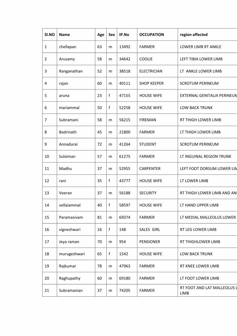
Sl.NO Name Age Sex IP.No OCCUPATION region affected predisposing factors comorbid disease Microoraganisms Identified antibiotic sensitivity mortality
1 chellapan 63 m 13492 FARMER LOWER LIMB RT ANKLE trauma CCF, ANAEMIA,SMOKING e.coli, streptococcus cefoperazone sulbactum NO
2 Arusamy 58 m 34642 COOLIE LEFT TIBIA LOWER LIMB nil HIV,DM,HT e.coli, streptococcus ,klebsiella cefoperazone sulbactum,gentamycin,amikacin, meropenem,ofloxacin
NO
3 Ranganathan 52 m 38518 ELECTRICIAN LT ANKLE LOWER LIMB trauma SMOKING,DM bacteroides,E.coli ceftriaxone, amoxycillin with clavulinic acid, ofloxacin NO
4 rajan 60 m 40111 SHOP KEEPER SCROTUM PERINEUM trauma SMOKING Streptococcus ciprofloxacin,cephalexin,linezolide YES
5 aruna 23 f 47155 HOUSE WIFE EXTERNAL GENITALIA PERINEUM episiotomy sepsis NIL klebsiella,staphylococcus piperacillin tazobactum ,amikacin NO
6 mariammal 50 f 52258 HOUSE WIFE LOW BACK TRUNK trauma NIL Streptococcus,E.coli ampicilllin, cefotaxime, ofloxacin YES
7 Subramani 58 m 56215 FIREMAN RT THIGH LOWER LIMB ALCOHOLISM DM , HT,SMOKING, OLD AWMI
E.coli,staphylococus meropenem, cotrimoxazole, linezolide NO
8 Badrinath 45 m 21800 FARMER LT THIGH LOWER LIMB trauma DM,HT,COPD Streptococcus,pseudomonas cefoperazone sulbactum NO
9 Annadurai 72 m 41264 STUDENT SCROTUM PERINEUM trauma NIL pseudomonas,Actinobacter cefoperazone sulbactum YES
10 Sulaiman 57 m 61275 FARMER LT INGUINAL REGION TRUNK nil DM Streptococcus,pseudomonas cefotaxime NO
11 Madhu 37 m 52955 CARPENTER LEFT FOOT DORSUM LOWER LIMB trauma NIL klebsiella Streptococcus ceftriaxone NO
12 rani 35 f 43777 HOUSE WIFE LT LOWER LIMB thron prick HT,COPD klebsiella,pseudomonas ciprofloxacin,cephalexin,linezolide NO
13 Veeran 37 m 56188 SECURITY RT THIGH LOWER LIMB AND ANKLE nil CKD klebsiella, Proteus cephalexin ,linezolide NO
14 vellaiammal 40 f 58597 HOUSE WIFE LT HAND UPPER LIMB trauma DM klebsiella ,proteus ,pseudomonas,enterococcus metronidazole, amoxycillin, netilimycin NO
15 Paramasivam 81 m 69374 FARMER LT MEDIAL MALLEOLUS LOWER LIMB injection HT,COPD klebsiella ,Streptococcus ceftriaxone, amikacin,gentamycin YES
16 vigneshwari 16 f 148 SALES GIRL RT LEG LOWER LIMB native drug allergy NIL Enterococcus cephalexin ,linezolide NO
17 Jeya raman 70 m 954 PENSIONER RT THIGHLOWER LIMB trauma DM,SMOKING E.coli,staphylococus piperacillin tazobactum ,amikacin YES
18 murugeshwari 65 f 1542 HOUSE WIFE LOW BACK TRUNK insect bite DM E.coli, streptococcus, proteus,pseudomonas linezolide NO
19 Rajkumar 78 m 47963 FARMER RT KNEE LOWER LIMB trauma DM,CKD E.coli,bacterioides piperacillin tazobactum ,amikacin NO
20 Raghupathy 60 m 69180 FARMER LT FOOT LOWER LIMB trauma NIL E.coli, staphyloccus, enterococcus ofloxacin, erythromycin, gentamycin NO
21 Subramanian 37 m 74205 FARMER RT FOOT AND LAT MALLEOLUS LOWER LIMB
thron prick OLD PT,DM, SMOKING Nil piperacillin tazobactum ,amikacin NO
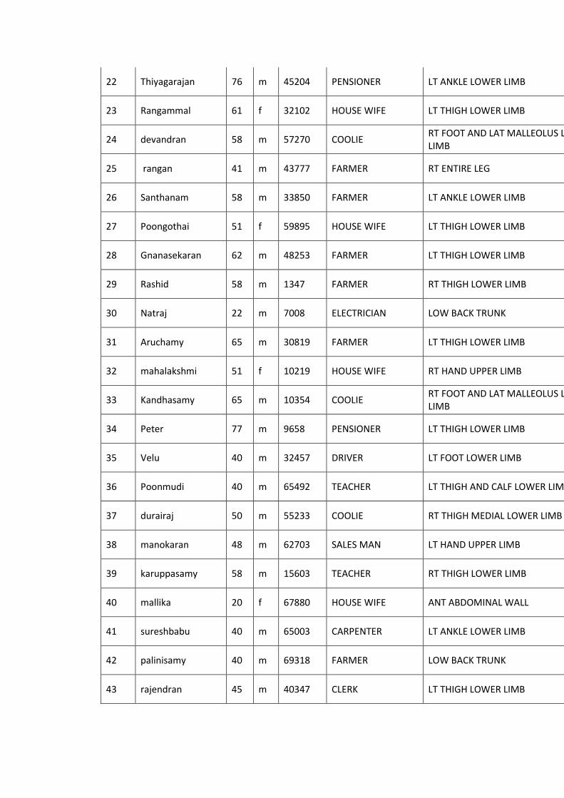
22 Thiyagarajan 76 m 45204 PENSIONER LT ANKLE LOWER LIMB insect bite DM,HT,COPD Nil ampicilllin, cefotaxime, ofloxacin YES
23 Rangammal 61 f 32102 HOUSE WIFE LT THIGH LOWER LIMB eczema CRF E.coli,staphylococus ampicilllin, cefotaxime, ofloxacin NO
24 devandran 58 m 57270 COOLIE RT FOOT AND LAT MALLEOLUS LOWER LIMB
ALCOHOLISM HT,COPD,SMOKING , E.coli,enterococcus ceftriaxone YES
25 rangan 41 m 43777 FARMER RT ENTIRE LEG trauma,alcoholism DM bacteroides,E.coli,staphylococcus cefoperazone sulbactum YES
26 Santhanam 58 m 33850 FARMER LT ANKLE LOWER LIMB insect bite HT pseudomonas,Actinobacter ampicilllin, cefotaxime, ofloxacin NO
27 Poongothai 51 f 59895 HOUSE WIFE LT THIGH LOWER LIMB insect bite DM,COPD E.coli, streptococcus doxycycline, metronidazole, gentamycin YES
28 Gnanasekaran 62 m 48253 FARMER LT THIGH LOWER LIMB abrasion ECZEMA pseudomonas,Actinobacter,staphylococcus,E.coli ampicilllin, cefotaxime, ofloxacin NO
29 Rashid 58 m 1347 FARMER RT THIGH LOWER LIMB rta,alcoholism NIL E.coli,Pseudomonas,proteus ampicilllin, cefotaxime, ofloxacin NO
30 Natraj 22 m 7008 ELECTRICIAN LOW BACK TRUNK insect bite HTN Nil piperacillin tazobactum ,amikacin NO
31 Aruchamy 65 m 30819 FARMER LT THIGH LOWER LIMB native drug allergy DM Staphylococcus, bacteroides cefotaxime YES
32 mahalakshmi 51 f 10219 HOUSE WIFE RT HAND UPPER LIMB post surgery DM,HT,COPD Enterococcus cefotaxime, gentamycin ,amikacin NO
33 Kandhasamy 65 m 10354 COOLIE RT FOOT AND LAT MALLEOLUS LOWER LIMB
native drug allergy NIL Streptococcus,E.coli,staphylococcus cefoperazone sulbactum NO
34 Peter 77 m 9658 PENSIONER LT THIGH LOWER LIMB nil HT Streptococcus,E.coli cefoperazone sulbactum YES
35 Velu 40 m 32457 DRIVER LT FOOT LOWER LIMB post surgery CAD,HT Enterococcus,klebsiella ceftriaxone, amoxycillin with clavulinic acid, ofloxacin NO
36 Poonmudi 40 m 65492 TEACHER LT THIGH AND CALF LOWER LIMB rta,alcoholism DM,HT,COPD Streptococcus,E.coli,proteus,staphylococcus ciprofloxacin,cephalexin,linezolide NO
37 durairaj 50 m 55233 COOLIE RT THIGH MEDIAL LOWER LIMB native drug allergy HT Nil ciprofloxacin,cephalexin,linezolide NO
38 manokaran 48 m 62703 SALES MAN LT HAND UPPER LIMB thron prick CRF klebsiella piperacillin tazobactum ,amikacin NO
39 karuppasamy 58 m 15603 TEACHER RT THIGH LOWER LIMB injection HIV,DM,SMOKING pseudomonas ampicilllin, cefotaxime, ofloxacin NO
40 mallika 20 f 67880 HOUSE WIFE ANT ABDOMINAL WALL septic abortion SEPSIS E.coli, streptococcus cefoperazone sulbactum NO
41 sureshbabu 40 m 65003 CARPENTER LT ANKLE LOWER LIMB ir rays OA KNEE klebsiella doxycycline, metronidazole, gentamycin NO
42 palinisamy 40 m 69318 FARMER LOW BACK TRUNK native drug allergy DM,CKD E.coli,pseudomonas metronidazole, amoxycillin, netilimycin NO
43 rajendran 45 m 40347 CLERK LT THIGH LOWER LIMB rta,alcoholism CRF E.coli,pseudomonas ampicilllin, cefotaxime, ofloxacin NO
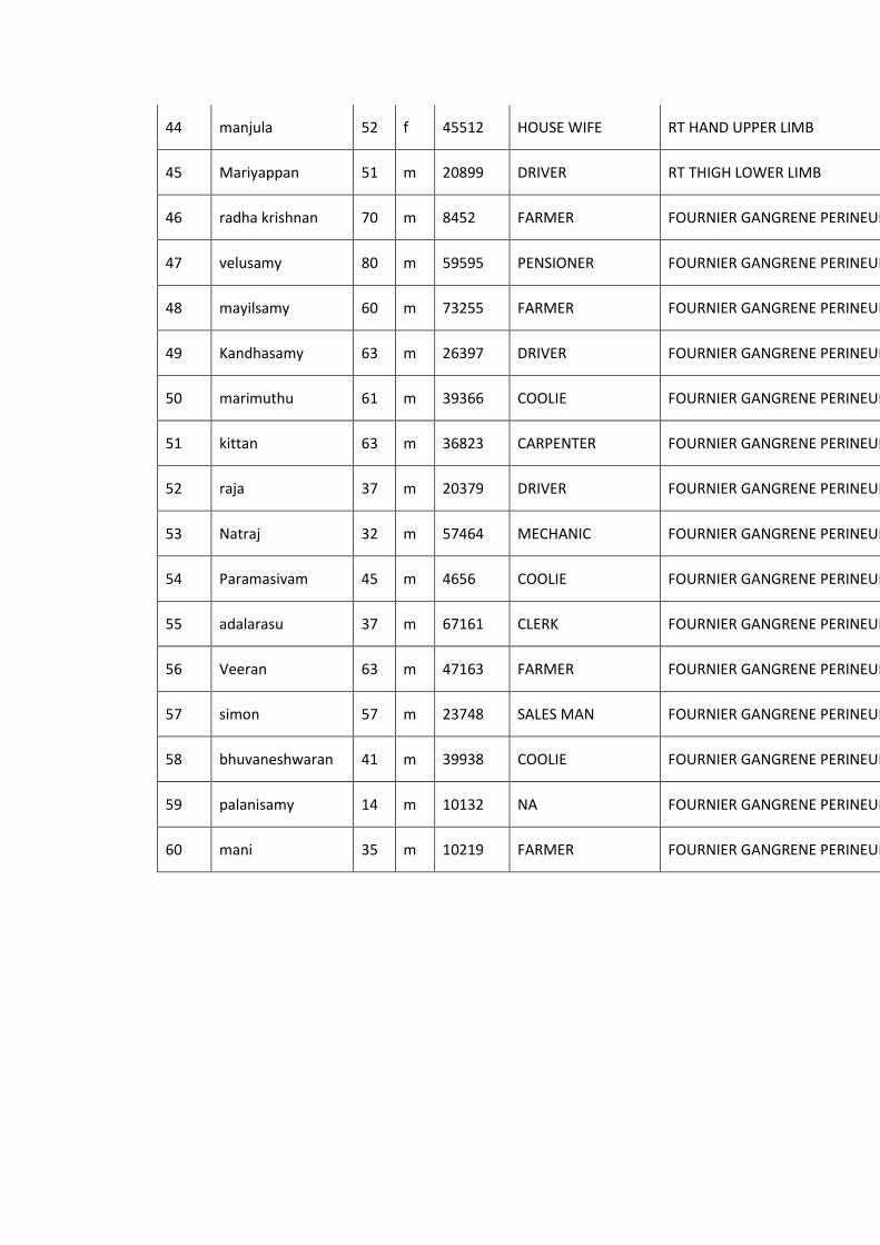
44 manjula 52 f 45512 HOUSE WIFE RT HAND UPPER LIMB native drug allergy HTN E.coli,streptococcus piperacillin tazobactum ,amikacin NO
45 Mariyappan 51 m 20899 DRIVER RT THIGH LOWER LIMB post surgery CAD klebsiella,pseudomonas,proteus ciprofloxacin,cephalexin,linezolide NO
46 radha krishnan 70 m 8452 FARMER FOURNIER GANGRENE PERINEUM trauma DM streptococcus,klebsiella doxycycline, metronidazole, gentamycin NO
47 velusamy 80 m 59595 PENSIONER FOURNIER GANGRENE PERINEUM rta,alcoholism CAD streptococccus,klebsiella cefoperazone sulbactum NO
48 mayilsamy 60 m 73255 FARMER FOURNIER GANGRENE PERINEUM insect bite DM,HT,COPD streptococcus,klebsiella cefotaxime NO
49 Kandhasamy 63 m 26397 DRIVER FOURNIER GANGRENE PERINEUM rta,alcoholism OLD MI,SMOKING streptococcus ofloxacin NO
50 marimuthu 61 m 39366 COOLIE FOURNIER GANGRENE PERINEUM trauma CIRRHOSIS E.coli ciprofloxacin,cephalexin,linezolide NO
51 kittan 63 m 36823 CARPENTER FOURNIER GANGRENE PERINEUM thron prick DM streptococcus,proteus linezolide YES
52 raja 37 m 20379 DRIVER FOURNIER GANGRENE PERINEUM trauma CRF E.coli,pseudomonas meropenem, cotrimoxazole, linezolide,metronidazole NO
53 Natraj 32 m 57464 MECHANIC FOURNIER GANGRENE PERINEUM nil OLD PT,,SMOKING streptoccous.klebsiella ceftriaxone, doxycycline, metronidazole NO
54 Paramasivam 45 m 4656 COOLIE FOURNIER GANGRENE PERINEUM native drug allergy HT,CIRRHOSIS klebsiella,pseudomonas,E.coli metronidazole, amoxycillin, netilimycin NO
55 adalarasu 37 m 67161 CLERK FOURNIER GANGRENE PERINEUM trauma DM,CKD klebsiella,enteroccus ampicilllin, cefotaxime, ofloxacin NO
56 Veeran 63 m 47163 FARMER FOURNIER GANGRENE PERINEUM rta,alcoholism DM,SMOKING E.coli, streptococcus ,klebsiella,pseudomonas doxycycline, metronidazole, gentamycin NO
57 simon 57 m 23748 SALES MAN FOURNIER GANGRENE PERINEUM trauma CAD klebsiella,pseudomonas amikacin ,ofloxacin, eryhromycin YES
58 bhuvaneshwaran 41 m 39938 COOLIE FOURNIER GANGRENE PERINEUM native drug allergy CRF E.coli,enteroccus piperacillin tazobactum ,amikacin NO
59 palanisamy 14 m 10132 NA FOURNIER GANGRENE PERINEUM trauma HTN,CIRRHOSIS klebsiella, Proteus amikacin ,ofloxacin, eryhromycin NO
60 mani 35 m 10219 FARMER FOURNIER GANGRENE PERINEUM native drug allergy NIL E.coli.streptococcus ,klebsiella ceftriaxone, amoxycillin with clavulinic acid, ofloxacin NO
Related Documents