A direct physical interaction between Nanog and Sox2 regulates embryonic stem cell self-renewal Alessia Gagliardi 1,5 , Nicholas P Mullin 1,5 , Zi Ying Tan 1,6 , Douglas Colby 1 , Anastasia I Kousa 1 , Florian Halbritter 1 , Jason T Weiss 1,7 , Anastasia Felker 1,8 , Karel Bezstarosti 2 , Rebecca Favaro 3 , Jeroen Demmers 2 , Silvia K Nicolis 3 , Simon R Tomlinson 1 , Raymond A Poot 4 and Ian Chambers 1, * 1 MRC Centre for Regenerative Medicine, Institute for Stem Cell Research, School of Biological Sciences, University of Edinburgh, Edinburgh, UK, 2 Proteomics Center, Erasmus MC, Rotterdam, The Netherlands, 3 Department of Biotechnology and Biological Sciences, University of Milano-Bicocca, Milano, Italy and 4 Department of Cell Biology, Erasmus MC, Rotterdam, The Netherlands Embryonic stem (ES) cell self-renewal efficiency is deter- mined by the Nanog protein level. However, the protein partners of Nanog that function to direct self-renewal are unclear. Here, we identify a Nanog interactome of over 130 proteins including transcription factors, chromatin modifying complexes, phosphorylation and ubiquitination enzymes, basal transcriptional machinery members, and RNA processing factors. Sox2 was identified as a robust interacting partner of Nanog. The purified Nanog–Sox2 complex identified a DNA recognition sequence present in multiple overlapping Nanog/Sox2 ChIP-Seq data sets. The Nanog tryptophan repeat region is necessary and sufficient for interaction with Sox2, with tryptophan residues required. In Sox2, tyrosine to alanine mutations within a triple-repeat motif (S X T/S Y) abrogates the Nanog–Sox2 interaction, alters expression of genes associated with the Nanog-Sox2 cognate sequence, and reduces the ability of Sox2 to rescue ES cell differentiation induced by endogen- ous Sox2 deletion. Substitution of the tyrosines with phenylalanine rescues both the Sox2–Nanog interaction and efficient self-renewal. These results suggest that aro- matic stacking of Nanog tryptophans and Sox2 tyrosines mediates an interaction central to ES cell self-renewal. The EMBO Journal (2013) 32, 2231–2247. doi:10.1038/ emboj.2013.161; Published online 26 July 2013 Subject Categories: signal transduction; development Keywords: DNA-independent interaction; hydrophobic stacking; pluripotency; protein interactome; SELEX Introduction Embryonic stem (ES) cell self-renewal efficiency depends on the level of expression of components of the pluripotency gene regulatory network. Among these, Oct4, Sox2 and Nanog play central roles. While the levels of Oct4 and Sox2 are relatively uniform in undifferentiated ES cells, the levels of Nanog vary considerably (Hatano et al, 2005; Chambers et al, 2007; Singh et al, 2007) with high levels of Nanog directing efficient self-renewal (Chambers et al, 2003, 2007). However, the mechanisms by which Nanog delivers this function in ES cells are not fully understood. In particular, although Nanog has been reported to interact with several proteins (Wang et al, 2006; Wu et al, 2006; Liang et al, 2008; Costa et al, 2013), the full extent of the Nanog interactome is not known. In the past few years, proteomic approaches have been employed to characterize and begin to understand the net- work of biochemical interactions controlling pluripotent cell function. This has resulted in the identification of additional proteins that interact with the key transcriptional factors Nanog, Sox2 and Oct4 to control and maintain the pluripotent state. Pioneering studies by Wang et al (2006) identified a Nanog-centred interactome of 17 proteins that extended to other transcription factors including Oct4, Zfp281, Nac1, Rex1 and Nr0b1. This list of Nanog-interacting proteins has since been extended (Liang et al, 2008) with a recent interactome identifying a total of 27 Nanog interactors (Costa et al, 2013). This relatively small number is in contrast to the larger number of interactors identified in recent Oct4 (Pardo et al, 2010; van den Berg et al, 2010; Ding et al, 2012) and Sox2 (Gao et al, 2012) interactomes. Interactome studies have the potential to contribute to the elucidation of the mechanisms by which specific factors function. Central to this is the identification of the interacting amino-acid side chains on partner proteins as well as the functional significance of their association. To date, biochemical characterization of protein– protein interactions in pluripotent cells has been relatively sparse with most effort analysing the interaction between Sox2 and Oct4 (Yuan et al, 1995; Ambrosetti et al, 1997, 2000; Remenyi et al, 2003; Kim et al, 2008; Chen et al, 2008a; Lam et al, 2012). From a biochemical perspective, little is known about how Nanog fits into the tight relationship between Oct4 and Sox2. Previously, we described a method to identify partner proteins interacting with nuclear proteins of interest in ES cells and used this to identify an extensive interaction net- work for the transcription factor Oct4 (van den Berg et al, 2010). Here, this technique is applied to Nanog, resulting in identification of a Nanog interactome which includes over 130 Nanog partners in ES cells. From this, the direct *Corresponding author. MRC Centre for Regenerative Medicine, Institute for Stem Cell Research, School of Biological Sciences, University of Edinburgh, 5 Little France Drive, Edinburgh EH16 4UU, UK. Tel.: þ44 (0)131 651 9500; Fax: þ44 (0)131 651 9501; E-mail: [email protected] 5 These authors contributed equally to this work. 6 Present address: Gene Regulation Laboratory, Genome Institute of Singapore, Singapore 138672, Singapore. 7 Present address: Edinburgh Cancer Research UK Centre, MRC Institute of Genetics and Molecular Medicine, University of Edinburgh, Crewe Road South, Edinburgh EH4 2XR, UK. 8 Present address: Institute of Molecular Life Sciences, University of Zurich, Winterthurerstrasse 190, 8057 Zurich, Switzerland. Received: 30 January 2013; accepted: 27 June 2013; published online: 26 July 2013 The EMBO Journal (2013) 32, 2231–2247 www.embojournal.org EMBO THE EMBO JOURNAL THE EMBO JOURNAL 2231 & 2013 European Molecular Biology Organization The EMBO Journal VOL 32 | NO 16 | 2013

Welcome message from author
This document is posted to help you gain knowledge. Please leave a comment to let me know what you think about it! Share it to your friends and learn new things together.
Transcript

A direct physical interaction between Nanog andSox2 regulates embryonic stem cell self-renewal
Alessia Gagliardi1,5, Nicholas P Mullin1,5,Zi Ying Tan1,6, Douglas Colby1,Anastasia I Kousa1, Florian Halbritter1,Jason T Weiss1,7, Anastasia Felker1,8,Karel Bezstarosti2, Rebecca Favaro3,Jeroen Demmers2, Silvia K Nicolis3,Simon R Tomlinson1, Raymond A Poot4
and Ian Chambers1,*1MRC Centre for Regenerative Medicine, Institute for Stem CellResearch, School of Biological Sciences, University of Edinburgh,Edinburgh, UK, 2Proteomics Center, Erasmus MC, Rotterdam,The Netherlands, 3Department of Biotechnology and BiologicalSciences, University of Milano-Bicocca, Milano, Italy and 4Departmentof Cell Biology, Erasmus MC, Rotterdam, The Netherlands
Embryonic stem (ES) cell self-renewal efficiency is deter-
mined by the Nanog protein level. However, the protein
partners of Nanog that function to direct self-renewal are
unclear. Here, we identify a Nanog interactome of over
130 proteins including transcription factors, chromatin
modifying complexes, phosphorylation and ubiquitination
enzymes, basal transcriptional machinery members, and
RNA processing factors. Sox2 was identified as a robust
interacting partner of Nanog. The purified Nanog–Sox2
complex identified a DNA recognition sequence present in
multiple overlapping Nanog/Sox2 ChIP-Seq data sets. The
Nanog tryptophan repeat region is necessary and sufficient
for interaction with Sox2, with tryptophan residues
required. In Sox2, tyrosine to alanine mutations within a
triple-repeat motif (S X T/S Y) abrogates the Nanog–Sox2
interaction, alters expression of genes associated with the
Nanog-Sox2 cognate sequence, and reduces the ability of
Sox2 to rescue ES cell differentiation induced by endogen-
ous Sox2 deletion. Substitution of the tyrosines with
phenylalanine rescues both the Sox2–Nanog interaction
and efficient self-renewal. These results suggest that aro-
matic stacking of Nanog tryptophans and Sox2 tyrosines
mediates an interaction central to ES cell self-renewal.
The EMBO Journal (2013) 32, 2231–2247. doi:10.1038/
emboj.2013.161; Published online 26 July 2013Subject Categories: signal transduction; development
Keywords: DNA-independent interaction; hydrophobic
stacking; pluripotency; protein interactome; SELEX
Introduction
Embryonic stem (ES) cell self-renewal efficiency depends on
the level of expression of components of the pluripotency
gene regulatory network. Among these, Oct4, Sox2 and
Nanog play central roles. While the levels of Oct4 and Sox2
are relatively uniform in undifferentiated ES cells, the levels
of Nanog vary considerably (Hatano et al, 2005; Chambers
et al, 2007; Singh et al, 2007) with high levels of Nanog
directing efficient self-renewal (Chambers et al, 2003, 2007).
However, the mechanisms by which Nanog delivers this
function in ES cells are not fully understood. In particular,
although Nanog has been reported to interact with several
proteins (Wang et al, 2006; Wu et al, 2006; Liang et al, 2008;
Costa et al, 2013), the full extent of the Nanog interactome is
not known.
In the past few years, proteomic approaches have been
employed to characterize and begin to understand the net-
work of biochemical interactions controlling pluripotent cell
function. This has resulted in the identification of additional
proteins that interact with the key transcriptional factors
Nanog, Sox2 and Oct4 to control and maintain the pluripotent
state. Pioneering studies by Wang et al (2006) identified a
Nanog-centred interactome of 17 proteins that extended to
other transcription factors including Oct4, Zfp281, Nac1, Rex1
and Nr0b1. This list of Nanog-interacting proteins has since
been extended (Liang et al, 2008) with a recent interactome
identifying a total of 27 Nanog interactors (Costa et al, 2013).
This relatively small number is in contrast to the larger
number of interactors identified in recent Oct4 (Pardo et al,
2010; van den Berg et al, 2010; Ding et al, 2012) and Sox2
(Gao et al, 2012) interactomes. Interactome studies have the
potential to contribute to the elucidation of the mechanisms
by which specific factors function. Central to this is the
identification of the interacting amino-acid side chains on
partner proteins as well as the functional significance of their
association. To date, biochemical characterization of protein–
protein interactions in pluripotent cells has been relatively
sparse with most effort analysing the interaction between
Sox2 and Oct4 (Yuan et al, 1995; Ambrosetti et al, 1997, 2000;
Remenyi et al, 2003; Kim et al, 2008; Chen et al, 2008a; Lam
et al, 2012). From a biochemical perspective, little is known
about how Nanog fits into the tight relationship between Oct4
and Sox2.
Previously, we described a method to identify partner
proteins interacting with nuclear proteins of interest in ES
cells and used this to identify an extensive interaction net-
work for the transcription factor Oct4 (van den Berg et al,
2010). Here, this technique is applied to Nanog, resulting
in identification of a Nanog interactome which includes
over 130 Nanog partners in ES cells. From this, the direct
*Corresponding author. MRC Centre for Regenerative Medicine, Institutefor Stem Cell Research, School of Biological Sciences, University ofEdinburgh, 5 Little France Drive, Edinburgh EH16 4UU, UK.Tel.: þ44 (0)131 651 9500; Fax: þ44 (0)131 651 9501;E-mail: [email protected] authors contributed equally to this work.6Present address: Gene Regulation Laboratory, Genome Institute ofSingapore, Singapore 138672, Singapore.7Present address: Edinburgh Cancer Research UK Centre, MRC Instituteof Genetics and Molecular Medicine, University of Edinburgh, CreweRoad South, Edinburgh EH4 2XR, UK.8Present address: Institute of Molecular Life Sciences, University ofZurich, Winterthurerstrasse 190, 8057 Zurich, Switzerland.
Received: 30 January 2013; accepted: 27 June 2013; published online:26 July 2013
The EMBO Journal (2013) 32, 2231–2247
www.embojournal.org
EMBO
THE
EMBOJOURNAL
THE
EMBOJOURNAL
2231&2013 European Molecular Biology Organization The EMBO Journal VOL 32 | NO 16 | 2013

interaction between Nanog and Sox2 was selected for further
characterization, pinpointing individual residues required
for the interaction and defining the functional consequences
of elimination of the interaction between these central
pluripotency regulators.
Results
Identification of a Nanog interactome
An ES cell line expressing epitope-tagged Nanog protein was
obtained by transfection of E14Tg2a cells with a construct in
which the constitutive CAG promoter directs expression of
(FLAG)3Nanog, linked via an IRES to puromycin resistance.
Puromycin-resistant colonies were expanded and the
resulting cell lines were analysed by immunoblotting. A cell
line was identified (hereafter called F-Nanog) that expressed
(FLAG)3Nanog at close to endogenous levels (Figure 1A). A
qRT-PCR analysis of F-Nanog and E14Tg2a wild-type cells
showed no significant difference in the expression level of the
ES cell-specific genes Oct4, Sox2 and Rex1 (Figure 1B). In
agreement with recent reports of autorepression by Nanog
(Fidalgo et al, 2012; Navarro et al, 2012b), F-Nanog cells
Figure 1 Characterization of E14Tg2a Flag Nanog cell line. (A) Expression levels of Nanog protein in E14Tg2a and E14Tg2a F-Nanog cellscompared by immunoblot analysis using b-actin as a loading control. Note the reduced expression of endogenous Nanog protein in cellstransfected with (Flag)3Nanog, consistent with autorepression of the Nanog gene by Nanog protein (Navarro et al, 2012a). (B) Expression levelsof Sox2, Oct4 and Rex1 in E14Tg2a F-Nanog relative to E14Tg2a which was set to 1. Error bars are s.e.m. of three biological replicates.(C) Coomassie-stained SDS–polyacrylamide gel of the FLAG immunoprecipitation from E14Tg2a F-Nanog and control E14Tg2a cells.(D) Proteins detected by mass spectrometry analysis are grouped in classes. Transcription factors are shown in blue circles, NuRD componentsare in green, Trrap/p400 complex is in yellow, PcG components are in red, E2F6 complex is in purple, Sin3a complex is in burgundy, N-CoRcomplex is in khaki, LSD1 complex is white, Mll complex is in blue green, chromatin remodelling/transcriptional regulation proteins are in darkorange, transcriptional machinery proteins are in pale green, proteins involved in phosphorylation are in pale blue, proteins involved inubiquitination are in amber, proteins involved in RNA processing are in fuschia, proteins involved in cell cycle or DNA replication are in coral,proteins involved in DNA repair are in pink and other proteins are in grey. (E) Nuclear extracts from E14Tg2a F-Nanog cells (top) or from RCNbH-B(t):F-Nanog (bottom) were immunoprecipitated as indicated and immunoblots analysed with the indicated antibodies. In the bottom panel, Crefers to control samples from RCNbH-B(t) parental cells. Source data for this figure is available on the online supplementary information page.
Proteomic identification of a Nanog–Sox2 complexA Gagliardi et al
2232 The EMBO Journal VOL 32 | NO 16 | 2013 &2013 European Molecular Biology Organization
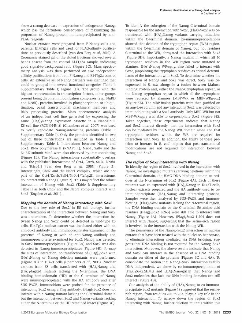
show a strong decrease in expression of endogenous Nanog,
which has the fortuitous consequence of maximizing the
proportion of Nanog protein immunoprecipitated by anti-
FLAG reagents.
Nuclear extracts were prepared from F-Nanog cells and
parental E14Tg2a cells and used for FLAG-affinity purifica-
tions as previously described (van den Berg et al, 2010). A
Coomassie-stained gel of the eluted fractions showed several
bands absent from the control E14Tg2a sample, indicating
good signal-to-background ratio (Figure 1C). Mass spectro-
metry analysis was then performed on two independent
affinity purifications from both F-Nanog and E14Tg2a control
cells. An extensive set of Nanog partners was identified that
could be grouped into several functional categories (Table I;
Supplementary Table I; Figure 1D). The group with the
highest representation is transcription factors, other groups
present being chromatin modification complexes (e.g., NuRD
and NcoR), proteins involved in phosphorylation or ubiqui-
tination, basal transcriptional machinery members and
RNA processing proteins. Mass spectrometric analysis
of an independent cell line generated by expressing the
same (Flag)3Nanog expression cassette in a Nanog-null
ES cell line (RCNbH-B(t)) (Chambers et al, 2007) was used
to verify candidate Nanog-interacting proteins (Table I;
Supplementary Table I). Only the proteins identified in two
out of three purifications are included in Table I and
Supplementary Table I. Interactions between Nanog and
Sox2, RNA polymerase II (RNAPolII), Nac-1, Sall4 and the
NuRD subunit Mta2 were also observed by immunoblotting
(Figure 1E). The Nanog interactome substantially overlaps
with the published interactome of Oct4, Esrrb, Sall4, Nr0b1
and Tcfcp2l1 (van den Berg et al, 2010) (Figure 2).
Interestingly, Chd7 and the Ncor1 complex, which are not
part of the Oct4/Esrrb/Sall4/Nr0b1/Tcfcp2l1 interactome,
do interact with Nanog (Figure 2). This may reflect the robust
interaction of Nanog with Sox2 (Table I; Supplementary
Table I) as both Chd7 and the Ncor1 complex interact with
Sox2 (Engelen et al, 2011).
Mapping the domain of Nanog interacting with Sox2
Due to the key role of Sox2 in ES cell biology, further
characterization of the interaction between Nanog and Sox2
was undertaken. To determine whether the interaction be-
tween Nanog and Sox2 could be detected in wild-type ES
cells, E14Tg2a nuclear extract was incubated either with an
anti-Sox2 antibody and immunoprecipitates examined for the
presence of Nanog or with an anti-Nanog antibody and
immunoprecipitates examined for Sox2. Nanog was detected
in Sox2 immunoprecipitates (Figure 3A) and Sox2 was also
detected in Nanog immunoprecipitates (Figure 3B). To map
the sites of interaction, co-transfections of (Flag)3Sox2 with
(HA)3Nanog or Nanog deletion mutants were performed
(Figure 3C) in E14/T cells (Chambers et al, 2003). Nuclear
extracts from ES cells transfected with (HA)3Nanog and
(HA)3-tagged mutants lacking the N-terminus, the DNA
binding homeodomain (HD) or the C-terminus of Nanog
were immunoprecipitated with the HA antibody and after
SDS–PAGE, immunoblots were probed for the presence of
interacting Sox2 using a Flag antibody. (Flag)3Sox2 does not
interact with a Nanog mutant lacking the C-terminal domain
but the interaction between Sox2 and Nanog variants lacking
either the N-terminus or the HD remained intact (Figure 3C).
To identify the subregion of the Nanog C-terminal domain
responsible for the interaction with Sox2, (Flag)3Sox2 was co-
transfected with (HA)3Nanog variants carrying mutations
within the C-terminal domain. Co-immunoprecipitations
showed that deletion of the tryptophan repeat (WR) region,
within the C-terminal domain of Nanog, but not residues
C-terminal to the WR, abrogated the interaction with Sox2
(Figure 3D). Importantly, a Nanog mutant in which all 10
tryptophan residues in the WR region were mutated to
alanines, (HA)3Nanog WRW10-A, also failed to interact with
Sox2, pinpointing the tryptophan residues as critical determi-
nants of the interaction with Sox2. To determine whether the
interaction of Nanog and Sox2 was direct, Sox2 was co-
expressed in E. coli alongside a fusion between Maltose
Binding Protein and, either the Nanog tryptophan repeat, or
the Nanog tryptophan repeat in which all the tryptophans
were replaced by alanines (MBP-WR or MBP-WRW10-A)
(Figure 3E). The MBP-fusion proteins were then purified on
an amylose column and any interacting Sox2 was detected by
immunoblotting with a Sox2 antibody. Only MBP-WR but not
MBP-WRW10-A was able to co-precipitate Sox2 (Figure 3E).
Taken together, these experiments indicate that Nanog
and Sox2 interact directly, that the interaction with Sox2
can be mediated by the Nanog WR domain alone and that
tryptophan residues within the WR are required for
interaction with Sox2. In addition, the ability of these pro-
teins to interact in E. coli implies that post-translational
modifications are not required for interaction between
Nanog and Sox2.
The region of Sox2 interacting with Nanog
To identify the region of Sox2 involved in the interaction with
Nanog, we investigated mutants carrying deletions within the
C-terminal domain, the HMG DNA binding domain or resi-
dues at the N-terminus of Sox2 (Figure 4A). Each of these
mutants was co-expressed with (HA)3Nanog in E14/T cells,
nuclear extracts prepared and the HA antibody used to co-
immunoprecipitate (HA)3Nanog and interacting proteins.
Samples were then analysed by SDS–PAGE and immuno-
blotting. (Flag)3Sox2 mutants lacking the N-terminal region,
the DNA binding domain or the C-terminal 56 amino acid
residues [(Flag)3Sox2 1-263] were still able to interact with
Nanog (Figure 4A). However, (Flag)3Sox2 1-204 does not
interact with Nanog, suggesting that the serine-rich region
is involved in the interaction with the Nanog WR.
The persistence of the Nanog–Sox2 interaction in nuclear
extracts that have been treated with the nuclease, benzonase,
to eliminate interactions mediated via DNA bridging, sug-
gests that DNA binding is not required for the Nanog–Sox2
interaction. Moreover, the above results indicate that Nanog
and Sox2 can interact in the absence of a DNA binding
domain on either of the proteins (Figures 3C and 4A). To
consolidate the notion that Nanog–Sox2 interaction is fully
DNA independent, we show by co-immunoprecipitation of
(Flag)3Sox2DHMG and (HA)3NanogDHD that Nanog and
Sox2 molecules that lack the DNA binding domains can still
interact (Figure 4B).
Our analysis of the ability of (HA)3Nanog to co-immuno-
precipitate Sox2 mutants (Figure 4) suggested that the serine-
rich region, from residues 205 to 263, plays a key role in the
Nanog interaction. To narrow down the region of Sox2
interacting with Nanog, further deletion mutants within this
Proteomic identification of a Nanog–Sox2 complexA Gagliardi et al
2233&2013 European Molecular Biology Organization The EMBO Journal VOL 32 | NO 16 | 2013
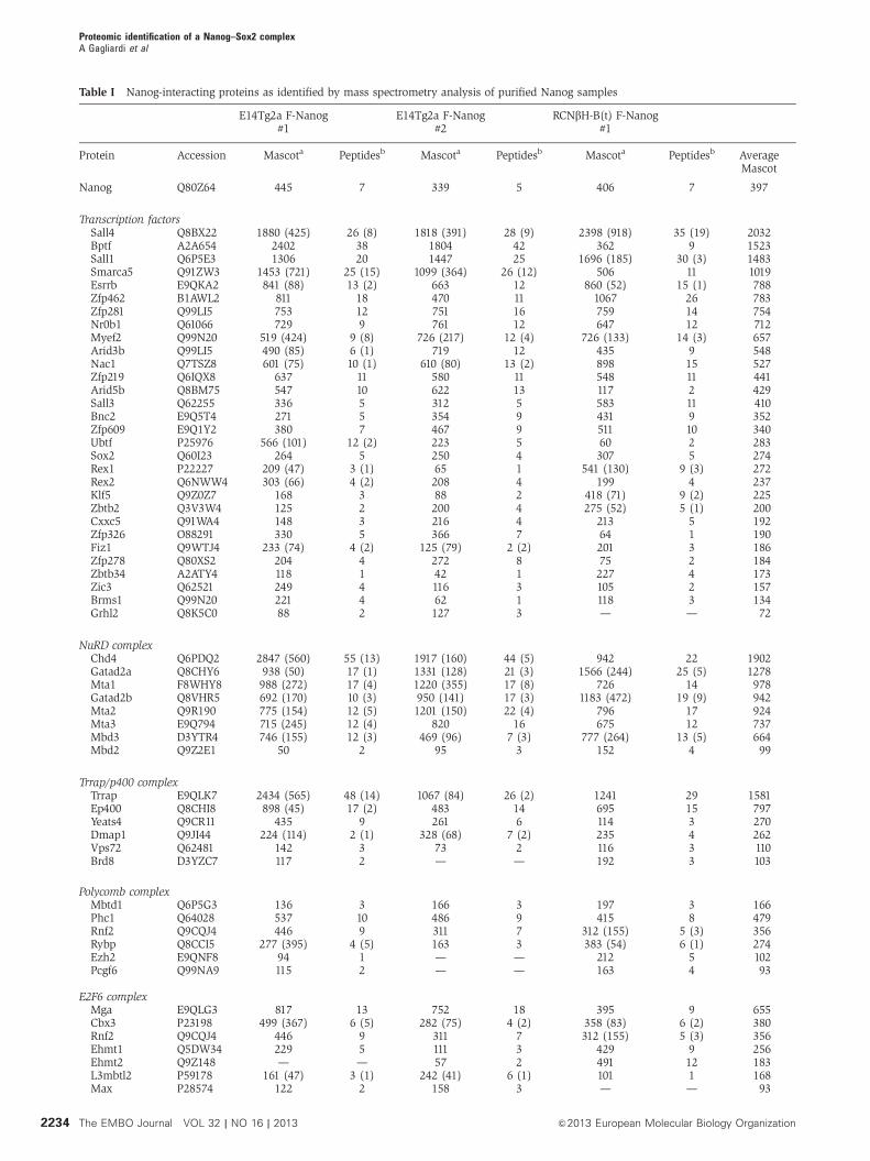
Table I Nanog-interacting proteins as identified by mass spectrometry analysis of purified Nanog samples
E14Tg2a F-Nanog#1
E14Tg2a F-Nanog#2
RCNbH-B(t) F-Nanog#1
Protein Accession Mascota Peptidesb Mascota Peptidesb Mascota Peptidesb AverageMascot
Nanog Q80Z64 445 7 339 5 406 7 397
Transcription factorsSall4 Q8BX22 1880 (425) 26 (8) 1818 (391) 28 (9) 2398 (918) 35 (19) 2032Bptf A2A654 2402 38 1804 42 362 9 1523Sall1 Q6P5E3 1306 20 1447 25 1696 (185) 30 (3) 1483Smarca5 Q91ZW3 1453 (721) 25 (15) 1099 (364) 26 (12) 506 11 1019Esrrb E9QKA2 841 (88) 13 (2) 663 12 860 (52) 15 (1) 788Zfp462 B1AWL2 811 18 470 11 1067 26 783Zfp281 Q99LI5 753 12 751 16 759 14 754Nr0b1 Q61066 729 9 761 12 647 12 712Myef2 Q99N20 519 (424) 9 (8) 726 (217) 12 (4) 726 (133) 14 (3) 657Arid3b Q99LI5 490 (85) 6 (1) 719 12 435 9 548Nac1 Q7TSZ8 601 (75) 10 (1) 610 (80) 13 (2) 898 15 527Zfp219 Q6IQX8 637 11 580 11 548 11 441Arid5b Q8BM75 547 10 622 13 117 2 429Sall3 Q62255 336 5 312 5 583 11 410Bnc2 E9Q5T4 271 5 354 9 431 9 352Zfp609 E9Q1Y2 380 7 467 9 511 10 340Ubtf P25976 566 (101) 12 (2) 223 5 60 2 283Sox2 Q60I23 264 5 250 4 307 5 274Rex1 P22227 209 (47) 3 (1) 65 1 541 (130) 9 (3) 272Rex2 Q6NWW4 303 (66) 4 (2) 208 4 199 4 237Klf5 Q9Z0Z7 168 3 88 2 418 (71) 9 (2) 225Zbtb2 Q3V3W4 125 2 200 4 275 (52) 5 (1) 200Cxxc5 Q91WA4 148 3 216 4 213 5 192Zfp326 O88291 330 5 366 7 64 1 190Fiz1 Q9WTJ4 233 (74) 4 (2) 125 (79) 2 (2) 201 3 186Zfp278 Q80XS2 204 4 272 8 75 2 184Zbtb34 A2ATY4 118 1 42 1 227 4 173Zic3 Q62521 249 4 116 3 105 2 157Brms1 Q99N20 221 4 62 1 118 3 134Grhl2 Q8K5C0 88 2 127 3 — — 72
NuRD complexChd4 Q6PDQ2 2847 (560) 55 (13) 1917 (160) 44 (5) 942 22 1902Gatad2a Q8CHY6 938 (50) 17 (1) 1331 (128) 21 (3) 1566 (244) 25 (5) 1278Mta1 F8WHY8 988 (272) 17 (4) 1220 (355) 17 (8) 726 14 978Gatad2b Q8VHR5 692 (170) 10 (3) 950 (141) 17 (3) 1183 (472) 19 (9) 942Mta2 Q9R190 775 (154) 12 (5) 1201 (150) 22 (4) 796 17 924Mta3 E9Q794 715 (245) 12 (4) 820 16 675 12 737Mbd3 D3YTR4 746 (155) 12 (3) 469 (96) 7 (3) 777 (264) 13 (5) 664Mbd2 Q9Z2E1 50 2 95 3 152 4 99
Trrap/p400 complexTrrap E9QLK7 2434 (565) 48 (14) 1067 (84) 26 (2) 1241 29 1581Ep400 Q8CHI8 898 (45) 17 (2) 483 14 695 15 797Yeats4 Q9CR11 435 9 261 6 114 3 270Dmap1 Q9JI44 224 (114) 2 (1) 328 (68) 7 (2) 235 4 262Vps72 Q62481 142 3 73 2 116 3 110Brd8 D3YZC7 117 2 — — 192 3 103
Polycomb complexMbtd1 Q6P5G3 136 3 166 3 197 3 166Phc1 Q64028 537 10 486 9 415 8 479Rnf2 Q9CQJ4 446 9 311 7 312 (155) 5 (3) 356Rybp Q8CCI5 277 (395) 4 (5) 163 3 383 (54) 6 (1) 274Ezh2 E9QNF8 94 1 — — 212 5 102Pcgf6 Q99NA9 115 2 — — 163 4 93
E2F6 complexMga E9QLG3 817 13 752 18 395 9 655Cbx3 P23198 499 (367) 6 (5) 282 (75) 4 (2) 358 (83) 6 (2) 380Rnf2 Q9CQJ4 446 9 311 7 312 (155) 5 (3) 356Ehmt1 Q5DW34 229 5 111 3 429 9 256Ehmt2 Q9Z148 — — 57 2 491 12 183L3mbtl2 P59178 161 (47) 3 (1) 242 (41) 6 (1) 101 1 168Max P28574 122 2 158 3 — — 93
Proteomic identification of a Nanog–Sox2 complexA Gagliardi et al
2234 The EMBO Journal VOL 32 | NO 16 | 2013 &2013 European Molecular Biology Organization

Table I Continued
E14Tg2a F-Nanog#1
E14Tg2a F-Nanog#2
RCNbH-B(t) F-Nanog#1
Sin3a complexSin3a Q60520 1123 (154) 19 (3) 765 (143) 16 (5) 1221 (223) 22 (6) 1036Hcfc1 Q61191 546 (291) 9 (7) 419 (114) 12 (3) 926 20 630Ncor1 E9Q2B2 237 4 358 7 254 5 283Sap130 Q8BIH0 144 3 69 2 220 4 144Mbd2 Q9Z2E1 50 2 95 3 152 4 99
N-CoR complexTbl1xr1 Q8BHJ5 962 (289) 13 (5) 837 (236) 13 (4) 292 5 697Tbl1x Q9QXE7 677 10 663 12 479 8 606Ncor1 E9Q2B2 237 4 358 7 254 5 283Hdac3 Q3UM33 226 6 250 5 69 2 182
LSD1 complexLsd1 Q6ZQ88 589 (237) 12 (4) 647 (106) 12 (3) 924 (58) 17 (2) 720Zmym2 Q9CU65 155 2 281 6 1005 21 480Rcor1 E9QK09 68 1 — — 91 2 53
MLL complexHcfc1 Q61191 546 (291) 9 (7) 419 (114) 12 (3) 926 20 630Bap18 Q9DCT6 663 (47) 14 (1) 933 19 222 4 606Mll2 O08550 668 16 195 7 133 3 332Mll3 Q8BRH4 670 14 259 8 — — 310Ash2l E9PU93 174 4 340 7 273 6 204Men1 O88559 68 2 125 3 245 4 146Myst1 Q9D1P2 131 3 100 3 205 4 145Rbbp5 Q8BX09 114 3 219 6 76 2 63
Chromatin remodelling/transcriptional regulationChd7 A2AJK6 1408 (154) 24 (4) 944 17 252 7 868Cnot1 Q6ZQ08 1471 (355) 33 (9) 710 (221) 18 (6) 245 (62) 6 (2) 809Tet 1 E9Q9Y4 1370 24 — — 390 9 587Arid1a E9QAQ7 83 3 182 5 1315 (179) 27 (4) 527Rcor2 Q8C796 240 (99) 4 (2) 423 (48) 7 (1) 694 12 452Morf4l1 P60762 458 (47) 8 (1) 446 10 367 7 424Wiz F6ZBR8 247 4 185 4 769 17 400Bcor Q8CGN4 322 7 454 11 279 6 352Hira Q61666 451 7 394 11 102 3 316Hist1h1c P15864 274 4 139 3 112 2 175
Transcriptional machineryInts1 Q6P4S8 721 (172) 14 (5) 608 (213) 13 (6) 248 6 526Wdr82 Q8BFQ4 592 (431) 9 (8) 320 (69) 8 (2) 377 (78) 8 (1) 430Supt6h Q62383 868 18 252 7 101 3 407Polr2a P08775 240 (93) 2 148 4 481 11 290Taf9 Q8VI33 317 7 158 4 261 7 245Polr2b Q8CFI7 281 5 122 3 — — 134
PhosphorylationAkap8 Q9DBR0 827 (190) 14 (3) 760 (162) 13 (3) 859 (75) 15 (1) 815Csnk2a Q60737 408 (118) 8 (3) 323 (45) 7 (2) 431 8 387Csnk1a1 E9Q2U6 247 5 202 5 151 4 200Ppp2r1a Q76MZ3 178 (67) 4 (2) 261 6 57 2 165
UbiquitinationUchl5 Q9WUP7 349 (101) 6 (2) 344 (96) 8 (2) 238 (88) 5 (2) 310Stub1 Q9WUD1 457 10 350 7 83 2 297Ranbp2 Q9ERU9 283 (90) 8 (2) 164 (44) 4 (1) 932 (42) 20 (3) 220RanGAP1 E9Q757 118 (47) 3 (1) 289 6 370 (85) 7 (2) 164Psmd11 Q8BG32 217 (70) 6 (2) 137 (43) 4 (2) — — 118
RNA processingHnrnph2 P70333 513 8 625 (174) 11 (5) 545 10 2437Dhx9 O70133 972 17 502 12 1126 (530) 22 (13) 867Ilf3 Q45VK5 1388 (689) 22 (14) 1022 (230) 21 (7) 670 13 803Raly Q3U3F6 480 (488) 9 (10) 392 (119) 7 (3) 271 7 381Zcchc8 Q9CYA6 425 5 377 6 274 5 359Ddx1 Q91VR5 167 (354) 4 (8) 333 (63) 7 (2) 259 6 253Wdr33 Q8K4P0 316 (42) 6 (1) 221 (77) 6 (3) 219 6 252Pum1 Q80U78 274 (120) 6 (3) 290 (66) 7 (2) 177 4 247Rbm9 Q8BP71 214 4 305 4 210 4 243
Proteomic identification of a Nanog–Sox2 complexA Gagliardi et al
2235&2013 European Molecular Biology Organization The EMBO Journal VOL 32 | NO 16 | 2013

region were generated (Figure 5A). Co-immunoprecipitation
analyses show that while a Sox2 mutant truncated after
residue 233 retained the ability to interact with Nanog,
Sox2 mutants with deletion of residues between 205 and
233, or truncated after residue 212 were unable to interact
with Nanog (Figure 5B). These analyses identify a critical
Nanog-interacting region in Sox2 between residues 212 and
233. This sequence is highly enriched for hydroxyamino acids
(12/21 residues) and, like the WR of Nanog, is devoid of
acidic and basic side chains. Moreover, careful examination
of this 21 amino-acid region highlighted three repeats of the
sequence S X T/S Y that may be responsible for mediating
the interaction with Nanog. To determine the potential im-
portance of these motifs for the Nanog–Sox2 interaction,
additional truncations were made after residues 218 and
226, which truncate Sox2 after repeat 1 or 2, respectively.
This indicates that repeat 1 is sufficient for interaction with
Nanog but that together repeats 1 and 2 interact with Nanog
with an efficiency approaching that of wild-type Sox2
(Figure 5B). To examine the sequences required on Sox2 in
more detail, a series of point mutations were generated
within the repeats (Figure 6). Individual or combinatorial
contributions of each of the three repeats to Nanog binding
were initially examined (Figure 6A and B). Mutation of
individual repeats suggests an order of importance for
Nanog interaction of repeat 14repeat 34repeat 2
(Figure 6A). This is supported by analyses of the combina-
torial mutants where mutation of repeats 1þ 3 almost
entirely eliminates the ability of Sox2 to interact with
Nanog (Figure 6B). Mutations of the amino acids at positions
1, 3 or 4 in all the three repeats were next analysed.
Combined mutations at positions 1 and 3 had negligible
effects (Figure 6C), implying that residues at these positions
are not required for Nanog interaction. In contrast, the
combined mutation of the tyrosines at position 4 indicates
that these residues play a key role in the interaction with
Nanog (Figure 6C). Together, these experiments suggest that
the tyrosines are the residues directly interacting with the
Nanog WR, with the tyrosines in repeats 1 and 3 being more
important in this regard than the tyrosine in repeat 2.
The Sox2 tyrosine residues could interact with Nanog
partially via the hydroxyl, the phenyl ring or both. Since
Table I Continued
E14Tg2a F-Nanog#1
E14Tg2a F-Nanog#2
RCNbH-B(t) F-Nanog#1
Mtr3 Q8BTW3 380 (74) 5 (1) 120 (108) 2 (2) 212 4 237Mov10 P23249 251 (45) 6 (1) 185 (58) 3 (2) — — 145Rbm4b Q8VE92 87 2 61 2 279 6 142Ints4 Q8CIM8 112 3 122 3 192 4 142Pum2 Q80U58 122 2 187 4 — — 103
Cell cycle/DNA replicationSgol2 Q7TSY8 747 (91) 12 (3) 387 (52) 8 (2) 735 13 623Mad1l1 Q9WTX8 493 (126) 9 (3) 520 11 732 (984) 14 (20) 582Rfc4 Q99J62 517 (171) 13 (5) 541 (265) 10 (7) 324 (78) 6 (2) 461Gtse1 Q8R080 635 (178) 9 (3) 587 11 64 1 429Tpx2 A2APB8 374 (40) 8 (1) 142 3 383 9 300Orc5l Q9WUV0 196 (45) 4 (1) 109 (140) 3 (4) 260 5 188Smc3 Q9CW03 124 3 — — 338 9 154Mat89Bb Q8QZV7 125 (30) 3 (1) 144 5 184 4 151Pogz D3YUW8 91 1 40 1 268 6 180
DNA repairMsh6 P54276 613 (112) 15 (3) 623 (139) 14 (4) 208 6 481Brca2 P97929 576 14 309 9 296 9 394Rad51 D6RCK1 328 5 208 4 232 5 256Brca1 A2A4Q4 74 2 157 4 466 12 239Emsy Q8BMB0 306 (52) 6 (2) 149 5 69 2 152
OtherSec16 E9QAT4 889 (44) 15 (1) 777 18 353 8 673Cad O54788 883 18 968 24 108 1 653Qser1 A2BIE1 669 (192) 11 (4) 433 9 826 16 643Zfr O88532 241 (80) 4 (1) 193 (67) 4 (1) 1155 21 530Dnaja1 P63037 515 (144) 9 (2) 345 (107) 8 (2) 514 8 4582810474O19RIK D3Z687 633 (47) 14 (1) 933 19 — — 380C130039O16Rik E9Q2I4 474 (41) 7 (1) 314 7 258 5 349Skt A2AQ25 247 5 665 15 60 2 324Wdr68 P61963 385 5 381 8 168 4 311Dnttip1 Q99LB0 442 (37) 8 (1) 167 3 177 3 262Sec13 Q9D1M0 498 (58) 7 (1) 207 5 76 2 260Kpna2 P52293 292 (90) 5 (1) 260 5 226 3 259Dpf2 Q61103 244 3 145 3 375 (148) 8 (4) 255
a
Mascot score for the specified protein in the Nanog sample, purified by FLAG affinity. Mascot score for the specified protein in thecorresponding control purification, if present, is in parentheses.b
Number of identified unique, nonredundant peptides for the specified protein in the Nanog sample. Number of identified unique peptides inthe control purification is in parentheses.
Proteomic identification of a Nanog–Sox2 complexA Gagliardi et al
2236 The EMBO Journal VOL 32 | NO 16 | 2013 &2013 European Molecular Biology Organization

the tryptophans in WR are critical for the Nanog/Sox2
interaction this raises the hypothesis that hydrophobic stacking
of the aromatic rings in the Sox2 tyrosines and the Nanog WR
tryptophans mediate the interaction. If these were the case,
then the tyrosines hydroxyl groups should be unimportant for
the interaction between Nanog and Sox2. To test this hypo-
thesis, the tyrosines were mutated to phenylalanine. The direct
comparison of the interaction between (HA)3Nanog and
(Flag)3Sox2:YYY4A or (Flag)3Sox2:YYY4F by co-immuno-
precipitations clearly shows that substitution of the tyrosine
residues with phenylalanines rescues the Nanog interaction,
indicating that it is the benzene ring of these amino-acid
residues that is required for the interaction to occur (Figure 6D).
Identification of Nanog/Sox2 binding motif in vitro
To investigate possible DNA sequences bound by the Nanog/
Sox2 complex, (His)6-tagged Nanog and unmodified Sox2
were co-expressed in E. coli for use in Systematic Evolution
of Ligands by Exponential Enrichment (SELEX). As controls,
MBP-Nanog and (His)6-Sox2 were expressed individually.
Purification from bacterial lysate containing co-expressed
proteins on a nickel column followed by elution with imida-
zole yielded two proteins of the expected size for Nanog and
Sox2. These were recognized by a-Nanog and a-Sox2 anti-
bodies (Figure 7A), with N-terminal sequencing establishing
the identities of the two bands as Nanog and Sox2. The
Nanog–Sox2 interaction is robust, since the proteins co-purify
through subsequent ion exchange (Figure 7B). The Nanog–
Sox2 complex bound to the Ni-agarose, MBP-Nanog bound to
amylose resin and (His)6-Sox2 bound to Ni-agarose were
used for SELEX, the bound oligonucleotides cloned and the
sequences determined (Figure 7C) used to derive the motifs
shown (Figure 7D). The motif obtained from Nanog alone has
a TAAT core sequence followed by CG, consistent with the
motif obtained previously by SELEX (Mitsui et al, 2003) and
the nucleotide preferences of the isolated Nanog HD in
EMSAs (Jauch et al, 2008). Sox2 also gives a motif highly
similar to that determined by SELEX (CA/TTTGA/T) (Harley
et al, 1994; Maruyama et al, 2005). The motif obtained from
the Nanog/Sox2 complex is bipartite with bases 10–15 similar
to the motif obtained by us and others for Sox2 alone (Harley
et al, 1994; Maruyama et al, 2005) and bases 5–7 showing
similarity to the central core of the Nanog motif identified by
SELEX (TAAT) in this work and by others (Mitsui et al, 2003).
However, the published Nanog motif has a high degree of
confidence over a four base sequence (TAAT) while the
Nanog–Sox2 binding sequence shows high certainty for
only three bases (TAA) with the preference for the 30-flanking
CG no longer apparent. This difference may reflect an altera-
tion in the binding specificity of Nanog when in complex with
Sox2. Interestingly, the SELEX motif shows high similarity to
a Nanog/Sox2 motif identified by de novo methods from
Figure 2 The Nanog interaction network. The Nanog interactome (pale blue) as part of a larger interactome encompassing the interactions ofOct4, Esrrb, Sall4, Nr0b1 and Tcfcp2l1 (van den Berg et al, 2010).
Proteomic identification of a Nanog–Sox2 complexA Gagliardi et al
2237&2013 European Molecular Biology Organization The EMBO Journal VOL 32 | NO 16 | 2013

ChIP-Seq data (Hutchins et al, 2013), which notably also
contains a 2-bp gap between the major binding nucleotide
groups (Figure 7D). Therefore, a combined motif was gene-
rated and used to search available ChIP-Seq data sets. Analysis
of three independent ChIP-Seq data sets (Chen et al, 2008b;
Marson et al, 2008; Whyte et al, 2013) identified 3257 Nanog/
Sox2 overlapping peaks, which are common to the three data
sets (out of a total of 16 454 from all Nanog/Sox2 overlapping
peaks in the three data sets). Of these 3257 high confidence
peaks, 29.1% (948 peaks) contain the motif. The motif
occurs in a significantly smaller fraction of the Nanog only
or Sox2 only peaks (4898 peaks out of a total of 31 271 peaks
(15.7%; hypergeometric P-value o1�10�10). Examples of
occurrences of the motif relative to the nearest gene are
shown (Figure 7E; Supplementary Table II).
The Nanog–Sox2 interaction is critical for Sox2 function
To investigate the functional significance of the interaction
between Nanog and Sox2, we took advantage of ES cells
carrying a conditional Sox2 knock-out allele (Sox2CKO). In
this cell line, one of the Sox2 alleles is flanked by loxP sites
(Favaro et al, 2009), while the other Sox2 allele has been
replaced with a b-geo cassette (Zappone et al, 2000;
Avilion et al, 2003). These cells also have a constitutively
expressed CreERT2-IRES-Puro transgene integrated randomly
in the genome. Upon addition of tamoxifen, CreERT2 is
Figure 3 Mutational analysis of the Sox2-interaction domain in Nanog. (A) Co-immunoprecipitation of endogenous Sox2 and Nanog fromE14Tg2a nuclear extract. Immunoprecipitation was performed with Sox2 antibody and immunoblot probed with anti-Nanog or anti-Sox2antibodies. (B) Co-immunoprecipitation of endogenous Nanog and Sox2 from E14Tg2a nuclear extract. Immunoprecipitation was performedwith Nanog antibody and immunoblot probed with anti-Sox2 or anti-Nanog antibodies. (C) Left, schematic representation of the (HA)3Nanogconstructs. Right, co-immunoprecipitations of Nanog variants with Sox2. (FLAG)3Sox2 and (HA)3Nanog deletion mutants were transfected intoE14/T cells. Immunoprecipitations were performed with an HA antibody as indicated and immunoblots probed with anti-FLAG or anti-HAantibodies. I is 1% of input. (D) Left, schematic representation of the (HA)3Nanog constructs. Right, co-immunoprecipitations of Nanogvariants with Sox2. (FLAG)3Sox2 and (HA)3Nanog deletion mutants were transfected into E14/T cells. Immunoprecipitations were performedwith an HA antibody as indicated and immunoblots probed with anti-FLAG or anti-HA antibodies. I is 1% of input. (E) Left, Sox2, co-expressedin E. coli with either a Maltose Binding Protein-tryptophan repeat (MBP-WR) fusion protein or a Maltose Binding Protein-tryptophan repeat inwhich all the tryptophans were mutated to alanine (MBP-WRW10-A). Right, MBP fusion proteins and associated proteins were purified onamylose resin, subjected to SDS–PAGE and immunoblots probed with Sox2 or MBP antibodies. Source data for this figure is available on theonline supplementary information page.
Proteomic identification of a Nanog–Sox2 complexA Gagliardi et al
2238 The EMBO Journal VOL 32 | NO 16 | 2013 &2013 European Molecular Biology Organization

translocated to the nucleus and excises the Sox2 gene
between the loxP sites (Figure 8A). As ES cells from which
Sox2 activity has been removed are unable to self-renew and
differentiate into trophectoderm-like cells (Masui et al, 2007),
this cell line was used to test whether Sox2 mutant molecules
impaired in Nanog binding could rescue the Sox2 null
phenotype (Figure 8B). Sox2CKO cells expressing a GFP
control plasmid completely differentiate upon Tamoxifen
treatment (Figure 8B). As expected, cells transfected with
an unmutated (Flag)3Sox2 cDNA rescued this differentiation
phenotype. In contrast, cells expressing (Flag)3Sox2:YYY4A
transgene showed a decrease in self-renewal activity with
50% fewer undifferentiated colonies compared to wild-type
Sox2 (Figure 8C). In accordance with the interaction data,
expression of (Flag)3Sox2:YYY4F fully rescued the differen-
tiation phenotype (Figure 8C). To examine the possibility that
the reduced colony formation by the Sox2:YYY4A cells was
due to a reduced expression level, an immunoblot for Sox2
was performed. However, the amount of Sox2 expressed is
comparable between Sox2:YYY4A and other lines and does
not differ from the endogenous Sox2 level expressed by the
parental line (Figure 8D). These data suggest that the inter-
action with Nanog is a key component in the function of Sox2
in ES cell self-renewal.
To further investigate the effect of disrupting the Nanog–
Sox2 interaction, the expression of genes present in the ChIP-
Seq data sets was examined in cell lines expressing wild-type
or mutant Sox2 (YYY4A). Of 13 genes analysed, 5 showed
consistent differences by qRT-PCR when the Nanog/Sox2
complex was disrupted (Figure 8E). The genes that show
altered expression include transcription factors reported to be
important for ES cell identity (Rex1 and Klf5 (Shi et al, 2006;
Parisi et al, 2010), the gene encoding the chromatin re-
modelling protein Myst4 (Ura et al, 2011) as well as the
cell-surface markers Ncam and Itga9 (Rugg-Gunn et al,
2012). In addition, Oct4, which does not contain the
Nanog/Sox2 motif, did not change expression level in
absence of a Nanog/Sox2 functional complex. It is therefore
likely that the effect of disrupting the Nanog/Sox2 complex
on self-renewal is a consequence of the misregulation of the
genes controlled by the two proteins in complex.
Discussion
By taking advantage of improved methodology (van den Berg
et al, 2010) the Nanog interactome has been expanded to over
130 proteins which can be subdivided into a number of
different categories (Table I; Supplementary Table I). Many
Figure 4 The serine-rich domain of Sox2 interacts with Nanog. (A) Top, schematic representation of the (FLAG)3Sox2 constructs. Bottom,(HA)3Nanog and the indicated (FLAG)3Sox2 deletion mutants were transfected into E14/T cells and immunoprecipitations were performedwith an HA antibody as indicated and immunoblots probed with anti-FLAG or anti-HA antibodies. I is 1% of input. (B) Left, schematicrepresentation of the (FLAG)3Sox2 and (HA)3Nanog constructs used to assess the DNA independence of the Nanog–Sox2 interaction. Right,E14/T cells were transfected with the indicated vectors. Immunoprecipitations were performed with an HA antibody as indicated andimmunoblots probed with anti-FLAG or anti-HA antibodies. I is 1% of input. Source data for this figure is available on the onlinesupplementary information page.
Proteomic identification of a Nanog–Sox2 complexA Gagliardi et al
2239&2013 European Molecular Biology Organization The EMBO Journal VOL 32 | NO 16 | 2013

of the proteins identified in the interactome are components of
large multi subunit complexes involved in chromatin modifica-
tion, for several of which, all the known subunits are detected.
Most of these are considered to be transcriptional repressors
(NuRD, Polycomb Group protein (PcG), the atypical Polycomb
complex E2F6, Sin3a and N-CoR) that bind to genomic sites
adjacent to differentiation-specific genes to mediate repression
(Jepsen and Rosenfeld, 2002; McDonel et al, 2009; Surface
et al, 2010; Qin et al, 2012). Emerging evidence suggests that
NuRD and PcG complexes are also found at sites that are
actively transcribed (Brookes et al, 2012; Reynolds et al, 2012).
How the NuRD complex is directed to target genes is not fully
understood but Nanog and/or other NuRD-interacting
transcription factors may target the complex to the relevant
sites in the genome. In this respect, it is interesting that
inducing Nanog protein results in enhanced binding of both
Nanog and NuRD to the Nanog enhancer (Fidalgo et al, 2012).
Another proposed role for the chromatin modification
complexes is to maintain repressed genes in a state that
allows a rapid response to external cues. Evidence for this
comes from the co-localization of enzymatically active PRC
complexes and the paused form of RNA PolII at a large
number of developmentally important genes (Brookes et al,
2012). This could allow alterations in the signalling
environment to promptly increase the level of gene
expression. The interaction of Nanog with both PRC and
RNA PolII may reflect this poised state of some genes. The
association with the chromatin modification machinery is
common to transcription factors involved in maintenance of
ES cell pluripotency (Wang et al, 2006; Liang et al, 2008;
Pardo et al, 2010; van den Berg et al, 2010; Ding et al, 2012).
However, the range of complexes binding to individual
factors differs with SWI/SNF not directly connecting to
Nanog (this study; Wang et al, 2006) but interacting with
other transcription factors (van den Berg et al, 2010). Recent
data showing that Esrrb can substitute for Nanog function in
ES cells (Festuccia et al, 2012) could in part be explained by
the fact that Esrrb and Nanog bind to a number of the same
chromatin modification complexes.
The Nanog interactome includes a number of proteins that
have not previously been identified in an ES cell transcription
factor interactome. In addition to TET-1, which has also been
shown to interact with Nanog (Costa et al, 2013), these
include the RNA processing proteins Ilf3, Rbm9, Pum1/2
and the transcription factors Zfp326, Arid5b, Zfp609.
Examining the function of these molecules and the signi-
ficance of their interaction with Nanog will provide further
detail on how the extensive protein interaction network
functions to control pluripotency.
In this study, we have focussed on the interaction between
Nanog and Sox2 because of the central role of these proteins
in the pluripotency gene regulatory network. Sox2 has been
shown to interact with another key pluripotency factor, Oct4
by interaction of side chains within the DNA binding domains
(Ambrosetti et al, 1997, 2000). In the case of Nanog and Sox2,
interaction occurs through sequences outwith the DNA
binding domains. Nevertheless, the sequence of the SELEX
motif suggests that this interaction results in a specific spatial
relationship of DNA binding domains of both proteins on
DNA. The sequence of Sox2 that mediates interaction with
Nanog is a triple repeat of the sequence S X S/T Y.
Experiments analysing Sox2 mutants for their ability to
rescue ES cells from differentiation induced by Sox2
deletion demonstrate the importance of the interaction of
Nanog with Sox2. Mutation of the tyrosines in the S X S/T Y
motifs to alanines reduces the formation of undifferentiated
Figure 5 A subregion of the Sox2 serine-rich domain interacts with Nanog. (A) Schematic representation of the (FLAG)3Sox2 constructs usedfor mapping the minimal Sox2 region involved in the interaction with Nanog. (B) E14/T cells were transfected with (HA)3Nanog and theindicated (FLAG)3Sox2 mutants. Immunoprecipitations were performed with an HA antibody as indicated and immunoblots probed with ananti-FLAG or anti-HA antibodies. I is 1% of input. Source data for this figure is available on the online supplementary information page.
Proteomic identification of a Nanog–Sox2 complexA Gagliardi et al
2240 The EMBO Journal VOL 32 | NO 16 | 2013 &2013 European Molecular Biology Organization

ES cell colonies to 50% of the level achieved using a non-
mutant Sox2 cDNA in the absence of any difference in protein
levels expressed by the transgene. Therefore, the 50% drop in
undifferentiated colonies observed in the presence of
Sox2:YYY4A is a result of the misregulation of Nanog/Sox2
gene targets. The use of the SELEX motif identified as a
Nanog/Sox2 target sequence together with a previously
published de novo target sequence (Hutchins et al, 2013)
allowed potential target genes of the Nanog/Sox2 complex
to be identified. A number of these genes show altered
expression upon abrogation of the Nanog/Sox2 interaction
(e.g., Ncam, Itga9, Klf5 and Myst4). However, not all the
genes tested are sensitive to loss of the interaction between
Nanog and Sox2 (Supplementary Table II). This could suggest
that in such cases the hydrophobic interaction of Nanog and
Sox2 proteins is not required for chromatin binding, or that
only in some cases is the associated gene sensitive to disrup-
tion of the interaction. The latter is reminiscent of our finding
that only a subset of loci that bind Nanog respond to the
presence of Nanog by modulating expression of a nearby
gene (Festuccia et al, 2012).
In ES cells, composite Oct/Sox binding sites have been
proposed to be redundantly regulated by Sox4, Sox11 and
Sox15 (Masui et al, 2007). However, this redundancy does not
extend to blockade of differentiation caused by Sox2 deletion.
Consistent with this, Sox4, Sox11 and Sox15 are not present
in the Nanog interactome and none of these Sox proteins
contains a sequence that matches the S X S/T Y motif.
The three copies of the S X S/T Y motif in Sox2 occur
within a 15-residue sequence in which 9 residues are hydro-
xyamino acids. Despite this preponderance of hydroxyamino
acids, it is the aromatic rings of the tyrosine residues that are
Figure 6 Identification of amino-acid residues within Sox2(213–233) interacting with Nanog. (A) Top, schematic representation of hydroxyaminoacid mutations in repeats 1, 2 or 3 in Sox2. Bottom, E14/T cells were transfected with (HA)3Nanog and the indicated (FLAG)3Sox2 mutants.Immunoblots of the HA immunoprecipitates were analysed by immunoblotting with an anti-FLAG or an anti-HA antibody. I is 1% of input.(B) Top, schematic representation of the combinatorial mutations of the hydroxyamino acids in repeats 1, 2 and 3 of Sox2. Bottom, E14/Tcellswere transfected with (HA)3Nanog and the indicated (FLAG)3Sox2 mutants. Immunoblots of the HA immunoprecipitates were analysed byimmunoblotting with an anti-FLAG or an anti-HA antibody. I is 1% of input. (C) Top, schematic representation of the mutations of thehydroxyamino acids in positions 1, 3 or 4 of repeats 1, 2 and 3 of Sox2. Bottom, E14/T cells were transfected with (HA)3Nanog and theindicated (FLAG)3Sox2 mutants. Immunoblots of the HA immunoprecipitates were analysed by immunoblotting with an anti-FLAG or an anti-HA antibody. I is 1% of input. (D) Top, schematic representation of the mutations of the hydroxyamino acids in position 4 of repeats 1, 2 and 3of Sox2. Bottom, E14/T cells were transfected with (HA)3Nanog and the indicated (FLAG)3Sox2 mutants. Immunoblots of the HAimmunoprecipitates were analysed by immunoblotting with an anti-FLAG or an anti-HA antibody. I is 1% of input. Source data for thisfigure is available on the online supplementary information page.
Proteomic identification of a Nanog–Sox2 complexA Gagliardi et al
2241&2013 European Molecular Biology Organization The EMBO Journal VOL 32 | NO 16 | 2013

critical mediators of the interaction with Nanog. This con-
clusion is derived from the fact that alanine substitution
of all three serines at position 1 of the repeats or all three
serines/threonines at position 3 of the repeats allowed
continued efficient binding to Nanog, whereas alanine
substitution of all three tyrosines decreased the Nanog inter-
action severely. Moreover, the fact that the Nanog interaction
could be rescued when the tyrosines were substituted by
phenylalanines indicates that the tyrosine hydroxyl groups
are not required for the interaction and is highly suggestive
that the two proteins interact by stacking of the aro-
matic rings. This is consistent with the fact that tyrosine
and tryptophan residues cluster at protein–protein inter-
action ‘hot spots’ (Bogan and Thorn, 1998; DeLano,
2002). Functionally relevant stacking of tryptophan and
tyrosine residues has also been demonstrated in the
Figure 7 Identification of a Nanog/Sox2 motif by SELEX. (A) Left, a Coomassie-stained SDS–PAGE gel of the imidazole eluate from theNi-agarose purification of lysate from E. coli expressing His6-Nanog and Sox2, showing the major two bands identified as containing Nanog(upper) and Sox2 (lower) by immunoblot analysis, as indicated on the right-hand panel. (B) Coomassie-stained SDS–PAGE gel of fractions fromion-exchange purification of Nanog/Sox2. Lanes 1–3 are protein from flow-through fractions and lanes 4–7 are from the eluted peak.(C) Sequence of 22 oligonucleotides that contribute to the motif generated by the de novo discovery program MEME. (D) Top panel, SELEXmotifs generated for Nanog and Sox2 expressed individually from a total of 19 (Nanog) and 15 (Sox2) sequences submitted to MEME; middlepanel, SELEX motif generated for Nanog/Sox2 complex from 38 sequences submitted to MEME; bottom panel, representation of the de novoNanog/Sox2 motif (Hutchins et al, 2013) and the combined motif from SELEX sequence for Nanog/Sox2 and de novo Nanog/Sox2 motif. Motifsin the bottom panels were generated with Web Logo 3.3. (E) Nanog and Sox2 ChIP-seq peaks located near the transcriptional start sites ofZfp42, Klf5, Ncam1 and Myst4. The peaks that contain the Nanog/Sox2 motif are highlighted in the shaded box; Nanog (N) and Sox2 (S) peaksin data sets from Chen (C), Marson (M) and Whyte (W) data sets. Source data for this figure is available on the online supplementaryinformation page.
Proteomic identification of a Nanog–Sox2 complexA Gagliardi et al
2242 The EMBO Journal VOL 32 | NO 16 | 2013 &2013 European Molecular Biology Organization

formation of an aromatic gate in apo flavodoxin (Genzor
et al, 1996), in the regulation of galactose oxidase activity
(Rogers et al, 2007) and, of particular relevance to this study,
in the interlocking of tyrosine and tryptophan residues at the
interaction interface of human nuclear receptor pregnane X
receptor (PXR) that mediates protein homodimerization
(Noble et al, 2006).
The lack of a requirement for the hydroxyl groups on Sox2
for the Nanog interaction is underscored by experiments using
bacterially expressed recombinant proteins that demonstrate
that the Nanog–Sox2 interaction occurs in the absence of post-
translational modifications. However, this does not mean that
post-translational modifications might not affect the interac-
tion between Nanog and Sox2. The interaction between the
Figure 8 The aromatic tyrosine side chains of the Nanog-interaction domain are required to fully rescue differentiation of Sox2 null ES cells. (A)Scheme of the Sox2 conditional knock-out (Sox2CKO) cell line. (B) Top, alkaline phosphatase staining of the Sox2 null rescued cells. Sox2CKOwas transfected with the indicated vectors, selected in hygromycin and cultured in the presence or absence of tamoxifen. Cells were grown atclonal density for 7 days before staining. Bottom, two representative fields of colonies obtained for the indicated treatments. (C) Quantificationof the data from B. The number of alkaline phosphatase-positive colonies formed following transfection with the indicated vector was calculatedas a proportion of the number obtained by transfection with (Flag)3Sox2, which was set as 100%. Error bars, s.e.m. of at least four independentexperiments. ***Differences between FSox2 and FSox2:YYY4A by Mann–Whitney test (Po0.001). (D) Expression level of Flag-Sox2 variants inclonal assay. Comparison of Sox2 expression in E14Tg2a (WT) with Sox2 CKO populations expressing eGFP, Flag-Sox2, FlagSox2:YYY4A,FlagSox2:YYY4F, plated at clonal density for 7 days. (E) Expression levels of Nanog/Sox2 target genes. Expression levels of Rex1, Myst4, Ncam,Itga9, Klf5 and Oct4 in cells plated at clonal density, treated with tamoxifen for 12h and grown for 7 days before RNA extraction. The expressionlevel in Sox2CKO F-Sox2 þ tamoxifen was set to 1. Error bars, s.e.m. of three biological replicates. *Differences between FSox2 andFSox2:YYY4A by two-tailed t-test (Po0.05). Source data for this figure is available on the online supplementary information page.
Proteomic identification of a Nanog–Sox2 complexA Gagliardi et al
2243&2013 European Molecular Biology Organization The EMBO Journal VOL 32 | NO 16 | 2013

two proteins occurs through polypeptide stretches devoid of
strongly charged amino-acid side chains. Potential modifica-
tion of hydroxyl groups on the Sox2 interaction surface,
whether on the tyrosine or on the neighbouring serine and
threonine residues, would introduce charged moieties that
would be expected to interfere with the interaction between
the hydrophobic interacting residues. Moreover, recent work
indicates that hydroxyl groups on Sox2 can also be modified
by addition of N-acetylglucosamine, although the effect on
Sox2 function is unclear (Jang et al, 2012). The fact that
Nanog interacts with proteins that mediate post-translational
modifications such as phosphorylation and ubiquitination is
consistent with the observation that Nanog is phosphorylated
(Yates and Chambers, 2005; Moretto-Zita et al, 2010) and
ubiquitinated (Moretto-Zita et al, 2010). In addition, Nanog
partners could also be affected by such modifications because
of physical proximity to the relevant enzymes. The role for
these modifications and how they influence interactions
between transcription factors and/or transcription factor
function in ES cells is an important area for future
investigation.
The high number of Nanog-interacting proteins identified in
this study suggest that Nanog acts as a ‘hub’ protein (Han
et al, 2004; Mullin and Chambers, 2012). The ability of
individual partner proteins to interact with a hub protein
like Nanog depends on the affinity of the interaction and the
availability of the binding sites on both the hub protein and
the partner, as has been discussed previously (Han et al, 2004;
Mullin and Chambers, 2012). Since both competitive and non-
competitive interactions are simultaneously possible, it will be
important to determine which factors compete for the same
regions of Nanog. Of particular relevance will be whether
factors that bind through the WR interact through a precise
subregion of the WR or if there is variability in the exact
sequence bound by a specific partner. To date, only Sox2 and
Nac1 have been demonstrated to interact directly with the
WR. Loss of the tryptophans of the WR has also been
demonstrated to abrogate the interaction of Nanog with
Sall4, Nr0b1, Zfp198 and Zfp281 (Wang et al, 2008) but a
direct interaction has not yet been shown for these proteins. In
the situation where multiple factors bind the WR it is possible
that binding of one factor could increase the affinity of another
protein for interaction with an adjacent site in the same region
resulting in co-operative binding of two or more factors. A
clear potential example of this could be Nac-1, which has been
reported to bind the C-terminal WR subunit (Ma et al, 2009). It
is possible that both competitive and non-competitive binding
to distinct sites on Nanog occurs simultaneously, allowing
the assembly of large, functionally active complexes. An
additional level of complexity arises from the possibility that
the Nanog/Sox2 interaction may occur with either monomeric
or dimeric Nanog (Mullin et al, 2008; Wang et al, 2008).
Potential mechanisms that affect the Nanog dimerization equi-
librium, such as covalent modifications, could thereby play an
important part in regulating interactions and subsequent
downstream events.
Materials and methods
ES cell cultureMouse ESC lines were cultured on gelatin-coated dishes withoutfeeders in GMEM/b-mercaptoethanol/10% FCS/LIF (GMEMb/FCS/
LIF) as described (Smith, 1991). Nanog null RCNbH-B(t) cells havebeen described (Chambers et al, 2007): briefly, these cells have anIRES-HygromycinR-pA or an IRES-bgeo-pA replacement of Nanogsequences from intron I through to the 3’UTR. Sox2 conditionalknock-out cells were obtained by re-targeting ES cells heterozygousfor a Sox2flox allele (Favaro et al, 2009) with a Sox2-b-geo ‘knock-in’targeting vector (Zappone et al, 2000; Avilion et al, 2003). This wasfollowed by stable transfection of a pPyCAG-CreERT2IP construct(Figure 8A). Puromycin-resistant clones were screened for efficientdeletion of the Sox2flox allele following tamoxifen treatment to selectthe Sox2CKO clone used here.E14Tg2a (Flag)3-Nanog and RCNbH-B(t) (Flag)3-Nanog cells were
generated by electroporating E14Tg2a (Hooper et al, 1987) andRCNbH-B(t) cells with pPyCAG (Flag)3NanogIP (Mullin et al, 2008)linearized with ScaI. Electroporated cells were plated in GMEMb/FCS/LIF and after 30 h, 1 mg/ml puromycin (Sigma, P9620) wasadded. Medium was replaced every 2 days and after 12 days,puromycin-resistant colonies were picked and Nanog expressionlevels were determined by immunoblotting with a-Nanog antibody(Chambers et al, 2007). Sox2CKO cells expressing (Flag)3-Sox2,(Flag)3-Sox2:YYY4A, (Flag)3-Sox2: YYY4F or eGFP control weregenerated by electroporating Sox2CKO cells with pPyCAG(Flag)3-Sox2IH, pPyCAG(Flag)3-Sox2:YYY4AIH or pPyCAG(Flag)3-Sox2:YYY4FIH linearized with FspI. Electroporated cells were plated inGMEMb/FCS/LIF medium and after 30h, 100mg/ml hygromycin B(Roche, 10843555001) was added. Medium was replaced every 2days and after 12 days, hygromycin-resistant colonies were pooledto generate populations for rescue assays.Colony-forming assays were as described (Chambers et al, 2003).
Plasmids(HA)3-Nanog, (HA)3-NanogDN, (HA)3-NanogDHD, (HA)3-NanogDC,(HA)3-NanogDC49 and (HA)3-NanogDWR have been described(Mullin et al, 2008). (HA)3-NanogWRW10A was generated byinserting synthetic DNA in which all 10 tryptophan codons werereplaced by alanine codons between the two SexAI sites in theNanog ORF. Flag-Sox2 was generated by cloning a Sox2 PCRproduct between the BamHI and NotI sites of pPyCAG(FLAG)3IP.(Flag)3-Sox2 1-263, (Flag)3-Sox2 1-204, (Flag)3-Sox2 1-233, (Flag)3-Sox2 1-226, (Flag)3-Sox2 1-218 and (Flag)3-Sox2 1-212 weregenerated by PCR using a forward primer containing a XhoI site 50
to the (Flag)3 tag and reverse primers containing a stop codon at thedesired position followed by a NotI site. (Flag)3-Sox2 DHMG and(Flag)3-Sox2 D205–233 were generated by PCR by overlap extension(Ho et al, 1989). PCR products were cloned between the XhoI andNotI sites in pPyCAGIP. (Flag)3-Sox2 DN120 was generated by PCRusing a forward bipartite primer, containing a BamHI site, whichanneals to the linker between the (Flag)3 tag and the Sox2 sequenceand a sequence starting at codon 121 and a reverse primercontaining a NotI site after the Sox2 stop codon. The PCR productwas cloned between the BamHI and the NotI sites inpPyCAGIP(Flag)3-Sox2 vector. The primers used for the abovePCRs are listed in Supplementary Table III.(Flag)3-Sox2:Rep14A, (Flag)3-Sox2:Rep24A, (Flag)3-Sox2:
Rep34A, (Flag)3-Sox2:Rep1þ 24A, (Flag)3-Sox2:Rep1þ 34A,(Flag)3-Sox2:Rep2þ 34A, (Flag)3-Sox2:Rep1þ 2þ 34A, (Flag)3-Sox2:SSS4A, (Flag)3-Sox2:TTS4A, (Flag)3-Sox2:YYY4A and(Flag)3-Sox2:YYY4F were generated by introducing synthetic DNAencoding the desired sequences (Genscript) between AgeI and ClaIsites in the Sox2 ORF. These two sites were introduced into Sox2 bysilent mutation of C(693)4G (AgeI) and C(810)4A, C(813)4G(ClaI) (NM_011443.3). The coding sequence of all plasmids gener-ated was checked by sequencing for absence of unwanted mutations.
Rescue experimentsSox2CKO cell lines, stably expressing (Flag)3-Sox2, (Flag)3-Sox2:YYY4A, (Flag)3-Sox2:YYY4F or eGFP were preselected for3–5 days with 200mg/ml G418 (PAA, P31-011), 0.75mg/ml puromycinand 100mg/ml hygromycin B. Cells were plated at clonal density(3�103 cells/63 cm2 dish) in hygromycin B (100mg/ml) for 24h. 4-OH Tamoxifen (1mM) (Sigma, H7904) was added (12h) to delete theendogenous Sox2 allele. Cells were cultured in 100mg/ml hygromy-cin B for 14 days before alkaline phosphatase staining (Sigma, 86R).The rescue efficiency was determined from the ratio of alkaline-positive ES cell colonies (plus Tamoxifen/minus Tamoxifen).
Proteomic identification of a Nanog–Sox2 complexA Gagliardi et al
2244 The EMBO Journal VOL 32 | NO 16 | 2013 &2013 European Molecular Biology Organization
Protein purificationPreparation of nuclear extracts and purification of Flag-tagged pro-teins were performed as described (van den Berg et al, 2010). Briefly,nuclear extract was prepared from cells (Dignam et al, 1983) andFlag-tagged protein purified using 60ml Flag-agarose beads per 1.5mlof nuclear extract, during which samples were treated with 150U/mlDNase Benzonase (41C, 3 h) to decrease spurious protein purificationdue to DNA bridging. Nanog and interacting proteins were theneluted using Flag peptide (0.2mg/ml). For production of proteins inE. coli MBP-WR/Sox2 and MBP-WRW104A/Sox2 were cloned intopET Duet (Novagen) and expressed in BL21(DE3) cells. MBP-taggedproteins were lysed in 10mM Tris pH 8.0, 100mM NaCl, passed overamylose resin (NEB, E8021S), washed and proteins eluted with10mM maltose. Co-purifying protein was detected by immuno-blotting. For SELEX, Nanog was cloned into pMalc2e (NEB) inframe with MBP and expressed in BL21 cells by addition of 1mMIPTG. Cells were lysed in 10mM Tris pH 8.0, 200mM NaCl andpurified on amylose resin. Sox2 was cloned into pET15b (Novagen)and expressed in BL21(DE3) by addition of 1mM IPTG. Proteinwas purified by lysing cells in 25mM Tris pH 8.0, 30mMimidazole, 500mM NaCl and passing lysate over nickel resin(His-select, Sigma, P6611). For co-expression, Nanog and Sox2were cloned into pET Duet, to encode (His)6-Nanog and un-modified Sox2 and expressed in BL21(DE3) cells induced with1mM IPTG. Cells were lysed in 25mM Hepes pH 7.6, 1M NaCl,5mM imidazole and lysate incubated in batch mode with nickelresin. Ion-exchange purification of Nanog/Sox2 was performed at pH7.6 on a 1-ml CM Sepharose FF column (GE Healthcare, 17-5056-01).Bound protein was eluted using a gradient of 0–1M NaCl over 20column volumes.
Co-immunoprecipitationsFor validation of Nanog interactors from E14Tg2a F-Nanog, 5mg ofFlag antibody (Sigma, F3165) or mouse IgG control (Santa Cruz, sc-2015) was added to 200ml of nuclear extract and incubated (41C,3 h). Immunocomplexes were purified by addition of Protein G,washed in 20mM Hepes pH 7.6, 10% glycerol, 100mM KCl, 1.5mMMgCl2, 0.2mM EDTA pH 8, 0.2% NP-40, 0.5mM DTTand eluted inLaemmli buffer (Laemmli, 1970). For probing extracts from RCNbH-B(t) and RCNbH-B(t) (Flag)3-Nanog, FLAG agarose (Sigma, A2220)was used to purify proteins. Complexes were eluted in Laemmlibuffer and analysed by immunoblotting.
For the characterization of the Nanog–Sox2 interaction, E14/Tcells were transfected using Lipofectamine 2000 (Invitrogen, 11668-019) with (HA)3-Nanog or Nanog mutants and (Flag)3Sox2 or Sox2mutants and nuclear extract was prepared. In all, 5mg of HAantibody (MMS-101P, Covance) was added (41C, 3 h) and immuno-complexes purified as above. Immunocomplexes were fractionatedon NuPage-Novex 10% Bis-Tris gels (Invitrogen, NP0301BOX) andco-immunoprecipitating proteins detected by immunoblotting usingthe following antibodies: a-Sall4 (gift of Matthias Treir), a-Mta2(8106, Abcam), a-Sox2 (sc-17320, Santa Cruz), a-Nanog (A300-397A, Bethyl Laboratories), a-Nac-1 (29047, Abcam), a-RNAPolII(PB-7C2, Euromedex) and a-Flag antibody (Sigma, F3165).
ImmunoblotsCells were lysed in 20mM Hepes pH 7.6, 20% glycerol, 250mM KCl,1.5mM MgCl2, 0.2mM EDTA, 0.5mM DTT, 0.5% NP-40 and 1�protease inhibitor cocktail (Roche). Protein extract was treated with150U/ml DNase Benzonase (Novagen) (30min, 41C), 60 mg oflysate run on SDS–PAGE (Laemmli, 1970), transferred ontonitrocellulose (Whatman(R) Protran(R)) and blots probed withantibodies diluted in 5% non-fat dry milk/TBS/0.01% Tween-20.Membranes were developed with Super-Signal West Pico (Pierce)and exposed to Hyperfilm (Amersham). Primary antibodies wereanti-Nanog (Chambers et al, 2007), 0.5 mg/ml, anti-Sox2 (sc-17320,Santa Cruz) 0.2mg/ml and anti-LaminB (sc-6216, cs-6217 SantaCruz) 0.2 mg/ml.
Protein interaction network criteriaCriteria for inclusion in Table I and Supplementary Table I as aNanog-interacting protein are present in two out of the threepurifications from E14Tg2a:F-Nanog and RCNbH-B(t):F-Nanogcells, with a Mascot score of 450 and at least three-fold higherthan in the corresponding control experiment. The Mascot score is a
statistical measure of confidence of correct identification of aprotein from its peptides (Perkins et al, 1999).
SELEXRecombinant proteins used in SELEX assays were purified asdescribed above. Purified protein identities were established byN-terminal sequencing (performed at the LIGHT Laboratories,Faculty of Biological Sciences, Leeds University by Edman degrada-tion). Resin was washed with purification buffer supplementedwith imidazole to 50mM and used directly in SELEX. The oligolibrary contained 25 bp of random sequence flanked by ACGTGGATCCACTGACGG and GCTAGCGCCTCGAGACTTG. The doublestranded library was synthesized by annealing the single-strandedlibrary to a reverse primer and incubating in a Klenow fragmentreaction. The initial round of SELEX consisted of incubation of10 pmol of protein (on bead) with 20pmol library in 20mMHepes pH 7.6, 200mM KCl, 10% glycerol for 1 h. After extensivewashing, bound oligonucleotides were eluted with imidazolecontaining buffer. PCR amplification of bound oligonucleotidewas performed and the enrichment cycle repeated with 300 ngof total DNA. Five enrichment rounds were performed afterwhich PCR products were cloned into TopoTA (Invitrogen).Individual clones were picked, DNA prepared and sequenced.Obtained sequences were submitted to MEME for motif searching(Bailey et al, 2009).
Combined ChIP-seq and motif analysisTo identify joint Nanog–Sox2 (NS) DNA-binding events, enrichedbinding events (‘peaks’) based on three ChIP-seq experiments(Marson et al, 2008; Chen et al, 2008b; Whyte et al, 2013) werecombined using GeneProf (http://www.geneprof.org; Halbritteret al, 2012). After centring peaks on ±50 bp surrounding thehighest point in the alignment, those overlapping in at least 1 bpwere taken forward as joint NS peaks. A regular expression(A[TGA]T..[TC][AT]TT[GCT][AT]) was used to find occurrencesof the NS motif. Peaks were linked to the closest transcriptionstart site (max. distancep30kb). The full analysis workflow isaccessible at gpXP_001309 (http://www.geneprof.org/show?id=gpXP_001309).
Quantitative PCRRNA was extracted with TRIZOL (Invitrogen), DNase treated(Qiagen) and reverse transcribed with SuperScriptIII (Invitrogen).(Q)PCRs were performed in 384-well plates with a 480 LightCycler(Roche) using LightCycler 480 SYBR Green I Master (Roche). Allprimer sequences are listed in Supplementary Table III.
Supplementary dataSupplementary data are available at The EMBO Journal Online(http://www.embojournal.org).
Acknowledgements
We are grateful to P Chambon for CreERT2 and to M Trier for Sall4antibody. Research in IC’s laboratory was funded by The WellcomeTrust and the IC and ST laboratories by The Medical ResearchCouncil of the UK (including a studentship to AG) and the EUFramework 7 project ‘EuroSyStem’; RP’s laboratory was supportedby a VIDI grant (NWO) and the Netherlands Institute ofRegenerative Medicine network; SKN’s laboratory was supportedby grants from ASTIL Regione Lombardia (SAL-19 Ref no 16874),Telethon (GGP12152), Cariplo (2010-0673) and AIRC (IG-5801).Author contributions: AG, NM and DC prepared cell lines and
performed the biochemical analysis. AG and ZYT performed muta-genesis and analysis of Sox2. AF, JW and NM performed SELEX. JDand KB performed MS analysis. RAP provided advice on proteinpurification and MS assistance. RF and SKN prepared the Sox2mutant cell line. AK, FH and SRT performed bioinformatics ana-lyses. IC conceived the project and with NM and AG analysed thedata. IC, NM and AG wrote the paper and with SKN, JD and RAPedited the manuscript.
Conflict of interest
The authors declare that they have no conflict of interest.
Proteomic identification of a Nanog–Sox2 complexA Gagliardi et al
2245&2013 European Molecular Biology Organization The EMBO Journal VOL 32 | NO 16 | 2013
References
Ambrosetti DC, Basilico C, Dailey L (1997) Synergistic activation ofthe fibroblast growth factor 4 enhancer by Sox2 and Oct-3depends on protein-protein interactions facilitated by a specificspatial arrangement of factor binding sites. Mol Cell Biol 17:6321–6329
Ambrosetti DC, Scholer HR, Dailey L, Basilico C (2000) Modulationof the activity of multiple transcriptional activation domains bythe DNA binding domains mediates the synergistic action of Sox2and Oct-3 on the fibroblast growth factor-4 enhancer. J Biol Chem275: 23387–23397
Avilion AA, Nicolis SK, Pevny LH, Perez L, Vivian N, Lovell-Badge R(2003) Multipotent cell lineages in early mouse developmentdepend on SOX2 function. Genes Dev 17: 126–140
Bailey TL, Boden M, Buske FA, Frith M, Grant CE, Clementi L, Ren J,Li WW, Noble WS (2009) MEME SUITE: tools for motif discoveryand searching. Nucleic Acids Res 37(Web Server issue):W202–W208
Bogan AA, Thorn KS (1998) Anatomy of hot spots in proteininterfaces. J Mol Biol 280: 1–9
Brookes E, de Santiago I, Hebenstreit D, Morris KJ, Carroll T,Xie SQ, Stock JK, Heidemann M, Eick D, Nozaki N, Kimura H,Ragoussis J, Teichmann SA, Pombo A (2012) Polycombassociates genome-wide with a specific RNA polymerase II var-iant, and regulates metabolic genes in ESCs. Cell Stem Cell 10:157–170
Chambers I, Colby D, Robertson M, Nichols J, Lee S, Tweedie S,Smith A (2003) Functional expression cloning of Nanog, apluripotency sustaining factor in embryonic stem cells. Cell 113:643–655
Chambers I, Silva J, Colby D, Nichols J, Nijmeijer B, Robertson M,Vrana J, Jones K, Grotewold L, Smith A (2007) Nanog safeguardspluripotency and mediates germline development. Nature 450:1230–1234
Chen X, Fang F, Liou YC, Ng HH (2008a) Zfp143 regulatesNanog through modulation of Oct4 binding. Stem Cells 26:2759–2767
Chen X, Xu H, Yuan P, Fang F, Huss M, Vega VB, Wong E, Orlov YL,Zhang W, Jiang J, Loh YH, Yeo HC, Yeo ZX, Narang V,Govindarajan KR, Leong B, Shahab A, Ruan Y, Bourque G,Sung WK et al (2008b) Integration of external signaling pathwayswith the core transcriptional network in embryonic stem cells.Cell 133: 1106–1117
Costa Y, Ding J, Theunissen TW, Faiola F, Hore TA, Shliaha PV,Fidalgo M, Saunders A, Lawrence M, Dietmann S, Das S,Levasseur DN, Li Z, Xu M, Reik W, Silva JC, Wang J (2013)NANOG-dependent function of TET1 and TET2 in establishmentof pluripotency. Nature 495: 370–374
DeLano WL (2002) Unraveling hot spots in binding interfaces:progress and challenges. Curr Opin Struct Biol 12: 14–20
Dignam JD, Lebovitz RM, Roeder RG (1983) Accurate transcriptioninitiation by RNA polymerase II in a soluble extract from isolatedmammalian nuclei. Nucleic Acids Res 11: 1475–1489
Ding J, Xu H, Faiola F, Ma’ayan A, Wang J (2012) Oct4 links multipleepigenetic pathways to the pluripotency network. Cell Res 22:155–167
Engelen E, Akinci U, Bryne JC, Hou J, Gontan C, Moen M,Szumska D, Kockx C, van Ijcken W, Dekkers DH, Demmers J,Rijkers EJ, Bhattacharya S, Philipsen S, Pevny LH, Grosveld FG,Rottier RJ, Lenhard B, Poot RA (2011) Sox2 cooperates with Chd7to regulate genes that are mutated in human syndromes. NatGenet 43: 607–611
Favaro R, Valotta M, Ferri AL, Latorre E, Mariani J, Giachino C,Lancini C, Tosetti V, Ottolenghi S, Taylor V, Nicolis SK (2009)Hippocampal development and neural stem cell maintenancerequire Sox2-dependent regulation of Shh. Nat Neurosci 12:1248–1256
Festuccia N, Osorno R, Halbritter F, Karwacki-Neisius V,Navarro P, Colby D, Wong F, Yates A, Tomlinson SR, Chambers I(2012) Esrrb is a direct Nanog target gene that can substitutefor Nanog function in pluripotent cells. Cell Stem Cell 11:477–490
Fidalgo M, Faiola F, Pereira CF, Ding J, Saunders A, Gingold J,Schaniel C, Lemischka IR, Silva JC, Wang J (2012) Zfp281mediates Nanog autorepression through recruitment of the
NuRD complex and inhibits somatic cell reprogramming. ProcNatl Acad Sci USA 109: 16202–16207
Gao Z, Cox JL, Gilmore JM, Ormsbee BD, Mallanna SK, WashburnMP, Rizzino A (2012) Determination of protein interactome oftranscription factor Sox2 in embryonic stem cells engineered forinducible expression of four reprogramming factors. J Biol Chem287: 11384–11397
Genzor CG, Perales-Alcon A, Sancho J, Romero A (1996) Closure ofa tyrosine/tryptophan aromatic gate leads to a compact fold inapo flavodoxin. Nat Struct Biol 3: 329–332
Halbritter F, Vaidya HJ, Tomlinson SR (2012) GeneProf: analysis ofhigh-throughput sequencing experiments. Nat Methods 9: 7–8
Han JD, Bertin N, Hao T, Goldberg DS, Berriz GF, Zhang LV,Dupuy D, Walhout AJ, Cusick ME, Roth FP, Vidal M (2004)Evidence for dynamically organized modularity in the yeastprotein-protein interaction network. Nature 430: 88–93
Harley VR, Lovell-Badge R, Goodfellow PN (1994) Definition of aconsensus DNA binding site for SRY. Nucleic Acids Res 22:1500–1501
Hatano SY, Tada M, Kimura H, Yamaguchi S, Kono T, Nakano T,Suemori H, Nakatsuji N, Tada T (2005) Pluripotential competenceof cells associated with Nanog activity. Mech Dev 122: 67–79
Ho SN, Hunt HD, Horton RM, Pullen JK, Pease LR (1989) Site-directed mutagenesis by overlap extension using the polymerasechain reaction. Gene 77: 51–59
Hooper M, Hardy K, Handyside A, Hunter S, Monk M (1987) HPRT-deficient (Lesch-Nyhan) mouse embryos derived from germlinecolonization by cultured cells. Nature 326: 292–295
Hutchins AP, Choo SH, Mistri TK, Rahmani M, Woon CT, Ng CK,Jauch R, Robson P (2013) Co-motif discovery identifies an Esrrb-Sox2-DNA ternary complex as a mediator of transcriptionaldifferences between mouse embryonic and epiblast stem cells.Stem Cells 31: 269–281
Jang H, Kim TW, Yoon S, Choi SY, Kang TW, Kim SY, Kwon YW,Cho EJ, Youn HD (2012) O-GlcNAc regulates pluripotency andreprogramming by directly acting on core components of thepluripotency network. Cell Stem Cell 11: 62–74
Jauch R, Ng CK, Saikatendu KS, Stevens RC, Kolatkar PR (2008)Crystal structure and DNA binding of the homeodomainof the stem cell transcription factor Nanog. J Mol Biol 376:758–770
Jepsen K, Rosenfeld MG (2002) Biological roles and mechanisticactions of co-repressor complexes. J Cell Sci 115(Pt 4): 689–698
Kim J, Chu J, Shen X, Wang J, Orkin SH (2008) An extendedtranscriptional network for pluripotency of embryonic stemcells. Cell 132: 1049–1061
Laemmli UK (1970) Cleavage of structural proteins during theassembly of the head of bacteriophage T4. Nature 227: 680–685
Lam CS, Mistri TK, Foo YH, Sudhaharan T, Gan HT, Rodda D,Lim LH, Chou C, Robson P, Wohland T, Ahmed S (2012) DNA-dependent Oct4-Sox2 interaction and diffusion properties char-acteristic of the pluripotent cell state revealed by fluorescencespectroscopy. Biochem J 448: 21–33
Liang J, Wan M, Zhang Y, Gu P, Xin H, Jung SY, Qin J, Wong J,Cooney AJ, Liu D, Songyang Z (2008) Nanog and Oct4 associatewith unique transcriptional repression complexes in embryonicstem cells. Nat Cell Biol 10: 731–739
Ma T, Wang Z, Guo Y, Pei D (2009) The C-terminal pentapeptide ofNanog tryptophan repeat domain interacts with Nac1 and reg-ulates stem cell proliferation but not pluripotency. J Biol Chem284: 16071–16081
Marson A, Levine SS, Cole MF, Frampton GM, Brambrink T,Johnstone S, Guenther MG, Johnston WK, Wernig M,Newman J, Calabrese JM, Dennis LM, Volkert TL, Gupta S,Love J, Hannett N, Sharp PA, Bartel DP, Jaenisch R, Young RA(2008) Connecting microRNA genes to the core transcriptionalregulatory circuitry of embryonic stem cells. Cell 134: 521–533
Maruyama M, Ichisaka T, Nakagawa M, Yamanaka S (2005)Differential roles for Sox15 and Sox2 in transcriptional controlin mouse embryonic stem cells. J Biol Chem 280: 24371–24379
Masui S, Nakatake Y, Toyooka Y, Shimosato D, Yagi R, Takahashi K,Okochi H, Okuda A, Matoba R, Sharov AA, Ko MS, Niwa H (2007)Pluripotency governed by Sox2 via regulation of Oct3/4 expres-sion in mouse embryonic stem cells. Nat Cell Biol 9: 625–635
Proteomic identification of a Nanog–Sox2 complexA Gagliardi et al
2246 The EMBO Journal VOL 32 | NO 16 | 2013 &2013 European Molecular Biology Organization
McDonel P, Costello I, Hendrich B (2009) Keeping things quiet: rolesof NuRD and Sin3 co-repressor complexes during mammaliandevelopment. Int J Biochem Cell Biol 41: 108–116
Mitsui K, Tokuzawa Y, Itoh H, Segawa K, Murakami M, TakahashiK, Maruyama M, Maeda M, Yamanaka S (2003) The homeopro-tein Nanog is required for maintenance of pluripotency in mouseepiblast and ES cells. Cell 113: 631–642
Moretto-Zita M, Jin H, Shen Z, Zhao T, Briggs SP, Xu Y (2010)Phosphorylation stabilizes Nanog by promoting its interactionwith Pin1. Proc Natl Acad Sci USA 107: 13312–13317
Mullin NP, Chambers I (2012) The function of Nanog in pluripo-tency. In Nuclear Reprogramming and Stem Cells, Ainscough J,Yamanaka S, Tada T (eds), pp 99–112. New York: Springer
Mullin NP, Yates A, Rowe AJ, Nijmeijer B, Colby D, Barlow PN,Walkinshaw MD, Chambers I (2008) The pluripotency rheostatNanog functions as a dimer. Biochem J 411: 227–231
Navarro P, Festuccia N, Colby D, Gagliardi A, Mullin NP, Zhang W,Karwacki-Neisius V, Osorno R, Kelly D, Robertson M, Chambers I(2012a) OCT4/SOX2-independent Nanog autorepression modu-lates heterogeneous Nanog gene expression in mouse ES cells.EMBO J 31: 4547–4562
Navarro P, Festuccia N, Colby D, Gagliardi A, Mullin NP, Zhang W,Karwacki-Neisius V, Osorno R, Kelly D, Robertson M, Chambers I(2012b) OCT4/SOX2-independent Nanog autorepression modu-lates heterogeneous Nanog gene expression in mouse ES cells.EMBO J 31: 4547–4562
Noble SM, Carnahan VE, Moore LB, Luntz T, Wang H, Ittoop OR,Stimmel JB, Davis-Searles PR, Watkins RE, Wisely GB, LeCluyseE, Tripathy A, McDonnell DP, Redinbo MR (2006) Human PXRforms a tryptophan zipper-mediated homodimer. Biochemistry45: 8579–8589
Pardo M, Lang B, Yu L, Prosser H, Bradley A, Babu MM, Choudhary J(2010) An expanded Oct4 interaction network: implications for stemcell biology, development, and disease. Cell Stem Cell 6: 382–395
Parisi S, Cozzuto L, Tarantino C, Passaro F, Ciriello S, Aloia L,Antonini D, De Simone V, Pastore L, Russo T (2010) Direct targetsof Klf5 transcription factor contribute to the maintenance ofmouse embryonic stem cell undifferentiated state. BMC Biol 8: 128
Perkins DN, Pappin DJ, Creasy DM, Cottrell JS (1999) Probability-based protein identification by searching sequence databasesusing mass spectrometry data. Electrophoresis 20: 3551–3567
Qin J, Whyte WA, Anderssen E, Apostolou E, Chen HH, Akbarian S,Bronson RT, Hochedlinger K, Ramaswamy S, Young RA, Hock H(2012) The polycomb group protein L3mbtl2 assembles an aty-pical PRC1-family complex that is essential in pluripotent stemcells and early development. Cell Stem Cell 11: 319–332
Remenyi A, Lins K, Nissen LJ, Reinbold R, Scholer HR, Wilmanns M(2003) Crystal structure of a POU/HMG/DNA ternary complexsuggests differential assembly of Oct4 and Sox2 on two enhan-cers. Genes Dev 17: 2048–2059
Reynolds N, Latos P, Hynes-Allen A, Loos R, Leaford D,O’Shaughnessy A, Mosaku O, Signolet J, Brennecke P, Kalkan T,Costello I, Humphreys P, Mansfield W, Nakagawa K, Strouboulis J,Behrens A, Bertone P, Hendrich B (2012) NuRD suppressespluripotency gene expression to promote transcriptional hetero-geneity and lineage commitment. Cell Stem Cell 10: 583–594
Rogers MS, Tyler EM, Akyumani N, Kurtis CR, Spooner RK,Deacon SE, Tamber S, Firbank SJ, Mahmoud K, Knowles PF,Phillips SE, McPherson MJ, Dooley DM (2007) The stackingtryptophan of galactose oxidase: a second-coordination sphere
residue that has profound effects on tyrosyl radical behavior andenzyme catalysis. Biochemistry 46: 4606–4618
Rugg-Gunn PJ, Cox BJ, Lanner F, Sharma P, Ignatchenko V,McDonald AC, Garner J, Gramolini AO, Rossant J, Kislinger T(2012) Cell-surface proteomics identifies lineage-specific markersof embryo-derived stem cells. Dev Cell 22: 887–901
Shi W, Wang H, Pan G, Geng Y, Guo Y, Pei D (2006) Regulation ofthe pluripotency marker Rex-1 by Nanog and Sox2. J Biol Chem281: 23319–23325
Singh AM, Hamazaki T, Hankowski KE, Terada N (2007) A hetero-geneous expression pattern for Nanog in embryonic stem cells.Stem Cells 25: 2534–2542
Smith AG (1991) Culture and differentiation of embryonic stemcells. J Tiss Cult Meth 13: 89–94
Surface LE, Thornton SR, Boyer LA (2010) Polycomb group proteinsset the stage for early lineage commitment. Cell Stem Cell 7: 288–298
Ura H, Murakami K, Akagi T, Kinoshita K, Yamaguchi S, Masui S,Niwa H, Koide H, Yokota T (2011) Eed/Sox2 regulatory loopcontrols ES cell self-renewal through histone methylation andacetylation. EMBO J 30: 2190–2204
van den Berg DL, Snoek T, Mullin NP, Yates A, Bezstarosti K,Demmers J, Chambers I, Poot RA (2010) An Oct4-centered proteininteraction network in embryonic stem cells. Cell Stem Cell 6:369–381
Wang J, Rao S, Chu J, Shen X, Levasseur DN, Theunissen TW,Orkin SH (2006) A protein interaction network for pluripotency ofembryonic stem cells. Nature 444: 364–368
Wang Z, Ma T, Chi X, Pei D (2008) Aromatic residues in theC-terminal domain 2 are required for Nanog to mediate LIF-independent self-renewal of mouse embryonic stem cells. J BiolChem 283: 4480–4489
Whyte WA, Orlando DA, Hnisz D, Abraham BJ, Lin CY, Kagey MH,Rahl PB, Lee TI, Young RA (2013) Master transcription factors andmediator establish super-enhancers at key cell identity genes. Cell153: 307–319
Wu Q, Chen X, Zhang J, Loh YH, Low TY, Zhang W, Zhang W, SzeSK, Lim B, Ng HH (2006) Sall4 interacts with Nanog and co-occupies Nanog genomic sites in embryonic stem cells. J BiolChem 281: 24090–24094
Yates A, Chambers I (2005) The homeodomain protein Nanog andpluripotency in mouse embryonic stem cells. Biochem Soc Trans33(Pt 6): 1518–1521
Yuan H, Corbi N, Basilico C, Dailey L (1995) Developmental-specificactivity of the FGF-4 enhancer requires the synergistic action ofSox2 and Oct-3. Genes Dev 9: 2635–2645
Zappone MV, Galli R, Catena R, Meani N, De Biasi S, Mattei E,Tiveron C, Vescovi AL, Lovell-Badge R, Ottolenghi S, Nicolis SK(2000) Sox2 regulatory sequences direct expression of a (beta)-geo transgene to telencephalic neural stem cells and precursors ofthe mouse embryo, revealing regionalization of gene expressionin CNS stem cells. Development 127: 2367–2382
The EMBO Journal is published by NaturePublishing Group on behalf of the European
Molecular Biology Organization. This article is licensedunder a Creative Commons Attribution 3.0 UnportedLicence. To view a copy of this licence visit http://creativecommons.org/licenses/by/3.0/.
Proteomic identification of a Nanog–Sox2 complexA Gagliardi et al
2247&2013 European Molecular Biology Organization The EMBO Journal VOL 32 | NO 16 | 2013
Related Documents