Diffusion Tensor Imaging Study of White Matter Damage in Chronic Meningitis Wei-Che Lin 1 , Pei-Chin Chen 1 , Hung-Chen Wang 2 , Nai-Wen Tsai 3 , Kun-Hsien Chou 4 , Hsiu-Ling Chen 1 , Yu- Jih Su 5 , Ching-Po Lin 4 , Shau-Hsuan Li 5 , Wen-Neng Chang 3 , Cheng-Hsien Lu 3,6 * 1 Department of Radiology, Kaohsiung Chang Gung Memorial Hospital, Chang Gung University College of Medicine, Kaohsiung, Taiwan, 2 Department of Neurosurgery, Kaohsiung Chang Gung Memorial Hospital, Chang Gung University College of Medicine, Kaohsiung, Taiwan, 3 Department of Neurology, Kaohsiung Chang Gung Memorial Hospital, Chang Gung University College of Medicine, Kaohsiung, Taiwan, 4 Institute of Neuroscience, National Yang-Ming University, Taipei, Taiwan, 5 Internal Medicine, Kaohsiung Chang Gung Memorial Hospital, Chang Gung University College of Medicine, Kaohsiung, Taiwan, 6 Department of Biological Science, National Sun Yat-Sen University, Kaohsiung, Taiwan Abstract Tuberculous meningitis (TBM) and cryptococcal meningitis (CM) are two of the most common types of chronic meningitis. This study aimed to assess whether chronic neuro-psychological sequelae are associated with micro-structure white matter (WM) damage in HIV-negative chronic meningitis. Nineteen HIV-negative TBM patients, 13 HIV-negative CM patients, and 32 sex- and age-matched healthy volunteers were evaluated and compared. The clinical relevance of WM integrity was studied using voxel-based diffusion tensor imaging (DTI) magnetic resonance imaging. All of the participants underwent complete medical and neurologic examinations, and neuro-psychological testing. Differences in DTI indices correlated with the presence of neuro-psychological rating scores and cerebrospinal fluid (CSF) analysis during the initial hospitalization. Patients with CM had more severe cognitive deficits than healthy subjects, especially in TBM. There were changes in WM integrity in several limbic regions, including the para-hippocampal gyrus and cingulate gyrus, and in the WM close to the globus pallidus. A decline in WM integrity close to the globus pallidus and anterior cingulate gyrus was associated with worse CSF analysis profiles. Poorer DTI parameters directly correlated with worse cognitive performance on follow-up. These correlations suggest that WM alterations may be involved in the psychopathology and pathophysiology of co-morbidities. Abnormalities in the limbic system and globus pallidus, with their close relationship to the CSF space, may be specific biomarkers for disease evaluation. Citation: Lin W-C, Chen P-C, Wang H-C, Tsai N-W, Chou K-H, et al. (2014) Diffusion Tensor Imaging Study of White Matter Damage in Chronic Meningitis. PLoS ONE 9(6): e98210. doi:10.1371/journal.pone.0098210 Editor: Joseph Najbauer, University of Pe ´cs Medical School, Hungary Received October 11, 2013; Accepted April 30, 2014; Published June 3, 2014 Copyright: ß 2014 Lin et al. This is an open-access article distributed under the terms of the Creative Commons Attribution License, which permits unrestricted use, distribution, and reproduction in any medium, provided the original author and source are credited. Funding: This study was supported by Chang Gung Memorial Hospital (Chang Gung Medical Research Project Grant/CMRPG 870482 to WC Lin, CMRPG 870991 to CH Lu, and CMRPG 890801 to HL Chen). The funders had no role in study design, data collection and analysis, decision to publish, or preparation of the manuscript. Competing Interests: The authors have declared that no competing interests exist. * E-mail: [email protected] Introduction Tuberculous meningitis (TBM) and cryptococcal meningitis (CM) are two of the most common types of chronic meningitis. They have similar clinical presentations and cerebrospinal fluid (CSF) features and despite the advent of new antimicrobial therapies, their morbidity and mortality remain high. The high rate of neurologic sequelae among survivors indicates that therapy is far from being satisfactory [1–3]. Moreover, detailed neuro- psychological evaluation to detect cognitive sequelae after complete treatment of chronic meningitis [4–6] or in co-morbidity with HIV infection [7] is limited. In the diagnosis of chronic meningitis, magnetic resonance imaging (MRI) provided greater inherent sensitivity and specificity than CT scan. Advanced MRI techniques, such as magnetization transfer imaging, diffusion imaging, and proton magnetic resonance spectroscopy may also provide better tissue character- ization in CNS chronic meningitis [6,8]. Diffusion tensor imaging (DTI) is a non-invasive technique that can explore and provide evidence of micro-structural features of WM that can be closely correlated with differences in cognitive functions [9–12]. It can also quantify peri-ventricular white matter (WM) changes in neonatal meningitis and suggest that patients with abnormal outcome have decreased anisotropy values [13]. A small pilot study has revealed significant WM ultra-structural damage in CM using DTI in multiple selected regions of interest, including the corpus callosum, peri-ventricular WM, and lentiform nucleus [6]. Higher CSF cryptococcal-antigen titer on admission is further associated with unfavorable DTI parameters. Although various causes have been proposed, hydrocephalus or high microbial CSF burden causing direct or indirect damage to vulnerable anatomical sites is regarded as the most likely explanation [6]. However, manual analysis of regions of interest in a spectrum of meningitis- related abnormalities based on previous reports may overlook and underestimate injury to the global brain parenchyma from meningitis. The limbic system consists of the phylogenetically old limbic system and other sub-cortical structures and their connections that have direct contact with the CSF system. Injury may cause neuro- psychological impairment in attention, memory, and emotions, but whether or not the limbic system may suffer from more damage than the neo-cortex and their association with the clinical PLOS ONE | www.plosone.org 1 June 2014 | Volume 9 | Issue 6 | e98210

Welcome message from author
This document is posted to help you gain knowledge. Please leave a comment to let me know what you think about it! Share it to your friends and learn new things together.
Transcript
Diffusion Tensor Imaging Study of White Matter Damagein Chronic MeningitisWei-Che Lin1, Pei-Chin Chen1, Hung-Chen Wang2, Nai-Wen Tsai3, Kun-Hsien Chou4, Hsiu-Ling Chen1, Yu-
Jih Su5, Ching-Po Lin4, Shau-Hsuan Li5, Wen-Neng Chang3, Cheng-Hsien Lu3,6*
1 Department of Radiology, Kaohsiung Chang Gung Memorial Hospital, Chang Gung University College of Medicine, Kaohsiung, Taiwan, 2 Department of Neurosurgery,
Kaohsiung Chang Gung Memorial Hospital, Chang Gung University College of Medicine, Kaohsiung, Taiwan, 3 Department of Neurology, Kaohsiung Chang Gung
Memorial Hospital, Chang Gung University College of Medicine, Kaohsiung, Taiwan, 4 Institute of Neuroscience, National Yang-Ming University, Taipei, Taiwan, 5 Internal
Medicine, Kaohsiung Chang Gung Memorial Hospital, Chang Gung University College of Medicine, Kaohsiung, Taiwan, 6 Department of Biological Science, National Sun
Yat-Sen University, Kaohsiung, Taiwan
Abstract
Tuberculous meningitis (TBM) and cryptococcal meningitis (CM) are two of the most common types of chronic meningitis.This study aimed to assess whether chronic neuro-psychological sequelae are associated with micro-structure white matter(WM) damage in HIV-negative chronic meningitis. Nineteen HIV-negative TBM patients, 13 HIV-negative CM patients, and 32sex- and age-matched healthy volunteers were evaluated and compared. The clinical relevance of WM integrity was studiedusing voxel-based diffusion tensor imaging (DTI) magnetic resonance imaging. All of the participants underwent completemedical and neurologic examinations, and neuro-psychological testing. Differences in DTI indices correlated with thepresence of neuro-psychological rating scores and cerebrospinal fluid (CSF) analysis during the initial hospitalization.Patients with CM had more severe cognitive deficits than healthy subjects, especially in TBM. There were changes in WMintegrity in several limbic regions, including the para-hippocampal gyrus and cingulate gyrus, and in the WM close to theglobus pallidus. A decline in WM integrity close to the globus pallidus and anterior cingulate gyrus was associated withworse CSF analysis profiles. Poorer DTI parameters directly correlated with worse cognitive performance on follow-up. Thesecorrelations suggest that WM alterations may be involved in the psychopathology and pathophysiology of co-morbidities.Abnormalities in the limbic system and globus pallidus, with their close relationship to the CSF space, may be specificbiomarkers for disease evaluation.
Citation: Lin W-C, Chen P-C, Wang H-C, Tsai N-W, Chou K-H, et al. (2014) Diffusion Tensor Imaging Study of White Matter Damage in Chronic Meningitis. PLoSONE 9(6): e98210. doi:10.1371/journal.pone.0098210
Editor: Joseph Najbauer, University of Pecs Medical School, Hungary
Received October 11, 2013; Accepted April 30, 2014; Published June 3, 2014
Copyright: � 2014 Lin et al. This is an open-access article distributed under the terms of the Creative Commons Attribution License, which permits unrestricteduse, distribution, and reproduction in any medium, provided the original author and source are credited.
Funding: This study was supported by Chang Gung Memorial Hospital (Chang Gung Medical Research Project Grant/CMRPG 870482 to WC Lin, CMRPG 870991to CH Lu, and CMRPG 890801 to HL Chen). The funders had no role in study design, data collection and analysis, decision to publish, or preparation of themanuscript.
Competing Interests: The authors have declared that no competing interests exist.
* E-mail: [email protected]
Introduction
Tuberculous meningitis (TBM) and cryptococcal meningitis
(CM) are two of the most common types of chronic meningitis.
They have similar clinical presentations and cerebrospinal fluid
(CSF) features and despite the advent of new antimicrobial
therapies, their morbidity and mortality remain high. The high
rate of neurologic sequelae among survivors indicates that therapy
is far from being satisfactory [1–3]. Moreover, detailed neuro-
psychological evaluation to detect cognitive sequelae after
complete treatment of chronic meningitis [4–6] or in co-morbidity
with HIV infection [7] is limited.
In the diagnosis of chronic meningitis, magnetic resonance
imaging (MRI) provided greater inherent sensitivity and specificity
than CT scan. Advanced MRI techniques, such as magnetization
transfer imaging, diffusion imaging, and proton magnetic
resonance spectroscopy may also provide better tissue character-
ization in CNS chronic meningitis [6,8]. Diffusion tensor imaging
(DTI) is a non-invasive technique that can explore and provide
evidence of micro-structural features of WM that can be closely
correlated with differences in cognitive functions [9–12]. It can
also quantify peri-ventricular white matter (WM) changes in
neonatal meningitis and suggest that patients with abnormal
outcome have decreased anisotropy values [13]. A small pilot
study has revealed significant WM ultra-structural damage in CM
using DTI in multiple selected regions of interest, including the
corpus callosum, peri-ventricular WM, and lentiform nucleus [6].
Higher CSF cryptococcal-antigen titer on admission is further
associated with unfavorable DTI parameters. Although various
causes have been proposed, hydrocephalus or high microbial CSF
burden causing direct or indirect damage to vulnerable anatomical
sites is regarded as the most likely explanation [6]. However,
manual analysis of regions of interest in a spectrum of meningitis-
related abnormalities based on previous reports may overlook and
underestimate injury to the global brain parenchyma from
meningitis.
The limbic system consists of the phylogenetically old limbic
system and other sub-cortical structures and their connections that
have direct contact with the CSF system. Injury may cause neuro-
psychological impairment in attention, memory, and emotions,
but whether or not the limbic system may suffer from more
damage than the neo-cortex and their association with the clinical
PLOS ONE | www.plosone.org 1 June 2014 | Volume 9 | Issue 6 | e98210
Ta
ble
1.
De
mo
gra
ph
icd
ata
and
ne
uro
-psy
cho
log
ical
asse
ssm
en
tso
fth
etu
be
rcu
lou
sm
en
ing
itis
(TB
M),
cryp
toco
ccal
me
nin
git
is(C
M),
and
no
rmal
con
tro
lg
rou
ps.
De
mo
gra
ph
icD
ata
No
rma
lco
ntr
ol
(n=
32
)T
BM
(n=
19
)C
M(n
=1
3)
To
tal
chro
nic
me
nin
git
is(T
BM
an
dC
M)
F1
or
X1
2p
1v
alu
eF
2o
rX
22
p2
va
lue
Ag
eat
follo
w-u
p5
0.5
61
5.3
51
.86
18
.55
2.0
61
3.3
51
.96
16
.40
.06
30
.93
90
.00
70
.93
1
Sex
(Mal
e/F
em
ale
)2
7M
/5F
15
M/4
F1
2M
/1F
27
M/5
F0
.49
90
.59
30
.00
01
.00
0
Ye
ars
of
Edu
cati
on
12
.66
4.5
9.2
64
.91
1.6
64
.81
0.2
64
.83
.36
80
.06
12
.73
80
.10
3
Gla
sgo
wC
om
aSc
ale
atad
mis
sio
n-
14
.46
1.5
13
.56
2.9
14
.06
2.2
3.7
45
0.2
36
--
Gla
sgo
wC
om
aSc
ale
atd
isch
arg
e-
14
.96
0.5
14
.26
2.2
14
.66
1.4
86
.91
40
.25
9-
-
Du
rati
on
of
follo
w-u
p(m
on
ths)a
-8
1(2
6,
10
8)
11
5(4
2,
12
5)
69
(28
,1
22
)0
.7
4-
-
CSF
Wh
ite
cell
cou
nt
(/m
m3)a
-1
04
(21
,1
40
)1
44
(34
,2
40
)1
09
(38
,2
19
)0
.65
1-
-
CSF
Pro
tein
(mg
/dL)
a-
12
9(7
5,
21
0)
14
3(9
3,
25
1)
12
9(8
7,
21
0)
0.7
61
--
CSF
Lact
ate
(mg
/dL)
a-
33
(17
,5
0)
25
(18
,4
9)
29
(18
,5
1)
0.7
00
--
CSF
Glu
cose
(mg
/dL)
a-
52
(36
,7
8)
37
(15
,4
9)
49
(28
,5
9)
0.0
18
--
CSF
/Se
rum
glu
cose
rati
oa
-0
.40
(0.2
3,
0.5
0)
0.3
2(0
.06
,0
.37
)0
.35
(0.2
1,
0.4
7)
0.0
30
--
Hyd
roce
ph
alu
sw
ith
V-P
shu
nti
ng
pro
ced
ure
-3
71
00
.04
7-
-
MR
Iim
ag
ing
du
rin
ga
cute
ph
ase
Me
nin
ge
ale
nh
ance
me
nt
-1
26
18
0.9
07
0.3
41
BG
infa
rcti
on
/V-R
sp
ace
sd
ilata
tio
n-
76
13
0.2
77
0.5
98
Ce
reb
riti
s-
82
10
2.5
65
0.1
09
Hyd
roce
ph
alu
s-
27
91
.75
80
.18
5
MR
Iim
ag
ing
du
rin
gfo
llo
w-u
p
Me
nin
ge
ale
nh
ance
me
nt
-2
03
.11
80
.15
7
BG
infa
rcti
on
/V-R
sp
ace
sd
ilata
tio
n-
00
--
Ce
reb
riti
s-
31
2.2
39
0.2
79
Hyd
roce
ph
alu
s-
47
0.1
26
1.0
00
Ne
uro
-psy
cho
log
ica
lA
sse
ssm
en
ts
Att
en
tio
nF
un
ctio
n
Dig
itsp
an1
1.4
2(2
.77
)8
.506
3.7
89
.546
3.1
58
.946
3.5
2.4
30
0.0
98
6.0
08
0.0
17
Att
en
tio
n7
.786
0.5
76
.846
0.2
97
.486
0.3
37
.006
1.6
52
.42
50
.09
82
.47
70
.12
1
Ori
en
tati
on
17
.936
0.2
61
5.7
96
4.4
01
6.9
26
2.1
81
6.2
56
3.6
61
.86
00
.16
54
.58
50
.03
6
Ex
ecu
tiv
eF
un
ctio
n
Dig
itsy
mb
ol
cod
ing
11
.546
2.9
2#
16
.946
2.8
6#
9.0
86
3.4
31
7.8
46
3.2
49
.65
5,
0.0
00
17
.63
6,
0.0
01
Sim
ilari
ty1
1.4
66
2.3
3#
7.8
46
3.2
4#
9.6
26
3.3
68
.566
3.3
56
.22
20
.00
41
1.6
97
0.0
01
Ari
thm
eti
c1
0.8
26
2.5
18
.066
2.6
91
0.0
86
3.2
88
.906
3.0
72
.41
90
.09
94
.97
00
.03
0
Pic
ture
arra
ng
em
en
t1
1.4
66
3.3
4#
7.9
46
3.3
9#
9.7
76
3.0
08
.716
3.3
14
.76
00
.01
27
.98
10
.00
6
Mat
rix
reas
on
ing
10
.616
3.3
58
.216
3.7
79
.546
2.9
98
.756
3.4
80
.84
60
.43
51
.94
90
.16
8
Ab
stra
ctth
inki
ng
10
.756
1.3
29
.476
2.6
51
0.0
86
2.1
49
.726
2.4
41
.23
80
.29
81
.70
10
.19
7
Me
nta
lM
anip
ula
tio
n9
.256
1.1
47
.846
3.0
48
.856
1.7
28
.256
2.6
00
.80
50
.45
21
.42
10
.23
8
Diffusion Tensor MRI for Chronic Meningitis
PLOS ONE | www.plosone.org 2 June 2014 | Volume 9 | Issue 6 | e98210
Ta
ble
1.
Co
nt.
De
mo
gra
ph
icD
ata
No
rma
lco
ntr
ol
(n=
32
)T
BM
(n=
19
)C
M(n
=1
3)
To
tal
chro
nic
me
nin
git
is(T
BM
an
dC
M)
F1
or
X1
2p
1v
alu
eF
2o
rX
22
p2
va
lue
Co
nce
pt
fro
mW
CST
a5
1.2
26
28
.23
37
.756
32
.04
50
.486
30
.89
42
.96
31
.72
1.0
39
0.3
60
0.6
12
0.4
37
No
n-p
ers
eve
rati
on
err
or
fro
mW
CST
a1
3.5
66
10
.01
18
.006
14
.54
13
.926
11
.67
16
.346
13
.40
0.4
29
0.6
53
0.3
03
0.5
84
Pe
rse
vera
tio
ne
rro
rfr
om
WC
STa
10
.846
5.0
11
3.2
16
11
.75
13
.326
7.5
71
3.2
66
10
.11
0.5
41
0.5
85
1.0
86
0.3
02
Pe
rse
vera
tio
nre
spo
nse
fro
mW
CST
a1
2.1
66
5.8
61
5.4
76
13
.26
14
.626
9.3
71
5.1
36
13
.22
0.5
37
0.5
88
1.0
38
0.3
12
Me
mo
ryF
un
ctio
n
Sho
rtte
rmm
em
ory
10
.586
2.0
28
.526
3.7
79
.356
2.6
08
.866
3.3
21
.71
60
.19
03
.14
60
.08
1
Lon
gte
rmm
em
ory
9.8
66
0.5
49
.266
1.6
69
.856
0.5
59
.506
1.3
40
.92
40
.40
31
.18
90
.28
0
Info
rmat
ion
11
.296
3.4
18
.426
2.4
39
.546
2.6
38
.886
2.5
42
.10
70
.13
26
.65
70
.01
2
Sp
ee
cha
nd
La
ng
ua
ge
Vo
cab
ula
ry1
1.3
96
3.1
19
.056
3.2
61
0.5
46
2.9
39
.666
3.1
70
.42
40
.65
71
.18
70
.28
0
Co
mp
reh
en
sio
n1
2.2
16
3.1
48
.796
3.2
81
0.1
56
3.5
19
.346
3.3
92
.55
30
.08
66
.06
90
.01
7
Lan
gu
age
9.9
76
0.1
19
.806
0.4
39
.316
1.1
39
.516
0.9
40
.89
40
.41
50
.55
00
.46
1
Sem
anti
cfl
ue
ncy
8.6
86
1.7
07
.476
2.5
78
.006
1.6
87
.696
2.2
40
.87
80
.42
11
.36
40
.24
8
Vis
uo
-co
nst
ruct
ion
Fu
nct
ion
Pic
ture
Co
mp
lete
10
.576
3.4
6#
7.0
56
3.3
7#
8.6
96
2.5
97
.726
3.1
45
.35
40
.00
71
0.1
36
0.0
02
Blo
ckd
esi
gn
11
.506
3.1
2#
18
.426
2.8
1#
9.1
56
3.8
91
8.7
26
3.2
65
.38
30
.00
71
0.9
37
0.0
02
Dra
win
g9
.966
0.1
98
.536
3.1
59
.236
1.3
68
.816
2.5
71
.82
30
.17
14
.48
30
.03
8
Be
ckD
ep
ress
ion
Inv
en
tory
6.8
86
6.5
21
0.0
56
9.1
91
1.2
36
10
.90
10
.536
10
.21
0.6
76
0.5
13
1.4
91
0.2
27
Dat
aar
ep
rese
nte
das
me
an6
SD.
aM
ed
ian
(in
ter-
qu
arti
lera
ng
e)
valu
e.
F 1,
X1
2an
dp
1re
pre
sen
tco
mp
aris
on
sam
on
gth
eT
BM
,C
M,
and
con
tro
lg
rou
ps.
F 2,
X2
2an
dp
2re
pre
sen
tco
mp
aris
on
be
twe
en
the
con
tro
lsan
dal
lch
ron
icm
en
ing
itis
pat
ien
tsin
the
stu
dy.
#Si
gn
ific
ant
dif
fere
nce
be
twe
en
the
con
tro
lan
dT
BM
gro
up
sin
po
st-h
oc
anal
ysis
wit
hB
on
ferr
on
i’sco
rre
ctio
n.
1Si
gn
ific
ant
dif
fere
nce
be
twe
en
the
con
tro
lan
dC
Mg
rou
ps
inp
ost
-ho
can
alys
isw
ith
Bo
nfe
rro
ni’s
corr
ect
ion
.B
old
face
dva
lue
sre
pre
sen
tsi
gn
ific
ant
dif
fere
nce
s(p
#0
.05
).d
oi:1
0.1
37
1/j
ou
rnal
.po
ne
.00
98
21
0.t
00
1
Diffusion Tensor MRI for Chronic Meningitis
PLOS ONE | www.plosone.org 3 June 2014 | Volume 9 | Issue 6 | e98210
sequelae of chronic meningitis remains unknown. Increasing the
understanding of cognitive impairment in such patients may help
improve treatment strategies.
Whole-brain voxel-based morphometry (VBM) analysis of DTI
is an operator-independent approach that allows for the analysis of
entire brain volumes without a priori hypothesis regarding the
anatomic location of between-group differences [14]. Among the
DTI indices, the MD (average diffusion coefficient, [(l1+l2+l3)/
3] is recognized as an isotropic diffusion with free movement of
water and an index of alterations in brain micro-structures. Axial
diffusivity (AD) is the diffusion coefficient along the direction of
maximal ‘‘apparent’’ diffusion (principal diffusion component, l1).
The second and third eigenvalues in the DTI can be averaged and
presented as radial diffusivity (RD) (transverse diffusion compo-
nent, [(l2+l3)/2]). Lastly, the relative ratio of axial to radial
diffusivities is known as FA, indicating the integrity of white matter
fibers [15,16].
To comprehensively explore the different types of diffusion
changes in chronic meningitis, brain integrity in this study was
measured by four diffusivity indices: MD, FA, RD, and AD. The
present study targeted brain micro-structure integrity and aimed to
explore the psychopathology and pathophysiology of co-morbid-
ities in chronic meningitis. First, CSF examination, cognition
functions, and the effects of meningitis on the brain were
investigated between chronic meningitis subjects and healthy
controls using VBM analysis of DTI. Second, micro-structure
differences from direct group comparisons associated with the
initial CSF examination and cognition decline during long-term
follow-up were determined in HIV-negative chronic meningitis
patients.
Patients and Methods
Inclusion CriteriaChang Gung Memorial Hospital’s Institutional Review Com-
mittee on Human Research approved the study and all patients
provided written informed consent. From January 2009 to
December 2010, 24 HIV-negative TBM and 20 HIV-negative
CM patients who had discontinued anti-TB and anti-fungal
therapy and had been discharged from the hospital for more than
three months were seen at the Outpatient Neurology Clinic.
Diagnostic CriteriaThis is an extension of a prior study [6]. The diagnostic criteria
for tuberculous and cryptococcal meningitis were according to
previously published data [6,17]. Tuberculous meningitis (TBM)
was defined as: (1) isolation of Mycobacterium tuberculosis (M.
tuberculosis) in one or more CSF cultures and/or positive
polymerase chain reaction with clinical features of chronic
meningitis; or (2) isolation of M. tuberculosis from outside the
central nervous system, with clinical presentations of chronic
meningitis, and typical CSF features, including pleocytosis with .
20 cells, predominantly lymphocytes (.60%), protein .100 mg%,
glucose ,60% of corresponding blood glucose, and negative India
ink studies and cytology for malignant cells [17].
Cryptococcal meningitis (CM) was defined as: (1) isolation of
Cryptococcus neoformans in one or more CSF cultures, positive CSF
cryptococcal antigen titer, or positive CSF India ink test and
clinical features of meningitis; or (2) isolation of C. neoformans in
blood culture with clinical presentations of meningitis and typical
CSF features [6].
Exclusion CriteriaPatients were excluded if they had any of the following: 1) age ,
20 years or .75 years; 2) evidence for alcoholism or any other
addictive disorders, or known affective or psychiatric diseases other
than that caused by sedatives or neuroleptics; and 3) known
neurologic disorders potentially affecting the central nervous
system, or severe recent life events that may interfere with neuro-
psychological testing. There were 44 patients with meningitis,
including 19 TBM and 13 CM patients, who were enrolled and
received both neuro-imaging and neuro-psychological follow-up
examination. Twelve patients were excluded, including seven with
severe neurologic sequelae and five with poor neuro-imaging
quality.
Conventional ImagingCranial computed tomography (CT) scans and/or MRI studies
were done on admission and repeated if there was clinical
deterioration before discharge. The serial imaging studies were
collected and analyzed by neuro-radiologists experienced in the
field of central nervous system (CNS) infections. Brain lesions were
recorded following a pre-established check-list and chronic
meningitis-related lesions were defined by any of the following:
meningeal enhancement, basal ganglia infarction/dilated
Virchow-Robin spaces, cerebritis, and hydrocephalus. Cerebritis
was diagnosed by the presence of focal hypo-intensity on T1,
hyper-intensity on T2, and small areas of patchy enhancement on
post-contrast scan [18].
Hydrocephalus was diagnosed by the presence of a dilated
temporal horn of the lateral ventricle without obvious brain
atrophy and/or an Evan’s ratio (the ratio of the ventricular width
of the bilateral frontal horn to the maximum bi-parietal diameter)
.0.3 on CT or MRI during admission [19]. Patients with
hydrocephalus and evidence of increased intracranial pressure or
clinical deterioration underwent ventriculo-peritoneal shunting.
Information on the Glasgow coma scale (GCS) was obtained at the
time of the recording. All of the patients underwent complete
medical and neurologic examinations, and neuro-psychological
testing. Neurologists integrated the clinical manifestations and
neuro-psychological findings.
For comparison, 32 sex- and age-matched healthy subjects
without a medical history of neurologic disease and with similar
lengths of school education were recruited and served as the
control group.
Neuro-psychological TestingA clinical psychologist blinded to the patients’ exposure status
performed the neuro-psychological battery of tests, which focused
on attention, execution, speech and language, and amnesic and
visuo-construction function. Attention functions were measured by
digit span score from the Wechsler Adult Intelligence scale-III
(WAIS-III) [20] and attention and orientation score from the
Cognitive Ability Screening Instrument (CASI) [21]. Executive
functions were measured using digit symbol coding, similarity,
arithmetic, picture arrangement, and matrix reasoning scores from
WAIS-III [20]; abstract thinking scores from CASI [21]; and
concept, errors, and perseveration scores from the Wisconsin Card
Sorting Test, WCST-64 (Computer Version Scoring Program)
[22].
Memory functions were measured using short- and long-term
memory scores from CASI [21] and information scores from
WAIS-III [20]. Speech and language ability were measured using
vocabulary and comprehension scores from WAIS-III [20], and
language and semantic fluency scores from CASI [21].
Diffusion Tensor MRI for Chronic Meningitis
PLOS ONE | www.plosone.org 4 June 2014 | Volume 9 | Issue 6 | e98210
Visuo-construction ability was assessed using the score of picture
completion and block design from WAIS-III, and the drawing
score from CASI [21]. The Beck Depression Inventory II (BDI)
and Beck Anxiety Inventory (BAI) were 21-item self-report
questionnaires used to evaluate the severity of depression [23].
Image AcquisitionThe MR data were acquired on a 3.0T whole body GE Signa
MRI system (General Electric Healthcare, Milwaukee, WI, USA).
To minimize motion artifacts generated during the scan, the
subject’s head was immobilized with foam pillows inside the coil.
The T1-weighted structured images were acquired parallel to the
anterior-posterior commissure (AC-PC) through the whole head
using the 3D-FSPGR sequence [repetition time (TR) = 9.492 ms,
echo time (TE) = 3.888 ms, flip angle 20u, field of view
(FOV) = 24624 cm, matrix size = 5126512, 110 continuous slices
with the slice thickness of 1.3 mm and in-plane spatial resolution
of 0.4760.47 mm) to aid the localization of fractional anisotropy
differences.
The DTI were acquired for whole-brain voxel-wise analysis on
the WM micro-structure by using a single-shot echo-planar
imaging sequence (TR = 15800 ms, TE = 77 ms, number of
excitation (NEX) = 3, matrix size = 1286128, field of view
(FOV) = 25.6 cm, voxel size = 26262.5 mm3, 55 axial oblique
slices without gaps). The DTI gradient encoding schemes included
13 non-collinear directions with a b-value of 1000 s/mm2 and a
non-diffusion weighted image volume (null image, b-value 0 s/
mm2).
Data Pre-processingThe FA maps for each subject were computed using an in-house
program registered to the ICBM 152 template (Montreal
Neurological Institute). First, to reduce the error term due to
image registration and bias in template selection, a specific
customized group template was created for the study. This
involved spatially normalizing each structural MR image to the
ICBM 152 template using the optimum 12-parameter affine
transformation. All of the normalized T1W images were then
averaged and smoothened with an isotropic 8 mm full-width at
half maximum Gaussian kernel, thereby creating the customized
template.
Second, non-diffusion weighted (b = 0) images of an individual
subject were co-registered to their T1W images as the cost
function based on normalized mutual information. The registra-
tion parameters were subsequently applied on the FA maps that
were inherently registered to other diffusion-weighted images
during the acquisition. These FA maps were also skull-stripped to
remove non-brain tissue and background noise by utilizing the
Brain Extraction Tool (BET) compiled in the FSL library 4.1
(Oxford Centre for Functional Magnetic Resonance Imaging of
the Brain, Oxford University, Oxford, UK).
Third, all of the 64 T1W scans were transformed to the same
stereotactic space as the customized template image by applying
an affine transformation with 12 degrees of freedom together with
a series of non-linear warps characterized by a linear combination
of three dimension discrete cosine transform (DCT) basis
functions. The transformation parameters derived from this step
were also applied to the FA maps, which were then effectively
registered to the MNI space.
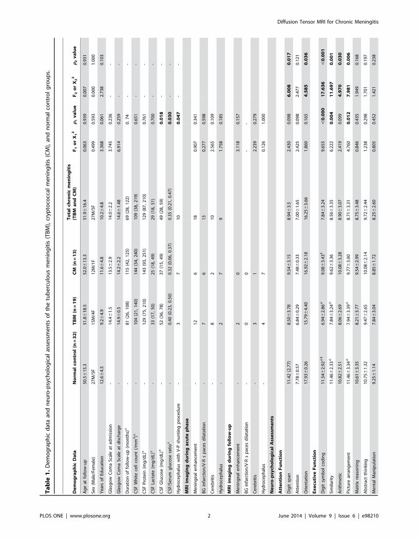
Statistical AnalysisBaseline Clinical Characteristics between
Groups. Clinical data and educational information were
compared by one-way analysis of variance (ANOVA). The GCS
Ta
ble
2.
Re
gio
ns
sho
win
glo
we
rfr
acti
on
alan
iso
tro
py
(FA
)va
lue
sin
HIV
-ne
gat
ive
chro
nic
me
nin
git
is(n
=3
2)
than
inn
orm
alco
ntr
ols
(n=
32
).
MN
Ia
tla
sco
ord
ina
tes
Vo
xe
lsi
ze
Wh
ite
ma
tte
rtr
act
Ne
are
stG
rey
Ma
tte
rYF
Am
ea
n(S
D)
t ma
xD
iffu
siv
ity
va
lue
s(M
en
ing
itis
-No
rma
l)
XY
ZN
orm
al
Me
nin
git
isM
DA
DR
D
22
22
62
22
29
Left
Infe
rio
rLo
ng
itu
din
alFa
scic
ulu
sP
ara-
hip
po
cam
pal
Gyr
us,
BA
19
0.3
4(0
.08
)0
.26
(0.0
7)
4.1
74
4.5
22
8.4
81
*
20
26
40
31
Rig
ht
Infe
rio
rLo
ng
itu
din
alFa
scic
ulu
sP
ara-
hip
po
cam
pal
Gyr
us,
BA
19
0.3
5(0
.07
)0
.28
(0.0
8)
4.1
14
4.3
*2
13
.67
3.2
*
81
62
83
7R
igh
tC
ing
ulu
mC
ing
ula
teG
yru
s,B
A2
40
.55
(0.1
0)
0.4
7(0
.12
)3
.82
17
0.5
82
.22
14
.7*
24
14
28
11
0R
igh
tSu
pe
rio
rC
oro
na
Rad
iata
Cin
gu
late
Gyr
us,
BA
32
0.4
1(0
.04
)0
.37
(0.0
4)
3.8
26
7.6
*4
5.9
78
.5*
22
22
70
16
27
Left
Forc
ep
sM
ajo
rC
un
eu
s,B
A3
00
.45
(0.1
1)
0.3
5(0
.16
)3
.76
34
.72
76
.3*
90
.2*
23
22
83
22
1Le
ftSu
pe
rio
rLo
ng
itu
din
alFa
scic
ulu
sP
re-c
en
tral
Gyr
us,
BA
60
.32
(0.0
5)
0.2
8(0
.04
)3
.73
39
.41
4.3
51
.9*
22
02
12
38
21
Left
Sup
eri
or
Co
ron
aR
adia
taC
ing
ula
teG
yru
s,B
A2
40
.35
(0.0
5)
0.3
1(0
.04
)3
.72
12
7.6
11
6.2
13
3.3
*
21
84
24
29
WM
clo
seto
Left
Glo
bu
sP
allid
us
Len
tifo
rmN
ucl
eu
s0
.38
(0.0
7)
0.3
1(0
.06
)3
.62
42
.92
19
.77
4.3
*
Th
ed
iffu
sivi
tyva
lue
sd
esc
rib
ed
iffe
ren
ces
(me
nin
git
isvs
.n
orm
al)
inm
ean
dif
fusi
vity
(MD
),ax
ial
dif
fusi
vity
(AD
),an
dra
dia
ld
iffu
sivi
tie
s(R
D)
(mm
2/s
)m
ult
iplie
db
y1
02
6.
*Sig
nif
ican
td
iffe
ren
ces
amo
ng
MD
,A
D,
and
RD
we
read
just
ed
wit
hag
e,
sex,
and
ed
uca
tio
nas
cova
riat
es
(p#
0.0
5).
YN
ear
est
Gra
yM
atte
rw
asn
ear
the
cen
ter
of
the
5-m
mra
diu
sse
arch
are
a.A
bb
revi
atio
ns:
MN
I,M
on
tre
alN
eu
rolo
gic
alIn
stit
ute
;B
A,
Bro
dm
ann
are
a.d
oi:1
0.1
37
1/j
ou
rnal
.po
ne
.00
98
21
0.t
00
2
Diffusion Tensor MRI for Chronic Meningitis
PLOS ONE | www.plosone.org 5 June 2014 | Volume 9 | Issue 6 | e98210
on initial admission and on discharge between the two patient
groups were analyzed by Wilcoxon rank sum test. Sex and the use
of ventriculo – peritoneal shunt between groups were analyzed by
Chi-square test or Fisher’s exact test, as appropriate. The Mann-
Whitney test was used to analyze the CSF examination, including
white cell count, lactate, protein and glucose levels, and CSF/
serum glucose ratio. Statistical differences in NP tests among the
groups were estimated by one-way analysis of covariance
(ANCOVA) with age, sex, and educational level as covariates.
Post-hoc analysis was performed with Bonferroni test. Statistical
significance was set at p,0.05.
Analysis of Group Comparison on FA Maps. All image
processing, including image registration, spatial normalization,
customized template creation, and voxel-wise statistical compar-
isons, were manipulated using the Statistical Parametric Mapping
8 (SPM8) (Wellcome Department of Cognitive Neurology,
London, UK) in MATLAB 7.8.0 (MathWorks, MA). Voxel-based
analysis on WM area was performed with SPM8 to investigate FA
differences among the groups [24]. First, differences in the FA
maps between the 32 chronic meningitis patients (TBM and CM)
and the controls were compared.
Second, differences in the FA maps among the three groups –
the TBM group/normal control group, the CM group/normal
control group, and the TBM group/CM group – were also
compared. Analysis of covariance (ANCOVA) was performed with
age and sex as covariates to investigate FA differences between
groups. In post-hoc tests, six contrasts were used to detect where
each voxel had a higher or lower fractional anisotropy when
comparing two of the three groups.
Since DTI was sensitive to WM alterations, a customized WM
mask threshold at 0.2 was used as an explicit mask to successfully
exclude voxels, which consisted of grey matter or cerebral spinal
fluid in the majority of subjects. The FA differences were
significant at the individual voxel level at p,0.001 and the
extended cluster size .20 voxels.
After the initial VBM analysis, all of the FA value differences
between groups based on the Johns Hopkins University DTI-
based WM atlas, which is included in FSL atlas tool (http://fsl.
fmrib.ox.ac.uk/fsl/fslwiki/Atlases), were reported. To identify the
significant WM clusters that corresponded to gray matter areas,
the GingleALE toolbox (The BrainMap Development Team;
http://brainmap.org/ale/index.html) and Talairach and Tour-
noux atlas (http://www.talairach.org/index.html) were used.
Correlation between Regional DTI-related Indices and
Clinical Evaluations. Partial Pearson correlation analysis with
age, sex, and years of formal education as nuisance covariates were
performed to correlate the clinical evaluations (i.e., GCS, CSF
study during admission, and cognitive function on follow-up) with
the regional DTI-related indices within the patient groups.
Statistical significance was set at p,0.05. All statistical analyses
were performed using the SPSS software, version 10.0 (SPSS Inc,
Chicago, IL).
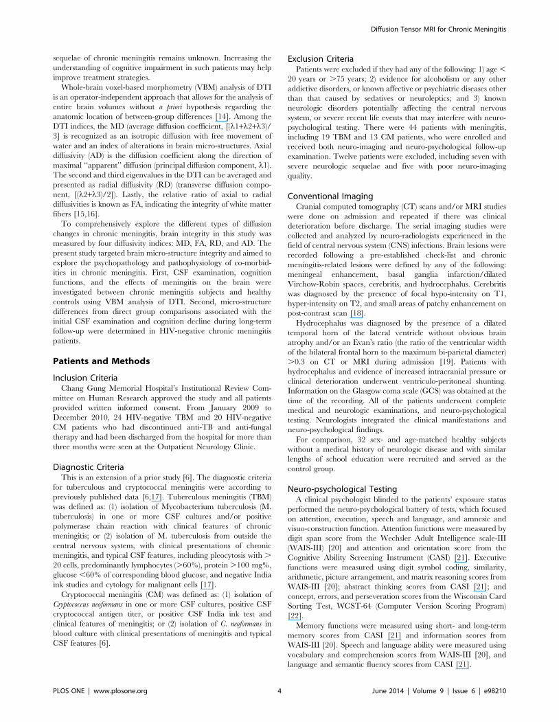
Figure 1. Comparison of fractional anisotropy (FA) between chronic meningitis patients and healthy controls. There was a closerelationship between Diffusion Tensor Imaging (DTI) deficits in the limbic system and basal ganglia, and their surrounding CSF space. The correlationbetween FA decline and CSF space were shown in three-dimensional configuration of the right lateral view (R), antero-posterior view (AP), and leftlateral view (L). Gray color, the ventricular system and deep brain arachnoid CSF space; Blue color, parts of the limbic system (cingulate gyrus andpara-hippocampal gyrus); Yellow voxels, regions with significantly lower FA value in chronic meningitis vs. normal control (p,0.001, corrected).doi:10.1371/journal.pone.0098210.g001
Diffusion Tensor MRI for Chronic Meningitis
PLOS ONE | www.plosone.org 6 June 2014 | Volume 9 | Issue 6 | e98210
Results
Baseline Clinical Characteristics between GroupsThe baseline clinical characteristics, neuro-imaging findings,
and cognitive function of all subjects were listed in Table 1. The
TBM and CM patients were followed-up for a median of
81 months (range, 26–108 months) and 115 months (range, 42–
125 months), respectively (p = 0.74). Statistical analysis of the
clinical manifestations and neuro-imaging findings between
patient groups were significant for CSF glucose level (p = 0.018),
CSF/serum glucose ratio (p = 0.030), and VP shunt use. There was
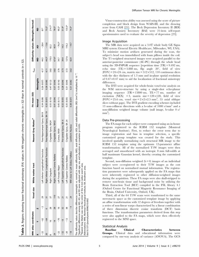
Figure 2. Comparisons of fractional anisotropy (FA) between groups. (A–F) Regions with significantly lower FA value in TBM vs. controls.(G–H) Regions with significantly lower FA value in CM vs. controls. (I–Q) Regions with significantly lower FA value in TBM vs. CM. (R–U) Regions withsignificantly lower FA value in CM vs. TBM.doi:10.1371/journal.pone.0098210.g002
Diffusion Tensor MRI for Chronic Meningitis
PLOS ONE | www.plosone.org 7 June 2014 | Volume 9 | Issue 6 | e98210
no significant difference in MRI findings between the acute phase
and follow-up.
In the two-group analyses, chronic meningitis had worse
cognitive examination, including attention, execution, memory,
speech, language, and visuo-construction function. In three-group
analysis, executive function [Digit symbol coding (F(2, 58) = 9.655; p,
0.001), similarity (F(2, 58) = 6.222; p = 0.004), picture arrangement (F(2,
58) = 4.760; p = 0.012)] and visuo-construction function [Picture
Complete (F(2, 58) = 5.354; p = 0.007), and block design, (F(2,
58) = 5.383; p = 0.007)] were worse in TBM patients than in
normal controls. Executive function [Digit symbol coding] and visuo-
construction function [Block Design] were also worse in CM patients
than in normal controls. There were no significant differences in
NP tests between the two groups.
Group Comparisons on FA MapsThe chronic meningitis group had significantly lower FA in
several WM regions than the control group. Together with
decreased FA, there were WMs with increased RD, and small or
no decreased AD in the para-hippocampus (bilateral inferior
longitudinal fasciculus), cingulate gyrus (right cingulum and
bilateral superior corona radiate), left pre-central gyrus (superior
longitudinal fasciculus), and left globus pallidus. Together with
decreased FA, there were WMs with increased RD and decreased
AD in the left cuneus (forceps major) (Table 2; Fig. 1).
Differences in regional WM integrity in the FA maps between
TBM and controls, between CM and controls, and between TBM
and CM patients were shown in Figure 2 and Table S1.
Correlation between Regional DTI-Related Indices andClinical Evaluations
DTI Indices and Baseline Clinical Characteris-
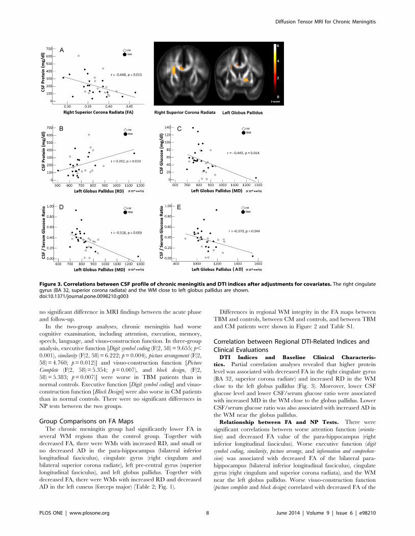
tics. Partial correlation analyses revealed that higher protein
level was associated with decreased FA in the right cingulate gyrus
(BA 32, superior corona radiate) and increased RD in the WM
close to the left globus pallidus (Fig. 3). Moreover, lower CSF
glucose level and lower CSF/serum glucose ratio were associated
with increased MD in the WM close to the globus pallidus. Lower
CSF/serum glucose ratio was also associated with increased AD in
the WM near the globus pallidus.
Relationship between FA and NP Tests. There were
significant correlations between worse attention function (orienta-
tion) and decreased FA value of the para-hippocampus (right
inferior longitudinal fasciculus). Worse executive function (digit
symbol coding, similarity, picture arrange, and information and comprehen-
sion) was associated with decreased FA of the bilateral para-
hippocampus (bilateral inferior longitudinal fasciculus), cingulate
gyrus (right cingulum and superior corona radiata), and the WM
near the left globus pallidus. Worse visuo-construction function
(picture complete and block design) correlated with decreased FA of the
Figure 3. Correlations between CSF profile of chronic meningitis and DTI indices after adjustments for covariates. The right cingulategyrus (BA 32, superior corona radiata) and the WM close to left globus pallidus are shown.doi:10.1371/journal.pone.0098210.g003
Diffusion Tensor MRI for Chronic Meningitis
PLOS ONE | www.plosone.org 8 June 2014 | Volume 9 | Issue 6 | e98210
Table 3. Correlation between diffusion tensor imaging (DTI) abnormalities and cognitive function after adjustments for age, sex,and education in HIV-negative chronic meningitis.
NP TestDTIMetrics Anatomical Lobe
BrodmannArea White Matter Tract
Correlation(r) p value
Attention Function
Digit Span MD Cingulate Gyrus 32 R Superior Corona Radiata 20.266 0.044
Orientation FA Para-hippocampal Gyrus 19 R Inferior Longitudinal Fasciculus 0.365 0.004
AD Para-hippocampal Gyrus 19 L Inferior Longitudinal Fasciculus 20.450 ,0.001
AD Cingulate Gyrus 32 R Superior Corona Radiata 20.428 0.001
RD Para-hippocampal Gyrus 19 L Inferior Longitudinal Fasciculus 20.529 ,0.001
RD Cingulate Gyrus 32 R Superior Corona Radiata 20.331 0.010
RD Para-hippocampal Gyrus 19 R Inferior Longitudinal Fasciculus 20.454 ,0.001
MD Para-hippocampal Gyrus 19 L Inferior Longitudinal Fasciculus 20.543 0.000
MD Cingulate Gyrus 32 R Superior Corona Radiata 20.380 0.003
MD Para-hippocampal Gyrus 19 R Inferior Longitudinal Fasciculus 20.434 0.001
Executive Function
Digit Symbol Coding FA Para-hippocampal Gyrus 19 L Inferior Longitudinal Fasciculus 0.275 0.040
RD Lentiform Nucleus L WM close to Globus Pallidus 20.413 0.002
MD Lentiform Nucleus L WM close to Globus Pallidus 20.370 0.005
Similarity FA Para-hippocampal Gyrus 19 L Inferior Longitudinal Fasciculus 0.343 0.009
AD Cingulate Gyrus 24 R Cingulum 20.362 0.006
AD Cingulate Gyrus 32 R Superior Corona Radiata 20.416 0.001
AD Precentral Gyrus 6 L Superior Longitudinal Fasciculus 20.268 0.044
AD Cingulate Gyrus 24 L Superior Corona Radiata 20.337 0.010
RD Cingulate Gyrus 24 R Cingulum 20.361 0.006
RD Cingulate Gyrus 32 R Superior Corona Radiata 20.418 0.001
RD Cingulate Gyrus 24 L Superior Corona Radiata 20.364 0.005
MD Cingulate Gyrus 24 R Cingulum 20.375 0.004
MD Cingulate Gyrus 32 R Superior Corona Radiata 20.432 0.001
MD Cingulate Gyrus 24 L Superior Corona Radiata 20.356 0.006
Picture Arrangement FA Para-hippocampal Gyrus 19 R Inferior Longitudinal Fasciculus 0.290 0.030
FA Lentiform Nucleus L WM close to Globus Pallidus 0.298 0.026
Information FA Para-hippocampal Gyrus 19 L Inferior Longitudinal Fasciculus 0.287 0.028
FA Cingulate Gyrus 24 R Cingulum 0.260 0.047
FA Cingulate Gyrus 32 R Superior Corona Radiata 0.282 0.031
FA Lentiform Nucleus L WM close to Globus Pallidus 0.319 0.014
RD Cingulate Gyrus 32 R Superior Corona Radiata 20.332 0.010
RD Cingulate Gyrus 24 L Superior Corona Radiata 20.268 0.040
MD Cingulate Gyrus 32 R Superior Corona Radiata 20.313 0.016
Comprehension FA Cingulate Gyrus 24 R Cingulum 0.279 0.033
AD Cingulate Gyrus 32 R Superior Corona Radiata 20.429 0.001
AD Cingulate Gyrus 24 L Superior Corona Radiata 20.322 0.013
RD Cingulate Gyrus 24 R Cingulum 20.285 0.029
RD Cingulate Gyrus 32 R Superior Corona Radiata 20.393 0.002
RD Cingulate Gyrus 24 L Superior Corona Radiata 20.328 0.011
MD Cingulate Gyrus 32 R Superior Corona Radiata 20.421 0.001
MD Cingulate Gyrus 24 L Superior Corona Radiata 20.329 0.011
Visuo-Construction Function
Picture Complete FA Para-hippocampal Gyrus 19 R Inferior Longitudinal Fasciculus 0.295 0.026
Block Design FA Para-hippocampal Gyrus 19 L Inferior Longitudinal Fasciculus 0.311 0.019
FA Para-hippocampal Gyrus 19 R Inferior Longitudinal Fasciculus 0.302 0.023
RD Para-hippocampal Gyrus 19 R Inferior Longitudinal Fasciculus 20.285 0.032
Diffusion Tensor MRI for Chronic Meningitis
PLOS ONE | www.plosone.org 9 June 2014 | Volume 9 | Issue 6 | e98210
bilateral para-hippocampus (bilateral inferior longitudinal fascic-
ulus) (Table 3).
Relationship between MD and NP Tests. There were
significant correlations between worse attention function (digit span,
and orientation) and increased MD value of the cingulate gyrus
(right superior corona radiate) and bilateral para-hippocampus
(bilateral inferior longitudinal fasciculus). Worse executive function
(digit symbol coding, similarity, information and comprehension) was
associated with increased MD value of the WM close to the left
globus pallidus and bilateral cingulate gyrus (right cingulum and
bilateral corona radiata).
Relationship between AD and NP Tests. There were
significant correlations between worse attention function (orienta-
tion) and decreased FA value of the right para-hippocampal gyrus
(right inferior longitudinal fasciculus) and cingulate gyrus (right
superior corona radiate). Worse executive function (similarity and
comprehension) was associated with increased AD in the bilateral
cingulate gyrus (right cingulum and bilateral corona radiata) and
left pre-central gyrus (superior longitudinal fasciculus).
Relationship between RD and NP Tests.. There were
significant correlations between worse attention function (orienta-
tion) and decreased FA value of the para-hippocampal gyrus
(bilateral right inferior longitudinal fasciculus) and cingulate gyrus
(right superior corona radiata). Worse executive function (digit
symbol coding, similarity, and information and comprehension) was
associated with increased RD of the WM close to the left globus
pallidus and bilateral cingulate gyrus (right cingulum and bilateral
corona radiate). Worse visuo-construction function (block design)
correlated with increased RD of the right para-hippocampal gyrus
(inferior longitudinal fasciculus) and WM near the left globus
pallidus.
Discussion
In the present study, HIV-negative chronic meningitis has
heterogeneous changes in micro-structures in the limbic system
and WM close to the globus pallidus, as revealed by multiple
diffusion tensor metrics (FA, MD, AD, and RD). These changes
are associated with the initial disease severity. There are attention,
execution, memory, speech, language, and visuo-construction
function deficits in chronic meningitis that are rarely reported.
Different limbic system deficits are also significantly correlated to
scores on these tests, revealing a direct relationship between
impaired cognitive functions and micro-structure abnormalities.
There is growing evidence, both experimentally and epidemi-
ologically, of the association of neuro-infection with cerebral WM
injury [25]. In histological study, abundantly infiltration of
Cryptococcus neoformans with prominent immune reaction were
revealed in cerebral WM and cortex adjacent to the leptomenin-
ges, but these findings were not observed in the subcortical and
cortical lesions [26]. Distinct WM disruption profiles demonstrate
heterogeneous pathologic processes affecting different WM areas
in chronic meningitis. Different alterations in WM integrity may
be the end result of interactions among (1) acute stage
inflammation, (2) ischemia after vessel occlusion, (3) pressure
stress from hydrocephalus, and (4) brain tissue vulnerabilities
[13,27]. Their contributions to DTI changes are not be fully
resolved in this cross-sectional analysis.
The FA is derived from directional diffusivities of diffusion
tensor imaging. Unfortunately, FA is a highly non-specific marker.
Consideration of the different directional diffusivities is important
because if changes in diffusion along the axial direction are
proportional to those along the radial directions, then FA (which is
a function of the ratio) will remain unchanged [28]. Decreased FA
can correspond to different pathologic findings like demyelin-
ation/dysmyelination, axonal loss, gliosis, and tissue inflammation
[29]. This decrease occurs after either an increase in RD or a
decrease in AD, or both. Increased RD reflects increased water
diffusion in the perpendicular direction.
In the present study, decreased FA, increased RD, and a much
smaller or no change in AD such as the para-hippocampus,
cingulate gyrus, pre-central gyrus, and WM close to globus
pallidus (Table 2), have been suggested as myelin injury [16].
However, decreased FA with reduced AD but increased RD in the
left cuneus (forceps major) may likewise be associated with axonal
damage and demyelination or the fiber re-organization found in
neuro-degenerative diseases [30].
Increased MD reflects isotropic diffusion with the free
movement of water that is commonly observed in gliosis after
infarction. In chronic meningitis, 30–50% of patients reportedly
experience cerebral infarction [2]. In TBM, the rich focus ruptures
into the sub-arachnoid space, causing meningitis. A thick,
gelatinous exudate infiltrates the cortical or meningeal blood
vessels, producing obliterative vasculitis or infarction. In CM,
inflammation is initially confined to the sub-arachnoid space and is
prone to infiltrating the Virchow-Robin peri-vascular spaces
which tend to exhibit a soap bubble appearance by MRI [31].
Thrombo-embolism from vasculitis within these small peri-
vascular spaces further leads to tissue ischemia via decreased
cerebral perfusion.
In the present study, the significantly decreased FA and increase
MD in the right para-hippocampal gyrus (BA 19) and right
cingulate gyrus (BA 32) may indicate the most severe WM
ischemia, with subsequent gliosis [29]. Thirteen of 32 patients
present with either basal ganglia infarction or prominent peri-
vascular space. However, decreased FA in the WM close to the
globus pallidus seem to primarily come from myelin injury
(increased RD but no change in MD), instead of gliosis. More
sensitive DTI for detecting WM than gray matter ischemia may
explain this phenomenon [32]. The true mechanism is unknown.
Hydrocephalus is the most common serious complication of
chronic meningitis and is strongly associated with long-term
outcome [3,33]. In chronic meningitis, basal meningitis eventually
leads to obstructive hydrocephalus from obstruction of the basilar
cisterns. In chronic hydrocephalus, WM loss is associated with
deficits in motor and cognitive functions in a previous animal and
human study [34]. In the present study, susceptible anatomies with
DTI differences between groups are consistent findings of a
previous study and suggest that mechanical injury from hydro-
cephalus may be part of the pathophysiology of WM injury.
Table 3. Cont.
NP TestDTIMetrics Anatomical Lobe
BrodmannArea White Matter Tract
Correlation(r) p value
RD Lentiform Nucleus L WM close to Globus Pallidus 20.309 0.019
doi:10.1371/journal.pone.0098210.t003
Diffusion Tensor MRI for Chronic Meningitis
PLOS ONE | www.plosone.org 10 June 2014 | Volume 9 | Issue 6 | e98210
However, most differential limbic system DTI changes result from
increased RD value, which points more to myelin loss. The
findings here are similar to another histologic study in neonatal
meningitis where there is myelin loss in the peri-ventricular region
showing infiltration with glial cells [35]. However, it is also possible
that the decreased FA and increased RD may indirectly represent
loss of axial diffusivity or axonal injury, although this is difficult to
substantiate given the absence of statistical significance. To date,
no conclusive findings on meningitis can be drawn from DTI
studies.
Malik et al. report that FA values decrease without information
about AD and RD in the peri-ventricular WM in the sub-acute
stage of neonatal meningitis, even in patients with normal
outcomes [13]. In the acute stage, hydrocephalus can compress
the WM with increased AD and decreased RD, resulting in an
overall increase in FA value in bacterial meningitis [36].
Alterations in DTI indices may be associated with a combination
of stretch injury, impaired blood circulation, and accumulated
damage from pathogens/waste products in the CSF [37].
Regardless of TBM or CM, CSF parameters like high CSF
lactate and protein levels, and low CSF glucose are identified as
poor prognostic indicators [38,39]. The association between worse
CSF profile in the acute stage and long-term decline in DTI
indices establish the possible etiology of their respective chronic
neuro-psychiatric sequelae. Results of the present study are
consistent with previous arguments that the initial stage of the
disease on presentation is a major prognostic indicator of
morbidity and mortality [40,41]. Poor neurologic outcomes can
be predicted based the presenting stage of the disease [40–42].
The high microbial burden in the sub-arachnoid space can
damage vessels in the circle of Willis and affect the globus pallidus
more directly than others. The results can explain the pathogenesis
of chronic meningitis.
Extensive impairment of neuro-psychological performance and
its significant correlation with WM damage in the limbic system
underscore the pathologic role of the limbic system in chronic
meningitis. The dorsal part of the anterior cingulate cortex (ACC)
(BA 24 and 32) is a central station for processing top-down and
bottom-up stimuli and for assigning appropriate control to other
brain cortices. The results here are consistent with previous reports
that ACC (BA 24 and 32) abnormalities in morphology and
histopathology constitute impaired executive and attention func-
tions in multiple neurologic and psychiatric disorders [43,44]. The
consistent damage in ACC in meningitis, chronic hydrocephalus
in animals and humans, and in the present study may make ACC
a specific pilot anatomy to help improve long-term outcomes in
chronic meningitis and guide future interventional treatment
strategies.
Abnormal DTI findings in the WM near the globus pallidus also
partly contribute to the neuro-psychiatric deficits in chronic
meningitis. There is growing evidence that the globus pallidus is
one of the neural substrates of executive cognitive function [45]
and that it plays an important role in the cortico – striatal –
pallidal – thalamo – cortical loops [46]. Its function is to serve as a
limbic-somatic motor interface in the planning and inhibition of
movements. The para-hippocampus (BA 19) is a histologically
delineated band antero-laterally abutting the visual area 18 and is
responsible for the heterogeneous visual information collection,
including feature-extracting, shape recognition, attention, and
multi-modal integrating functions [47]. Together with the
cingulate gyrus and globus pallidus, the para-hippocampus reveals
an association between declined DTI indices and the relative
decline in attention, execution, and visuo-construction functions.
The interpretation of the findings here must be tempered by
some limitations. The results are based on a relatively small cohort
for both TBM and CM. The study did not enroll patients with
severe sequelae. Thus, there is uncertainty in assessing the DTI
findings of critically-ill patients and those with poor prognosis.
Second, the DTI findings may be influenced by the duration of
hydrocephalus, increased intracranial pressure, antimicrobial
therapy, and follow-up period. These may be also influence by
other drugs (e.g. steroids and hyper-osmolarity agents) that are
commonly used in patients with meningitis. These drugs may
cause potential bias in the interpretation of DTI findings.
Moreover, further studies to determine whether the observed
effects are due to pre-existing neurologic or psychiatric illnesses
and micro-angiopathy in elderly subjects after long-term follow-up
are warranted as these may be unrelated to the disease process.
Lastly, a cross-sectional study must be performed on two sample
groups rather than a longitudinal study following treatment
because when the WM damage begins remains unsolved.
Recurrent infection is also an important risk factor for progressive
white matter injury [48].
In conclusion, there are significant WM differences in the limbic
system-associated anatomy and the WM close to the globus
pallidus between chronic meningitis and healthy controls. After
exploring the possible pathophysiology and psychopathology with
initial disease severity and long-term neuro-psychiatric sequelae,
abnormalities in the ACC, para-hippocampus, and globus pallidus
may be specific biomarkers for disease evaluation. A longitudinal
study combined with evaluation of the timing of medical and
intervention therapy is warranted.
Supporting Information
Table S1 Comparison of fractional anisotropy (FA) values
between tuberculous meningitis (TBM) and controls, between
cryptococcal meningitis (CM) and controls, and between TBM
and CM
(DOC)
Author Contributions
Conceived and designed the experiments: WCL CHL. Performed the
experiments: HCW NWT HLC. Analyzed the data: PCC KHC CPL.
Contributed reagents/materials/analysis tools: YJS CPL SHL WNC.
Wrote the paper: WCL CHL.
References
1. Lu CH, Chang WN, Chang HW, Chuang YC (1999) The prognostic factors of
cryptococcal meningitis in HIV-negative patients. J Hosp Infect 42: 313–320.
2. Lan SH, Chang WN, Lu CH, Lui CC, Chang HW (2001) Cerebral infarction in
chronic meningitis: a comparison of tuberculous meningitis and cryptococcal
meningitis. QJM 94: 247–253.
3. Liliang PC, Liang CL, Chang WN, Chen HJ, Su TM, et al. (2003) Shunt surgery
for hydrocephalus complicating cryptococcal meningitis in human immunode-
ficiency virus-negative patients. Clin Infect Dis 37: 673–678.
4. Sa’adah MA, Araj GF, Diab SM, Nazzal M (1995) Cryptococcal meningitis and
confusional psychosis. A case report and literature review. Trop Geogr Med 47:
224–226.
5. Hoffmann M, Muniz J, Carroll E, De Villasante J (2009) Cryptococcal
meningitis misdiagnosed as Alzheimer’s disease: complete neurological and
cognitive recovery with treatment. J Alzheimers Dis 16: 517–520.
6. Lu CH, Chen HL, Chang WN, Tsai NW, Wang HC, et al. (2011) Assessing the
chronic neuropsychologic sequelae of human immunodeficiency virus-negative
cryptococcal meningitis by using diffusion tensor imaging. AJNR
Am J Neuroradiol 32: 1333–1339.
7. Gumbo T, Kadzirange G, Mielke J, Gangaidzo IT, Hakim JG (2002)
Cryptococcus neoformans meningo-encephalitis in African children with
acquired immunodeficiency syndrome. Pediatr Infect Dis J 21: 54–56.
Diffusion Tensor MRI for Chronic Meningitis
PLOS ONE | www.plosone.org 11 June 2014 | Volume 9 | Issue 6 | e98210
8. Trivedi R, Saksena S, Gupta RK (2009) Magnetic resonance imaging in central
nervous system tuberculosis. Indian J Radiol Imaging 19: 256–265.
9. Lim KO, Helpern JA (2002) Neuropsychiatric applications of DTI – a review.
NMR Biomed 15: 587–593.
10. Moseley M (2002) Diffusion tensor imaging and aging – a review. NMR Biomed
15: 553–560.
11. Lin WC, Lu CH, Lee YC, Wang HC, Lui CC, et al. (2009) White matter
damage in carbon monoxide intoxication assessed in vivo using diffusion tensor
MR imaging. AJNR Am J Neuroradiol 30: 1248–1255.
12. Lin WC, Chou KH, Chen CC, Huang CC, Chen HL, et al. (2012) White matter
abnormalities correlating with memory and depression in heroin users under
methadone maintenance treatment. PLoS One 7: e33809.
13. Malik GK, Trivedi R, Gupta A, Singh R, Prasad KN, et al. (2008) Quantitative
DTI assessment of peri-ventricular white matter changes in neonatal meningitis.
Brain Dev 30: 334–341.
14. Catani M (2006) Diffusion tensor magnetic resonance imaging tractography in
cognitive disorders. Curr Opin Neurol 19: 599–606.
15. Song SK, Sun SW, Ju WK, Lin SJ, Cross AH, et al. (2003) Diffusion tensor
imaging detects and differentiates axon and myelin degeneration in mouse optic
nerve after retinal ischemia. Neuroimage 20: 1714–1722.
16. Song SK, Yoshino J, Le TQ, Lin SJ, Sun SW, et al. (2005) Demyelination
increases radial diffusivity in corpus callosum of mouse brain. Neuroimage 26:
132–140.
17. Lu CH, Chang WN, Chang HW, Chung KJ, Tsai NW, et al. (2007) Clinical
relevance of intracranial arterial stenosis in tuberculous and cryptococcal
meningitis. Infection 35: 359–363.
18. Tung GA, Rogg JM (2003) Diffusion-weighted imaging of cerebritis. AJNR
Am J Neuroradiol 24: 1110–1113.
19. MS G (1997) Hydrocephalus; Greenberg MS e, editor. Lakeland, FL: Greenberg
Graphics.
20. Wechsler D (1981) Wechsler Adapt Intelligence Scale-reised [manual]. San
Antonio, TX: Psychological Cop.
21. Chang CC, Liu JS, Chang YY, Chang WN, Chen SS, et al. (2008) (99m)Tc-
ethyl cysteinate dimer brain SPECT findings in early stage of dementia with
Lewy bodies and Parkinson’s disease patients: a correlation with neuropsycho-
logical tests. Eur J Neurol 15: 61–65.22.
22. Nyhus E, Barcelo F (2009) The Wisconsin Card Sorting Test and the cognitive
assessment of prefrontal executive functions: a critical update. Brain Cogn 71:
437–451.
23. Beck A, Steer R, Brown G (1996) Manual for the Beck Depression Inventory-II.
San Antonio, TX: Psychological Corporation.
24. Bai YM, Chou KH, Lin CP, Chen IY, Li CT, et al. (2009) White matter
abnormalities in schizophrenia patients with tardive dyskinesia: a diffusion tensor
image study. Schizophr Res 109: 167–181.
25. Dammann O, Leviton A (2000) Brain damage in pre-term newborns: biological
response modification as a strategy to reduce disabilities. J Pediatr 136: 433–438.
26. Kuwahara H, Tsuchiya K, Kobayashi Z, Inaba A, Akiyama H, et al. (2014)
Cryptococcal meningitis accompanying lymphocytic inflammation predomi-
nantly in cerebral deep white matter: a possible manifestation of immune
reconstitution inflammatory syndrome. Neuropathology 34: 45–48.
27. Lee SC, Dickson DW, Casadevall A (1996) Pathology of cryptococcal meningo-
encephalitis: analysis of 27 patients with pathogenetic implications. Hum Pathol
27: 839–847.
28. Acosta-Cabronero J, Williams GB, Pengas G, Nestor PJ (2010) Absolute
diffusivities define the landscape of white matter degeneration in Alzheimer’sdisease. Brain 133: 529–539.
29. Le Bihan D, Mangin JF, Poupon C, Clark CA, Pappata S, et al. (2001) Diffusion
tensor imaging: concepts and applications. J Magn Reson Imaging 13: 534–546.30. Beaulieu C (2002) The basis of anisotropic water diffusion in the nervous system
– a technical review. NMR Biomed 15: 435–455.31. Vieira MA, Costa CH, Ribeiro JC, Nunes-Filho LP, Rabelo MG, et al. (2013)
Soap bubble appearance in brain magnetic resonance imaging: cryptococcal
meningoencephalitis. Rev Soc Bras Med Trop 46: 658–659.32. Mukherjee P, Bahn MM, McKinstry RC, Shimony JS, Cull TS, et al. (2000)
Differences between gray matter and white matter water diffusion in stroke:diffusion-tensor MR imaging in 12 patients. Radiology 215: 211–220.
33. Misra UK, Kalita J, Srivastava M, Mandal SK (1996) Prognosis of tuberculousmeningitis: a multivariate analysis. J Neurol Sci 137: 57–61.
34. Del Bigio MR, Wilson MJ, Enno T (2003) Chronic hydrocephalus in rats and
humans: white matter loss and behavior changes. Ann Neurol 53: 337–346.35. Volpe JJ (2001) Neurology of the newborn; Volpe JJ, editor. London: WB
Saunders. 774–810 p.36. Trivedi R, Malik GK, Gupta RK, Gupta A, Nath K, et al. (2007) Increased
anisotropy in neonatal meningitis: an indicator of meningeal inflammation.
Neuroradiology 49: 767–775.37. Del Bigio MR (1993) Neuropathological changes caused by hydrocephalus. Acta
Neuropathol 85: 573–585.38. Diamond RD, Bennett JE (1974) Prognostic factors in cryptococcal meningitis.
A study in 111 cases. Ann Intern Med 80: 176–181.39. Thwaites GE, Simmons CP, Than Ha Quyen N, Thi Hong Chau T, Phuong
Mai P, et al. (2003) Pathophysiology and prognosis in Vietnamese adults with
tuberculous meningitis. J Infect Dis 188: 1105–1115.40. Girgis NI, Sultan Y, Farid Z, Mansour MM, Erian MW, et al. (1998)
Tuberculosis meningitis, Abbassia Fever Hospital-Naval Medical Research UnitNo. 3-Cairo, Egypt, from 1976 to 1996. Am J Trop Med Hyg 58: 28–34.
41. Thwaites GE, Nguyen DB, Nguyen HD, Hoang TQ, Do TT, et al. (2004)
Dexamethasone for the treatment of tuberculous meningitis in adolescents andadults. N Engl J Med 351: 1741–1751.
42. Rock RB, Olin M, Baker CA, Molitor TW, Peterson PK (2008) Central nervoussystem tuberculosis: pathogenesis and clinical aspects. Clin Microbiol Rev 21:
243–261, table of contents.43. Szeszko PR, Bilder RM, Lencz T, Ashtari M, Goldman RS, et al. (2000)
Reduced anterior cingulate gyrus volume correlates with executive dysfunction
in men with first-episode schizophrenia. Schizophr Res 43: 97–108.44. Carter CS, Mintun M, Nichols T, Cohen JD (1997) Anterior cingulate gyrus
dysfunction and selective attention deficits in schizophrenia: [15O]H2O PETstudy during single-trial Stroop task performance. Am J Psychiatry 154: 1670–
1675.
45. Bhatia KP, Marsden CD (1994) The behavioural and motor consequences offocal lesions of the basal ganglia in man. Brain 117 (Pt 4): 859–876.
46. Alexander GE, DeLong MR, Strick PL (1986) Parallel organization offunctionally segregated circuits linking basal ganglia and cortex. Annu Rev
Neurosci 9: 357–381.47. Wojciulik E, Kanwisher N (1999) The generality of parietal involvement in
visual attention. Neuron 23: 747–764.
48. Glass HC, Bonifacio SL, Chau V, Glidden D, Poskitt K, et al. (2008) Recurrentpostnatal infections are associated with progressive white matter injury in
premature infants. Pediatrics 122: 299–305.
Diffusion Tensor MRI for Chronic Meningitis
PLOS ONE | www.plosone.org 12 June 2014 | Volume 9 | Issue 6 | e98210
Related Documents