A CYTOPATHIC EFFECT-BASED HIGH-THROUGHPUT SCREENING ASSAY IDENTIFIED TWO NOVEL COMPOUNDS THAT INHIBIT DENGUE INFECTION: STREPTOVITACIN A AND NAGILACTONE C by Kevin Dylan McCormick BS, Seton Hill University, 2007 Submitted to the Graduate Faculty of Graduate School of Public Health in partial fulfillment of the requirements for the degree of Master of Science University of Pittsburgh 2011

Welcome message from author
This document is posted to help you gain knowledge. Please leave a comment to let me know what you think about it! Share it to your friends and learn new things together.
Transcript
A CYTOPATHIC EFFECT-BASED HIGH-THROUGHPUT SCREENING ASSAY
IDENTIFIED TWO NOVEL COMPOUNDS THAT INHIBIT DENGUE INFECTION:
STREPTOVITACIN A AND NAGILACTONE C
by
Kevin Dylan McCormick
BS, Seton Hill University, 2007
Submitted to the Graduate Faculty of
Graduate School of Public Health in partial fulfillment
of the requirements for the degree of
Master of Science
University of Pittsburgh
2011

ii
UNIVERSITY OF PITTSBURGH
GRADUATE SCHOOL OF PUBLIC HEALTH
This thesis was presented
by
Kevin Dylan McCormick
It was defended on
April 21, 2011 and
approved by
Ernesto T.A. Marques, Jr., MD, Ph.D. Associate Professor
Department of Infectious Diseases and Microbiology, Graduate School of Public Health Center for Vaccine Research
University of Pittsburgh
Nicolas Sluis-Cremer, Ph.D. Associate Professor of Medicine
Director of Basic Research, Division of Infectious Diseases University of Pittsburgh
Thesis Director: Tianyi Wang, PhD
Assistant Professor Department of Infectious Diseases and Microbiology, Graduate School of Public Health
University of Pittsburgh

iii
Copyright © by Kevin Dylan McCormick
2011

iv
Tianyi Wang, PhD
A CYTOPATHIC EFFECT-BASED HIGH-THROUGHPUT SCREENING ASSAY
IDENTIFIED TWO NOVEL COMPOUNDS THAT INHIBIT DENGUE INFECTION:
STREPTOVITACIN A AND NAGILACTONE C
Kevin Dylan McCormick, M.S.
University of Pittsburgh, 2011
Dengue is an emerging infectious disease and is spreading world-wide at exponential levels.
Two billion people in over 100 countries are at risk for infection from one of the four serotypes
of the dengue virus. Those infected with dengue may develop diseases such as dengue fever and
dengue hemorrhagic fever (DHF) and of the 500,000 cases that progress to DHF each year, more
than 22,000 will result in fatality. Discovering new antivirals to treat DHF is essential to
reducing this disease burden. Here, we have developed a cytopathic effect-based high-
throughput screen (HTS) to discover possible inhibitors of Dengue viral infection of hepatocytes
in vitro. Dengue virus infection of hepatocytes induces massive cell death, “cytopathic effect
(CPE)”, which we converted into a screening assay whereby inhibitors of Dengue infection
prevent cells from dying. In this assay, the viral induced CPE is quantitated by monitoring
cellular ATP levels, which positively correlates with cellular viability. ATP in the cell culture
will drive the oxidation of luciferin resulting in the emission of light that is quantitated using a
luminometer. The assay is simple and highly reproducible yielding a screening window
coefficient, Z- factor, of 0.78±0.12 between plates. The Z-factor is a statistical parameter
commonly accepted as an assay quality assessment and is reported as a value 0 to 1 and
anything over 0.5 is considered excellent quality. This assay is advantageous to current
v
methodology as it simultaneously screens possible inhibitory compounds while controlling for
any unwanted toxicity triggered by these drugs. Our initial HTS of a 288 small compound
library yielded a total of eleven hits that prevented the CPE of dengue infection. Further
evaluation with an immunofluorescence assay showed that two of these compounds,
Streptovitacin A and Nagilactone C, are highly potent inhibitors of dengue infection. At
effective inhibitory doses, they did not appear to be cytotoxic, and therefore both of these
compounds are possible antivirals and could be used to elucidate various cellular mechanisms
utilized during the dengue life cycle. The discovery of these two inhibitors demonstrates the
efficacy of our newly developed assay and the public health significance of this project.
vi
TABLE OF CONTENTS
ACKNOWLEDGMENTS .......................................................................................................... XI
LIST OF ABBREVIATIONS .................................................................................................. XII
1.0 INTRODUCTION........................................................................................................ 1
1.1 DESCRIPTION OF THE PROBLEM .............................................................. 1
1.1.1 Dengue Virus.................................................................................................... 1
1.1.2 Dengue Pathogenesis ....................................................................................... 3
1.1.3 Dengue Prevention and Treatment ................................................................ 5
2.0 STATEMENT OF THE PROJECT ........................................................................... 7
3.0 MATERIALS AND METHODS ................................................................................ 8
3.1 PROPAGATION AND IDENTIFICATION OF PROTOTYPE VIRUS ...... 8
3.1.1 Cell lines and Reagents.................................................................................... 8
3.1.2 Propagation of Virions .................................................................................... 8
3.1.3 Western Blot Assay.......................................................................................... 9
3.1.4 Immunofluorescence Assay ............................................................................ 9
3.1.5 Virus Tittering (Reed & Muench method) .................................................. 10
3.2 LUCIFERASE-BASED HIGH THROUGHPUT SCREEN .......................... 10
3.2.1 Cytopathic Effect - Luciferase Reagent ....................................................... 10
3.2.2 CPE-based high-throughput screening assay. ............................................ 11

vii
3.2.3 Cell Culture Derived (HCVcc) Luciferase Assay ....................................... 11
3.2.4 New Castle Disease Virus-Green Fluorescence Protein Assay .................. 12
3.2.5 High-Throughput Screening Assay Data Analysis ..................................... 12 4.0 RESULTS ................................................................................................................... 14
4.1 SPECIFIC AIM 1 RESULTS ........................................................................... 14
4.1.1 Experimental Scheme of Dengue propagation and detection.................... 15
4.1.2 Detection of the DV2 Infection of Vero and C6/36 ..................................... 16
4.1.3 Tittering o f D V1-4 using the Reed and Muench method to calculate
TCID50/ml .................................................................................................................. 18
4.1.4 The Infection of Human Hepatoma Cell Lines Huh7.5.1 and HepG2...... 19
4.2 SPECIFIC AIM 2 RESULTS ........................................................................... 20
4.2.1 Experimental Scheme of the Cytopathic-Based High-Throughput
Screening Assay .......................................................................................................... 21
4.2.2 Optimization of Multiplicity of Infection an d Duration of the Cytopathic
Effect-Based HTS Assay. ........................................................................................... 23
4.3 SPECIFIC AIM 3 RESULTS ........................................................................... 26
4.3.1 Screening a Small Compound Library ........................................................ 26
4.3.2 Validation of the Positive Hits with an Immunofluorescence Assay ........ 27
4.3.3 Calculating an IC50 and CC50 for the Two “Lead” Compounds................ 28
5.0 DISCUSSION ............................................................................................................. 35
BIBLIOGRAPHY ....................................................................................................................... 39

viii
LIST OF TABLES Table 1. TCID50/ml of each strains of propagated prototyped virus.. ........................................... 19
ix
LIST OF FIGURES
Figure 1. Dengue Genome and Polyprotein.................................................................................... 2
Figure 2. Dengue Replication Cycle ............................................................................................... 3
Figure 3. Dengue Pathogenesis flow chart ..................................................................................... 4
Figure 4. Overall scheme of the propogation and identification of prototype strains of DEN ..... 16
Figure 5.The infection of propagated virus in C6/36 and Vero cells............................................ 17
Figure 6. Virus Titering using an IFA and the Reed & Muench method of calculating the
TCID50/ml...................................................................................................................................... 18
Figure 7. P ropagated DV2 virus infection of hepatocytes cell lines was confirmed by IFA and
Western Blot Assay....................................................................................................................... 20
Figure 8. Cytopathic effect of the Dengue virus on HuH7.5.1 ..................................................... 22
Figure 9. Optimizing the concentration of virus and duration of the CPE-screening assay ......... 23
Figure 10. Efficiency of the CPE-based screening assay to show inhibition ............................... 24
Figure 11. Demonstration of the assays simultaneous detection of inhibition and toxicity ......... 25
Figure 12. High-throughput Screening of Small Compound Library........................................... 26
Figure 13. IFA of two positive hits from the CPE assay .............................................................. 28
Figure14.Inhibitory concentration IC50 of two inhibitors identified in the preliminary screening
of 264 NCI compounds ................................................................................................................. 29
x
Figure15. Cytotoxicity concentration CC50of the two inhibitors identified the a preliminary
screening of 264 NCI compounds................................................................................................. 30
Figure 16. Streptovitacin A and Nagilactone C inhibited the infection of all serotypes of DEN on Huh7.5.1 cells ............................................................................................................................... 32
Figure 17. Streptovitacin A and Nagilactone C inhibited the HCVcc and NDV-GFP infection of
Huh7.5.1 cells ............................................................................................................................... 34
Figure 18. Chemical structures of the two compounds identified to inhibit dengue infection,
streptovitacin A and nagilactone C. .............................................................................................. 37
xi
ACKNOWLEDGMENTS
I would first like to thank my PI Dr. Tianyi Wang for everything he has done for me over
the past two years. He has been a friend as well as a mentor and I will never forget his support.
I would also like to thank Dr. Ernesto Torres De Azeved Marques Jr. for providing dengue
prM/E plasmids and clinical dengue virus isolates and Nicolas Sluis-Cremer, PhD for providing
the small compound library. In addition I would like to thank Drs. Marques Jr. and Sluis-Cremer
for serving on my committee and for all of the support and suggestions that they offered.
I would also like to thank my first year advisor, Dr. Velpandi Ayyavoo, for her guidance
during my first year of graduate school. In addition, I am grateful to Judith Ann Malenka,
Joanne Pegher, and Suzanne McCusker for all of the administrative work they did to ensure that
I graduated with my fellow classmates.
I would also like to thank my loving wife, Kathryn McCormick. Without her support I
am not sure if I could have made it this far. Special thanks to our cat Kiki lala (RIP 04.19.2011)
she will always be remembered.
xii
LIST OF ABBREVIATIONS
ATP, Adenosine-5'-triphosphate Baf A, Bafilomycin A
C, Capsid Protein
CC50, 50% Cytotoxicity Concentration CD, Cluster of Differentiation
CPE, Cytopathic Effect
DAPI, 4',6-diamidino-2-phenylindole
DC-SIGN, Dendritic Cell-Specific Intercellular adhesion molecule-3-Grabbing Non-integrin
DS-RNA, Double-Stranded Ribonucleic acid
DEN, Dengue Virus
DV, Dengue Virus Serotype
E, Envelope Protein
ER, Endoplasmic Reticulum
FBS, Fetal Bovine Serum
GFP, Green Fluorescence Protein
HCV, Hepatitis C Virus
HCVcc, Cell Culture derived Hepatitis C Virus
HTS, High-Throughput Screening Assay

xiii
HSP, Heat Shock Protein IC50, 50% Inhibitory Concentration
IFA, Immunofluorescence Assay
IFN-α, Interferon α
LARII, Luciferase Assay Reagent II
LUC, Luciferase
M, Membrane Protein MOI, Multiplicity of Infection
MYA, Mycophenolic Acid
NCI, National Cancer Institute
NEAA, Non-Essential Amino Acids NDV, New Castle Disease Virus
NS, Non-Structural Protein
PI, Post Infection
prM/E, Pre-Membrane/Envelope Protein
RDRP, RNA-dependent-RNA-polymerase
RLU, Relative Light Units
RLUexp, luciferase counts obtained from inhibitor treated wells
RLUmax, luciferase counts obtained from uninfected wells
RLUmin, luciferase counts obtained from untreated infected wells
SARS, Severe Acute Respiratory Syndrome Coronavirus
SS-RNA, Single-Stranded Ribonucleic acid StDev, Standard Deviation
TCID50, 50% Tissue Culture Infectious Dose YPLL, Years of Potential Life Lost
XIV

1
1.0 INTRODUCTION
1.1 DESCRIPTION OF THE PROBLEM Two billion people in over 100 countries are at risk for infection from one of the four serotypes
of Dengue virus (DEN) making it the most prevalent mosquito-borne virus worldwide (10, 23).
Those infected by this virus may develop diseases ranging from the self-limited febrile illness
dengue fever (DF) to the more severe dengue shock syndrome (DSS) and dengue hemorrhagic
fever (DHF). Of the 500,000 cases that progress to DHF each year, more than 22,000 will result
in fatality (31). Unfortunately there are currently no vaccines or antiviral therapies approved for
the treatment of infection. Novel therapeutics, therefore, are in urgent need to fight the infection.
1.1.1 Dengue Virus
DEN is an arthropod-borne virus (arbovirus) and is transmitted to humans by the imbibing of a
mosquito, more specifically the Aedes genera of mosquito and most often from Aedes aegypti
(31). It is an enveloped positive sense single-stranded RNA virion that belongs to the genus
Flavivirus, family Flaviviridae. The 10.7kb genome of DEN is enclosed in the structural
proteins, capsid (C), membrane (M) and envelope (E) forming the ~50um spherical lipid-
enveloped particles (23).

2
Figure 1. Dengue Genome and Polyprotein.
The positive-sense single-stranded genome of DEN is translated into a polyprotein
containing three structural and seven nonstructural proteins. This polyprotein contains several
transmembrane regions and is integrated into the lipid bilayer of the ER where it is cleaved by
both host and viral proteases (Figure 1) (1, 24). Virus assembly occurs in the ER after cleavage
and the virions are exocytosed through the Golgi secretary vesicle system (24).
It has not yet conclusively been shown what the primary sites of infection are by the virus
given that viral antigens are found in many host cell lines during infection (8). In reality, dengue
has abroad cellular tropism as it uses a n umber of ubiquitous receptors and cellular factors for
entry including DC-SIGN, manose receptor, heparan sulfate, GRP78/BiP, CD14, Hsp90, and a
37/67-kDa high affinity laminin receptor (3, 4, 13, 18-20, 23, 28, 29). After the virion has
attached to the required cellular receptors it is thought to migrate to pre-existing clathrin coated
pits on the host cell before it enters via clathrin-mediated endocytosis (30). Once inside the
3
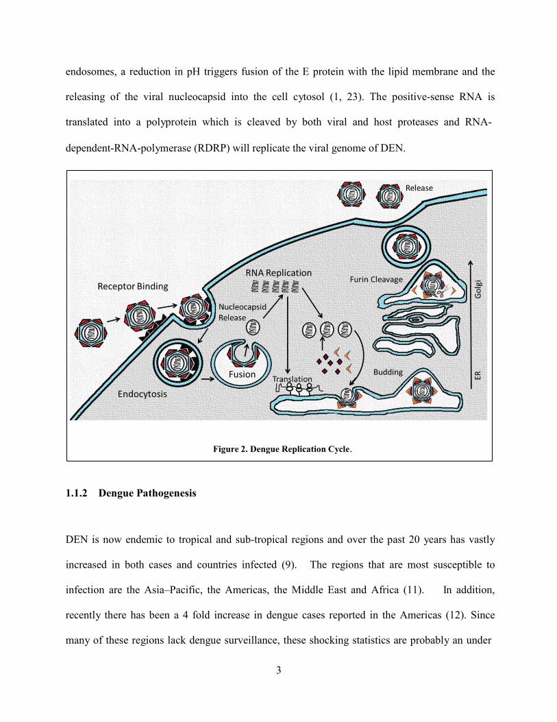
endosomes, a reduction in pH triggers fusion of the E protein with the lipid membrane and the
releasing of the viral nucleocapsid into the cell cytosol (1, 23). The positive-sense RNA is
translated into a polyprotein which is cleaved by both viral and host proteases and RNA-
dependent-RNA-polymerase (RDRP) will replicate the viral genome of DEN.
Figure 2. Dengue Replication Cycle. 1.1.2 Dengue Pathogenesis
DEN is now endemic to tropical and sub-tropical regions and over the past 20 years has vastly
increased in both cases and countries infected (9). The regions that are most susceptible to
infection are the Asia–Pacific, the Americas, the Middle East and Africa (11). In addition,
recently there has been a 4 fold increase in dengue cases reported in the Americas (12). Since
many of these regions lack dengue surveillance, these shocking statistics are probably an under
4
estimate to the true problem. There are four serotypes (DV1-4) all of which share the same
structural and pathogenic characteristics but have distinct genetic features. It is thought that
DV2 and DV3 contribute most to the severity of disease (11).
Symptomatic Asymptomatic
Dengue Fever Undifferentiated
Fever
Dengue Hemorrhagic Fever
V iral Clearance
Dengue Shock Syndrome
Death
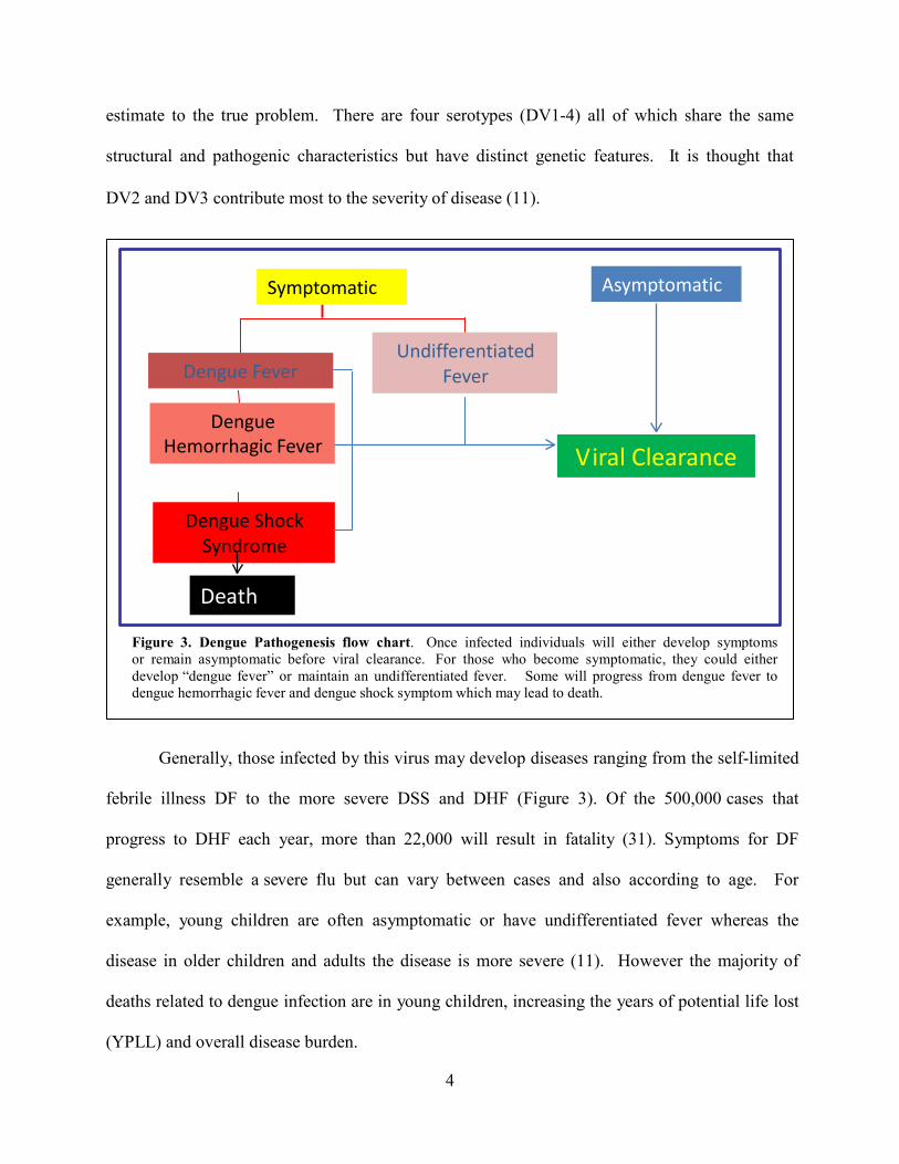
Figure 3. Dengue Pathogenesis flow chart. Once infected individuals will either develop symptoms or remain asymptomatic before viral clearance. For those who become symptomatic, they could either develop “dengue fever” or maintain an undifferentiated fever. Some will progress from dengue fever to dengue hemorrhagic fever and dengue shock symptom which may lead to death.
Generally, those infected by this virus may develop diseases ranging from the self-limited
febrile illness DF to the more severe DSS and DHF (Figure 3). Of the 500,000 cases that
progress to DHF each year, more than 22,000 will result in fatality (31). Symptoms for DF
generally resemble a severe flu but can vary between cases and also according to age. For
example, young children are often asymptomatic or have undifferentiated fever whereas the
disease in older children and adults the disease is more severe (11). However the majority of
deaths related to dengue infection are in young children, increasing the years of potential life lost
(YPLL) and overall disease burden.

5
The overall pathogesis of dengue infection is poorly understood. For example, it has not
conclusively been shown what the primary sites of infection are and exactly what cells are
involved in disease progression. However, recent data indicates that dengue may infect liver
sinusoidal endothelial cells and hepatocytes in vivo suggesting that the liver may play a key role
in dengue pathogenesis (34).
1.1.3 Dengue Prevention and Treatment
One might imagine an obvious solution to preventing this global disease could be to
eliminate the mosquito vector; however mosquito control measures have failed on numerous
occasions and attempts often create more problems (12). For example although eradicating
mosquitoes will reduce the amount of infections that occur, it also limits the number of people
who possess antibodies to fight the infection. This limitation in herd immunity may cause
detrimental effects if the mosquitoes have a chance to multiply at exponential levels for example
after a natural disaster.
Although a few compounds, ribavirin and mycophenolic acid, have shown to be potent
inhibit Dengue infection in vitro, there are no antiviral therapies approved for the treatment of
infection (27). Since there is no vaccine to prevent infection novel therapeutics are in urgent
need to treat the disease. High-throughput screening assays are a rapid, easy, cost-efficient way
to evaluate large libraries of compounds to find possible inhibitors of infection from viruses.
One such system, the CPE-based high-throughput assay, has proven to be robust and
reproducible systems to screen inhibitors for a number of viruses including influenza, hepatitis
C, bluetongue, human rhinovirus and severe acute respiratory syndrome coronavirus (SARS) (5,
16, 21, 22, 26). Because we have observed a similar cytopathic effect in DEN, we hypothesized

6
that novel inhibitors of DEN infection could be identified through a similar CPE-based
larger scale screening assay.
7
2.0 STATEMENT OF THE PROJECT While propagating the DV2, we noticed that it induces rapid cell death in the human hepatoma
cell line, HuH7.5.1. T his virus-induced cell death (cytopathic-effect) can be converted into a
screening assay in which inhibitors of Dengue infection will prevent cells from dying. In this
assay, viral induced cytopathic effects are quantitated by monitoring cellular ATP levels, which
positively correlate with cellular viability. ATP in the cell culture will drive the oxidation of
luciferin resulting in the emission of light that is quantitated using a luminometer. Since this
assay measures overall cellular viability, it simultaneously screens for inhibitory compounds
while controlling for unwanted cytopathic effects caused by the drugs (drugs that are toxic will
add to the viral educed cytopathic effect). We hypothesize that novel inhibitors of DEN
infection can be identified through a cytopathic effect-based larger scale screening. To test the
hypothesis, our objectives were:
Specific Aim 1 : The development and optimization of a CPE-based high-throughput
screening assay to identify novel inhibitory compounds of Dengue infection of hepatocytes.
Specific Aim 2: Screening a small compound library and the characterization of two novel
inhibitors Streptovitacin A and Nagilactone C.
We believe that this high-throughput screen will contribute to developing “lead” compounds of
novel antivirals and could be used to elucidate mechanisms of the dengue life-cycle.

8
3.0 MATERIALS AND METHODS
3.1 PROPAGATION AND IDENTIFICATION OF PROTOTYPE VIRUS 3.1.1 Cell lines and Reagents
Both Vero and C6/36 cells were purchased from ATCC and maintained as instructed by the
manufacturer. The Huh7.5.1 line was generated from a cured HCV replicon and was provided
by Dr. Francis Chisari (Scripps Research Institute). All cell lines were maintained in DMEM
supplemented with 5% Penicillin and streptomycin, 1% NEAA and 10% fetal bovine serum
(FBS) (Gemini Bio-Products).
Antibodies were produced from hybridoma cells purchased from ATCC (HB-112 and HB-
114). Both of these cell lines were maintained as instructed by the manufacturer. Secondary
antibodies were purchased from molecular probes (Invitrogen). M ycophenolic acid and
Bafilomycin A were purchased from Sigma.
3.1.2 Propagation of Virions
Prototype Dengue strains were propagated in either Vero or C6/36 cells depending on their
passage in previous cell lines. Cells were seeded 24 hour s prior to infection in a T175 cell
culture flask. When the cells had reached ~60-80% confluence they were infected with virus at
9
an MOI of 0.01. The cells were incubated for 7 days or until they began to die from the
cytopathic effect caused by the virus. After propagation, the virus containing media was
collected and centrifuged at 1500g for 5 minutes to remove extracellular debris. The supernatant
was then passed through a 0.22um filter syringe and alloquated before it was frozen at -80C for
long term storage.
3.1.3 Western Blot Assay
Cell Lysates (25µl) were subjected to electrophoresis through a 8% sodium dodecyl sulfate
(SDS)-polyacrylamide gel and transferred to nitrocellulose membranes by using the Mini Trans-
Blot electrophoretic transfer cell (Bio-Rad Laboratories, Richmond, Calif.) in transfer buffer
(15.6mM Tris base, 120mM glycine). The membrane containing transferred proteins was
blocked with 5% skim milk in Wash Buffer at room temperature for 10 m inutes. T he
membranes were incubated for 1h at room temperature with either a monoclonal antibody
produced by hybridoma cell line HB-114 (2H2) or HB-112 (4G2) against and actin at a dilution
of 1:10and 1:500 in 5% skim milk in Wash buffer, respectively. After incubation for 2 h at room
temperature, membranes were incubated with horseradish peroxidase-conjugated mouse anti-
goat IgG1:500 in 5% skim milk in Wash buffer for 1 h at room temperature. Finally, the signal
was developed using the ECL.
3.1.4 Immunofluorescence Assay
All cells were seeded 24 hours before infection to reach an 80% confluence. T he cells were
washed for 5 minutes 3 times with 1X PBS. After washing the cells, they were fixed using a 2%
10
paraformaldehyde and permeablized using 0.2% Triton or fixed and permeablized and fixed with 100% methanol. The dengue envelope proteins were detected using a 1:10 dilution of the 4G2 or
2H2 primary antibody isolated from a hybridoma culture and 1:500 of a HRP secondary
antibody. Nuclei were stained using Draq5 or DAPI as indicated.
3.1.5 Virus Tittering (Reed & Muench method)
Viruses were tittered using a Reed & Muench Tissue Culture Infectious Dose50 Assay
(TCID50/ml) system. A n example of the application of this assay is demonstrated in Figure 6.
Cells were seeded so that they will reach 80% confluence in 24 hour s. O n the day of the
experiment, serial dilutions of virus are made in media and a total of 8 wells were infected with
each serial dilution of the virus. After 48 hours incubation, an IFA was completed for all of the
used wells. T he wells were then examined under an epifluorescence for any positive wells.
Anything greater than one positive cell indicated a positive infected well. Interestingly though
where one cell is infected there is a small grouping of infected cells surrounding forming a foci.
3.2 LUCIFERASE-BASED HIGH THROUGHPUT SCREEN 3.2.1 Cytopathic Effect - Luciferase Reagent
The CPE Luciferase reagent was prepared by combining a 1ml of a 5X D-Luciferin Stock
Solution (1mM D-luciferin, 25mM Glycylglycine, 10mM DTT), 1ml luciferase enzyme
(1mg/ml), 0.5ml 250mM Glycylglycine and 3.5ml H2O in 9ml Luciferase Assay Buffer
11
(25mMGlycylglycine pH 7.8, 15mM Potassium Phosphate pH 7.8, 15mM MgSO4, 4mM EGTA
in H2O). The solution was then aliquoted into 1.8ml eppendorf tubes and kept at -80 C for long-
term storage.
3.2.2 CPE-based high-throughput screening assay.
Huh7.5.1 cells are counted with a hemacytometer and 1.8*104 cells are seeded per well in a 9 6
well format 24 hours prior to the experiment. After cells had reached an 80% confluence they
were infected with DV2 in the presence or absence of a compound for two hours. After the
infection the virus was replaced with fresh compound containing media. After a set duration, the
CPE solution was thawed at room temperature just before the assay. All media was removed
from the cells. A 5X passive lysis buffer was diluted to 1X in 1X PBS and then 70μl of the 1X
lysis buffer was added to each well. The plates were incubated in the 37 degree incubator for 10
minutes and then 50μl of the lysate was transferred to a luminometer plate. At least 50μl of the
CPE buffer was then quickly added to all wells in the luminometer plate and the luciferase
reading was quantified with an Auto Lumat luminometer.
3.2.3 Cell Culture Derived (HCVcc) Luciferase Assay
Production procedure of HCVcc (JFH-1 strain) expressing firefly luciferase that was inserted
between NS5A and NS5B (sequence available upon request) was described elsewhere (17, 32,
33). A 5X passive lysis buffer was diluted to 1X in 1X PBS and then 70μl of the 1X lysis buffer
was added to each well. The plates were incubated in the 37 degree incubator for 10 minutes and
then 50μl of the lysate was transferred to a luminometer plate. A t least 50μl of the LARII
12
luciferin buffer was then quickly added to all wells in the luminometer plate and the luciferase
reading was quantified with an Auto Lumat luminometer.
3.2.4 New Castle Disease Virus-Green Fluorescence Protein Assay
NDV-GFP virus production- NDV-GFP virus was a generous gift from Dr. Chris Basler (Mount
Sinai School of Medicine, NYC) and was produced in 10-days old embryonated eggs (Charles
River Laboratories International, Inc. Wilmington, MA). The inoculated eggs were incubated for
two days at 37 οC and the allantoic fluid containing virus was harvested, filtered and stored in -
80οC until use. The use of this virus has been described elsewhere [48].
NDV infection assay- 1.8*104Huh7.5.1 cells were seeded in 96 w ell plates. Cells were
washed with PBS and 20 µl (MOI 1) of NDV-GFP virus was added in 180 µl of PBS (with Ca
and Mg) and left at 37 οC for 1 hour. Cells were then washed with PBS and imaged. GFP signal
started to appear after 12 hours but peaked at 24hours post-infection. DAPI was added to stain
nuclei.
3.2.5 High-Throughput Screening Assay Data Analysis
The percentage of inhibition was calculated using a formula: % inhibition= (RLUexp-
RLUmin)/(RLUmax-RLUmin)*100 where RLUexp represents the luciferase counts obtained
from inhibitor treated wells, and RLUmax typically comes from uninfected wells whereas
RLUmin comes from DEN infected and treated with DMSO.
13
Since signal-to-background does not report any information on data variation, a screening
window coefficient, called "Z- factor," was used here to show the reproducibility and robustness
of the assay (35). T he Z’ factor = 1−((3SdDevS+3 SdDevN)/(|MeanS – MeanN|)) where S is the
luciferase counts from uninfected wells and N is the counts from infected wells. The Z’ values
are calculated taking luciferase counts in triplicate from within the same experiment. The signal-
to-background was calculated as a ratio between the RLUmax/RLUmin. Our data showed an
average Z-factor of 0.78±0.12 between plates.
14
4.0 RESULTS We hypothesize that novel inhibitors of DEN infection can be identified through a larger scale
screening. To test the hypothesis, our objectives were:
Aim 1: Propagation and tittering of prototype strains of Dengue virus. Aim 2 : The development and optimization of a CPE-based high-throughput screening
assay to identify novel inhibitory compounds of Dengue infection of hepatocytes.
Aim 3: Screening a small compound library and the characterization of two novel
inhibitors Streptovitacin A and Nagilactone C.
4.1 SPECIFIC AIM 1 RESULTS Aim 1: Propagation, detection and tittering of prototype strains of Dengue virus.
Until recently our lab was primarily focused on hepatitis C virus work. However we recently
decided to branch out and begin working on Dengue, another RNA virus that that belongs to the
Flavivirus family. We obtained all four serotypes of prototyped virus as a gift from Dr. Ernesto
Torres De Azeved Marques Jr. The following experiments outline the steps we took to
propagate the newly obtained virus using Vero and C6/36, two cell lines that were new to our
lab, and methods we utilized to confirm the infection of our cell lines. In addition these
15
experiments show that we successfully produced antibodies from hybridoma cells received from ATCC.
4.1.1 Experimental Scheme of Dengue propagation and detection
Because we obtained only a small quantity of DV2 (<1ml) we first needed to amplify the virus to
obtain enough to complete several experiments. The DV2 Thailand strain was propagated as
illustrated in Figure 4. Briefly, C6/36 cells were seeded 24 hours prior to infection in a T175 cell
culture flask. W hen the cells had reached 80% confluence they were infected with virus at an
MOI of 0.01. The cells were incubated for 7 days before the media was collected. Several 1 ml
aliquots were then prepared to prevent the loss of viable virus from repeated freeze-thaws. After
harvesting the virus we infected Vero and C6/36 cells to determine if we could distinguish
infection with our hybridoma produced antibodies on an IFA and western blot.

16
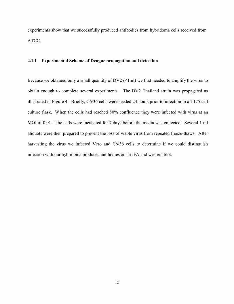
Figure 4. Overall scheme of the propogation and identification of prototype strains of DEN. C6/36 mosquito cells are infected with the prototype virus at an MOI of 0.01 and incubated for ~7days. The virus can then be identified on a western and IFA using antibodies produced by hybridoma cells.
4.1.2 Detection of the DV2 Infection of Vero and C6/36 We were able to detect infection from the newly propagated virus using both an IFA and western
blot (Figure 5). As shown in Figure 5A there was no prM/E signal in the mock (media) infection
for both C6/36 and Vero cells, however there are still a large number of viable cells present as
indicated by the blue DAPI nucleus stain. Conversely, in the cells infected with the DV2 strain
we see a distinct red signal surrounding the nucleus (cytoplasm) of the cell which is a distinct
characteristic for RNA viruses such as Dengue. Incubation of the western blot (Figure 5B) with
the hybridoma produced prM/E antibodies indicated a band in both positive lanes, but not the
negative lanes of C6/36 and Vero cells. Β-actin was included as a loading control.
17
A
B
Figure 5. The infection of propagated virus in C6/36 and Vero cells. (A) IFA of C6/36 and Vero cells infected or uninfected (Mock) with the DEN2 virus. The prM/E was detected with the 4G2 primary antibody conjugated with Alexa Fluor 568 as a secondary. (B) Western blot of C6/36 and Vero cells infected (+) of uninfected (-) with the DV2 clinical isolate virus. The prM/E was detected in both of these infected cell lines with the 4G2 primary antibody, but not in the uninfected. β-Actin was used as a loading control.

18
4.1.3 Tittering of DV1-4 using the Reed and Muench method to calculate TCID50/ml.
Since we had shown that we could detect infection of DV2 on an IFA, we attempted to propagate
and titer all 4 serotypes of DEN. To this aim we used the Reed and Muench method to calculate
a TCID50/ml as demonstrated in Figure 6. Briefly, serial dilutions are made of the virus and
then used to infect 8 wells of Vero cells. The wells are then counted as infected or uninfected
and these values are extrapolated into a virus titer.
Figure 6. Virus Titering using an IFA and the Reed & Muench method of calculating the TCID50/ml. The TCID50 is calculated using the formula:
lgTCID50=lgdo + (f + 1) lgR
Where do is the dilution that gives you a positive well , f is a number derived from then number of positive wells calculated by a moving average and R is the dilution factor.
As shown in Table 1, we obtained similar concentration to the initial for all viruses except the
Vero propagated DV1 Hawaii strain. Only the DV2 serotype caused a cytopathic effect during
the propagation procedure the remaining strains were propagated for a total of 7 days before
harvesting.
19
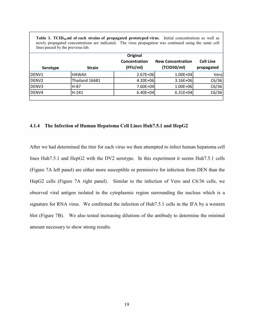
Table 1. TCID50/ml of each strains of propagated prototyped virus. Initial concentrations as well as newly propagated concentrations are indicated. The virus propagation was continued using the same cell lines passed by the previous lab.
Original Concentration New Concentration Cell Line
Serotype Strain (FFU/ml) (TCID50/ml) propagated
DENV1 HAWAII 2.67E+06 1.00E+04 Vero DENV2 Thailand 16681 4.20E+06 3.16E+06 C6/36 DENV3 H-87 7.60E+04 1.00E+06 C6/36 DENV4 H-241 6.40E+04 6.31E+04 C6/36
4.1.4 The Infection of Human Hepatoma Cell Lines Huh7.5.1 and HepG2
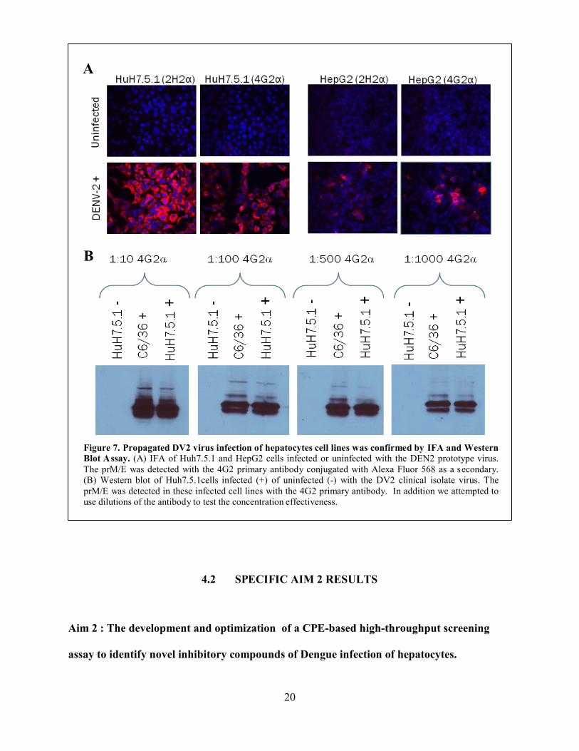
After we had determined the titer for each virus we then attempted to infect human hepatoma cell
lines Huh7.5.1 and HepG2 with the DV2 serotype. In this experiment it seems Huh7.5.1 cells
(Figure 7A left panel) are either more susceptible or permissive for infection from DEN than the
HepG2 cells (Figure 7A right panel). Similar to the infection of Vero and C6/36 cells, we
observed viral antigen isolated in the cytoplasmic region surrounding the nucleus which is a
signature for RNA virus. We confirmed the infection of Huh7.5.1 cells in the IFA by a western
blot (Figure 7B). We also tested increasing dilutions of the antibody to determine the minimal
amount necessary to show strong results.
20
A
B
Figure 7. Propagated DV2 virus infection of hepatocytes cell lines was confirmed by IFA and Western Blot A ssay. (A) IFA of Huh7.5.1 and HepG2 cells infected or uninfected with the DEN2 prototype virus. The prM/E was detected with the 4G2 primary antibody conjugated with Alexa Fluor 568 as a s econdary. (B) Western blot of Huh7.5.1cells infected (+) of uninfected (-) with the DV2 clinical isolate virus. The prM/E was detected in these infected cell lines with the 4G2 primary antibody. In addition we attempted to use dilutions of the antibody to test the concentration effectiveness.
4.2 SPECIFIC AIM 2 RESULTS Aim 2 : The development and optimization of a CPE-based high-throughput screening
assay to identify novel inhibitory compounds of Dengue infection of hepatocytes.
21
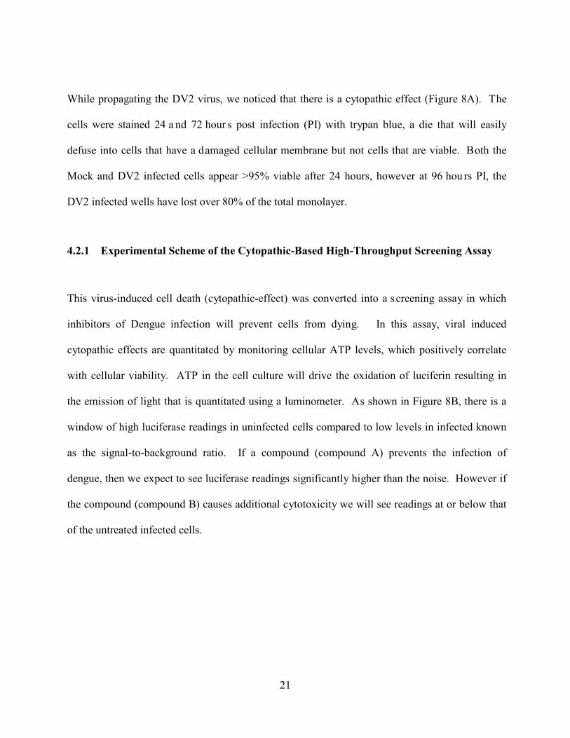
While propagating the DV2 virus, we noticed that there is a cytopathic effect (Figure 8A). The
cells were stained 24 a nd 72 hour s post infection (PI) with trypan blue, a die that will easily
defuse into cells that have a d amaged cellular membrane but not cells that are viable. B oth the
Mock and DV2 infected cells appear >95% viable after 24 hours, however at 96 hou rs PI, the
DV2 infected wells have lost over 80% of the total monolayer.
4.2.1 Experimental Scheme of the Cytopathic-Based High-Throughput Screening Assay
This virus-induced cell death (cytopathic-effect) was converted into a s creening assay in which
inhibitors of Dengue infection will prevent cells from dying. In this assay, viral induced
cytopathic effects are quantitated by monitoring cellular ATP levels, which positively correlate
with cellular viability. ATP in the cell culture will drive the oxidation of luciferin resulting in
the emission of light that is quantitated using a luminometer. As shown in Figure 8B, there is a
window of high luciferase readings in uninfected cells compared to low levels in infected known
as the signal-to-background ratio. If a compound (compound A) prevents the infection of
dengue, then we expect to see luciferase readings significantly higher than the noise. However if
the compound (compound B) causes additional cytotoxicity we will see readings at or below that
of the untreated infected cells.
22
A.
B.
Figure 8. Cytopathic effect of the Dengue virus on HuH7.5.1. (A). DEN infection of human hepatoma cell line Huh7.5.1 (MOI 1) induced massive cell death at 72 hours post-infection (PI). Huh7.5.1 were seeded in a 96 well format at a concentration of 1.8*104 and 72 hours after infection all wells were exposed to 1% Trypan Blue. We observed 80-90% cell death in the infected as compared to uninfected. (B) Overall design of the screening assay. The cell death resulting from dengue infection causes a loss of cellular ATP and by monitoring the ATP levels in vitro we can deduce the level of viral infection and replication before and after drug treatments. Huh7.5.1 cells are exposed to possible candidate drugs and infected with the dengue virus at an MOI of 1. After 48 hours infection the cells are lysed in a buffer containing both the luciferase enzyme and luciferin substrate. Any remaining ATP in the cell culture will drive the oxidation of luciferin resulting in the emission of light that is quantitated using a luminometer.

23
4.2.2 Optimization of Multiplicity of Infection and Duration of the Cytopathic Effect- Based HTS Assay.
Figure 9. Optimizing the concentration of virus and duration of the CPE-screening assay. Huh7.5.1 cells were infected with different multiplicities of infection (MOIs) for various lengths of time before taking a luciferase reading to find the shortest duration with the highest signal-to-background ratio.
Figure 9 shows the optimization of this screening assay to find the MOI with the broadest signal-
to-background ratio in the shortest duration. Having a broad signal-to-background will increase
the overall sensitivity of our assay and allow us to find hits that have a high probability of
inhibiting infection. The duration is important to optimize as well since too short of an infection
will not cause cell death and too long of a period will cause the chemicals to degrade and lose
effect. We therefore reasoned that infecting cells with an MOI of 1 for a total of 72 hours is an
24
optimal condition. Although the signal/noise ratio is >16, which is somewhat lower than the 22
fold at 96 hours, it is still a much broader window than at 48 hours or at any of the other MOIs.
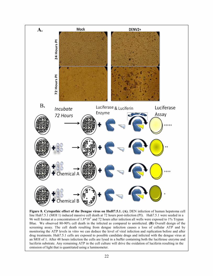
The optimization experiment from Figure 9 was repeated using IFN-α, a known inhibitor
of dengue infection, to show that the assay will in fact detect inhibition (Figure 10). Although
the inhibition was lowest at an MOI of 1 and duration of 72 hours, we reasoned that this
demonstrated the robustness of the assay, as it will only show positive hits that inhibit infection
more than a known non-specific inhibitor.
Figure 10. Efficiency of the CPE-based screening assay to show inhibition. IFN-α, a known non-specific inhibitor of dengue infection was used to show that the CPE assay will detect the inhibition of infection. Cells were infected using the same conditions as in the optimization experiment. An MOI of 1 would be optimal to ensure that only the compounds with strongest inhibition are defined as positive hits.

25
Since this assay measures overall cellular viability, it simultaneously screens for
inhibitory compounds while controlling for unwanted cytopathic effects caused by the drugs
(drugs that are toxic will add to the viral educed cytopathic effect). This is demonstrated with
Bafilomycin A in figure 11. In a TCID50/ml assay (Figure11A), Baf A inhibited the infection of
nearly 100%, however Baf A causes immense toxicity to the hepatocytes (Figure 11B). Since
our assay measures total cellular viability after treatment with compounds and virus, this drug
would have been screen out (Figure 11C).
Figure 11. Demonstration of the assays simultaneous detection of inhibition and toxicity. Baf A inhibits
nearly 100% infection in a TCID50 assay (A), however it caused cell death in over 50% of the cells by 72
hours post infection (B). Because our assay monitors cell viability and inhibition together, this compound
would have been screened out (C).

26
4.3 SPECIFIC AIM 3 RESULTS
Aim 3: Sreening a small compound library and the characterization of two novel
inhibitors Streptovitacin A and Nagilactone C.
4.3.1 Screening a Small Compound Library
Results from the initial CPE-based high-throughput screen on the National Cancer Institute
(NCI) 264 c ompound library are shown in Figure 12. A total of 11 c ompounds inhibited the
infection over our 50% cut off value. The compounds that fell below 0% mark are as such
because of the high level of toxicity. Two of the eleven compounds, Streptovitacin A and
Nagilactone C, were shown to inhibit infection with an IFA and were therefore further
characterized in the following experiments.
Figure 12. High-throughput Screening of Small Compound Library. The initial high throughput screen of the NCI 264 small compound library yielded 11 positive hits (over 50% inhibition). The two compounds that were found to inhibit in the following immunostaining assays, Streptovitacin A and Nagilactone C, are shown.
27
4.3.2 Validation of the Positive Hits with an Immunofluorescence Assay We attempted to validate the positive hits from the high-throughput screen using an IFA. Light
microscopy images (Figure 13 left) were taken to show the cell viability and morphology after
exposure to these chemicals. Untreated cells infected with media (mock) or DV2 48 hours prior
to staining were used as a control. There is no fluorescent signal in the mock infected cells,
however the DV2 cells show a high level of fluorescence that is localized to the cytoplasm
surrounding the cell nucleus. Compound 377 B 11 (streptovitacin A) inhibited infection at all
concentrations tested and 3977 G 11 (nagilactone C inhibited at all except the lowest 1μM
concentration. W e included results from one of the other compounds tested (3978 G8) as a
control as it did not inhibit infection at any of the concentrations.

28
Figure 13. IFA of two positive hits from the CPE assay. Compounds 3977 B 11 and G11 but not G8 inhibited DEN infection of Huh7.5.1 cells. Shown are light microscopy images of cells and immunofluorescence (IFA) staining of DEN pr/M (MOI= 1; 48 h rs post-infection) using the 2H2 antibody (red).
4.3.3 Calculating an IC50 and CC50 for the Two “Lead” Compounds.
After confirming that Streptovitacin A and Nagilactone C inhibited the infection of Huh7.5.1
cells by means of IFA, we then attempted to further characterize these compounds by creating
inhibitory curves to calculate the IC50 and CC50 values (Figure 14). To construct these curves we
infected cells in the presence of serial dilutions of compounds. Mycophenolic acid was used as a
control for this experiment and the IC50 value that we obtained from the curve (0.8uM Figure
14A) is analogous to the value provided in previous research (7). Streptovitacin A had a similar
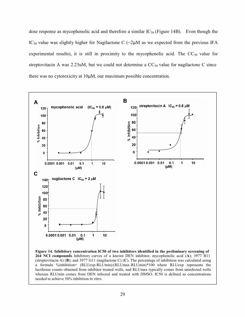
29
dose response as mycophenolic acid and therefore a similar IC50 (Figure 14B). Even though the
IC50 value was slightly higher for Nagilactone C (~2μM as we expected from the previous IFA
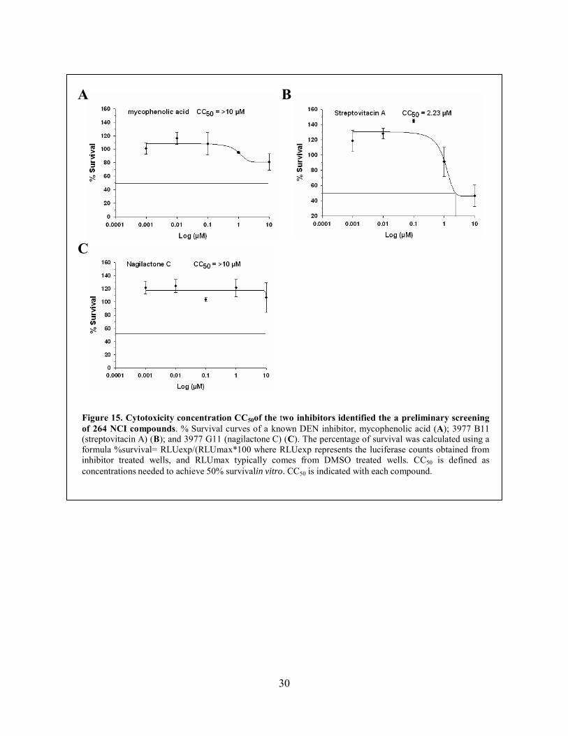
experimental results), it is still in proximity to the mycophenolic acid. The CC50 value for
streptovitacin A was 2.23uM, but we could not determine a CC50 value for nagilactone C since
there was no cytotoxicity at 10μM, our maximum possible concentration.
Figure 14. Inhibitory concentration IC50 of two inhibitors identified in the preliminary screening of 264 NCI compounds. Inhibitory curves of a known DEN inhibitor, mycophenolic acid (A); 3977 B11 (streptovitacin A) (B); and 3977 G11 (nagilactone C) (C). The percentage of inhibition was calculated using a formula %inhibition= (RLUexp-RLUmin)/(RLUmax-RLUmin)*100 where RLUexp represents the luciferase counts obtained from inhibitor treated wells, and RLUmax typically comes from uninfected wells whereas RLUmin comes from DEN infected and treated with DMSO. IC50 is defined as concentrations needed to achieve 50% inhibition in vitro.
30
A B
C
Figure 15. Cytotoxicity concentration CC50of the two inhibitors identified the a preliminary screening of 264 NCI compounds. % Survival curves of a known DEN inhibitor, mycophenolic acid (A); 3977 B11 (streptovitacin A) (B); and 3977 G11 (nagilactone C) (C). The percentage of survival was calculated using a formula %survival= RLUexp/(RLUmax*100 where RLUexp represents the luciferase counts obtained from inhibitor treated wells, and RLUmax typically comes from DMSO treated wells. CC50 is defined as concentrations needed to achieve 50% survivalin vitro. CC50 is indicated with each compound.
31
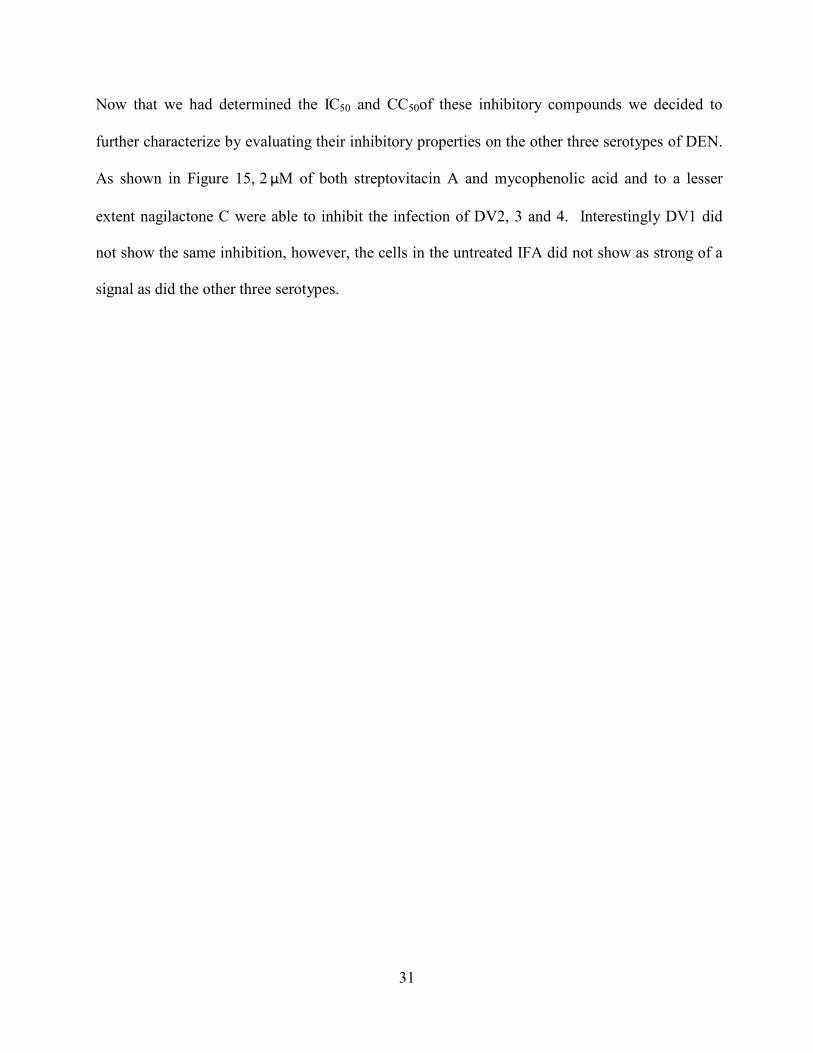
Now that we had determined the IC50 and CC50of these inhibitory compounds we decided to
further characterize by evaluating their inhibitory properties on the other three serotypes of DEN.
As shown in Figure 15, 2 μM of both streptovitacin A and mycophenolic acid and to a lesser
extent nagilactone C were able to inhibit the infection of DV2, 3 and 4. Interestingly DV1 did
not show the same inhibition, however, the cells in the untreated IFA did not show as strong of a
signal as did the other three serotypes.
32
Figure 16. Streptovitacin A and Nagilactone C inhibited the infection of all serotypes of DEN on Huh7.5.1 cells. Shown are light microscopy images of cells and immunofluorescence (IFA) staining of DEN pr/M (MOI= 1; 48 hrs post-infection) using the 2H2 antibody (red).
33
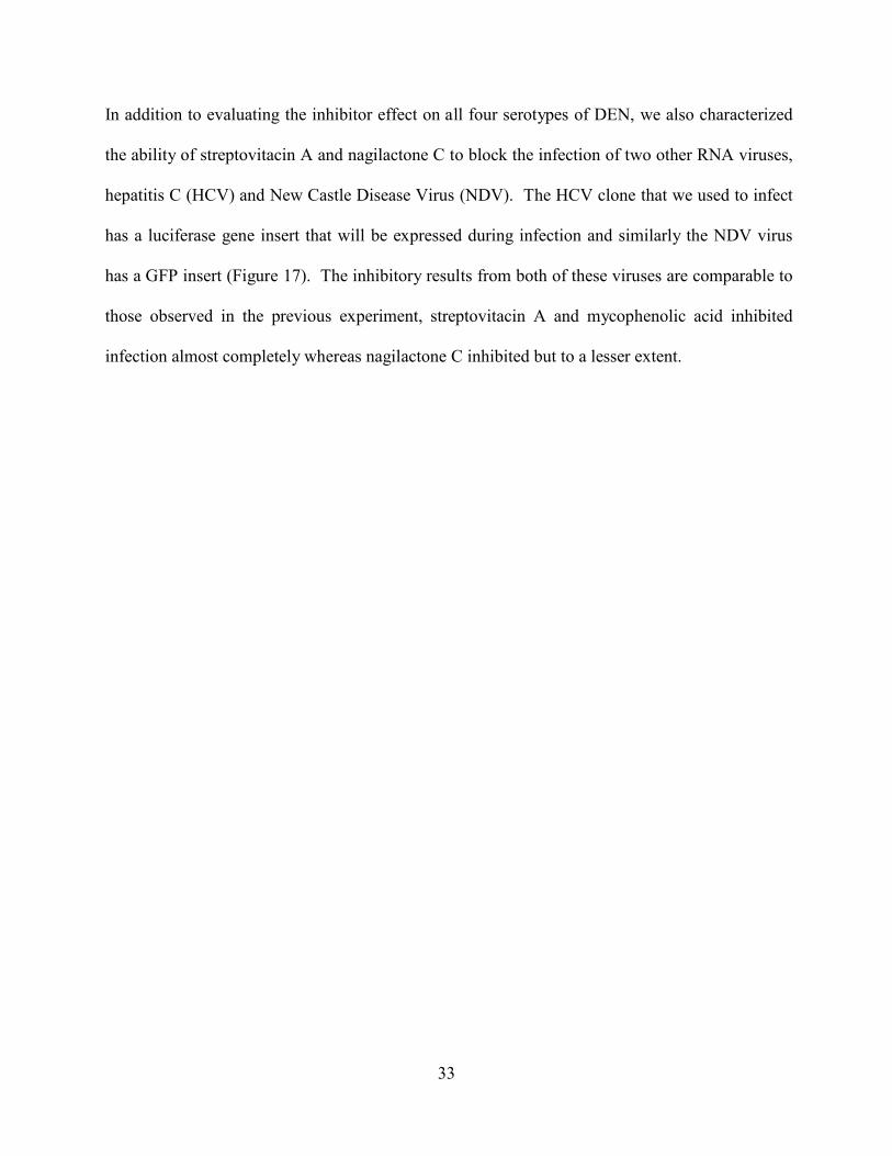
In addition to evaluating the inhibitor effect on all four serotypes of DEN, we also characterized
the ability of streptovitacin A and nagilactone C to block the infection of two other RNA viruses,
hepatitis C (HCV) and New Castle Disease Virus (NDV). The HCV clone that we used to infect
has a luciferase gene insert that will be expressed during infection and similarly the NDV virus
has a GFP insert (Figure 17). The inhibitory results from both of these viruses are comparable to
those observed in the previous experiment, streptovitacin A and mycophenolic acid inhibited
infection almost completely whereas nagilactone C inhibited but to a lesser extent.
34
Figure 17. Streptovitacin A and Nagilactone C inhibited the HCVcc and NDV-GFP infection of Huh7.5.1 cells. (A) Huh7.5.1 cells were infected with a h epatitis C luciferase expressing clone in the presence or absence of compounds. (B)Huh7.5.1 cells were infected with a New castle virus – green fluorescence expressing clone in the presence or absence of compounds. Mycophenolic acid, a known inhibitor of DEN infection was used as a control.
A.
B.
35
5.0 DISCUSSION Despite the severe disease burden of dengue infection each year, no va ccines or antivirals have
been found to treat this disease. In this study, we have successfully established a robust
technological high-throughput screening assay to find compounds that prevent the cytopathic
effect of dengue on he patocytes. We hypothesized that novel inhibitors of DEN infection could
be identified through this larger scale screening assay. These inhibitory compounds could be
used to elucidate cellular pathways that DEN may utilize for establishing its infection and possibly in
the long term as new therapeutics to combat the infection.
We have optimized the CPE-based assay using a range of virus MOIs for various
durations to find the largest signal-to-background ratio. Having a broad window for signal-to-
background permitted us to indentify compounds that have a potent effect in preventing this
cytopathic effect. At a MOI of 1 and a duration of 72 hou rs we observed the largest signal-to-
background ratio (>16) in the shortest amount of time. Using IFN-α as a positive control under
these experimental conditions, we see that there is less that 50% inhibition of cell death. We
reasoned that this would be an appropriate MOI and time period to demonstrate the potency of
the compounds as a strong inhibitor of dengue infection like IFN-α could not inhibit at this
concentration.
This assay is advantageous to current methodology because it simultaneously filters out
compounds that inhibit infection but at the same time are toxic to cells. This simultaneous effect
36
is demonstrated with Bafilomycin A in figure 11. In a TCID50/ml assay previously carried out
on Baf A showed almost complete inhibition of infection, however because it is somewhat toxic
it would be screened out in our assay. One obvious limitation to this assay is that rather than the
compounds directly inhibiting virus infection, they could be acting on and inhibiting a pathway
or mechanism required for cell death. However, data validation is crucial in modern high-
throughput screening. By setting a relatively stringent criterion, we anticipate to only
characterize those compounds that potently inhibit DEN infection without high levels of
cytotoxicity. The proposed functional characterizations include IFA-based detection of viral
protein pr/M, determination of CC50 and IC50 as well as therapeutic window.
We screened a 264 compound library and found a number of hits that inhibit at least 50% of this cytopathic effect. Further evaluation of these positive hits with an IFA showed that two
compounds, streptovitacin A and nagilactone C, block dengue infection as is demonstrated in
Figure 9. W e then did a dose response curve to find the IC50 for streptovitacin A and
nagilactone C and found the IC50 for each was comparable to mycophenolic acid, a known
inhibitor of dengue (7). Unfortunately were not able to determine the CC50 value as our highest
concentration available for each compound did not cause cytotoxicity.
Streptovitacin A and nagilactone C have been extensively studied for their use as possible
antitumor reagents. The chemical structures for both of these compounds are provided in Figure
18. Streptovitacin A is a hydrolated form of cycloheximide, a strong antifungal inhibitor
compound produced by the bacterium Streptomyces griseus, and also functions as an inhibitor of
protein biosynthesis (25). However unlike its parental compound, streptovitacin A does not
cause a depression of DNA or RNA synthesis and has low cytotoxicity and thus it was
previously studied as a possible antitumor agent. In addition this compound was used to study
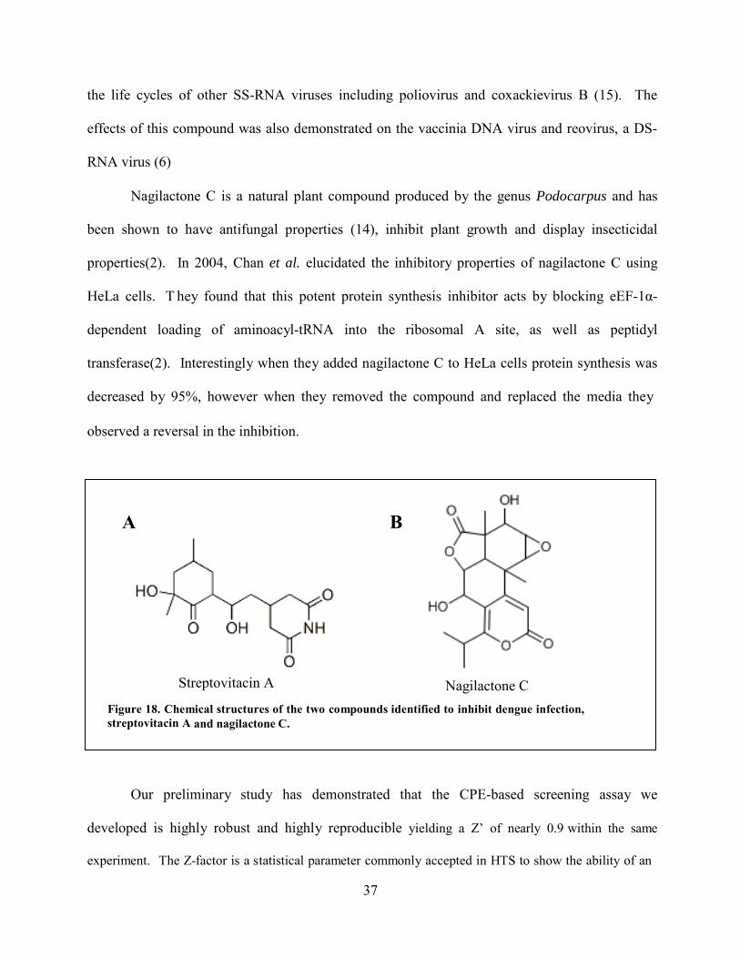
37
the life cycles of other SS-RNA viruses including poliovirus and coxackievirus B (15). The
effects of this compound was also demonstrated on the vaccinia DNA virus and reovirus, a DS-
RNA virus (6)
Nagilactone C is a natural plant compound produced by the genus Podocarpus and has
been shown to have antifungal properties (14), inhibit plant growth and display insecticidal
properties(2). In 2004, Chan et al. elucidated the inhibitory properties of nagilactone C using
HeLa cells. T hey found that this potent protein synthesis inhibitor acts by blocking eEF-1α-
dependent loading of aminoacyl-tRNA into the ribosomal A site, as well as peptidyl
transferase(2). Interestingly when they added nagilactone C to HeLa cells protein synthesis was
decreased by 95%, however when they removed the compound and replaced the media they
observed a reversal in the inhibition.
A B
Streptovitacin A Nagilactone C
Figure 18. Chemical structures of the two compounds identified to inhibit dengue infection, streptovitacin A and nagilactone C.
Our preliminary study has demonstrated that the CPE-based screening assay we
developed is highly robust and highly reproducible yielding a Z’ of nearly 0.9 within the same
experiment. The Z-factor is a statistical parameter commonly accepted in HTS to show the ability of an
38
assay to identify “hits”. The Z-factor is reported as a number 0 to 1 and anything over 0.5 is considered
excellent (35). We therefore reason that this method can be used to identify novel inhibitors of
DEN infection. Unfortunately, the current study is of limited scope given that we only performed
the initial screening using a 264 compound library. To prove this assay is adaptable to a high-
throughput manner, in the future we plan to screen a natural product library from TimTec (700
compounds, gift of Dr. Nicholas Sluis-Cremer) and a homemade mini pathway inhibitor library
(100 inhibitors) comprising inhibitors of commons kinases, cell trafficking, cell death). The first
library will provide an excellent non-biased library to verify the assay robustness. The goal of
screening the mini-pathway inhibitor library is to identify cellular pathways that DEN may
utilize for establishing its infection.
39
BIBLIOGRAPHY
1. Bartenschlager, R., and S. Miller. 2008. Molecular aspects of Dengue virus replication. Future Microbiol 3:155-65.
2. Chan, J., S. N. Khan, I. Harvey, W. Merrick, and J. Pelletier. 2004. Eukaryotic
protein synthesis inhibitors identified by comparison of cytotoxicity profiles. RNA 10:528-43.
3. Chen, Y., T. Maguire, R. E. Hileman, J. R. Fromm, J. D. Esko, R. J. Linhardt, and
R. M. Marks. 1997. Dengue virus infectivity depends on envelope protein binding to target cell heparan sulfate. Nat Med 3:866-71.
4. Chen, Y. C., S. Y. Wa ng, an d C. C. K ing. 1999. Bacterial lipopolysaccharide inhibits
dengue virus infection of primary human monocytes/macrophages by blockade of virus entry via a CD14-dependent mechanism. J Virol 73:2650-7.
5. Chockalingam, K., R. L. Simeon, C. M. Ri ce, and Z . Chen. 2010. A cell protection
screen reveals potent inhibitors of multiple stages of the hepatitis C virus life cycle. Proc Natl Acad Sci U S A 107:3764-9.
6. Dales, S . 1965. Effects of streptovitacin A on the initial events in the replication of
vaccinia and reovirus. Proc Natl Acad Sci U S A 54:462-8. 7. Diamond, M. S., M. Zachariah, and E. Harris. 2002. Mycophenolic acid inhibits
dengue virus infection by preventing replication of viral RNA. Virology 304:211-21.
8. Gibbons, R. V. 2010. Dengue conundrums. Int J Antimicrob Agents 36 Suppl 1:S36-9. 9. Gibbons, R. V., and D. W. Vaughn. 2002. Dengue: an escalating problem. BMJ
324:1563-6. 10. Gubler, D. J. 2006. Dengue/dengue haemorrhagic fever: history and current status.
Novartis Found Symp 277:3-16; discussion 16-22, 71-3, 251-3. 11. Guzman, A., and R. E. Isturiz. 2010. Update on the global spread of dengue. Int J
Antimicrob Agents 36 Suppl 1:S40-2.
40
12. Guzman, M. G., S. B. Halstead, H. Artsob, P. Buchy, J. Farrar, D. J. Gubler, E. Hunsperger, A. Kroeger, H. S. Margolis, E. Martinez, M. B. Nathan, J. L. Pelegrino, C. Simmons, S. Yoksan, and R. W. Peeling. 2010. Dengue: a continuing global threat. Nat Rev Microbiol 8:S7-S16.
13. Jindadamrongwech, S ., C. Thepparit, and D. R. Smith. 2004. Identification of GRP
78 (BiP) as a l iver cell expressed receptor element for dengue virus serotype 2. Arch Virol 149:915-27.
14. Kubo, I., H. Muroi, and M. Himejima. 1993. Combination effects of antifungal
nagilactones against Candida albicans and two other fungi with phenylpropanoids. J Nat Prod 56:220-6.
15. Levitt, N. H., and R. L. Crowell. 1967. Comparative studies of the regeneration of HeLa
cell receptors for poliovirus T1 and coxsackievirus B3. J Virol 1:693-700. 16. Li, Q., C. Maddox, L. Rasmussen, J. V. Hobrath, and L. E. White. 2009. Assay
development and high-throughput antiviral drug screening against Bluetongue virus. Antiviral Res 83:267-73.
17. Liu, S., W. Yang, L. Shen, J. R. Turner, C. B. Coyne, and T. Wang. 2009. Tight
junction proteins claudin-1 and occludin control hepatitis C virus entry and are downregulated during infection to prevent superinfection. J Virol 83:2011-4.
18. Lozach, P. Y., L. Burleigh, I. Staropoli, E . Navarro-Sanchez, J. Harriague, J. L .
Virelizier, F. A. Rey, P. Despres, F. Arenzana-Seisdedos, and A. Amara. 2005. Dendritic cell-specific intercellular adhesion molecule 3-grabbing non-integrin (DC- SIGN)-mediated enhancement of dengue virus infection is independent of DC-SIGN internalization signals. J Biol Chem 280:23698-708.
19. Miller, J. L., B. J. de Wet, L. Martinez-Pomares, C. M. Radcliffe, R. A. Dwek, P. M.
Rudd, and S. Gordon. 2008. The mannose receptor mediates dengue virus infection of macrophages. PLoS Pathog 4:e17.
20. Navarro-Sanchez, E., R. Altmeyer, A . Amara, O . Schwartz, F. Fieschi, J. L.
Virelizier, F. Arenzana-Seisdedos, and P. Despres. 2003. Dendritic-cell-specific ICAM3-grabbing non-integrin is essential for the productive infection of human dendritic cells by mosquito-cell-derived dengue viruses. EMBO Rep 4:723-8.
21. Noah, J. W., W. Severson, D. L. Noah, L. Rasmussen, E. L. White, and C. B.
Jonsson. 2007. A cell-based luminescence assay is effective for high-throughput screening of potential influenza antivirals. Antiviral Res 73:50-9.
22. Phillips, T., L. Jenkinson, C. McCrae, B. Thong, and J. Unitt. 2011. Development of
a high-throughput human rhinovirus infectivity cell-based assay for identifying antiviral compounds. J Virol Methods.
41
23. Rodenhuis-Zybert, I. A.., J. Wilschut, an d J. M. Smit. 2010. Dengue virus life cycle: viral and host factors modulating infectivity. Cell Mol Life Sci 67:2773-86.
24. Ross, T. M. 2010. Dengue virus. Clin Lab Med 30:149-60.
25. Sabin, A. B. 1966. Different effects of chloramphenicol, dactinomycin, and
streptovitacin A on synthesis of tumor and virion antigens in SV40 virus-infected cells. Proc Natl Acad Sci U S A 55:1141-8.
26. Severson, W. E., N. Shindo, M. Sosa, T. Fletcher, 3rd, E. L. White, S. Ananthan, and
C. B . Jonsson. 2007. Development and validation of a high-throughput screen for inhibitors of SARS CoV and its application in screening of a 100,000-compound library. J Biomol Screen 12:33-40.
27. Takhampunya, R ., S. Ubol, H. S. Houng, C. E. Cameron, an d R . Padmanabhan.
2006. Inhibition of dengue virus replication by mycophenolic acid and ribavirin. J Gen Virol 87:1947-52.
28. Tassaneetrithep, B., T. H. Burgess, A. Granelli-Piperno, C. Trumpfheller, J. Finke,
W. Sun, M. A. Eller, K . Pattanapanyasat, S. Sarasombath, D. L. Birx, R. M. Steinman, S . Schlesinger, an d M. A. Marovich. 2003. DC-SIGN (CD209) mediates dengue virus infection of human dendritic cells. J Exp Med 197:823-9.
29. Thepparit, C., and D. R. Smith. 2004. Serotype-specific entry of dengue virus into liver
cells: identification of the 37-kilodalton/67-kilodalton high-affinity laminin receptor as a dengue virus serotype 1 receptor. J Virol 78:12647-56.
30. van der Schaar, H. M., M. J. Rust, C. Chen, H. vander Ende-Metselaar, J.
Wilschut, X. Zhuang, and J. M. Smit. 2008. Dissecting the cell entry pathway of dengue virus by single-particle tracking in living cells. PLoS Pathog 4:e1000244.
31. World-Health-Organization 2011, posting date. WHO report on g lobal surveillance of
epidemic-prone infectious diseases—dengue and dengue haemorrhagic fever. World Health Organization. [Online.]
32. Yang, W., B. L. Hood, S. L. Chadwick, S. Liu, S. C. Watkins, G. Luo, T. P. Conrads,
and T. Wang. 2008. Fatty acid synthase is up-regulated during hepatitis C virus infection and regulates hepatitis C virus entry and production. Hepatology 48:1396-403.
33. Yang, W., C. Qiu, N. Biswas, J. Jin, S. C. Watkins, R. C. Montelaro, C. B. Co yne,
and T. Wang. 2008. Correlation of the tight junction-like distribution of Claudin-1 to the cellular tropism of hepatitis C virus. J Biol Chem 283:8643-53.
34. Zellweger, R. M., T. R. Prestwood, and S. Shresta. 2010. Enhanced infection of liver
sinusoidal endothelial cells in a mouse model of antibody-induced severe dengue disease. Cell Host Microbe 7:128-39.
42
35. Zhang, J. H., T. D. Chung, and K. R. Oldenburg. 1999. A Simple Statistical Parameter for Use in Evaluation and Validation of High Throughput Screening Assays. J Biomol Screen 4:67-73.
Related Documents