PLEASE SCROLL DOWN FOR ARTICLE This article was downloaded by: [Gamal-Eldeen, Amira] On: 6 February 2010 Access details: Access Details: [subscription number 919112745] Publisher Taylor & Francis Informa Ltd Registered in England and Wales Registered Number: 1072954 Registered office: Mortimer House, 37- 41 Mortimer Street, London W1T 3JH, UK Natural Product Research Publication details, including instructions for authors and subscription information: http://www.informaworld.com/smpp/title~content=t713398545 A crystal lapiferin derived from Ferula vesceritensis induces apoptosis pathway in MCF-7 breast cancer cells Amira M. Gamal-Eldeen a ; M. -E. F. Hegazy b a Cancer Biology Unit, Biochemistry Department, Center of Excellence for Advanced Sciences, National Research Center, Dokki, Cairo, Egypt b Chemistry of Medicinal Plant Department, National Research Centre, Dokki, Giza, Egypt Online publication date: 05 February 2010 To cite this Article Gamal-Eldeen, Amira M. and Hegazy, M. -E. F.(2010) 'A crystal lapiferin derived from Ferula vesceritensis induces apoptosis pathway in MCF-7 breast cancer cells', Natural Product Research, 24: 3, 246 — 257 To link to this Article: DOI: 10.1080/14786410802685398 URL: http://dx.doi.org/10.1080/14786410802685398 Full terms and conditions of use: http://www.informaworld.com/terms-and-conditions-of-access.pdf This article may be used for research, teaching and private study purposes. Any substantial or systematic reproduction, re-distribution, re-selling, loan or sub-licensing, systematic supply or distribution in any form to anyone is expressly forbidden. The publisher does not give any warranty express or implied or make any representation that the contents will be complete or accurate or up to date. The accuracy of any instructions, formulae and drug doses should be independently verified with primary sources. The publisher shall not be liable for any loss, actions, claims, proceedings, demand or costs or damages whatsoever or howsoever caused arising directly or indirectly in connection with or arising out of the use of this material.

Welcome message from author
This document is posted to help you gain knowledge. Please leave a comment to let me know what you think about it! Share it to your friends and learn new things together.
Transcript
PLEASE SCROLL DOWN FOR ARTICLE
This article was downloaded by: [Gamal-Eldeen, Amira]On: 6 February 2010Access details: Access Details: [subscription number 919112745]Publisher Taylor & FrancisInforma Ltd Registered in England and Wales Registered Number: 1072954 Registered office: Mortimer House, 37-41 Mortimer Street, London W1T 3JH, UK
Natural Product ResearchPublication details, including instructions for authors and subscription information:http://www.informaworld.com/smpp/title~content=t713398545
A crystal lapiferin derived from Ferula vesceritensis induces apoptosispathway in MCF-7 breast cancer cellsAmira M. Gamal-Eldeen a; M. -E. F. Hegazy b
a Cancer Biology Unit, Biochemistry Department, Center of Excellence for Advanced Sciences,National Research Center, Dokki, Cairo, Egypt b Chemistry of Medicinal Plant Department, NationalResearch Centre, Dokki, Giza, Egypt
Online publication date: 05 February 2010
To cite this Article Gamal-Eldeen, Amira M. and Hegazy, M. -E. F.(2010) 'A crystal lapiferin derived from Ferulavesceritensis induces apoptosis pathway in MCF-7 breast cancer cells', Natural Product Research, 24: 3, 246 — 257To link to this Article: DOI: 10.1080/14786410802685398URL: http://dx.doi.org/10.1080/14786410802685398
Full terms and conditions of use: http://www.informaworld.com/terms-and-conditions-of-access.pdf
This article may be used for research, teaching and private study purposes. Any substantial orsystematic reproduction, re-distribution, re-selling, loan or sub-licensing, systematic supply ordistribution in any form to anyone is expressly forbidden.
The publisher does not give any warranty express or implied or make any representation that the contentswill be complete or accurate or up to date. The accuracy of any instructions, formulae and drug dosesshould be independently verified with primary sources. The publisher shall not be liable for any loss,actions, claims, proceedings, demand or costs or damages whatsoever or howsoever caused arising directlyor indirectly in connection with or arising out of the use of this material.
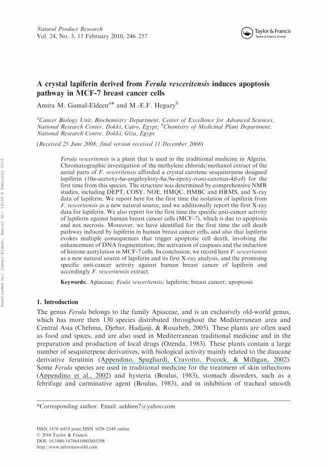
Natural Product ResearchVol. 24, No. 3, 15 February 2010, 246–257
A crystal lapiferin derived from Ferula vesceritensis induces apoptosispathway in MCF-7 breast cancer cells
Amira M. Gamal-Eldeena* and M.-E.F. Hegazyb
aCancer Biology Unit, Biochemistry Department, Center of Excellence for Advanced Sciences,National Research Center, Dokki, Cairo, Egypt; bChemistry of Medicinal Plant Department,National Research Centre, Dokki, Giza, Egypt
(Received 25 June 2008; final version received 11 December 2008)
Ferula vesceritensis is a plant that is used in the traditional medicine in Algeria.Chromatographic investigation of the methylene chloride/methanol extract of theaerial parts of F. vesceritensis afforded a crystal carotene sesquiterpene designedlapiferin (10�-acetoxy-6�-angeloyloxy-8�,9�-epoxy-trans-caxotan-4�-ol) for thefirst time from this species. The structure was determined by comprehensive NMRstudies, including DEPT, COSY, NOE, HMQC, HMBC and HRMS, and X-raydata of lapiferin. We report here for the first time the isolation of lapiferin fromF. vesceritensis as a new natural source, and we additionally report the first X-raydata for lapiferin. We also report for the first time the specific anti-cancer activityof lapiferin against human breast cancer cells (MCF-7), which is due to apoptosisand not necrosis. Moreover, we have identified for the first time the cell deathpathway induced by lapiferin in human breast cancer cells, and also that lapiferinevokes multiple consequences that trigger apoptotic cell death, involving theenhancement of DNA fragmentation, the activation of caspases and the inductionof histone acetylation inMCF-7 cells. In conclusion, we record here F. vesceritensisas a new natural source of lapiferin and its first X-ray analysis, and the promisingspecific anti-cancer activity against human breast cancer of lapiferin andaccordingly F. vesceritensis extract.
Keywords: Apiaceae; Feula vesceritensis; lapiferin; breast cancer; apoptosis
1. Introduction
The genus Ferula belongs to the family Apiaceae, and is an exclusively old-world genus,which has more then 130 species distributed throughout the Mediterranean area andCentral Asia (Chehma, Djebar, Hadjaiji, & Rouabeh, 2005). These plants are often usedas food and spices, and are also used in Mediterranean traditional medicine and in thepreparation and production of local drugs (Ozenda, 1983). These plants contain a largenumber of sesquiterpene derivatives, with biological activity mainly related to the daucanederivative ferutinin (Appendino, Spagliardi, Cravotto, Pocock, & Milligan, 2002).Some Ferula species are used in traditional medicine for the treatment of skin inftections(Appendino et al., 2002) and hysteria (Boulus, 1983), stomach disorders, such as afebrifuge and carminative agent (Boulus, 1983), and in inhibition of tracheal smooth
*Corresponding author. Email: [email protected]
ISSN 1478–6419 print/ISSN 1029–2349 online
� 2010 Taylor & Francis
DOI: 10.1080/14786410802685398
http://www.informaworld.com
Downloaded By: [Gamal-Eldeen, Amira] At: 14:04 6 February 2010
muscle (Aqel, Al-Khalil, Afifi, & Al-Eisawi, 1991). The species Ferula asafoetida has beenreported to exhibit anti-carcinogenic properties and to afford protection against free-radical-mediated diseases (Saleem, Alam, & Sultana, 2001) and exhibited anti-leishmanialactivity against promastigotes (Iranshahi et al., 2007). Previous phytochemical studiesof plants from this genus revealed that the main constituents are sesquiterpenes andsesquiterpene coumarins (reviewed in Ahmed et al., 2007).
Ferula vesceritensis (Batt.), which has the synonym F. tingitana L. var, is an endemicplant to Algeria and Libya, where it is used extensively in traditional medicine to treatcancer and inflammatory diseases (Chehma et al., 2005; Ozenda, 1983).
In our recent communication, we reported the isolation and structure elucidation oftwo new sesquiterpene coumarins (Ahmed et al., 2007; Lahouel et al., 2007) from amethylene chloride extract of F. vesceritensis. It is evident from the literature and previousinvestigations that the genus Ferula possesses high biological activities (Ahmed et al.,2007), which prompts us to study the anti-cancer activity for the major isolated compound,lapiferin, from F. vesceritensis with different extraction methods.
Lapiferin is a complex ester of sesquiterpene alcohols with aliphatic acid and it does notpossess ionophoric properties (Andrey et al., 2001). In an earlier report, lapiferin waspurified from the roots of F. lapidosa and its structure and configuration have beenestablished on the basis of chemical transformations and the analysis of spectralcharacteristics (Diaz, Fragam, Gonzalaz, Gonzalaz, & Hernandez, 1984; Golovina,Saidkhodzhaev, Abdullaev, Malikov, & Yagudaev, 1983). Recently, lapiferin extractedfrom F. communis and F. arrigonii sardinian showed antiproliferative activity against humancolon cancer, and the interaction with type II estrogen-binding sites underlies this activity(Poli et al., 2005).
Herein, we report for the first time the isolation of lapiferin from F. vesceritensis, andwe additionally report the first X-ray data for lapiferin. We also report here for the firsttime the specific anti-cancer activity of lapiferin against breast cancer cells, and themechanistic pathway of this cell death.
2. Methodology
2.1. General1H NMR (600MHz, CDCl3),
13C NMR (150MHz, CDCl3) and 2D spectra were recordedon a JEOL ECA 500 spectrometer, with TMS as an internal standard. FABMS andHRFABMS were recorded on a JEOL SX102A mass spectrometer. IR spectra wererecorded on a JASCO FT/IR-5300 spectrometer.
2.2. Plant material
Ferula vesceritensis was collected during the flowering stage in March 2004 near Biskra,approximately 300 miles south-east of Algiers, Algeria, by Amar Zellagui, Departmentof Chemistry, Constantine University, where a voucher specimen has been deposited(AM#112).
2.3. Extraction and isolation
Roots of F. vesceritensis (800 g) were crushed and extracted with CH2Cl2 :MeOH (1 : 1)at room temperature. The extract was concentrated in vacuo to obtain a residue (30 g).
Natural Product Research 247
Downloaded By: [Gamal-Eldeen, Amira] At: 14:04 6 February 2010
The residue was fractionated by silica gel CC (6� 120 cm) eluted with hexane (3 L),followed by a gradient of n-hexane–CH2Cl2 up to 100% CH2Cl2 and CH2Cl2–MeOH upto 15% MeOH (2L of each solvent mixture). The n-hexane : CH2Cl2 (3 : 1) fraction wassubjected to silica gel CC (3� 60 cm) eluted with n-hexane–CH2Cl2–MeOH to give 13 sub-fractions. On the basis of TLC chromatographies, fractions 6 and 7 were further purifiedon sephadex LH-20 (2� 40 cm) eluted with n-hexane : EtOAc (6 : 1) to afford lapiferin(30mg) in a crystal form.
2.4. Cell culture
Several human cell lines were used in the testing of anti-cancer activity, including:lymphoblastic leukemia (1301 cells), a generous gift from The Training Center ofDakoCytomation, Elly, UK, hepatocellular carcinoma (Hep-G2) and colon carcinoma(HCT-116) (ATCC, VA, USA). Cells were routinely cultured in DMEM (Dulbeco’smodified Eagle’s medium) at 37�C in humidified air containing 5% CO2. Media weresupplemented with 10% foetal bovine serum (FBS), 2mML-glutamine, containing100 unitsmL�1 penicillin G sodium, 100 unitsmL�1 streptomycin sulphate and250 ngmL�1 amphotericin B. Monolayer cells were harvested by trypsin/EDTA treatment.The tested compound was dissolved in dimethyl sulphoxide (DMSO, 99.9%, HPLC grade)and diluted 1000-fold in the assays. In all the cellular experiments, results were comparedwith DMSO-treated cells. Compound dilutions were tested before assays for endotoxinusing Pyrogent� Ultra gel clot assay, and they were found to be free of endotoxin. Allexperiments were repeated four times, unless mentioned, and the data was represented as(mean� SD). Except the above mentioned, all culture materials were obtained fromCambrex BioScience (Copenhagen, Denmark), and all chemicals were from Sigma (USA).
2.5. Cytotoxicity assay
Anti-proliferative activity against hepatocellular carcinoma (Hep-G2), breast carcinoma(MCF-7) and lymphoblastic leukaemia 1301 cells (T-lymphocytes) was estimated by a3-(4,5-dimethyl-2-thiazolyl)-2,5-diphenyl-2H-tetrazolium bromide (MTT) assay (Hansen,Nielsen, & Berg, 1989). The yellow tetrazolium salt of MTT is reduced by mitochondrialdehydrogenases in metabolically active cells to form insoluble purple formazan crystals,which are solubilised by the addition of a detergent. Cells (5� 104 cells per well) wereincubated with various concentrations of the compound at 37�C in a FBS-free mediumbefore being submitted to the MTT assay. The absorbance was measured with an ELISAreader (BioRad, Munchen, Germany) at 570 nm. The relative cell viability was determinedby the amount of MTT converted to the insoluble formazan salt. The data are expressed asthe mean percentage of viable cells as compared to the respective control cultures treatedwith the solvent. The half maximal growth inhibitory concentration (IC50) values werecalculated from the line equation of the dose-dependent curve of each compound.
2.6. Apoptosis and necrosis staining
The type of the cell death in MCF-7 cells was investigated in the treated and untreated cellsusing acridine orange/ethidium bromide staining. In brief, a mixture of 100 mgmL�1
acridine orange and 100 mgmL�1 ethidium bromide was prepared in PBS. The cell uptake
248 A.M. Gamal-Eldeen and M.-E.F. Hegazy
Downloaded By: [Gamal-Eldeen, Amira] At: 14:04 6 February 2010
of the stain was monitored under a fluorescence microscope and the apoptotic, necroticand viable cells were counted.
2.7. DNA fragmentation
DNA fragmentation was essentially assayed as reported previously (Messmer et al., 1998).Briefly, the pellets of the treated and untreated MCF-7 cells were re-suspended in 250 mL10mM Tris, 1mM EDTA, pH 8.0 (TE-buffer), and incubated with an additional volumelysis buffer (5mM Tris, 20mM EDTA, pH 8.0, 0.5% Triton X-100) for 30min at 48�C.After lysis, the intact chromatin (pellet) was separated from DNA fragments (supernatant)by centrifugation for 15min at 13,000� g. Pellets were re-suspended in 500 mL TE-bufferand samples were precipitated by adding 500 mL of 10% trichloroacetic acid at 48�C.Samples were pelleted at 4000 rpm for 10min and the supernatant was removed. Afteraddition of 300 mL of 5% trichloroacetic acid, samples were boiled for 15min. DNAcontents were quantified using the diphenylamine reagent (Burton, 1956). The percentageof DNA fragmented was calculated as the ratio of the DNA content in the supernatant tothe amount in the pellet.
2.8. Histone deacetylase activity
The activity of HDAC was measured in MCF-7 cells, using a colourimetric assay kit(BioVision, Mountain View, Kit No. K331-100). The procedure involves the use of theHDAC colourimetric substrate (Boc-Lys(Ac)-pNA), which comprises an acetylated lysineside chain and is incubated with a sample containing nuclear extract. Deactivationsensitises the substrate, and treatment with the lysine developer produces a chromophore,which can be analysed using a colourimetric plate reader. An HeLa cell nuclear extract wasused as a positive control. A standard curve was prepared using the known amount of thedeacetylated standard (Boc-Lys-pNA) included in the kit. A similar volume of controlsample was added to 100 ngmL�1 trichostatin A (TSA), as a known inhibitor of HDACactivity.
2.9. Evaluation of caspase activity
The lysates of the treated and untreated MCF-7 cells were submitted to a total caspasekit according to the manufacturer’s instructions. A Red Multi-Caspase Staining Kit(# PK-CA577-K190), PromoKine, Heidelberg, Germany was used for analysis of totalcaspases in a black microtiter plate with fluorescence plate reader at Ex.¼ 540 nm andEm.¼ 570 nm. The assay utilises the caspase family inhibitor VAD-FMK conjugated tosulfo-rhodamine (Red-VAD-FMK) as the fluorescent in situ marker. A comparisonbetween the fluorescence readings of the treated sample and the untreated cells allowsdetermination of the fold increase in the total caspases.
2.10. Statistical analysis
All values were expressed at the mean� SD of four measurements. Data were statisticallyanalysed using an IBM computer supplied with SPSS 10.00 for Windows (SPSS Inc.,Chicago, USA). The Student’s unpaired t-test as well as the one-way analysis of variance
Natural Product Research 249
Downloaded By: [Gamal-Eldeen, Amira] At: 14:04 6 February 2010
(ANOVA) test was used to detect the statistical significance. p-Values of more than 0.05were considered insignificant.
3. Results and discussion
Ferula vesceritensis roots were air dried, ground to a powder and thoroughly extractedwith CHCl3 :MeOH (1 : 1, v/v), which was then separated by column chromatography (Sigel) using mixtures of hexane, CHCl3 and MeOH of increasing polarity, giving 13fractions. On the basis thin layer chromatographies, fractions 6 and 7 were collected andpurified by a combination of column chromatography (Si gel, Sephadex LH-20) affordinglapiferin (10�-acetoxy-6�-angeloyloxy-8�,9�-epoxy-trans-caxotan-4�-ol) for the first timefrom F. vesceritensis species.
Compound 1 was isolated as white crystal, ½��25D : þ53.0� (c 0.6, MeOH). Its IRspectrum showed absorption bands at 3421 cm�1 (OH) and 1714 cm�1 (C¼O). Themolecular formula of 1, C22H34O6, was determined from HRCIMS (395.50156) and 13CNMR spectral data. The 1H and 13C NMR (Table 1) exhibited signals for a daucanesesquiterpene nucleus, including a doublet at �H 4.91 (1H, d, J¼ 5.5Hz, H-10) thatcorrelated in the 1H–1H COSY spectrum with a signal at �H 2.93 (1H, d, J¼ 5.5Hz, H-9).In the same experiment, a signal at �H 5.23 (1H, ddd, J¼ 11.0, 11.0, 3.0Hz, H-6) exhibited
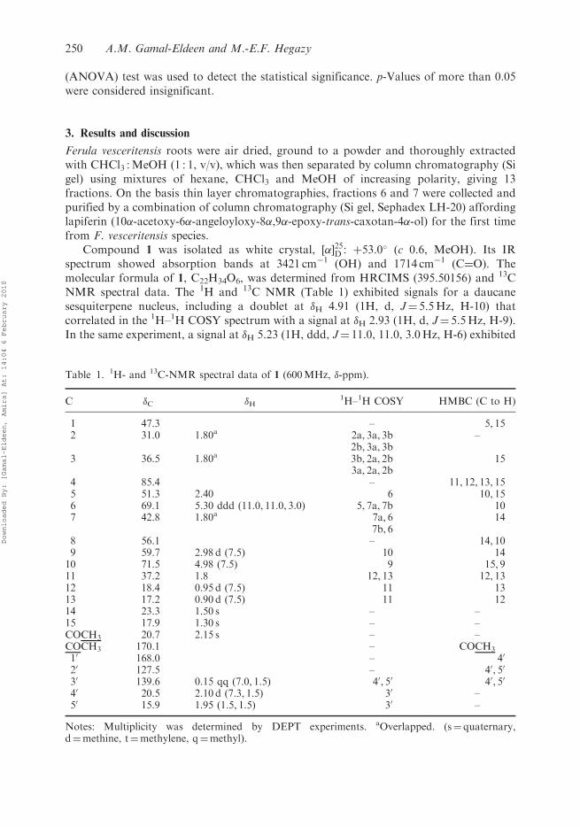
Table 1. 1H- and 13C-NMR spectral data of 1 (600MHz, �-ppm).
C �C �H1H–1H COSY HMBC (C to H)
1 47.3 – 5, 152 31.0 1.80a 2a, 3a, 3b –
2b, 3a, 3b3 36.5 1.80a 3b, 2a, 2b 15
3a, 2a, 2b4 85.4 – 11, 12, 13, 155 51.3 2.40 6 10, 156 69.1 5.30 ddd (11.0, 11.0, 3.0) 5, 7a, 7b 107 42.8 1.80a 7a, 6 14
7b, 68 56.1 – 14, 109 59.7 2.98 d (7.5) 10 1410 71.5 4.98 (7.5) 9 15, 911 37.2 1.8 12, 13 12, 1312 18.4 0.95 d (7.5) 11 1313 17.2 0.90 d (7.5) 11 1214 23.3 1.50 s – –15 17.9 1.30 s – –COCH3 20.7 2.15 s – –COCH3 170.1 – COCH3
10 168.0 – 40
20 127.5 – 40, 50
30 139.6 0.15 qq (7.0, 1.5) 40, 50 40, 50
40 20.5 2.10 d (7.3, 1.5) 30 –50 15.9 1.95 (1.5, 1.5) 30 –
Notes: Multiplicity was determined by DEPT experiments. aOverlapped. (s¼ quaternary,d¼methine, t¼methylene, q¼methyl).
250 A.M. Gamal-Eldeen and M.-E.F. Hegazy
Downloaded By: [Gamal-Eldeen, Amira] At: 14:04 6 February 2010
correlations with three signals at �H 2.39 (1H, d, J¼ 11.0Hz, H-5), 2.11 (1H,d, J¼ 14.0Hz,
H-7a) and 1.92 (1H, ddd, J¼ 14.0, 14.0, 6.8Hz, H-7b). The two methyl doublet signals
at �H 0.89 (3H, d, J¼ 7Hz, H-12) and 0.87 (3H, d, J¼ 7Hz, H-13) coupled in the 1H–1H
COSY spectrum with a signal at �H 1.90 (1H, m), which accounted for an isopropyl
moiety. Additionally, there were three singlet signals for methyl groups at 2.10, 1.44 and
1.28, for O(CO)CH3, H-14 and H-15, respectively. The 1H NMR spectrum also exhibited
signals typical for an angelate moiety at �H 6.10 (H-30), 1.99 (H-40) and 1.87 (H-50). Table 1
lists the other protons, which were assigned by 1H–1H COSY.The 13C NMR spectral data of 1 (Table 1) exhibited 22 carbon signals that were
resolved by DEPT experiment into six methines, three methylenes, seven methyls and six
quaternary carbons. The two signals at �C 170.1 and 168.0 could be assigned by HMBC to
the two carbonyl carbons of the acetate and angelate groups, respectively.Five oxygen bearing carbons were assigned at �C 85.4 (C-4), 69.1 (C-6), 56.1 (C-8), 59.7
(C-9) and 71.5 (C-10) (Diaz et al., 1984; Golovina et al., 1983). Other proton and carbon
signals were determined by HMQC and HMBC. In a HMBC experiment, H-10 (�H 4.98)
showed a cross peak with the carbon signal at �C 51.3 (C-5), 56.1 (C-8) locating the acetyl
moiety at C-10; H-11 (�H 1.80) exhibited a cross peak with the carbon signal at �C 85.4
(C-4), placing the isopropyl moiety at C-4, and H-9 (�H 2.98) showed a cross peak with the
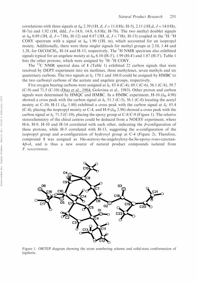
carbon signal at �C 71.5 (C-10), placing the epoxy group at C-8/C-9 (Figure 1). The relative
stereochemistry of the chiral centres could be deduced from a NOESY experiment, where
H-6, H-9, H-10 and H-14 correlated with each other, indicating the �-configuration of
these protons, while H-5 correlated with H-13, suggesting the �-configuration of the
isopropyl group and �-configuration of hydroxyl group at C-4 (Figure 2). Therefore,
compound 1 was assigned as 10�-acetoxy-6�-angeloyloxy-8�,9�-epoxy-trans-caxotan-4�-ol, and is thus a new source of natural product compounds isolated from
F. vesceritensis.
Figure 1. ORTEP diagram showing the atom numbering scheme and solid-state conformation oflapiferin.
Natural Product Research 251
Downloaded By: [Gamal-Eldeen, Amira] At: 14:04 6 February 2010
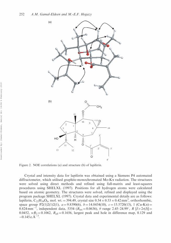
Crystal and intensity data for lapiferin was obtained using a Siemens P4 automateddiffractometer, which utilised graphite-monochromated Mo-Ka radiation. The structureswere solved using direct methods and refined using full-matrix and least-squaresprocedures using SHELXL (1997). Positions for all hydrogen atoms were calculatedbased on atomic geometry. The structures were solved, refined and displayed using theprogram package SHELXL (1997). Crystal data and experimental details are as follows:lapiferin, C22H34O6, mol. wt.¼ 394.49, crystal size 0.54� 0.53� 0.42mm3, orthorhombic,space group P2(1)2(1)2(1), a¼ 9.8390(6), b¼ 14.8434(10), c¼ 15.5720(13), l (Cu-Ka)¼0.824mm�1, independent data, 5354 (Rint¼ 0.0636), � range 2.45–24.99�, R [I42r(I)]¼0.0452, wR2¼ 0.1062, Rall¼ 0.1656, largest peak and hole in difference map, 0.129 and�0.145 e.A�3.
(a)
(b)
O
O
O
HO
12
35
67
8
4
910
11
1213
14
15
1′2′
4′
5′
3′
O
CH3
O
H
Figure 2. NOE correlations (a) and structure (b) of lapiferin.
252 A.M. Gamal-Eldeen and M.-E.F. Hegazy
Downloaded By: [Gamal-Eldeen, Amira] At: 14:04 6 February 2010
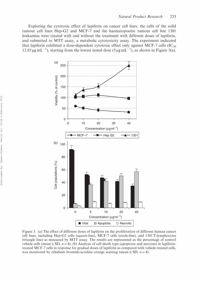
Exploring the cytotoxic effect of lapiferin on cancer cell lines, the cells of the solidtumour cell lines Hep-G2 and MCF-7 and the haematopoeitic tumour cell line 1301leukaemia were treated with and without the treatment with different doses of lapiferin,and submitted to MTT assay, a metabolic cytotoxicity assay. The experiment indicatedthat lapiferin exhibited a dose-dependent cytotoxic effect only against MCF-7 cells (IC50
12.85mgmL�1), starting from the lowest tested dose (5 mgmL�1), as shown in Figure 3(a).
(a)
0
50
100
150
200
250
0 10 20 30 40
Concentration (mgml−1)
Via
bilit
y (%
of c
ontr
ol)
MCF–7 Hep G2 1301
(b)
0
20
40
60
80
100
0 10 20 40
Concentration (mgml−1)
Cel
l pop
ulat
ions
(%
)
Vital Apoptotic Necrotic
5
Figure 3. (a) The effect of different doses of lapiferin on the proliferation of different human cancercell lines, including Hep-G2 cells (square-line), MCF-7 cells (circle-line), and 1301T-lymphocytes(triangle line) as measured by MTT assay. The results are represented as the percentage of controlvehicle cells (mean� SD, n¼ 4). (b) Analysis of cell death type (apoptosis and necrosis) in lapiferin-treated MCF-7 cells in response for gradual doses of lapiferin as compared with vehicle-treated cells,was monitored by ethidium bromide/acridine orange staining (mean� SD, n¼ 4).
Natural Product Research 253
Downloaded By: [Gamal-Eldeen, Amira] At: 14:04 6 February 2010
Screening of lapiferin cytotoxicity against another solid tumour cells; Hep-G2 cellsindicated that lapiferin did not affect the cell growth. On the other hand, submission ofhaematopoeitic tumour cells; 1301 cells, to MTT assay described that lapiferin induced theproliferation of lymphocytes in a dose-dependant manner up to 2.48-fold of control at adose of 40 mgmL�1 (Figure 3(a)). This finding suggested that lapiferin is animmunostimulant agent that can drastically induce T-lymphocytes proliferation. Asuggestion that needs to be explored further.
Cell death is generally classified into two large categories: apoptosis, representing‘active’ programmed cell death, and necrosis, representing ‘passive’ cell death without(known) underlying regulatory mechanisms; both are distinguished by well-definedmorphological and biochemical features. Necrosis is characterised by cell swelling,disruption and rapid disintegration of the cell membrane (Kalka, Ahmad, Criswell,Boothman, & Mukhtar, 2000). In contrast, during apoptosis the cells undergo nuclearand cytoplasmic shrinkage, the chromatin is condensed and fragmented, and the cellsare finally broken into multiple membrane-surrounded bodies (apoptotic bodies)(Kalka et al., 2000).
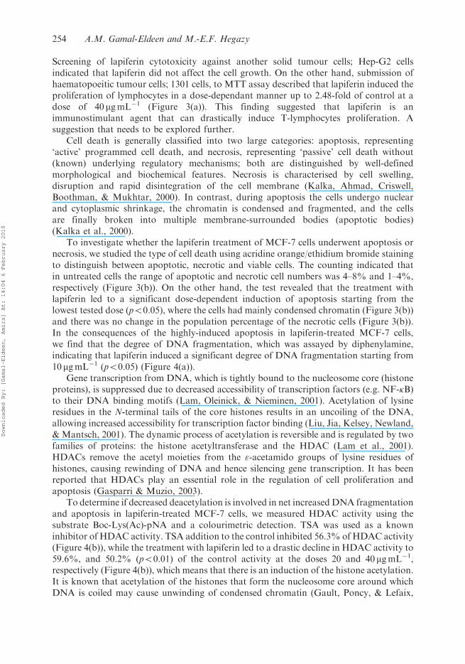
To investigate whether the lapiferin treatment of MCF-7 cells underwent apoptosis ornecrosis, we studied the type of cell death using acridine orange/ethidium bromide stainingto distinguish between apoptotic, necrotic and viable cells. The counting indicated thatin untreated cells the range of apoptotic and necrotic cell numbers was 4–8% and 1–4%,respectively (Figure 3(b)). On the other hand, the test revealed that the treatment withlapiferin led to a significant dose-dependent induction of apoptosis starting from thelowest tested dose (p50.05), where the cells had mainly condensed chromatin (Figure 3(b))and there was no change in the population percentage of the necrotic cells (Figure 3(b)).In the consequences of the highly-induced apoptosis in lapiferin-treated MCF-7 cells,we find that the degree of DNA fragmentation, which was assayed by diphenylamine,indicating that lapiferin induced a significant degree of DNA fragmentation starting from10 mgmL�1 (p50.05) (Figure 4(a)).
Gene transcription from DNA, which is tightly bound to the nucleosome core (histoneproteins), is suppressed due to decreased accessibility of transcription factors (e.g. NF-�B)to their DNA binding motifs (Lam, Oleinick, & Nieminen, 2001). Acetylation of lysineresidues in the N-terminal tails of the core histones results in an uncoiling of the DNA,allowing increased accessibility for transcription factor binding (Liu, Jia, Kelsey, Newland,& Mantsch, 2001). The dynamic process of acetylation is reversible and is regulated by twofamilies of proteins: the histone acetyltransferase and the HDAC (Lam et al., 2001).HDACs remove the acetyl moieties from the "-acetamido groups of lysine residues ofhistones, causing rewinding of DNA and hence silencing gene transcription. It has beenreported that HDACs play an essential role in the regulation of cell proliferation andapoptosis (Gasparri & Muzio, 2003).
To determine if decreased deacetylation is involved in net increased DNA fragmentationand apoptosis in lapiferin-treated MCF-7 cells, we measured HDAC activity using thesubstrate Boc-Lys(Ac)-pNA and a colourimetric detection. TSA was used as a knowninhibitor of HDAC activity. TSA addition to the control inhibited 56.3% of HDAC activity(Figure 4(b)), while the treatment with lapiferin led to a drastic decline in HDAC activity to59.6%, and 50.2% (p50.01) of the control activity at the doses 20 and 40 mgmL�1,respectively (Figure 4(b)), which means that there is an induction of the histone acetylation.It is known that acetylation of the histones that form the nucleosome core around whichDNA is coiled may cause unwinding of condensed chromatin (Gault, Poncy, & Lefaix,
254 A.M. Gamal-Eldeen and M.-E.F. Hegazy
Downloaded By: [Gamal-Eldeen, Amira] At: 14:04 6 February 2010
2004). Decondensation of the chromatin allows access of transcription factors to consensussites on the DNA, leading to the transcription of inflammatory mediators and therebyenhancing the inflammatory response (Gault & Lefaix, 2003; Messmer et al., 1998).
Caspases are cysteine proteases produced as inactive zymogens that cleave theirsubstrates at aspartic acid residues contained within a tetrapeptide recognition motif.Activation of initiator caspases (procaspase-8,-9,-10) leads to the proteolytic activation ofdownstream effector caspases (caspase-3,-6,-7) that cleave specific substrates (reviewed inPozzi et al., 2007). DNA fragmentation is due to cleavage and inactivation of ICAD, theinitiator of CAD (caspase-activated DNase). In addition, the activation of several kinases
(a)
0
10
20
30
40
50
60
70
0 10 20 40
mgmL−1
% o
f DN
A fr
agm
enta
tion
(b)
0
20
40
60
80
100
120
140
160
180
200
TSA 10 20 40
HD
AC
act
ivity
(µM
dea
cety
late
dsu
bstr
ate/
mg
prot
ein)
5
mgmL−1
0
Figure 4. (a) The effect of lapiferin on the percentage of DNA fragmentation in lapiferin-treatedMCF-7 cells, compared with vehicle-treated cells. The amount of fragmented DNA was determinedwith the diphenylamine reaction. Values are means of three measurements (mean� SD). (b) Theeffect of different doses of lapiferin or vehicle on HDAC activity of MCF-7 cells. The data wascompared with the HDAC activity of the control cell pellets that were treated with TSA(100 ngmL�1), as a known inhibitor.
Natural Product Research 255
Downloaded By: [Gamal-Eldeen, Amira] At: 14:04 6 February 2010
by caspase cleavage contributes to the membrane remodelling and active blebbing
observed in apoptotic cells (Pozzi et al., 2007). Exploring the possible involvement of the
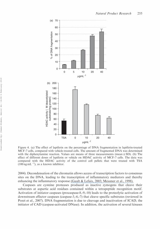
caspase activation in lapiferin-associated apoptosis, total caspases were investigated and
this revealed that there was only a significant increase at in MCF-7 cells (p50.05) when
treated with 20 and 40 mgmL�1 (Figure 5) as similar in case of HDAC inhibition.
Accordingly, lapiferin-induced apoptosis is associated with induced activation of caspases.Taken together, we reported here for the first time the isolation of lapiferin from
F. vesceritensis, and we additionally reported here the first X-ray data for lapiferin. We
also reported for the first time the specific anti-cancer activity of lapiferin against breast
cancer cells, which was due to apoptosis and not necrosis. Moreover, we have identified for
the first time the cell death pathway induced by lapiferin in human breast cancer cells and
that lapiferin evoked a multiple consequences that triggers apoptotic cell death, involving
the enhancement of DNA fragmentation, the activation of caspases and the induction of
histone acetylation.
Acknowledgement
This work was financially supported by the National Research Center, Cairo, Egypt.
References
Ahmed, A.A., Hegazy, M.E., Zellagui, A., Rhouati, S., Mohamed, T.A., Sayed, A.A., et al. (2007).
Ferulsinaic acid, a sesquiterpene coumarin with a rare carbon skeleton from Ferula species.
Phytochemistry, 68(5), 680–686.
0.0
0.5
1.0
1.5
2.0
2.5
3.0
0 10 20 40
mgmL−1
Tot
al c
aspa
se a
ctiv
ity (
fold
s)
5
Figure 5. Effect of lapiferin of the activity of total caspase activity of MCF-7 cells. Cells werecultured with vehicle or with various concentrations of lapiferin. Data are represented as folds of thecontrol (mean� SD, n¼ 4).
256 A.M. Gamal-Eldeen and M.-E.F. Hegazy
Downloaded By: [Gamal-Eldeen, Amira] At: 14:04 6 February 2010
Andrey, Y., Abramov, A., Maria, V., Zamaraeva, A., Albert, I., Hagelgans, A., et al. (2001).Influence of plant terpenoids on the permeability of mitochondria and lipid bilayers.Biochimica et Biophysica Acta Biomembranes, 1512(1), 98–11.
Appendino, G., Spagliardi, P., Cravotto, G., Pocock, V., & Milligan, S. (2002). Daucane
phytoestrogens: A structure–activity study. Journal of Natural Products, 65(11), 1612–1615.Aqel, M.B., Al-Khalil, S., Afifi, F., & Al-Eisawi, D. (1991). Relaxant effects of Ferula sinaica root
extract on rabbit and guinea pig smooth muscle. Journal of Ethnopharmacology, 31(3), 373–381.
Boulus, L. (1983). Medicinal plants of North Africa (p. 183). ML: Algomae.Burton, K.A. (1956). Study of the conditions and mechanisms of the diphenylamine reaction for the
estimation of deoxyribonucleic acid. Biochemical Journal, 62(2), 315–323.
Chehma, A., Djebar, M.R., Hadjaiji, F., & Rouabeh, L. (2005). The floristic composition of theSaharan rangelands of South East Algeria. Science et Changements Planetaires/Secheresse,16(4), 275–285.
Diaz, J.G., Fragam, B.M., Gonzalaz, A.G., Gonzalaz, P., & Hernandez, M.G. (1984). Eightcarotane sesquiterpenes from Ferula linkii. Phytochemistry, 23(11), 2541–2544.
Gasparri, F., & Muzio, M. (2003). Monitoring of apoptosis of HL60 cells by Fourier-transforminfrared spectroscopy. The Biochemical Journal, 369(2), 239–248.
Gault, N., & Lefaix, J.L. (2003). Infrared microspectroscopic characteristics of radiation-inducedapoptosis in human lymphocytes. Radiation Research, 160(2), 238–250.
Gault, N., Poncy, J.L., & Lefaix, J.L. (2004). Radiation-induced apoptosis: A new approach using
infrared microspectroscopy. Canadian Journal of Physiology and Pharmacology, 82(1), 38–49.Golovina, A., Saidkhodzhaev, A.I., Abdullaev, N.D., Malikov, V.M., & Yagudaev, M.R. (1983).
Structure and stereochemistry of lapiferin. Chemistry of Natural Compounds, 19(3), 296–301.
Hansen, M.B., Nielsen, S.E., & Berg, K. (1989). Re-examination and further development of aprecise and rapid dye method for measuring cell growth/cell kill. Journal of ImmunologicalMethods, 119(2), 203–210.
Iranshahi, M., Arfa, P., Ramezani, M., Jaafari, M.R., Sadeghian, H., Bassarello, C., et al. (2007).
Sesquiterpene coumarins from Ferula szowitsiasna and in vitro antileishmanial activity of7-prenyloxycoumarins against promastigotes. Phytochemistry, 68(4), 554–561.
Kalka, K., Ahmad, N., Criswell, T., Boothman, D., & Mukhtar, H. (2000). Up-regulation of
clusterin during phthalocyanine-4 photodynamic therapy mediated apoptosis of tumor cellsand ablation of mouse skin tumors. Cancer Research, 60(21), 5984–5987.
Lahouel, M., Zini, R., Zellagui, A., Rhouati, S., Carrupt, P.A., & Morin, D. (2007). Ferulenol
specifically inhibits succinate ubiquinone reductase at the level of the ubiquinone cycle.Biochemical Biophysical Research Communications, 355(1), 252–257.
Lam, M., Oleinick, N.L., & Nieminen, A.-L. (2001). Photodynamic therapy-induced apoptosis in
epidermoid carcinoma cells. The Journal of Biological Chemistry, 276(50), 47379–47386.Liu, K.Z., Jia, L., Kelsey, S.M., Newland, A.C., & Mantsch, H.H. (2001). Quantitative
determination of apoptosis on leukemia cells by infrared spectroscopy. Apoptosis, 6(4),269–278.
Messmer, U.K., Reed, U.K., & Brune, B. (1998). Bcl-2 protects macrophages from nitric oxide-induced apoptosis. Journal of Biological Chemistry, 271(33), 20192–20197.
Ozenda, P. (1983). Flore du Sahara (2nd ed, p. 622, 359). Paris: Centre National de la Recherche
Scientifique (CNRS).Poli, F., Appendino, G., Sacchetti, G., Ballero, M., Maggiano, N., & Ranelletti, F.O. (2005).
Antiproliferative effects of daucane esters from Ferula communis and F. Arrigonii on human
colon cancer cell lines. Phytotherapy Research, 19(2), 152–157.Pozzi, D., Grimaldi, P., Gaudenzi, S., Di Giambattista, L., Silvestri, I., Morrone, S., et al. (2007).
UVB-radiation-induced apoptosis in Jurkat cells: A coordinated Fourier transform infraredspectroscopy-flow cytometry study. Radiation Research, 168(6), 698–705.
Saleem, M., Alam, A., & Sultana, S. (2001). Asafoetida inhibits early events of carcinogenesis:A chemopreventive study. Life Science, 68(16), 1913–1921.
Natural Product Research 257
Downloaded By: [Gamal-Eldeen, Amira] At: 14:04 6 February 2010
Related Documents


