Int. J. Mol. Sci. 2011, 12, 8661-8694; doi:10.3390/ijms12128661 International Journal of Molecular Sciences ISSN 1422-0067 www.mdpi.com/journal/ijms Review A Concise Review on Epigenetic Regulation: Insight into Molecular Mechanisms Shahram Golbabapour *, Mahmood Ameen Abdulla and Maryam Hajrezaei Department of Molecular Medicine, Faculty of Medicine, University of Malaya, Kuala Lumpur 50603, Malaysia; E-Mails: [email protected] (M.A.A.); [email protected] (M.H.) * Author to whom correspondence should be addressed; E-Mail: [email protected]; Tel.: +603-7967-6604; Fax: +603-7967-6600. Received: 9 August 2011; in revised form: 7 November 2011 / Accepted: 10 November 2011 / Published: 30 November 2011 Abstract: Epigenetic mechanisms are responsible for the regulation of transcription of imprinted genes and those that induce a totipotent state. Starting just after fertilization, DNA methylation pattern undergoes establishment, reestablishment and maintenance. These modifications are important for normal embryo and placental developments. Throughout life and passing to the next generation, epigenetic events establish, maintain, erase and reestablish. In the context of differentiated cell reprogramming, demethylation and activation of genes whose expressions contribute to the pluripotent state is the crux of the matter. In this review, firstly, regulatory epigenetic mechanisms related to somatic cell nuclear transfer (SCNT) reprogramming are discussed, followed by embryonic development, and placental epigenetic issues. Keywords: epigenetic; pluripotency; SCNT; embryogenesis; gametogenesis; polycomb; methylation; histone modification 1. Introduction The transition from the differentiated somatic cell to the embryonic stage through somatic cell nuclear transfer (SCNT) requires activation energy to efficiently reprogram the resultant zygote to a proper pluripotent state [1,2]. SCNT is a tool to clone nuclear material into the enucleated cytoplasm of an unfertilized oocyte and thereby create genetically identical animals (Figure 1). SCNT not only OPEN ACCESS

Welcome message from author
This document is posted to help you gain knowledge. Please leave a comment to let me know what you think about it! Share it to your friends and learn new things together.
Transcript

Int. J. Mol. Sci. 2011, 12, 8661-8694; doi:10.3390/ijms12128661
International Journal of
Molecular Sciences ISSN 1422-0067
www.mdpi.com/journal/ijms
Review
A Concise Review on Epigenetic Regulation: Insight into Molecular Mechanisms
Shahram Golbabapour *, Mahmood Ameen Abdulla and Maryam Hajrezaei
Department of Molecular Medicine, Faculty of Medicine, University of Malaya, Kuala Lumpur 50603,
Malaysia; E-Mails: [email protected] (M.A.A.); [email protected] (M.H.)
* Author to whom correspondence should be addressed; E-Mail: [email protected];
Tel.: +603-7967-6604; Fax: +603-7967-6600.
Received: 9 August 2011; in revised form: 7 November 2011 / Accepted: 10 November 2011 /
Published: 30 November 2011
Abstract: Epigenetic mechanisms are responsible for the regulation of transcription of
imprinted genes and those that induce a totipotent state. Starting just after fertilization,
DNA methylation pattern undergoes establishment, reestablishment and maintenance.
These modifications are important for normal embryo and placental developments.
Throughout life and passing to the next generation, epigenetic events establish, maintain,
erase and reestablish. In the context of differentiated cell reprogramming, demethylation
and activation of genes whose expressions contribute to the pluripotent state is the crux of
the matter. In this review, firstly, regulatory epigenetic mechanisms related to somatic cell
nuclear transfer (SCNT) reprogramming are discussed, followed by embryonic
development, and placental epigenetic issues.
Keywords: epigenetic; pluripotency; SCNT; embryogenesis; gametogenesis; polycomb;
methylation; histone modification
1. Introduction
The transition from the differentiated somatic cell to the embryonic stage through somatic cell
nuclear transfer (SCNT) requires activation energy to efficiently reprogram the resultant zygote to a
proper pluripotent state [1,2]. SCNT is a tool to clone nuclear material into the enucleated cytoplasm
of an unfertilized oocyte and thereby create genetically identical animals (Figure 1). SCNT not only
OPEN ACCESS
Int. J. Mol. Sci. 2011, 12
8662
benefits agricultural applications, but has the potential for great advances in the field of medicine. In
addition, SCNT has paved the way to better understand the changes in cell differentiation and
reprogramming. Despite many investigations that have been done by numerous laboratories, the
efficiency (i.e., the ability to create a live born animal per nuclear transfer) by this technique is still
below 5% and several abnormalities have been reported [3]. One of the main reasons for these
abnormalities is the failure in reprogramming/remodeling of differentiated cells to the stage that will
evolve to a normal neonate. In the other words, programs involved in differentiated cells should be
replaced with totipotency to ensure nuclear cloning and production of healthy offspring. Gene
regulatory pathways are the critical network that could redefine SCNT. Clones, on the other hand, have
to change expression profiles to embryo-specific, global rearrangement of chromatin structure. As a
result, the cloning study is a way to understand epigenetic mechanisms and reprogram differentiated
nuclei. Epigenetic modifications in the donor cells remodel the gene expression profile to the extent
that is similar to the normal embryo. However, the epigenetic mechanisms that are responsible for the
transformation from a differentiated somatic cell into a pluripotent state remain mysterious. In this
review, we explore the epigenetic regulatory events that occur during the gametogenesis, embryogenesis
and placental development. The epigenetic modifications that modulate expression of genes and
subsequent reprogramming of the somatic nucleus to pluripotent state are also briefly discussed. The
purpose of review is to summarize effective epigenetic events that could increase efficiency of SCNT
and to emphasize recent epigenetic findings. In this regards, we briefly look into transition techniques
and highlight epigenetic modifications that happen during the nucleus reprogramming.
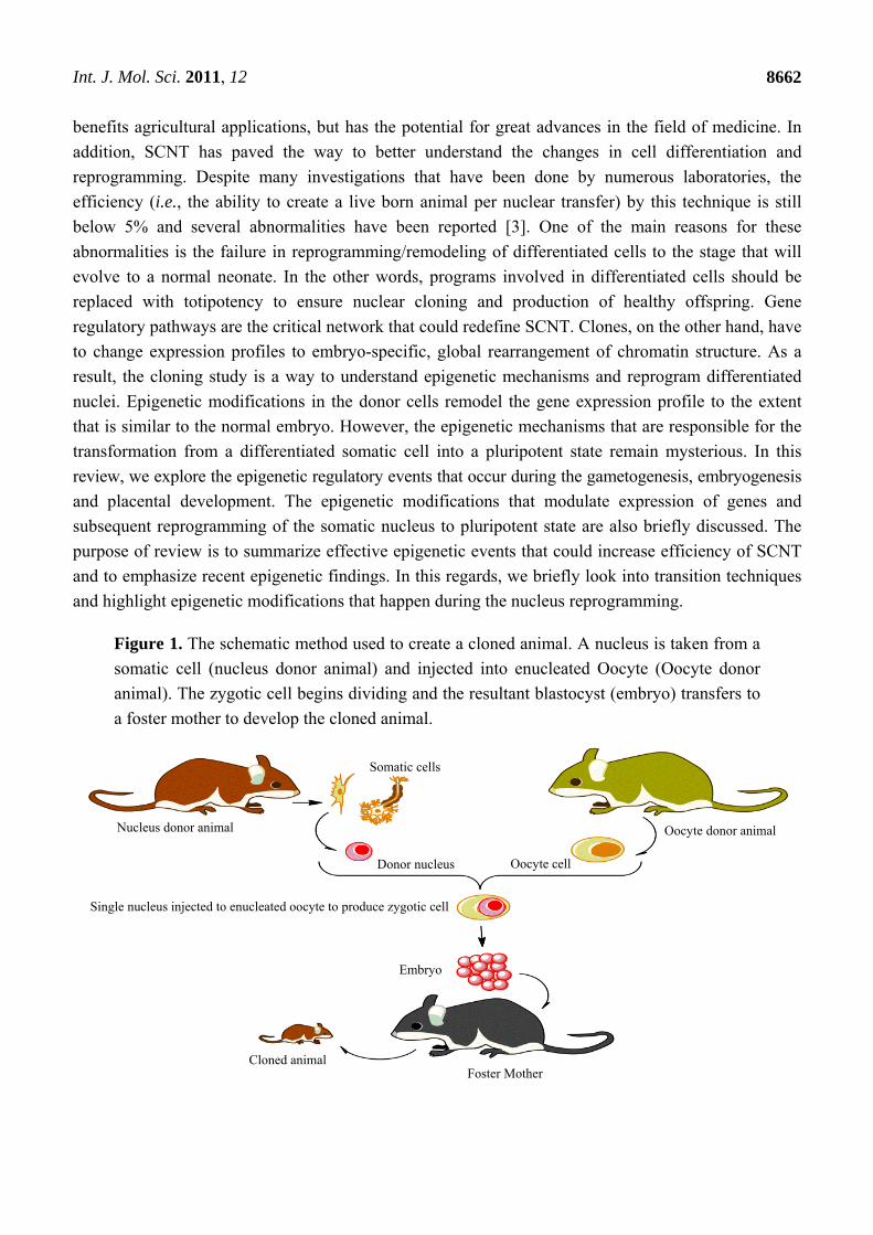
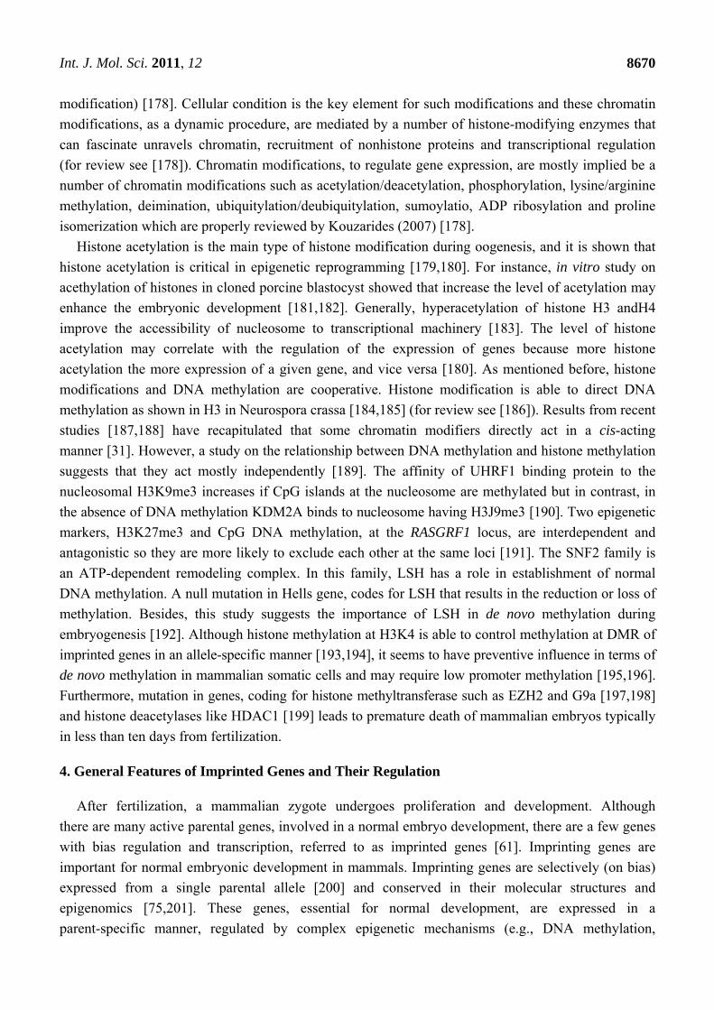
Figure 1. The schematic method used to create a cloned animal. A nucleus is taken from a
somatic cell (nucleus donor animal) and injected into enucleated Oocyte (Oocyte donor
animal). The zygotic cell begins dividing and the resultant blastocyst (embryo) transfers to
a foster mother to develop the cloned animal.
Donor nucleus Oocyte cell
Somatic cells
Single nucleus injected to enucleated oocyte to produce zygotic cell
Embryo
Foster Mother Cloned animal
Nucleus donor animal Oocyte donor animal
Int. J. Mol. Sci. 2011, 12
8663
2. Transition to Pluripotency
SCNT provides new insight into gene manipulation to achieve defined purposes. This technique is
to reprogram the differentiated somatic cell to a pluripotent state by transferring the nucleus of a
somatic cell into an enucleated oocyte and produce a zygote, which results in a live offspring.
In mammals, genomes of differentiated cell have to reprogram to a totipotent state to establish SCNT
during pre-implantation. Consequently, the development of a zygote initiates and follows with blastocyst
and the subsequent embryonic stages. Cloned embryos derived from less differentiated cells
(as nucleus donors), such as embryonic stem cells, show better implantation than those derived from
more differentiated somatic cells probably due to minimum or no reprogramming requirement [4]. It
was shown that the efficiency of bovine SCNT is relatively higher than the other experienced species
(for see review [5]) and pregnancy in Bos taurus is very similar to that of human in terms of length
and development.
Generation of induced pluripotent stem cell is to transport defined regulatory signals, influence the
epigenetic state and change it to another state (plasticity) which emphasizes the mutual reliance between
cell identity and epigenetic states [6,7]. This is, especially true during early embryo development and
gametogenesis [1]. Pluripotent stem cells are driven from somatic cells that are introduced by specific
reprogramming factors through either cell fusion or delivery of defined biochemical and/or chemical
factors which are also categorized as a reprogramming approach. The fusion technique produces
hybrid cells from differentiated somatic cells by nuclear reprogramming through the reactivation of
embryo-specific genes, whose expressions are suppressed in somatic cells [8]. In 2006, a four-gene set
was introduced to reprogram somatic cells to a pluripotent state [7]. Hybrid cells produced by fusion
technique show a pluripotent state by expression of the pluripotent markers such as OCT4 [9]. Moreover,
a number of other genes such as Nanong, Sox2, Lin28, Klf4, c-Myc and AID have been correlated to
the pluripotent state of a cell. The expressions of these genes result in cell reprogramming [7,10–13].
Based on these evidences, identification of embryo-specific genes is crucial to defining their expression
profiles during embryogenesis, functions during different stages of embryogenesis and the development
of placenta. These regulations, actually, are defined epigenetic regulation for which molecular signals
modulate the modifications.
Several morphological abnormalities such as hydroallantois, placentomegaly, cardiomegaly,
enlarged umbilical cord, abdominal ascites and placental dysfunctions [14,15], have been observed in
the cloned offsprings. Large offspring syndrome (LOS) is a developmental disorder mostly seen in
SCNT driven embryos. This syndrome in addition to the failure in the development of embryo and
placenta and other abnormalities is attributed to inappropriate and/or inadequate somatic nuclear
reprogramming events. Significant increase in genomic methylation in liver of cloned bovine fetuses is
attributed to fetal overgrowth [16]. LOS and failure in the normal development of an embryo that are
seen in cloned animals could be due to abnormal epigenetic patterns [17]. In fact, assisted reproductive
techniques appear to be accompanied by several anomalies, especially in the second half of the
gestation [14,18–20].
Int. J. Mol. Sci. 2011, 12
8664
3. Molecular Signals in Epigenetic Regulation
Cells’ information is inherited to the next generation through genetic and epigenetic routes. Genetic
information is encoded in the DNA sequence while, epigenetic information is defined basically by
DNA modification (DNA methylation) and chromatin modifications (methylation, phosphorylation,
acetylation and ubiquitination of histone cores). Combination of these modifications characterizes the
chromatin configuration and the accessibility of genes to the transcription machinery and consequently,
transcriptional regulation of the expression of genes. Cheng [21] introduced three categories by which
transcriptional function is generally initiated and controlled: First, general intrinsic promoter and
transcriptional machinery [22–24], second, specific transcriptional regulatory factors [25–27] and, third,
the configuration and accessibility of chromatin structure and DNA to the transcriptional machinery
through posttranslational modifications of histone and post replicational modification of DNA [27–29].
3.1. Main Epigenetic Regulatory Mechanisms
Complex epigenetic regulation comprises several molecular signals that direct the expression of
genes based on environmental changes and developmental status. Transcription factors, non-coding
RNAs (ncRNAs) [30], DNA methylation, histone modification and chromatin remodeling are such
epigenetic signals that mediate accessibility and expression of genes as needed. Transcription
mainly defines a self-propagating state mediated by cis-acting and/or trans-acting regulatory
mechanisms [31], and are able to establish epigenetic states through cis-acting [32] and non-coding
polycomb domains [31,33]. Reinforcement of epigenetic states happens through mutual relationship
between DNA methylation and histone modifications [34]. DNA methylation postulates a reinforcing
signal for other regulatory mechanisms whose functions are not that much strong [31].
DNA methylation and histone modification are two important mechanisms for modulating the
chromatin structure and regulating the expressions of the genes (for review see [35]). Epigenetic
regulation is a complex phenomenon that consists of a variety of different processes [21] such as
imprinting [36], X chromosome inactivation [37] and gene silencing [38,39]. In addition it encompasses
the development of an embryo [40–43] and placenta [44–46], nuclear reprogramming in SCNT
embryos [3,6] and carcinogenesis [47,48].
3.2. Transcriptional Regulation
The transcriptional regulation of genes is mainly directed by different strategies. These include the
state of genomic methylation [21], chromatin configuration [49,50], chromatin structural variations
(euchromatin and heterochromatin) [51,52], and chromatin modifications [53]. Chromatin modification
in turn is influenced by methylation, acetylation and phosphorylation, as well as polycomb proteins [54]
and matrix attached region [55]. Transcriptional regulation is mostly controlled by the methylation
pattern of the genome. DNA methylation on specific CpG dinucleotide (CpG) located in a cluster
(CpG islands) is the regulatory mechanism by which expression of gene is either activated or suppressed
(for review see [56]). Moreover, chemical modifications of chromatin histone cores are mediated by
DNA methylation of CpG islands [57]. These modifications have a mutual relationship with each
Int. J. Mol. Sci. 2011, 12
8665
other [58]. Germ cells and embryonic cells during early development are two epigenetic sites where
methylation patterns erase, establish and reestablish [59].
3.3. Epigenetic Reprogramming During Embryogenesis
In mammals, epigenetic reprogramming in germ cells and during preimplantation, especially its
effects on imprinting genes, predominantly establishes developmental stages [60]. The DNA methylation
patterns characterize developmental status during cell differentiation. In the concept of epigenomics,
molecular signals are responsible to establish the proper expression of embryo-specific genes, mainly
during gametogenesis and embryogenesis. Therefore, the main issue for a successful SCNT is the
establishment of these modifications, occurring during embryogenesis, which should be similar and
ideally identical to its normal embryo counterpart. However, undoubtedly, several lessons are still to
be learnt regarding epigenetic modifications during gametogenesis.
3.4. Epigenetic Features of DNA Methylation
As mentioned before, DNA methylation and histone modification are the main epigenetic factors,
by which gene expression could be regulated, and have important roles in nuclear reprogramming
during embryogenesis. DNA methylation is a heritable epigenetic marker by which expression of a
gene may be regulated through alteration in the local chromatin structure that mostly happen within the
CpG islands and imprinted genes at cytosine carbon 5 within palindromic dinucleotide 5′-CpG-3′ and
differentially methylated domains (DMDs) respectively (see review [60]). Cytosine residue of CpG is
the site for DNA methylation by which gene expression is regulated. Generally, DNA methylation
at CpG sequences suppresses the expression of the methylated gene [61]. CpG islands are usually
located within repetitive elements such as centromic repeats, satellite sequences and ribosomal RNA
genes [62,63]. DNA methylation can be varied in terms of patterns and the level of global/regional
DNA methylation, is specific to developmental stages [64] and origin of tissue [16,65]. In fact, the
mature parental gametes at fertilization are significantly methylated. For instance, DNA of sperm, in
comparison with that of the oocyte, is more methylated [66,67] and, undergoes demethylation after
fertilization [68–70]. However, imprinted genes and some retrotransposons mostly remain methylated.
In mouse, hypermethylation pattern in repetitive regions and heterochromatin region has been
observed; whilst in gene-specific region of DNA hypomethylation is predominant [17,71,72].
Abnormal DNA methylation of various repetitive elements in cloned blastocysts was reported for the
first time by Kang and coworkers [73]. Methylation of imprints, monoallelic expressed genes [74,75],
on the other hand, is a maintained (not de novo) and highly conserved event [76,77]. Recently, in a
human study, comparison between embryonic stem cells and differentiated cells illustrated that there
are a number of methylated cytosine in non-CpG regions of the embryonic stem cells [78].
3.5. DNA Methylation Signals
DNA methylation is under the control of two types of signals: cis-acting signals and trans-acting
signals. IGF2R, SNRPN, H19 and RASGRF1 are genes regulated by cis-acting signals (see review [79]).
Global DNA methylation takes place especially after fertilization and with different rate of

Int. J. Mol. Sci. 2011, 12
8666
demethylation that is specific to either parental genome [61]. Cell cycle observations reveal that
paternal demethylation generally happens during the first cell cycle but maternal alleles take a few
cycles to be demethylated [80]. After fertilization, imprinting control regions (ICRs) methylation is
established in a sex-dependent manner [61]. Despite the maintained methylation pattern in somatic
cells, methylation pattern in germ cells needs to be appropriately reestablished to provide a
methylation pattern that is heritable to the next generation. This suggests that methylation is modulated
in a sex-dependent manner [81]. Although hypomethylation of female germ line seems not to be
correlated to the sex chromosomes, their regulation is thought to be associated with genital ridge.
However, in the male germ line it is regulated by both mechanisms [81,82].
3.6. DNA Methylation Analysis
Epigenetic studies are strongly involved in DNA methylation. Analysis of the methylation patterns
is the main approach in different studies that focuses on gene regulation. The cytosine 5 methylation in
the context of CpGs mostly takes place within CpGs islands at the promoter region of genes and this
leads to suppression of the expression of the gene. Several studies correlate aberrant methylation
pattern of DNA to developmental failure during embryogenesis [83–85] and placentation [86,87] as
well as several diseases and disorders [88–91] (for review see [92]). DNA methylation techniques
cover a wide range of analysis from gene specific, locus specific to entire genome analysis using
proper methods categorized in four groups based on DNA methylation analysis techniques [93]: in the
first category cytosine residues are converted to Uralic by a bisulfite conversion, the second category is
based on methylation-sensitive restriction endonuclease, enrichment based methods and the last is the
capturing method based on the affinity to retain methylated DNA [94,95].
3.7. Regulatory Factors in DNA Methylation
As mentioned before, DNA methylation is a common epigenetic modification taking place by
enzymatic reactions to be added to the cytosines at CpG, mostly known as repetitive elements and
imprinted genes [79]. Generally, DNA methylation is classified either as de novo methylation or
maintained methylation. Therefore, there are two classes of enzymatic: de novo methyltransferases and
maintenance methyltransferases [96,97]. In mammals, DNA methylation occurs by the addition of a
methyl group from S-adenosylmethionine to Cytosine using DNA methyltransferases (DNMTs).
DNMTs are trans-acting factors targeting DNA sites for methylation using cis-acting signals. There
are a number of mammalian DNMTs (see Table 1) that have been identified since 1980s [98]
(for review see [99]).
Table 1. Types of DNA methyltransferases and their epigenetic functions.
DNMT # types [100] Functions
DNMT1 Maintaining methylation pattern [21,101,102] Essential for chromosome replication and repair [21,103,104] Essential for de novo methylation [105]
DNMT2 Effective in DNA and RNA methylation (for review see [106])
Int. J. Mol. Sci. 2011, 12
8667
Table 1. Cont.
DNMT3a Establishment of de novo methylation pattern [107,108] especially during gametogenesis [109] Maintaining methylation pattern [101]
DNMT3b Establishment of de novo methylation [107,108]
DNMT3L
Essential for de novo methylation [110] Enhance de novo methylation activity of DNMT3a [111] and DNMT3b [112] Establishment of de novo methylation pattern especially during gametogenesis [113]
# DNA methyltransferase
3.8. DNA Methyltransferases
DNA methylation at cytosine 5 nucleotide is catalyzed by DNMTs. This family of enzyme is vitally
important in epigenetic regulation which modulates the expression of genes especially imprinted ones
as well as X chromosome inactivation [114,115]. There are five main DNMTs that are important in
de novo and/or maintenance DNA methylation: DNMT1, DNMT10, DNMT3a, DNMT3b and
DNMT3L [99]. DNMT1, DNMT2 and DNMT3 are mostly characterized DNMTs that can categorize
either maintenance or de novo DNMT. DNMT1 is a maintenance DNMT that methylates both imprints
and non-imprints genes. DNMT2 seems to have a regulatory role in DNA methylation but the
mechanism and its role in methylation maintenance or de novo remains unclear. DNMT3 as a de novo
DNMT (DNMT3a) is a key factor in imprints’ methylation. Its isoforms are suggested to have roles in
global DNA methylation in germ cells [116].
DNMT3L is defined as an imprints’ regulatory candidate for DNA methylation by regulating
NMT3a/b [117]. The expression of DNMT1 gene has a positive correlation with DNA methylation
status on the satellite I region. Consequently, it has been shown that in vitro development of bovine
SCNT embryos to the blastocysts state can be enhanced through down regulation of DNMT1 [118].
It is also shown that the DNMT is responsible for ICRs methylation [109]. Moreover, transcriptional
analysis on the pS2/TFF1 during cell cycles reveals that DNMTs carry out two distinct actions, namely
methylation and demethylation of CpGs [119].
In comparison to the male germ line in which the establishment of ICR methylation of an imprinted
gene, H19, is regulated by DNMT3a and DNMTL [109,113], DNMT3L is the de novo methylating
regulatory factor for the female germ line [109]. In the female germ line, DNMT3L establishes the
methylation of ICRs that selectively interact with histone H3 [120]. This evidence in addition to
DNMT3L’s stimulating role for DNMT3a and DNMT3b [117] shows its potential in chromatin
mark-specific recognition and methylation establishment [61]. Promoter methylation-mediated
DNMTs show down regulation of DNMT1 and up regulation of DNMT3L in the human placenta and
brings strength to the capability of DNMT family in the establishment of de novo DNA methylation in
extraembryonic tissue [46]. A novel DNMT3b splice variant, DNMT3B3Δ5, is highly expressed in
pluripotent embryonic stem cell and in contrast, is repressed during differentiation [121].
In a human study on DNMTS, global hypomethylation is shown to be induced by significant
reduction in the expression of DNMT3A, DNMT3b and DNMT1 using microRNA-29b [122].
Int. J. Mol. Sci. 2011, 12
8668
DNMT3L by itself has no methyltransferase activity; however, its association with the DNMT3 family
seems essential for de novo methylation in mice [123]. There are some evidence on the activity of
DNMT1 in the establishment of methylation at non-CpG regions [55] and CpG islands [21,124,125].
Histone modification and CpG spacing are able to direct DMR methylation of imprints [21].
Crystallographic analysis showed that de novo DNA methylation might be controlled by specific
histone modifications in that a heterotetramer structure, assembled from DNMT3a and DNMTL,
provides two active sites of CpG that are 8–19 base-pair distance from each other [126–128]. A study
on chromosome 21 also Reinforces the crucial role of CpG spacing in DNA methylation [129]. Active
demethylation in mammalian genome seems promising, however, there has been no report of an
enzyme that can catalyze this reaction [130]. Some studies have emphasized an active demethylation
process, independent from DNA replication [131,132]. In fact, demethylation of the paternal alleles is
an active event that happens rapidly after fertilization. The maternal genome, however, demethylates
during first cell cycles in which demethylation mostly appears to be an inactive process. In addition to
DNA methylation and histone modification, ncRNAs and regulatory proteins are the most studied
epigenetic mechanisms that modulate epigenetic reprogramming. Small interfering RNAs (siRNAs)
transfection is a technique to silence DNMT mRNA and modify the DNA methylation pattern in
cells [6]. In a recent study, To examine the efficacy of the technique in SCNT embryos, DNMT1 RNA
was silenced using siRNA in SCNT bovine embryo which demonstrated the capability in nucleus
reprogramming through inducing DNA methylation [118]. In the expression of H19 in the male germ
line, DNMT3a and DNMTL are counterparts and reached their maximum [133,134] on embryonic
day 13 while there is no methylation on the H19 ICR, in mouse [135]. In addition, these enzymes seem
not to be specific for DNA binding [99], suggesting direct/indirect interactions with specific chromatin
modifications [61,136,137].
3.9. Epigenetic Features of ncRNAs
The cluster-oriented imprinted genes are laid in ~1Mb length base pair, containing parental expressed
genes, ncRNA sequences that regulate the nearby imprinted genes [138–141]. ncRNAs are mostly
placed in clusters and regulated by ICRs [142]. The GNAS and KCNQ1 are examples of such imprints;
containing ncRNAs that mediate the gene expression [143,144]. ncRNAs are divided into two groups,
small ncRNAs and long ncRNAs. Small ncRNAs attach chromatin modifiers to specific genome
sequence [145] and may interact with either RNA, single stranded DNA or double stranded
DNA [1,146]. Long ncRNAs have complex tertiary structure and act globally to bridge chromatin
modifiers to the genome [147]. But there are some evidences for local function of long ncRNAs, which
is considered to function in cis-acting regulation of parental imprinted gene and inactivation of
chromosome X [1].
In mammalian transcription of ncRNA genes is an important feature. ncRNAs usually are classified
based on their mature length, location and orientation according to the nearest protein-coding gene,
and their function which could be cis or trans [148–150]. Macro RNAs, such as inactive X-specific
transcript (Xist) and X (inactive)-specific transcript, antisense (Tsix), are categorized as cis-acting
ncRNAs that usually locate within clusters of imprinted genes. On the other hand, short ncRNAs such
as short interfering RNAs, micro RNAs, piwi-interacting RNAs and short nucleolar RNAs are

Int. J. Mol. Sci. 2011, 12
8669
categorized as trans-acting ncRNAs (for review see [151]) [148]. Koerner (2009) concluded that
chromosomes express macro ncRNA usually do not express imprinted mRNA genes and the expression
of imprinted macro ncRNAs may be regulated by an unmethylated imprint control element [148].
Moreover, there are a number of evidences that show trans-acting regulators for imprinted small
ncRNAs such as Snurf-SNRPN and Dlk1-Gtl2 [152,153]. It has been shown that ncRNAs have a
critical role during development. For instance, two ncRNAs, Dicer1 and Dgcr8, show developmental
impact in mice [154,155]. Moreover, studies on effects of ncRNAs during animal embryogenesis show
their specific and crucial role during embryonic development (for review see [156]). Micro ncRNAs,
specifically, show precise control on expression of imprinted genes during development [157]. For
instances, miR-15 and miR-16 are important in early embryonic development [158], miR-1, miR-133 and
miR-206 in development of skeletal and heart muscle [159,160], miR-124 in neuronal development [161]
(for review see [162]). During placentation, ncRNAs such as KCNQ1OT1, a long ncRNA, also
illustrate a leading role in imprinted genes regulation [163–165]. Regulation of ncRNAs is an
important silencing mechanism in plancenta (for review see [148]). It was shown that the repression of
imprinted genes during gestation is directly regulated by micro RNAs during placentation and
embryogenesis [166,167]. It seems that ncRNAs targets placental histone methyltransferases by
ncRNAs through chromatin modification [148].
3.10. Epigenetic Features of Small RNAs
Small RNAs (terminologically different from ncRNAs), generated by activity of RNaseIII enzymes
(reviewed in [168]), have variety of biological functions such as heterochromatin formation, mRNA
inactivation and transcriptional regulation [169,170]. Generally, their bioactivity is due to their
association with Argonaute (Ago)-family proteins [171]. microRNAs (miRNAs), endogenous small
interfering RNAs and Piwi-interacting RNAs (piRNAs) are classes of small RNAs. In mammalians,
small RNA-associated Ago proteins are mostly classified into Piwi subfamily and Ago subfamily
(for review see [171]). miRNAs to do their biological activity, which is post translational regulation by
acting on mRNAs, needs to be bound by Ago subfamily proteins (for review see [170]). Moreover,
regulation of most miRNAs may control by developmental signaling [172]. piRNAs are mostly bound
by Piwi subfamily proteins and have a critical role during gametogenesis [173] in germ line [174]. This
subfamily protein has shown to have a critical role in regulation of germline stem cells [175].
3.11. Epigenetic Features of Chromatin Modifications
Chromatin structure is crucial for gene regulation/expression, which is carried out by exploiting
recruitment of protein complexes [29]. Euchromatin structure of embryonic stem cells is a
predominant chromatic structure that allows for global gene expression accessibility [176] and
facilitates reprogramming to the pluripotent state. It is not surprising that histone modifications might
in turn influence the global gene expression by modulating chromatin configuration [177]. Covalent
modification of the core histone has a critical role in the regulation of gene expression through
acetylation and methylation. Chromatin modification and their function are important especially for
gene regulation. Kouzarides (2007) reviewed a number of chromatin modifications characterized by
mass spectrometry (for nucleosomal modification) and specific antibodies (for global histone
Int. J. Mol. Sci. 2011, 12
8670
modification) [178]. Cellular condition is the key element for such modifications and these chromatin
modifications, as a dynamic procedure, are mediated by a number of histone-modifying enzymes that
can fascinate unravels chromatin, recruitment of nonhistone proteins and transcriptional regulation
(for review see [178]). Chromatin modifications, to regulate gene expression, are mostly implied be a
number of chromatin modifications such as acetylation/deacetylation, phosphorylation, lysine/arginine
methylation, deimination, ubiquitylation/deubiquitylation, sumoylatio, ADP ribosylation and proline
isomerization which are properly reviewed by Kouzarides (2007) [178].
Histone acetylation is the main type of histone modification during oogenesis, and it is shown that
histone acetylation is critical in epigenetic reprogramming [179,180]. For instance, in vitro study on
acethylation of histones in cloned porcine blastocyst showed that increase the level of acetylation may
enhance the embryonic development [181,182]. Generally, hyperacetylation of histone H3 andH4
improve the accessibility of nucleosome to transcriptional machinery [183]. The level of histone
acetylation may correlate with the regulation of the expression of genes because more histone
acetylation the more expression of a given gene, and vice versa [180]. As mentioned before, histone
modifications and DNA methylation are cooperative. Histone modification is able to direct DNA
methylation as shown in H3 in Neurospora crassa [184,185] (for review see [186]). Results from recent
studies [187,188] have recapitulated that some chromatin modifiers directly act in a cis-acting
manner [31]. However, a study on the relationship between DNA methylation and histone methylation
suggests that they act mostly independently [189]. The affinity of UHRF1 binding protein to the
nucleosomal H3K9me3 increases if CpG islands at the nucleosome are methylated but in contrast, in
the absence of DNA methylation KDM2A binds to nucleosome having H3J9me3 [190]. Two epigenetic
markers, H3K27me3 and CpG DNA methylation, at the RASGRF1 locus, are interdependent and
antagonistic so they are more likely to exclude each other at the same loci [191]. The SNF2 family is
an ATP-dependent remodeling complex. In this family, LSH has a role in establishment of normal
DNA methylation. A null mutation in Hells gene, codes for LSH that results in the reduction or loss of
methylation. Besides, this study suggests the importance of LSH in de novo methylation during
embryogenesis [192]. Although histone methylation at H3K4 is able to control methylation at DMR of
imprinted genes in an allele-specific manner [193,194], it seems to have preventive influence in terms of
de novo methylation in mammalian somatic cells and may require low promoter methylation [195,196].
Furthermore, mutation in genes, coding for histone methyltransferase such as EZH2 and G9a [197,198]
and histone deacetylases like HDAC1 [199] leads to premature death of mammalian embryos typically
in less than ten days from fertilization.
4. General Features of Imprinted Genes and Their Regulation
After fertilization, a mammalian zygote undergoes proliferation and development. Although
there are many active parental genes, involved in a normal embryo development, there are a few genes
with bias regulation and transcription, referred to as imprinted genes [61]. Imprinting genes are
important for normal embryonic development in mammals. Imprinting genes are selectively (on bias)
expressed from a single parental allele [200] and conserved in their molecular structures and
epigenomics [75,201]. These genes, essential for normal development, are expressed in a
parent-specific manner, regulated by complex epigenetic mechanisms (e.g., DNA methylation,

Int. J. Mol. Sci. 2011, 12
8671
post-translational histone modification) using epigenetic markers (e.g., DNA methylation) [61]. The
conflicting interests of parental, imprinted genes are hypothesized as maternally and paternally
expressed imprints suppress and enhance the fetal growth respectively (see review [60]). In the nucleus,
imprinted genes are mostly placed in a cluster orientation but some are identified as isolated ones [72]
such as Nap1l5, Nnat, Inpp5f_v2 [202–206] and Gatm, Dcn and Htr2a (for review see [207]). Imprints
that are placed within CpG rich region are mostly in clusters, controlled by imprinting the control
regions through DNA methylation and histone modifications [58,208,209]. Regulations of imprinted
genes are generally proceeded through DNA methylation, post translational histone modification and
ncRNAs [210]. In addition, active imprinted genes (expressed allele) contains the allele-discriminating
signal (ADS) and the de novo methylation signal (DNS) that are necessary for establishing or
maintaining methylation [211,212]. For instance, SNRPN is a paternally expressed imprint whose
regulation is similar to that of IGF2r [212]. Human SNRPB contains two DNS signals; an ADS signal
and a signal to maintain paternal imprint (MPI) [213].
Methylation of Imprinted Genes and Its Abnormalities in Cloned Animals
Short regions of DNA, described as differentially methylated regions (DMRs), are marked by
methylation in a parental specific manner and therefore the expressions of such genes are monoallelic.
Regulation of clusters of imprinted genes and their activities are mostly controlled by differentially
methylated ICRs. In fact, ICRs are DMRs that obtain methylation on one allele (bios) and regulate
clustered imprinted genes [138]. In the other words, those DMRs that have a critical role in
maintaining imprinting are known as ICRs [81]. CpG spacing suggests a potential influence on ICRs
recognition and DMRs methylation in imprints [126]. Moreover, the transcriptional system, especially
those traversing ICRs, are considered a common requirement to open chromatin domains, and make
targets available for methylation specially in the germ line [32]. Besides, an in vivo study in a mouse
model illustrated a novel cis-acting function for the H19 ICR [214]. This study shows changes in the
size and CpG density that coincide with biallelic expression of the H19 without any detectable
alteration in the methylation pattern. The researchers concluded that, in addition to CTCF sites, there
are sequences within the ICR that are essential for its regulatory function. Moreover, the ICR size and
CpG density are of determinant elements.
Maternal alleles are dramatically more exposed to ICRs methylation than paternal ones [61,138].
Maternal alleles are mostly methylated on promoters of antisense transcripts but those of paternal
alleles are placed between genes (non-promoter regions), suggesting that parental imprinting
methylation acts differently [215]. In general, there is higher degree of methylation of the maternal
ICRs allele in comparison with that of paternal allele [61]. The H19 is an example of imprinted genes
whose preference is to be expressed from maternal allele. Methylation of the DMDs of H19,
maternally expressed imprinted gene, is needed for maintenance methylation [141].
DMRs of imprints, mostly, epigenetically signal for monoallelic expression of the gene. IGF2
encodes a fetal growth-factor and is predominantly expressed from the paternal allele, while H19 is
expressed from the maternal allele and encodes a transcript which may reduce cellular proliferation. In
mouse, IGF2 has a few identified DMRs named DMR0, DMR1, DMR2 and DMR3 among which the
first two DMRs are positioned upstream and DMR2 within the IGF2 gene [216–218]. Recently, an

Int. J. Mol. Sci. 2011, 12
8672
intragenic regulatory DMR has been reported within the last exon of the IGF2 gene [81]. The
comparison between methylation patterns of IGF2 DMR from parthenogenetic and androgenetic
blastocysts on one hand and that evolved from a normal zygote suggests that in normal embryos,
paternal allele significantly contributes in the DNA methylation at the locus [81]. Methylation on
DMDs of imprints are initially established during gametogenesis and prior to parental pronucleus
fusion in the zygote [61,219]. After fertilization, the intergenic DMR of bovine IGF2 undergoes
demethylation followed by low level remethylation before blastocyst stage, which in turn precedes
implantation by which the DMR is heavily remethylated. The study speculates that global methylation
pattern of SCNT blastocysts is reprogrammed and maintained in a sex specific manner, similar to its
normal counterpart. A recent study shows that, except for RASGRF1 DMR (paternally expressed
imprinted gene), methylation of the most imprinted genes during mouse embryonic cleavage stages
(preimplantation phase) are mainly controlled by maternal and zygotic DNA methyltransferase 1
(DNMT1) protein family [220].
Abnormalities at imprinted loci have been observed in cloned mammals. In cloned cattle abnormal
imprinted gene profiles have been observed especially in the expression of IGF2 and H19 [221]. In the
Bos taurus model, the IGF2 and H19 (IGF2/H19), a conserved cluster of imprinted gene, showed
significant variations from the normal pattern, mostly hypomethylation, associated with abnormal
expressions of the H19 (but not IGF2) from both alleles in methylation pattern of DMRs [17,222].
Moreover, methylation pattern which mostly occurs in early embryogenesis is dependent on
developmental stage and specific to different tissues, as was studied in IGF2/H19 [218].
Super ovulation, also, can cause abnormal imprinting patterns in oocytes [223] that might be
attributed to the reduced expression of imprinted parental alleles, SNRPN, PEG3 and KCNQ1OT1, but
to increased methylation of H19 [224]. MII oocytes of cloned porcine showed mostly unmethylated
profiles of DMR [225]. A recent study on bovine SCNT showed that significant demethylation at the
H19 DMD is attributed to biallelic expression of the imprint which might lead to decline in the rate of
implantation [226]. Moreover, biallelic expression of H19 in bovine is correlated to hypermethylation
of the paternal H19 differentially methylated domain and locus anomalies cause low SCNT efficiency
in cattle [226].
5. Control of Gene Expression During Gametogenesis
Gametogenesis and embryogenesis involve epigenetic reprogramming to establish proper epigenetic
marks and gene regulation. Generally, epigenetic pattern of the genome first reprograms and
reestablishes during gametogenesis. The second round of reprogramming and maintenance happens
after fertilization, especially during preimplantation of the embryo [61] (Figure 2). Gametogenesis in
both sexes involves methylation of DMRs, reestablished in a parent-specific manner [60]. Epigenetic
reprogramming is mostly characterized during gametogenesis and early embryonic development
especially prior to the zygotic implantation [227]. In fact, during gametogenesis (spermatogenesis and
oogenesis) the methylation patterns of these genes are erased and reestablished. These modifications
are continued after fertilization and during preimplantation specifically within non-imprinted
genes [225] (for review see [180]). Gametogenesis involves sex-specific, epigenetic remodeling of
male and female germ lines that matures the gametes for fertilization and constitutes proper regulatory
Int. J. Mol. Sci. 2011, 12
8673
processes [180]. Epigenetic reprogramming in sperm begins with DNA demethylation, followed by
DNA remethylation and de novo methylation to chromatin modification and histone-to-promatine
transition [180,228]. Moreover, during spermatogenesis, testis specific linker histones occupy somatic
linker variants. Among the histone variants centromere protein A appears to be epigenetically
important during spermatogenesis [180]. Spermatozoa have a transcriptionally inactive and highly
condensed chromatin structure. During spermatogenesis in rats, paternal-specific imprinted genes are
prone to hypomethylation due to estrogen-associated signaling [229]. In the male germ cells, DMR of
IGF2/H19 acquires DNA methylation during spermatogenesis, however, in the female germ cells, the
DMR possesses the zinc finger protein CTCF by which the DMR defends against methylation so the
allele is able to be expressed [230]. Through fertilization, paternal genome undergoes a series of
remodeling events which are controlled by the activity of the oocyte, and the protamine replaced by
oocyte-supplied histone and possessing maternal chromatin related proteins [231].
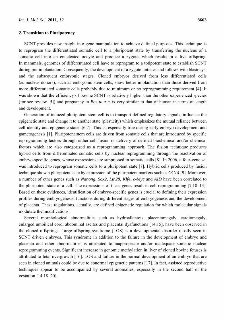
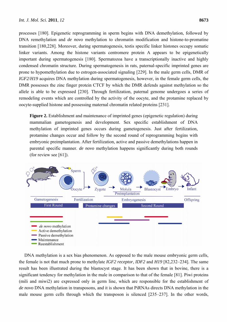
Figure 2. Establishment and maintenance of imprinted genes (epigenetic regulation) during
mammalian gametogenesis and development. Sex specific establishment of DNA
methylation of imprinted genes occurs during gametogenesis. Just after fertilization,
protamine changes occur and follow by the second round of reprogramming begins with
embryonic preimplantation. After fertilization, active and passive demethylations happen in
parental specific manner. de novo methylation happens significantly during both rounds
(for review see [61]).
DNA methylation is a sex bias phenomenon. As opposed to the male mouse embryonic germ cells,
the female is not that much prone to methylate IGF2 receptor, IDF2 and H19 [82,232–234]. The same
result has been illustrated during the blastocyst stage. It has been shown that in bovine, there is a
significant tendency for methylation in the male in comparison to that of the female [81]. Piwi proteins
(mili and miwi2) are expressed only in germ line, which are responsible for the establishment of
de novo DNA methylation in transposons, and it is shown that PiRNAs directs DNA methylation in the
male mouse germ cells through which the transposon is silenced [235–237]. In the other words,
Int. J. Mol. Sci. 2011, 12
8674
Piwi/PiRNA complex appears to guide the de novo methylation at transposons [235,237] and
deactivate transposons within a germline [238]. PiRNAs and siRNAs such as the one located within
AU76, a pseudogene of RANGAP1, negatively regulates transposons through their cis-acting function
in mouse oocytes as well as establishes the methylation of retrotransposons in the male mouse germ
line [235,237,239].
Somatic environment of the male/female germ line shows their influence in DNA methylation of
imprints [82]. Using sex-reversed mice to evaluate sex-specific methylation pattern in vivo, the germ
cells are found to be responsible for female/male imprints during oogenesis/spermatogenesis,
though sex chromosome constitution shows significant influence on male germ line for imprint
methylation [82]. It seems probable that somatic environment of the genital ridge and that of
chromosomal constitution have key roles in the establishment of imprinted genes. RASGRF1,
paternally expressed imprinted gene, is essential in the male germ line [240,241] suggesting a
regulatory mechanism containing DMD methylation and the repeat sequences, by which methylation
of the germ line is established [79].
6. Epigenetic Regulation During Gestation
Normal fetal development is dependent on proper development of embryo and placenta. These
developments are modulated through epigenetic signals during gestation. Although these molecular
signals are controlled by the same epigenetic mechanisms, their regulation is independent of each other
and may follow different patterns during embryogenesis in comparison to placental development.
During pregnancy, most monoallelic expressed genes carry out in extraembryonic tissues, such as
trophoblast and yolk sac, regulate the development and function of placenta [44,201]. In placenta, this
regulation seems to be directed by histone modification and ncRNAs through DNA methylation [201].
Embryo-placental development is a complex modulating phenomenon through which imprints undergo
necessary maintenance, establishment and/or reestablishment. Placental development is under the control
of IGF2 and its degradation receptor, IGF2r [242]. IGF2, paternally expressed imprint, codes for
embryo-placental growth factors. However, its receptor seems an unorthodox, imprinted gene [243].
Embryogenesis involves global methylation to erase and remethylate the methylation pattern.
During early embryogenesis, methylation re-establishment occurs mostly within CpG islands and the
imprints regulate in a sex-specific manner based on the new gender [79,244,245]. The demethylation
mostly happens during primordial germ cells (PGCs) migration towards the genital ridge [79,246,247].
It is hypothesized that histone replacement and chromatin changes, using DNA repair mechanisms, are
in accordance with the epigenetic reprogramming of PGC [61]. Evidences for such associations come
from the chromatin modification markers, for instance H3K9me2/3, H3K27me3, H3K4me2/3, H3K9ac,
NAP-1 and HIRA, during early embryogenesis [248].
Just after fertilization, pre-implantation phase, promatines replace with histones and some level of
histone modifications occur. Active demethylation of paternal pronucleus of the zygote starts and
follows with passive demethylation during the cleavage states. Re-activation of the inactive X
chromosome inactivation is the last significant change of the female embryo during pre-implantation
development (for review see [43]). Chromatin modifications during germ line development begin with
demethylation of imprinted genes in primordial germ cells, in a sex-dependent manner [246]. Then,
Int. J. Mol. Sci. 2011, 12
8675
during female gametogenesis, this modification proceeds to form primary oocytes, and follows to
reestablishment of maternal-specific methylation pattern during growth and maturation of oocyte [123].
In male gametogenesis, a number of modification factors are involved. During spermatogenesis, histones
undergo hypoacetylation. Especially, DNMTs are significantly important in the alteration of Leptotene
to Pachytene. In this transformation, DNA methylation, histone methylation and histone deacetylation
are counterparts [249]. The last modification to produce a mature sperm is the promatine formation
(for review see [53]).
7. Epigenetic Regulation During Embryogenesis
Early embryonic mouse shows high level of DNA methylation and expression of imprinted genes.
The epigenetic pattern is maintained in somatic cells but erased in the PGC about 11.5–12.5 embryonic
day [61]. At this time, the expression of the imprinted genes in PGC is biallelic and reestablishes
during prenatal (in a male embryo) and postnatal stages (in a female embryo) [61,246,250].
Mammalian promoters enriched in H3 K27 trimethylation [251] and H3 K4 trimethylation [252] are
mostly occupied by polycomb group (PcG). Besides, PcG proteins influence the pluripotency of
embryonic stem cell [253–257]. Study on mouse embryos revealed the regulatory mechanism for
imprints in which DNA configuration is the key silencing factor. An imprinted gene, KCNQ1, is
paternally repressed by ncRNA, KCNQLOT1, in association with PcG proteins (EZH2 and Rnf2) at
Cdkn1c, Cd81 and Tssc4 cis genes [167]. An in vitro study on the expression of imprinted genes, H19
and SNRPN, in male mouse [258], suggested a mostly intrinsic, sex-specific reestablishment of DNA
methylation (after DNA demethylation) in the male germ line. However, there is still a probability that
somatic cells at earlier stages may influence DNA methylation [61].
8. Epigenetic Regulation and Placental Development
In mammals, imprinting has an important role in extraembryonic tissue. Their activation pattern
seems to be tissue-specific as they are varied between embryonic imprints and placental imprints, for
instance in mouse, placental imprints are mostly paternally repressed [201]. The authors suggested an
evolutionary relation between placenta imprints and that of chromosome X repression. These findings
propose that independent regulatory mechanisms are active in the embryo and in the placenta [259].
The regulatory mechanism suggested for imprints expression, is DNA methylation through histone
modification and ncRNAs [201]. Research on a mouse model postulates that the regulation of the
expression of imprints is not very firm in the trophoblast as it is in the embryo [260]. In fact, histone
methylation maintains the silencing of the inactive allele of the imprints in mouse extraembryonic
tissue, placenta. Further, the absence of histone methyltransferase G9a that has aberrant effects on
placental imprinted cluster, Kcnq1. Retrotransposon-derived Peg 11 (paternally expressed 11) or Rtl1
(Retrotransposon-like q) is a paternally expressed imprinted gene responsible for the maintenance of
placental development especially in fetal capillary development [261,262]. In placenta, repressive
histone modification seems more crucial for the maintenance of imprinted genes [201,260].
Moreover, ethanol-induced growth inhibitory effects on the methylation of paternal allele H19,
suggests CCCTC-binding factor site as a epigenetic switch in placenta [263]. A recent study on
methylation status of placental PEG10 emphasizes the importance of normal methylation of placental
Int. J. Mol. Sci. 2011, 12
8676
imprints for normal development of SCNT in cloned cattle. The placental PEG10 shows a similar
methylation pattern in the cloned calves, which survived and were healthy, in comparison with normal
calves. Further, the cloned calves that died because of developmental failure, showed hypermethylation
in PEG10 [264]. The importance of epigenetic regulation for proper placental development is obvious.
Thus, unsurprisingly, SCNT paves the way to have a better understanding of placental development
and molecular signals that modulate epigenetic regulation.
9. Transcriptional Regulation by Polycomb Protein
DNA methylation and PcG proteins are two main silencing epigenetic pathways that are in
accordance with each other [265]. PcG proteins are epigenetic regulatory proteins combined in
numerous protein complexes as well as individual PcG proteins and usually interact with histones.
They are classified as polycomb repressive complexes 1 and 2 classes. EED is a PcG, which can
modify histone and change chromatin structure. EZH2, a PcG protein, is a regulatory element for the
methylation of CpG in which the protein is in direct contact with DNMTs. PcG proteins play a critical
role in epigenetic regulation such as in higher organisms X-chromosome inactivation, imprinting
regulation and restoring to pluripotent status [266,267]. Their epigenetic role is more in maintaining
chromatin structure as well as reestablishment of transcriptional regulation (for review see [268]),
especially during differentiation and development [255,269]. PcG are responsible for maintenance of
repression of specific developmental genes [180]. CTCF is an essential factor in insulator’s function
that regulates transcription in mammals [270]. It contains 11 zinc-finger DNA binding protein [271]
with versatile functions [272,273] as well as a transcriptional activator [274] and a repressor [271,275]
(see review [276] for more information). Studies show epigenetic activities of CTCF in regulation of
imprints [277] and X chromosome inactivation [148,278,279]. However, post-fertilization methylation
of the H19 ICR in a transgenic mouse model shows the necessity of the CTCF binding sites for the
maintenance of the imprint pattern after implantation but not during pre-implantation phase [280].
A recent study on H19 ICR in SCNT bovine embryos reconfirms that significant demethylation of the
gene prevents successful implantation of the embryo [226]. These investigators showed that the CTCF
binding sites of the paternal allele are mostly unmethylated, and coincided with the expression of the
H19, though during postimplantation period the methylation pattern and the expression profile of the
gene was similar to control. Transcriptional regulation of genes is also associated with chromatin
modification enzymes, such as HDAC1 [265], G9a [281] which are associated with DNMTs [282].
10. Conclusion
Creating live and healthy offspring through SCNT technique is only partially explained through
epigenetic modifications. Clearly, somatic nuclei need to appropriately reprogram to the pluripotent
state from which embryogenesis embarks. Most of the epigenetic modifications are probably mediated
by DNA methylation and histone modifications. The epigenetic modifications reviewed here might
explain some of the transcriptional regulatory mechanisms in SCNT reprogramming, which could
influence embryonic gene expressions and might also affect the placental development during
gestation. In contrast to genetic alterations, most epigenetic modifications are reversible, and the
modulation of such modifications by reprogramming pluripotent genes in an embryo and placenta
Int. J. Mol. Sci. 2011, 12
8677
increase the amount of successes in animal cloning. In this review, we have highlighted the importance
and possible epigenetic modifications that probably influences the efficiency of animal cloning. Proper
regulation of these could further influence the life span of the cloned livestock via the epigenetic
modulation of somatic gene expression. In addition to unraveling the mechanisms that have been
described in the past decade, several other mechanisms would require additional careful investigation.
As discussed, somatic profiles of DNA methylation, histone modifications and chromatin
configuration must be erased and reprogrammed in a precise manner in terms of both timing and
location. Embryo-specific marks must be acquired in cloned embryos, similar to its natural counterpart.
For proper acquisition epigenetic marks take place during embryogenesis, and this is why
understanding of the epigenetic modification through gametogenesis up to fertilization is crucial.
Inappropriate modification and reprogramming would affect the embryo-placental development and,
consequently, could lead to failure during gestation, abnormalities and syndromes. This review is
intended to emphasize the importance of understanding nuclear reprogramming for proper SCNT and
the importance of DNA methylation and chromatin modification in nuclear reprogramming. Failures in
the reprogramming will influence normal development of embryo and placenta and cause several
abnormalities. All efforts to illuminate the complexity of epigenetic reprogramming that produces
healthy cloned offsprings are necessary in order to have a better insight into the interaction between
genomics and epigenomics.
Acknowledgments
This work was funded by research grant UM/MOHE High Impact Research Grant (HIR Grant
No. F000009-21001) from the University of Malaya. The authors thank V. Bhavananthan, Nazia
Abdul Majid and Sheila Golbabapour for their critical advice on the manuscript.
References
1. Bonasio, R.; Tu, S.; Reinberg, D. Molecular signals of epigenetic states. Science 2010, 330,
612–616.
2. Mikkelsen, T.S.; Hanna, J.; Zhang, X.; Ku, M.; Wernig, M.; Schorderet, P.; Bernstein, B.E.;
Jaenisch, R.; Lander, E.S.; Meissner, A. Dissecting direct reprogramming through integrative
genomic analysis. Nature 2008, 454, 49–55.
3. Yang, X.; Smith, S.L.; Tian, X.C.; Lewin, H.A.; Renard, J.-P.; Wakayama, T. Nuclear
reprogramming of cloned embryos and its implications for therapeutic cloning. Nat. Genet. 2007,
39, 295–302.
4. Rideout, W.M.; Eggan, K.; Jaenisch, R. Nuclear cloning and epigenetic reprogramming of the
genome. Science 2001, 293, 1093–1098.
5. Keefer, C.L. Lessons learned from nuclear transfer (cloning). Theriogenology 2008, 69, 48–54.
6. Yamanaka, S.; Blau, H.M. Nuclear reprogramming to a pluripotent state by three approaches.
Nature 2010, 465, 704–712.
7. Takahashi, K.; Yamanaka, S. Induction of pluripotent stem cells from mouse embryonic and
adult fibroblast cultures by defined factors. Cell 2006, 126, 663–676.
Int. J. Mol. Sci. 2011, 12
8678
8. Pells, S.; Di Domenico, A.I.; Gallagher, E.J.; McWhir, J. Multipotentiality of neuronal cells after
spontaneous fusion with embryonic stem cells and nuclear reprogramming in vitro. Cloning Stem
Cells 2002, 4, 331–338.
9. Tada, M.; Takahama, Y.; Abe, K.; Nakatsuji, N.; Tada, T. Nuclear reprogramming of somatic
cells by in vitro hybridization with ES cells. Curr. Biol. 2001, 11, 1553–1558.
10. Takahashi, K.; Tanabe, K.; Ohnuki, M.; Narita, M.; Ichisaka, T.; Tomoda, K.; Yamanaka, S.
Induction of pluripotent stem cells from adult human fibroblasts by defined factors. Cell 2007,
131, 861–872.
11. Yu, J.Y.; Vodyanik, M.A.; Smuga-Otto, K.; Antosiewicz-Bourget, J.; Frane, J.L.; Tian, S.;
Nie, J.; Jonsdottir, G.A.; Ruotti, V.; Stewart, R.; et al. Induced pluripotent stem cell lines derived
from human somatic cells. Science 2007, 318, 1917–1920.
12. Bhutani, N.; Brady, J.J.; Damian, M.; Sacco, A.; Corbel, S.Y.; Blau, H.M. Reprogramming
towards pluripotency requires AID-dependent DNA demethylation. Nature 2010, 463,
1042–1047.
13. Morgan, H.D.; Dean, W.; Coker, H.A.; Reik, W.; Petersen-Mahrt, S.K. Activation-induced
cytidine deaminase deaminates 5-methylcytosine in DNA and is expressed in pluripotent
tissues—Implications for epigenetic reprogramming. J. Biol. Chem. 2004, 279, 52353–52360.
14. Constant, F.; Guillomot, M.; Heyman, Y.; Vignon, X.; Laigre, P.; Servely, J.L.; Renard, J.P.;
Chavatte-Palmer, P. Large offspring or large placenta syndrome? Morphometric analysis of late
gestation bovine placentomes from somatic nuclear transfer pregnancies complicated by
hydrallantois. Biol. Reprod. 2006, 75, 122–130.
15. Tamashiro, K.L.K.; Wakayama, T.; Blanchard, R.J.; Blanchard, D.C.; Yanagimachi, R. Postnatal
growth and behavioral development of mice cloned from adult cumulus cells. Biol. Reprod. 2000,
63, 328–334.
16. Hiendleder, S.; Wirtz, M.; Mund, C.; Klempt, M.; Reichenbach, H.-D.; Stojkovic, M.;
Weppert, M.; Wenigerkind, H.; Elmlinger, M.; Lyko, F.; et al. Tissue-specific effects of in vitro
fertilization procedures on genomic cytosine methylation levels in overgrown and normal sized
bovine fetuses. Biol. Reprod. 2006, 75, 17–23.
17. Curchoe, C.L.; Zhang, S.; Yang, L.; Page, R.; Tian, X.C. Hypomethylation trends in the
intergenic region of the imprinted IGF2 and H19 genes in cloned cattle. Anim. Reprod. Sci. 2009,
116, 213–225.
18. Paoloni-Giacobino, A. Implications of reproductive technologies for birth and developmental
outcomes: Imprinting defects and beyond. Expert Rev. Mol. Med. 2006, 8, 1–14.
19. Smith, L.; Suzuki, J., Jr.; Goff, A.; Filion, F.; Therrien, J.; Murphy, B.; Kohan-Ghadr, H.;
Lefebvre, R.; Brisville, A.; Buczinski, S. Epigenetic anomalies associated with prenatal survival
and neonatal morbidity in cloned calves. Anim. Reprod. 2010, 7, 197–203.
20. Heyman, Y.; Chavatte-Palmer, P.; LeBourhis, D.; Camous, S.; Vignon, X.; Renard, J.P.
Frequency and occurrence of late-gestation losses from cattle cloned embryos. Biol. Reprod.
2002, 66, 6–13.
21. Cheng, X.; Hashimoto, H.; Horton, J.R.; Zhang, X. Mechanisms of DNA Methylation,
Methyl-CpG Recognition, and Demethylation in Mammals. In Handbook of Epigenetics;
Trygve, T., Ed.; Academic Press: San Diego, CA, USA, 2011; pp. 9–627.
Int. J. Mol. Sci. 2011, 12
8679
22. Dvir, A.; Conaway, J.W.; Conaway, R.C. Mechanism of transcription initiation and promoter
escape by RNA polymerase II. Curr. Opin. Genet. Dev. 2001, 11, 209–214.
23. Sandelin, A.; Carninci, P.; Lenhard, B.; Ponjavic, J.; Hayashizaki, Y.; Hume, D.A. Mammalian
RNA polymerase II core promoters: Insights from genome-wide studies. Nat. Rev. Genet. 2007,
8, 424–436.
24. Tran, K.; Gralla, J.D. Control of the timing of promoter escape and RNA catalysis by the
transcription factor IIB fingertip. J. Biol. Chem. 2008, 283, 15665–15671.
25. Malik, S.; Roeder, R.G. Dynamic regulation of pol II transcription by the mammalian Mediator
complex. Trends Biochem. Sci. 2005, 30, 256–263.
26. Hoffmann, A.; Natoli, G.; Ghosh, G. Transcriptional regulation via the NF-κB signaling module.
Oncogene 2006, 25, 6706–6716.
27. Carrera, I.; Treisman, J.E. Message in a nucleus: Signaling to the transcriptional machinery.
Curr. Opin. Genet. Dev. 2008, 18, 397–403.
28. Li, B.; Carey, M.; Workman, J.L. The role of chromatin during transcription. Cell 2007, 128,
707–719.
29. Berger, S.L. The complex language of chromatin regulation during transcription. Nature 2007,
447, 407–412.
30. Taft, R.J.; Pang, K.C.; Mercer, T.R.; Dinger, M.; Mattick, J.S. Non-coding RNAs: Regulators of
disease. J. Pathol. 2010, 220, 126–139.
31. Bonasio, R.; Tu, S.J.; Reinberg, D. Molecular signals of epigenetic states. Science 2010, 330,
612–616.
32. Chotalia, M.; Smallwood, S.A.; Ruf, N.; Dawson, C.; Lucifero, D.; Frontera, M.; James, K.;
Dean, W.; Kelsey, G. Transcription is required for establishment of germline methylation marks
at imprinted genes. Genes Dev. 2009, 23, 105–117.
33. Schmitt, S.; Prestel, M.; Paro, R. Intergenic transcription through a polycomb group response
element counteracts silencing. Genes Dev. 2005, 19, 697–708.
34. Cedar, H.; Bergman, Y. Linking DNA methylation and histone modification: Patterns and
paradigms. Nat. Rev. Genet. 2009, 10, 295–304.
35. Hanley, B.; Dijane, J.; Fewtrell, M.; Grynberg, A.; Hummel, S.; Junien, C.; Koletzko, B.; Lewis, S.;
Renz, H.; Symonds, M.; et al. Metabolic imprinting, programming and epigenetics—A review of
present priorities and future opportunities. Br. J. Nutr. 2010, 104, S1–S25.
36. Hore, T.A.; Rapkins, R.W.; Graves, J.A.M. Construction and evolution of imprinted loci in
mammals. Trends Genet. 2007, 23, 440–448.
37. Yen, Z.C.; Meyer, I.M.; Karalic, S.; Brown, C.J. A cross-species comparison of X-chromosome
inactivation in Eutheria. Genomics 2007, 90, 453–463.
38. Miranda, T.B.; Jones, P.A. DNA methylation: The nuts and bolts of repression. J. Cell. Physiol.
2007, 213, 384–390.
39. Lande-Diner, L.; Zhang, J.; Ben-Porath, I.; Amariglio, N.; Keshet, I.; Hecht, M.; Azuara, V.;
Fisher, A.G.; Rechavi, G.; Cedar, H. Role of DNA methylation in stable gene repression.
J. Biol. Chem. 2007, 282, 12194–12200.
40. Shi, L.J.; Wu, J. Epigenetic regulation in mammalian preimplantation embryo development.
Reprod. Biol. Endocrinol. 2009, 7, doi:10.1186/1477-7827-7-59.
Int. J. Mol. Sci. 2011, 12
8680
41. Wang, J.L.; Zhang, M.; Zhang, Y.; Kou, Z.H.; Han, Z.M.; Chen, D.Y.; Sun, Q.Y.; Gao, S.R. The
histone demethylase JMJD2C is stage-specifically expressed in preimplantation mouse embryos
and is required for embryonic development. Biol. Reprod. 2010, 82, 105–111.
42. Badr, H.; Bongioni, G.; Abdoon, A.S.S.; Kandil, O.; Puglisi, R. Gene expression in the
in vitro-produced preimplantation bovine embryos. Zygote 2007, 15, 355–367.
43. Hajkova, P. Epigenetic reprogramming—Taking a lesson from the embryo. Curr. Opin. Cell Biol.
2010, 22, 342–350.
44. Coan, P.M.; Burton, G.J.; Ferguson-Smith, A.C. Imprinted genes in the placenta—A review.
Placenta 2005, 26, S10–S20.
45. Fowden, A.L.; Coan, P.M.; Angiolini, E.; Burton, G.J.; Constancia, M. Imprinted genes and the
epigenetic regulation of placental phenotype. Prog. Biophys. Mol. Biol. 2011, 106, 281–288.
46. Ng, H.K.; Novakovic, B.; Hiendleder, S.; Craig, J.M.; Roberts, C.T.; Saffery, R. Distinct patterns
of gene-specific methylation in mammalian placentas: Implications for placental evolution and
function. Placenta 2010, 31, 259–268.
47. Schär, P.; Fritsch, O. DNA Repair and the Control of DNA Methylation. In Epigenetics and
Disease; Gasser, S.M., Li, E., Eds.; Springer: Basel, Switzerland, 2011; Volume 67, pp. 51–68.
48. Gronbaek, K.; Hother, C.; Jones, P.A. Epigenetic changes in cancer. APMIS 2007, 115,
1039–1059.
49. Baumann, C.; Daly, C.M.; McDonnell, S.M.; Viveiros, M.M.; de la Fuente, R. Chromatin
configuration and epigenetic landscape at the sex chromosome bivalent during equine
spermatogenesis. Chromosoma 2011, 120, 227–244.
50. Ho, L.; Crabtree, G.R. Chromatin remodelling during development. Nature 2010, 463, 474–484.
51. Lucia, P.; Fanti, L.; Negri, R.; Del Vescovo, V.; Fatica, A.; Pimpinelli, S. The Heterochromatin
Protein 1 positively regulates euchromatic gene expression by RNA binding. Aviable online:
http://hdl.handle.net/10101/npre.2008.2687.1 (accessed on 27 July 2011).
52. Girton, J.R.; Johansen, K.M. Chromatin Structure and the Regulation of Gene Expression: The
Lessons of PEV in Drosophila. In Advances in Genetics; van Veronica, H., Robert, E.H., Eds.;
Academic Press: San Diego, CA, USA, 2008; Volume 61, pp. 1–43.
53. Li, E. Chromatin modification and epigenetic reprogramming in mammalian development.
Nat. Rev. Genet. 2002, 3, 662–673.
54. Simon, J.A.; Kingston, R.E. Mechanisms of Polycomb gene silencing: Knowns and unknowns.
Nat. Rev. Mol. Cell Biol. 2009, 10, 697–708.
55. Girod, P.-A.; Nguyen, D.-Q.; Calabrese, D.; Puttini, S.; Grandjean, M.; Martinet, D.; Regamey, A.;
Saugy, D.; Beckmann, J.S.; Bucher, P.; et al. Genome-wide prediction of matrix attachment
regions that increase gene expression in mammalian cells. Nat. Methods 2007, 4, 747–753.
56. Shiota, K. DNA methylation profiles of CpG islands for cellular differentiation and development
in mammals. Cytogenet. Genome Res. 2004, 105, 325–334.
57. Kim, J.K.; Samaranayake, M.; Pradhan, S. Epigenetic mechanisms in mammals. Cell. Mol. Life
Sci. 2009, 66, 596–612.
58. Vaissiere, T.; Sawan, C.; Herceg, Z. Epigenetic interplay between histone modifications and
DNA methylation in gene silencing. Mutat. Res. 2008, 659, 40–48.
Int. J. Mol. Sci. 2011, 12
8681
59. Hanna, J.H.; Saha, K.; Jaenisch, R. Pluripotency and cellular reprogramming: Facts, hypotheses,
unresolved issues. Cell 2010, 143, 508–525.
60. Reik, W.; Dean, W.; Walter, J. Epigenetic reprogramming in mammalian development. Science
2001, 293, 1089–1093.
61. Weaver, J.R.; Susiarjo, M.; Bartolomei, M.S. Imprinting and epigenetic changes in the early
embryo. Mamm. Genome 2009, 20, 532–543.
62. Ooi, S.K.T.; O’Donnell, A.H.; Bestor, T.H. Mammalian cytosine methylation at a glance.
J. Cell Sci. 2009, 122, 2787–2791.
63. Walsh, C.P.; Bestor, T.H. Cytosine methylation and mammalian development. Genes Dev. 1999,
13, 26–34.
64. Gopalakrishnan, S.; van Emburgh, B.O.; Robertson, K.D. DNA methylation in development and
human disease. Mutat. Res. 2008, 647, 30–38.
65. Chang, H.; Zhang, T.; Zhang, Z.; Bao, R.; Fu, C.; Wang, Z.; Bao, Y.; Li, Y.; Wu, L.;
Zheng, X.; et al. Tissue-specific distribution of aberrant DNA methylation associated with
maternal low-folate status in human neural tube defects. J. Nutr. Biochem. 2011, in press.
66. Howlett, S.K.; Reik, W. Methylation levels of maternal and paternal genomes during
preimplantation development. Development 1991, 113, 119–127.
67. Monk, M.; Boubelik, M.; Lehnert, S. Temporal and regional changes in dna methylation in the
embryonic, extraembryonic and germ-cell lineages during mouse embryo development.
Development 1987, 99, 371–382.
68. Gehring, M.; Reik, W.; Henikoff, S. DNA demethylation by DNA repair. Trends Genet. 2009, 25,
82–90.
69. Mayer, W.; Niveleau, A.; Walter, J.; Fundele, R.; Haaf, T. Embryogenesis: Demethylation of the
zygotic paternal genome. Nature 2000, 403, 501–502.
70. Oswald, J.; Engemann, S.; Lane, N.; Mayer, W.; Olek, A.; Fundele, R.; Dean, W.; Reik, W.;
Walter, J. Active demethylation of the paternal genome in the mouse zygote. Curr. Biol. 2000, 10,
475–478.
71. Doherty, A.S.; Mann, M.R.W.; Tremblay, K.D.; Bartolomei, M.S.; Schultz, R.M. Differential
effects of culture on imprinted H19 expression in the preimplantation mouse embryo.
Biol. Reprod. 2000, 62, 1526–1535.
72. Mann, M.R.W.; Chung, Y.G.; Nolen, L.D.; Verona, R.I.; Latham, K.E.; Bartolomei, M.S.
Disruption of imprinted gene methylation and expression in cloned preimplantation stage mouse
embryos. Biol. Reprod. 2003, 69, 902–914.
73. Kang, Y.K.; Lee, K.K.; Han, Y.M. Reprogramming DNA methylation in the preimplantation
stage: Peeping with Dolly’s eyes. Curr. Opin. Cell Biol. 2003, 15, 290–295.
74. Jones, P.A.; Takai, D. The role of DNA methylation in mammalian epigenetics. Science 2001,
293, 1068–1070.
75. Ferguson-Smith, A.C.; Surani, M.A. Imprinting and the epigenetic asymmetry between parental
genomes. Science 2001, 293, 1086–1089.
76. Mayer, W.; Niveleau, A.; Walter, J.; Fundele, R.; Haaf, T. Embryogenesis—Demethylation of
the zygotic paternal genome. Nature 2000, 403, 501–502.
Int. J. Mol. Sci. 2011, 12
8682
77. Tremblay, K.D.; Saam, J.R.; Ingram, R.S.; Tilghman, S.M.; Bartolomei, M.S. A paternal-specific
methylation imprint marks the alleles of the mouse H19 gene. Nat. Genet. 1995, 9, 407–413.
78. Lister, R.; Pelizzola, M.; Dowen, R.H.; Hawkins, R.D.; Hon, G.; Tonti-Filippini, J.; Nery, J.R.;
Lee, L.; Ye, Z.; Ngo, Q.M.; et al. Human DNA methylomes at base resolution show widespread
epigenomic differences. Nature 2009, 462, 315–322.
79. Holmes, R.; Soloway, P.D. Regulation of imprinted DNA methylation. Cytogenet. Genome Res.
2006, 113, 122–129.
80. Santos, F.; Hendrich, B.; Reik, W.; Dean, W. Dynamic reprogramming of DNA methylation in
the early mouse embryo. Dev. Biol. 2002, 241, 172–182.
81. Gebert, C.; Wrenzycki, C.; Herrmann, D.; Groger, D.; Thiel, J.; Reinhardt, R.; Lehrach, H.;
Hajkova, P.; Lucas-Hahn, A.; Carnwath, J.W.; et al. DNA methylation in the IGF2 intragenic
DMR is re-established in a sex-specific manner in bovine blastocysts after somatic cloning.
Genomics 2009, 94, 63–69.
82. Durcova-Hills, G.; Hajkova, P.; Sullivan, S.; Barton, S.; Surani, M.A.; McLaren, A. Influence of
sex chromosome constitution on the genomic imprinting of germ cells. Proc. Natl. Acad. Sci. USA
2006, 103, 11184–11188.
83. Horsthemke, B. Genomic imprinting and imprinting defects. Med. Genet. 2010, 22, 385–391.
84. Hou, J.; Cui, X.H.; Lei, T.H.; Liu, L.; An, X.R.; Chen, Y.F. Aberrant DNA methylation patterns
in cultured mouse embryos. Prog. Nat. Sci. 2005, 15, 1079–1083.
85. Beaujean, N.; Taylor, J.; Gardner, J.; Wilmut, I.; Meehan, R.; Young, L. Effect of limited DNA
methylation reprogramming in the normal sheep embryo on somatic cell nuclear transfer.
Biol. Reprod. 2004, 71, 185–193.
86. Wei, Y.; Zhu, J.; Huan, Y.; Liu, Z.; Yang, C.; Zhang, X.; Mu, Y.; Xia, P.; Liu, Z. Aberrant
expression and methylation status of putatively imprinted genes in placenta of cloned piglets.
Cell. Reprogram. 2010, 12, 213–222.
87. Bourque, D.K.; Avila, L.; Penaherrera, M.; von Dadelszen, P.; Robinson, W.P. Decreased
placental methylation at the H19/IGF2 imprinting control region is associated with normotensive
intrauterine growth restriction but not preeclampsia. Placenta 2010, 31, 197–202.
88. Balassiano, K.; Lima, S.; Jenab, M.; Overvad, K.; Tjonneland, A.; Boutron-Ruault, M.C.;
Clavel-Chapelon, F.; Canzian, F.; Kaaks, R.; Boeing, H.; et al. Aberrant DNA methylation of
cancer-associated genes in gastric cancer in the european prospective investigation into cancer
and nutrition (EPIC-EURGAST). Cancer Lett. 2011, 311, 85–95.
89. Chung, J.-H.; Lee, H.J.; Kim, B.-h.; Cho, N.-Y.; Kang, G.H. DNA methylation profile during
multistage progression of pulmonary adenocarcinomas. Virchows Arch. 2011, 459, 201–211.
90. Estecio, M.R.H.; Issa, J.-P.J. Dissecting DNA hypermethylation in cancer. FEBS Lett. 2011, 585,
2078–2086.
91. Tada, Y.; Yokomizo, A.; Shiota, M.; Tsunoda, T.; Plass, C.; Naito, S. Aberrant DNA methylation
of T-cell leukemia, homeobox 3 modulates cisplatin sensitivity in bladder cancer. Int. J. Oncol.
2011, 39, 727–733.
92. Shames, D.S.; Minna, J.D.; Gazdar, A.F. DNA methylation in health, disease, and cancer.
Curr. Mol. Med. 2007, 7, 85–102.
Int. J. Mol. Sci. 2011, 12
8683
93. Acevedo, L.G.; Sanz, A.; Jelinek, M.A. Novel DNA binding domain-based assays for detection
of methylated and nonmethylated DNA. Epigenomics 2011, 3, 93–101.
94. Laird, P.W. Principles and challenges of genome-wide DNA methylation analysis. Nat. Rev.
Genet. 2010, 11, 191–203.
95. Harris, R.A.; Wang, T.; Coarfa, C.; Nagarajan, R.P.; Hong, C.B.; Downey, S.L.; Johnson, B.E.;
Fouse, S.D.; Delaney, A.; Zhao, Y.J.; et al. Comparison of sequencing-based methods to profile
DNA methylation and identification of monoallelic epigenetic modifications. Nat. Biotechnol.
2010, 28, 1097–1105.
96. Razin, A.; Kantor, B. DNA Methylation in Epigenetic Control of Gene Expression.
In Epigenetics and Chromatin; Jeanteur, P., Ed.; Springer: Berlin, Germany, 2005; Volume 38,
pp. 151–167.
97. Singal, R.; Ginder, G.D. DNA methylation. Blood 1999, 93, 4059–4070.
98. Bestor, T.; Laudano, A.; Mattaliano, R.; Ingram, V. Cloning and sequencing of a cDNA-encoding
DNA methyltransferase of mouse cells: The carboxyl-terminal domain of the mammalian
enzymes is related to bacterial restriction methyltransferases. J. Mol. Biol. 1988, 203, 971–983.
99. Chen, T.P.; Li, E. Structure and Function of Eukaryotic DNA Methyltransferases. In Stem Cells
in Development and Disease; Schatten, G.P., Ed.; Academic Press: San Diego, CA, USA, 2004;
Volume 60, pp. 55–89.
100. Bestor, T.H. The DNA methyltransferases of mammals. Hum. Mol. Genet. 2000, 9, 2395–2402.
101. Feng, J.; Zhou, Y.; Campbell, S.L.; Le, T.; Li, E.; Sweatt, J.D.; Silva, A.J.; Fan, G.P. Dnmt1 and
Dnmt3a maintain DNA methylation and regulate synaptic function in adult forebrain neurons.
Nat. Neurosci. 2010, 13, 423–430.
102. Robert, M.F.; Morin, S.; Beaulieu, N.; Gauthier, F.; Chute, I.C.; Barsalou, A.; MacLeod, A.R.
DNMT1 is required to maintain CpG methylation and aberrant gene silencing in human cancer
cells. Nat. Genet. 2003, 33, 61–65.
103. Chen, T.; Li, E. Establishment and maintenance of DNA methylation patterns in mammals.
Curr. Top. Microbiol. Immunol. 2006, 301, 179–201.
104. Mortusewicz, O.; Schermelleh, L.; Walter, J.; Cardoso, M.C.; Leonhardt, H. Recruitment of
DNA methyltransferase I to DNA repair sites. Proc. Natl. Acad. Sci. USA 2005, 102, 8905–8909.
105. Grandjean, V.; Yaman, R.; Cuzin, F.; Rassoulzadegan, M. Inheritance of an epigenetic mark: The
CpG DNA methyltransferase 1 is required for de novo establishment of a complex pattern of
non-CpG methylation. PLoS One 2007, 2, doi:10.1371/journal.pone.0001136.
106. Schaefer, M.; Lyko, F. Solving the Dnmt2 enigma. Chromosoma 2010, 119, 35–40.
107. Chedin, F. The DNMT3 family of mammalian de novo DNA methyltransferases. In
Modifications of Nuclear DNA and Its Regulatory Proteins; Cheng, X.D., Blumenthal, R.M., Eds.;
Academic Press: San Diego, CA, USA, 2011; Volume 101, pp. 255–285.
108. Okano, M.; Bell, D.W.; Haber, D.A.; Li, E. DNA methyltransferases Dnmt3a and Dnmt3b are
essential for de novo methylation and mammalian development. Cell 1999, 99, 247–257.
109. Kaneda, M.; Okano, M.; Hata, K.; Sado, T.; Tsujimoto, N.; Li, E.; Sasaki, H. Essential role for
de novo DNA methyltransferase Dnmt3a in paternal and maternal imprinting. Nature 2004, 429,
900–903.

Int. J. Mol. Sci. 2011, 12
8684
110. Bourc’his, D.; Xu, G.L.; Lin, C.S.; Bollman, B.; Bestor, T.H. Dnmt3L and the establishment of
maternal genomic imprints. Science 2001, 294, 2536–2539.
111. Chedin, F.; Lieber, M.R.; Hsieh, C.L. The DNA methyltransferase-like protein DNMT3L
stimulates de novo methylation by Dnmt3a. Proc. Natl. Acad. Sci. USA 2002, 99, 16916–16921.
112. Gowher, H.; Liebert, K.; Hermann, A.; Xu, G.L.; Jeltsch, A. Mechanism of stimulation of
catalytic activity of Dnmt3A and Dnmt3B DNA-(cytosine-C5)-methyltransferases by Dnmt3L.
J. Biol. Chem. 2005, 280, 13341–13348.
113. Webster, K.E.; O’Bryan, M.K.; Fletcher, S.; Crewther, P.E.; Aapola, U.; Craig, J.; Harrison, D.K.;
Aung, H.; Phutikanit, N.; Lyle, R.; et al. Meiotic and epigenetic defects in Dnmt3L-knockout
mouse spermatogenesis. Proc. Natl. Acad. Sci. USA 2005, 102, 4068–4073.
114. Klose, R.J.; Bird, A.P. Genomic DNA methylation: The mark and its mediators. Trends Biochem.
Sci. 2006, 31, 89–97.
115. Jurkowska, R.Z.; Jurkowski, T.P.; Jeltsch, A. Structure and function of mammalian DNA
methyltransferases. Chembiochem 2011, 12, 206–222.
116. Sakai, Y.; Suetake, I.; Shinozaki, F.; Yamashina, S.; Tajima, S. Co-expression of de novo DNA
methyltransferases Dnmt3a2 and Dnmt3L in gonocytes of mouse embryos. Gene Expr. Patterns
2004, 5, 231–237.
117. Suetake, I.; Shinozaki, F.; Miyagawa, J.; Takeshima, H.; Tajima, S. DNMT3L stimulates the
DNA methylation activity of Dnmt3a and Dnmt3b through a direct interaction. J. Biol. Chem.
2004, 279, 27816–27823.
118. Yamanaka, K.I.; Sakatani, M.; Kubota, K.; Balboula, A.Z.; Sawai, K.; Takahashi, M. Effects of
downregulating DNA methyltransferase 1 transcript by RNA interference on DNA methylation
status of the satellite I region and in vitro development of bovine somatic cell nuclear transfer
embryos. J. Reprod. Dev. 2011, 57, 393–402.
119. Metivier, R.; Gallais, R.; Tiffoche, C.; Le Peron, C.; Jurkowska, R.Z.; Carmouche, R.P.; Ibberson,
D.; Barath, P.; Demay, F.; Reid, G.; et al. Cyclical DNA methylation of a transcriptionally active
promoter. Nature 2008, 452, 45–50.
120. Ooi, S.L.; Henikoff, S. Germline histone dynamics and epigenetics. Curr. Opin. Cell Biol. 2007,
19, 257–265.
121. Gopalakrishnan, S.; Van Emburgh, B.O.; Shan, J.X.; Su, Z.; Fields, C.R.; Vieweg, J.; Hamazaki, T.;
Schwartz, P.H.; Terada, N.; Robertson, K.D. A novel DNMT3B splice variant expressed in
tumor and pluripotent cells modulates genomic DNA methylation patterns and displays altered
DNA binding. Mol. Cancer Res. 2009, 7, 1622–1634.
122. Garzon, R.; Liu, S.J.; Fabbri, M.; Liu, Z.F.; Heaphy, C.E.A.; Callegari, E.; Schwind, S.; Pang, J.X.;
Yu, J.H.; Muthusamy, N.; et al. MicroRNA-29b induces global DNA hypomethylation and
tumor suppressor gene reexpression in acute myeloid leukemia by targeting directly DNMT3A
and 3B and indirectly DNMT1. Blood 2009, 113, 6411–6418.
123. Hata, K.; Okano, M.; Lei, H.; Li, E. Dnmt3L cooperates with the Dnmt3 family of de novo DNA
methyltransferases to establish maternal imprints in mice. Development 2002, 129, 1983–1993.
124. Feltus, F.A.; Lee, E.K.; Costello, J.F.; Plass, C.; Vertino, P.M. Predicting aberrant CpG island
methylation. Proc. Natl. Acad. Sci. USA 2003, 100, 12253–12258.
Int. J. Mol. Sci. 2011, 12
8685
125. Jair, K.W.; Bachman, K.E.; Suzuki, H.; Ting, A.H.; Rhee, I.; Yen, R.W.C.; Baylin, S.B.;
Schuebel, K.E. De novo CpG island methylation in human cancer cells. Cancer Res. 2006, 66,
682–692.
126. Jia, D.; Jurkowska, R.Z.; Zhang, X.; Jeltsch, A.; Cheng, X.D. Structure of Dnmt3a bound to
Dnmt3L suggests a model for de novo DNA methylation. Nature 2007, 449, 248–251.
127. Ooi, S.K.T.; Qiu, C.; Bernstein, E.; Li, K.Q.; Jia, D.; Yang, Z.; Erdjument-Bromage, H.;
Tempst, P.; Lin, S.P.; Allis, C.D.; et al. DNMT3L connects unmethylated lysine 4 of histone H3
to de novo methylation of DNA. Nature 2007, 448, 714–717.
128. Ferguson-Smith, A.C.; Greally, J.M. Epigenetics: Perceptive enzymes. Nature 2007, 449,
148–149.
129. Zhang, Y.Y.; Rohde, C.; Tierling, S.; Jurkowski, T.P.; Bock, C.; Santacruz, D.; Ragozin, S.;
Reinhardt, R.; Groth, M.; Walter, J.; et al. DNA methylation analysis of chromosome 21 gene
promoters at single base pair and single allele resolution. PLoS Genet. 2009, 5, doi:10.1371/
journal.pgen.1000438.
130. Ooi, S.K.T.; Bestor, T.H. The colorful history of active DNA demethylation. Cell 2008, 133,
1145–1148.
131. Hattori, N.; Imao, Y.; Nishino, K.; Ohgane, J.; Yagi, S.; Tanaka, S.; Shiota, K. Epigenetic
regulation of Nanog gene in embryonic stem and trophoblast stem cells. Genes Cells 2007, 12,
387–396.
132. Simonsson, S.; Gurdon, J. DNA demethylation is necessary for the epigenetic reprogramming of
somatic cell nuclei. Nat. Cell Biol. 2004, 6, 984–990.
133. La Salle, S.; Mertineit, C.; Taketo, T.; Moens, P.B.; Bestor, T.H.; Trasler, J.M. Windows for
sex-specific methylation marked by DNA methyltransferase expression profiles in mouse germ
cells. Dev. Biol. 2004, 268, 403–415.
134. Lees-Murdock, D.J.; Shovlin, T.C.; Gardiner, T.; De Felici, M.; Walsh, C.P. DNA
methyltransferase expression in the mouse germ line during periods of de novo methylation.
Dev. Dyn. 2005, 232, 992–1002.
135. Davis, T.L.; Yang, G.J.; McCarrey, J.R.; Bartolomei, M.S. The H19 methylation imprint is
erased and re-established differentially on the parental alleles during male germ cell development.
Hum. Mol. Genet. 2000, 9, 2885–2894.
136. Fedoriw, A.M.; Stein, P.; Svoboda, P.; Schultz, R.M.; Bartolomei, M.S. Transgenic RNAi
reveals essential function for CTCF in H19 gene imprinting. Science 2004, 303, 238–240.
137. Jelinic, P.; Stehle, J.C.; Shaw, P. The testis-specific factor CTCFL cooperates with the protein
methyltransferase PRMT7 in H19 imprinting control region methylation. PLoS Biol. 2006,
4, e355.
138. Verona, R.I.; Mann, M.R.W.; Bartolomei, M.S. Genomic imprinting: Intricacies of epigenetic
regulation in clusters. Annu. Rev. Cell Dev. Biol. 2003, 19, 237–259.
139. Fitzpatrick, G.V.; Soloway, P.D.; Higgins, M.J. Regional loss of imprinting and growth
deficiency in mice with a targeted deletion of KvDMR1. Nat. Genet. 2002, 32, 426–431.
140. Mancini-DiNardo, D.; Steele, S.J.S.; Ingram, R.S.; Tilghman, S.M. A differentially methylated
region within the gene KCNQ1 functions as an imprinted promoter and silencer. Hum. Mol.
Genet. 2003, 12, 283–294.
Int. J. Mol. Sci. 2011, 12
8686
141. Thorvaldsen, J.L.; Duran, K.L.; Bartolomei, M.S. Deletion of the H19 differentially methylated
domain results in loss of imprinted expression of H19 and IGF2. Genes Dev. 1998, 12,
3693–3702.
142. Zhang, Y.J.; Qu, L.H. Non-coding RNAs and the acquisition of genomic imprinting in mammals.
Sci. China C Life Sci. 2009, 52, 195–204.
143. Peters, J.; Robson, J.E. Imprinted noncoding RNAs. Mamm. Genome 2008, 19, 493–502.
144. Latos, P.A.; Barlow, D.P. Regulation of imprinted expression by macro non-coding RNAs.
RNA Biol. 2009, 6, 100–106.
145. Bourc’his, D.; Voinnet, O. A small-RNA perspective on gametogenesis, fertilization, and early
zygotic development. Science 2010, 330, 617–622.
146. Moazed, D. Small RNAs in transcriptional gene silencing and genome defence. Nature 2009, 457,
413–420.
147. Rinn, J.L.; Kertesz, M.; Wang, J.K.; Squazzo, S.L.; Xu, X.; Brugmann, S.A.; Goodnough, L.H.;
Helms, J.A.; Farnham, P.J.; Segal, E.; et al. Functional demarcation of active and silent chromatin
domains in human HOX loci by Noncoding RNAs. Cell 2007, 129, 1311–1323.
148. Koerner, M.V.; Pauler, F.M.; Huang, R.; Barlow, D.P. The function of non-coding RNAs in
genomic imprinting. Development 2009, 136, 1771–1783.
149. Erhard, F.; Zimmer, R. Classification of ncRNAs using position and size information in deep
sequencing data. Bioinformatics 2010, 26, i426–i432.
150. Childs, L.; Nikoloski, Z.; May, P.; Walther, D. Identification and classification of ncRNA
molecules using graph properties. Nucleic Acids Res. 2009, 37, doi:10.1093/nar/gkp206.
151. Wutz, A.; Gribnau, J. X inactivation Xplained. Curr. Opin. Genet. Dev. 2007, 17, 387–393.
152. Seitz, H.; Royo, H.; Lin, S.P.; Youngson, N.; Ferguson-Smith, A.C.; Cavaille, J. Imprinted small
RNA genes. Biol. Chem. 2004, 385, 905–911.
153. Royo, H.; Bortolin, M.L.; Seitz, H.; Cavaille, J. Small non-coding RNAs and genomic imprinting.
Cytogenet. Genome Res. 2006, 113, 99–108.
154. Wang, Y.; Medvid, R.; Melton, C.; Jaenisch, R.; Blelloch, R. DGCR8 is essential for microRNA
biogenesis and silencing of embryonic stem cell self-renewal. Nat. Genet. 2007, 39, 380–385.
155. Kanellopoulou, C.; Muljo, S.A.; Kung, A.L.; Ganesan, S.; Drapkin, R.; Jenuwein, T.;
Livingston, D.M.; Rajewsky, K. Dicer-deficient mouse embryonic stem cells are defective in
differentiation and centromeric silencing. Genes Dev. 2005, 19, 489–501.
156. Stefani, G.; Slack, F.J. Small non-coding RNAs in animal development. Nat. Rev. Mol. Cell Biol.
2008, 9, 219–230.
157. Santoro, F.; Barlow, D.P. Developmental control of imprinted expression by macro non-coding
RNAs. Semin. Cell Dev. Biol. 2011, 22, 328–335.
158. Martello, G.; Zacchigna, L.; Inui, M.; Montagner, M.; Adorno, M.; Mamidi, A.; Morsut, L.;
Soligo, S.; Tran, U.; Dupont, S.; et al. MicroRNA control of nodal signalling. Nature 2007, 449,
183–188.
159. Kwon, C.; Han, Z.; Olson, E.N.; Srivastava, D. MicroRNA1 influences cardiac differentiation in
Drosophila and regulates notch signaling. Proc. Natl. Acad. Sci. USA 2005, 102, 18986–18991.
160. Deng, Z.; Chen, J.-F.; Wang, D.-Z. Transgenic overexpression of miR-133a in skeletal muscle.
BMC Musculoskelet. Disord. 2011, 12, doi:10.1186/1471-2474-12-115.

Int. J. Mol. Sci. 2011, 12
8687
161. Sempere, L.F.; Freemantle, S.; Pitha-Rowe, I.; Moss, E.; Dmitrovsky, E.; Ambros, V. Expression
profiling of mammalian microRNAs uncovers a subset of brain-expressed microRNAs with
possible roles in murine and human neuronal differentiation. Genome Biol. 2004, 5, R13–R23.
162. Vo, N.K.; Cambronne, X.A.; Goodman, R.H. MicroRNA pathways in neural development and
plasticity. Curr. Opin. Neurobiol. 2010, 20, 457–465.
163. Hudson, Q.J.; Kulinski, T.M.; Huetter, S.P.; Barlow, D.P. Genomic imprinting mechanisms in
embryonic and extraembryonic mouse tissues. Heredity 2010, 105, 45–56.
164. Lewis, A.; Mitsuyaj, K.; Constancia, M.; Reik, W. Tandem repeat hypothesis in imprinting:
Deletion of a conserved direct repeat element upstream of H19 has no effect on imprinting in the
IGF2-H19 region. Mol. Cell. Biol. 2004, 24, 5650–5656.
165. Umlauf, D.; Goto, Y.; Cao, R.; Cerqueira, F.; Wagschal, A.; Zhang, Y.; Feil, R. Imprinting along
the KCNQ1 domain on mouse chromosome 7 involves repressive histone methylation and
recruitment of Polycomb group complexes. Nat. Genet. 2004, 36, 1296–1300.
166. Pandey, R.R.; Mondal, T.; Mohammad, F.; Enroth, S.; Redrup, L.; Komorowski, J.; Nagano, T.;
Mancini-DiNardo, D.; Kanduri, C. Antisense noncoding RNA mediates lineage-specific
transcriptional silencing through chromatin-level regulation. Mol. Cell 2008, 32, 232–246.
167. Terranova, R.; Yokobayashi, S.; Stadler, M.B.; Otte, A.P.; van Lohuizen, M.; Orkin, S.H.;
Peters, A.H.F.M. Polycomb group proteins EZH2 and Rnf2 direct genomic contraction and
imprinted repression in early mouse embryos. Dev. Cell 2008, 15, 668–679.
168. Carmell, M.A.; Hannon, G.J. RNase III enzymes and the initiation of gene silencing. Nat. Struct.
Mol. Biol. 2004, 11, 214–218.
169. Chu, C.-Y.; Rana, T.M. Small RNAs: Regulators and guardians of the genome. J. Cell. Physiol.
2007, 213, 412–419.
170. Filipowicz, W.; Bhattacharyya, S.N.; Sonenberg, N. Mechanisms of post-transcriptional
regulation by microRNAs: Are the answers in sight? Nat. Rev. Genet. 2008, 9, 102–114.
171. Kim, V.N.; Han, J.; Siomi, M.C. Biogenesis of small RNAs in animals. Nat. Rev. Mol. Cell Biol.
2009, 10, 126–139.
172. Landgraf, P.; Rusu, M.; Sheridan, R.; Sewer, A.; Iovino, N.; Aravin, A.; Pfeffer, S.; Rice, A.;
Kamphorst, A.O.; Landthaler, M.; et al. A mammalian microRNA expression atlas based on
small RNA library sequencing. Cell 2007, 129, 1401–1414.
173. Girard, A.; Sachidanandam, R.; Hannon, G.J.; Carmell, M.A. A germline-specific class of small
RNAs binds mammalian Piwi proteins. Nature 2006, 442, 199–202.
174. Klattenhoff, C.; Theurkauf, W. Biogenesis and germline functions of piRNAs. Development
2008, 135, 3–9.
175. Szakmary, A.; Cox, D.N.; Wang, Z.; Lin, H.F. Regulatory relationship among piwi, pumilio, and
bag-of-marbles in Drosophila germline stem cell self-renewal and differentiation. Curr. Biol.
2005, 15, 171–178.
176. Efroni, S.; Duttagupta, R.; Cheng, J.; Dehghani, H.; Hoeppner, D.J.; Dash, C.; Bazett-Jones, D.P.;
Le Grice, S.; McKay, R.D.G.; Buetow, K.H.; et al. Global transcription in pluripotent embryonic
stem cells. Cell Stem Cell 2008, 2, 437–447.
177. Kimura, H.; Tada, M.; Nakatsuji, N.; Tada, T. Histone code modifications on pluripotential
nuclei of reprogrammed somatic cells. Mol. Cell. Biol. 2004, 24, 5710–5720.
Int. J. Mol. Sci. 2011, 12
8688
178. Kouzarides, T. Chromatin modifications and their function. Cell 2007, 128, 693–705.
179. Kim, J.M.; Liu, H.L.; Tazaki, M.; Nagata, M.; Aoki, F. Changes in histone acetylation during
mouse oocyte meiosis. J. Cell Biol. 2003, 162, 37–46.
180. Kimmins, S.; Sassone-Corsi, P. Chromatin remodelling and epigenetic features of germ cells.
Nature 2005, 434, 583–589.
181. Yamanaka, K.; Sugimura, S.; Wakai, T.; Kawahara, M.; Sato, E. Acetylation level of histone H3
in early embryonic stages affects subsequent development of miniature pig somatic cell nuclear
transfer embryos. J. Reprod. Dev. 2009, 55, 638–644.
182. Zhang, Y.H.; Li, J.; Villemoes, K.; Pedersen, A.M.; Purup, S.; Vajta, G. An epigenetic modifier
results in improved in vitro blastocyst production after somatic cell nuclear transfer. Cloning
Stem Cells 2007, 9, 357–363.
183. Birney, E.; Stamatoyannopoulos, J.A.; Dutta, A.; Guigo, R.; Gingeras, T.R.; Margulies, E.H.;
Weng, Z.P.; Snyder, M.; Dermitzakis, E.T.; Thurman, R.E.; et al. Identification and analysis of
functional elements in 1% of the human genome by the ENCODE pilot project. Nature 2007, 447,
799–816.
184. Tamaru, H.; Selker, E.U. A histone H3 methyltransferase controls DNA methylation in
Neurospora crassa. Nature 2001, 414, 277–283.
185. Jackson, J.P.; Lindroth, A.M.; Cao, X.F.; Jacobsen, S.E. Control of CpNpG DNA methylation by
the KRYPTONITE histone H3 methyltransferase. Nature 2002, 416, 556–560.
186. Fuks, F. DNA methylation and histone modifications: Teaming up to silence genes. Curr. Opin.
Genet. Dev. 2005, 15, 490–495.
187. Francis, N.J.; Follmer, N.E.; Simon, M.D.; Aghia, G.; Butler, J.D. Polycomb proteins remain
bound to chromatin and DNA during DNA replication in vitro. Cell 2009, 137, 110–122.
188. Blobel, G.A.; Kadauke, S.; Wang, E.; Lau, A.W.; Zuber, J.; Chou, M.M.; Vakoc, C.R.
A reconfigured pattern of Mll occupancy within mitotic chromatin promotes rapid transcriptional
reactivation following mitotic exit. Mol. Cell 2009, 36, 970–983.
189. Hahn, M.A.; Wu, X.W.; Li, A.X.; Hahn, T.; Pfeifer, G.P. Relationship between gene body DNA
methylation and intragenic H3K9me3 and H3K36me3 chromatin marks. PLoS One 2011, 6,
doi:10.1371/journal.pone.0018844.
190. Bartke, T.; Vermeulen, M.; Xhemalce, B.; Robson, S.C.; Mann, M.; Kouzarides, T.
Nucleosome-interacting proteins regulated by DNA and histone methylation. Cell 2010, 143,
470–484.
191. Lindroth, A.M.; Park, Y.J.; McLean, C.M.; Dokshin, G.A.; Persson, J.M.; Herman, H.; Pasini, D.;
Miro, X.; Donohoe, M.E.; Lee, J.T.; et al. Antagonism between DNA and H3K27 methylation at
the imprinted RASGRF1 locus. PLoS Genet. 2008, 4, doi:10.1371/journal.pgen.1000145.
192. Myant, K.; Termanis, A.; Sundaram, A.Y.M.; Boe, T.; Li, C.; Merusi, C.; Burrage, J.;
de Las Heras, J.I.; Stancheva, I. LSH and G9a/GLP complex are required for developmentally
programmed DNA methylation. Genome Res. 2011, 21, 83–94.
193. Fournier, C.; Goto, Y.J.; Ballestar, E.; Delaval, K.; Hever, A.M.; Esteller, M.; Feil, R.
Allele-specific histone lysine methylation marks regulatory regions at imprinted mouse genes.
EMBO J. 2002, 21, 6560–6570.
Int. J. Mol. Sci. 2011, 12
8689
194. Rougeulle, C.; Navarro, P.; Avner, P. Promoter-restricted H3 Lys 4 di-methylation is an
epigenetic mark for monoallelic expression. Hum. Mol. Genet. 2003, 12, 3343–3348.
195. Appanah, R.; Dickerson, D.R.; Goyal, P.; Groudine, M.; Lorincz, M.C. An unmethylated
3′ promoter-proximal region is required for efficient transcription initiation. PLoS Genet. 2007, 3,
241–253.
196. Weber, M.; Hellmann, I.; Stadler, M.B.; Ramos, L.; Paabo, S.; Rebhan, M.; Schubeler, D.
Distribution, silencing potential and evolutionary impact of promoter DNA methylation in the
human genome. Nat. Genet. 2007, 39, 457–466.
197. O’Carroll, D.; Erhardt, S.; Pagani, M.; Barton, S.C.; Surani, M.A.; Jenuwein, T. The
Polycomb-group gene EZH2 is required for early mouse development. Mol. Cell. Biol. 2001, 21,
4330–4336.
198. Tachibana, M.; Sugimoto, K.; Nozaki, M.; Ueda, J.; Ohta, T.; Ohki, M.; Fukuda, M.; Takeda, N.;
Niida, H.; Kato, H.; et al. G9a histone methyltransferase plays a dominant role in euchromatic
histone H3 lysine 9 methylation and is essential for early embryogenesis. Genes Dev. 2002, 16,
1779–1791.
199. Lagger, G.; O’Carroll, D.; Rembold, M.; Khier, H.; Tischler, J.; Weitzer, G.; Schuettengruber, B.;
Hauser, C.; Brunmeir, R.; Jenuwein, T.; et al. Essential function of histone deacetylase 1 in
proliferation control and CDK inhibitor repression. EMBO J. 2002, 21, 2672–2681.
200. Tilghman, S.M. The sins of the fathers and mothers: Genomic imprinting in mammalian
development. Cell 1999, 96, 185–193.
201. Wagschal, A.; Feil, R. Genomic imprinting in the placenta. Cytogenet. Genome Res. 2006, 113,
90–98.
202. Davies, W.; Smith, R.J.; Kelsey, G.; Wilkinson, L.S. Expression patterns of the novel imprinted
genes Nap1l5 and Peg13 and their non-imprinted host genes in the adult mouse brain. Gene Expr.
Patterns 2004, 4, 741–747.
203. Smith, R.J.; Dean, W.; Konfortova, G.; Kelsey, G. Identification of novel imprinted genes in a
genome-wide screen for maternal methylation. Genome Res. 2003, 13, 558–569.
204. Kagitani, F.; Kuroiwa, Y.; Wakana, S.; Shiroishi, T.; Miyoshi, N.; Kobayashi, S.; Nishida, M.;
Kohda, T.; KanekoIshino, T.; Ishino, F. Peg5/Neuronatin is an imprinted gene located on
sub-distal chromosome 2 in the mouse. Nucleic Acids Res. 1997, 25, 3428–3432.
205. Kikyo, N.; Williamson, C.M.; John, R.M.; Barton, S.C.; Beechey, C.V.; Ball, S.T.;
Cattanach, B.M.; Surani, M.A.; Peters, J. Genetic and functional analysis of neuronatin in mice
with maternal or paternal duplication of distal Chr 2. Dev. Biol. 1997, 190, 66–77.
206. Choi, J.D.; Underkoffler, L.A.; Wood, A.J.; Collins, J.N.; Williams, P.T.; Golden, J.A.;
Schuster, E.F.; Loomes, K.M.; Oakey, R.J. A novel variant of Inpp5f is imprinted in brain, and
its expression is correlated with differential methylation of an internal CpG island. Mol. Cell.
Biol. 2005, 25, 5514–5522.
207. Peters, J.; Beechey, C. Identification and characterisation of imprinted genes in the mouse.
Brief. Funct. Genomics Proteomics 2004, 2, 320–333.
208. Branco, M.R.; Oda, M.; Reik, W. Safeguarding parental identity: Dnmt1 maintains imprints
during epigenetic reprogramming in early embryogenesis. Genes Dev. 2008, 22, 1567–1571.
Int. J. Mol. Sci. 2011, 12
8690
209. Ikegami, K.; Ohgane, J.; Tanaka, S.; Yagi, S.; Shiota, K. Interplay between DNA methylation,
histone modification and chromatin remodeling in stem cells and during development.
Int. J. Dev. Biol. 2009, 53, 203–214.
210. Royo, H.; Cavaille, J. Non-coding RNAs in imprinted gene clusters. Biol. Cell 2008, 100,
149–166.
211. Wutz, A.; Smrzka, O.W.; Schweifer, N.; Schellander, K.; Wagner, E.F.; Barlow, D.P. Imprinted
expression of the IGF2r gene depends on an intronic CpG island. Nature 1997, 389, 745–749.
212. Birger, Y.; Shemer, R.; Perk, J.; Razin, A. The imprinting box of the mouse IGF2r gene. Nature
1999, 397, 84–88.
213. Kantor, B.; Makedonski, K.; Green-Finberg, Y.; Shemer, R.; Razin, A. Control elements within
the PWS/AS imprinting box and their function in the imprinting process. Hum. Mol. Genet. 2004,
13, 751–762.
214. Ideraabdullah, F.Y.; Abramowitz, L.K.; Thorvaldsen, J.L.; Krapp, C.; Wen, S.C.; Engel, N.;
Bartolomei, M.S. Novel cis-regulatory function in ICR-mediated imprinted repression of H19.
Dev. Biol. 2011, 355, 349–357.
215. Edwards, C.A.; Ferguson-Smith, A.C. Mechanisms regulating imprinted genes in clusters.
Curr. Opin. Cell Biol. 2007, 19, 281–289.
216. Brandeis, M.; Kafri, T.; Ariel, M.; Chaillet, J.R.; McCarrey, J.; Razin, A.; Cedar, H.
The ontogeny of allele-specific methylation associated with imprinted genes in the mouse.
EMBO J. 1993, 12, 3669–3677.
217. Moore, T.; Constancia, M.; Zubair, M.; Bailleul, B.; Feil, R.; Sasaki, H.; Reik, W. Multiple
imprinted sense and antisense transcripts, differential methylation and tandem repeats in a
putative imprinting control region upstream of mouse IGF2. Proc. Natl. Acad. Sci. USA 1997, 94,
12509–12514.
218. Feil, R.; Walter, J.; Allen, N.D.; Reik, W. Developmental control of allelic methylation in the
imprinted mouse IGF2 and H19 genes. Development 1994, 120, 2933–2943.
219. Kacem, S.; Feil, R. Chromatin mechanisms in genomic imprinting. Mamm. Genome 2009, 20,
544–556.
220. Hirasawa, R.; Chiba, H.; Kaneda, M.; Tajima, S.; Li, E.; Jaenisch, R.; Sasaki, H. Maternal and
zygotic Dnmt1 are necessary and sufficient for the maintenance of DNA methylation imprints
during preimplantation development. Genes Dev. 2008, 22, 1607–1616.
221. Yang, L.; Chavatte-Palmer, P.; Kubota, C.; O’Neill, M.; Hoagland, T.; Renard, J.P.; Taneja, M.;
Yang, X.Z.; Tian, X.C. Expression of imprinted genes is aberrant in deceased newborn cloned
calves and relatively normal in surviving adult clones. Mol. Reprod. Dev. 2005, 71, 431–438.
222. Zhang, S.Q.; Kubota, C.; Yang, L.; Zhang, Y.Q.; Page, R.; O’Neill, M.; Yang, X.Z.; Tian, X.C.
Genomic imprinting of H19 in naturally reproduced and cloned cattle. Biol. Reprod. 2004, 71,
1540–1544.
223. Sato, A.; Otsu, E.; Negishi, H.; Utsunomiya, T.; Arima, T. Aberrant DNA methylation of
imprinted loci in superovulated oocytes. Hum. Reprod. 2007, 22, 26–35.
224. Market-Velker, B.A.; Zhang, L.Y.; Magri, L.S.; Bonvissuto, A.C.; Mann, M.R.W. Dual effects
of superovulation: Loss of maternal and paternal imprinted methylation in a dose-dependent
manner. Hum. Mol. Genet. 2010, 19, 36–51.
Int. J. Mol. Sci. 2011, 12
8691
225. Park, C.H.; Kim, H.S.; Lee, S.G.; Lee, C.K. Methylation status of differentially methylated
regions at IGF2/H19 locus in porcine gametes and preimplantation embryos. Genomics 2009, 93,
179–186.
226. Suzuki, J.; Therrien, J.; Filion, F.; Lefebvre, R.; Goff, A.K.; Perecin, F.; Meirelles, F.V.;
Smith, L.C. Loss of methylation at H19 DMD is associated with biallelic expression and reduced
development in cattle derived by somatic cell nuclear transfer. Biol. Reprod. 2011, 84, 947–956.
227. Morgan, H.D.; Santos, F.; Green, K.; Dean, W.; Reik, W. Epigenetic reprogramming in mammals.
Hum. Mol. Genet. 2005, 14, R47–R58.
228. Sassone-Corsi, P. Unique chromatin remodeling and transcriptional regulation in spermatogenesis.
Science 2002, 296, 2176–2178.
229. Pathak, S.; Kedia-Mokashi, N.; Saxena, M.; D’Souza, R.; Maitra, A.; Parte, P.; Gill-Sharma, M.;
Balasinor, N. Effect of tamoxifen treatment on global and insulin-like growth factor 2-H19
locus-specific DNA methylation in rat spermatozoa and its association with embryo loss.
Fertil. Steril. 2009, 91, 2253–2263.
230. Delaval, K.; Feil, R. Epigenetic regulation of mammalian genomic imprinting. Curr. Opin. Genet.
Dev. 2004, 14, 188–195.
231. McLay, D.W.; Clarke, H.J. Remodelling the paternal chromatin at fertilization in mammals.
Reproduction 2003, 125, 625–633.
232. Labosky, P.A.; Barlow, D.P.; Hogan, B.L.M. Mouse embryonic germ (EG) cell-lines:
Transmission through the germline and differences in the methylation imprint of insulin-like
growth-factor 2 receptor (IGF2r) gene compared with embryonic stem (ES) cell-lines.
Development 1994, 120, 3197–3204.
233. Tada, T.; Tada, M.; Hilton, K.; Barton, S.C.; Sado, T.; Takagi, N.; Surani, M.A. Epigenotype
switching of imprintable loci in embryonic germ cells. Dev. Genes Evol. 1998, 207, 551–561.
234. Durcova-Hills, G.; Burgoyne, P.; McLaren, A. Analysis of sex differences in EGC imprinting.
Dev. Biol. 2004, 268, 105–110.
235. Aravin, A.A.; Sachidanandam, R.; Bourc’his, D.; Schaefer, C.; Pezic, D.; Toth, K.F.; Bestor, T.;
Hannon, G.J. A piRNA pathway primed by individual transposons is linked to de novo DNA
methylation in mice. Mol. Cell 2008, 31, 785–799.
236. Carmell, M.A.; Girard, A.; van de Kant, H.J.G.; Bourc’his, D.; Bestor, T.H.; de Rooij, D.G.;
Hannon, G.J. MIWI2 is essential for spermatogenesis and repression of transposons in the mouse
male germline. Dev. Cell 2007, 12, 503–514.
237. Kuramochi-Miyagawa, S.; Watanabe, T.; Gotoh, K.; Totoki, Y.; Toyoda, A.; Ikawa, M.; Asada, N.;
Kojima, K.; Yamaguchi, Y.; Ijiri, T.W.; et al. DNA methylation of retrotransposon genes is
regulated by Piwi family members MILI and MIWI2 in murine fetal testes. Genes Dev. 2008, 22,
908–917.
238. Nicholas, C.R.; Chavez, S.L.; Baker, V.L.; Pera, R.A.R. Instructing an embryonic stem
cell-derived oocyte fate: Lessons from endogenous oogenesis. Endocr. Rev. 2009, 30, 264–283.
239. Watanabe, T.; Totoki, Y.; Toyoda, A.; Kaneda, M.; Kuramochi-Miyagawa, S.; Obata, Y.;
Chiba, H.; Kohara, Y.; Kono, T.; Nakano, T.; et al. Endogenous siRNAs from naturally formed
dsRNAs regulate transcripts in mouse oocytes. Nature 2008, 453, 539–543.
Int. J. Mol. Sci. 2011, 12
8692
240. Yoon, B.J.; Herman, H.; Sikora, A.; Smith, L.T.; Plass, C.; Soloway, P.D. Regulation of DNA
methylation of RASGRF1. Nat. Genet. 2002, 30, 92–96.
241. Holmes, R.; Chang, Y.J.; Soloway, P.D. Timing and sequence requirements defined for
embryonic maintenance of imprinted DNA methylation at RASGRF1. Mol. Cell. Biol. 2006, 26,
9564–9570.
242. Constancia, M.; Hemberger, M.; Hughes, J.; Dean, W.; Ferguson-Smith, A.; Fundele, R.; Stewart,
F.; Kelsey, G.; Fowden, A.; Sibley, C.; et al. Placental-specific IGF-II is a major modulator of
placental and fetal growth. Nature 2002, 417, 945–948.
243. Kalscheuer, V.M.; Mariman, E.C.; Schepens, M.T.; Rehder, H.; Ropers, H.H. The insulin-like
growth factor type-2 receptor gene is imprinted in the mouse but not in humans. Nat. Genet. 1993,
5, 74–78.
244. Gardner, D.K.; Larman, M.G.; Thouas, G.A. Sex-related physiology of the preimplantation
embryo. Mol. Hum. Reprod. 2010, 16, 539–547.
245. Biliya, S.; Bulla, L.A. Genomic imprinting: The influence of differential methylation in the two
sexes. Exp. Biol. Med. 2010, 235, 139–147.
246. Hajkova, P.; Erhardt, S.; Lane, N.; Haaf, T.; El-Maarri, O.; Reik, W.; Walter, J.; Surani, M.A.
Epigenetic reprogramming in mouse primordial germ cells. Mech. Dev. 2002, 117, 15–23.
247. Yamazaki, Y.; Mann, M.R.W.; Lee, S.S.; Marh, J.; McCarrey, J.R.; Yanagimachi, R.; Bartolomei,
M.S. Reprogramming of primordial germ cells begins before migration into the genital ridge,
making these cells inadequate donors for reproductive cloning. Proc. Natl. Acad. Sci. USA 2003,
100, 12207–12212.
248. Hajkova, P.; Ancelin, K.; Waldmann, T.; Lacoste, N.; Lange, U.C.; Cesari, F.; Lee, C.; Almouzni,
G.; Schneider, R.; Surani, M.A. Chromatin dynamics during epigenetic reprogramming in the
mouse germ line. Nature 2008, 452, 877–881.
249. Hazzouri, M.; Pivot-Pajot, C.; Faure, A.K.; Usson, Y.; Pelletier, R.; Sele, B.; Khochbin, S.;
Rousseaux, S. Regulated hyperacetylation of core histones during mouse spermatogenesis:
Involvement of histone-deacetylases. Eur. J. Cell Biol. 2000, 79, 950–960.
250. Szabo, P.E.; Mann, J.R. Biallelic expression of imprinted genes in the mouse germ-line:
Implications for erasure, establishment, and mechanisms of genomic imprinting. Genes Dev.
1995, 9, 1857–1868.
251. Boyer, L.A.; Plath, K.; Zeitlinger, J.; Brambrink, T.; Medeiros, L.A.; Lee, T.I.; Levine, S.S.;
Wernig, M.; Tajonar, A.; Ray, M.K.; et al. Polycomb complexes repress developmental
regulators in murine embryonic stem cells. Nature 2006, 441, 349–353.
252. Ku, M.; Koche, R.P.; Rheinbay, E.; Mendenhall, E.M.; Endoh, M.; Mikkelsen, T.S.; Presser, A.;
Nusbaum, C.; Xie, X.H.; Chi, A.S.; et al. Genomewide analysis of PRC1 and PRC2 occupancy
identifies two classes of bivalent domains. PLoS Genet. 2008, 4, doi:10.1371/journal.pgen.1000242.
253. Kashyap, V.; Rezende, N.C.; Scotland, K.B.; Shaffer, S.M.; Persson, J.L.; Gudas, L.J.;
Mongan, N.P. Regulation of stem cell pluripotency and differentiation involves a mutual
regulatory circuit of the Nanog, OCT4, and SOX2 pluripotency transcription factors with
polycomb repressive complexes and stem cell microRNAs. Stem Cells Dev. 2009, 18, 1093–1108.
254. Atkinson, S.; Armstrong, L. Epigenetics in embryonic stem cells: Regulation of pluripotency and
differentiation. Cell Tissue Res. 2008, 331, 23–29.
Int. J. Mol. Sci. 2011, 12
8693
255. Pasini, D.; Bracken, A.P.; Hansen, J.B.; Capillo, M.; Helin, K. The polycomb group protein
Suz12 is required for embryonic stem cell differentiation. Mol. Cell. Biol. 2007, 27, 3769–3779.
256. Herranz, N.; Pasini, D.; Diaz, V.M.; Franci, C.; Gutierrez, A.; Dave, N.; Escriva, M.;
Hernandez-Munoz, I.; di Croce, L.; Helin, K.; et al. Polycomb complex 2 is required for
E-cadherin repression by the snail1 transcription factor. Mol. Cell. Biol. 2008, 28, 4772–4781.
257. Yuzyuk, T.; Fakhouri, T.H.I.; Kiefer, J.; Mango, S.E. The polycomb complex protein mes-2/E(z)
promotes the transition from developmental plasticity to differentiation in C. elegans embryos.
Dev. Cell 2009, 16, 699–710.
258. Iwahashi, K.; Yoshioka, H.; Low, E.W.; McCarrey, J.R.; Yanagimachi, R.; Yamazaki, Y.
Autonomous regulation of sex-specific developmental programming in mouse fetal germ cells.
Biol. Reprod. 2007, 77, 697–706.
259. Feil, R.; Berger, F. Convergent evolution of genomic imprinting in plants and mammals.
Trends Genet. 2007, 23, 192–199.
260. Wagschal, A.; Sutherland, H.G.; Woodfine, K.; Henckel, A.; Chebli, K.; Schulz, R.; Oakey, R.J.;
Bickmore, W.A.; Feil, R. G9a histone methyltransferase contributes to imprinting in the mouse
placenta. Mol. Cell. Biol. 2008, 28, 1104–1113.
261. Ono, R.; Kobayashi, S.; Wagatsuma, H.; Aisaka, K.; Kohda, T.; Kaneko-Ishino, T.; Ishino, F.
A retrotransposon-derived gene, PEG10, is a novel imprinted gene located on human chromosome
7q21. Genomics 2001, 73, 232–237.
262. Sekita, Y.; Wagatsuma, H.; Nakamura, K.; Ono, R.; Kagami, M.; Wakisaka, N.; Hino, T.;
Suzuki-Migishima, R.; Kohda, T.; Ogura, A.; et al. Role of retrotransposon-derived imprinted
gene, Rtl1, in the feto-maternal interface of mouse placenta. Nat. Genet. 2008, 40, 243–248.
263. Haycock, P.C.; Ramsay, M. Exposure of mouse embryos to ethanol during preimplantation
development: Effect on dna methylation in the H19 imprinting control region. Biol. Reprod. 2009,
81, 618–627.
264. Su, J.M.; Xu, W.B.; Li, Y.Y.; Wang, L.J.; Wang, Y.S.; Zhang, Y. The methylation status of
PEG10 in placentas of cloned transgenic calves. Yi Chuan 2011, 33, 533–538.
265. Vire, E.; Brenner, C.; Deplus, R.; Blanchon, L.; Fraga, M.; Didelot, C.; Morey, L.; van Eynde, A.;
Bernard, D.; Vanderwinden, J.M.; et al. The Polycomb group protein EZH2 directly controls
DNA methylation. Nature 2006, 439, 871–874.
266. Pasini, D.; Bracken, A.P.; Agger, K.; Christensen, J.; Hansen, K.; Cloos, P.A.C.; Helin, K.
Regulation of Stem Cell Differentiation by Histone Methyltransferases and Demethylases. Cold
Spring Harb. Symp. Quant. Biol. 2008, 73, 253–263.
267. Schuettengruber, B.; Chourrout, D.; Vervoort, M.; Leblanc, B.; Cavalli, G. Genome regulation
by polycomb and trithorax proteins. Cell 2007, 128, 735–745.
268. Bantignies, F.; Cavalli, G. Cellular memory and dynamic regulation of polycomb group proteins.
Curr. Opin. Cell Biol. 2006, 18, 275–283.
269. Ross, P.J.; Ragina, N.P.; Rodriguez, R.M.; Iager, A.E.; Siripattarapravat, K.; Lopez-Corrales, N.;
Cibelli, J.B. Polycomb gene expression and histone H3 lysine 27 trimethylation changes during
bovine preimplantation development. Reproduction 2008, 136, 777–785.
270. Dunn, K.L.; Davie, J.R. The many roles of the transcriptional regulator CTCF. Biochem. Cell Biol.
2003, 81, 161–167.
Int. J. Mol. Sci. 2011, 12
8694
271. Filippova, G.N.; Fagerlie, S.; Klenova, E.M.; Myers, C.; Dehner, Y.; Goodwin, G.; Neiman, P.E.;
Collins, S.J.; Lobanenkov, V.V. An exceptionally conserved transcriptional repressor, CTCF,
employs different combinations of zinc fingers to bind diverged promoter sequences of avian and
mammalian c-Myc oncogenes. Mol. Cell. Biol. 1996, 16, 2802–2813.
272. Soshnikova, N.; Montavon, T.; Leleu, M.; Galjart, N.; Duboule, D. Functional analysis of CTCF
during mammalian limb development. Dev. Cell 2010, 19, 819–830.
273. Essien, K.; Vigneau, S.; Apreleva, S.; Singh, L.N.; Bartolomei, M.S.; Hannenhalli, S. CTCF
binding site classes exhibit distinct evolutionary, genomic, epigenomic and transcriptomic features.
Genome Biol. 2009, 10, doi:10.1186/gb-2009-10-11-r131.
274. Vostrov, A.A.; Quitschke, W.W. The zinc finger protein CTCF binds to the APBβ domain of the
amyloid β-protein precursor promoter. J. Biol. Chem. 1997, 272, 33353–33359.
275. Cuddapah, S.; Jothi, R.; Schones, D.E.; Roh, T.Y.; Cui, K.R.; Zhao, K.J. Global analysis of the
insulator binding protein CTCF in chromatin barrier regions reveals demarcation of active and
repressive domains. Genome Res. 2009, 19, 24–32.
276. Phillips, J.E.; Corces, V.G. CTCF: Master weaver of the genome. Cell 2009, 137, 1194–1211.
277. Han, L.; Lee, D.H.; Szabo, P.E. CTCF is the master organizer of domain-wide allele-specific
chromatin at the H19/IGF2 imprinted region. Mol. Cell. Biol. 2008, 28, 1124–1135.
278. Kim, T.H.; Abdullaev, Z.K.; Smith, A.D.; Ching, K.A.; Loukinov, D.I.; Green, R.D.; Zhang, M.Q.;
Lobanenkov, V.V.; Ren, B. Analysis of the vertebrate insulator protein CTCF-binding sites in the
human genome. Cell 2007, 128, 1231–1245.
279. Ideraabdullah, F.Y.; Vigneau, S.; Bartolomei, M.S. Genomic imprinting mechanisms in mammals.
Mutat. Res. 2008, 647, 77–85.
280. Matsuzaki, H.; Okamura, E.; Fukamizu, A.; Tanimoto, K. CTCF binding is not the epigenetic
mark that establishes post-fertilization methylation imprinting in the transgenic H19 ICR.
Hum. Mol. Genet. 2010, 19, 1190–1198.
281. Esteve, P.-O.; Chin, H.G.; Smallwood, A.; Feehery, G.R.; Gangisetty, O.; Karpf, A.R.;
Carey, M.F.; Pradhan, S. Direct interaction between DNMT1 and G9a coordinates DNA and
histone methylation during replication. Genes Dev. 2006, 20, 3089–3103.
282. Robertson, A.K.; Geiman, T.M.; Sankpal, U.T.; Hager, G.L.; Robertson, K.D. Effects of
chromatin structure on the enzymatic and DNA binding functions of DNA methyltransferases
DNMT1 and Dnmt3a in vitro. Biochem. Biophys. Res. Commun. 2004, 322, 110–118.
© 2011 by the authors; licensee MDPI, Basel, Switzerland. This article is an open access article
distributed under the terms and conditions of the Creative Commons Attribution license
(http://creativecommons.org/licenses/by/3.0/).
Related Documents