www.cmaj.ca March 13, 2007, Vol. 176, No. 6 • Le 13 mars 2007, vol. 176, n o 6 CMAJ • JAMC A comprehensive view of sex-specific issues related to cardiovascular disease CMAJ 2007;176(6 suppl):S1-44

Welcome message from author
This document is posted to help you gain knowledge. Please leave a comment to let me know what you think about it! Share it to your friends and learn new things together.
Transcript

www.cmaj.ca
March 13, 2007, Vol. 176, No. 6 • Le 13 mars 2007, vol. 176, no 6
CMAJ•JAMC
A comprehensive view of sex-specific issues related
to cardiovascular disease
CMAJ 2007;176(6 suppl):S1-44

Burden of cardiovascular disease in women and menDoreen Rabi and Jafna Cox
CMAJ • March 13, 2007 • 176(6) | S1© 2007 Canadian Medical Association or its licensors
DO
I:10
.150
3/cm
aj.0
5145
5
Louise Pilote, Kaberi Dasgupta, Veena Guru, Karin H. Humphries, Jennifer McGrath, Colleen Norris, Doreen Rabi, Johanne Tremblay, Arsham Alamian, Tracie Barnett, Jafna Cox,William Amin Ghali, Sherry Grace, Pavel Hamet, Teresa Ho, Susan Kirkland, Marie Lambert,Danielle Libersan, Jennifer O’Loughlin, Gilles Paradis, Milan Petrovich, Vicky Tagalakis
A comprehensive view of sex-specific issues relatedto cardiovascular disease
Cardiovascular disease (CVD) is the leading cause of mortality in women. In fact, CVD is responsible for a third of all deaths ofwomen worldwide and half of all deaths of women over 50 years of age in developing countries. The prevalence of CVD risk factorprecursors is increasing in children. Retrospective analyses suggest that there are some clinically relevant differences betweenwomen and men in terms of prevalence, presentation, management and outcomes of the disease, but little is known about whyCVD affects women and men differently. For instance, women with diabetes have a significantly higher CVD mortality rate thanmen with diabetes. Similarly, women with atrial fibrillation are at greater risk of stroke than men with atrial fibrillation. Histori-cally, women have been underrepresented in clinical trials. The lack of good trial evidence concerning sex-specific outcomes hasled to assumptions about CVD treatment in women, which in turn may have resulted in inadequate diagnoses and suboptimalmanagement, greatly affecting outcomes. This knowledge gap may also explain why cardiovascular health in women is not im-proving as fast as that of men. Over the last decades, mortality rates in men have steadily declined, while those in women re-mained stable. It is also becoming increasingly evident that gender differences in cultural, behavioural, psychosocial and socio-economic status are responsible, to various degrees, for the observed differences between women and men. However, theinteraction between sex- and gender-related factors and CVD outcomes in women remains largely unknown.
Abstract
CMAJ 2007;176(6):S1–44
Although cardiovascular disease (CVD) is common, signifi-cant sex-related differences in its epidemiology have only re-cently been appreciated. The objective of this section is todemonstrate that there are sex-specific differences in theprevalence, complications and burden of CVD in terms ofmortality, hospital admissions and quality of life.
Search strategy
A MEDLINE search was conducted using the MeSH terms“cardiovascular disease” OR “atrial fibrillation” OR “conges-tive heart failure.” A second search used the terms “preva-lence” OR “incidence” OR “mortality” and the final searchcombined the results of the first 2 searches and added theterms “gender” OR “sex.” Articles identified in this mannerwere retrieved and their reference lists searched for additionalrelevant articles. The search was limited to English-languagepublications, but no other restrictions were applied. Otherdata sources included Web sites of the World Health Organi-zation, the Canadian Institute for Health Information and theNational Centre for Health Statistics. Thirty-three original
studies were reviewed. Studies were included if they were co-hort studies, case–control studies or nested cohort studiesthat examined the incidence, prevalence or mortality of CVD,congestive heart failure or atrial fibrillation. The studies had toinclude data on both men and women.
Cardiovascular disease
Prevalence
CVD is ubiquitous. Determining the extent of the burden thisdisease places on society is difficult as most databases andstudies base the presence of CVD on presentation with anacute event, making the prevalence of asymptomatic diseasedifficult to establish. Public health surveys have been used todetermine the prevalence of CVD in the ambulatory popula-tion. North American surveys indicate that CVD is diagnosedmore frequently in men, with 5.4% of Canadian men (com-pared with 4.6% of women)1 and 8.4% of US men (comparedwith 5.6% of women)2 reporting a prior diagnosis of CVD.
Trends in CVD vary considerably from region to region.3 In
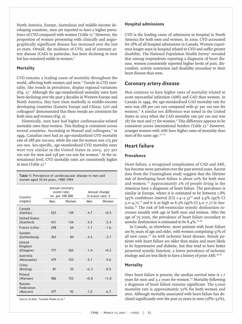
North America, Europe, Australasia and middle-income de-veloping countries, men are reported to have a higher preva-lence of CVD compared with women (Table 1).4 However, theproportion of women presenting with clinically and angio-graphically significant disease has increased over the last20 years. Overall, the incidence of CVD, and of coronary ar-tery disease (CAD) in particular, has been declining in menbut has remained stable in women.5
Mortality
CVD remains a leading cause of mortality throughout theworld, affecting both women and men.6 Trends in CVD mor-tality, like trends in prevalence, display regional variations(Fig. 1).7 Although the age-standardized mortality rates havebeen declining over the past 3 decades in Western Europe andNorth America, they have risen markedly in middle-incomedeveloping countries (Eastern Europe and China). Levi andcolleagues8 demonstrated that these trends are consistent forboth men and women (Fig. 2).
Historically, men have had higher cardiovascular-relatedmortality rates than women. This finding is consistent acrossseveral countries. According to Manuel and colleagues,9 in1999, Canadian men had an age-standardized CVD mortalityrate of 288 per 100 000, while the rate for women was 175 per100 000. Sex-specific, age-standardized CVD mortality rateswere very similar in the United States in 2003: 307 per100 000 for men and 158 per 100 000 for women.2 At the in-ternational level, CVD mortality rates are consistently higherin men (Table 2).6
Hospital admissions
CVD is the leading cause of admission to hospital in NorthAmerica for both men and women. In 2000, CVD accountedfor 18% of all hospital admissions in Canada. Women experi-ence longer stays in hospital related to CVD and suffer greaterdisability. The National Population Health Survey1 revealedthat among respondents reporting a diagnosis of heart dis-ease, women consistently reported higher levels of pain, dis-comfort, activity restriction and disability secondary to theirheart disease than men.
Coronary artery disease
Men continue to have higher rates of mortality related toacute myocardial infarction (AMI) and CAD than women. InCanada in 1999, the age-standardized CAD mortality rate formen was 188 per 100 000 compared with 97 per 100 000 forwomen.9 A similar sex difference was noted in the UnitedStates in 2003 when the CAD mortality rate per 100 000 was187 for men and 77 for women.2 This difference appears to beconsistent across international borders (Table 2).6 However,younger women with AMI have higher rates of mortality thanmen of the same age.10–12
Heart failure
Prevalence
Heart failure, a recognized complication of CAD and AMI,has become more prevalent over the past several years. Recentdata from the Framingham study suggest that the lifetimerisk of developing heart failure is about 20% for both menand women.13 Approximately 2% of people living in theAmericas have a diagnosis of heart failure. The prevalence issimilar in Europe, where it is estimated to be between 1.8%(95% confidence interval [CI] 1.4–2.3)14 and 3.9% (95% CI3.0–4.7),15 and it is as high as 6.3% (95% CI 5.0–7.7) in Aus-tralia.16 The risk of left-ventricular systolic dysfunction in-creases steadily with age in both men and women. After theage of 75 years, the prevalence of heart failure secondary tosystolic dysfunction is estimated to be 8.4%.17,18
In Canada, as elsewhere, most patients with heart failureare 65 years of age and older, with women comprising 51% ofall new cases.19 As with ischemic heart disease, female pa-tients with heart failure are older than males and more likelyto be hypertensive and diabetic, but they tend to have betterpreserved systolic function, a lower prevalence of ischemicetiology and are less likely to have a history of prior AMI.20,21
Mortality
Once heart failure is present, the median survival time is 1.7years for men and 3.2 years for women.13 Mortality followinga diagnosis of heart failure remains significant. The 5-yearmortality rate is approximately 50% for both women andmen. Although mortality associated with heart failure has de-clined significantly over the past 20 years in men (28%–52%),
CMAJ • March 13, 2007 • 176(6) | S2
Table 1: Prevalence of cardiovascular disease in men and women aged 35–64 years, 1980–1994
Annual coronary event rate;
no. per 100 000 Annual change
in event rate; % Country (region) Men Women Men Women
Canada (Halifax) 523 139 –4.7 +0.5
United States (Stanford) 431 134 –4.2 –2.4
France (Lille) 298 64 –1.1 –1.6
Sweden (Gothenburg) 363 84 –4.2 –3.7
United Kingdom (Glasgow) 777 265 –1.4 +0.2
Australia (Newcastle) 479 153 –5.1 –5.6
China (Beijing) 81 35 +2.3 –0.5
Poland (Warsaw) 586 153 +0.8 +1.0
Russian Federation (Moscow) 477 92 –1.0 –6.7
Source of data: Tunstall-Pedoe et al.4

the decrease has not been as significant in women(6%–33%).22
Hospital admissions
Heart failure is a leading cause of hospital admissionthroughout the world, and women account for about 50% ofthese admissions. In North America, admissions to hospitalwith a primary diagnosis of heart failure have increased by34% since 1990, but this increase is more notable in women(39%) than men (29%). In fact, a review of the American Na-tional Hospital Discharge Survey23 noted that age-adjustedheart failure admission rates have been constant since 1991for men, but have continued to increase for women (19%)(Fig. 3), suggesting that the increase in hospital admissionsin recent years is largely the result of more women presentingwith clinically significant disease. Once discharged from hos-pital, men and women appear to have similar rates of read-mission (hazard ratio [HR] 0.89; 95% CI 0.71–1.11).21 Sex dif-
ferences in length of hospital stays have also been noted, withwomen having significantly longer hospital stays than men.24
Atrial fibrillation
Prevalence
Atrial fibrillation is the most common cardiac arrhythmia andis an established risk factor for stroke and premature death.North American and European studies indicate that about0.95%–1.1% of the population experiences atrial fibrilla-tion.25–27 The Framingham study demonstrated that men were1.5 times more likely to develop atrial fibrillation thanwomen,28 but because the number of women older than75 years is almost twice that of men, the absolute numbers ofthose with atrial fibrillation are roughly equal.26 Incidence in-creases with age; the prevalence of atrial fibrillation at age 55is 0.1% and increases to 9.0% in those over 80 years ofage.26,29 This age-related increase in prevalence is most strik-
ing in men (Table 3).30
Mortality
Mortality rates are significantly higher inboth women and men with atrial fibrilla-tion. In longitudinal studies, mortality rateswere approximately 25% higher than age-and sex-matched controls.31,32 In the UnitedStates, in-hospital mortality rate followingadmission for atrial fibrillation is estimatedat 0.8%. Men have a modestly higher (butstatistically significant) risk of in-hospitalmortality. Using administrative data (Na-tional Hospital Discharge Survey), Kairallahand colleagues33 demonstrated that malesex was an independent predictor of in-hospital mortality (odds ratio [OR] 1.10;95% CI 1.06–1.14). However, analysis of ad-ministrative data from Scotland suggeststhat 1-year mortality may be higher inwomen; Stewart and colleagues34 found the1-year case fatality rate in men to be 11.9% v.16.2% in women. The distribution of agesin this cohort is not known; the higher mor-tality rate seen in women may reflect agreater number of very elderly women diag-nosed with atrial fibrillation. Wattigney andcolleagues35 have demonstrated in a US co-hort that mortality remains higher amongmen after age standardization.
Hospital admission and stroke
Reflecting the sex differences in prevalence,admissions to hospital for atrial fibrillationare also more common in men at every age(Table 4).36 Among patients with atrial fib-rillation, women seem to be at greater risk
CMAJ • March 13, 2007 • 176(6) | S3
WomenMen
Australia Norway
New Zealand United Kingdom
Ireland Finland
Luxembourg Italy
Germany Portugal
France
United States Chile Spain
Slovenia Hungary
–80 –60 –40 –20 0 20 40 60 80
% decrease % increase
Armenia Georgia Mexico Japan
Romania Russian Federation
Belarus Kazakhstan
Ukraine
Fig. 1: Changes in rates of death from cardiovascular disease among men andwomen aged 35–74 years between 1990 and 2000 in selected countries. Reproducedwith permission from the World Health Organization.7

of stroke than men. In a cohort of US Medicaid patients, Wolfand colleagues31 found that, after controlling for other estab-lished risk factors, women with atrial fibrillation had a22%–25% greater risk of stroke than women without atrialfibrillation (Table 5). In contrast, the increased risk of strokein men with atrial fibrillation was completely attenuated bycontrolling for other stroke risk factors.31 Women have alsobeen found to have higher readmission rates followingstroke.36
CMAJ • March 13, 2007 • 176(6) | S4
0
50
100
150
200
250
300
350
1965–69 1970–74 1975–79 1980–84 1985–89 1990–94 1995–97
No.
per
100 0
00
Men
Women
European UnionEastern countries
JapanUnited States
0
50
100
150
200
250
300
350
1965–69 1970–74 1975–79 1980–84 1985–89 1990–94 1995–97
No.
per
100 0
00
European UnionEastern countries
JapanUnited States
Fig. 2: Trends in age-standardized (world population) cardio-vascular disease mortality among men and women in the Euro-pean Union, Eastern Europe (Bulgaria, Czech Republic, Hun-gary, Poland, Romania and Slovakia), the United States andJapan from 1965 to 1972. Reproduced with permission from BMJPublishing Group (Heart 2002;88:119-24).8
Table 2: Cardiovascular-related mortality rates per 100 000 people for men and women aged 35–74 years
Men Women
Country (yr) CVD CAD CVA Total CVD CAD CVA Total
Russian Federation (1998) 1167 639 361 2502 540 230 229 1001
China, rural (1999) 413 64 243 1260 286 110 84 748
China, urban (1999) 389 106 217 1003 273 71 147 663
United States (2003) 367 187 36 943 158 77 28 593
England/ Wales (2002) 301 196 49 811 138 68 36 509
Australia (2001) 206 138 31 679 92 47 22 396
Canada (2001) 222 150 29 757 97 51 21 455
Note: CAD = coronary artery disease, CVA = cerebrovascular accident, CVD = cardiovascular disease. Source of data: World Health Organization.6
Table 3: Prevalence of atrial fibrillation in adults aged 65–84 years (% of total population), 1968–1989
1968–1970
1971–1973
1975–1977
1979–1981
1983–1985
1987–1989
Men 3.2 5.3 6.5 7.8 7.5 9.1
Women 2.8 3.3 4.3 4.3 3.9 4.7
Source of data: Wolf et al.30
Table 4: Age- and sex-specific hospital admission rates for atrial fibrillation or flutter in Canada, 1997/98–1999/2000
Hospital admission rate; no. per 100 000
Age, yr Men Women
20–49 40.1 17.1
50–64 478 245
65–74 2096.7 1297.6
75–84 4891 3548.9
85+ 7645.9 5924.5
Source of data: Humphries et al.36
1 200 000
1 000 000
800 000
600 000
400 000
200 000
1990 1992 1994 1996 1998 2000
No.
of
hos
pit
al a
dm
issi
ons
All adultsFemalesMales
Fig. 3: American National Hospital Discharge Survey data onannual hospital admissions because of heart failure amongadults 35 years and older, 1990–2000. Reproduced with per-mission from Elsevier (Am Heart J 2004;147:74-8).23

Cardiovascular risk factors in girls and boysJennifer McGrath, Tracie Barnett, Marie Lambert, Jennifer O’Loughlin, Gilles Paradis, Arsham Alamian and Teresa Ho
CMAJ • March 13, 2007 • 176(6) | S5
Although most cardiovascular events occur in adulthood, theprecursors of CVD manifest during childhood and adoles-cence.38,39 CVD is partly attributable to modifiable lifestyle be-haviours, and childhood is a critical developmental periodwhen these habits are established.40 Further, risk factors suchas smoking, sedentary behaviour and poor diet in childrenand adolescents persist through young adulthood and are im-portant predictors of subsequent risk of CVD.41–44 Conse-quently, it is essential to promote cardiovascular health anddirect primary prevention efforts toward children and adoles-cents to disrupt the progression of CVD risk factors andthereby offset both the risk of CVD in adulthood and the un-precedented potential burden on health care systems.
Search strategy
Studies included in this qualitative review of childhood cardio-vascular risk factors were identified through the use of widelyavailable computer databases (Ovid MEDLINE, Ovid EMBASE,PubMed, PsycInfo, the Cochrane Library). Boolean searcheswere carried out by combining the keyword (“boy” OR “girl”OR “child” OR “pediatric” OR “adolescent” OR “youngadult”) with each of the following keyword combinations us-ing the AND operator: (“atherosclerosis” OR “cardiovascular”OR “coronary” OR “heart”), (“obesity” OR “overweight”),(“lipids” OR “lipoprotein”), (“hypertension” OR “blood pres-sure” OR “systolic” OR “diastolic”), (“cigarettes” OR “smok-ing” OR “tobacco”), (“diet” OR “nutrition”), (“exercise” OR
“physical activity” OR “sedentary”), (“clustering” OR “behav-ior” OR “lifestyle”). Web sites of several well-known organiza-tions, such as the World Health Organization, Centers for Dis-ease Control and Prevention (CDC), Heart and StrokeFoundation of Canada and the American Heart Association,were reviewed for additional information and current recom-mendations. Finally, to obtain information from national andinternational statistical databases as well other “grey litera-ture” and nonconventional documents, government Web sites(e.g., Statistics Canada, Health Canada, CDC) were examined.To reduce the copious amount of information this searchstrategy produced, the review emphasized children or adoles-cent populations; large studies with representative samples;longitudinal studies; and recent publications that reportedsex- or gender-specific findings.
Overweight and obesity
Overweight and obesity are the most frequent nutritional dis-orders in industrialized countries in children as well as inadults; the prevalence of obesity has increased almost 3-foldover the past 2 decades.45,46 Although it is generally thoughtthat girls are more likely to be overweight than boys, there areno differences in prevalence between girls and boys.
In a recent report based on heights and weights measuredin the National Longitudinal Survey of Children and Youth47
and using Cole and colleagues’48 age- and sex-specific bodymass index (BMI) threshold values for overweight and obesity,
Health-related quality of life
Although men are more likely to die from CAD, women aremore likely to live with CAD-related disability. In NorthAmerican cohort studies, women with a history of CAD, heart
failure or atrial fibrillation consistently report lower health-related quality of life and greater disability related to theirheart disease than men. Women are also less likely to returnto work following admission to hospital for complications re-lated to CVD.37
Summary
• CVD is prevalent among both women and men.• Women have a lower CVD mortality rate than men.• CAD and atrial fibrillation are more prevalent among men
than among women.• Women appear to have a relatively higher risk of atrial fib-
rillation-related stroke than men.• Clinically significant heart failure is on the rise in women.• Women are more likely to live with more CVD-related dis-
abilities and have a lower health-related quality of life.
Knowledge gap
• Why are CVD incidence and mortality rates decreasingamong men but stable among women?
Table 5: Adjusted* mortality and stroke risk associated with atrial fibrillation following hospital admission by sex in adults aged 75–84 years
Outcome; group Risk ratio 95% confidence
interval
Mortality
Men 1.07 0.96–1.19
Women 1.20 1.09–1.32
Stroke
Men 1.05 0.94–1.16
Women 1.25 1.14–1.37
*Adjusted for prior history of acute myocardial infarction, unstable angina, stable angina, congestive heart failure, hypertension, diabetes, valvular disease, stroke or chronic obstructive pulmonary disease. Patients were eliminated at death from stroke analysis. Source of data: Wolf et al.31

Shields46 estimated that the prevalence of overweight and obe-sity of Canadian boys and girls aged 2–17 years was 27% and25%, respectively; the corresponding figures for obesity onlywere 9% and 7%, respectively. Although the overall prevalenceof overweight and obesity was similar for boys and girls,trends varied for different age groups. The percentage of over-weight and obese children 2–5 years old remained unchanged(21%) between 1978–1979 and 2004. However, the prevalenceof overweight and obesity doubled among those 6–11 years(from 13% to 26%) and those 12–17 years (from 14% to 29%),and the prevalence of obesity tripled among those 12–17 years(from 3% to 9%). Secular trends in body mass of Canadianchildren are shown in Fig. 4.49 Part of these sex-based differ-ences may be attenuated by the fact that age- and sex-specificthresholds are used to define overweight and obesity.
In the United States, using height and weight measurementsobtained in 1999–2000 as part of the National Health and Nutri-tion Examination Survey (NHANES) and the 2000 CDC growthchart reference values, Ogden and colleagues45 reported that theprevalence of overweight and obesity was 20.5%, 26.2% and26.5% among non-Hispanic white youth aged 2–5, 6–11 and12–19 years, respectively. The corresponding figures for obesityonly were 10.1%, 11.8% and 12.7%. The prevalence of obesitywas not significantly different for boys and girls.
Longitudinal studies have shown that measures of BMItaken during childhood and adolescence predict adult values.In the Bogalusa Heart Study,50 childhood and adult BMI weremoderately correlated (Spearman’s rank correlation coeffi-cient 0.58), and this relation did not vary significantly withage, ethnic origin or sex. Similar results were observed in theChild and Adolescent Trial for Cardiovascular Health wherethe Kendall index of concordance for BMI was 0.86 over afollow-up period of 6 years and tracking was similar for bothsexes.51
Summary
• The prevalence of obesity has increased almost 3-fold overthe past 2 decades.
• There are no significant differences between girls and boysin the prevalence of overweight and obesity.
• A large proportion of obese adolescents will become obeseadults.
Knowledge gap
• Additional research is needed on factors that contribute tothe onset of overweight or obesity in childhood and ado-lescence and factors that contribute to its persistence intoadulthood.
• The basic biologic characteristics of appetite, weight con-trol, genetic susceptibility and environmental triggers re-main elusive.
• We do not know specific prevention or treatment strate-gies that have sustained benefits in a broad spectrum ofindividuals.
Lipids and insulin resistance
Overweight and obesity are associated with significant healthproblems in the pediatric population and are important earlyrisk factors for much of the adult morbidity and mortality as-sociated with type 2 diabetes mellitus and CVD.
A large number of studies have consistently shown associ-ations among childhood obesity, dyslipidemia, hyperinsu-linemia and high blood pressure.52–54 The clustering of theseCVD risk factors defines the metabolic syndrome called in-sulin resistance syndrome (IRS). The likelihood of IRS is thesame for girls and boys. In the 1999 Quebec Child and Ado-lescent Health and Social Survey (QCAHS), a representativecross-sectional survey of Quebec youth, the overall prevalenceof IRS was 11.5% in youth aged 9, 13 and 16 years.55 This is theonly report on the prevalence of IRS in a provincially repre-sentative sample of youth in Canada. Findings from the thirdNHANES survey show a prevalence of IRS of 9.2% (95% CI7.8–10.6) in US youth aged 12–19 years.56 Prevalence wascomparable for girls (8.9%; 95% CI 7.1–10.7) and boys (9.5%;95% CI 7.5–11.5) and for older (8.3%; 95% CI 6.5–10.1) andyounger (10.3%; 95% CI 8.3–12.3) adolescents. The ethnicdistribution was similar to that in adults: Mexican Americans(12.9%; 95% CI 10.4–15.4) and non-Hispanic white people(10.9%; 95% CI 8.4–13.4) had a greater prevalence of IRScompared with non-Hispanic black people (2.5%; 95% CI1.3–3.7). Nearly a third (31.2%; 95% CI 28.3–34.1) of over-weight or obese adolescents had IRS.
Given the link between excess weight and dyslipidemia,the increase in overweight and obesity among youth in NorthAmerica is expected to affect trends in lipid and glucose levelsadversely. However, comparing data from 2 NHANES surveysof US youth aged 4–17 years in 1988–1994 and 1999–2000,Ford and colleagues57 found that the mean concentrations oftotal cholesterol, high-density lipoprotein (HDL) cholesteroland low-density lipoprotein (LDL) cholesterol were almost
CMAJ • March 13, 2007 • 176(6) | S6
25
30
35
40
45
50
1996198819811966
Bod
y m
ass,
kg
Boys 12 yrGirls 12 yr
Boys 10 yrGirls 10 yr
Boys 8 yrGirls 8 yr
Fig. 4: Trends in body mass of Canadian boys and girls.Source: Adapted from Tremblay and Willms.49

the same in the 2 groups. However, in 1999–2000, meantriglyceride concentration was almost 10% lower than in1988–1994 and mean glucose concentration decreased by 3%.These inconsistent trends in CVD risk factors, especially inHDL cholesterol and triglyceride levels, are difficult to ex-plain. Changes in mean levels may not be sensitive enough todetect variations occurring at the extremities of the distribu-tions; it would have been interesting to compare selected per-centiles in the 2 surveys.
Although longitudinal data for plasma lipid levels in Cana-dian youth are not available, mean concentrations of plasmalipids in the 1999 QCAHS and in the 1999–2000 NHANES weresimilar.55,57 Body composition and fat distribution are differentbetween boys and girls and across ages, and these differencesmay influence the relation between fatness and lipids.
Summary
• There are no significant differences between girls and boysin prevalence of IRS.
• There are no significant differences in the trends in lipidand glucose profiles of girls and boys over the last decade.
Knowledge gap
• Little is known about the natural history of the metabolicconsequences of excess adiposity in childhood and adoles-cence.
• Surveillance of trends in obesity and the potential effectson CVD risk factors is needed.
• Better understanding of the relative importance of genetic,biologic, environmental and psychosocial determinants ofmetabolic abnormalities associated with excess fat isrequired.
Blood pressure
Knowledge of blood pressure distributions in youth is impor-tant: both systolic and diastolic pressure persist from child-hood to adulthood42,58,59 and the current youth obesity epi-demic has important effects on blood pressure distribution inthis segment of the population.60
There are few data on blood pressure distribution and theprevalence of elevated blood pressure in Canadian children.The 1999 QCAHS reported important increases in mean sys-tolic blood pressure and in height-specific systolic bloodpressure percentile values compared with reference valuesfrom the National High Blood Pressure Education ProgramWorking Group on Hypertension Control in Children andAdolescents61 for both boys and girls in all age groups.52 Theproportion of children aged 9, 13, and 16 years with high-normal or elevated systolic pressure was 12%, 22% and 30%,respectively, for boys and 14%, 19% and 17% for girls. Ele-vated systolic pressure occurred in almost twice as many 16-year-old boys as girls. The mean systolic pressure of 13- and16-year-old boys was 2 mm Hg (p = 0.004) and 10 mm Hg(p < 0.0001) higher, respectively, than that of girls. Less than1% of youth had elevated or borderline diastolic blood pres-
sure, and this did not differ between sexes. These findingswere recently confirmed in a longitudinal study of Canadianadolescents. The likelihood of high systolic blood pressurevalues among boys compared with girls was 1.29 (95% CI0.77–2.16) in grade 7, 1.98 (95% CI 1.35–2.93) in grade 9, and2.74 (95% CI 1.52–4.94) in grade 11.
62
A similar trend in high blood pressure has been observedin US children (Fig. 5).63 In age-matched boys and girls,64
cross-sectional analyses of the baseline data from the Na-tional Heart, Lung, and Blood Institute Growth and HealthStudy showed significantly higher systolic (105 v.100 mm Hg) and diastolic (71 v. 65 mm Hg) blood pressurelevels among overweight white girls aged 9 years than amongthose who were not overweight.65
Limited data are available from representative samples ofchildren from other parts of the world. Comparisons are fur-ther complicated by variations in sampling design and blood
CMAJ • March 13, 2007 • 176(6) | S7
Systolic
95
100
105
110
1988–1994 1999–2000
Blo
od p
ress
ure
, m
m H
g
Girls 8–17 yr
Boys 8–17 yr
Diastolic
50
55
60
65
70
1988–1994 1999–2000
Blo
od p
ress
ure
, m
m H
g
Boys 8–17 yr
Girls 8–17 yr
Fig. 5: Trends in systolic and diastolic blood pressure amongchildren and adolescents in the United States. Source: Adaptedfrom Muntner et al.63

pressure measurement. For example, a recent survey of 809boys and 842 girls aged 7–14 years from Belgrade, Serbia andMontenegro revealed average blood pressures of113/70 mm Hg in boys and 115/71 mm Hg in girls. High sys-tolic pressure was present in 5% of boys and girls, and highdiastolic pressure was found in 6% of boys and 5% of girls.66
Conversely, a study of over 1200 children aged 6–11 years inMilan reported a significantly higher prevalence of elevatedblood pressure in girls (5%) than in boys (3%).58 Sex differ-ences in blood pressure may be due to differences in BMI be-tween boys and girls at any given age, differences in activitylevels and differences in pubertal stage at any given age.
Summary
• Elevated blood pressure persists from childhood to adult-hood.
• Elevated blood pressure is prevalent in both girls and boys.• Boys have higher systolic blood pressure than girls.
Knowledge gap
• Criterion-related reference values are necessary to clarifythe significance of blood pressure levels in youth.
• More information is needed on blood pressure distribu-tion and the prevalence of elevated blood pressure ingroups of Canadian girls and boys.
• Sex differences in blood pressure require explanation at bi-ologic, environmental and behavioural levels.
Smoking
Although data from the Global Youth Tobacco Survey67 and
the National Tobacco Information Online System68 suggestthat, in many developing countries, proportionately moreboys than girls smoke, there have been few notable differ-ences in the prevalence of smoking by boys and girls in de-veloped countries over the last decade.69 Recent data forCanadian youth70,71 concur (Fig. 6).72 In Canada, 25% ofgirls 15–17 years old smoke compared with 19% of same-ageboys, but by age 19 the prevalence is equal (31%).73 How-ever, there appear to be sex differences in the number of cig-arettes smoked per day, at least among young daily smok-ers. In 2002, Canadian boys in grades 5–9 smoked 8.8cigarettes a day on average, compared with 7.3 cigarettesamong girls.70 There are currently no nationally representa-tive data comparing the incidence of smoking initiation bysex, although 1 prospective Canadian study suggests thatboys are more likely than girls to escalate cigarette con-sumption rapidly after initiation.74 Although smoking ces-sation in youth is understudied, several surveys suggest thatthere are few differences in cessation attempts and success-ful cessation by sex.70
Although prevalence does not differ markedly by sex,girls and boys may smoke for different reasons. Amongadolescent girls, body image, eating disorders and tar-geted advertising by tobacco companies likely relate toinitiation of and sustained smoking, whereas aggressionand conduct disorders appear to be fairly consistent pre-dictors of smoking among boys.75 However, the currentliterature on sex differences in the determinants of smok-ing is generally inconclusive because many studies arecross-sectional, the definitions of smoking and of the po-tential determinants of smoking are widely divergentacross studies and most studies investigated only smallsubsets of potential determinants.
CMAJ • March 13, 2007 • 176(6) | S8
15
20
25
30
35
1985 1991 1994/95 1994/95 1996 1996/97 1998/99 1999 2000 2000/01 2001
Pre
vale
nce
, %
Girls Boys
Fig. 6: Trends in current-smoker prevalence among Canadian adolescents, 15–19 years of age. Source: Adapted from Gilmore.72
Summary
• The prevalence of smoking is similar in girls and boys.• Boys who smoke daily smoke more cigarettes a day than
girls who smoke daily.• Determinants of smoking differ with sex.
Knowledge gap
• The sex-specific incidence and prevalence of smokingshould be monitored throughout the life course.
• Longitudinal life-course studies based on socioecologicmodels of health behaviour are needed to determinewhether girls smoke for different reasons than boys.
• Researchers should examine the relative importance of in-dividual (genetic, sociodemographic, psychosocial, behav-ioural) and environmental (social influences, policy, ad-vertising) factors that influence smoking.
Diet
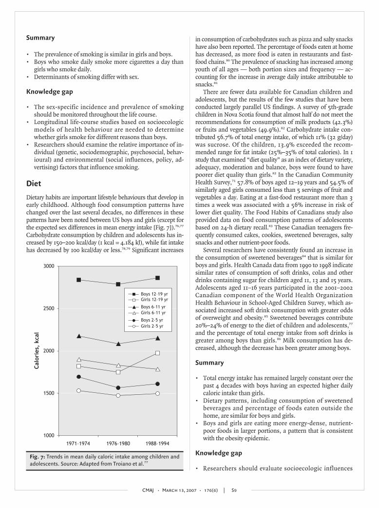
Dietary habits are important lifestyle behaviours that develop inearly childhood. Although food consumption patterns havechanged over the last several decades, no differences in thesepatterns have been noted between US boys and girls (except forthe expected sex differences in mean energy intake [Fig. 7]).76,77
Carbohydrate consumption by children and adolescents has in-creased by 150–200 kcal/day (1 kcal = 4.184 kJ), while fat intakehas decreased by 100 kcal/day or less.78,79 Significant increases
in consumption of carbohydrates such as pizza and salty snackshave also been reported. The percentage of foods eaten at homehas decreased, as more food is eaten in restaurants and fast-food chains.80 The prevalence of snacking has increased amongyouth of all ages — both portion sizes and frequency — ac-counting for the increase in average daily intake attributable tosnacks.81
There are fewer data available for Canadian children andadolescents, but the results of the few studies that have beenconducted largely parallel US findings. A survey of 5th-gradechildren in Nova Scotia found that almost half do not meet therecommendations for consumption of milk products (42.3%)or fruits and vegetables (49.9%).82 Carbohydrate intake con-tributed 56.7% of total energy intake, of which 11% (32 g/day)was sucrose. Of the children, 13.9% exceeded the recom-mended range for fat intake (25%–35% of total calories). In 1study that examined “diet quality” as an index of dietary variety,adequacy, moderation and balance, boys were found to havepoorer diet quality than girls.82 In the Canadian CommunityHealth Survey,71 57.8% of boys aged 12–19 years and 54.5% ofsimilarly aged girls consumed less than 5 servings of fruit andvegetables a day. Eating at a fast-food restaurant more than 3times a week was associated with a 56% increase in risk oflower diet quality. The Food Habits of Canadians study alsoprovided data on food consumption patterns of adolescentsbased on 24-h dietary recall.83 These Canadian teenagers fre-quently consumed cakes, cookies, sweetened beverages, saltysnacks and other nutrient-poor foods.
Several researchers have consistently found an increase inthe consumption of sweetened beverages84 that is similar forboys and girls. Health Canada data from 1990 to 1998 indicatesimilar rates of consumption of soft drinks, colas and otherdrinks containing sugar for children aged 11, 13 and 15 years.Adolescents aged 11–16 years participated in the 2001–2002Canadian component of the World Health OrganizationHealth Behaviour in School-Aged Children Survey, which as-sociated increased soft drink consumption with greater oddsof overweight and obesity.85 Sweetened beverages contribute20%–24% of energy to the diet of children and adolescents,77
and the percentage of total energy intake from soft drinks isgreater among boys than girls.86 Milk consumption has de-creased, although the decrease has been greater among boys.
Summary
• Total energy intake has remained largely constant over thepast 4 decades with boys having an expected higher dailycaloric intake than girls.
• Dietary patterns, including consumption of sweetenedbeverages and percentage of foods eaten outside thehome, are similar for boys and girls.
• Boys and girls are eating more energy-dense, nutrient-poor foods in larger portions, a pattern that is consistentwith the obesity epidemic.
Knowledge gap
• Researchers should evaluate socioecologic influences
CMAJ • March 13, 2007 • 176(6) | S9
1000
1500
2000
2500
3000
1971–1974 1976–1980 1988–1994
Cal
orie
s, k
cal
Girls 2–5 yr
Girls 6–11 yr
Boys 2–5 yr
Boys 6–11 yr
Boys 12–19 yr Girls 12–19 yr
Fig. 7: Trends in mean daily caloric intake among children andadolescents. Source: Adapted from Troiano et al.77

(e.g., parental dietary habits, psychosocial and behaviouralfactors, social influences, media and advertising) on di-etary behaviour.
• Researchers should improve assessment procedures to de-termine more accurately children’s dietary intake (advanc-ing 24-h dietary recall, food frequency questionnaires andprospective food records or diaries).
• The sex-specific prevalence and trends in dietary and totalenergy intake should be monitored throughout the lifecourse.
Physical activity and sedentary behaviour
Data from recent national surveys suggest a favourable trendin physical activity among Canadian adolescents.87 Between1994 and 2003, the proportion of 12–19-year-olds classified asactive (i.e., average energy expenditure ≥ 3.0 kcal/kg bodyweight a day) increased from 44.6% to 54.6% for boys andfrom 27.3% to 39.5% for girls. Despite this encouragingtrend, the physical activity gap between boys and girls re-mains wide (Fig. 8).87 Girls report less physical activity thanboys, both before and during adolescence.88–92 Sex differencesare particularly apparent for vigorous physical activity, withgirls less likely than boys to engage in such activity duringtheir free time or in the context of organized physical activity,during school and outside school.93–95
Although girls and boys are equally likely to be enrolled inorganized physical activity and lessons outside school, girlsare less likely to belong to sports clubs or participate in unor-ganized physical activity or in school sports outside physicaleducation classes.96 Not surprisingly, boys and girls differsubstantially in their physical activity preferences97,98 and intheir patterns of involvement in physical activity.99,100 Boys
and girls also differ in their attitudes and beliefs regardingphysical activity,101,102 as well as their motivation for97,103 andbarriers to104,105 engaging in physical activity.
Individual and environmental factors do not influenceboys and girls equally.106,107 There is evidence that genetic ef-fects are associated with sports participation and with leisuretime physical activity to a greater extent in boys than ingirls.108 Boys’ and girls’ physical activity levels are similarly af-fected by peer and family social support,109,110 but appear to beinfluenced by different neighbourhood factors. For example,perceived neighbourhood opportunities for physical activityare associated with girls’ but not boys’ activity levels,111 andphysical features of the school environment appear to influ-ence physical activity to a greater extent in boys.112,113 Finally,significant effects by sex are frequently observed in interven-tions promoting physically active lifestyles among youth.104,114
Data on sedentary pursuits are limited. A recent reviewconcluded that total media use among youth in industrializedcountries has remained stable in the past decades at approxi-mately 5 h/day.104,115 The most recent data show that the pro-portion of adolescents aged 12–17 years who spend 30 h ormore a week in sedentary activities (i.e., watching television,playing video games, spending time on the computer) is30.0% in boys and 18.2% in girls.116 Most of the discrepancybetween older boys and girls concerning time spent in seden-tary pursuits relates to greater computer use and video gameplay in boys, not to television viewing.115,116
Sex and gender differences in physical activity and seden-tary behaviour may be largely due to interactions between in-dividual characteristics (e.g., physical maturation, personalmotivation) and responses to environmental cues that en-hance or inhibit involvement in these pursuits.
Summary
• Despite recent increases in physical activity in all youth,sex-related disparities in involvement in physical activityhave not diminished.
• Boys are consistently more active than girls at all ages, andage-related decreases in physical activity occur earlier ingirls than in boys.
• Most girls are not active enough to meet guidelines for op-timal growth and development.
Knowledge gap
• Refine methods of physical activity assessment to capturethe different dimensions and contexts of both sexes, in-cluding low- and moderate-intensity activities of daily liv-ing and active transportation, as well as the more tradi-tional structured, free play and vigorous physical activities.
• Investigate the possible role of genetic inheritance inadaptation to sedentary or active lifestyles.
• Investigate how individual, familial, school and neigh-bourhood characteristics interact with sex and gender todetermine involvement in physical activity.
• An increased understanding of the clustering of active andsedentary behaviours over the life course is required to
CMAJ • March 13, 2007 • 176(6) | S10
0
10
20
30
40
50
60
1994/95 1996/97 1998/99 2000/01 2002/03
Pro
por
tion
act
ive,
%
Boys 12–14 yr
Girls 12–14 yr
Boys 15–19 yr
Girls 15–19 yr
Fig. 8: Trends in leisure-time physical activity among Canadianadolescents. Proportion active are those with an average dailyenergy expenditure of at least 3.0 kcal/kg. Source: Adaptedfrom Statistics Canada.87

help devise more effective sex-specific prevention and pro-motion programs.
Clustering of behavioural risk factors
According to the Canadian Cardiovascular Society’s 1998consensus on the prevention of CVD, the major CVD behav-ioural risk factors in youth include smoking, physical inac-tivity and obesity.117 These modifiable risk factors persistfrom childhood into adulthood41–44 and tend to clusteramong youth.118–121 In the longitudinal Cardiovascular Riskin Young Finns Study, Raitakari and colleagues118 found that15- and 18-year-old boys and girls who smoked were morelikely to be regular users of alcohol and physically inactivecompared with non-smokers. Obesity was more prevalentamong physically inactive compared with active males (14%v. 8%, p < 0.05), female drinkers compared with non-drinkers (20% v. 9%, p < 0.001) and smokers compared withnon-smokers (males 15% v. 9%, p < 0.05; females 16% v.9%, p < 0.01). In males, those with 4 selected CVD behav-ioural risk factors, including smoking, physical inactivity,obesity and intake of dietary fat, had a 5.5 times greater riskof having an atherogenic lipid profile and high diastolicblood pressure compared with those with 0 or 1 behaviouralrisk factor.118 In another prospective study investigating theassociation between family socioeconomic status and an ad-verse cardiovascular risk profile among 14- and 17-year-oldboys and girls in Sweden, Bergstrom and colleagues119 re-ported clustering of high BMI, low physical fitness and dailysmoking among girls living in families of low socioeco-nomic status compared with girls of the same age in familiesof medium or high socioeconomic status.
In the United States, Pate and colleagues120 investigatedthe association between physical activity and other health-related behaviours, including smoking and dietary habits, ina representative sample of adolescents aged 12–18 years. Boysand girls who smoked 1 or more cigarettes over the past30 days and who did not eat fruits or vegetables on the previ-ous day were 1.5 and 2 times more likely to be less active thanthose who did not report these behaviours, respectively. In amore recent study, Pronk and colleagues121 reported that only31% of US adolescents aged 13–17 years met recommendedguidelines for multiple healthy lifestyle factors includingphysical activity, non-smoking, high-quality diet and healthyweight. This implies that more than two-thirds of US adoles-cents have 1 or more CVD behavioural risk factors, an esti-mate that is quite alarming given the potential synergistic ef-fects associated with the presence of multiple behaviouralrisk factors on the risk of chronic diseases in adult life.122
Pronk and colleagues121 also found that depression is associ-ated with clustering of health-related behaviours in adoles-cents. Specifically, non-depressed adolescents were 2.15times more likely to engage in 4 healthy lifestyle factors. Theclustering of health-related behaviours in US adolescents wassimilar for boys and girls.
Data on the prevalence and potential determinants of mul-tiple CVD behavioural risk factors in Canada are scarce. Thestudies reviewed in this section suggest that CVD behavioural
risk factors cluster in children and adolescents. Certain socio-demographic characteristics, including age and sex,109,110 andpsychosocial variables, such as depression and family socio-economic status,112 seem to be associated with the clusteringof CVD behavioural risk factors in youth, but evidence in thisarea remains limited and inconsistent.
Critical analysis of studies
All the studies reviewed were observational, cross-sectionalcohort or longitudinal studies. Wherever possible, results ofstudies specifically focusing on girls or on boys or providingsex comparisons have been included. The areas not suffi-ciently addressed by existing studies have been highlighted asknowledge gaps.
Why are there sex differences?
When sex differences in risk factors are apparent, they appearto be attributable to a combination of biologic (sex) and be-havioural (gender) factors. Compared with age-matchedboys, girls have lower systolic blood pressure. However, thissex difference is attenuated with age and may be partly attrib-utable to sex hormones or their receptors. In terms of healthbehaviours, the prevalence of smoking does not differmarkedly by sex, although boys smoke more cigarettes a daythan girls, and they are more likely than girls to escalate ciga-rette consumption rapidly after initiation. Boys and girls ap-pear to smoke for different reasons. Body image, eating dis-orders and targeted advertising by tobacco companies likelyrelate to initiation and sustained smoking among adolescentgirls, while aggression and conduct disorders are predictorsof smoking among boys. Sex differences in physical activityare also apparent; girls are less likely to engage in physical ac-tivity than boys, both before and during adolescence. Thismay be partly due to sex and gender factors.
Summary
• The major CVD behavioural risk factors, including smok-ing, physical inactivity and obesity, cluster among youngboys and girls.
• Having multiple CVD behavioural risk factors increasesthe risk of atherogenic profile and high blood pressureamong boys.
• Age, gender, depression and family socioeconomic statusare associated with clustering of CVD behavioural risk fac-tors in youth.
Knowledge gap
• Studies identifying the prevalence and determinants ofmultiple CVD behavioural risk factors in youth are war-ranted.
• A better understanding of the frequency and clusteringpatterns of CVD behavioural risk factors in young girls andboys is needed to facilitate health professionals’ efforts toreduce the incidence of CVD.
CMAJ • March 13, 2007 • 176(6) | S11
Cardiovascular risk factors in women and menKaberi Dasgupta, Susan Kirkland, Doreen Rabi and Vicky Tagalakis
CMAJ • March 13, 2007 • 176(6) | S12
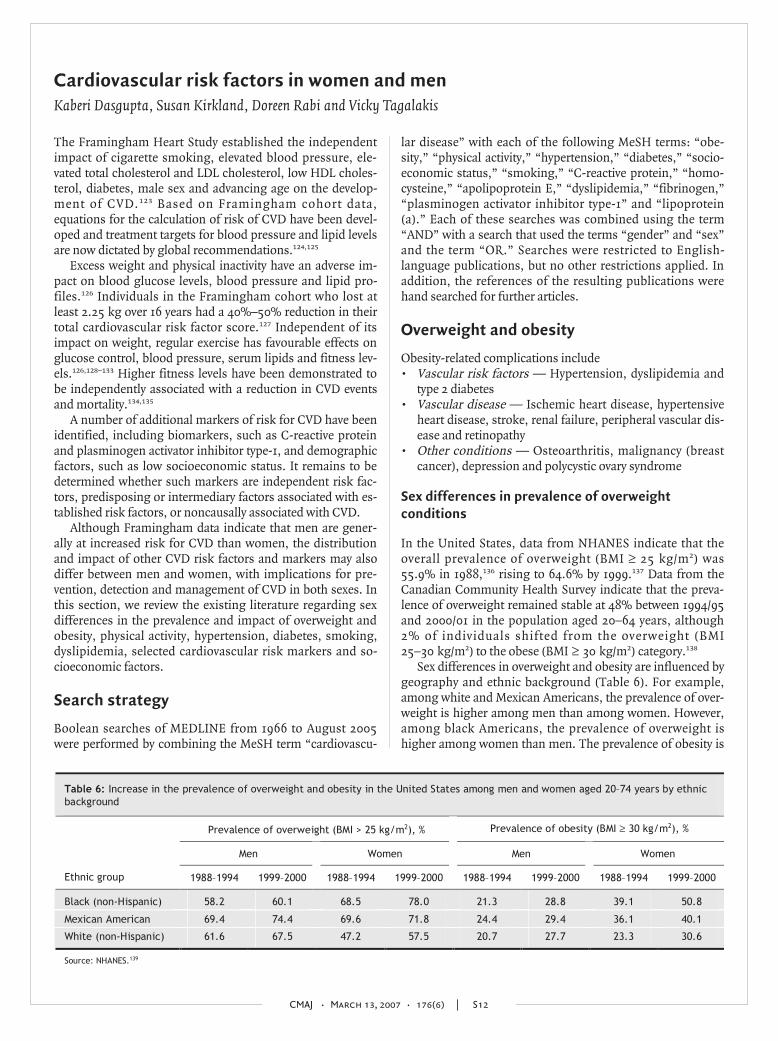
Table 6: Increase in the prevalence of overweight and obesity in the United States among men and women aged 20–74 years by ethnic background
Prevalence of overweight (BMI > 25 kg/m2), % Prevalence of obesity (BMI ≥ 30 kg/m2), %
Men Women Men Women
Ethnic group 1988–1994 1999–2000 1988–1994 1999–2000 1988–1994 1999–2000 1988–1994 1999–2000
Black (non-Hispanic) 58.2 60.1 68.5 78.0 21.3 28.8 39.1 50.8
Mexican American 69.4 74.4 69.6 71.8 24.4 29.4 36.1 40.1
White (non-Hispanic) 61.6 67.5 47.2 57.5 20.7 27.7 23.3 30.6
Source: NHANES.139
The Framingham Heart Study established the independentimpact of cigarette smoking, elevated blood pressure, ele-vated total cholesterol and LDL cholesterol, low HDL choles-terol, diabetes, male sex and advancing age on the develop-ment of CVD.123 Based on Framingham cohort data,equations for the calculation of risk of CVD have been devel-oped and treatment targets for blood pressure and lipid levelsare now dictated by global recommendations.124,125
Excess weight and physical inactivity have an adverse im-pact on blood glucose levels, blood pressure and lipid pro-files.126 Individuals in the Framingham cohort who lost atleast 2.25 kg over 16 years had a 40%–50% reduction in theirtotal cardiovascular risk factor score.127 Independent of itsimpact on weight, regular exercise has favourable effects onglucose control, blood pressure, serum lipids and fitness lev-els.126,128–133 Higher fitness levels have been demonstrated tobe independently associated with a reduction in CVD eventsand mortality.134,135
A number of additional markers of risk for CVD have beenidentified, including biomarkers, such as C-reactive proteinand plasminogen activator inhibitor type-1, and demographicfactors, such as low socioeconomic status. It remains to bedetermined whether such markers are independent risk fac-tors, predisposing or intermediary factors associated with es-tablished risk factors, or noncausally associated with CVD.
Although Framingham data indicate that men are gener-ally at increased risk for CVD than women, the distributionand impact of other CVD risk factors and markers may alsodiffer between men and women, with implications for pre-vention, detection and management of CVD in both sexes. Inthis section, we review the existing literature regarding sexdifferences in the prevalence and impact of overweight andobesity, physical activity, hypertension, diabetes, smoking,dyslipidemia, selected cardiovascular risk markers and so-cioeconomic factors.
Search strategy
Boolean searches of MEDLINE from 1966 to August 2005were performed by combining the MeSH term “cardiovascu-
lar disease” with each of the following MeSH terms: “obe-sity,” “physical activity,” “hypertension,” “diabetes,” “socio-economic status,” “smoking,” “C-reactive protein,” “homo-cysteine,” “apolipoprotein E,” “dyslipidemia,” “fibrinogen,”“plasminogen activator inhibitor type-1” and “lipoprotein(a).” Each of these searches was combined using the term“AND” with a search that used the terms “gender” and “sex”and the term “OR.” Searches were restricted to English-language publications, but no other restrictions applied. Inaddition, the references of the resulting publications werehand searched for further articles.
Overweight and obesity
Obesity-related complications include • Vascular risk factors — Hypertension, dyslipidemia and
type 2 diabetes• Vascular disease — Ischemic heart disease, hypertensive
heart disease, stroke, renal failure, peripheral vascular dis-ease and retinopathy
• Other conditions — Osteoarthritis, malignancy (breastcancer), depression and polycystic ovary syndrome
Sex differences in prevalence of overweightconditions
In the United States, data from NHANES indicate that theoverall prevalence of overweight (BMI ≥ 25 kg/m2) was55.9% in 1988,136 rising to 64.6% by 1999.137 Data from theCanadian Community Health Survey indicate that the preva-lence of overweight remained stable at 48% between 1994/95and 2000/01 in the population aged 20–64 years, although2% of individuals shifted from the overweight (BMI25–30 kg/m2) to the obese (BMI ≥ 30 kg/m2) category.138
Sex differences in overweight and obesity are influenced bygeography and ethnic background (Table 6). For example,among white and Mexican Americans, the prevalence of over-weight is higher among men than among women. However,among black Americans, the prevalence of overweight ishigher among women than men. The prevalence of obesity is
higher among US women than men, with the highest preva-lence among black American women.139
In Canada, for both men and women, the prevalence ofoverweight is lower among black Canadians (50%) thanwhite Canadians (60%).140 Consistent with this, among men,the prevalence of obesity is lower among black Canadians(10%) than white Canadians (15%), although among Cana-dian women, the prevalence of obesity is higher among blackpeople (20%) than white people (15%). The ethnic group withthe highest rates of overweight and obesity is the Aboriginalpopulation (60% overweight in women and 65% in men;25%–30% obesity in men and women). Between 1994/95 and2000/01, the prevalence of obesity increased in all age and sexgroups in Canada, with the exception of women 20–34 yearsof age. These trends are consistent with those documented inother countries.141–144
Overweight and obesity are also prevalent in many devel-oping countries. Nishida and Mucavele145 found that theprevalence of obesity was higher in women than in men inthe following countries: Brazil, 11.7% v. 4.8%; Egypt,33.0% v. 12.6%; South Africa, 30.1% v. 9.4%; Seychelles,28.2% v. 8.5%. The prevalence of overweight was similarfor the 2 sexes, but countries reporting higher levels ofoverweight among women were located in Africa, LatinAmerica, Asia and Oceania, whereas male overweight wasmore prevalent than women’s in countries of Europe andNorth America.
Recommendations for the prevention and treatment of obesity
• Reduced consumption of energy-dense foods• Regular physical activity• Weight loss for those with BMI ≥ 25 kg/m2 (diet and exer-
cise counselling, behavioural counselling)• Those with BMI ≥ 35 kg/m2 or with BMI ≥ 30 kg/m2 and
obesity-related complications may consider bariatricsurgery
Periods of risk
Women appear to be particularly susceptible to significantweight increase during adolescence,146 pregnancy136 andmenopause.147 The weight increase that occurs duringmenopause has been shown to be associated with a signifi-cant increase in blood pressure.148 The period after marriageappears to be a period of risk for weight gain among men.149
Knowledge gap
• Does the utility of weight loss for the prevention of CVDdiffer between women and men?
• Why do obese people appear to have lower rates of fatal re-current cardiovascular events?
• What weight-loss strategies are particularly effectiveamong men?
• What weight-loss strategies are particularly effectiveamong women?
• Why are black women in both the United States andCanada at high risk of obesity?
Physical activity
Sex differences in activity levels
Many studies suggest that women are more likely to be seden-tary than men. A questionnaire administered by Pitsavos andcolleagues150 in the Attica region of Greece revealed that,overall, 53% of men and 48% of women were physically ac-tive, and men tended to be more physically active than womenacross all age groups. In an interview-based survey conductedin Portugal, 79% (95% CI 75.7–81.6) of men and 86% (95%CI 84.0–88.0) of women were found to be sedentary.151 InFinland, the proportion of people classified as sedentary oronly moderately active during their leisure time was 75%among males and 82% among females.152 Men in Japan havealso been found to be more active than women.153
There is some evidence that, although both men andwomen are both less likely to be active when weather condi-tions are unfavourable, women are less likely to increase ac-tivity levels when weather conditions become morefavourable. In community-dwelling adults in Massachusetts,mean physical activity during the summer increased by51 minutes/day (95% CI 20–82) in men, but only by 16 min-utes/day (95% CI -12–45) in women.154
Potential mechanisms
Low levels of physical activity render weight maintenance dif-ficult, contribute to the development of insulin resistance,with associated increases in blood pressure, blood glucoselevel, dyslipidemia and thrombogenic factors. In addition,there is increasing evidence that low levels of activity and fit-ness are directly related to increased CVD risk. In a prospec-tive cohort study that examined the impact of physical activityon mortality, Blair and colleagues155 found that women in thelowest tertile of physical activity had a greater than 5-fold in-creased risk of mortality compared with women in the high-est physical activity tertile. Men in the lowest physical activitytertile were at a 3-fold higher increase in risk of mortalitycompared with men in the highest fitness tertile.
Barriers to physical activity in women
Findings from Canada’s National Population Health Surveydemonstrate that the presence of children in the household isa significant deterrent to becoming active for women, but notfor men.156 The most commonly reported barrier to women’sparticipation in physical activity is lack of time due to familyresponsibilities.157,158 Middle-aged and older women appearto have positive attitudes toward exercise, but are unable orunwilling to take action.159
Physical activity recommendations
• To remain healthy and maintain body weight — moderate
CMAJ • March 13, 2007 • 176(6) | S13

exercise (e.g., walking) 30 minutes daily or vigorous exer-cise (e.g., jogging) 20 minutes daily.
• To lose weight — vigorous exercise 30 minutes daily.
Knowledge gap
• What are the barriers to physical activity in women?• Are the barriers largely related to child care or competing
work–home responsibilities or both?• How can activity levels among women be increased?
Hypertension
Excess body weight and physical inactivity may promote thedevelopment of a number of CVD risk factors, including highblood pressure (see Table 7 for the general classification ofblood pressure levels).
Hypertension-related comorbidities
• ischemic heart disease• hypertensive cardiomyopathy• stroke• renal failure
Sex differences in blood pressure
In the third NHANES (1988–1994) evaluation, among USadults under 45 years of age, men had higher systolic bloodpressure levels than women.160 By 60–69 years of age, non-Hispanic white women had blood pressure levels similar tothose of men and by 70–79 years of age, had higher levelsthan men.161 By 60–69 years of age, non-Hispanic black andHispanic women had higher blood pressure levels than menof similar ethnic background.161 Overall, among those45 years of age and older, systolic blood pressure levels werehigher among women.160 In a cohort study conducted in Den-mark, 24-h mean blood pressure levels were 6–10 mm Hghigher among men than women until 70–79 years of age, butsimilar thereafter.162 Women from developing countries havehigher mean systolic blood pressure than their male counter-
parts.163 It is also noteworthy that women from these coun-tries have higher blood pressure than women from developedcountries. Women from the African region have the highestmean systolic blood pressure.
Among young and middle-aged adults, population surveysreport hypertension to be more frequent among men com-pared with women, with sex differences of 4% in the UnitedStates, 8% in Canada and 11% in Western Europe.164 After60 years of age, however, the prevalence of hypertension ap-pears to be higher among women than among men.
Potential mechanisms
Androgen is thought to play a role in the sex differences inblood pressure. One possible mechanism may be the blunt-ing of the pressure-natriuresis relation.165 Female sex hor-mones and their receptors may also be implicated in bloodpressure differences between men and women. A genetic as-sociation study by the Victorian Family Heart Study investiga-tors found that men inheriting the “a” allele on the estrogenreceptor a gene had significantly higher systolic blood pres-sure levels (5 mm Hg) than men with other genotypes.166 Nosignificant associations between estrogen receptor genes andblood pressure were detected among women.
Knowledge gap
• How can the postmenopausal increase in hypertension beprevented?
• Should the threshold for hypertension diagnosis in wo-men be lower to prevent the postmenopausal increase inhypertension?
• Can earlier detection of hypertension in boys and youngmen reduce the sex–gender differential in incidence ofCVD between men and premenopausal women?
Diabetes
Diabetes is highly prevalent; over 151 million people live withthis condition worldwide (Table 8). Diabetes is an established
CMAJ • March 13, 2007 • 176(6) | S14
Table 7: General classification of blood pressure
Classification Blood pressure
Normal Systolic 120–129 mm Hg Diastolic 80–84 mm Hg
High normal* Systolic 130–139 mm Hg Diastolic 85–89 mm Hg
Grade 1 Systolic 140–159 mm Hg Diastolic 90–99 mm Hg
Grade 2 Systolic 160–179 mm Hg Diastolic 100–109 mm Hg
Grade 3 Systolic ≥ 180 mm Hg Diastolic ≥ 110 mm Hg
*Considered high in the context of diabetes or renal disease.
Table 8: Crude prevalence of diabetes among adults aged 20–79 years in various countries
Country (year) Men, % Women, %
Canada (1999) 5.0 4.6
United States (2002) 6.9 6.1
Mexico (2003) 2.7 4.7
United Kingdom (1998) 2.2 2.0
Spain (2003) 4.0 5.9
Sweden (2003) 3.3 3.9
Australia (2003) 3.4 2.7
China (Hong Kong) (2003) 4.3 4.6
Japan (2003) 3.6 3.3
Source of data: Haffner et al.167
risk factor for the development of CVD.167 People with dia-betes have a 2- to 4-fold greater risk of developing CVD com-pared with those without diabetes.168 CVD is the leadingcause of morbidity and mortality for those living with dia-betes.
Although the prevalence of diabetes is lower in developingcountries, these countries have experienced the greatest in-crease in diabetes. The prevalence is highest in the EasternMediterranean and Middle East (7.0%), South and CentralAmerica (5.6%), Southeast Asia (5.6%), Western Pacific(3.1%) and Africa (2.4%). The prevalence of diabetes is higheramong women than among men in Latin America (57.5% v.42.5%) and in the Western Pacific (53.7% v. 46.3%).169
Diabetes-related vasculopathy
Microvasculature• retinopathy• nephropathy
Macrovasculature• peripheral vascular disease• cerebrovascular disease• ischemic heart disease
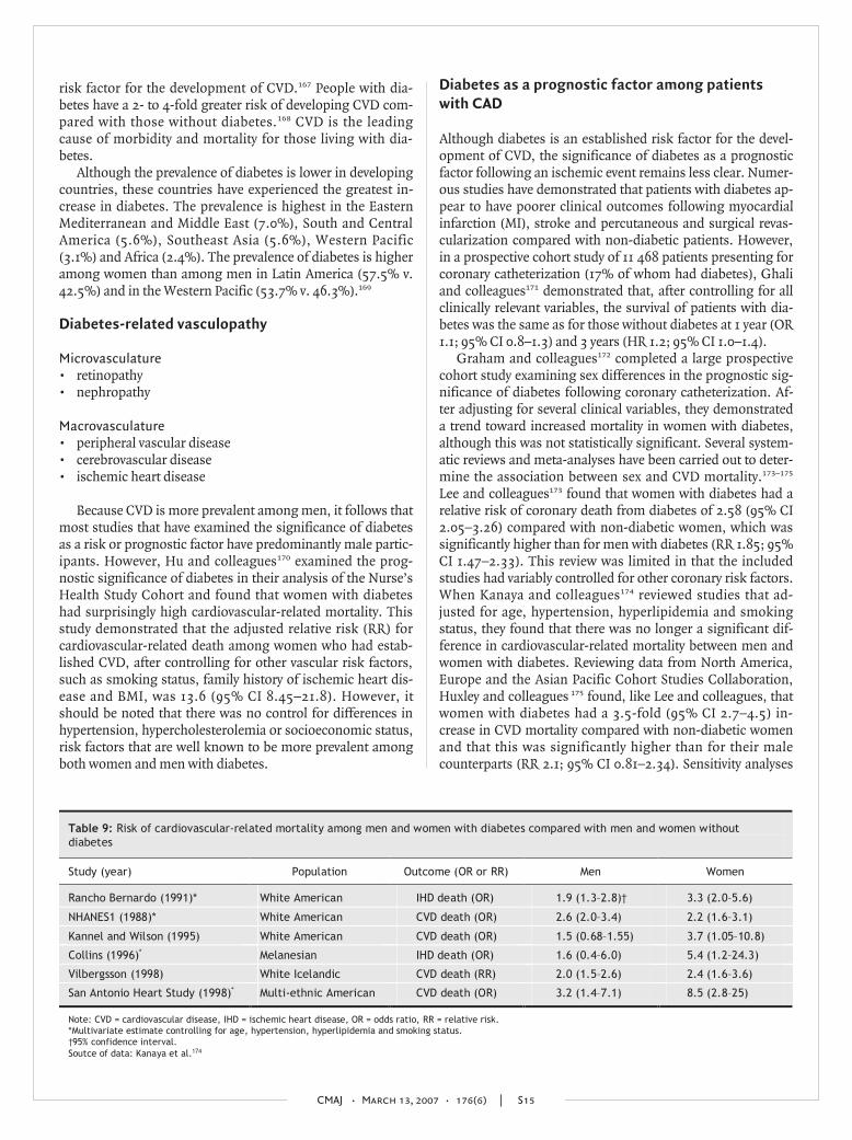
Because CVD is more prevalent among men, it follows thatmost studies that have examined the significance of diabetesas a risk or prognostic factor have predominantly male partic-ipants. However, Hu and colleagues170 examined the prog-nostic significance of diabetes in their analysis of the Nurse’sHealth Study Cohort and found that women with diabeteshad surprisingly high cardiovascular-related mortality. Thisstudy demonstrated that the adjusted relative risk (RR) forcardiovascular-related death among women who had estab-lished CVD, after controlling for other vascular risk factors,such as smoking status, family history of ischemic heart dis-ease and BMI, was 13.6 (95% CI 8.45–21.8). However, itshould be noted that there was no control for differences inhypertension, hypercholesterolemia or socioeconomic status,risk factors that are well known to be more prevalent amongboth women and men with diabetes.
Diabetes as a prognostic factor among patientswith CAD
Although diabetes is an established risk factor for the devel-opment of CVD, the significance of diabetes as a prognosticfactor following an ischemic event remains less clear. Numer-ous studies have demonstrated that patients with diabetes ap-pear to have poorer clinical outcomes following myocardialinfarction (MI), stroke and percutaneous and surgical revas-cularization compared with non-diabetic patients. However,in a prospective cohort study of 11 468 patients presenting forcoronary catheterization (17% of whom had diabetes), Ghaliand colleagues171 demonstrated that, after controlling for allclinically relevant variables, the survival of patients with dia-betes was the same as for those without diabetes at 1 year (OR1.1; 95% CI 0.8–1.3) and 3 years (HR 1.2; 95% CI 1.0–1.4).
Graham and colleagues172 completed a large prospectivecohort study examining sex differences in the prognostic sig-nificance of diabetes following coronary catheterization. Af-ter adjusting for several clinical variables, they demonstrateda trend toward increased mortality in women with diabetes,although this was not statistically significant. Several system-atic reviews and meta-analyses have been carried out to deter-mine the association between sex and CVD mortality.173–175
Lee and colleagues173 found that women with diabetes had arelative risk of coronary death from diabetes of 2.58 (95% CI2.05–3.26) compared with non-diabetic women, which wassignificantly higher than for men with diabetes (RR 1.85; 95%CI 1.47–2.33). This review was limited in that the includedstudies had variably controlled for other coronary risk factors.When Kanaya and colleagues174 reviewed studies that ad-justed for age, hypertension, hyperlipidemia and smokingstatus, they found that there was no longer a significant dif-ference in cardiovascular-related mortality between men andwomen with diabetes. Reviewing data from North America,Europe and the Asian Pacific Cohort Studies Collaboration,Huxley and colleagues 175 found, like Lee and colleagues, thatwomen with diabetes had a 3.5-fold (95% CI 2.7–4.5) in-crease in CVD mortality compared with non-diabetic womenand that this was significantly higher than for their malecounterparts (RR 2.1; 95% CI 0.81–2.34). Sensitivity analyses
CMAJ • March 13, 2007 • 176(6) | S15
Table 9: Risk of cardiovascular-related mortality among men and women with diabetes compared with men and women without diabetes
Study (year) Population Outcome (OR or RR) Men Women
Rancho Bernardo (1991)* White American IHD death (OR) 1.9 (1.3–2.8)† 3.3 (2.0–5.6)
NHANES1 (1988)* White American CVD death (OR) 2.6 (2.0–3.4) 2.2 (1.6–3.1)
Kannel and Wilson (1995) White American CVD death (OR) 1.5 (0.68–1.55) 3.7 (1.05–10.8)
Collins (1996)* Melanesian IHD death (OR) 1.6 (0.4–6.0) 5.4 (1.2–24.3)
Vilbergsson (1998) White Icelandic CVD death (RR) 2.0 (1.5–2.6) 2.4 (1.6–3.6)
San Antonio Heart Study (1998)* Multi-ethnic American CVD death (OR) 3.2 (1.4–7.1) 8.5 (2.8–25)
Note: CVD = cardiovascular disease, IHD = ischemic heart disease, OR = odds ratio, RR = relative risk. *Multivariate estimate controlling for age, hypertension, hyperlipidemia and smoking status. †95% confidence interval. Soutce of data: Kanaya et al.174

that corrected for coronary risk factors revealed that this sexdifference was attenuated, but remained statistically signifi-cant (Table 9).
A recent cohort study demonstrated that a history of dia-betes in women was associated with a 37% increase in CVD-related mortality compared with a history of AMI. However,the presence of a previous AMI in men increased the risk ofCVD-related mortality by 43% compared with a history ofdiabetes.176
Potential mechanisms
Several mechanisms may explain the apparent sex differencein CVD mortality among patients with diabetes. There is evi-dence that different pathophysiologic processes — in termsof endothelial function, dyslipidemia and thrombosis — mayresult in different cardiovascular outcomes among men andwomen with diabetes.
Diabetes has been shown to abrogate the vascular protec-tion afforded to premenopausal women. Steinberg and col-leagues177 elegantly demonstrated that premenopausal womenhave an enhanced vasodilatory response to endogenously pro-duced nitric oxide compared with men. This study also illus-trated that the development of diabetes is associated with ab-normal endothelial-dependent vasodilation in both sexes.This loss of vasodilation is most striking in women. Sowers178
has further demonstrated that hyperglycemia significantly
decreases estrogen-mediated nitric oxide production. It seemsthat a unique interaction between diabetes and sex makeswomen more vulnerable to endothelial dysfunction.
The noted sex differences in CVD outcomes may be influ-enced by factors beyond biology. A study by Wexler and col-leagues179 suggests that sex differences in CVD mortality maybe due to disparities in medical management. This prospec-tive cohort study demonstrated that women with diabeteswere less likely to be treated until they reached establishedtherapeutic targets than men with diabetes, even if thewomen had established CVD. Whether these disparities arerelated to physician factors (underappreciation of cardiovas-cular risk in women, hesitation to use vasoprotective medica-tions in reproductive women) or patient factors (adherence toor tolerance of prescribed medications) remains unclear.
Dyslipidemia
Abnormal levels of lipoprotein cholesterols are significantpredictors of atherosclerosis in all populations, with a fifth ofglobal stroke events and about 56% of global heart disease at-tributable to high cholesterol levels. In particular, elevated lev-els of total cholesterol, LDL cholesterol and triglycerides andlow levels of HDL cholesterol have been associated with CAD,stroke and peripheral vascular disease and are often associatedwith such significant comorbidities as diabetes, hypertensionand obesity. In both US and Canadian populations, abnormalcholesterol levels are highly prevalent across different age andethnic groups (Table 10 and Table 11),180 and there appear tobe sex differences in the prevalence of elevated choles-terol181,182 (see Table 12 for classification of lipoprotein levels).
Low-density lipoprotein
LDL cholesterol is believed to be the principal lipoprotein inthe development of atherosclerosis and remains the primary
CMAJ • March 13, 2007 • 176(6) | S16
Table 10: Percentage of United States population with high blood cholesterol,* 1991–2003 — Behavioural Risk Factor Surveillance System
Variable 1991 1995 1999 2003 % change†
Age group, yr
20–44 17.6 19.1 18.6 20.3 15.3‡
45–64 33.5 35.3 36.8 41.3 23.3‡
≥ 65 33.4 38.4 42.3 47.5 42.2‡
Sex§
Women 25.4 27.3 27.6 29.4 15.7‡
Men 24.9 27.1 28.3 33.0 32.5‡
Ethnic group§
White, non-Hispanic 25.4 27.6 28.2 31.5 24.0‡
Black, non-Hispanic 24.0 25.7 26.8 28.9 20.4‡
Hispanic 23.4 26.3 27.0 29.9 27.8‡
Asian/Pacific Islander 28.4 28.8 32.2 29.2 2.8
Native American 26.3 21.5 31.6 31.2 18.6
Total§ 25.3 27.3 28.0 31.1 22.9‡
*Total cholesterol > 6.21 mmol/L.180
†Difference in prevalence of high cholesterol level between 1991 and 2003 as percentage of 1991 prevalence. ‡t-test is significant at p < 0.05.
§Age-standardized to the 2000 United States population. Source: Centers for Disease Control and Prevention.181
Table 11: Distribution (%) of blood cholesterol in Canadians aged 55–74 years, 1986–1992
Total cholesterol, mmol/L; population estimate, %
Group; age, yr < 5.20
5.20–6.19
6.20–6.49 ≥ 6.85
No. of participants
Men
55–64 41 41 11 7 843
65–74 38 38 16 7 1713
Total 40 40 13 7 2556
Women
55–64 22 41 18 18 833
65–74 22 38 18 22 1586
Total 23 40 18 20 2419
Overall total 30 40 16 14 4975
Reprinted with permission from the Canadian Medical Association (CMAJ 1999;161[8 Suppl]:S3-9).182

target of therapy for the prevention of CVD. Elevated LDLcholesterol levels are more predictive of coronary risk in menthan in women, particularly premenopausal women.183 Thismight be due in part to lower levels of LDL cholesterol in pre-menopausal women than in middle-aged men (35–65 years).However, after age 50 years, LDL cholesterol levels plateau inmen and increase in women between ages 40 and 60 years atan average rate of 0.05 mmol/L a year.184 This increase in LDLcholesterol at menopause is thought to be partly the result ofadvancing age and declining levels of estrogen, which resultin downregulation of LDL receptors in the liver leading to de-creased clearance of LDL cholesterol from the serum.
High-density lipoprotein
HDL cholesterol is an important independent predictor ofCVD in both men and women,185,186 but may have greater pre-dictive potential in women than men.185,187 In the Framing-ham Heart Study, a 0.025-mmol/L increase in HDL choles-terol level was associated with a 3% decrease in the incidenceof CAD in women compared with a 2% decrease in men.185 Inthe Lipid Research Clinics Prevalence Mortality Follow-upStudy, a 0.025-mmol/L increase in HDL cholesterol was asso-ciated with a 4.7% reduction in CVD mortality among women(p = 0.002) compared with a 3.7% reduction among men(p < 0.001).188 On average, HDL cholesterol levels are0.25 mmol/L higher in premenopausal women than in men,which may account for the lower incidence of CVD before age50 in women compared with men. With menopause, HDLcholesterol levels have been shown to decrease, although theadministration of exogenous estrogen can increase levels.189
However, the protective effect of exogenous estrogens re-mains controversial. Although oral estrogens can increaseHDL cholesterol and decrease LDL cholesterol, they alsoincrease the potential for coagulation and possibly forinflammation.
Triglycerides
There is some evidence to suggest that high levels of triglyc-erides are a significant independent risk factor for CVD inboth sexes, but more so in women than men.185,190 In a meta-analysis of 17 prospective population-based studies, elevatedtriglyceride levels adjusted for HDL cholesterol levels were as-sociated with a 37% (95% CI 13–66) increase in risk of CVD-related events in women compared with a 14% (95% CI 5–28)increase in men.191 The mechanism for increased risk of CVDassociated with hypertriglyceridemia is unclear, but elevatedtriglycerides are often accompanied by other metabolic dis-turbances that may predispose to CVD, including reducedHDL cholesterol, increased levels of very low-density lipopro-tein cholesterol and insulin resistance, which makes it diffi-cult to assess the independent risk associated with triglyc-erides. Moreover, some analyses suggest that elevatedtriglycerides interact with some of these other risk factors tomodulate the risk of CVD and that the interaction may differbetween men and women. For example, a study of 174 pa-tients with type 2 diabetes mellitus who were not receiving
lipid-lowering therapy concluded that the severity of CAD (asexamined by angiography) was related to the number oftriglyceride-rich lipoproteins and that the relation wasstronger for women than men, independent of HDL and LDLcholesterol.192
Knowledge gap
• Triglyceride and HDL cholesterol levels must be measuredand addressed, especially in women with other metabolicdisturbances.
• Changes in triglycerides, HDL cholesterol and other lipidmetabolites may be indicators of unrecognized metabolicdisturbances, especially in women.
• Pharmacologic interventions to elevate HDL cholesterol inwomen have not been undertaken.
Smoking
Smoking has been identified as the primary preventable causeof morbidity and mortality in Canada, contributing to 27% ofall deaths among men and 17% of all deaths among women,the majority being due to CVD.193 Trends in smoking-attributable mortality reflect the smoking behaviour of thepopulation 2–3 decades earlier. Whereas smoking rates formen peaked in the mid 1960s, the rates for women did not be-gin to decline until the late 1970s. As a result, deaths due toCVD among women have yet to decrease.193,194
Ongoing national and provincial surveillance initiativesmonitor smoking trends and the effect on health. Smokingprevalence has steadily declined over the last few decades,
CMAJ • March 13, 2007 • 176(6) | S17
Table 12: Classification of levels of low-density lipoprotein (LDL) cholesterol, high-density lipoprotein (HDL) cholesterol and triglycerides
Cholesterol or triglyceride level Classification
LDL cholesterol, mmol/L
< 2.59 Optimal
2.59–3.36 Near–above optimal
3.37–4.14 Borderline high
4.15–4.90 High
> 4.90 Very high
HDL cholesterol, mmol/L
< 1.04 Low for men
< 1.30 Low for women
> 1.55 High (protective against heart disease)
Triglyceride, mmol/L
< 1.70 Desirable
1.70–2.25 Borderline high
2.26–5.64 High
≥ 5.65 Very high
Source of data: National Cholesterol Education Program.180

with a stronger decrease in men than women (Fig. 9).195 Ac-cording to the 2004 Canadian Tobacco Use Monitoring Sur-vey (CTUMS),196 approximately 22% of men and 17% ofwomen aged 15 years and older are current smokers. Al-though women experiment with smoking at a younger agethan men, smoking rates are higher for men than women inall age groups. Prevalence peaks at ages 20–29 years in bothsexes — at 37.3% in men and 30.9% in women. For bothsexes, the prevalence begins to decline at age 30–49 years andsmoking prevalence among those aged 55 years and older ishalf that of 20–29 year olds.195,197,198 The overall smoking rateof 20% is among the lowest in the OECD countries and isfavourably consistent with trends in other developed coun-tries. However, we may have reached a point at which theprevalence rates reflect hard-to-reach populations, and fur-ther decreases may be difficult to achieve. In a survey similarto CTUMS conducted in the Northwest Territories, the preva-lence of smoking was 44% and 38% among men and women,respectively, yet Aboriginal residents had a prevalence of overtwice that of non-Aboriginal residents, 60% v. 25%.199
In developing countries, the prevalence of smoking is sub-stantially higher among men (50%) than among women (9%).However, female smoking is increasing rapidly due, at least inpart, to targeted promotional strategies of the tobacco industry.Worldwide, the lowest smoking prevalence for both men andwomen is in African countries, while the highest prevalence formen is in Eastern Europe (where rates exceed 60%) and forwomen in Central Europe and parts of South America.200 Thegreatest increases in smoking prevalence over the next decadeare expected to occur in Africa and the Middle East.
The determinants of smoking primarily reflect social andeconomic disadvantage. A higher prevalence of smoking hasbeen found among women and men who live in low-incomehouseholds, have low-status jobs or are unemployed, are single
parents or divorced, and have low levels of education.194,201 Thisis consistent with the epidemic description of smoking as itspreads through a population, reverting from a positive associ-ation to a negative association with socioeconomic status, firstamong men and then among women in later stages of the epi-demic, as seen in developed countries.202 Qualitative researchhas shown that men and women may smoke for different rea-sons. For example, women use smoking as a coping mecha-nism to deal with gendered stresses such as child care.203 Theymay also encounter greater difficulties in quitting.199
Epidemiologic studies have consistently documented therisk of CVD associated with smoking in both sexes. It is gen-erally thought that the CVD risk is similar, except amongwomen smokers taking oral contraceptives, where the risk ofstroke is much greater. However in ARIC, a large prospectivepopulation study of 15 792 men and women aged 45–64 yearsat entry, investigators found that the hazard ratio for coronaryheart disease was greater for women than men (2.95 v. 1.55for current smoking).204
Potential mechanisms
The precise mechanisms connecting increase in CVD riskwith smoking are not completely clear. Possible deleteriousimpacts of smoking include endothelial dysfunction, lipid ab-normalities, increased concentration of fibrinogen andplatelet aggregation.205,206 Few studies have investigated thepossible mechanisms underlying sex differences in CVD as-sociated with smoking. Interactions between smoking andhormonal factors may be involved in development of CVD. Itis possible that smoking may increase the risk of CVD inwomen smokers via an anti-estrogenic effect.205
Cardiovascular risk markers
C-reactive protein
Inflammatory mechanisms have been shownto play an important role in the pathogenesisof CVD, and increasing attention has beenpaid to the measurement of inflammatorybiomarkers, particularly C-reactive protein(CRP).207–209 Indeed, of 12 plasma markersevaluated simultaneously, Ridker and col-leagues207 found CRP to be the most signifi-cant predictor of CVD risk in healthy post-menopausal women. Adjusted for otherplasma markers and conventional risk fac-tors, a 1-quartile increase in CRP concentra-tion increased the risk of a CVD event by 50%(OR 1.5; 95% CI 1.1–2.1). These findings weresupported in a meta-analysis of 22 prospec-tive studies conducted in general (apparentlyhealthy) populations.208 Those in the topthird of the study group, with respect to CRPlevel, had a 58% increase (OR 1.58; 95% CI1.48–1.68) in risk of CAD compared with thebottom third, after adjusting for the major
CMAJ • March 13, 2007 • 176(6) | S18
0
5
10
15
20
25
All 15–19 20–24 25–34 35–44 45–54 ≥ 55
Age group, yr
Pre
vale
nce
, %
Males
Females
Average, ages ≥ 15
Fig. 9: Prevalence of current smoking by age and sex, 2003. Source: Adapted fromCanadian Tobacco Use Monitoring Survey.195

conventional CVD risk factors. Although most studies werepredominantly in men, the summary odds ratio reportedseparately for women in 3 studies did not differ markedlyfrom the summary odds ratio based on 12 studies that re-ported their findings for men only. Studies in groups withestablished heart disease have also demonstrated that highCRP levels are associated with increased AMI or mortality.210
A number of other studies have reported higher adjustedbaseline CRP values for women than men.209,211 Bertran andcolleagues211 found that, on average, CRP concentrationswere 29% higher in women than men. An age–sex interactionhas also been demonstrated, whereby CRP was predictive ofdeath or AMI, or both, in women and men under 55 years ofage, but only for men over the age of 55 years.209
Homocysteine
Elevated levels of homocysteine have also been implicated inCVD. Blood levels of homocysteine have been found to behigher in men than in women.212,213 For example, homocys-teine levels in the control group were 9.7 ± 4.9 µmol/L formen and 7.6 ± 4.1 µmol/L for women (p < 0.001) in a case–control study of French-Canadians.213 Men and women withCAD had significantly higher homocysteine levels (11.7± 5.8 µmol/L and 12.0 ± 6.3 µmol/L, respectively), but no sexdifference in CAD was observed.
A systematic review214 of the association between plasmahomocysteine level and CVD suggested that moderately in-creased homocysteine levels are prevalent in the general pop-ulation and are independently associated with an increasedrisk of CVD. In a more recent meta-analysis,215 elevated ho-mocysteine level was found to be a modest independent pre-dictor of ischemic heart disease in healthy populations; a de-cline of 3 µmol/L in homocysteine level was associated withan 11% decrease in risk of ischemic heart disease (OR 0.89;95% CI 0.83–0.96).
Apolipoprotein E
Apolipoprotein E (APOE) is a component of lipoproteins,with considerable variation as a result of cysteine–arginineexchanges in 3 alleles known as ε2, ε3 and ε4. Numerouspopulation studies have implicated APOE as a major modula-tor of total cholesterol and LDL cholesterol.216–218 The pres-ence of the ε4 allele has been associated with elevations inLDL cholesterol, whereas the ε2 allele has been associatedwith decreased levels of LDL cholesterol.216–218 Data from theFramingham Offspring Study have also indicated that the as-sociation of APOE phenotype with LDL cholesterol level issignificantly greater in postmenopausal women than in pre-menopausal women or in men.218
Some studies have indicated that the APOE polymorphismmay be a sex-specific predictor of CVD.219,220 Most recent datafrom the Framingham Heart Study suggest an increased riskin men with the ε2 and ε4 alleles compared with those withthe ε3:ε3 genotype. After adjustment for all risk factors in-cluding age and lipids, the increase in risk of CVD with an ε4allele was 51% for men (OR 1.51; 95% CI 1.05–2.18) and 48%
for women (OR 1.48; 95% CI 0.93–2.34). The association be-tween the ε2 allele and CVD remained significant in men afteradjusting for all risk factors, including lipids (OR 1.94; 95%CI 1.23–3.04). However, no association between the ε2 alleleand increased CVD risk was found in women.219 Neither themechanism underlying the excess in ε4 allele in CVD patientsnor the effect of interaction between sex and APOE polymor-phisms on CVD is completely clear.
Fibrinogen
Increased plasma fibrinogen levels have been independentlyassociated with an increased risk of AMI and stroke inprospective studies of healthy people and patients with pre-existing CVD.221–223 Both men and women in the highest thirdfor plasma fibrinogen have a 2- to 3-fold higher risk thanthose in the lowest third.224 In a meta-analysis of 31 prospec-tive studies that included 154 211 participants from WesternEurope, North America and Japan, the age- and sex-adjustedhazard ratio per 1-g/L increase in usual fibrinogen level was2.42 (95% CI 2.24–2.60) for coronary heart disease and 2.06(95% CI 1.83–2.33) for stroke.225 Although the risk of CVD issimilar for men and women with elevated fibrinogen levels,there are some small sex differences with regard to modula-tors of fibrinogen levels; women tend to have higher levels offibrinogen than men when stratified for age and ethnic-ity.226,227 Plasma fibrinogen increases with menopause, preg-nancy and the use of oral contraceptives; treatment with hor-mone replacement therapy may decrease fibrinogen levels,and the effect of smoking, which is regarded as the strongestdeterminant of high fibrinogen levels,226 is not as consistentin women as it is in men.228,229 Possible reasons include differ-ences in nutritional habits and more powerful determinantsof fibrinogen in women, such as hormonal status, that couldmask the smoking effect. Although the clinical significanceof these sex differences remains to be elucidated, they may beuseful in the development of sex-specific interventions tolower fibrinogen levels.
Plasminogen activator inhibitor type-1
Increased levels of plasminogen activator inhibitor type-1(PAI-1) have been associated with CAD.223,230–232 In a recentprospective nested case–control study of 2246 Swedish pa-tients with a first AMI matched for age and sex, elevated levelsof PAI-1 were significantly associated with a 2-fold increasedrisk of AMI in both men and women.233 A few studies haveshown that premenopausal women have lower plasma PAI-1levels than postmenopausal women.234 In addition, post-menopausal women receiving hormone replacement therapyhave lower plasma PAI-1 levels than those not receiving suchtherapy.235,236
The presence of polymorphisms in the PAI-1 gene hasbeen found to correlate with increased plasma levels of PAI-1and may help explain differences among men and womenand even among pre- and postmenopausal women. For ex-ample, a large cohort study of men and women with recentAMI revealed that the risk of AMI, after adjusting for common
CMAJ • March 13, 2007 • 176(6) | S19

CVD risk factors, was significantly associated with 4G/5Gpolymorphism in women compared with men.237 More re-cently, a few case–control studies have shown a higher preva-lence of this mutation in postmenopausal women with CADcompared with premenopausal women with and withoutCAD, and the addition of hormone replacement therapy de-creases PAI-1 levels in women with CAD, which correlateswith the presence of the 4G allele.238
Lipoprotein (a)
Lipoprotein (a) (Lp(a)) has been postulated to be highlyatherothrombotic. A recent meta-analysis of 18 prospectivepopulation-based studies, which included over 3500 women,largely from Nordic countries, Germany, the United King-dom and the United States, showed a combined risk ratio forCVD of 1.7 (95% CI 1.4–1.8) for those in the top third of base-line Lp(a) concentrations compared with those in the bottomthird.239 Adjustment for conventional risk factors includingLDL cholesterol did not significantly change the association.Moreover, data from the Quebec Cardiovascular Study240 andthe Italian Longitudinal Study on Aging241 suggest that,among men, elevated Lp(a) levels interact positively with highLDL cholesterol levels, hypertension, hyperhomocysteinemiaand elevated fibrinogen concentrations. In a recent cohortstudy242 using data from the Nurses’ Health Study Initiative —which showed that women in the highest quintile of Lp(a)were twice as likely to develop CVD compared with women inthe lowest quintile, independent of other conventional riskfactors — no significant interactions with LDL cholesterol,HDL cholesterol or fibrinogen were found. This may be partlyexplained by the fact that levels of Lp(a) tend to increase withage, more so in women than men,243 which parallels the age-related increase in LDL cholesterol and fibrinogen levels seenin women.
Socioeconomic status
There is a clear socioeconomic gradient in risk of CVD, withthose in the lowest socioeconomic strata having the highestrisk of cardiovascular events.244,245 Although this gradient ex-ists for both men and women, there is increasing evidence tosuggest that there may be an interaction between sex and so-cioeconomic status in the development of both cardiovascu-lar risk factors and CVD.
Socioeconomic status is a complex construct that is influ-enced by financial, employment and education experiences.The protective effect of high social standing was convinc-ingly and elegantly demonstrated in the Whitehall study.Marmot and colleagues246 clearly showed that, in a cohort ofBritish Civil servants, those in the lowest social strata were2.6 times more likely to die from CVD than those in thewealthiest class. This negative relation between CVD and so-cioeconomic status has been demonstrated consistently innumerous studies throughout the developed world. Recentstudies indicate that there may be a gender–socioeconomicstatus interaction in terms of CVD mortality. Gerward andcolleagues247 examined the effect of neighbourhood socio-
economic factors on mortality in the first 28 days after anAMI in a Swedish cohort. They demonstrated that low so-cioeconomic status and early CVD mortality were mostclosely associated in men over 75 years of age (p value fortrend 0.007). This analysis was limited in that it used eco-logic measures of both socioeconomic status and cardiacrisk. However, examining the influence of social class onCVD mortality in another large Swedish cohort, Baigi andcolleagues248 demonstrated that although labourers havemuch higher CVD mortality than their white-collar counter-parts, female labourers have a CVD mortality risk that is dou-ble that of male labourers. Results from the NHANEScohort249 were similar: low-income women had a 61% in-crease in risk of CVD mortality compared with high-incomewomen. Low income also increased risk of CVD mortality inmen, but the elevation (29%) was less striking. This analysisadjusted for several other risk factors that may vary with in-come, including smoking status, blood pressure, cholesterollevel and diabetes status.
Health care access
People with low income do not appear to obtain health careas effectively as the wealthy.250,251 Barriers to health care ap-pear to be larger for poor women than men in both single andmultipayer systems. In universal systems in Canada, womenare more likely to visit their primary care physician, but areless likely to be referred to specialty care and are more likelyto be admitted to hospital.251,252 In private systems, such asthose in the United States, women are less likely to be able toafford essential medications. This inability to obtain evi-dence-based therapy is associated with a 50% increase in theincidence of angina, non-fatal AMI and non-fatal strokeamong US women.252
CVD risk factors and health behaviours
Diabetes, obesity, sedentary lifestyle, smoking and hyperten-sion are more prevalent in populations at low socioeconomiclevels.253 Lower social class has consistently been found to beassociated with lower levels of HDL cholesterol and APO-A1in both men and women.254 No consistent relation betweensocioeconomic status and LDL cholesterol has been found.The interaction between socioeconomic status and genderappears to have an impact on specific cardiovascular risk fac-tors. Although low socioeconomic status women are at muchgreater risk of diabetes mellitus and obesity than men,255,256
the negative relation between smoking behaviour and wealthis more evident in men.
Psychosocial factors
There is increasing awareness of the influence of psychologicand social factors on the development of heart disease.257 InNorth America, people who experience depressive symptomsare more likely to be women and are more likely to have lowincome.258 US women who are involuntarily unemployed havebeen found to have higher blood pressure and higher rates of
CMAJ • March 13, 2007 • 176(6) | S20

anginal pain compared with employed women and home-makers. The effect is most striking in African-Americanwomen. An unemployed African-American woman is 2.6times more likely to have CVD compared with an unemployednon-Hispanic white woman.259 Van Lenthe and colleagues260
showed the independent and significant relation between so-cial and financial deprivation and heart disease in the GLOBEstudy. Even after controlling for the behavioural risk factorssmoking, alcohol use, physical inactivity and obesity, materialfactors such as unemployment, poor housing and crowdingwere associated with a 25%–80% increase in the incidence ofAMI in this Dutch cohort. Wang and colleagues261 suggestthat different components of the socioeconomic status con-struct affect men and women differently. They examined theassociation of self-reported health with various aspects of so-cioeconomic status (income, education, social support andconnectedness) in a large Japanese cohort (n = 9650). Theyfound that high income was associated with better self-reported health in both men and women. Among those inlower social classes, poor self-reported health was moreclosely associated with low income in men, but in women,poor self-reported health was more closely associated withsocial isolation and lack of social support.
Critical analysis of studies
The studies reviewed above include observational cohortstudies and cross-sectional assessments. Results of studiesspecifically focusing on women, on men, or providing sexcomparisons were included. Areas not sufficiently ad-dressed by existing studies are highlighted as knowledgegaps.
Why are there sex differences?
Review of the existing literature suggests that sex differencesin cardiovascular risk factors are attributable to a combina-tion of biologic and behavioural factors that may be furthermodified by age, geography and ethnic background. Com-pared with their male counterparts, adolescent girls and pre-menopausal women tend to have more favourable risk pro-files, not only in terms of blood pressure and serum lipids,but also in terms of emerging cardiovascular risk factors.However, this “sex protection” is much attenuated with ageamong postmenopausal women. There is a possibility thatthe evolution of sex differences in risk profiles is at leastpartly attributable to sex hormones or their receptors. Interms of health behaviour, women tend to be less physicallyactive and are more likely to be obese than men, but smokingrates are higher among adult men than women. Possible rea-
sons for these behavioural differences include child care andhousehold duties limiting physical activity in women, post-partum retention of weight and lower likelihood of smokingbecause of concerns about its impact during pregnancy. Al-though lower levels of activity and higher rates of obesity maybe offset by better overall cardiovascular risk profiles beforemenopause, they may contribute to a marked deterioration inrisk profile following menopause.
Summary
• Men are more likely to be overweight than women.• The prevalence of obesity is higher among women, partic-
ularly among black women.• Women tend to be less physically active than men.• Prior to menopause, women have better cardiovascular
risk profiles than men, with lower levels of blood pressure,LDL cholesterol and plasminogen activator inhibitor type-1 and higher levels of HDL cholesterol.
• Following menopause, cardiovascular risk profiles ofwomen and men become more similar.
• Diabetes appears to abrogate the vascular protection af-forded to premenopausal women.
• Rates of absolute CVD are higher among men with dia-betes than among women with diabetes. However, dia-betes accounts for a larger proportion of the CVD that oc-curs among women than men.
• Smoking rates are higher for men than women.• The association of APOE phenotype with LDL cholesterol
level is significantly greater in postmenopausal womenthan in premenopausal women or in men.
• Low socioeconomic status has an adverse impact on ac-cess to health care and cardiovascular risk profiles.
• Low socioeconomic status is associated with high preva-lence of obesity and diabetes in women, but not consis-tently so among men.
• Women are less likely to be referred for specialty care thanmen.
Knowledge gap
• What strategies can be employed to optimize detectionand treatment of hypertension and dyslipidemia amongmen and women?
• What are the barriers to improving physical activity levelsamong women?
• Can the relative risk of CVD among women with diabetesbe reduced through optimal diabetes management?
• How can we improve access to health care among womenof low socioeconomic status?
CMAJ • March 13, 2007 • 176(6) | S21
Increasing evidence suggests that there are sex or gender dif-ferences in the presentation and prodromal symptoms ofacute coronary syndrome (ACS).262–264 However, the modelused to identify the symptoms of ACS continues to be consis-tently based on data sets in which men are the normativestandard.265 Furthermore, this phenomenon, described as the“Yentl syndrome,” has been evidenced by lower use of diag-nostic and therapeutic procedures for women and, perhapsmore important, a decreased ability to identify female pa-tients at risk for acute coronary events.265 Consequently, ifwomen do in fact experience symptoms that deviate fromwhat is understood as “normal,” the presenting symptomsmay not be recognized as prodromal to ACS, resulting in de-layed treatment. As well, if women seek attention for the “ab-normal” symptoms, they may be misinterpreted by medicalpersonnel, delaying or giving rise to misdiagnosis and lack oftreatment typically provided to men.
Search strategy
A comprehensive review of the literature from 1999 to 2005was undertaken using the following combination of MeSHheadings: “myocardial ischemia,” “myocardial infarction,”“unstable angina,” “stable angina” and “presenting symp-toms,” “prodromal symptoms” and “gender” and “women.”
Sex differences in symptoms of ACS
Sex differences in anatomic, physiologic, biologic and psy-chologic characteristics may explain sex differences insymptoms of ACS.264,266 Women have smaller coronary ar-tery lumens and less collateral circulation than men, whichmay lead to an increase in ischemia, particularly during ex-ertion or stress. From a physiologic perspective, the syn-drome X, possibly the result of dysfunctional coronary mi-crovasculature or smooth muscle vasomotor tone, maycontribute to the difference in symptoms experienced bywomen and men. Sex hormones may partly account forsymptom differences. For example, diminishing estrogenlevels in premenopausal and menopausal women may affectlipid metabolism and the activation of the coagulation cas-cade and vasoactive mediators. Finally, psychosocial differ-ences between women and men may also explain differ-ences in the experience of symptoms. Depression is 3 timesmore common in women than men.267 The similaritiesamong symptoms for anxiety, depression and ACS maycomplicate the diagnostic process.
Sex differences in the diagnosis of ACS
The prevalence of ACS diagnosed in emergency departmentsis lower for women than men,268 with rates dependent onclinical presentation at admission. The percentage of womendiagnosed with ACS can range from 33% to 45%. Most stud-
ies addressing sex differences in ACS have focused specifi-cally on AMI.268–270 Framingham data indicate that 69% ofwomen initially present with unstable angina compared with30% of men.271 As women are less likely to present with AMI,fewer women than men have been studied with respect toACS presentation. There is a need for studies addressingsymptomatology among women with unstable angina, amore common ACS presentation in women.
Sex differences in prodromal symptomsof ACS
Atypical prodromal symptoms in women include fatigue; dif-ficulty breathing, shortness of breath and dyspnea; neck andjaw pain; palpitations; cough; nausea and vomiting; and indi-gestion. Few studies have addressed prodromal symptomsfrom a sex perspective. Recently, Bahr and colleagues272 re-viewed the “state of the science” about the phenomenontermed “prodromal.” Their search included literature from1937 to 2000 although “an effort was made to identify themore rigorous studies (in English) from the last 10 years thatincluded randomized prospective trials.” The results high-lighted the fact that, although prodromal symptoms appearto play an important role in the evolution of ACS, there is nostandard definition of symptoms, nor any consistent patternin their reporting. More important, of the 28 studies that fo-cused on patients who were diagnosed with AMI, the propor-tion of women in the study sample, when reported, variedfrom 11% to 28%, making it impossible to determine whetherwomen and men experienced or described different symp-toms. With the exception of the work of McSweeney and col-leagues263 whose sample included only women, the studiesreported on by Bahr and colleagues did not address differ-ences between women and men in either the presence or pro-portion of prodromal or acute symptoms reported.
In contrast, in their seminal work on women’s prodromaland acute symptoms of AMI, McSweeney and colleagues263
identified a number of important issues that may explain thelack of consistent findings regarding the differences betweenwomen and men. The foremost issue was the fact that thetools used to describe the symptoms of ACS are typicallyclosed-ended or structured questions based on retrospectivechart reviews from studies conducted primarily on men.Therefore, women are restricted to choices that inaccuratelydescribe their symptoms. In addition, there may be a seman-tic difference in the definitions of the symptoms themselves.Questions concerning pain “in the back” may be interpretedas lower back pain and may not elicit descriptions of discom-fort in the upper back, posterior to the shoulder. The timeframe, specifically whether symptoms were prodromal oracute, has not been well described among women. Finally,studies that included only questions on symptoms that oc-curred during hospital stays and gathered this information
CMAJ • March 13, 2007 • 176(6) | S22
Differences in cardiovascular presentation in women and menColleen Norris, Kaberi Dasgupta and Susan Kirkland

retrospectively from hospital records (including the majorityof the 28 studies identified by Bahr and colleagues) wouldhave excluded sex differences in symptoms that may have be-come apparent had patients been queried prospectively.
Having identified these issues, McSweeney and colleagueswent on to develop and test the McSweeney Acute and Pro-dromal Myocardial Infarction Symptom Survey (MAPMISS).In a multicentre study,273 95% of women reported prodromalsymptoms before AMI, but only 29.7% reported chest dis-comfort, a hallmark symptom in men.
Evidence for sex differences in symptomspresentation
Among the 37 studies identified in our search, 8 addressedthe diagnosis of ACS;274–281 4 reported on treatment or out-comes;282–285 2 addressed the predictive relation between pro-dromal symptoms and infarct size;286,287 4 discussed the clini-cal presentation for other coronary conditions including stentrestenosis,288 hemostasis and vascular reactivity,289 calcifica-tion290 and nonobstructive coronary disease in women;291 and1 investigated the presence of “distressed personality.”292 Ofthe remaining 18 studies, 3 were restricted to women,273,293,294
and 7 observed that women with ACS experience more “atypi-cal” symptoms than men.262–264,266,273,293,295 Ryan and col-leagues296 found that patients, namely older adults andwomen, presenting with atypical symptoms including chestpain in areas other than substernum or left side or no chestpain, often had delayed diagnosis, less-aggressive treatmentand higher rates of in-hospital mortality. Similarly, Boccardiand Verde’s review265 of gender differences in clinical presen-tation to the emergency department for chest pain found thatwomen with ACS were older and had a higher prevalence ofdiabetes and hypertension, longer delays from symptom on-set to presentation and higher in-hospital mortality comparedwith men.
Herein lies the source of the paucity of data concerning sexdifferences in prodromal and presenting symptoms of ACS.
The fact that 69% of women initially presenting with anginacompared with 30% of men,264 combined with data that sug-gest that women with ACS experience more “atypical” symp-toms,262–264,266,273,293,295 means that fewer women with ACS arestudied, and there is a lack of understanding as to whetherthe sex–gender differences in prodromal and presentingsymptoms of ACS are significant enough to require change inclinical practice.
As shown in reviews,264,297 the data are inconsistent withregard to sex differences in the prodromal and presentingsymptoms of ACS. Methodologic issues around the collec-tion of the data, particularly the reliability of retrospective de-signs that rely on medical records, the understanding of“typical” and “atypical” symptoms of ACS and the present-ing diagnostic or inclusion criteria may have significantly al-tered the results. A greater understanding of the clinical rele-vance of sex differences in ACS will only be achieved byevaluating both male and female patients with both unstableangina and AMI.
Summary
• Women are more likely than men to present with unstableangina.
• Women experience more atypical symptoms than men.• Delayed diagnosis and treatment are more likely to occur
in women than in men.• Depression is more likely to occur in women than in
men.
Knowledge gap
• A better understanding of typical and atypical symptomsof ACS in women is needed.
• Public health educational programs are needed to raiseawareness of factors involved in treating women.
• Research is needed to determine whether gender and sexdifferences in the symptoms of ACS are significant enoughto require a change in clinical practice.
CMAJ • March 13, 2007 • 176(6) | S23
Genetic and sex determinants of hypertension and CVDJohanne Tremblay, Milan Petrovich and Pavel Hamet
Sex differences have been virtually ignored in genetic studiesof hypertension and other CVD, and few studies have focusedexclusively on females or have examined the 2 sexes sepa-rately. The sexual dichotomy observed in the prevalence of hy-pertension may be related to genetic factors, including the Xand Y chromosomes and the genes that control sex steroids.This section highlights recent studies using genomic ap-proaches to help elucidate the causes of sexual dimorphismin patients and animal models of CVD.
Search strategy
Literature searches were conducted using PubMed with the
following keywords: “genomics,” “genetics,” “sex” and “car-diovascular disease,” and “sex” and “genetics.” We also men-tion our recent studies in rats, in which we were first tosearch the whole genome for sex differences in the determi-nants of stress response.
Y chromosome
The Y chromosome is passed intact from father to son. Origi-nally it was thought that most of the genes on the Y chromo-some contribute to male differentiation, but it now appearsthat they may also be responsible for differences in bloodpressure and stress responses between males and females.

Several groups have investigated the pathophysiologicmechanisms of the Y chromosome effect. Ely and col-leagues298 demonstrated its possible involvement in bloodpressure regulation through activation of the sympatheticnervous system. Higher sympathetic nervous system indices,such as plasma norepinephrine and adrenal chromogranin Alevels, were observed in male rats, which could result in dif-ferences in renal and cardiac norepinephrine turnover. Ourstudies on stress response have revealed the essential role ofthe kidney in the Y chromosome effect, as the Y chromosomeinfluences modulation of the response to dietary salt via al-tered sodium and potassium excretion.299
In humans, Uehara and colleagues300 suggested that a hy-pertensive father, but not a hypertensive mother, determinesblood pressure in male offspring, providing indirect evidenceof the importance of the Y chromosome. The impact of the Ychromosome was assessed directly in 2 populations of Cau-casian origin301,302 by looking at the association between mi-crosatellite markers located in the centromeric region of the Ychromosome and blood pressure. Some specific markerswere significantly associated with diastolic blood pressureand with serum cholesterol levels in Polish males303 and in aJapanese population that was genotyped for an Alu insertionpolymorphism.304 Men with the Alu insertion polymorphismhad significantly higher HDL levels than those without thevariant. Familial data and association studies confirmed thatthe Y chromosome harbours genes affecting cardiovascularphenotypes. However, such an association was not found inall populations,305 and it is most probable that the genes lo-cated within the nonrecombinant Y locus that are closely re-lated to hypertension and other CVD factors may interact withother genes and environmental factors. In addition, the mag-nitude of the effect differs with ethnic origin.
Only 5% of the Y chromosome participates in meiotic re-combination by pairing with the X chromosome; the remain-ing 95% is not recombined and is male specific. These genesare broadly expressed in various tissues and may thus deter-mine other phenotypic differences between males and fe-males. They also interact with the environment, includingrisk factors for CVD (such as smoking, higher serum choles-terol, low HDL cholesterol and obesity). The Y chromosomeis also suspected of interacting with androgens and their re-ceptors303,306,307 and hormones of the renin-angiotensin-aldosterone system303 in blood pressure regulation.
Although evidence supports the contribution of the Ychromosome to the regulation of blood pressure and othercardiovascular phenotypes in males, no genes on the Y chro-mosome have been found to contribute directly to hyperten-sion, although confirmation is still required for some candi-date genes.308 Genetic heterogeneity (i.e., relevance limited tosome families but not to others) at other blood pressure locitends to obscure the isolated Y chromosome effect.
X chromosome
Whereas the Y chromosome is male specific, the X chromo-some is shared by both sexes. Although women have 2 Xchromosomes, 1 is inactive. The X chromosome is associated
with quantitative trait loci (QTLs) that might play an impor-tant role in hypertension and other diseases, such as CVD,cardiovascular malformation, dilated cardiomyopathy, renaldiseases and Turner syndrome. QTLs for blood pressure havebeen mapped on rat chromosomes 10 and X309 and QTLs forhypertensive renal disease, but not for hypertension per sehave been mapped on the X chromosome.310,311 Interestingly,the angiotensin type-2 receptor subtype (AT2-R) is located onthe X chromosome.312
X-linked disorders
Turner syndrome: Turner syndrome is a result of complete orpartial loss of 1 of the 2 X chromosomes in females and is as-sociated with short stature, gonadal dysgenesis, somatic stig-mata, cardiovascular and renal anomalies and a large spec-trum of other disorders. The increased mortality associatedwith Turner syndrome is primarily a result of cardiovascularcomplications. Congenital cardiac anomalies (coarctation ofthe aorta, bicuspid aortic valve, anomalous venous drainage)are present in 23%–40% of patients. There is an increasedrisk of aortic dilatation (42%) and dissection, and of ischemicheart disease, and the risk of hypertension is increased 3-fold. In addition, insulin resistance may be present in up to50% of women with Turner syndrome, and an atherogeniclipid profile favours the development of CAD.313 It has alsobeen shown that systolic, but not diastolic, blood pressure ishigher in women with Turner syndrome.314
Dilated cardiomyopathy: Genetic transmission of disease hasbeen identified in a significant proportion of patients with di-lated cardiomyopathy. Autosomal dominant transmission isthe most frequent mechanism, and several candidate diseaseloci have been identified by genetic linkage analysis. Threedisease genes are currently known: the cardiac actin gene, thedesmin gene and the lamin A/C gene. The last-named has re-cently been found to be responsible for both the autosomaldominant form of dilated cardiomyopathy with subclinicalskeletal muscle disease (8% of cases) and the familial formwith conduction defects (3% of cases) or the autosomal vari-ant of Emery–Dreifuss muscular dystrophy. The autosomalrecessive form of dilated cardiomyopathy accounts for 16% ofcases and is characterized by a worse prognosis. An X-linkedform of dilated cardiomyopathy manifests in the adult popu-lation and is due to mutations in the dystrophin gene. Clinicaland experimental evidence suggests that in a large number ofcases, dilated cardiomyopathies are diseases of the cytoskele-ton.315
Genes of interest on X chromosomes
The renin–angiotensin system plays a pivotal role in bloodpressure regulation by influencing salt–water homeostasisand vascular tone. Angiotensin II exerts its effect via 2 recep-tor types, AT1-R and AT2-R. Both receptor genes are consid-ered good candidates for producing hypertension andCVD.316 A polymorphism in this receptor has been associatedwith hypertension in women but not in men. 317 Moreover,
CMAJ • March 13, 2007 • 176(6) | S24
this female-specific association was more pronounced in pre-menopausal than in postmenopausal women.
Angiotensin-converting enzyme type 2 (ACE2) is anothergenetic anomaly that may affect hypertension. ACE2, whichwas discovered recently, is 42% identical to ACE.318 It hasbeen suggested that ACE2 might negatively regulate the acti-vated renin–angiotensin system by diverting the generation ofthe vasoconstrictor angiotensin II toward the inactive an-giotensin 1–9 and vasodilatory angiotensin 1–7 peptides.Competition between ACE2 and ACE for the same substratecould thus serve as a counterbalancing function.319,320 TheACE2 gene maps to a QTL for blood pressure on the X chro-mosome in hypertensive rats. ACE2 mRNA and protein levelsare greatly reduced in the kidney and inversely correlated withelevation in blood pressure, and salt loading was also shownto reduce ACE2 further and raise blood pressure.
Although Benjafield and colleagues321 did not find any sig-nificant association between ACE2 polymorphism and essen-tial hypertension, we found a significant association withearly onset hypertension in French-Canadian families.322 Theimpact of the ACE2 variant on hypertension was muchstronger in hemizygous males than in heterozygous or ho-mozygous females. We also showed that this variant is asso-ciated with obesity. In fact, obesity and smoking appear to beimportant modifiers of the ACE2 association with predisposi-tion to early onset hypertension.
Autosomal chromosomes and sex differences
Genetic sex differences in the development of hypertensionand CVD are not confined to the sex chromosomes. For in-stance, in the rat, blood pressure is controlled by QTLs affect-ing different regulatory systems. In a genome scan to searchfor genetic determinants of blood pressure, we identified spe-cific loci located on different chromosomes for males and fe-males.323,324 Other QTL mapping studies in rats also showedquite different results in males and females.325 Blood pressureQTLs in the female population differed considerably fromthose previously identified in males, and the same appears tobe true in humans. We recently reanalyzed our total genomescans of a cohort of French-Canadian families326 and foundsex-specific QTLs for many metabolic phenotypes related tohypertension. Recently, Weiss and colleagues327 expandedthis notion into the sex-specific architecture of quantitativetraits for various diseases, including CVD and hypertension.
Although sexual specificity of QTL effects can be explainedby action of the sex chromosome, hormonal interaction at thetranscriptional and post-transcriptional levels represents analternative possibility that needs to be addressed.
Mitochondrial DNA
Mitochondria have their own genome. No recombination orexchange takes place between mitochondrial DNA (mtDNA),which is transmitted from the mother. This allows tracing ofa direct genetic line; if a mutation arises in a woman’smtDNA, every one of her descendants will have that muta-tion. Mutations of mtDNA can lead to a number of illnesses.
Several studies have shown excess maternal transmission ofessential hypertension, suggesting possible mitochondrialinvolvement.328 To assess the contribution of mtDNA to hu-man essential hypertension, Schwartz and colleagues328 car-ried out systematic, extended screening of hypertensive indi-viduals to identify potentially pathogenic mtDNA mutations.Watson and colleagues329 described a possible mechanisminvolving energy-driven sodium transport in the renaltubules, which is largely responsible for sodium homeosta-sis. The energy is provided by numerous mitochondria lo-cated in the renal tubular walls. Mutations could lead toaberrant energy production, which, in turn, would lead tosodium retention and ultimately contribute to or cause hy-pertension. Watson and colleagues’ results appear to sup-port the hypothesis that mutations in mtDNA causing renaltubule dysfunction may be partly responsible for the suscep-tibility to hypertension of African-Americans who have pro-gressed to end-stage renal disease. Moreover, Fukuda andcolleagues330 suggest that mtDNA mutations in Japanese dia-betic patients are related to the development of diabetes andthat these mutations are associated not only with a decreasein insulin secretion but also with advanced diabetic mi-crovascular complications.
Future research
A better understanding of the pathogenetic mechanisms ofsexual dimorphism in the development of CVD is clearlyneeded. The rapid access to vertiginously increasing novel ge-nomic information; the development of powerful tools forfine mapping, including single nucleotide polymorphism mi-croarrays, offering dense coverage of the whole genome; anddevelopment of novel genetically designed animal modelswill help unlock the secrets of sexual dimorphism of CVD,but only if studies are performed in a targeted way in bothsexes in humans and animal models. The combination oflinkage disequilibrium and functional studies in human andgenetically designed rat strains will help identify the genomicdeterminants implicated in CVD and hypertension. Genderand genoethnic stratification may help decrease the geneticheterogeneity of the studied populations. A major enigmathat remains to be unravelled concerns the differences in thecontribution of distinct loci in each sex to the same pheno-type. Is it all in genes or should we look more carefully at the99% non-coding material of our genome to find new clues tosex–gender differences in CVD? With the magnificent toolsnow available, sex-specific therapeutic approaches and, even-tually, preventive measures are now foreseeable.
Why are there sex differences?
Sex is a biologic status, determined by sex chromosomes andtheir interactions with autosomes, X chromosome inactiva-tion, sex hormones and related determinants. It was only re-cently realized that genes show variation in expression andaction in men and women. Sex-specific transcriptional regu-lation could be due to different growth hormone and sex hor-mone profiles in the 2 sexes, which can affect the expression
CMAJ • March 13, 2007 • 176(6) | S25

Delivery of cardiac care in women and menVeena Guru, Jafna Cox, William Amin Ghali, Sherry Grace and Karin H. Humphries
CMAJ • March 13, 2007 • 176(6) | S26
of numerous genes, and these genes in turn affect the tran-scriptome of many tissues.
Gender is in part determined by the above, but also in-volves social, cultural and educational shaping resulting fromsocietal influences and geoethnic modulation. In this context,the observable traits in humans are a result of both sex andgender influences, even in the presence of a strong geneticcomponent, as the eventual phenotype expression may bemodulated differentially in men and women by society. How-ever, many questions remain regarding molecular contribu-tions to sexual dimorphism.
Summary
• The Y chromosome contributes to differences in bloodpressure and stress responses between males and females.
• The X chromosome may play an important role in hyper-tension, cardiovascular malformation, dilated cardiomyo-pathy, renal disease and Turner syndrome.
• Sex-specific genetic architecture of human traits con-tributing to CVD is a result of interplay between sex andautosomal chromosomes.
• A polymorphism in the angiotensin II receptor has beenassociated with hypertension in women but not in men.
• The impact of the ACE2 variant on hypertension is muchstronger in males than females, yet the role played by thisnewly discovered gene in the therapeutic response to anti-hypertensive agents remains to be determined.
Knowledge gap
• Research addressing sex differences in genetic susceptibil-ity to the development of CVD or CVD outcomes is needed.
• Studies should be performed in a targeted way in bothsexes.
• Hormonal interaction at the transcriptional and post-tran-scriptional levels must be addressed to assess the impact ofautosomes on the sexual dimorphism of CVD determinants.
Search strategy
A literature search using MEDLINE was conducted using thekeywords “gender,” “women” or “female” in combinationwith “acute myocardial infarction” and “treatment,” “second-ary prevention,” “cardiac catheterization,” “coronary arterybypass surgery” and “percutaneous coronary intervention.”Data from the Canadian Cardiovascular Research OutcomesTeam (CCORT) atlas were obtained to elucidate sex-relatedtrends in the delivery of cardiac care in Canada and were ag-gregated to provide an overview of observed sex differences.
Acute myocardial infarction
Many studies have documented sex differences in the treatmentof AMI. Some have found that women present to hospital laterafter symptom onset, wait longer for initiation of treatment,are less likely to receive treatment and are less likely to be ad-mitted to monitored intensive care settings.331,332 Others sug-gest that women tend to present when they are older and aremore likely to have comorbidities that may make them ineligi-ble for certain therapies.282,333,334 Women are also less likely toreceive any type of reperfusion therapy (thrombolysis, angio-plasty),335,336 and elderly women in particular have lower ratesof thrombolysis compared with men.335 These observationshave been documented in other regions of the world337–344 aswell as in Canada and the United States (Table 13). However,there are also data suggesting no sex bias in treatment alloca-tion345 and overall rates of reperfusion once they are adjustedfor various clinical factors.343,344 Women have also been ob-served to consume more hospital resources than men.346
Canadian survey data suggest that the delay in seeking
treatment may relate to a lack of public awareness of the ur-gency associated with experiencing “heart attack–like”symptoms.347 This delay has been observed in both men andwomen, with as many as 50% waiting more than 2 h and25% waiting more than 5 h.348 The sociodemographic fac-tors that describe those most likely to present late to hospitalinclude female gender, older age, ethnic minority, low in-come, lower education level and living alone.349 Women aremore likely to be concerned about “not troubling others,”whereas men are more likely to have delayed treatment dueto no prior knowledge of the option of thrombolysis.350 Ithas also been suggested that women may be less likely to re-alize they are having a heart attack for 2 reasons: primarilybecause their symptoms are more likely to differ from thoseclassically reported and studied in men and secondarily be-cause of sex differences in pain thresholds (i.e., women havebeen shown to have a higher pain threshold).351 This theorymay be supported by the Framingham study observation thatthe rate of silent or unrecognized AMI is higher in womenthan men.352
Secondary prevention after acutemyocardial infarction
There are no visible sex differences in secondary preventionpractice patterns in Canada, at least in terms of medicationprescription, such as the use of beta-blockers, statins andACE inhibitors following admission to hospital for AMI353
(Table 14). This is similar to findings observed in other re-gions of the world.354–356 In Sweden, no sex-specific differ-ences in access to medical therapy were observed, and no dif-

ferences in LDL cholesterol levels by sex were documented at1 year following the implementation of a national quality im-provement program targeting secondary prevention follow-ing AMI.357 However, this same study demonstrated highersystolic blood pressure values for women suggesting ineffec-tive dosing of antihypertensive drugs relative to men.357
In Canada, older patients are less likely to receive medica-tions for secondary prevention, and this becomes a sex issueas approximately 45% of patients with AMI aged 65 years orolder are women.353 This is a significant issue for both sexesand, in fact, some have argued that age is a stronger gradientinfluencing the bias in secondary prevention than sex.
Sex differences are evident in the lower participation ofwomen in cardiac rehabilitation programs. It is unclear
whether this is due to patient-specific issues, such as greatersusceptibility of women to depressive symptoms followingAMI, which could limit participation in such programs, or tophysician factors.358 Physician referral biases have been docu-mented; for example, in one French study, female gender wasindependently associated with a 56% lower rate of referral to acardiac rehabilitation program at discharge.355 A recent Cana-dian study found that after an ischemic coronary event, morewomen than men reported receiving much less informationthan they wanted from health professionals in understandingtheir treatment decisions.359 Whatever the barriers, cardiac re-habilitation is an important part of AMI care that has severalphysical and psychologic benefits for the elderly and womenand, as such, should be more readily available to them.360
Cardiac catheterization
There is a body of literature suggesting that women are lesslikely to undergo cardiac catheterization after an AMI.361,362
Some have speculated that this may be due to the fact that ex-ercise stress tests are less likely to be positive in women withdisease and that the predictive value of these tests is corre-spondingly lower than in men. When such test characteristicsare taken into account, the sex differences disappear.363,364
Nonetheless, sex-based patterns have been noted in the refer-ral of patients for diagnostic testing.365,366 Men are more likelyto undergo exercise testing and angiography, whereaswomen are more likely to receive echocardiography.365 Evenafter risk adjustment for clinical characteristics, women arestill less likely to undergo noninvasive studies and coronaryangiography than men (OR for the underuse of angiographywithin 12 h of an AMI, 1.28; 95% CI 1.15–1.41), with the sex
CMAJ • March 13, 2007 • 176(6) | S27
Table 13: Overall arrival times and rates of treatment of ST-elevation myocardial infarction by sex in Canada and other regions of the world*
% of patients†
Country Measure of difference Men Women
Canada335
(1999–2002) Symptom onset at arrival
0–6 h 77 72
6–12 h 9 10
> 12 h 14 19
USA338
(1993–1994) Symptom onset at arrival
< 6 h 90 65
6–24 h 10 35
Germany340
(1999–2002) Median time to treatment 2 h 2.6 h
Spain342
(1990–1995) Median time to treatment 6 h 8 h
Canada335
(1999–2002) Any reperfusion therapy 69 58
Germany340
(1999–2002) Any reperfusion therapy 69 50
Germany339
(1994–1997) Reperfusion within 48 h of arrival 63 49
Spain342
(1990–1995) Any reperfusion therapy 61 45
Italy343
(2000–2001) Any reperfusion therapy 58 49
Canada335
(1999–2002) Median door-to-needle time
< 30 min 43 33
30–40 min 15 16
40–60 min 18 21
> 60 min 22 27
France341
(1993–1995) Median time to medical treatment 135 min 180 min
Canada335
(1999–2002) Median door-to-balloon time 90 min 92 min
*Studies in other regions of the world are not national. †Unless stated otherwise.
Table 14: Post-discharge use of beta-blockers, ACE inhibitors and statins for acute myocardial infarction in patients aged 65 years and older in Canada (1997–2000) and Sweden (1998)
Rate of use, %* Group; variable Men Women
Canada353 (90 days post discharge)
Age, yr 65–74 75–84 ≥ 85 65–74 75–84 ≥ 85
Beta-blocker
70 60 48 67 62 51
ACE inhibitor
54 59 57 56 59 57
Statin 45 29 10 47 27 8
Sweden357 (1 year post discharge)
Age, yr < 70 ≥ 70 < 70 ≥ 70
Beta-blocker
87 83 86 86
ACE inhibitor†
43 44 41 48
Statin‡ 91 85 90 86
Note: ACE = angiotensin-converting enzyme. *Unless stated otherwise. †Includes angiotensin II blocker. ‡Includes other lipid-lowering agent.

gap widest in the most elderly cohort of patients.342,366 Otherresearch has shown that with changes in techniques over thelast few decades, these differences are no longer evident.367
Women admitted to hospital with an AMI are known tohave lower rates of severe coronary disease, and this may in-fluence physician referral practices.368 Yet, a recent Dutchstudy uncovered no sex differences in the extent of coronarydisease or in referral patterns for cardiac catheterization.369
Women undergoing stress myocardial perfusion imaginghave lower pretest probabilities of coronary disease and,when this is taken into account, there is little evidence for asex bias in subsequent referral for angiography.370,371 It is im-portant to emphasize, however, that the prognostic implica-tions of a positive result at stress imaging are the same inboth men and women.372,373 Finally, as with secondary pre-vention practices, some argue that age is the major driver fordifferences in referral for cardiac catheterization after anAMI, not sex.374
Cardiac procedures after acute myocardialinfarction
Women in Canada are less likely to undergo percutaneouscoronary intervention (PCI) or coronary artery bypass grafting(CABG) after AMI and are more likely to be treated with med-ical therapy than men (Table 15). This may relate to the factthat women tend to be older than men when presenting withAMI, and referral for revascularization in Canada becomesless likely with advancing age.375 Some reports from theUnited States have similarly demonstrated lower rates ofrevascularization for women following AMI.364 Others havefound no sex differences in referrals for PCI and CABG, atleast not if the cohort examined is limited to those who havealready received angiography following AMI.269
In Canada, wait times for revascularization procedureshave not been found to differ between sexes.375 However,there is a large age gradient in wait time; elderly women waitmuch longer than younger women for either PCI or CABG.375
Cardiac procedure rates in all patientswith CAD
Studies have repeatedly reported that PCI and CABG rates arehigher in men than in women (Table 16) in Canada, theUnited States376,377 and other regions of the world, includingthe United Kingdom.378 Reasons commonly given for thelower CABG rates in women include more comorbidity,which augments their operative risk, and smaller coronary ar-teries, which present greater technical challenges and in-crease the potential for incomplete revascularization.379,380
A conflicting view has been put forward by investigatorswho have shown that PCI rates in women are similar to thosefor men after adjustment for age.368,381 A report from Al-berta382 found that apparent sex differences in the rates ofrevascularization were eliminated once demographic andclinical characteristics were taken into account. Thus, in theirview, the observed variation in access reflected appropriatedecision-making rather than sex bias. Similarly, other studieshave shown that although women were more likely to havePCI compared with CABG, when both procedures were con-
CMAJ • March 13, 2007 • 176(6) | S28
Table 16: Sex- and age-specific rates of percutaneous coronary intervention (PCI) and coronary artery bypass graft (CABG) in Canada (1997–2001) and the United States (2001)
No. per 100 000 population
Age, yr Procedure Men Women
Canada376
20–34 PCI 3.4 0.9
CABG 0.9 0.3
35–49 PCI 98.1 21.9
CABG 40.4 7.9
50–64 PCI 396.5 117.4
CABG 300.2 64.4
65–74 PCI 541.8 244.3
CABG 613.0 197.7
≥ 75 PCI 368.8 185.3
CABG 400.6 129.5
United States377
< 45 PCI 20 6
CABG 6 2
45–54 PCI 325 93
CABG 136 34
55–64 PCI 740 253
CABG 430 103
65–74 PCI 1022 464
CABG 752 233
75–84 PCI 1004 557
CABG 751 248
≥ 85 PCI 492 318
CABG 257 60
Table 15: Aggregated sex-specific revascularization* rates (%) 1 year after acute myocardial infarction in Canada by province (1997/98 to 1999/2000)
Province Men Women
Prince Edward Island 6.2 5.3
Nova Scotia 30 21
New Brunswick 28 20
Quebec 35 24
Ontario 27 17
Manitoba 25 18
Saskatchewan† 33 33
Alberta 39 30
British Columbia 37 23
*Percutaneous coronary intervention and coronary artery bypass graft. †Only province in which rates are not lower for women than men. Source of data: Pilote et al.375
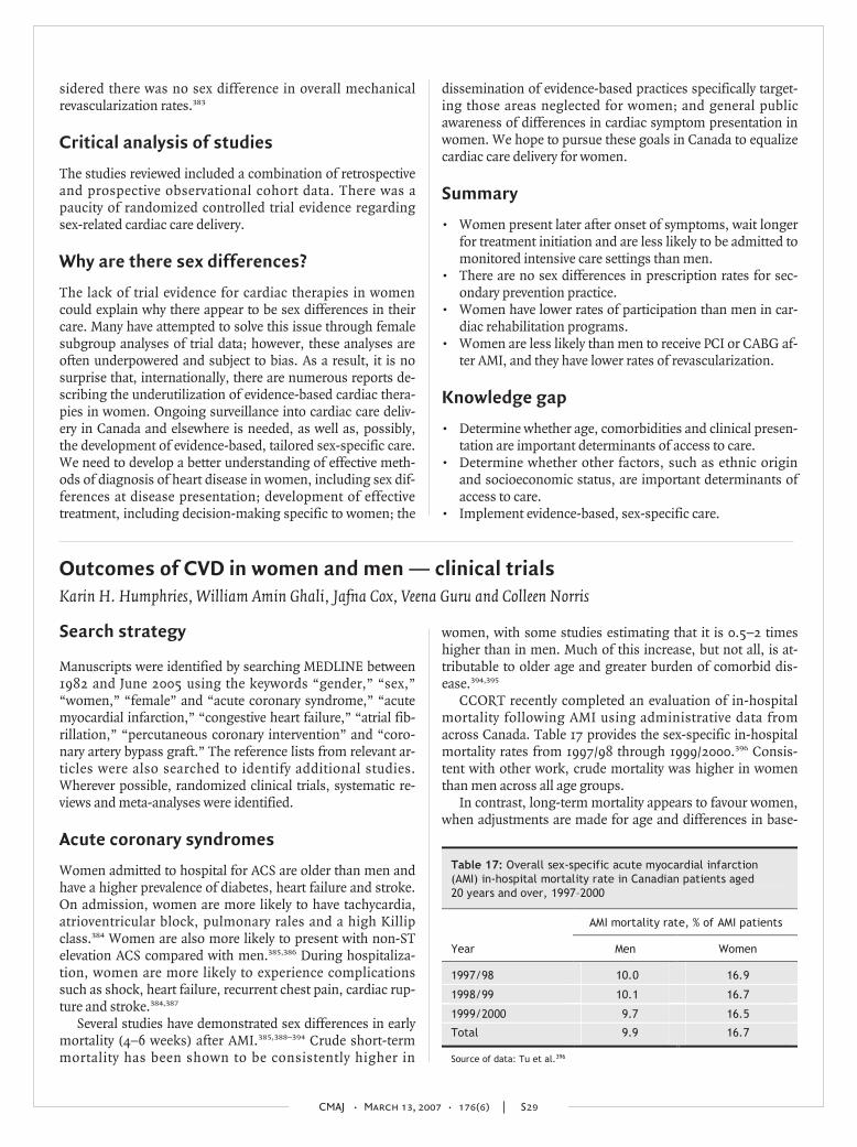
sidered there was no sex difference in overall mechanicalrevascularization rates.383
Critical analysis of studies
The studies reviewed included a combination of retrospectiveand prospective observational cohort data. There was apaucity of randomized controlled trial evidence regardingsex-related cardiac care delivery.
Why are there sex differences?
The lack of trial evidence for cardiac therapies in womencould explain why there appear to be sex differences in theircare. Many have attempted to solve this issue through femalesubgroup analyses of trial data; however, these analyses areoften underpowered and subject to bias. As a result, it is nosurprise that, internationally, there are numerous reports de-scribing the underutilization of evidence-based cardiac thera-pies in women. Ongoing surveillance into cardiac care deliv-ery in Canada and elsewhere is needed, as well as, possibly,the development of evidence-based, tailored sex-specific care.We need to develop a better understanding of effective meth-ods of diagnosis of heart disease in women, including sex dif-ferences at disease presentation; development of effectivetreatment, including decision-making specific to women; the
dissemination of evidence-based practices specifically target-ing those areas neglected for women; and general publicawareness of differences in cardiac symptom presentation inwomen. We hope to pursue these goals in Canada to equalizecardiac care delivery for women.
Summary
• Women present later after onset of symptoms, wait longerfor treatment initiation and are less likely to be admitted tomonitored intensive care settings than men.
• There are no sex differences in prescription rates for sec-ondary prevention practice.
• Women have lower rates of participation than men in car-diac rehabilitation programs.
• Women are less likely than men to receive PCI or CABG af-ter AMI, and they have lower rates of revascularization.
Knowledge gap
• Determine whether age, comorbidities and clinical presen-tation are important determinants of access to care.
• Determine whether other factors, such as ethnic originand socioeconomic status, are important determinants ofaccess to care.
• Implement evidence-based, sex-specific care.
CMAJ • March 13, 2007 • 176(6) | S29
Outcomes of CVD in women and men — clinical trialsKarin H. Humphries, William Amin Ghali, Jafna Cox, Veena Guru and Colleen Norris
Search strategy
Manuscripts were identified by searching MEDLINE between1982 and June 2005 using the keywords “gender,” “sex,”“women,” “female” and “acute coronary syndrome,” “acutemyocardial infarction,” “congestive heart failure,” “atrial fib-rillation,” “percutaneous coronary intervention” and “coro-nary artery bypass graft.” The reference lists from relevant ar-ticles were also searched to identify additional studies.Wherever possible, randomized clinical trials, systematic re-views and meta-analyses were identified.
Acute coronary syndromes
Women admitted to hospital for ACS are older than men andhave a higher prevalence of diabetes, heart failure and stroke.On admission, women are more likely to have tachycardia,atrioventricular block, pulmonary rales and a high Killipclass.384 Women are also more likely to present with non-STelevation ACS compared with men.385,386 During hospitaliza-tion, women are more likely to experience complicationssuch as shock, heart failure, recurrent chest pain, cardiac rup-ture and stroke.384,387
Several studies have demonstrated sex differences in earlymortality (4–6 weeks) after AMI.385,388–394 Crude short-termmortality has been shown to be consistently higher in
women, with some studies estimating that it is 0.5–2 timeshigher than in men. Much of this increase, but not all, is at-tributable to older age and greater burden of comorbid dis-ease.394,395
CCORT recently completed an evaluation of in-hospitalmortality following AMI using administrative data fromacross Canada. Table 17 provides the sex-specific in-hospitalmortality rates from 1997/98 through 1999/2000.396 Consis-tent with other work, crude mortality was higher in womenthan men across all age groups.
In contrast, long-term mortality appears to favour women,when adjustments are made for age and differences in base-
Table 17: Overall sex-specific acute myocardial infarction (AMI) in-hospital mortality rate in Canadian patients aged 20 years and over, 1997–2000
AMI mortality rate, % of AMI patients
Year Men Women
1997/98 10.0 16.9
1998/99 10.1 16.7
1999/2000 9.7 16.5
Total 9.9 16.7
Source of data: Tu et al.396

line conditions.10,392,394 Vaccarino and colleagues10–12 alsonoted an age–sex interaction in which younger women, butnot older women, had higher rates of both long- and short-term mortality compared with men of the same age. These re-sults suggest that younger women with ACS are a particularlyhigh-risk group. At older age, there were no differences inlong-term mortality and, in the very oldest group, women hadbetter outcomes than men.
Summary
• Women are more likely than men to present with non-STelevation ACS.
• Short-term mortality from ACS is higher among womenthan among men, in part because of their older age atpresentation and greater burden of comorbid disease.
• Women have lower long-term mortality from ACS.• Women < 50 years old with AMI are at particularly high
risk of death.
Percutaneous coronary intervention
PCI is the mainstay of treatment for ACS. Although womenreferred for this procedure have a lower prevalence of priorAMI and left-ventricular dysfunction than men, they havemore heart failure that presumably relates to diastolic dys-function. Of interest, despite the greater burden of comorbiddisease, advanced age and severe angina, the extent of angio-graphic epicardial disease appears to be less than in men.397
In the early days of PCI, registry data demonstrated higherprocedural risk and decreased efficacy in women. More re-cent results from the 1997–98 National Heart, Lung, andBlood Institute Registry suggest that sex differences in out-comes have decreased, and the proportion of women under-going this procedure has increased to 35%. Despite increasedage and more comorbid conditions, including diabetes, hy-pertension and heart failure, rates of procedural success(95.0% v. 94.2%), in-hospital death (2.2% v. 1.3%), AMI(2.3% v. 3.0%) and emergency CABG (1.3% v. 1.4%) were notdifferent between women and men, respectively. At 1 year,mortality was significantly higher in women than men (6.5%v. 4.3%), as was the combined end point of death–MI–CABG(18.3% v. 14.4%). However, after controlling for baseline dif-ferences, sex was not a significant predictor of death, but re-mained independently associated with the combined endpoint. This result was driven by the need for subsequentrevascularization and may reflect the lower use of stents inwomen.398
No sex-specific results have been published from the nu-merous trials comparing bare metal stents with balloon an-gioplasty. A recent report evaluating in-hospital mortality andtarget vessel revascularization in patients undergoing electivePCI using 3.0–4.0-mm stents showed no sex differences.399 Intrials of drug-eluting stents (DESs), both the SIRIUS andTAXUS IV trials demonstrated similar reductions in resteno-sis, target vessel revascularization and major adverse cardiacevents at 1 year in women and men.400,401 Evidence for long-term outcomes with DESs are lacking, but early data suggest
that DESs result in favourable outcomes in small vessels inboth women and men.402
In a CCORT evaluation of outcomes after PCI in Canadabetween 1992/93 and 2000/01, female sex emerged as an in-dependent predictor of in-hospital mortality (OR 1.28; 95%CI 1.15–1.42).403 This result is difficult to interpret as the time-frame of this evaluation covers the balloon angioplasty era forwhich outcomes in women are known to be poor, as well asthe stent era and the start of the DES era, where results havebeen shown to be better.
Despite evidence of improved outcomes in the contempo-rary PCI era, vascular complications remain more prevalentamong women. Although the development of less aggressiveanticoagulation regimens, increased use of weight-adjustedheparin dosing and the use of smaller sheath sizes havereduced vascular complications over time, women still havea 2-fold higher risk of access-site hematomas, bleedingcomplications requiring transfusion and retroperitonealbleeds.400
Primary PCI and thrombolysis in STelevation myocardial infarction
Prompt coronary reperfusion, by PCI or thrombolysis, is thestandard of care in ST elevation MI. Clinical trials examiningthe efficacy of thrombolysis have shown similar benefits inwomen and men after adjustment for baseline differences,but a greater risk of bleeding in women.385,389–391,393 Hochmanand colleagues385 examined outcomes in GUSTO IIb patientsby type of coronary syndrome and determined that womenwith ST elevation had a nonsignificant trend toward greaterrisk of death or recurrent MI (OR 1.27; 95% CI 0.98–1.63)compared with men, while there was no difference in thenon-ST elevation group (OR 0.93; 95% CI 0.72–1.2) and aprotective effect in those with unstable angina (OR 0.65; 95%CI 0.49–0.87). A recent meta-analysis of primary PCI v.thrombolysis in patients with ST elevation MI found that pri-mary PCI was significantly better at reducing overall short-term death, non-fatal reinfarction, stroke and the combinedend point of death–non-fatal MI–stroke.404 However, sex-specific outcomes were not reported.
Early revascularization in non-ST elevationmyocardial infarction
A meta-analysis of early invasive strategy (PCI or CABG) v.conservative treatment in non-ST elevation MI establishedthat primary PCI reduced reinfarction, severe angina andreadmission to hospital over a mean follow-up period of17 months, but with an early mortality hazard that only be-came a mortality benefit over the long term.405 Again, thismeta-analysis did not report sex-specific outcomes; however,2 of the trials included in the analysis did investigate sex-specific outcomes. The Fragmin and Fast Revascularizationduring Instability in Coronary artery disease (FRISC II) trial,which enrolled 749 women (30.5%), reported a significant
CMAJ • March 13, 2007 • 176(6) | S30

interaction between sex and early invasive strategy; men, butnot women, experienced a reduction in death and recurrentAMI.406 In a subsequent analysis of the data from this trial, itwas noted that the sex difference in outcome was only in theCABG group (RR 3.74; 95% CI 1.89–7.40), not the PCI group(RR 1.03; 95% CI 0.60–1.80).406
RITA 3, a British Heart Foundation randomized trial ofearly invasive strategy v. conservative management,407 found asignificant interaction between sex and intervention strategy(p = 0.007), with men benefiting from an early invasive ap-proach (OR 0.63; 95% CI 0.41–0.98), but not women (OR1.79; 95% CI 0.95–3.35).408 In contrast, the TACTICS-TIMI 18study did not find that women were less likely than men tobenefit from an early invasive strategy.409 A cohort study ofearly revascularization (50% PCI) in 1450 patients with non-ST elevation MI demonstrated that women had comparableshort-term outcomes (HR 0.81; 95% CI 0.44–1.48) and betterlong-term outcomes (HR 0.65; 95% CI 0.42–0.99) comparedwith men.410 Comparisons of in-hospital and late mortalityrates between women and men after primary PCI for AMI arepresented in Fig. 10.400
Summary
• After elective PCI, short-term mortality, target vessel revas-cularization and restenosis rates are similar in women andmen.
• Thrombolysis is equally effective in women and men.• Women have a 2-fold higher risk of bleeding complica-
tions than men.
Knowledge gap
• What is the biologic and pathophysiologic basis for less-extensive angiographic disease in women?
• Do women with non-ST elevation MI benefit from an earlyinvasive strategy?
• Is primary PCI superior to thrombolysis in women with STelevation MI?
Coronary artery bypass graft surgery
The Society of Thoracic Surgeons (STS) database in theUnited States shows an increase in the proportion of fe-males undergoing CABG from 25.7% in 1990 to 28.7% in1999.411 In Canada, women constitute 22.3% of CABG pa-tients, and this remained stable between 1992/93 and2000/01.412
30-day mortality
Sex differences in operative mortality following CABG remaincontroversial. The issue is difficult to resolve given the lack ofwomen in the few randomized trials that have evaluated theefficacy of CABG compared with medical therapy or PCI.413–415
Observational studies comparing short-term mortality (in-hospital or 30-day) between men and women provide con-flicting evidence, with several studies reporting increasedmortality in women,379,388,412,416–418 but some demonstratingequivalent outcomes after adjustment for baseline differ-ences.419–423 However, the weight of evidence indicates that
CMAJ • March 13, 2007 • 176(6) | S31
Unadjusted
mortality rate
Study N % women Women Men p value
In hospital
ACC-NCDR 59 792 32 5.4 3.1 —
Watanabe 36 765 36 4.0 2.0 < 0.0001
CARS 1 044 30 7.9 2.3 < 0.05
CADILLAC 2 082 27 3.4 1.0 0.003
Late
Mehilli 1 937 26 13.8 12.9 —
CADILLAC 2 082 27 7.8 3.0 < 0.001
Antoniucci 1 019 23 12.0 7.0 0.03
Adjusted OR (95% CI)
Higher among women
0 0.5 1 2 3 4 5
Fig. 10: In-hospital and late mortality rates among women versus men after primary percutaneous coronary intervention for acute myo-cardial infarction. Note: ACC–NCDR = American College of Cardiology National Cardiovascular Data Registry; CARS = Coumadin AspirinReinfarction Study; CADILLAC = Controlled Abciximab and Device Investigation to Lower Late Angioplasty Complications; OR = odds ra-tio; CI = confidence interval. Reproduced with permission from the American Heart Association (Circulation 2005;111:940-53).400
short-term mortality is higher in women. The most recentdata from the STS National Cardiac Surgery Database for2002 indicate an operative mortality of 3.54% in women com-pared with 2.15% in men.424 It is important to note that mor-tality rates have fallen significantly over time, with a greaterdecrease in women than men.425
Several theories have been advanced to explain the highermortality in women, including older age, greater burden ofcomorbid disease and more urgent revascularization. How-ever, women had better preserved left-ventricular functionand less extensive coronary disease.384,388 Smaller coronary ar-teries in women have also been proposed as a possible expla-nation for the higher mortality rate,426 but more recent re-search indicates that the proportion of women with smallcoronary arteries (< 1.5 mm) is no different than the propor-tion of men, 57% and 59% respectively.427
Long-term mortality
In contrast, female sex does not appear to confer increasedrisk of long-term mortality.379,422,428–431 In some studies,women had even better long-term survival than men after ad-justing for risk factors.379,420 The reasons for improved long-term, but not short-term, survival are speculative. Survivalbias may be a factor or the improved long-term survival maybe a reflection of longer life expectancy in women.
Another indication for CABG is to obtain symptom reliefand long-term improvement in function and quality of life.Although studies in these areas have not been consistent, theevidence suggests that after CABG women have worse symp-tom status, poorer physical function, more depression andlower quality of life compared with men.432,433 Depression atthe time of CABG has been shown to influence functionaloutcomes negatively, and this effect is stronger in womenthan in men.434
Off-pump outcomes
Women may have better outcomes with off-pump sur-gery.435–438 In women-only cohorts, those undergoing off-pump surgery had better outcomes than women undergoingconventional CABG.436,437 Studies that compared women andmen undergoing off-pump surgery demonstrated that femalesex was not associated with worse outcomes.435,438 The resultsof the latter studies should be interpreted with caution as theywere retrospective and the sample sizes were small. However,larger, all-female studies are also not definitive, as patient se-lection cannot be ruled out. Specifically, there is evidence thatinternal mammary arteries are more commonly used in off-pump patients compared with conventional CABG. Thus, theimproved outcomes may be associated with increased use ofthese arteries.424
Summary
• Although mortality rates after CABG surgery have fallensignificantly over time, short-term mortality remainshigher among women than among men.
• Women appear to have better long-term survival afterCABG surgery.
• Women may have better outcomes with off-pump sur-gery.
Knowledge gap
• Further assessment of the potential role of vessel size inshort-term outcomes is warranted.
• Further evaluation of the benefits of off-pump surgery inwomen is warranted.
Heart failure
Information on sex differences in heart failure outcomes islimited and not consistent. Among epidemiologic studies,women appear to have better survival rates than men.439,440 Inthe Framingham study, the 1-year mortality rate was 36% inwomen compared with 43% in men (HR 0.64; 95% CI0.54–0.77).439 In NHANES-I, the 1-year mortality rate was23.8% and 54.4% in women and men, respectively.440 Simi-larly, in 2 retrospective cohort studies of elderly Medicare pa-tients, women had better survival than men.441,442 Vaccarinoand colleagues442 reported lower age-adjusted 6-month mor-tality in women compared with men (RR 0.81; 95% CI0.68–0.95), which persisted at 1 year, whereas 30-day mortal-ity did not differ. However, after adjustment for baseline dif-ferences, the 6-month mortality risk for women was compa-rable with that of men (RR 0.90; 95% CI -0.75–1.08). Incontrast, women in the Studies of Left Ventricular Dysfunc-tion (SOLVD) registry had a higher 1-year mortality rate (22%)than men (17%).442
Atrial fibrillation
Evidence from the Framingham study suggests that there isno sex difference in mortality rate in patients with atrial fibril-lation (OR 1.2; 95% CI 0.98–1.49).32 Analysis of data from theCanadian Registry of Atrial Fibrillation (CARAF), which en-rolled patients at the time of their first ECG-documentedepisode of atrial fibrillation, found that women were, on aver-age, 5 years older than men and more likely to be sympto-matic.36 During annual follow-up visits, women were also sig-nificantly more likely to report episodes of paroxysmal atrialfibrillation, but progression to permanent atrial fibrillationdid not vary by sex.
CMAJ • March 13, 2007 • 176(6) | S32

There is mounting evidence to suggest that women andmen respond differently to drug therapy. This sex-based dif-ference may affect the outcomes of patients with CVD.
Search strategy
Manuscripts were identified by searching MEDLINE between1982 and June 2005, using the keywords “gender,” “sex,”“women,” “female,” “acute myocardial infarction,” “conges-tive heart failure,” “atrial fibrillation” AND “ACE inhibitors,”“aspirin,” “GPIIb/IIIa inhibitors,” “beta-blockers,” “statins,”“digoxin,” “spironolactone,” “warfarin” and “rate andrhythm control therapy.” The reference lists from relevant ar-ticles were also searched to identify additional studies. Wher-ever possible, randomized clinical trials, systematic reviewsand meta-analyses were identified.
Acute myocardial infarction
Practice guidelines based on evidence from randomizedclinical trials support the use of ACE inhibitors, ASA, beta-blockers and statins in patients with AMI without contraindi-cations.443–445
ACE inhibitors
A systematic overview of 100 000 patients, randomly as-signed to short-term treatment with ACE inhibitors follow-ing AMI, failed to demonstrate a statistically significant re-duction in risk for women. This may be a reflection of thesmall number of women enrolled in these trials (22%–27%),rather than lack of effect of ACE inhibition in women.446 Incontrast, a meta-analysis of long-term ACE inhibitiondemonstrated comparable outcomes in men and womenwith significant reductions in the composite end point(death–heart failure–MI) in men (OR 0.71; 95% CI0.65–0.77) and women (OR 0.79; 95% CI 0.67–0.93)(pinteraction = 0.34).447
ASA
The Antiplatelet Trialists’ Collaboration reviewed 145 trials ofASA use and found that among high-risk patients, womenand men benefited equally from the use of ASA in terms of re-duction in vascular events.448 ASA use prevented vascularevents in 37 out of 100 men, compared with 33 of 100women. Sex-specific outcomes were not presented in themost recent update from the Antiplatelet Trialists’ Collabora-tion, which reviewed 287 studies of antiplatelet therapy in-volving 135 000 patients.449 In contrast, ASA use for primaryprevention has not been shown to be of benefit in women. Inthe large Women’s Health Study, ASA had a nonsignificanteffect on AMI (RR 1.02; 95% CI 0.84–1.25; p = 0.83) anddeath from cardiovascular causes (RR 0.95; 95% CI
0.74–1.22; p = 0.68), but there was weak evidence that ASAreduced the risk of stroke (RR 0.83; 95% CI 0.69–0.99;p = 0.04).450 In a recent sex-specific meta-analysis of ASA forthe primary prevention of cardiovascular events, ASA wasshown to reduce the risk of ischemic stroke in women (OR0.76; 95% CI 0.63–0.93), but not in men (OR 1.00; 95% CI0.72–1.41).451 Consistent with the Women’s Health Study,ASA use in women was not associated with a decreased riskof MI (OR 1.01; 95% CI 0.84–1.21) in contrast with men (OR0.68; 95% CI 0.54–0.86).451
Platelet glycoprotein IIb/IIIa inhibitors
These agents have been shown to reduce cardiac complica-tions in patients undergoing PCI by blocking the glycoproteinIIb/IIIa receptor, which is the final common pathway leadingto platelet aggregation. The literature reports that womenhave increased platelet function compared with men and,thus, might be expected to benefit more than men from gly-coprotein IIb/IIIa inhibition.452 A pooled analysis of 3 largePCI trials — Evaluation of 7E3 for the Prevention of IschemicComplications (EPIC), Evaluation in Percutaneous Translu-minal Coronary Angioplasty to Improve Long-Term Outcomewith Abciximab GP IIb/IIIa blockade (EPILOG) and Evalua-tion of Platelet IIb/IIIa Inhibitor for Stent (EPISTENT) —demonstrated no gender difference in the benefit derivedfrom glycoprotein IIb/IIIa use. Men had a 41% reduction inthe composite end point of death–MI–revascularization(p < 0.001) whereas women had a 38% reduction(p = 0.01).453
Three recent meta-analyses of platelet glycoprotein IIb/IIIainhibitors were identified.454–456 One comparing glycoproteinIIb/IIIa inhibitors with placebo or standard therapy in 3266patients treated with primary PCI for AMI demonstrated a sig-nificant reduction in the 30-day composite end pointdeath–reinfarction–ischemic or urgent target-vessel revascu-larization (OR 0.54; 95% CI 0.40–0.72). A sex-specific analy-sis of outcomes was not provided, even though the study pop-ulation included 26.1% women (n = 852).454 Similarly, ameta-analysis of glycoprotein IIb/IIIa inhibitors use in 27 115patients with ST elevation MI did not provide sex-specific out-comes, nor did it indicate how many women were included inthe evaluation.455 The third analysis by Boersma and col-leagues456 evaluated the efficacy of glycoprotein IIb/IIIa in-hibitors in patients with ACS who were not routinely sched-uled to undergo early revascularization. In this population of31 402 patients (35% female), glycoprotein IIb/IIIa inhibitorswere shown to be efficacious with a 9% 30-day reduction indeath or MI (OR 0.91; 95% CI 0.85–0.98). A highly significantsex–treatment interaction was noted (pinteraction < 0.001), withmen experiencing benefit (OR 0.81; 95% CI 0.75–0.89), whilewomen were at increased risk of death or MI (OR 1.15; 95% CI1.01–1.30). The sex difference remained, even after adjustingfor baseline differences. However, when patients were strati-
CMAJ • March 13, 2007 • 176(6) | S33
Outcomes of CVD in women and men — post-admission drug therapyKarin H. Humphries, William Amin Ghali, Jafna Cox, Veena Guru and Colleen Norris

fied by baseline troponin levels, the risk estimates suggestedthat, in the troponin-positive group, both sexes benefitedfrom treatment, whereas, in the troponin-negative group,neither benefited. The results of this stratified analysis shouldbe interpreted with caution, as 65% of patients were excludeddue to missing troponin values.
Beta-blockers
Information on sex differences in outcomes following treat-ment with beta-blockers is lacking. A meta-analysis of beta-blocker use after AMI by Freemantle and colleagues457 did notprovide any sex-specific outcomes; however, 2 observationalstudies, the Worcester Heart Attack Study458 and the NationalCooperative Cardiovascular Project,459 did report sex-specificoutcomes. In a community-wide study of 10 374 patients ad-mitted to hospital with confirmed AMI between 1975 and1999, Silvet and colleagues458 reported beneficial effects ofbeta-blocker use in both men (OR 0.22; 95% CI 0.18–0.27)and women (OR 0.32; 95% CI 0.26–0.39) in terms of in-hospital mortality. Krumholz and colleagues459 evaluated out-comes in 115 015 patients 65 years and older who survivedhospital admission with a confirmed AMI between 1994 and1995 and reported comparable 1-year mortality in men (RR0.83; 95% CI 0.76–0.91) and women (RR 0.89; 95% CI0.81–0.97).
Statins
The first major randomized controlled trial of statins in pa-tients with AMI was the Scandinavian Simvastatin SurvivalStudy (4S).460,461 Women constituted only 19% of the 4444 pa-tients in the trial. The primary end point, mortality, was sig-nificantly reduced in men (RR 0.66; 95% CI 0.53–0.80), butnot in women (RR 1.12; 95% CI 0.65–1.93). The secondaryend point, CHD death–MI–resuscitated cardiac arrest, wassignificantly reduced in both men (RR 0.66; 95% CI0.58–0.76) and women (RR 0.65; 95% CI 0.47–0.91). Anothermajor trial in patients with AMI, but average cholesterol lev-els, was the Cholesterol and Recurrent Events (CARE) trial.Again, women made up only 14% of the 4159 participants,who were randomly chosen to receive pravastatin orplacebo.462 In a separate evaluation of outcomes in women,treatment with pravastatin resulted in a 43% (95% CI 4–66)reduction in the primary end point, CAD death and MI, and a46% (95% CI 22–62) reduction in the combined end point,which also included CABG or PCI.463 In men, the risk reduc-tion for the combined end point was only 20%. A test for in-teraction between sex and therapy was significant (p = 0.048)suggesting that women had better outcomes than men, butthis result should be interpreted with caution, as this was notan a priori hypothesis. The more recent Medical ResearchCouncil/British Heart Foundation Heart Protection Study ofsimvastatin demonstrated comparable efficacy in men andwomen, who were at high risk of death from CAD.464 In asubgroup analysis by sex, the relative risk reduction inwomen was 18.9% compared with 21.7% in men, a differencethat was not significant (pinteraction = 0.76).
In a recent meta-analysis of 10 randomized controlled tri-als of statins for primary and secondary prevention, womenmade up 18.9% of the total sample (n = 23 447).465 The sum-mary relative risk ratios (RRR) were not significantly differentfor women (RRR 0.77; 95% CI 0.64–0.94) and men (RRR0.74; 95% CI 0.69–0.79; pinteraction = 0.37). Although the di-chotomy of primary and secondary prevention may be some-what artificial, most of the evidence for the efficacy of statinscomes from secondary prevention trials. A systematic reviewof 6 primary prevention trials concluded that lipid loweringdid not reduce total mortality (RR 0.95; 95% CI 0.62–1.46) orCHD events RR 0.87; 95% CI 0.69–1.09) in women.466
Summary
• Evidence of the benefit of short-term treatment with ACEinhibitors in women is lacking; however, long-term treat-ment reduces the risk of recurrent events.
• ASA use after AMI is equally efficacious in women andmen, but ASA use for primary prevention is not of benefitin women.
• There is evidence that primary prevention with ASA mayreduce the risk of stroke in women.
• Although clinical trial evidence for use of beta-blockers af-ter AMI in women is lacking, observational trials havedemonstrated short- and long-term effectiveness.
• Statin therapy is equally effective in women and men.
Knowledge gap
• The benefits of ASA use in women appear to be limited tosecondary prevention. The lack of effectiveness in primaryprevention requires further exploration.
Heart failure
Current clinical guidelines recommend therapy with multipledrugs, including ACE inhibitors and beta-blockers to reducemorbidity and mortality, digoxin to reduce readmission tohospital, and the addition of spironolactone in patients withmore severe symptoms.467,468
ACE inhibitors
As in other areas of cardiovascular research, women havebeen underrepresented in heart failure trials. In an overviewof 30 randomized trials of ACE inhibitors, neither the reduc-tion in mortality nor the combined end point of mortality andheart failure hospitalizations was significant in women.469
This finding is difficult to interpret, as it may be a reflectionof the underenrolment of women in the earlier trials, ratherthan evidence of less benefit of ACE inhibitors in women withdecreased left-ventricular ejection fraction.
The Heart Outcomes Prevention Evaluation (HOPE) trialundertook a preplanned analysis of the effect of ramipril inhigh-risk women without heart failure and with preservedleft-ventricular function. Women constituted 21% of thestudy cohort, and 37.5% had a prior AMI. Women experi-
CMAJ • March 13, 2007 • 176(6) | S34

enced similar benefits to men. Specifically, women treatedwith ramipril had significant reductions in the primary endpoint of MI, stroke or cardiovascular death (RR 0.77; 95% CI0.62–0.96), as well as fewer strokes (RR 0.64; 95% CI 0.43–0.96) and fewer cardiovascular deaths (RR 0.62; 95% CI0.44–0.88).470
Beta-blockers
In a post-hoc analysis of the 898 (23%) women enrolled inthe Metoprolol CR/XL Randomised Intervention Trial in Con-gestive Heart Failure (MERIT-HF) trial of extended-releasemetoprolol added to optimal therapy with a diuretic and anACE inhibitor, women on metoprolol experienced a signifi-cant (21%) reduction in the primary combined end point ofall-cause mortality–all-cause hospitalization.471 Similarly, in apost hoc analysis of Cardiac Insufficiency Bisoprolol Study(CIBIS) II, a study of bisoprolol added to a diuretic and ACEinhibitors, women not only benefited from beta-blockade,but mortality was significantly lower in women comparedwith men (HR 0.64; 95% CI 0.47–0.86).472 A pooled analysisof 3 trials of beta-blockade — MERIT-HF, CIBIS II, CarvedilolProspective Randomized Cumulative Survival (COPERNI-CUS) — revealed a significant female survival advantage (RR0.69; 95% CI 0.51–0.93).471
Digoxin
In a post hoc analysis of the Digitalis Investigation Grouptrial of digoxin in patients with heart failure and depressedventricular systolic function, women had increased risk of all-cause mortality (HR 1.23; 95% CI 1.02–1.47), which was notseen in men assigned to digoxin (HR 0.93; 95% CI0.85–1.02).473 A recent retrospective analysis of this study de-termined that excess mortality is seen only when serum con-centrations of digoxin exceed 1.2 ng/mL (HR 1.33; 95% CI1.00–1.76); at concentrations of 0.5–0.9 ng/mL, there was noexcess mortality in women (HR 0.8; 95% CI 0.62–1.13).474 Atest for interaction between sex, digoxin concentration andmortality was not significant, but this result should be inter-preted with caution, as the sample size in this subgroupanalysis was small (n = 89 women).
Spironolactone
In the Randomized Aldactone Evaluation Study (RALES), aretrospective analysis by sex revealed that the reduction inrisk of death among patients on spironolactone (0.70; 95% CI0.60–0.82) was similar for men and women.475 Sex-specificrelative risks were not provided.
Summary
• The efficacy of ACE inhibitors is uncertain given the smallnumber of women in trials of these drugs and the lack ofdirect comparisons with men.
• Post hoc analyses suggest that beta-blockers and spirono-lactone are equally efficacious in women and men.
Knowledge gap
• Most clinical trials have focused on patients with de-creased left-ventricular ejection fraction, whereas womenare more likely to have preserved ventricular systolic func-tion. Further research is required to evaluate the effective-ness of therapies in this patient group, which comprisespredominantly elderly women.
Atrial fibrillation
Warfarin
Undoubtedly the most important advance in atrial fibrillationtherapy has been the decreased risk of stroke in patientstreated with warfarin. Some evidence suggests that elderlywomen are less likely to receive warfarin than their malecounterparts. According to CARAF, women 75 years of ageand older were 54% less likely to receive warfarin, comparedwith men in this age group.36 This is consistent with resultsfrom a British community survey of warfarin use in patientswith atrial fibrillation, where the lowest warfarin use wasamong elderly women.476
In an analysis of pooled data from the 5 major antithrom-botic trials, women comprised 26% of the study cohort.477 Fe-male participation ranged from no women in the Stroke Pre-vention in Nonrheumatic Atrial Fibrillation (SPINAF) trial to47% women in the Atrial Fibrillation, Aspirin, and Anticoag-ulation (AFASAK) trial.478,479 In women, warfarin treatmentreduced the risk of stroke by 84% (95% CI 55–95), comparedwith 60% (95% CI 35–76) in men. Although ASA resulted in asignificant decreased risk of stroke in men (44%), in womenthe 23% reduction did not achieve statistical significance.
In CARAF, women on warfarin therapy, predominantlythose less than 75 years of age, were 3.35 times more likelythan men on warfarin to experience a major bleed.36 Althoughincreased female risk of bleeds has also been described in peo-ple undergoing thrombolytic therapy for AMI and oral antico-agulation after hospital admission for deep vein thrombo-sis,385 increased risk of bleeds was not reported in any of thetrials of warfarin use in atrial fibrillation patients. This may re-flect the difference in the intensity of anticoagulation monitor-ing in clinical trials compared with “real world” experience.
Rate and rhythm control therapy
There were no sex differences in response to treatment in theAtrial Fibrillation Follow-up Investigation of Rhythm Man-agement (AFFIRM) study, which investigated the efficacy of 2treatment strategies — rate control v. rhythm control — inhigh-risk atrial fibrillation patients. Both men (adjusted HR1.20) and women (adjusted HR 1.30) had a nonsignificant in-creased risk of death in the rhythm control arm comparedwith the rate control arm.480
Critical analysis of studies
Wherever possible, we have evaluated randomized controlled
CMAJ • March 13, 2007 • 176(6) | S35

trial evidence or meta-analyses based on randomized con-trolled trials. In some instances, trial evidence was lackingand prospective cohort studies were reviewed. Universally,women were underrepresented in clinical trials and conse-quently in the meta-analyses, thus limiting the power to de-tect differences if they do exist. Indeed, the risk of a type-II er-ror is quite large and must be considered when evaluatingtrials and analyses where no differences were noted.
Where sex differences were noted, these may be due to in-adequate adjustment for baseline differences in age and co-morbidities, as women enrolled in clinical trials are on aver-age older than men and present with a greater burden ofcomorbid disease. The most important limitation was theoverall lack of sex-specific information in published studies.Although most studies now enrol women, few provide sex-specific outcomes or test for interactions to determinewhether sex differences do exist.
Summary
• Warfarin use in women with atrial fibrillation is effica-cious for the prevention of stroke, but warfarin is under-used in elderly women.
• There is some evidence that women with atrial fibrillationmay not benefit from ASA use.
• There appear to be no sex differences in response to rateand rhythm control therapy.
Knowledge gap
• The apparent increased risk of bleeding in women treatedwith anticoagulants merits further investigation.
Conclusions
CVD affects both women and men, but it is the leadingcause of mortality in women. Although many aspects of thedisease are similar in women and men, there is a growingbody of evidence to support sex and gender dimorphismsin the prevalence, presenting symptoms, management andoutcomes of CVD. However, because data are retrievedfrom retrospective, observational and cross-sectional co-hort studies, definitive conclusions about why there are sexdifferences cannot be drawn. It is imperative that well-designed studies be conducted in which women are ade-quately represented. These studies must be sufficientlypowered to allow statistical inferences and identification ofsex-specific outcomes. Understanding how sex and genderdifferences translate into CVD will help improve the healthof women and men.
REFERENCES1. The changing face of heart disease and stroke in Canada 2000. Ottawa: Heart and
Stroke Foundation; 1999. Available: www.phac-aspc.gc.ca/ccdpc-cpcmc/cvd-mcv/publications/pdf/card2ke.pdf (accessed 2000 Dec. 19).
2. Thom T, Haase N, Rosamond W, et al. Heart disease and stroke statistics — 2006update: a report from the American Heart Association Statistics Committee andStroke Statistics Subcommittee. Circulation 2005;113:e85-151.
3. Marrugat J, Elosua R, Aldasoro E, et al.; IBERICA Investigators. Regional variabilityin population acute myocardial infarction cumulative incidence and mortality ratesin Spain 1997 and 1998. Eur J Epidemiol 2004;19:831-9.
4. Tunstall-Pedoe H, Kuulasmaa K, Mahonen M, et al. Contribution of trends in sur-vival and coronary-event rates to changes in coronary heart disease mortality: 10-year results from 37 MONICA Project populations. Lancet 1999;353:1547-57.Available: www.heartstats.org/temp/TABsp2.2spweb04.xls (accessed 2006Nov 24).
5. Arciero TJ, Jacobsen SJ, Reeder GS, et al. Temporal trends in the incidence of coro-nary disease. Am J Med 2004;117:228-33.
6. World health statistics 2006. Geneva: World Health Organization; 2006. Available:www.who.int/whosis/whostat2006.pdf (accessed 2006 Dec 19).
7. World Health Organization. Changes in death rates from CHD, men and womenaged 35–74, between 1990 and 2000, selected countries. [Web site of the BritishHeart Foundation], 2006. Available: www.heartstats.org/temp/Figsp1.5bspweb06.xls (accessed 2006 Dec 5).
8. Levi F, Lucchini F, Negri E, et al. Trends in mortality from cardiovascular and cere-brovascular diseases in Europe and other areas of the world. Heart 2002;88:119-24.
9. Manuel DG, Leung M, Nguyen K, et al. Burden of cardiovascular disease inCanada. Can J Cardiol 2003;19:997-1004.
10. Vaccarino V, Krumholz HM, Yarzebski J, et al. Sex differences in 2-year mortalityafter hospital discharge for myocardial infarction. Ann Intern Med 2001;134:173-81.
11. Vaccarino V, Parsons L, Every NR, et al. Sex-based differences in early mortality af-ter myocardial infarction. National Registry of Myocardial Infarction 2 Partici-pants. N Engl J Med 1999;341:217-25.
12. Vaccarino V, Horwitz RI, Meehan TP, et al. Sex differences in mortality after my-ocardial infarction: evidence for a sex-age interaction. Arch Intern Med 1998;158:2054-62.
13. Lloyd-Jones DM. The risk of congestive heart failure: sobering lessons from theFramingham Heart Study. Curr Cardiol Rep 2001;3:184-90.
14. Davies M, Hobbs F, Davis R, et al. Prevalence of left-ventricular systolic dysfunc-tion and heart failure in the Echocardiographic Heart of England Screening study:a population based study. Lancet 2001;358:439-44.
15. Mosterd A, Hoes AW, de Bruyne MC, et al. Prevalence of heart failure and left ven-tricular dysfunction in the general population: the Rotterdam study. Eur Heart J1999;20:447-55.
CMAJ • March 13, 2007 • 176(6) | S36
This project is part of GENESIS Interdisciplinary Capacity EnhancementTeam, an interdisciplinary enhancement study of the gender and sex determi-nants of cardiovascular disease funded by the Canadian Institutes of HealthResearch and the Heart and Stroke Foundation of Canada, grant no. 72565.
From the Divisions of Internal Medicine (Pilote, Dasgupta) and Clinical Epi-demiology (Pilote, Dasgupta, Libersan), The McGill University Health CentreResearch Institute, McGill University, Montréal, Que.; Division of Cardiovas-cular Surgery, Sunnybrook and Women’s College Health Sciences Centre, In-sititute for Clinical Evaluative Sciences (Guru), University of Toronto,Toronto, Ont.; Centre for Health Evaluation and Outcome Sciences, St. Paul’s
This article has been peer reviewed.
Hospital (Humphries), University of British Columbia, Vancouver, BC; De-partment of Psychology (McGrath), Concordia University, Montréal, Que.;University of Alberta (Norris), Edmonton, Alta.; Faculty of Medicine, Depart-ment of Community Health Sciences (Rabi), Calgary, Alta.; Centre hospitalierde l’Université de Montréal Research Centre, Hôtel-Dieu (Tremblay, Hamet,Petrovich) and Groupe de recherche interdisciplinaire en santé, Départementde médecine sociale et préventive (Barnett, O’Loughlin), University of Mon-treal, Montréal, Que.; Institut national de santé publique du Québec(Alamian, Paradis), Québec, Que.; New Halifax Infirmary Site, QEII HealthSciences Center (Cox), Dalhousie University, Halifax, NS; Department ofMedicine and Community Health Sciences (Ghali), University of Calgary, Cal-gary, Alta.; York University (Grace), University Health Network, Toronto,Ont.; Department of Epidemiology and Biostatistics (Ho), McGill University,Montréal, Que.; Department of Community Health and Epidemiology (Kirk-land), Dalhousie University, Halifax, NS; Département de pédiatrie (Lam-bert), Hôpital Ste-Justine and University of Montreal, Montréal, Que.; Centrefor Clinical Epidemiology & Community Studies (Tagalakis), Jewish GeneralHospital-Sir Mortimer B. Davis, McGill University, Montréal, Que.
Competing interests: Dr. Pilote received financial assistance from MerckFrosst to travel to the Canadian Cardiovascular Society in Ottawa in 2006.None declared by the other authors.
Each author has contributed substantially to conception and design, or acqui-sition of data, or analysis and interpretation of data; drafted the article or re-vised it critically for important intellectual content and gave final approval ofthe version to be published.

16. Abhayaratna WP, Smith WT, Becker NG, et al. Prevalence of heart failure and sys-tolic ventricular dysfunction in older Australians: the Canberra Heart Study. Med JAust 2006;184:151-4.
17. Redfield MM, Jacobsen SJ, Burnett JC Jr, et al. Burden of systolic and diastolic ven-tricular dysfunction in the community: appreciating the scope of the heart failureepidemic. JAMA 2003;289:194-202.
18. Jong P, Vowinckel E, Liu PP, et al. Prognosis and determinants of survival in pa-tients newly hospitalized for heart failure: a population-based study. Arch InternMed 2002;162:1689-94.
19. Lee DS, Johansen H, Gong Y, et al. Regional outcomes of heart failure in Canada.Can J Cardiol 2004;20:599-607.
20. Wenger NK. Women, heart failure, and heart failure therapies. Circulation 2002;105:1526-8.
21. Lee WY, Capra AM, Jensvold NG, et al. for the Epidemiology, Practice, Outcomes,and Cost of Heart Failure (EPOCH) study. Gender and risk of adverse outcomes inheart failure. Am J Cardiol 2004;94:1147-52.
22. Roger VL, Weston SA, Redfield MM, et al. Trends in heart failure incidence andsurvival in a community-based population. JAMA 2004;292:344-50.
23. Koelling TM, Chen RS, Lubwama RN, et al. The expanding national burden ofheart failure in the United States: the influence of heart failure in women. AmHeart J 2004;147:74-8.
24. Philbin EF, DiSalvo TG. Influence of race and gender on care process, resourceuse, and hospital-based outcomes in congestive heart failure. Am J Cardiol 1998;82:76-81.
25. Ryder KM, Benjamin EJ. Epidemiology and significance of atrial fibrillation. Am JCardiol 1999;84(9A):131R-8R.
26. Feinberg WM, Blackshear JL, Laupacis A, et al. Prevalence, age distribution, andgender of patients with atrial fibrillation. Analysis and implications. Arch InternMed 1995;155:469-73.
27. Kannel WB, Wolf PA, Benjamin EJ, et al. Prevalence, incidence, prognosis, andpredisposing conditions to atrial fibrillation: population-based estimates. Am JCardiol 1998;82(8A):2N-9N.
28. Benjamin EJ, Levy D, Vaziri SM, et al. Independent risk factors for atrial fibrillationin a population-based cohort. The Framingham Heart Study. JAMA 1994;271:840-4.
29. Friberg J, Scharling H, Gadsboll N, et al. Sex-specific increase in the prevalence ofatrial fibrillation (the Copenhagen City Heart Study). Am J Cardiol 2003;92:1419-23.
30. Wolf PA, Benjamin EJ, Belanger AJ, et al. Secular trends in the prevalence of atrialfibrillation: the Framingham study. Am Heart J 1996;131:790-5.
31. Wolf PA, Mitchell JB, Baker CS, et al. Impact of atrial fibrillation on mortality,stroke, and medical costs. Arch Intern Med 1998;158:229-34.
32. Benjamin EJ, Wolf PA, D’Agostino RB, et al. Impact of atrial fibrillation on the riskof death: the Framingham Heart Study. Circulation 1998;98:946-52.
33. Khairallah F, Ezzedine R, Ganz LI, et al. Epidemiology and determinants of out-come of admissions for atrial fibrillation in the United States from 1996 to 2001.Am J Cardiol 2004;94:500-4.
34. Stewart S, MacIntyre K, MacLeod MM, et al. Trends in hospital activity, morbidityand case fatality related to atrial fibrillation in Scotland, 1986-1996. Eur Heart J2001;22:693-701.
35. Wattigney WA, Mensah GA, Croft JB. Increased atrial fibrillation mortality: UnitedStates, 1980-1998. Am J Epidemiol 2002;155:819-26.
36. Humphries KH, Jackevicius C, Gong Y, et al. Population rates of hospitalization foratrial fibrillation/flutter in Canada. Can J Cardiol 2004;20:869-76.
37. Norris CM, Ghali WA, Galbraith PD, et al. Women with coronary artery disease re-port worse health-related quality of life outcomes compared to men. Health QualLife Outcomes 2004;2:21.
38. Berenson GS, Srinivasan SR, Bao W, et al. Association between multiple cardiovas-cular risk factors and atherosclerosis in children and young adults. The BogalusaHeart Study. N Engl J Med 1998;338:1650-6.
39. Mahoney LT, Burns TL, Stanford W, et al. Coronary risk factors measured in child-hood and young adult life are associated with coronary artery calcification in youngadults: the Muscatine study. J Am Coll Cardiol 1996;27:277-84.
40. Chen E, Matthews KA, Boyce WT. Socioeconomic differences in children’s health:how and why do these relationships change with age? Psychol Bull 2002;128:295-329.
41. Janz KF, Dawson JD, Mahoney LT. Tracking physical fitness and physical activityfrom childhood to adolescence: the Muscatine Study. Med Sci Sports Exerc 2000;32:1250-7.
42. Bao W, Threefoot SA, Srinivasan SR, et al. Essential hypertension predicted bytracking of elevated blood pressure from childhood to adulthood: the BogalusaHeart Study. Am J Hypertens 1995;8:657-65.
43. Guo SS, Huang C, Maynard LM, et al. Body mass index during childhood, adoles-cence, and young adulthood in relation to adult overweight and adiposity: the FelsLongitudinal Study. Int J Obes Relat Metab Disord 2000;24:1628-35.
44. Nicklas TA, von Duvillard SP, Berenson GS. Tracking of serum lipids and lipopro-teins from childhood to dyslipidemia in adults: the Bogalusa Heart Study. Int JSports Med 2002;23(suppl 1):S39-43.
45. Ogden CL, Flegal KM, Carroll MD, et al. Prevalence and trends in overweightamong US children and adolescents, 1999-2000. JAMA 2002;288:1728-32.
46. Shields M. Measured obesity: overweight Canadian children and adolescents.Ottawa: Statistics Canada; 2005. Available: www.statcan.ca/english/research/82-620-MIE/2005001/pdf/cobesity.pdf (accessed 2006 Dec 3).
47. Willms JD, Tremblay MS, Katzmarzyk PT. Geographic and demographic variationin the prevalence of overweight Canadian children. Obes Res 2003;11:668-73.
48. Cole TJ, Bellizzi MC, Flegal KM, et al. Establishing a standard definition for childoverweight and obesity worldwide: International survey. BMJ 2000;320:1240-3.
49. Tremblay MS, Willms JD. Secular trends in the body mass index of Canadian chil-dren. CMAJ 2000;163:1429-33.
50. Freedman DS, Khan LK, Dietz WH, et al. Relationship of childhood obesity tocoronary heart disease risk factors in adulthood: the Bogalusa Heart Study. Pedi-atrics 2001;108:712-8.
51. Kelder SH, Osganian SK, Feldman HA, et al. Tracking of physical and physiologi-cal risk variables among ethnic subgroups from third to eighth grade: the Childand Adolescent Trial for Cardiovascular Health cohort study. Prev Med 2002;34:324-33.
52. Paradis G, Lambert M, O’Loughlin J, et al. Blood pressure and adiposity in childrenand adolescents. Circulation 2004;110:1832-8.
53. Freedman DS, Dietz WH, Srinivasan SR, et al. The relation of overweight to cardio-vascular risk factors among children and adolescents: the Bogalusa Heart Study.Pediatrics 1999;103:1175-82.
54. Stan S, Levy E, Delvin EE, et al. Distribution of LDL particle size in a population-based sample of children and adolescents and relationship with other cardiovascu-lar risk factors. Clin Chem 2005;51:1192-200.
55. Paradis G, Lambert M, O’Loughlin J, et al. The Quebec Child and AdolescentHealth and Social Survey: design and methods of a cardiovascular risk factor sur-vey for youth. Can J Cardiol 2003;19:523-31.
56. de Ferranti SD, Gauvreau K, Ludwig DS, et al. Prevalence of the metabolic syn-drome in American adolescents. Findings from the Third National Health and Nu-trition Examination Survey. Circulation 2004;110:2494-7.
57. Ford ES, Mokdad AH, Ajani UA. Trends in risk factors for cardiovascular diseaseamong children and adolescents in the United States. Pediatrics 2004;114:1534-44.
58. Genovesi S, Giussani M, Pieruzzi F, et al. Results of blood pressure screening in apopulation of school-aged children in the province of Milan: role of overweight.J Hypertens 2005;23:493-7.
59. Lauer RM, Clarke WR. Childhood risk factors for high adult blood pressure: theMuscatine Study. Pediatrics 1989;84:633-41.
60. Paradis G, Lambert M, O’Loughlin J, et al. Facteurs de risque de maladies cardio-vasculaires. In: Enquête sociale et de santé auprès des enfants et adolescentsquébécois 1999. Québec: Institut de la statistique du Québec; 2002. p. 345-70.
61. National High Blood Pressure Education Program Working Group on Hyperten-sion Control in Children and Adolescents. Update on the 1987 task force report onhigh blood pressure in children and adolescents. Pediatrics 1996;98:649-58.
62. Dasgupta K, O’Loughlin J, Chen S, et al. Emergence of sex differences in preva-lence of high systolic blood pressure. Analysis of a longitudinal adolescent cohort.Circulation 2006;114:2663-70.
63. Muntner P, He J, Cutler JA, et al. Trends in blood pressure among children andadolescents. JAMA 2004;291:2107-13.
64. Rosner B, Prineas R, Daniels SR, et al. Blood pressure differences between blacksand whites in relation to body size among US children and adolescents. Am J Epi-demiol 2000;151:1007-19.
65. Morrison JA, Sprecher DL, Barton BA, et al. Overweight, fat patterning, and cardio-vascular disease risk factors in black and white girls: the National Heart, Lung, andBlood Institute Growth and Health Study. J Pediatr 1999;135:458-64.
66. Vlajinac H, Miljus D, Adanja B, et al. Blood pressure levels in 7 to 14-year-old Bel-grade children. J Hum Hypertens 2003;17:761-5.
67. Global youth tobacco survey (GYTS). [Web site of the Centers for Disease Controland Prevention], 2006. Available: www.cdc.gov/tobacco/global/gyts/GYTS_fact-sheets.htm (accessed 2006 Nov 24).
68. National Tobacco Information Online System: country data. [Web site of the Cen-ters for Disease Control and Prevention], 2003. Available: apps.nccd.cdc.gov/na-tions/nations/Country_Specific_indicators.asp (accessed 2006 Nov 24).
69. Global Youth Tobacco Survey Collaborating Group. Differences in worldwide to-bacco use by gender: findings from the Global Youth Tobacco Survey. J Sch Health2003;73:207-15.
70. 2002 youth smoking survey — technical report. Ottawa: Health Canada, Ministerof Supply and Services Canada; 2005. Available: www.hc-sc.gc.ca/hl-vs/pubs/to-bac-tabac/yss-etj-2002/index_e.html (accessed 2006 Nov 24).
71. Canadian Community Health Survey profiles. [Web site of Statistics Canada],2003. Available: www.statcan.ca/bsolc/english/bsolc?catno=82-576-X&CHROPG=1 (accessed 2006 Dec 3).
72. Gilmore J. Report on smoking in Canada, 1985 to 2001. Ottawa: Minister of Indus-try, Statistics Canada; 2002. Available: www.statcan.ca/english/research/82F0077XIE/82F0077XIE2001001.pdf (accessed 2006 Nov 29). Cat. no.82F0077XIE-01.
73. Canadian tobacco use monitoring survey (CTUMS), February-June 2004. [Web siteof Health Canada.] Available: www.hc-sc.gc.ca/hl-vs/tobac-tabac/research-recherche/stat/ctums-esutc/fs-if/2000/2000youth_e.html (accessed 2006 Dec 19).
74. Karp I, O’Loughlin J, Paradis G, et al. Smoking trajectories of adolescent novicesmokers in a longitudinal study of tobacco use. Ann Epidemiol 2005;15:445-52.
75. Turner L, Mermelstein R, Flay B. Individual and contextual influences on adoles-cent smoking. Ann N Y Acad Sci 2004;1021:175-97.
76. Demory-Luce D, Morales M, Nicklas T, et al. Changes in food group consumptionpatterns from childhood to young adulthood: the Bogalusa Heart Study. J Am DietAssoc 2004;104:1684-91.
77. Troiano RP, Briefel RR, Carroll MD, et al. Energy and fat intakes of children andadolescents in the United States: data from the National Health and Nutrition Ex-amination Surveys. Am J Clin Nutr 2000;72(Suppl):1343S-53S.
78. Nicklas TA, Elkasabany A, Srinivasan SR, et al. Trends in nutrient intake of 10-year-old children over two decades (1973-1994): the Bogalusa Heart Study. Am J
CMAJ • March 13, 2007 • 176(6) | S37
Epidemiol 2001;153:969-77.79. Sturm R. Childhood obesity — what we can learn from existing data on societal
trends, part 2. Prev Chronic Dis 2005;2(2):A20. Available: www.cdc.gov/pcd/issues/2005/apr/04_0039.htm (accessed 2006 Nov 29).
80. Nielsen SJ, Siega-Riz AM, Popkin BM. Trends in food locations and sourcesamong adolescents and young adults. Prev Med 2002;35:107-13.
81. Jahns L, Siega-Riz AM, Popkin BM. The increasing prevalence of snacking amongUS children from 1977 to 1996. J Pediatr 2001;138:493-8.
82. Veugelers PJ, Fitzgerald AL, Johnston E. Dietary intake and risk factors for poordiet quality among children in Nova Scotia. Can J Public Health 2005;96:212-6.
83. Phillips S, Jacobs Starkey LJ, Gray-Donald K. Food habits of Canadians: foodsources of nutrients for the adolescent sample. Can J Diet Pract Res 2004;65:81-4.
84. Cavadini C, Siega-Riz AM, Popkin BM. US adolescent food intake trends from1965 to 1996. Arch Dis Child 2000;83:18-24.
85. Janssen I, Katzmarzyk P, Boyce WF, et al. Overweight and obesity in Canadian ado-lescents and their associations with dietary habits and physical activity patterns.J Adolesc Health 2004;35:360-7.
86. French SA, Lin B-H, Guthrie JF. National trends in soft drink consumption amongchildren and adolescents age 6 to 17 years: prevalence, amounts, and sources,1977/1978 to 1994/1998. J Am Diet Assoc 2003;103:1326-31.
87. Leisure-time physical activity, by age group and sex, household population aged 12and over, Canada, 2003. Ottawa: Statistics Canada; 2003. Catalogue no. 82-221,Vol. 2004, No. 1.
88. US Department of Health and Human Services. Physical activity and health: a re-port of the Surgeon General. Atlanta: US Department of Health and Human Ser-vices, Centers for Disease Control and Prevention, National Center for ChronicDisease Prevention and Health Promotion; 1996. Available: www.cdc.gov/nccdphp/sgr/pdf/sgrfull.pdf (accessed 2006 Nov 9).
89. Higgins JW, Gaul C, Gibbons S, et al. Factors influencing physical activity levelsamong Canadian youth. Can J Public Health 2003;94:45-51.
90. Eisenmann JC, Bartee RT, Wang MQ. Physical activity, TV viewing, and weight inU.S. youth: 1999 Youth Risk Behavior Survey. Obes Res 2002;10:379-85.
91. Andersen RE, Crespo CJ, Bartlett SJ, et al. Relationship of physical activity and tele-vision watching with body weight and level of fatness among children: resultsfrom the Third National Health and Nutrition Examination Survey. JAMA1998;279:938-42.
92. Simons-Morton BG, McKenzie TJ, Stone E, et al. Physical activity in a multiethnicpopulation of third graders in four states. Am J Public Health 1997;87:45-50.
93. Young people in Canada: their health and well-being. Health Behaviour of School-Aged Children (HBSC) Study. Ottawa: Health Canada; 2004. Available: www.phac-aspc.gc.ca/dca-dea/publications/hbsc-2004/pdf/hbsc_summary_2004_e.pdf (ac-cessed 2006 Dec 3).
94. Centers for Disease Control and Prevention. Participation in high school physicaleducation — United States, 1991–2003. MMWR Morb Mortal Wkly Rep 2004;53:844-7.
95. Nader PR. National Institute of Child Health and Human Development Study ofEarly Child Care and Youth Development Network. Frequency and intensity of ac-tivity of third-grade children in physical education. Arch Pediatr Adolesc Med2003;157:185-90.
96. Health Canada. Exercise and leisure activities. In: Trends in the health of Canadianyouth. Ottawa: Health Canada; 1999. Available: www.phac-aspc.gc.ca/dca-dea/publications/pdf/hbsc_08ch8_e.pdf (accessed 2006 Nov 9).
97. Wilson DK, Williams J, Evans A, et al. Brief report: a qualitative study of genderpreferences and motivational factors for physical activity in underserved adoles-cents. J Pediatr Psychol 2005;30:293-7.
98. Aaron DJ, Storti KL, Robertson RJ, et al. Longitudinal study of the number andchoice of leisure time physical activities from mid to late adolescence: implicationsfor school curricula and community recreation programs. Arch Pediatr AdolescMed 2002;156:1075-80.
99. Jago R, Anderson CB, Baranowski T, et al. Adolescent patterns of physical activitydifferences by gender, day, and time of day. Am J Prev Med 2005;28:447-52.
100. Nelson MC, Gordon-Larsen P, Adair LS, et al. Adolescent physical activity andsedentary behavior: patterning and long-term maintenance. Am J Prev Med 2005;28:259-66.
101. Vilhjalmsson R, Kristjansdottir G. Gender differences in physical activity in olderchildren and adolescents: the central role of organized sport. Soc Sci Med 2003;56:363-74.
102. Garcia AW, Broda MA, Frenn M, et al. Gender and developmental differences inexercise beliefs among youth and prediction of their exercise behavior. J SchHealth 1995;65:213-9.
103. Wang CK, Chatzisarantis NL, Spray CM, et al. Achievement goal profiles in schoolphysical education: differences in self-determination, sport ability beliefs, andphysical activity. Br J Educ Psychol 2002;72:433-45.
104. Biddle SJ, Gorely T, Stensel DJ. Health-enhancing physical activity and sedentarybehaviour in children and adolescents. J Sports Sci 2004;22:679-701.
105. Tergerson JL, King KA. Do perceived cues, benefits, and barriers to physical activ-ity differ between male and female adolescents? J Sch Health 2002;72:374-80.
106. Brodersen NH, Steptoe A, Williamson S, et al. Sociodemographic, developmental,environmental, and psychological correlates of physical activity and sedentary be-havior at age 11 to 12. Ann Behav Med 2005;29:2-11.
107. Sallis JF, Zakarian JM, Hovell MF, et al. Ethnic, socioeconomic, and sex differencesin physical activity among adolescents. J Clin Epidemiol 1996;49:125-34.
108. Maia JA, Thomis M, Beunen G. Genetic factors in physical activity levels: a twinstudy. Am J Prev Med 2002;23(2 Suppl):87-91.
109. Anderssen N, Wold B. Parental and peer influences on leisure-time physical activ-
ity in young adolescents. Res Q Exerc Sport 1992;63:341-8.110. Duncan SC, Duncan TE, Strycker LA. Sources and types of social support in youth
physical activity. Health Psychol 2005;24:3-10.111. Hume C, Salmon J, Ball K. Children’s perceptions of their home and neighbor-
hood environments, and their association with objectively measured physical activ-ity: a qualitative and quantitative study. Health Educ Res 2005;20:1-13.
112. Sallis JF, Conway TL, Prochaska JJ, et al. The association of school environmentswith youth physical activity. Am J Public Health 2001;91:618-20.
113. McKenzie TL, Marshall SJ, Sallis JF, et al. Leisure-time physical activity in schoolenvironments: an observational study using SOPLAY. Prev Med 2000;30:70-7.
114. Stone EJ, McKenzie TL, Welk GJ, et al. Effects of physical activity interventions inyouth: review and synthesis. Am J Prev Med 1998;15:298-315.
115. Marshall SJ, Gorely T, Biddle SJ. A descriptive epidemiology of screen-based mediause in youth: a review and critique. J Adolesc 2006;29:333-49.
116. Statistics Canada. Children’s participation in sedentary activities, in hours perweek, by age group and sex, household population aged 12 to 17, Canadian Com-munity Health Survey cycle 2.2, Canada and provinces. Ottawa: Statistics Canada;2004.
117. Canadian Cardiovascular Society 1998 Consensus Conference on the Prevention ofCardiovascular Diseases: the role of the cardiovascular specialist. Can J Cardiol1999;15(Suppl G):1G-119G.
118. Raitakari OT, Porkka KV, Rasanen L, et al. Relations of life-style with lipids, bloodpressure and insulin in adolescents and young adults. The Cardiovascular Risk inYoung Finns Study. Atherosclerosis 1994;111:237-46.
119. Bergstrom E, Hernell O, Persson LA. Cardiovascular risk indicators cluster in girlsfrom families of low socio-economic status. Acta Paediatr 1996;85:1083-90.
120. Pate RR, Heath GW, Dowda M, et al. Associations between physical activity andother health behaviors in a representative sample of US adolescents. Am J PublicHealth 1996;86:1577-81.
121. Pronk NP, Anderson LH, Crain AL, et al. Meeting recommendations for multiplehealthy lifestyle factors. Prevalence, clustering, and predictors among adolescent,adult, and senior health plan members. Am J Prev Med 2004;27:25-33.
122. McKenna M, Taylor W, Marks J, et al. Current issues and challenges in chronic dis-ease control. In: Brownson R, Remington P, Davis J, editors. Chronic disease epi-demiology and control. Washington: American Public Health Association; 1998.p.1-26.
123. Grundy S, Pasternak R, Greenand P, et al. Assessment of cardiovascular risk by useof multiple-risk-factor assessment equations. A statement for healthcare profes-sionals from the American Heart Association and the American College of Cardiol-ogy. J Am Coll Cardiol 1999;34:1348-59.
124. Genest J, Frohlich J, Fodor G, et al.; Working Group on Hypercholesterolemia andOther Dyslipidemias. Recommendations for the management of dyslipidemia andthe prevention of cardiovascular disease: summary of the 2003 update. CMAJ2003;169:921-4.
125. National Cholesterol Education Program (NCEP) Expert Panel on Detection; Evalu-ation and Treatment of High Blood Cholesterol in Adults. (Adult Treatment PanelIII). Executive summary of the third report of the national cholesterol educationprogram (NCEP) expert panel on detection, evaluation, and treatment of highblood cholesterol in adults (Adult Treatment Panel III). JAMA 2001;285:2486-97.
126. A national dialogue on healthy body weights, December 7–9, 2001, Toronto: sum-mary of proceedings. Ottawa: Obesity Canada and the Canadian Institutes ofHealth Research Institute of Nutrition, Metabolism, and Diabetes; 2001. Available:www.cihr-irsc.gc.ca/e/pdf_24010.htm (accessed 2006 Dec 3).
127. Vidal J. Updated review on the benefits of weight loss. Int J Obes Relat Metab Dis-ord 2002;26(Suppl 4):S25-8.
128. Boule N, Haddad E, Kenny G, et al. Effects of exercise on glycemic control andbody mass in type 2 diabetes mellitus: a meta-analysis of controlled clinical trials.JAMA 2001;286:1218-27.
129. Fagard R. Physical activity in the prevention and treatment of hypertension in theobese. Med Sci Sports Exerc 1999;31(Suppl 11):S624-30.
130. American Diabetes Association. Diabetes mellitus and exercise. Diabetes Care2002;25(Suppl 1):S64-8. Available: care.diabetesjournals.org/cgi/content/full/25/suppl_1/s64 (accessed 2006 Dec 3).
131. American Diabetes Association. Management of dyslipidemia in adults with dia-betes. Diabetes Care 2002;25(Suppl 1):S74-7. Available: care.diabetesjournals.org/cgi/content/full/25/suppl_1/s74 (accessed 2006 Dec 3).
132. American Diabetes Association. Treatment of hypertension in adults with diabetes.Diabetes Care 2002;25(Suppl 1):S71-3. Available: care.diabetesjournals.org/cgi/content/full/25/suppl_1/s71 (accessed 2006 Dec 3).
133. Wallace J. Exercise in hypertension. A clinical review. Sports Med 2003;33:585-98.134. Ekelund L, Haskell W, Johnson J, et al. Physical fitness as predictor of cardiovascu-
lar mortality in asymptomatic North American men. The Lipid Research ClinicsMortality Follow-up Study. N Engl J Med 1988;319:1379-84.
135. Mora S, Redberg R, Cui Y, et al. Ability of exercise testing to predict cardiovascularand all-cause death in asymptomatic women: a 20-year follow-up of the Lipid Re-search Clinics Prevalence Study. JAMA 2003;290:1600-7.
136. Williamson D, Madans J, Pamuk E, et al. A prospective study of childbearing and10-year weight gain in US white women 25 to 45 years of age. Int J Obes RelatMetab Disord 1994;18:561-9.
137. Flegal KM, Carroll MD, Ogden CL, et al. Prevalence and trends in obesity amongUS adults, 1999-2000. JAMA 2002;288:1723-7.
138. Canadian community health survey: a first look. The Daily May 2002. [Web site ofStatistics Canada], 2002. Available: www.statcan.ca/Daily/English/020508/d020508a.htm (accessed 2006 Dec 3).
139. National Health and Nutrition Examination Survey: Healthy weight, overweight,
CMAJ • March 13, 2007 • 176(6) | S38

and obesity among U.S. adults. [Web site of the Centers for Disease Control andPrevention]. Available: www.cdc.gov/nchs/data/nhanes/databriefs/adultweight.pdf(accessed 2006 Dec. 19)
140. Tremblay MS, Pérez CE, Ardern CI, et al. Obesity, overweight and ethnicity. HealthRep 2005;16:23-34.
141. Visscher TL, Seidell JC. Time trends (1993–1997) and seasonal variation in bodymass index and waist circumference in the Netherlands. Int J Obes Relat MetabDisord 2004;28:1309-16.
142. Milewicz A, Jedrzejuk D, Lwow F, et al. Prevalence of obesity in Poland. Obes Rev2005;6:113-4.
143. Kaluski DN, Berry EM. Prevalence of obesity in Israel. Obes Rev 2005;6:115-6.144. Kim DM, Ahn CW, Nam SY. Prevalence of obesity in Korea. Obes Rev 2005;6:
117-21.145. Nishida C, Mucavele P. Monitoring the rapidly emerging public health problem of
overweight and obesity: the WHO Global Database on Body Mass Index. SCNNews 2005;(29):5-12.
146. Serdula MK, Ivery D, Coates RJ, et al. Do obese children become obese adults? Areview of the literature. Prev Med 1993;22:167-77.
147. Wing R, Matthews K, Kuller L, et al. Weight gain at the time of menopause. ArchIntern Med 1991;151:97-102.
148. Bidlingmeyer I, Burnier M, Bidlingmeyer M, et al. Isolated office hypertension: aprehypertensive state? J Hypertens 1996;14:327-32.
149. Sobal J, Rauschenbach BS, Frongillo EA Jr. Marital status, fatness and obesity. SocSci Med 1992;35:915-23.
150. Pitsavos C, Panagiotakos D, Lentzas Y, et al. Epidemiology of leisure-time physicalactivity in socio-demographic, lifestyle and psychological characteristics of menand women in Greece: the ATTICA Study. BMC Public Health 2005;5:37.
151. Gal DL, Santos AC, Barros H. Leisure-time versus full-day energy expenditure: across-sectional study of sedentarism in a Portuguese urban population. BMC Pub-lic Health 2005;5:16.
152. Barengo NC, Nissinen A, Tuomilehto J, et al. Twenty-five-year trends in physicalactivity of 30- to 59-year-old populations in eastern Finland. Med Sci Sports Exerc2002;34:1302-7.
153. Iwai N, Yoshiike N, Saitoh S, et al. Leisure-time physical activity and relatedlifestyle characteristics among middle-aged Japanese. Japan Lifestyle MonitoringStudy Group. J Epidemiol 2000;10:226-33.
154. Matthews CE, Freedson PS, Hebert JR, et al. Seasonal variation in household, occu-pational, and leisure time physical activity: longitudinal analyses from the seasonalvariation of blood cholesterol study. Am J Epidemiol 2001;153:172-83.
155. Blair SN, Kohl HW III, Paffenbarger RS Jr, et al. Physical fitness and all-cause mor-tality. A prospective study of healthy men and women. JAMA 1989;262:2395-401.
156. Chen J, Millar WJ. Starting and sustaining physical activity. Health Rep 2001;12:33-43.
157. Brownson RC, Baker EA, Housemann RA, et al. Environmental and policy deter-minants of physical activity in the United States. Am J Public Health 2001;91:1995-2003.
158. King AC, Castro C, Wilcox S, et al. Personal and environmental factors associatedwith physical inactivity among different racial-ethnic groups of U.S. middle-agedand older-aged women. Health Psychol 2000;19:354-64.
159. Lee C. Factors related to the adoption of exercise among older women. J BehavMed 1993;16:323-34.
160. Martins D, Nelson K, Pan D, et al. The effect of gender on age-related blood pres-sure changes and the prevalence of isolated systolic hypertension among olderadults: data from NHANES III. J Gend Specif Med 2001;4:10-3, 20.
161. Burt VL, Whelton P, Roccella EJ, et al. Prevalence of hypertension in the US adultpopulation. Results from the Third National Health and Nutrition ExaminationSurvey, 1988-1991. Hypertension 1995;25:305-13.
162. Wiinberg N, Hoegholm A, Christensen HR, et al. 24-h ambulatory blood pressurein 352 normal Danish subjects, related to age and gender. Am J Hypertens 1995;8:978-86.
163. The SURF report 2: surveillance of chronic disease risk factors. Country-level dataand comparable estimates. Geneva: World Health Organization; 2005. Available:www.who.int/ncd_surveillance/infobase/web/surf2/start.html (accessed 2006Dec 3).
164. Wolf-Maier K, Cooper RS, Banegas JR, et al. Hypertension prevalence and bloodpressure levels in 6 European countries, Canada, and the United States. JAMA2003;289:2363-9.
165. Reckelhoff JF. Gender differences in the regulation of blood pressure. Hyperten-sion 2001;37:1199-208.
166. Ellis JA, Wong ZY, Stebbing M, et al. Sex, genes and blood pressure. Clin Exp Phar-macol Physiol 2001;28:1053-5.
167. Haffner S, Lehto S, Ronnemaa T, et al. Mortality from coronary heart disease insubjects with type 2 diabetes and in nondiabetic subjects with and without priormyocardial infarction. N Engl J Med 1998 339:229-34.
168. Haffner SM. Epidemiology of insulin resistance and its relation to coronary arterydisease. Am J Cardiol 1999;84:11J-4J.
169. Estimated prevalence of diabetes and numbers of people with diabetes, 2003 and2025, selected countries, the world. Brussels: International Diabetes Federation;2003. [Web site of the British Heart Foundation]. Available: www.heartstats.org/temp/TABsp12.8spweb06.xls (accessed 2006 Dec. 1).
170. Hu FB, Stampfer MJ, Solomon CG, et al. The impact of diabetes mellitus on mor-tality from all causes and coronary heart disease in women: 20 years of follow-up.Arch Intern Med 2001;161:1717-23.
171. Ghali WA, Quan H, Norris CM, et al. Prognostic significance of diabetes as a pre-dictor of survival after cardiac catheterization. Am J Med 2000;109:543-8.
172. Graham MM, Ghali WA, Faris PD, et al.; APPROACH Investigators. Sex differencesin the prognostic importance of diabetes in patients with ischemic heart diseaseundergoing coronary angiography. Diabetes Care 2003;26:3142-7.
173. Lee WL, Cheung AM, Cape D, et al. Impact of diabetes on coronary artery diseasein women and men: a meta-analysis of prospective studies. Diabetes Care 2000;23(7):962-8.
174. Kanaya AM, Grady D, Barrett-Connor E. Explaining the sex difference in coronaryheart disease mortality among patients with type 2 diabetes mellitus. A meta-analy-sis. Arch Intern Med 2002;162:1737-45.
175. Huxley R, Barzi F, Woodward M. Excess risk of fatal coronary heart disease associ-ated with diabetes in men and women: meta-analysis of 37 prospective cohortstudies. BMJ 2006 332:73-8.
176. Hu G, Jousilahti P, Qiao Q, et al. The gender-specific impact of diabetes and my-ocardial infarction at baseline and during follow-up on mortality from all causesand coronary heart disease. J Am Coll Cardiol 2005;45:1413-8.
177. Steinberg HO, Paradisi G, Cronin J, et al. Type II diabetes abrogates sex differencesin endothelial function in premenopausal women. Circulation 2000;101:2040-6.
178. Sowers JR. Insulin and insulin-like growth factors in normal and pathologic car-diovascular physiology. Hypertension 1997;29:691-9.
179. Wexler D, Grant RW, Meigs JB, et al. Sex disparities in the treatment of cardiac riskfactors in patients with type 2 diabetes. Diabetes Care 2005;28:514-20.
180. National Cholesterol Education Program: ATP III guidelines at-a-glance quickdesk reference. Bethesda, MD: National Institutes of Health; 2001. Available:www.nhlbi.nih.gov/guidelines/cholesterol/atglance.pdf (accessed 2006 Dec 19).
181. Centers for Disease Control and Prevention. Trends in cholesterol screening andawareness of high blood cholesterol — United States, 1991–2003. MMWR MorbMortal Wkly Rep 2005;54:865-70.
182. Langille DB, Joffres MR, MacPherson KM, et al. Prevalence of risk factors for car-diovascular disease in Canadians 55 to 74 years of age: results from the CanadianHeart Health Surveys, 1986-1992. CMAJ 1999;161(8 Suppl):S3-9.
183. Bass KM, Newschaffer CJ, Klag MJ, et al. Plasma lipoprotein levels as predictors ofcardiovascular death in women. Arch Intern Med 1993;153:2209-16.
184. Johnson CL, Rifkind BM, Sempros CT, et al. Declining serum total cholesterol lev-els among US adults: the National Health and Nutrition Examination Surveys.JAMA 1993;269:3002-8.
185. Castelli WP, Anderson K, Willson PW, et al. Lipids and risk of coronary heart dis-ease: the Framingham Study. Ann Epidemiol 1992;2:23-8.
186. Sharrett AR, Ballantyne CM, Coady SA, et al. Coronary heart disease predictionfrom lipoprotein cholesterol levels, triglycerides, lipoprotein(a), apolipoproteinsA-1 and B, and HDL density subfractions: the Atherosclerosis Risk in Communities(ARIC) Study. Circulation 2001;104:1108-13.
187. Castelli WP. Epidemiology of triglycerides: a view from Framingham. Am J Cardiol1992;70:3H-9H.
188. Gordon DJ, Probstfield JL, Garrison RJ, et al. High-density lipoprotein cholesteroland cardiovascular disease: four prospective American studies. Circulation 1989;79:8-15.
189. Knopp RH, Zhu X, Bonet B. Effects of estrogens on lipoprotein metabolism andcardiovascular disease in women. Atherosclerosis 1994;110:S83-91.
190. Krauss RM. Dense low density lipoproteins and coronary artery disease. Am J Car-diol 1995;75:53B-7B.
191. Hokanson JE, Austin MA. Plasma triglyceride level is a risk factor for cardiovascu-lar disease independent of high-density lipoprotein cholesterol level: a meta-analy-sis of population-based prospective studies. J Cardiovasc Risk 1996;3:213-9.
192. Tkac I, Kimball BP, Lewis G, et al. The severity of coronary atherosclerosis in type 2diabetes mellitus is related to the number of circulating triglyceride-rich lipopro-tein particles. Arterioscler Thromb Vasc Biol 1997;17:3633-8.
193. Makomaski Illing EM, Kaiserman MJ. Mortality attributable to tobacco use inCanada and its regions, 1998. Can J Public Health 2004;95:38-44.
194. Kirkland S, Greaves L, Devichand P. Gender differences in smoking and self re-ported indicators of health. BMC Womens Health 2004;4(Suppl 1):S7.
195. Canadian Tobacco Use Monitoring Survey (CTUMS), 2003, wave 1. Summary of re-sults. [Web site of Health Canada], 2005. Available: www.hc-sc.gc.ca/hl-vs/tobac-tabac/research-recherche/stat/ctums-esutc/2003/index_e.html (accessed 2006Dec 1).
196. Canadian Tobacco Use Monitoring Survey (CTUMS), 2004, wave 1. Summary of re-sults. [Web site of Health Canada], 2005. Available: www.hc-sc.gc.ca/hl-vs/tobac-tabac/research-recherche/stat/ctums-esutc/2004/index_e.html (accessed 2006Dec 1).
197. Health and social support, 1985. General Social Survey, Cycle 1. Ottawa: StatisticsCanada; 1988. Cat. no. 11-612E, no.1.
198. Canadian Tobacco Use Monitoring Survey (CTUMS), 2002, wave 1. Summary of re-sults. [Web site of Health Canada], 2005. Available: www.hc-sc.gc.ca/hl-vs/tobac-tabac/research-recherche/stat/ctums-esutc/2002/index_e.html (accessed 2006Dec 1).
199. The national strategy: moving forward — the 2005 progress report on tobaccocontrol: smoking prevalence in Canada. [Web site of Health Canada], 2005. Avail-able: www.hc-sc.gc.ca/hl-vs/pubs/tobac-tabac/foward-avant/part3_e.html (ac-cessed 2006 Dec 1).
200. Mackay J, Eriksen M. The tobacco atlas. Geneva: World Health Organization;2002. Available: www.who.int/tobacco/statistics/tobacco_atlas/en/ (accessed 2006Dec 1).
201. Watson JM, Scarinci IC, Klesges RC, et al. Relationships among smoking status,ethnicity, socioeconomic indicators, and lifestyle variables in a biracial sample ofwomen. Prev Med 2003;37:138-47.
202. Cavelaars AE, Kunst AE, Geurts JJ, et al. Educational differences in smoking: Inter-
CMAJ • March 13, 2007 • 176(6) | S39

national comparison. BMJ 2000;320:1102-7.203. Stewart MJ, Brosky G, Gillis A, et al. Disadvantaged women and smoking. Can J
Public Health 1996;87:257-60.204. Richey Sharrett A, Coady SA, Folsom AR, et al. Smoking and diabetes differ in their
associations with subclinical atherosclerosis and coronary heart disease —theARIC Study. Atherosclerosis 2004;172:143-9.
205. Bolego C, Poli A, Paoletti R. Smoking and gender. Cardiovasc Res 2002;53:568-76.206. Newby DE, Wright RA, Labinjoh C, et al. Endothelial dysfunction, impaired en-
dogenous fibrinolysis, and cigarette smoking: a mechanism for arterial thrombo-sis and myocardial infarction. Circulation 1999;99:1411-5.
207. Ridker PM, Hennekens CH, Buring JE, et al. C-reactive protein and other markersof inflammation in the prediction of cardiovascular disease in women. N Engl JMed 2000;342:836-43.
208. Danesh J, Wheeler JG, Hirschfield GM, et al. C-reactive protein and other circulat-ing markers of inflammation in the prediction of coronary heart disease. N Engl JMed 2004;350:1387-97.
209. Khor LL, Muhlestein JB, Carlquist JF, et al. Sex- and age-related differences in theprognostic value of C-reactive protein in patients with angiographic coronary ar-tery disease. Am J Med 2004;117:657-64.
210. Muhlestein JB, Horne BD, Carlquist JF, et al. Cytomegalovirus seropositivity and C-reactive protein have independent and combined predictive value for mortality inpatients with angiographically demonstrated coronary artery disease. Circulation2000;102:1917-23.
211. Bertran N, Camps J, Fernandez-Ballart J, et al. Diet and lifestyle are associated withserum C-reactive protein concentrations in a population-based study. J Lab ClinMed 2005;145:41-6.
212. Panagiotakos DB, Pitsavos C, Zeimbekis A, et al. The association between lifestyle-related factors and plasma homocysteine levels in healthy individuals from the“ATTICA” Study. Int J Cardiol 2005;98:471-7.
213. Dalery K, Lussier-Cacan S, Selhub J, et al. Homocysteine and coronary artery dis-ease in French Canadian subjects: relation with vitamins B12, B6, pyridoxal phos-phate, and folate. Am J Cardiol 1995;75:1107-11.
214. Eikelboom JW, Lonn E, Genest J Jr, et al. Homocyst(e)ine and cardiovascular dis-ease: a critical review of the epidemiologic evidence. Ann Intern Med 1999;131:363-75.
215. Homocysteine Studies Collaboration. Homocysteine and risk of ischemic heartdisease and stroke: a meta-analysis. JAMA 2002;288:2015-22.
216. Srinivasan SR, Ehnholm C, Elkasabany A, et al. Influence of apolipoprotein E poly-morphism on serum lipids and lipoprotein changes from childhood to adulthood:the Bogalusa Heart Study. Atherosclerosis 1999;143:435-43.
217. Ballantyne CM, Herd JA, Stein EA, et al. Apolipoprotein E genotypes and responseof plasma lipids and progression-regression of coronary atherosclerosis to lipid-lowering drug therapy. J Am Coll Cardiol 2000;36:1572-8.
218. Schaefer EJ, Lamon-Fava S, Johnson S, et al. Effects of gender and menopausal sta-tus on the association of apolipoprotein E phenotype with plasma lipoprotein lev-els. Results from the Framingham Offspring Study. Arterioscler Thromb 1994;14:1105-13.
219. Lahoz C, Schaefer EJ, Cupples LA, et al. Apolipoprotein E genotype and cardiovas-cular disease in the Framingham Heart Study. Atherosclerosis 2001;154:529-37.
220. Moore JH, Reilly SL, Ferrell RE, et al. The role of the apolipoprotein E polymor-phism in the prediction of coronary artery disease age of onset. Clin Genet 1997;51:22-5.
221. Danesh J, Collins R, Appleby P, et al. Association of fibrinogen, C-reactive protein,albumin, or leukocyte count with coronary heart disease: meta-analysis of prospec-tive studies. JAMA 1998;279:1477-82.
222. Maresca G, Di Blasio A, Marchioli R, et al. Measuring plasma fibrinogen to predictstroke and myocardial infarction. An update. Arterioscler Thromb Vasc Biol 1999;19:1368-77.
223. Rajecki M, Pajunen P, Jousilahti P, et al. Hemostatic factors as predictors of strokeand cardiovascular diseases: the FINRISK ’92 Hemostasis Study. Blood CoagulFibrinolysis 2005;16:119-24.
224. Voetsch B, Loscalzo J. Genetics of thombophilia: impact on atherogenesis. CurrOpin Lipidol 2004;15:129-43.
225. Danesh J, Lewington S, Thompson SG, et al.; Fibrinogen Studies Collaboration.Plasma fibrinogen level and the risk of major cardiovascular diseases and nonvas-cular mortality: an individual participant meta-analysis. JAMA 2005;294:1799-809.
226. Vorster HH. Fibrinogen and women’s health. Thromb Res 1999;95:137-54.227. Ossei-Gerning N, Wilson IJ, Grant PJ. Sex differences in coagulation and fibrinoly-
sis in subjects with coronary artery disease. Thromb Haemost 1998;79:736-40.228. Vorster HH, Jerling JC, Steyn K, et al. Plasma fibrinogen of black South Africans:
the BRISK study. Public Health Nutr 1998;1:169-76.229. Krobot K, Hense HW, Cremer P, et al. Determinants of plasma fibrinogen: relation
to body weight, waist-to-hip ratio, smoking, alcohol, age, and sex. Results fromthe second MONICA Augsburg Survey, 1989-1990. Arterioscler Thromb 1992;12:780-8.
230. Thompson SG, Kienast J, Pyke SD, et al. Hemostatic factors and the risk of my-ocardial infarction or sudden death in patients with angina pectoris. N Engl J Med1995;332:635-41.
231. Hamsten A, de Faire U, Walldius G, et al. Plasminogen activator inhibitor inplasma: risk factor for recurrent myocardial infarction. Lancet 1987;2:3-9.
232. Scarabin PY, Aillaud MF, Amouyel P, et al. Associations of fibrinogen, factor VIIand PAI-1 with baseline findings among 10,500 male participants in a prospectivestudy of myocardial infarction: the PRIME Study. Thromb Haemost 1998;80:749-56.
233. Nordenhem A, Leander K, Hallqvist J, et al. The complex between tPA and PAI-1:
risk factor for myocardial infarction as studied in the SHEEP project. Thromb Res2005;116:223-32.
234. Kohler HP, Grant PJ. Plasminogen-activator inhibitor type 1 and coronary arterydisease. N Engl J Med 2000;342:1792-801.
235. Kroon UB, Silfverstolpe G, Tengborn L. The effects of transdermal estradiol andoral conjugated estrogens on haemostasis variables. Thromb Haemost 1994;71:420-3.
236. Gebara OC, Mittleman MA, Sutherland P, et al. Association between increased es-trogen status and increased fibrinolytic potential in the Framingham OffspringStudy. Circulation 1995;91:1952-8.
237. Yamada Y, Izawa H, Ichihara S, et al. Prediction of the risk of myocardial infarctionfrom polymorphisms in candidate genes. N Engl J Med 2002;347:1916-23.
238. Grancha S, Estellés A, Tormo G, et al. Plasminogen activator inhibitor-1 (PAI-1)promoter 4G/5G genotype and increased PAI-1 circulating levels in postmenopausal women with coronary artery disease. Thromb Haemost 1999;81:516-21.
239. Danesh J, Collins R, Peto R. Lipoprotein(a) and coronary heart disease. Meta-analysis of prospective studies. Circulation 2000;102:1082-5.
240. Cantin B, Despres JP, Lamarche B, et al. Association of fibrinogen and lipopro-tein(a) as a coronary heart disease risk factor in men (the Quebec CardiovascularStudy). Am J Cardiol 2002;89:662-6.
241. Solfrizzi V, Panza F, Colacicco AM, et al. Relation of lipoprotein(a) as coronary riskfactor to type 2 diabetes mellitus and low-desity lipoprotein cholesterol in patients> or = 65 years of age (the Italian Longitudinal Study on Aging). Am J Cardiol2002;89:825-9.
242. Shai I, Rimm EB, Hankinson SE, et al. Lipoprotein(a) and coronary heart diseaseamong women: beyond a cholesterol carrier? Eur Heart J 2005;26:1633-9.
243. LaRosa JC. Lipids and cardiovascular disease: do the findings and therapy applyequally to men and women? Womens Health Issues 1992;2:102-11.
244. Hattersley L. Trends in life expectancy by social class: an update. Health Stat Q1999;2:16-24.
245. Winkleby MA, Cubbin C. Influence of individual and neighbourhood socioeco-nomic status on mortality among black, Mexican-American and white women andmen in the United States. J Epidemiol Community Health 2003;57:444-52.
246. Marmot MG, Smith GD, Stansfeld S, et al. Health inequalities among British civilservants: the Whitehall II study. Lancet 1991;337:1387-93.
247. Gerward S, Tyden P, Hansen O, et al. Survival rate 28 days after hospital admissionwith first myocardial infarction. Inverse relationship with socio-economic circum-stances. J Intern Med 2006;259:164-72.
248. Baigi A, Holmen A, Hogstedt B, et al. Birthplace and social characteristics as riskfactors for acute myocardial infarction in Halland, Sweden. Public Health 2002;116:279-84.
249. Thurston RC, Kubzansky LD, Kawachi I, et al. Is the association between socioeco-nomic position and coronary heart disease stronger in women than in men? Am JEpidemiol 2005;162:57-65.
250. Dunlop S, Coyte PC, McIsaac W. Socio-economic status and the utilisation ofphysicians’ services: results from the Canadian National Population Health Survey.Soc Sci Med 2000;51:123-33.
251. Alter DA, Naylor CD, Austin P, et al. Effects of socioeconomic status on access toinvasive cardiac procedures and on mortality after acute myocardial infarction. NEngl J Med 1999;341:1359-67.
252. Heisler M, Langa KM, Eby EL, et al. The health effects of restricting prescriptionmedication use because of cost. Med Care 2004;42:626-34.
253. Colhoun HM, Hemingway H, Poulter NR. Socio-economic status and blood pres-sure: an overview analysis. J Hum Hypertens 1998;12:91-110.
254. Brunner EJ, Marmot MG, White IR, et al. Gender and employment grade differ-ences in blood cholesterol, apolipoproteins and haemostatic factors in the White-hall II study. Atherosclerosis 1993;102:195-207.
255. Robbins J, Vaccarino V, Zhang H, et al. Socioeconomic status and type 2 diabetesin African American and non-Hispanic white women and men: evidence from theThird National Health and Nutrition Examination Survey. Am J Public Health 2001;91:76-84.
256. Kelly C, Booth GL. Diabetes in Canadian women. BMC Womens Health 2004;4(Suppl 1):S16.
257. Yusuf S, Hawken S, Ounpuu S, et al. Effect of potentially modifiable risk factors as-sociated with myocardial infarction in 52 countries (the INTERHEART study):case-control study. Lancet 2004;364:937-52.
258. Inaba A, Thoits PA, Ueno K, et al. Depression in the United States and Japan: gen-der, marital status, and SES patterns. Soc Sci Med 2005;61:2280-92.
259. Hopson SD, Marshall-Williams S. The relationship between employment statusand women’s physical and psychological health. Second International Conferenceon Women, Heart Disease, and Stroke, 2005 Feb 16–19; Orlando. Abstract P49.
260. Van Lenthe FJ, Gevers E, Joung IM, et al. Material and behavioral factors in the ex-planation of educational differences in incidence of acute myocardial infarction:the GLOBE study. Ann Epidemiol 2002;12:535-42.
261. Wang N, Iwasaki M, Otani T, et al. Perceived health as related to income, socio-economic status, lifestyle, and social support factors in a middle-aged Japanese.J Epidemiol 2005;15:155-62.
262. McSweeney JC. Women’s narratives: evolving symptoms of myocardial infarction.J Women Aging 1998;10:67-83.
263. McSweeney JC, Cody M, Crane PB. Do you know them when you see them?Women’s prodromal and acute symptoms of myocardial infarction. J CardiovascNurs 2001;15:26-38.
264. DeVon HA, Zerwic JJ. Symptoms of acute coronary syndromes: are there genderdifferences? A review of the literature. Heart Lung 2002;31:235-45.
CMAJ • March 13, 2007 • 176(6) | S40

265. Boccardi L, Verde M. Gender differences in the clinical presentation to the emer-gency department for chest pain. Ital Heart J 2003;4:371-3.
266. DeVon HA, Zerwic JJ. The symptoms of unstable angina: do women and men dif-fer? Nurs Res 2003;52:108-18.
267. Glassman AH, O’Connor CM, Califf RM, et al. Sertraline treatment of major de-pression in patients with acute MI or unstable angina. JAMA 2002;288:701-9.
268. Goldberg RJ, O’Donnell C, Yarzebski J, et al. Sex differences in symptom presenta-tion associated with acute myocardial infarction: a population-based perspective.Am Heart J 1998;136:189-95.
269. Maynard C, Litwin PE, Martin JS, et al. Gender differences in the treatment andoutcome of acute myocardial infarction. Results from the Myocardial InfarctionTriage and Intervention Registry. Arch Intern Med 1992;152:972-6.
270. Meischke H, Larsen MP, Eisenberg MS. Gender differences in reported symptomsfor acute myocardial infarction: impact on prehospital delay time interval. Am JEmerg Med 1998;16:363-6.
271. Lerner DJ, Kannel WB. Patterns of coronary heart disease morbidity and mortalityin the sexes: a 26-year follow-up of the Framingham population. Am Heart J1986;111:383-90.
272. Bahr R, Christenson R, Farin H, et al. Prodromal symptoms of acute myocardialinfarction: overview of evidence. Md Med 2001;(Suppl):49-59.
273. McSweeney JC, Cody M, O’Sullivan P, et al. Women’s early warning symptoms ofacute myocardial infarction. Circulation 2003;108:2619-23.
274. Biagini E, Elhendy A, Bax JJ, et al. Seven-year follow-up after dobutamine stressechocardiography: impact of gender on prognosis. J Am Coll Cardiol 2005;45:93-7.
275. Al-Khalili F, Wamala SP, Orth-Gomer K, et al. Prognostic value of exercise testingin women after acute coronary syndromes (the Stockholm Female Coronary RiskStudy). Am J Cardiol 2000;86:211-3.
276. Gulati M, Pratap P, Kansal P, et al. Gender differences in the value of ST-segmentdepression during adenosine stress testing. Am J Cardiol 2004;94:997-1002.
277. Imran MB, Palinkas A, Pasanisi EM, et al. Optimal reading criteria in stressechocardiography. Am J Cardiol 2002;90:444-5.
278. Mehta RH, Eagle KA. Missed diagnosis of acute coronary syndromes in the emer-gency room — Continuing challenges. N Engl J Med 2000;342:1207-10.
279. Al-Khalili F, Svane B, Wamala SP, et al. Clinical importance of risk factors and ex-ercise testing for prediction of significant coronary artery stenosis in women recov-ering from unstable coronary artery disease: the Stockholm Female Coronary RiskStudy. Am Heart J 2000;139:971-8.
280. Balk EM, Ioannidis JP, Salem D, et al. Accuracy of biomarkers to diagnose acutecardiac ischemia in the emergency department: a meta-analysis. Ann Emerg Med2001;37:478-94.
281. Lee TH, Goldman L. Evaluation of the patient with acute chest pain. N Engl J Med2000;342:1187-95.
282. Williams RI, Fraser AG, West RR. Gender differences in management after acutemyocardial infarction: not ‘sexism’ but a reflection of age at presentation. J PublicHealth (Oxf) 2004;26:259-63.
283. Bowker TJ, Turner RM, Wood DA, et al. A national Survey of Acute Myocardial In-farction and Ischaemia (SAMII) in the U.K.: characteristics, management and in-hospital outcome in women compared to men in patients under 70 years. EurHeart J 2000;21:1458-63.
284. Marrugat J, Garcia M, Elosua R, et al. Short-term (28 days) prognosis between gen-ders according to the type of coronary event (Q-wave versus non-Q-wave acute my-ocardial infarction versus unstable angina pectoris). Am J Cardiol 2004;94:1161-5.
285. Taneva E, Bogdanova V, Shtereva N. Acute coronary syndrome, comorbidity, andmortality in geriatric patients. Ann N Y Acad Sci 2004;1019:106-10.
286. Christenson RH, Leino EV, Giugliano RP, et al. Usefulness of prodromal unstableangina pectoris in predicting better survival and smaller infarct size in acute my-ocardial infarction (the InTIME-II Prodromal Symptom Substudy). Am J Cardiol2003;92:598-600.
287. Bahr RD, Leino EV, Christenson RH. Prodromal unstable angina in acute myocar-dial infarction patients: prognostic value of short- and long-term outcome and pre-dictor of infarct size. Am Heart J 2000;140:126-33.
288. Chen L-C, Chen Y-W, Lin S-J, et al. Clinical and angiographic determinants of ad-verse cardiac events in patients with stent restenosis. Catheter Cardiovasc Interv2002;55:331-7.
289. Schwertz DW, Penckofer S. Sex differences and the effects of sex hormones on he-mostasis and vascular reactivity. Heart Lung 2001;30:401-26.
290. Beckman JA, Ganz J, Creager MA, et al. Relationship of clinical presentation andcalcification of culprit coronary artery stenoses. Arterioscler Thromb Vasc Biol2001;21:1618-22.
291. Redberg RF, Cannon RO III, Bairey Merz N, et al. Women’s ischemic syndromeevaluation: current status and future research directions: report of the NationalHeart, Lung and Blood Institute Workshop: October 2-4, 2002: Section 2: stable is-chemia: pathophysiology and gender differences. Circulation 2004;109:e47-9.
292. Pedersen SS, Middel B. Increased vital exhaustion among type-D patients with is-chemic heart disease. J Psychosom Res 2001;51:443-9.
293. Méthot J, Hamelin BA, Bogaty P, et al. Does hormonal status influence the clinicalpresentation of acute coronary syndromes in women? J Womens Health (Larchmt)2004;13:695-702.
294. McSweeney JC, Crane PB. An act of courage: women’s decision-making processesregarding outpatient cardiac rehabilitation attendance. Rehabil Nurs 2001;26:132-40.
295. Kimble LP, McGuire DB, Dunbar SB, et al. Gender differences in pain characteris-tics of chronic stable angina and perceived physical limitation in patients withcoronary artery disease. Pain 2003;101:45-53.
296. Ryan CJ, DeVon HA, Zerwic JJ. Typical and atypical symptoms: diagnosing acutecoronary syndromes accurately. Am J Nurs 2005;105:34-6.
297. Pope JH, Aufderheide TP, Ruthazer R, et al. Missed diagnoses of acute cardiac is-chemia in the emergency department. N Engl J Med 2000;342:1163-70.
298. Ely D, Caplea A, Dunphy G, et al. Spontaneously hypertensive rat Y chromosome in-creases indexes of sympathetic nervous system activity. Hypertension 1997;29:613-8.
299. Dumas P, Kren V, Krenova D, et al. Identification and chromosomal localization ofecogenetic components of electrolyte excretion. J Hypertens 2002;20:209-17.
300. Uehara Y, Shin WS, Watanabe T, et al. A hypertensive father, but not hypertensivemother, determines blood pressure in normotensive male offspring through bodymass index. J Hum Hypertens 1998;12:441-5.
301. Ellis JA, Stebbing M, Harrap SB. Association of the human Y chromosome withhigh blood pressure in the general population. Hypertension 2000;36:731-3.
302. Charchar FJ, Tomaszewski M, Padmanabhan S, et al. The Y chromosome effect onblood pressure in two European populations. Hypertension 2002;39:353-6.
303. Charchar FJ, Tomaszewski M, Lacka B, et al. Association of the human Y chromo-some with cholesterol levels in the general population. Arterioscler Thromb VascBiol 2004;24:308-12.
304. Shoji M, Tsutaya S, Shimada J, et al. Lack of association between Y chromosomeAlu insertion polymorphism and hypertension. Hypertens Res 2002;25:1-3.
305. Garcia EC, Gonzalez P, Castro MG, et al. Association between genetic variation inthe Y chromosome and hypertension in myocardial infarction patients. Am J MedGenet A 2003;122:234-7.
306. Ely D, Turner M, Milsted A. Review of the Y chromosome and hypertension. Braz JMed Biol Res 2000;33:679-91.
307. McBride MW, Carswell HV, Graham D, et al. Genetic and gender determinants ofcerebrovascular disease. Semin Nephrol 2002;22:127-34.
308. Monastyrskaia EA, Murashev AN, Khokhlova ON, et al. [Genetic aspects of arterialhypertension: the role of Y chromosome and mitochondrial DNA]. Ross Fiziol ZhIm I M Sechenova 2000;86:671-80. [In Russian.]
309. Hilbert P, Lindpaintner K, Beckmann JS, et al. Chromosomal mapping of two ge-netic loci associated with blood-pressure regulation in hereditary hypertensiverats. Nature 1991;353:521-9.
310. Herrera VL, Traverse S, Lopez LV, et al. X-linked locus associated with hypertensiverenal disease susceptibility in Dahl rats. J Hypertens 2003;21:67-71.
311. Yagil C, Sapojnikov M, Kreutz R, et al. Role of chromosome X in the Sabra ratmodel of salt-sensitive hypertension. Hypertension 1999;33:261-5.
312. Martin MM, Elton TS. The sequence and genomic organization of the human type2 angiotensin receptor. Biochem Biophys Res Commun 1995;209:554-62.
313. Lichiardopol C, Mota M. Cardiovascular risk factors in Turner syndrome. Rom JIntern Med 2004;42:371-9.
314. Landin-Wilhelmsen K, Bryman I, Wilhelmsen L. Cardiac malformations and hy-pertension, but not metabolic risk factors, are common in Turner syndrome. J ClinEndocrinol Metab 2001;86:4166-70.
315. Sinagra G, Di Lenarda A, Brodsky GL, et al. New insights into the molecular basisof familial dilated cardiomyopathy. Ital Heart J 2001;2:280-6.
316. Schmieder RE, Erdmann J, Delles C, et al. Effect of the angiotensin II type 2-recep-tor gene (+1675 G/A) on left ventricular structure in humans. J Am Coll Cardiol2001;37:175-82.
317. Jin JJ, Nakura J, Wu Z, et al. Association of angiotensin II type 2 receptor gene vari-ant with hypertension. Hypertens Res 2003;26:547-52.
318. Bernstein KE. Two ACEs and a heart. Nature 2002;417:799-802.319. Danilczyk U, Eriksson U, Crackower MA, et al. A story of two ACEs. J Mol Med
2003;81:227-34.320. Oudit GY, Crackower MA, Backx PH, et al. The role of ACE2 in cardiovascular
physiology. Trends Cardiovasc Med 2003;13:93-101.321. Benjafield AV, Wang WY, Morris BJ. No association of angiotensin-converting en-
zyme 2 gene (ACE2) polymorphisms with essential hypertension. Am J Hypertens2004;17:624-8.
322. Labuda M, Uwabo J, Sun Y, et al. Polymorphism in ACE2 gene is associated withhypertension and hypertension-related phenotypes in French-Canadians. [Ab-stract]. Can J Cardiol 2004;20(Suppl D):48D.
323. Ueno T, Tremblay J, Kunes J, et al. Rat model of familial combined hyperlipidemiaas a result of comparative mapping. Physiol Genomics 2004;17:38-47.
324. Ueno T, Tremblay J, Kunes J, et al. Gender-specific genetic determinants of bloodpressure and organ weight: pharmacogenetic approach. Physiol Res 2003;52:689-700.
325. Moreno C, Dumas P, Kaldunski ML, et al. Genomic map of cardiovascular pheno-types of hypertension in female Dahl S rats. Physiol Genomics 2003;15:243-57.
326. Hamet P, Merlo E, Seda O, et al. Quantitative founder-effect analysis of FrenchCanadian families identifies specific loci contributing to metabolic phenotypes ofhypertension. Am J Hum Genet 2005;76:815-32.
327. Weiss LA, Pan L, Abney M, et al. The sex-specific genetic architecture of quantita-tive traits in humans. Nat Genet 2006;38:218-22.
328. Schwartz F, Duka A, Sun F, et al. Mitochondrial genome mutations in hypertensiveindividuals. Am J Hypertens 2004;17:629-35.
329. Watson B Jr, Khan MA, Desmond RA, et al. Mitochondrial DNA mutations in blackAmericans with hypertension-associated end-stage renal disease. Am J Kidney Dis2001;38:529-36.
330. Fukuda M, Nakano S, Imaizumi N, et al. Mitochondrial DNA mutations are associ-ated with both decreased insulin secretion and advanced microvascular complica-tions in Japanese diabetic subjects. J Diabetes Complications 1999;13:277-83.
331. Heston TF, Lewis LM. Gender bias in the evaluation and management of acutenontraumatic chest pain. The St.Louis Emergency Physicians’ Association Re-search Group. Fam Pract Res J 1992;12:383-9.
CMAJ • March 13, 2007 • 176(6) | S41

332. Dellborg M, Swedberg K. Acute myocardial infarction: difference in the treatmentbetween men and women. Qual Assur Health Care 1993;5:261-5.
333. Bakler T, Baburin A, Teesalu R, et al. Comparison of management and 30-daymortality of acute myocardial infarction in men versus women in Estonia. ActaCardiol 2004;59:275-81.
334. Zubaid M, Rashed WA, Thalib L, et al. Differences in thrombolytic treatment andin-hospital mortality between women and men after acute myocardial infarction.Jpn Heart J 2001;42:669-76.
335. Jackevicius CA, Alter D, Cox J, et al. Acute treatment of myocardial infarction inCanada 1999-2002. Can J Cardiol 2005;21:145-52.
336. Austin PC, Tu JV, Daly PA, et al. The use of quantile regression in health care re-search: a case study examining gender differences in the timeliness of throm-bolytic therapy. Stat Med 2005;24:791-816.
337. Feliciano J, Fiarresga AJ, Timoteo AT, et al. Differences in outcome in primary an-gioplasty in women. Rev Port Cardiol 2005;24:193-201.
338. Kaplan KL, Fitzpatrick P, Cox C, et al. Use of thrombolytic therapy for acute my-ocardial infarction: effects of gender and age on treatment rates. J Thromb Throm-bolysis 2002;13:21-6.
339. Heer T, Schiele R, Schneider S, et al. Gender differences in acute myocardial in-farction in the era of reperfusion (the MITRA registry). Am J Cardiol 2002;89:511-7.
340. Theres H, Maier B, Matteucci Gothe R, et al. Influence of gender on treatment andshort-term mortality of patients with acute myocardial infarction in Berlin. Z Kar-diol 2004;93:954-63.
341. de Gevigney G, Mosnier S, Ecochard R, et al. Are women with acute myocardial in-farction managed as well as men? Does it have consequences on in-hospital mor-tality? Analysis of an unselected cohort of 801 women and 1,718 men. Acta Cardiol2001;56:169-79.
342. Martinez-Selles M, Lopez-Palop R, Perez-David E, et al. Influence of age on genderdifferences in the management of acute inferior or posterior myocardial infarction.Chest 2005;128:792-7.
343. Carrabba N, Santoro GM, Balzi D, et al. In-hospital management and outcome inwomen with acute myocardial infarction (data from the AMI-Florence Registry).Am J Cardiol 2004;94:1118-23.
344. Hanratty B, Lawlor DA, Robinson MB, et al. Sex differences in risk factors, treat-ment and mortality after acute myocardial infarction: an observational study. J Epi-demiol Community Health 2000;54:912-6.
345. Vacek JL, Handlin LR, Rosamond TL, et al. Gender-related differences in reperfu-sion treatment allocation and outcome for acute myocardial infarction. Am J Car-diol 1995;76:226-9.
346. Wolinsky FD, Wyrwich KW, Gurney JG. Gender differences in the sequelae of hos-pitalization for acute myocardial infarction among older adults. J Am Geriatr Soc1999;47:151-8.
347. Wyatt PA, Ratner PA. Evaluating treatment-seeking for acute myocardial infarctionin women. Can J Cardiovasc Nurs 2004;14:39-45.
348. Goff DC Jr, Feldman HA, McGovern PG, et al. Prehospital delay in patients hospi-talized with heart attack symptoms in the United States: the REACT trial. RapidEarly Action for Coronary Treatment (REACT) Study Group. Am Heart J 1999;138:1046-57.
349. Lefler LL, Bondy KN. Women’s delay in seeking treatment with myocardial infarc-tion: a meta-synthesis. J Cardiovasc Nurs 2004;19:251-68.
350. Moser DK, McKinley S, Dracup K, et al. Gender differences in reasons patients de-lay in seeking treatment for acute myocardial infarction symptoms. Patient EducCouns 2005;56:45-54.
351. Goldberg R, Goff D, Cooper L, et al. Age and sex differences in presentation ofsymptoms among patients with acute coronary disease: the REACT trial. CoronArtery Dis 2000;11:399-407.
352. Kannel WB, Abbott RD. Incidence and prognosis of unrecognized myocardial in-farction. An update on the Framingham study. N Engl J Med 1984;311:1144-7.
353. Pilote L, Beck CA, Karp I, et al. Secondary prevention after acute myocardial infarc-tion in four Canadian provinces, 1997-2000. Can J Cardiol 2004;20:61-7.
354. Harrold LR, Lessard D, Yarzebski J, et al. Age and sex differences in the treatmentof patients with initial acute myocardial infarction: a community-wide perspective.Cardiology 2003;99:39-46.
355. Bongard V, Grenier O, Ferrieres J, et al. Drug prescriptions and referral to cardiacrehabilitation after acute coronary events: comparison between men and women inthe French PREVENIR Survey. Int J Cardiol 2004;93:217-23.
356. Ben-Ami T, Gilutz H, Porath A, et al. No gender difference in the clinical manage-ment and outcome of unstable angina. Isr Med Assoc J 2005;7:228-32.
357. Nilsson P, Brandstrom H, Lingfors H, et al. Gender differences in secondary pre-vention of coronary heart disease: reasons to worry or not? Scand J Prim HealthCare 2003;21:37-42.
358. Norrman S, Stegmayr B, Eriksson M, et al. Depressive mood after a cardiac event:gender inequality and participation in rehabilitation programme. Eur J CardiovascNurs 2004;3:295-302.
359. Stewart DE, Abbey SE, Shnek ZM, et al. Gender differences in health informationneeds and decisional preferences in patients recovering from an acute ischemiccoronary event. Psychosom Med 2004;66:42-8.
360. Lavie CJ, Milani RV. Benefits of cardiac rehabilitation and exercise training in eld-erly women. Am J Cardiol 1997;79:664-6.
361. Weitzman S, Cooper L, Chambless L, et al. Gender, racial, and geographic differ-ences in the performance of cardiac diagnostic and therapeutic procedures for hos-pitalized acute myocardial infarction in four states. Am J Cardiol 1997;79:722-6.
362. Garg PP, Landrum MB, Normand SL, et al. Understanding individual and smallarea variation in the underuse of coronary angiography following acute myocardial
infarction. Med Care 2002;40:614-26.363. Wong Y, Rodwell A, Dawkins S, et al. Sex differences in investigation results and
treatment in subjects referred for investigation of chest pain. Heart 2001;85:149-52.
364. Rathore SS, Wang Y, Radford MJ, et al. Sex differences in cardiac catheterizationafter acute myocardial infarction: the role of procedure appropriateness. Ann In-tern Med 2002;137:487-93.
365. Chiriboga DE, Yarzebski J, Goldberg RJ, et al. A community-wide perspective ofgender differences and temporal trends in the use of diagnostic and revasculariza-tion procedures for acute myocardial infarction. Am J Cardiol 1993;71:268-73.
366. Gan SC, Beaver SK, Houck PM, et al. Treatment of acute myocardial infarction and30-day mortality among women and men. N Engl J Med 2000;343:8-15.
367. Harrold LR, Esteban J, Lessard D, et al. Narrowing gender differences in procedureuse for acute myocardial infarction: insights from the Worcester Heart AttackStudy. J Gen Intern Med 2003;18:423-31.
368. Krumholz HM, Douglas PS, Lauer MS, et al. Selection of patients for coronary an-giography and coronary revascularization early after myocardial infarction: is thereevidence for a gender bias? Ann Intern Med 1992;116:785-90.
369. Roeters van Lennep JE, Zwinderman AH, Roeters van Lennep HW, et al. Genderdifferences in diagnosis and treatment of coronary artery disease from 1981 to1997. No evidence for the Yentl syndrome. Eur Heart J 2000;21:911-8.
370. Miller TD, Roger VL, Hodge DO, et al. Gender differences and temporal trends inclinical characteristics, stress test results and use of invasive procedures in patientsundergoing evaluation for coronary artery disease. J Am Coll Cardiol 2001;38:690-7.
371. Mark DB, Shaw LK, DeLong ER, et al. Absence of sex bias in the referral of patientsfor cardiac catheterization. N Engl J Med 1994;330:1101-6.
372. Lauer MS, Pashkow FJ, Snader CE, et al. Gender and referral for coronary angiog-raphy after treadmill thallium testing. Am J Cardiol 1996;78:278-83.
373. Travin MI, Johnson LL. Assessment of coronary artery disease in women. CurrOpin Cardiol 1997;12:587-94.
374. Alter DA, Naylor CD, Austin PC, et al. Biology or bias: practice patterns and long-term outcomes for men and women with acute myocardial infarction. J Am CollCardiol 2002;39:1909-16.
375. Pilote L, Merrett P, Karp I, et al. Cardiac procedures after an acute myocardial in-farction across nine Canadian provinces. Can J Cardiol 2004;20:491-500.
376. King KM, Ghali WA, Faris PD, et al. Sex differences in outcomes after cardiaccatheterization: effect modification by treatment strategy and time. JAMA 2004;291:1220-5.
377. Ulrich MR, Brock DM, Ziskind AA. Analysis of trends in coronary artery bypassgrafting and percutaneous coronary intervention rates in Washington state from1987 to 2001. Am J Cardiol 2003;92:836-9.
378. Petticrew M, McKee M, Jones J. Coronary artery surgery: are women discriminatedagainst? BMJ 1993;306:1164-6.
379. Guru V, Fremes SE, Tu JV. Time-related mortality for women after coronary arterybypass graft surgery: a population-based study. J Thorac Cardiovasc Surg 2004;127:1158-65.
380. Malenka DJ, Wennberg DE, Quinton HA, et al. Gender-related changes in the prac-tice and outcomes of percutaneous coronary interventions in Northern New Eng-land from 1994 to 1999. J Am Coll Cardiol 2002;40:2092-101.
381. Davis KB, Chaitman B, Ryan T, et al. Comparison of 15-year survival for men andwomen after initial medical or surgical treatment for coronary artery disease: aCASS registry study. Coronary Artery Surgery Study. J Am Coll Cardiol 1995;25:1000-9.
382. Ghali WA, Faris PD, Galbraith PD, et al. Sex differences in access to coronaryrevascularization after cardiac catheterization: importance of detailed clinical data.Ann Intern Med 2002;136:723-32.
383. Bell MR, Berger PB, Holmes DR Jr, et al. Referral for coronary artery revasculariza-tion procedures after diagnostic coronary angiography: evidence for gender bias?J Am Coll Cardiol 1995;25:1650-5.
384. Wenger NK. Coronary heart disease: the female heart is vulnerable. Prog Cardio-vasc Dis 2003;46:199-229.
385. Hochman JS, Tamis JE, Thompson TD, et al. Sex, clinical presentation, and out-come in patients with acute coronary syndromes. Global Use of Strategies to OpenOccluded Coronary Arteries in Acute Coronary Syndromes IIb Investigators.N Engl J Med 1999;341:226-32.
386. Rosengren A, Wallentin L. K Gitt A, Behar S, Battler A, Hasdai D. Sex, age, andclinical presentation of acute coronary syndromes. Eur Heart J 2004;25:663-70.
387. Greenland P, Reicher-Reiss H, Goldbourt U, et al. In-hospital and 1-year mortalityin 1,524 women after myocardial infarction. Comparison with 4,315 men. Circula-tion 1991;83:484-91.
388. Vaccarino V, Abramson JL, Veledar E, et al. Sex differences in hospital mortality af-ter coronary artery bypass surgery: evidence for a higher mortality in youngerwomen. Circulation 2002;105:1176-81.
389. White HD, Barbash GI, Modan M, et al. After correcting for worse baseline charac-teristics, women treated with thrombolytic therapy for acute myocardial infarctionhave the same mortality and morbidity as men except for a higher incidence of he-morrhagic stroke. The Investigators of the International Tissue Plasminogen Acti-vator/Streptokinase Mortality Study. Circulation 1993;88:2097-103.
390. Stone GW, Grines CL, Browne KF, et al. Comparison of in-hospital outcome inmen versus women treated by either thrombolytic therapy or primary coronary an-gioplasty for acute myocardial infarction. Am J Cardiol 1995;75:987-92.
391. Weaver WD, White HD, Wilcox RG, et al. Comparisons of characteristics and out-comes among women and men with acute myocardial infarction treated withthrombolytic therapy. GUSTO-I investigators. JAMA 1996;275:777-82.
CMAJ • March 13, 2007 • 176(6) | S42
392. Kober L, Torp-Pedersen C, Ottesen M, et al. Influence of gender on short- andlong-term mortality after acute myocardial infarction. TRACE study group. Am JCardiol 1996;77:1052-6.
393. Malacrida R, Genoni M, Maggioni AP, et al. A comparison of the early outcome ofacute myocardial infarction in women and men. The Third International Study ofInfarct Survival Collaborative Group. N Engl J Med 1998;338:8-14.
394. Gottlieb S, Harpaz D, Shotan A, et al. Sex differences in management and outcomeafter acute myocardial infarction in the 1990s: a prospective observational commu-nity-based study. Israeli Thrombolytic Survey Group. Circulation 2000;102:2484-90.
395. Vaccarino V, Krumholz HM, Berkman LF, et al. Sex differences in mortality aftermyocardial infarction. Is there evidence for an increased risk for women? Circula-tion 1995;91:1861-71.
396. Tu JV, Austin PC, Filate WA, et al. Outcomes of acute myocardial infarction inCanada. Can J Cardiol 2003;19:893-901.
397. Jacobs AK. Coronary revascularization in women in 2003: sex revisited. Circulation2003;107:375-7.
398. Jacobs AK, Johnston JM, Haviland A, et al. Improved outcomes for women under-going contemporary percutaneous coronary intervention: a report from the Na-tional Heart, Lung, and Blood Institute Dynamic Registry. J Am Coll Cardiol 2002;39:1608-14.
399. Chauhan MS, Ho KK, Baim DS, et al. Effect of gender on in-hospital and one-yearoutcomes after contemporary coronary artery stenting. Am J Cardiol 2005;95:101-4.
400. Lansky AJ, Hochman JS, Ward PA, et al. Percutaneous coronary intervention andadjunctive pharmacotherapy in women: a statement for healthcare professionalsfrom the American Heart Association. Circulation 2005;111:940-53.
401. Holmes DR Jr, Leon MB, Moses JW, et al. Analysis of 1-year clinical outcomes inthe SIRIUS trial: a randomized trial of a sirolimus-eluting stent versus a standardstent in patients at high risk for coronary restenosis. Circulation 2004;109:634-40.
402. Ardissino D, Cavallini C, Bramucci E, et al. Sirolimus-eluting v. uncoated stents forprevention of restenosis in small coronary arteries: a randomized trial. JAMA 2004;292:2727-34.
403. Jamal SM, Shrive FM, Ghali WA, et al. In-hospital outcomes after percutaneouscoronary intervention in Canada: 1992/93 to 2000/01. Can J Cardiol 2003;19:782-9.
404. Keeley EC, Boura JA, Grines CL. Primary angioplasty versus intravenous throm-bolytic therapy for acute myocardial infarction: a quantitative review of 23 ran-domised trials. Lancet 2003;361:13-20.
405. Mehta SR, Cannon CP, Fox KA, et al. Routine vs selective invasive strategies in pa-tients with acute coronary syndromes: a collaborative meta-analysis of randomizedtrials. JAMA 2005;293:2908-17.
406. Lagerqvist B, Safstrom K, Stahle E, et al. Is early invasive treatment of unstablecoronary artery disease equally effective for both women and men? FRISC II StudyGroup Investigators. J Am Coll Cardiol 2001;38:41-8.
407. Fox KA, Poole-Wilson PA, Henderson RA, et al. Interventional versus conservativetreatment for patients with unstable angina or non-ST-elevation myocardial infarc-tion: the British Heart Foundation RITA 3 randomised trial. Randomized Interven-tion Trial of unstable Angina. Lancet 2002;360:743-51.
408. Clayton TC, Pocock SJ, Henderson RA, et al. Do men benefit more than womenfrom an interventional strategy in patients with unstable angina or non-ST-eleva-tion myocardial infarction? The impact of gender in the RITA 3 trial. Eur Heart J2004;25:1641-50.
409. Cannon CP, Weintraub WS, Demopoulos LA, et al. Comparison of early invasiveand conservative strategies in patients with unstable coronary syndromes treatedwith the glycoprotein IIb/IIIa inhibitor tirofiban. N Engl J Med 2001;344:1879-87.
410. Mueller C, Neumann FJ, Roskamm H, et al. Women do have an improved long-term outcome after non-ST-elevation acute coronary syndromes treated very earlyand predominantly with percutaneous coronary intervention: a prospective studyin 1,450 consecutive patients. J Am Coll Cardiol 2002;40:245-50.
411. Ferguson TB Jr, Hammill BG, Peterson ED, et al. A decade of change–risk profilesand outcomes for isolated coronary artery bypass grafting procedures, 1990-1999:a report from the STS National Database Committee and the Duke Clinical Re-search Institute. Society of Thoracic Surgeons. [discussion 489-90]. Ann ThoracSurg 2002;73:480-9.
412. Ghali WA, Quan H, Shrive FM, et al. Outcomes after coronary artery bypass graftsurgery in Canada: 1992/93 to 2000/01. Can J Cardiol 2003;19:774-81.
413. Yusuf S, Zucker D, Peduzzi P, et al. Effect of coronary artery bypass graft surgery onsurvival: overview of 10-year results from randomised trials by the Coronary ArteryBypass Graft Surgery Trialists Collaboration. Lancet 1994;344:563-70.
414. Solomon AJ, Gersh BJ. Management of chronic stable angina: medical therapy,percutaneous transluminal coronary angioplasty, and coronary artery bypass graftsurgery. Lessons from the randomized trials. Ann Intern Med 1998;128:216-23.
415. Varnauskas E. Twelve-year follow-up of survival in the randomized EuropeanCoronary Surgery Study. N Engl J Med 1988;319:332-7.
416. Fisher LD, Kennedy JW, Davis KB, et al. Association of sex, physical size, and oper-ative mortality after coronary artery bypass in the Coronary Artery Surgery Study(CASS). J Thorac Cardiovasc Surg 1982;84:334-41.
417. Edwards FH, Carey JS, Grover FL, et al. Impact of gender on coronary bypass oper-ative mortality. Ann Thorac Surg 1998;66:125-31.
418. Aldea GS, Gaudiani JM, Shapira OM, et al. Effect of gender on postoperative out-comes and hospital stays after coronary artery bypass grafting. Ann Thorac Surg1999;67:1097-103.
419. Koch CG, Khandwala F, Nussmeier N, et al. Gender and outcomes after coronaryartery bypass grafting: a propensity-matched comparison. J Thorac CardiovascSurg 2003;126:2032-43.
420. Abramov D, Tamariz MG, Sever JY, et al. The influence of gender on the outcomeof coronary artery bypass surgery. [discussion 806]. Ann Thorac Surg2000;70:800-5.
421. Carey JS, Cukingnan RA, Singer LK. Health status after myocardial revasculariza-tion: inferior results in women. Ann Thorac Surg 1995;59:112-7.
422. Hammar N, Sandberg E, Larsen FF, et al. Comparison of early and late mortality inmen and women after isolated coronary artery bypass graft surgery in Stockholm,Sweden, 1980 to 1989. J Am Coll Cardiol 1997;29:659-64.
423. Woods SE, Noble G, Smith JM, et al. The influence of gender in patients undergo-ing coronary artery bypass graft surgery: an eight-year prospective hospitalized co-hort study. J Am Coll Surg 2003;196:428-34.
424. Edwards FH, Ferraris VA, Shahian DM, et al. Gender-specific practice guidelinesfor coronary artery bypass surgery: perioperative management. Ann Thorac Surg2005;79:2189-94.
425. O’Rourke DJ, Malenka DJ, Olmstead EM, et al. Improved in-hospital mortality inwomen undergoing coronary artery bypass grafting. Northern New England Car-diovascular Disease Study Group. Ann Thorac Surg 2001;71:507-11.
426. Christakis GT, Weisel RD, Buth KJ, et al. Is body size the cause for poor outcomesof coronary artery bypass operations in women? [discussion 1356-8]. J Thorac Car-diovasc Surg 1995;110:1344-56.
427. Mickleborough LL, Carson S, Ivanov J. Gender differences in quality of distal ves-sels: effect on results of coronary artery bypass grafting. J Thorac Cardiovasc Surg2003;126:950-8.
428. Jacobs AK, Kelsey SF, Brooks MM, et al. Better outcome for women compared withmen undergoing coronary revascularization: a report from the Bypass AngioplastyRevascularization Investigation (BARI). Circulation 1998;98:1279-85.
429. Brandrup-Wognsen G, Berggren H, Hartford M, et al. Female sex is associatedwith increased mortality and morbidity early, but not late, after coronary artery by-pass grafting. Eur Heart J 1996;17:1426-31.
430. Risum O, Abdelnoor M, Nitter-Hauge S, et al. Coronary artery bypass surgery inwomen and in men; early and long-term results. A study of the Norwegian popula-tion adjusted by age and sex. Eur J Cardiothorac Surg 1997;11:539-46.
431. Herlitz J, Brandrup-Wognsen G, Karlson BW, et al. Mortality, risk indicators ofdeath, mode of death and symptoms of angina pectoris during 5 years after coro-nary artery bypass grafting in men and women. J Intern Med 2000;247:500-6.
432. Vaccarino V, Lin ZQ, Kasl SV, et al. Gender differences in recovery after coronaryartery bypass surgery. J Am Coll Cardiol 2003;41:307-14.
433. Herlitz J, Wiklund I, Sjoland H, et al. Relief of symptoms and improvement ofhealth-related quality of life five years after coronary artery bypass graft in womenand men. Clin Cardiol 2001;24:385-92.
434. Mallik S, Krumholz HM, Lin ZQ, et al. Patients with depressive symptoms havelower health status benefits after coronary artery bypass surgery. Circulation 2005;111:271-7.
435. Athanasiou T, Al-Ruzzeh S, Del Stanbridge R, et al. Is the female gender an inde-pendent predictor of adverse outcome after off-pump coronary artery bypass graft-ing? Ann Thorac Surg 2003;75:1153-60.
436. Mack MJ, Brown P, Houser F, et al. On-pump versus off-pump coronary artery by-pass surgery in a matched sample of women: a comparison of outcomes. Circula-tion 2004;110:II1-6.
437. Brown PP, Mack MJ, Simon AW, et al. Outcomes experience with off-pump coro-nary artery bypass surgery in women. [discussion 2120]. Ann Thorac Surg 2002;74:2113-9.
438. Capdeville M, Chamogeogarkis T, Lee JH. Effect of gender on outcomes of beatingheart operations. Ann Thorac Surg 2001;72:S1022-5.
439. Ho KK, Anderson KM, Kannel WB, et al. Survival after the onset of congestiveheart failure in Framingham Heart Study subjects. Circulation 1993;88:107-15.
440. Schocken DD, Arrieta MI, Leaverton PE, et al. Prevalence and mortality rate of con-gestive heart failure in the United States. J Am Coll Cardiol 1992;20:301-6.
441. Rathore SS, Foody JM, Wang Y, et al. Sex, quality of care, and outcomes of elderlypatients hospitalized with heart failure: findings from the National Heart FailureProject. Am Heart J 2005;149:121-8.
442. Vaccarino V, Chen YT, Wang Y, et al. Sex differences in the clinical care and out-comes of congestive heart failure in the elderly. Am Heart J 1999;138:835-42.
443. Antman EM, Anbe DT, Armstrong PW, et al. ACC/AHA guidelines for the manage-ment of patients with ST-elevation myocardial infarction–executive summary. A re-port of the American College of Cardiology/American Heart Association TaskForce on Practice Guidelines (Writing Committee to Revise the 1999 Guidelines forthe Management of Patients with Acute Myocardial Infarction). J Am Coll Cardiol2004;44:671-719.
444. Fallen EL, Cairns J, Dafoe W, et al. Management of the postmyocardial infarctionpatient: a consensus report–revision of 1991 CCS guidelines. Can J Cardiol1995;11:477-86.
445. Ryan TJ, Antman EM, Brooks NH, et al. 1999 update: ACC/AHA guidelines for themanagement of patients with acute myocardial infarction. A report of the Ameri-can College of Cardiology/American Heart Association Task Force on PracticeGuidelines (Committee on Management of Acute Myocardial Infarction). J AmColl Cardiol 1999;34:890-911.
446. ACE Inhibitor Myocardial Infarction Collaborative Group. Indications for ACE in-hibitors in the early treatment of acute myocardial infarction: systematic overviewof individual data from 100 000 patients in randomized trials. Circulation 1998;97:2202-12.
447. Flather MD, Yusuf S, Kober L, et al. Long-term ACE-inhibitor therapy in patientswith heart failure or left-ventricular dysfunction: a systematic overview of datafrom individual patients. ACE-Inhibitor Myocardial Infarction CollaborativeGroup. Lancet 2000;355:1575-81.
CMAJ • March 13, 2007 • 176(6) | S43

CMAJ • March 13, 2007 • 176(6) | S44
448. Collaborative overview of randomised trials of antiplatelet therapy—I: Preventionof death, myocardial infarction, and stroke by prolonged antiplatelet therapy invarious categories of patients. Antiplatelet Trialists’ Collaboration. BMJ 1994;308:81-106.
449. Collaborative meta-analysis of randomised trials of antiplatelet therapy for preven-tion of death, myocardial infarction, and stroke in high risk patients. BMJ 2002;324:71-86.
450. Ridker PM, Cook NR, Lee IM, et al. A randomized trial of low-dose aspirin in theprimary prevention of cardiovascular disease in women. N Engl J Med 2005;352:1293-304.
451. Berger JS, Roncaglioni MC, Avanzini F, et al. Aspirin for the primary prevention ofcardiovascular events in women and men: a sex-specific meta-analysis of random-ized controlled trials. JAMA 2006;295:306-13.
452. Faraday N, Goldschmidt-Clermont PJ, Bray P. Gender differences in platelet GPIIb/IIIa activation. Thromb Haemost 1997;77:748-54.
453. Cho L, Topol EJ, Balog C, et al. Clinical benefit of glycoprotein IIb/IIIa blockadewith abciximab is independent of gender. Pooled analysis from EPIC, EPILOG andEPISTENT trials. J Am Coll Cardiol 2000;36:381-6.
454. Kandzari DE, Hasselblad V, Tcheng JE, et al. Improved clinical outcomes with ab-ciximab therapy in acute myocardial infarction: a systematic overview of random-ized clinical trials. Am Heart J 2004;147:457-62.
455. De Luca G, Suryapranata H, Stone GW, et al. Abciximab as adjunctive therapy toreperfusion in acute ST-segment elevation myocardial infarction: a meta-analysisof randomized trials. JAMA 2005;293:1759-65.
456. Boersma E, Harrington RA, Moliterno DJ, et al. Platelet glycoprotein IIb/IIIa in-hibitors in acute coronary syndromes: a meta-analysis of all major randomisedclinical trials. Lancet 2002;359:189-98.
457. Freemantle N, Cleland J, Young P, et al. beta Blockade after myocardial infarction:systematic review and meta regression analysis. BMJ 1999;318:1730-7.
458. Silvet H, Spencer F, Yarzebski J, et al. Communitywide trends in the use and out-comes associated with beta-blockers in patients with acute myocardial infarction:the Worcester Heart Attack Study. Arch Intern Med 2003;163:2175-83.
459. Krumholz HM, Radford MJ, Wang Y, et al. National use and effectiveness of{beta}-blockers for the treatment of elderly patients after acute myocardial infarc-tion: National Cooperative Cardiovascular Project. JAMA 1998;280:623-9.
460. Randomised trial of cholesterol lowering in 4444 patients with coronary heart dis-ease: the Scandinavian Simvastatin Survival Study (4S). Lancet 1994;344:1383-9.
461. Miettinen TA, Pyorala K, Olsson AG, et al. Cholesterol-lowering therapy in womenand elderly patients with myocardial infarction or angina pectoris: findings fromthe Scandinavian Simvastatin Survival Study (4S). Circulation 1997;96:4211-8.
462. Sacks FM, Pfeffer MA, Moye LA, et al. The effect of pravastatin on coronary eventsafter myocardial infarction in patients with average cholesterol levels. Cholesteroland Recurrent Events Trial Investigators. N Engl J Med 1996;335:1001-9.
463. Lewis SJ, Sacks FM, Mitchell JS, et al. Effect of pravastatin on cardiovascular eventsin women after myocardial infarction: the cholesterol and recurrent events (CARE)trial. J Am Coll Cardio 1998;32:140-6.
464. MRC/BHF Heart Protection Study of cholesterol lowering with simvastatin in20,536 high-risk individuals: a randomised placebo-controlled trial. Lancet 2002;360:7-22.
465. Cheung BM, Lauder IJ, Lau CP, et al. Meta-analysis of large randomized controlledtrials to evaluate the impact of statins on cardiovascular outcomes. Br J Clin Phar-macol 2004;57:640-51.
466. Walsh JM, Pignone M. Drug treatment of hyperlipidemia in women. JAMA 2004;291:2243-52.
467. Hunt S, Abraham WT, Chin MH, et al. ACC/AHA 2005 guideline update for the di-agnosis and management of chronic heart failure in the adult — summary article:a report of the American College of Cardiology/American Heart Association TaskForce on Practice Guidelines (Writing Committee to Update the 2001 Guidelinesfor the evaluation and Management of Heart Failure). J Am Coll Cardiol 2005;46:1116-43.
468. Liu P, Arnold JM, Belenkie I, et al. The 2002/3 Canadian Cardiovascular Societyconsensus guideline update for the diagnosis and management of heart failure.Can J Cardiol 2003;19:347-56.
469. Garg R, Yusuf S. Overview of randomized trials of angiotensin-converting enzymeinhibitors on mortality and morbidity in patients with heart failure. CollaborativeGroup on ACE Inhibitor Trials. JAMA 1995;273:1450-6.
470. Lonn E, Roccaforte R, Yi Q, et al. Effect of long-term therapy with ramipril in high-risk women. J Am Coll Cardiol 2002;40:693-702.
471. Ghali JK, Pina IL, Gottlieb SS, et al.; MERIT-HF Study Group. Metoprolol CR/XL infemale patients with heart failure: analysis of the experience in Metoprolol Ex-tended-Release Randomized Intervention Trial in Heart Failure (MERIT-HF). Cir-culation 2002;105:1585-91.
472. Simon T, Mary-Krause M, Funck-Brentano C, et al. Sex differences in the progno-sis of congestive heart failure: results from the Cardiac Insufficiency BisoprololStudy (CIBS II). Circulation 2001;103:375-80.
473. Rathore SS, Wang Y, Krumholz HM. Sex-based differences in the effect of digoxinfor the treatment of heart failure. N Engl J Med 2002;347:1403-11.
474. Adams KF Jr, Patterson JH, Gattis WA, et al. Relationship of serum digoxin con-centration to mortality and morbidity in women in the Digitalis InvestigationGroup Trial: a retrospective analysis. J Am Coll Cardiol 2005;46:497-504.
475. Pitt B, Zannad F, Remme WJ, et al. The effect of spironolactone on morbidity andmortality in patients with severe heart failure. Randomized Aldactone EvaluationStudy Investigators. N Engl J Med 1999;341:709-17.
476. Sudlow M, Thomson R, Thwaites B, et al. Prevalence of atrial fibrillation and eligi-bility for anticoagulants in the community. Lancet 1998;352:1167-71.
477. Risk factors for stroke and efficacy of antithrombotic therapy in atrial fibrillation.Analysis of pooled data from five randomized controlled trials. Arch Intern Med1994;154:1449-57.
478. Petersen P, Boysen G, Godtfredsen J, et al. Placebo-controlled, randomised trial ofwarfarin and aspirin for prevention of thromboembolic complications in chronicatrial fibrillation. The Copenhagen AFASAK study. Lancet 1989;1:175-9.
479. Stroke Prevention in Atrial Fibrillation Study. Final results. Circulation 1991;84:527-39.
480. Curtis AB, Gersh BJ, Corley SD, et al. Clinical factors that influence response totreatment strategies in atrial fibrillation: the Atrial Fibrillation Follow-up Investiga-tion of Rhythm Management (AFFIRM) study. Am Heart J 2005;149:645-9.
Correspondence to: Dr. Louise Pilote, The McGill UniversityHealth Centre Research Institute, Divisions of Internal Medicineand Clinical Epidemiology, 687 av. Des Pins Ouest, A4.23,Montréal QC H3A 1A1; fax 514 934-8293; [email protected]
Related Documents