International Journal of Innovation and Scientific Research ISSN 2351‐8014 Vol. 13 No. 2 Feb. 2015, pp. 659‐665 © 2015 Innovative Space of Scientific Research Journals http://www.ijisr.issr‐journals.org/ Corresponding Author: H. M. Ferdousi 659 A Comprehensive Review of Recent Trend in Nano-Dental Materials Research A. K. M. Salah Uddina 1 , Md. Khairul Islam 2 , H. M. Ferdousi 3 , and Ahmed Sharif 2 1 Department of Science of Dental Materials, Chattagram International Dental College & Hospital, Chittagong‐4000, Bangladesh 2 Department of Materials and Metallurgical Engineering, Bangladesh University of Engineering and Technology (BUET), Dhaka‐1000, Bangladesh 3 Dhaka Dental College & Hospital, Mirpur, Dhaka‐1206, Bangladesh Copyright © 2015 ISSR Journals. This is an open access article distributed under the Creative Commons Attribution License, which permits unrestricted use, distribution, and reproduction in any medium, provided the original work is properly cited. ABSTRACT: The development of sophisticated material for application in dentistry has become a crying need at the present age. Although a significant numbers of materials including metals, ceramic, polymers, composites and now some nanocomposites exist for current and potential applications as dental implants, restoration and filling tooth roots, further rigorous research works are essential to make these promising materials commercially available as well as clinically feasible. However, mechanical properties, biological compatibility and antibacterial properties play vital role in determining the suitability of a particular material to be used. This review focuses on some newly developed potential dental materials in terms of their compatibility issues, sustainability in bacterial attack and mechanical characteristics. In addition, the limitation of the material and further research on it will also be discussed. KEYWORDS: MTA nanotechnology, Ag nanoparticles, Nanocomposite, biocompatibility, Antimicrobial. 1 INTRODUCTION According to World Health Organization (WHO) worldwide, 60–90% of school children and nearly 100% of adults have dental cavities. Dental cavities can be prevented by maintaining a constant low level of fluoride in the oral cavity. Severe periodontal (gum) disease, which may result in tooth loss, is found in 15–20% of middle‐aged (35‐44 years) adults. Moreover, globally, about 30% of people aged 65–74 have no natural teeth. Hence, demand for artificial teeth , implants, adhesives and filling materials which would be non‐toxic, bioactive, disinfectant and have desired mechanical properties is in growth. Currently, replacement of missing teeth is being carried out widely by dental implants [1]. Despite this wide scale use, failures leading to dental implants’ removal still do occur. Several risk factors, such as loose trabecular bone, excessive occlusal loading, tobacco use, diabetes, have been considered to contribute to implant failures [2]. In general, direct bone apposition onto the material surface almost decides the fate of dental implants. Therefore, optimized fabrication of dental implant–bone interface is attracting many dentists’ attention for its great potential in osseointegration and subsequent clinical success. Pure Ti and Ti alloys are attractive materials for biomedical applications due to their light weight, high strength, relatively low Young's modulus and good biocompatibility. Currently Ti–6Al–4V (Ti64) is the most widely used commercial Ti alloy for dental and orthopedic applications [3‐5]. Due to much higher young’s modulus of Ti alloys there appear problems such as implants’ failure, loosening of implants and increased possibility of revision surgery [6, 7]. Furthermore, the passive film of Ti64 can slowly leach‐out toxic V ions [8]. Therefore, the current research sought to design a new generation of Ti alloys that has similar strength, but lower Young's modulus and better biocompatibility than Ti64.

Welcome message from author
This document is posted to help you gain knowledge. Please leave a comment to let me know what you think about it! Share it to your friends and learn new things together.
Transcript
International Journal of Innovation and Scientific Research ISSN 2351‐8014 Vol. 13 No. 2 Feb. 2015, pp. 659‐665 © 2015 Innovative Space of Scientific Research Journals http://www.ijisr.issr‐journals.org/
Corresponding Author: H. M. Ferdousi 659
A Comprehensive Review of Recent Trend in Nano-Dental Materials Research
A. K. M. Salah Uddina1, Md. Khairul Islam
2, H. M. Ferdousi
3, and Ahmed Sharif
2
1Department of Science of Dental Materials,
Chattagram International Dental College & Hospital, Chittagong‐4000, Bangladesh
2Department of Materials and Metallurgical Engineering,
Bangladesh University of Engineering and Technology (BUET), Dhaka‐1000, Bangladesh
3Dhaka Dental College & Hospital, Mirpur, Dhaka‐1206, Bangladesh
Copyright © 2015 ISSR Journals. This is an open access article distributed under the Creative Commons Attribution License, which permits unrestricted use, distribution, and reproduction in any medium, provided the original work is properly cited.
ABSTRACT: The development of sophisticated material for application in dentistry has become a crying need at the present
age. Although a significant numbers of materials including metals, ceramic, polymers, composites and now some nanocomposites exist for current and potential applications as dental implants, restoration and filling tooth roots, further rigorous research works are essential to make these promising materials commercially available as well as clinically feasible. However, mechanical properties, biological compatibility and antibacterial properties play vital role in determining the suitability of a particular material to be used. This review focuses on some newly developed potential dental materials in terms of their compatibility issues, sustainability in bacterial attack and mechanical characteristics. In addition, the limitation of the material and further research on it will also be discussed.
KEYWORDS: MTA nanotechnology, Ag nanoparticles, Nanocomposite, biocompatibility, Antimicrobial.
1 INTRODUCTION
According to World Health Organization (WHO) worldwide, 60–90% of school children and nearly 100% of adults have dental cavities. Dental cavities can be prevented by maintaining a constant low level of fluoride in the oral cavity. Severe periodontal (gum) disease, which may result in tooth loss, is found in 15–20% of middle‐aged (35‐44 years) adults. Moreover, globally, about 30% of people aged 65–74 have no natural teeth. Hence, demand for artificial teeth , implants, adhesives and filling materials which would be non‐toxic, bioactive, disinfectant and have desired mechanical properties is in growth.
Currently, replacement of missing teeth is being carried out widely by dental implants [1]. Despite this wide scale use, failures leading to dental implants’ removal still do occur. Several risk factors, such as loose trabecular bone, excessive occlusal loading, tobacco use, diabetes, have been considered to contribute to implant failures [2]. In general, direct bone apposition onto the material surface almost decides the fate of dental implants. Therefore, optimized fabrication of dental implant–bone interface is attracting many dentists’ attention for its great potential in osseointegration and subsequent clinical success. Pure Ti and Ti alloys are attractive materials for biomedical applications due to their light weight, high strength, relatively low Young's modulus and good biocompatibility. Currently Ti–6Al–4V (Ti64) is the most widely used commercial Ti alloy for dental and orthopedic applications [3‐5]. Due to much higher young’s modulus of Ti alloys there appear problems such as implants’ failure, loosening of implants and increased possibility of revision surgery [6, 7]. Furthermore, the passive film of Ti64 can slowly leach‐out toxic V ions [8]. Therefore, the current research sought to design a new generation of Ti alloys that has similar strength, but lower Young's modulus and better biocompatibility than Ti64.
A Comprehensive Review of Recent Trend in Nano-Dental Materials Research
ISSN : 2351-8014 Vol. 13 No. 2, Feb. 2015 660
Emerging nanotechnology and all of its outstanding features have the potential to add new dimension in dentistry. Nanoparticles (molecular units typically defined as having diameters of between 0.1 and 100 nm) of various compositions represent the most widespread use of nanoscale units in dentistry. They are currently being used in resin‐based composite restorations (RBC). Considerable research related to nanocomposites is focused on tailoring newer types of silane bonding agents for optimal use with nanoparticles in RBC. Organosilanes, [9] such as allyltriethoxysilane, have demonstrated good compatibility with nanoparticle fillers, such as TiO2. Nanoparticles and associated modifications of existing RBC systems have a considerable record of demonstrated clinical utility and widespread use.
There have been found a no of promising dental materials reported in literature, some are being used while others are not still clinically being used. In vitro studies of material may not always represent the real scenario of the particular material while set in application. The mechanical properties are also investigated in low loading condition. Hence, further studies required to explore the actual situation when exposed to other oral bacteria. Extensive studies on some promising materials are essential in terms of bio compatibility, non‐toxicity, mechanical properties in order to make them clinically viable. This review focuses on some potential and promising dental materials and ongoing research on them.
2 DISCUSSION
2.1 NOVEL ALBO-MPCA
Missing, fractured or aesthetically poor anterior teeth and restorations may cause problems during eating and also can affect the personality development. As a satisfactory solution of such kind of problem, recent researches suggest the application of mineral trioxide aggregate (MTA) as an apical plug. The usage of MTA reduces the application of calcium hydroxide as traditional material in such treatments, in the teeth with open and divergent apical morphology and thin and weak dentine walls, with serious risk of cervical fracture during root canal treatments [10–12].
MTA has been used in enormous endodontic treatment modalities [10–13] due to its bioactive properties, as internal resorption and very promising process of root‐end induction [14]. Despite these advantages, MTA also incurs very pronounced disadvantages including difficulties of manipulation due to its dry consistency 6 and very long setting time. The usage of MTA has become difficult during orthograde canal filling procedures because of these undesired characteristics. With a view to overcoming these difficulties, Jokanovi et al. [15] developed a strategy of synthesis based on nano‐structured highly active calcium silicates and calcium carbonates for MTA like materials, mineral polyoxide carbonate aggregate, ALBO‐MPCA, using nanotechnology.
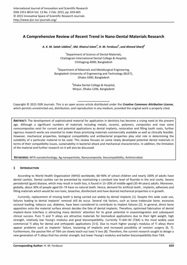
SEM micrographs of ALBO‐MPCA are shown in Fig. 1. The sizes of particles were mostly distributed between 300 and 500 nm. Particles were of polygonal shape, elongated in one direction, interconnected together in larger aggregates with sizes about 3µm. Agglomerates were of various shapes: spherical and rod like, as it is shown in Fig. 1a, b.
Fig.1 Typical appearance of structure of ALBO-MPCA: (a) prevailing calcium silicate phase, (b) prevailing calcite phase [15].
Satisfactory mechanical properties as well as setting time of ALBO‐MPCA compared to other competitive materials suggest suitable application of this material in dentistry. The compressive strengths were: for cylinders with dimensions 20×10 mm: 23.1 MPa for the samples that were aging for 1 and 3 days, 35 MPa for the aging time of 7 days, and 42.5 MPa for the aging time of 28 days. The obtained results are very promising, because beside very easy preparation and manipulation
A. K. M. Salah Uddina, Md. Khairul Islam, H. M. Ferdousi, and Ahmed Sharif
ISSN : 2351-8014 Vol. 13 No. 2, Feb. 2015 661
with ALBO‐MPCA, satisfied mechanical properties were achieved. ALBO‐MPCA was very plastic and useful for filling of teeth roots.
Experimental results from published literature revealed that the setting time of Portland cement was 235 min, with initial set about 262 min, for sample in the form of cube cylinder with dimensions 70.7×70.7 mm [16, 17].Similar result was obtained by J. Camilleri for cylinder with dimensions 6×12 mm (initial set time was 270 min, while setting time was 198 min). In addition, setting time of MTA has also pretty high value (234 min), which is comparable with setting time of Portland cements. On the other hand, in the case of ALBO‐MPCA, these values were 20 min for initial set time, while setting time was 50 min. ALBO‐MPCA evidently shows much better setting properties in comparison with Portland cement and MTA. With experimental evidence the authors also claimed that, the cytotoxicity of ALBO‐MPCA can indeed be neglected. Therefore, ALBO‐MPCA can be used in dental practice.
2.2 DENTAL RESTORATIVE NANOCOMPOSITE
Structure and biocompatibility are key parameters that determine the usefulness of dental materials for clinical use. Khan et al. [18] prepared Novel polyurethane (PU) nanocomposite material by chemically binding nanohydroxyapatite (nHA) to the diisocyanate component of the PU backbone by solvent‐polymerization. In this study, nHA was incorporated into PU by the stepwise addition of monomeric units of the PU. They studied mechanical and biological compatibility as well as the response to bacterial attack of the novel composites for dental applications.
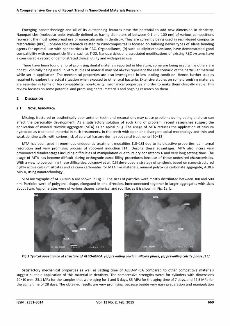
The elastic modulus and tensile strength of dentin are about 16 GPa and 36–100 MPa,[19] respectively. Theoretically, a material bonded to dentin should have a similar elastic modulus to that of dentin to avoid stress concentration along the interface. The higher elastic modulus of PU/ nHA20 composite (127.8 MPa) should contribute a greater reinforcing effect to the roots. In addition, the tensile strength (fig 2) of PU/nHA composite (33.4 MPa) is also closer to that of dentin than those of Gutta‐percha (6.0 MPa) and Resilon (8.1 MPa). As the cells were in direct contact with the samples, it was shown that no toxic substance was released from the samples that would cause cellular damage.
Fig.2 (a) Ultimate tensile strength and (b) elastic modulus of PU and PU/nHA composites, where significant difference (P≤0.05) was observed between PU and PU/nHA20 values [18].
A Comprehensive Review of Recent Trend in Nano-Dental Materials Research
ISSN : 2351-8014 Vol. 13 No. 2, Feb. 2015 662
The results of their study showed that the samples with nHA, which are hydrophobic in nature, exhibited attachment and proliferation of cells, which confirmed its level of biocompatibility. However, Clinical studies have indicated that plaque accumulated at lesser extent on ceramic material as compared to the surfaces of polymers.
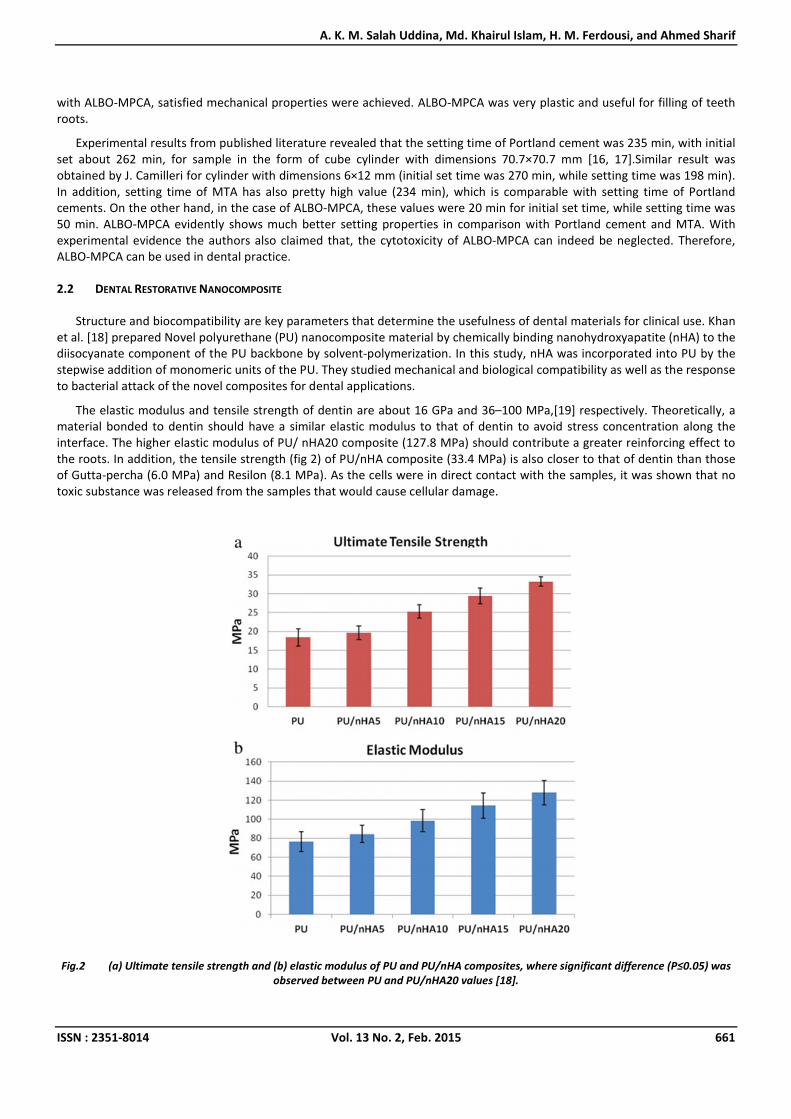
Adhesion of bacteria to dental materials is the initial event leading to colonization, potentially resulting in infection, and caries [20]. Osteoblast culture has been commonly used to evaluate the material’s surface characterization on biocompatibility and Streptococcus sanguinis is one of first bacterium to colonize tooth surfaces, by forming dental plaque. In comparison there was 97.09% reduction in bacteria adhering to the grafted composite as compared to PU (Fig.3).
Fig.3 Bacterial adhesion values for PU and PU/nHA20 [18].
However, it is worth to note that in the in vitro study the osteoblast‐like cells and Streptococcus sanguinis may not be able to present the real situation as occurred in vivo. Primary investigation shows significant improvement of the bioactive, bonding and mechanical properties. In addition, the authors [18] suggested further studies to be carried out to clarify the biocompatibility with connective tissues, fibroblasts, and other oral bacteria in order to understand the real underlying.
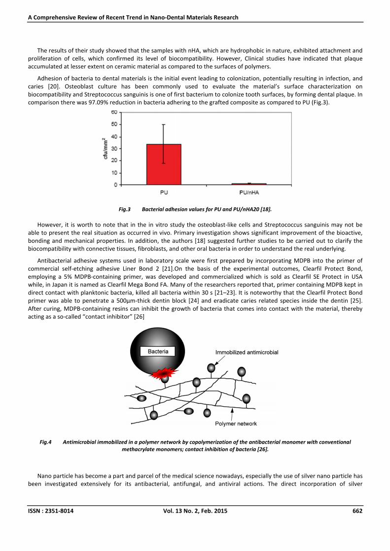
Antibacterial adhesive systems used in laboratory scale were first prepared by incorporating MDPB into the primer of commercial self‐etching adhesive Liner Bond 2 [21].On the basis of the experimental outcomes, Clearfil Protect Bond, employing a 5% MDPB‐containing primer, was developed and commercialized which is sold as Clearfil SE Protect in USA while, in Japan it is named as Clearfil Mega Bond FA. Many of the researchers reported that, primer containing MDPB kept in direct contact with planktonic bacteria, killed all bacteria within 30 s [21–23]. It is noteworthy that the Clearfil Protect Bond primer was able to penetrate a 500µm‐thick dentin block [24] and eradicate caries related species inside the dentin [25]. After curing, MDPB‐containing resins can inhibit the growth of bacteria that comes into contact with the material, thereby acting as a so‐called “contact inhibitor” [26]
Fig.4 Antimicrobial immobilized in a polymer network by copolymerization of the antibacterial monomer with conventional methacrylate monomers; contact inhibition of bacteria [26].
Nano particle has become a part and parcel of the medical science nowadays, especially the use of silver nano particle has been investigated extensively for its antibacterial, antifungal, and antiviral actions. The direct incorporation of silver
A. K. M. Salah Uddina, Md. Khairul Islam, H. M. Ferdousi, and Ahmed Sharif
ISSN : 2351-8014 Vol. 13 No. 2, Feb. 2015 663
nanoparticles into a polymer matrix is a common strategy for preparing antibacterial resinous materials [27]. However, silver nanoparticles are difficult to disperse, as nanosized particles tend to aggregate. Cheng et al. reported a new technique for preparing dental polymers with evenly dispersed silver nanoparticles using coupling photo‐initiated free radical polymerization of dimethacrylates with in situ silver ion reduction [28]. The experimental composites containing 0.08% of silver nanoparticles exhibited a 40% reduction in bacterial coverage [28]. Experimental adhesives containing both Quaternary ammonium compound (QAC) monomers and silver nanoparticles exhibited significantly enhanced antibacterial potency before and after curing compared with adhesives that used either agent alone [29‐31].
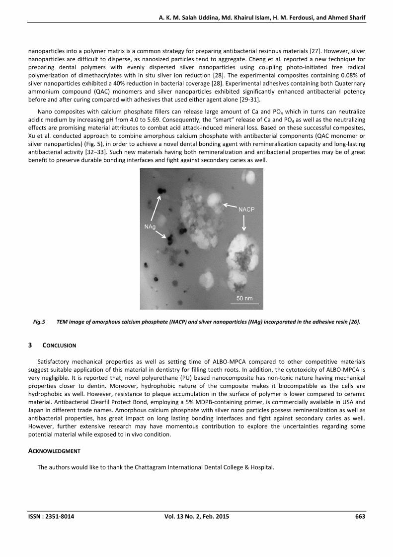
Nano composites with calcium phosphate fillers can release large amount of Ca and PO4 which in turns can neutralize acidic medium by increasing pH from 4.0 to 5.69. Consequently, the “smart” release of Ca and PO4 as well as the neutralizing effects are promising material attributes to combat acid attack‐induced mineral loss. Based on these successful composites, Xu et al. conducted approach to combine amorphous calcium phosphate with antibacterial components (QAC monomer or silver nanoparticles) (Fig. 5), in order to achieve a novel dental bonding agent with remineralization capacity and long‐lasting antibacterial activity [32–33]. Such new materials having both remineralization and antibacterial properties may be of great benefit to preserve durable bonding interfaces and fight against secondary caries as well.
Fig.5 TEM image of amorphous calcium phosphate (NACP) and silver nanoparticles (NAg) incorporated in the adhesive resin [26].
3 CONCLUSION
Satisfactory mechanical properties as well as setting time of ALBO‐MPCA compared to other competitive materials suggest suitable application of this material in dentistry for filling teeth roots. In addition, the cytotoxicity of ALBO‐MPCA is very negligible. It is reported that, novel polyurethane (PU) based nanocomposite has non‐toxic nature having mechanical properties closer to dentin. Moreover, hydrophobic nature of the composite makes it biocompatible as the cells are hydrophobic as well. However, resistance to plaque accumulation in the surface of polymer is lower compared to ceramic material. Antibacterial Clearfil Protect Bond, employing a 5% MDPB‐containing primer, is commercially available in USA and Japan in different trade names. Amorphous calcium phosphate with silver nano particles possess remineralization as well as antibacterial properties, has great impact on long lasting bonding interfaces and fight against secondary caries as well. However, further extensive research may have momentous contribution to explore the uncertainties regarding some potential material while exposed to in vivo condition.
ACKNOWLEDGMENT
The authors would like to thank the Chattagram International Dental College & Hospital.
A Comprehensive Review of Recent Trend in Nano-Dental Materials Research
ISSN : 2351-8014 Vol. 13 No. 2, Feb. 2015 664
REFERENCES
[1] Hasegawa, H., Ozawa, S., Hashimoto, K., Takeichi, T., & Ogawa, T. (2007). Type 2 diabetes impairs implant osseointegration capacity in rats. The International journal of oral & maxillofacial implants, 23(2), 237‐246.
[2] Wang, F., Song, Y. L., Li, D. H., Li, C. X., Wang, Y., Zhang, N., & Wang, B. G. (2010). Type 2 diabetes mellitus impairs bone healing of dental implants in GK rats. Diabetes research and clinical practice, 88(1), e7‐e9.
[3] Geetha, M., Singh, A. K., Asokamani, R., & Gogia, A. K. (2009). Ti based biomaterials, the ultimate choice for orthopaedic implants–a review. Progress in Materials Science, 54(3), 397‐425.
[4] Wapner, K. L. (1991). Implications of metallic corrosion in total knee arthroplasty. Clinical orthopaedics and related research, 271, 12‐20.
[5] Nakai, M., Niinomi, M., Akahori, T., Ohtsu, N., Nishimura, H., Toda, H., ... & Ogawa, M. (2008). Surface hardening of biomedical Ti–29Nb–13Ta–4.6 Zr and Ti–6Al–4V ELI by gas nitriding. Materials Science and Engineering: A, 486(1), 193‐201.
[6] Geetha, M., Singh, A. K., Asokamani, R., & Gogia, A. K. (2009). Ti based biomaterials, the ultimate choice for orthopaedic implants–a review. Progress in Materials Science, 54(3), 397‐425.
[7] Elias, C. N., Lima, J. H. C., Valiev, R., & Meyers, M. A. (2008). Biomedical applications of titanium and its alloys. Jom, 60(3), 46‐49.
[8] Aragon, P. J., & Hulbert, S. F. (1972). Corrosion of Ti‐6Al‐4V in simulated body fluids and bovine plasma. Journal of biomedical materials research, 6(3), 155‐164.
[9] Xia, Y., Zhang, F., Xie, H., & Gu, N. (2008). Nanoparticle‐reinforced resin‐based dental composites. Journal of dentistry, 36(6), 450‐455.
[10] Soares, J., Santos, S., César, C., Silva, P., Sá, M., Silveira, F., & Nunes, E. (2008). Calcium hydroxide induced apexification with apical root development: a clinical case report. International endodontic journal, 41(8), 710‐719.
[11] Hayashi, M., Shimizu, A., & Ebisu, S. (2004). MTA for obturation of mandibular central incisors with open apices: case report. Journal of Endodontics, 30(2), 120‐122.
[12] Al‐Hezaimi, K., Naghshbandi, J., Oglesby, S., Simon, J. H., & Rotstein, I. (2005). Human saliva penetration of root canals obturated with two types of mineral trioxide aggregate cements. Journal of endodontics, 31(6), 453‐456.
[13] Martin, R. L., Monticelli, F., Brackett, W. W., Loushine, R. J., Rockman, R. A., Ferrari, M., ... & Tay, F. R. (2007). Sealing properties of mineral trioxide aggregate orthograde apical plugs and root fillings in an in vitro apexification model. Journal of endodontics, 33(3), 272‐275.
[14] Bogen, G., & Kuttler, S. (2009). Mineral trioxide aggregate obturation: a review and case series. Journal of endodontics, 35(6), 777‐790.
[15] Jokanović, V., Čolović, B., Mitrić, M., Marković, D., & Ćetenović, B. (2014). Synthesis and Properties of a New Dental Material Based on Nano‐Structured Highly Active Calcium Silicates and Calcium Carbonates. International Journal of Applied Ceramic Technology, 11(1), 57‐64.
[16] Camilleri, J., Montesin, F. E., Curtis, R. V., & Ford, T. R. P. (2006). Characterization of Portland cement for use as a dental restorative material. Dental Materials, 22(6), 569‐575.
[17] Camilleri, J. (2008). The physical properties of accelerated Portland cement for endodontic use. International endodontic journal, 41(2), 151‐157.
[18] Khan, A. S., Wong, F. S. L., McKay, I. J., Whiley, R. A., & Rehman, I. U. (2013). Structural, mechanical, and biocompatibility analyses of a novel dental restorative nanocomposite. Journal of Applied Polymer Science, 127(1), 439‐447.
[19] Sano, H., Ciucchi, B., Matthews, W. G., & Pashley, D. H. (1994). Tensile properties of mineralized and demineralized human and bovine dentin. Journal of Dental Research, 73(6), 1205‐1211.
[20] Berlot‐Moirez, S., Pavon‐Djavid, G., Montdargent, B., Jozefowicz, M., & Migonney, V. (2002). Modulation of< i> Staphylococcus aureus</i> adhesion by biofunctional copolymers derived from polystyrene. ITBM‐RBM, 23(2), 102‐108.
[21] Imazato S, Kinomoto Y, Tarumi H, Torii M, Russell RRB,McCabe JF. Incorporation of antibacterial monomer MDPB into dentin primer. Journal of Dental Research 1997;76:768–72.
[22] Imazato, S., Torii, Y., Takatsuka, T., Inoue, K., Ebi, N., & Ebisu, S. (2001). Bactericidal effect of dentin primer containing antibacterial monomer methacryloyloxydodecylpyridinium bromide (MDPB) against bacteria in human carious dentin. Journal of oral rehabilitation, 28(4), 314‐319.
[23] Imazato, S., Kuramoto, A., Takahashi, Y., Ebisu, S., & Peters, M. C. (2006). In vitro antibacterial effects of the dentin primer of Clearfil Protect Bond. Dental Materials, 22(6), 527‐532.
[24] Schmalz, G., Ergücü, Z., & Hiller, K. A. (2004). Effect of dentin on the antibacterial activity of dentin bonding agents. Journal of endodontics, 30(5), 352‐358.
A. K. M. Salah Uddina, Md. Khairul Islam, H. M. Ferdousi, and Ahmed Sharif
ISSN : 2351-8014 Vol. 13 No. 2, Feb. 2015 665
[25] Imazato, S., Walls, A. W., Kuramoto, A., & Ebisu, S. (2002). Penetration of an antibacterial dentine‐bonding system into demineralized human root dentine in vitro. European journal of oral sciences, 110(2), 168‐174.
[26] Imazato, S., Ma, S., Chen, J. H., & Xu, H. H. (2014). Therapeutic polymers for dental adhesives: Loading resins with bio‐active components. Dental Materials, 30(1), 97‐104.
[27] Kassaee, M. Z., Akhavan, A., Sheikh, N., & Sodagar, A. (2008). Antibacterial effects of a new dental acrylic resin containing silver nanoparticles. Journal of applied polymer science, 110(3), 1699‐1703.
[28] Cheng, Y. J., Zeiger, D. N., Howarter, J. A., Zhang, X., Lin, N. J., Antonucci, J. M., & Lin‐Gibson, S. (2011). In situ formation of silver nanoparticles in photocrosslinking polymers. Journal of Biomedical Materials Research Part B: Applied Biomaterials, 97(1), 124‐131.
[29] Cheng, L., Zhang, K., Melo, M. A. S., Weir, M. D., Zhou, X., & Xu, H. H. K. (2012). Anti‐biofilm dentin primer with quaternary ammonium and silver nanoparticles. Journal of dental research, 91(6), 598‐604.
[30] Cheng, L., Weir, M. D., Zhang, K., Arola, D. D., Zhou, X., & Xu, H. H. (2013). Dental primer and adhesive containing a new antibacterial quaternary ammonium monomer dimethylaminododecyl methacrylate. Journal of dentistry, 41(4), 345‐355.
[31] Zhang, K., Cheng, L., Imazato, S., Antonucci, J. M., Lin, N. J., Lin‐Gibson, S., ... & Xu, H. H. (2013). Effects of dual antibacterial agents MDPB and nano‐silver in primer on microcosm biofilm, cytotoxicity and dentine bond properties. Journal of dentistry, 41(5), 464‐474.
[32] Cheng, L., Weir, M. D., Xu, H. H., Antonucci, J. M., Lin, N. J., Lin‐Gibson, S., ... & Zhou, X. (2012). Effect of amorphous calcium phosphate and silver nanocomposites on dental plaque microcosm biofilms. Journal of Biomedical Materials Research Part B: Applied Biomaterials, 100(5), 1378‐1386.
[33] Melo, M. A. S., Cheng, L., Zhang, K., Weir, M. D., Rodrigues, L. K., & Xu, H. H. (2013). Novel dental adhesives containing nanoparticles of silver and amorphous calcium phosphate. Dental Materials, 29(2), 199‐210.
Related Documents