A complex of species related to Paradiscogaster glebulae (Digenea: Faustulidae) in chaetodontid fishes (Teleostei: Perciformes) of the Great Barrier Reef Pablo E. Diaz a, ⁎, Rodney A. Bray b , Scott C. Cutmore a , Selina Ward a , Thomas H. Cribb a a The University of Queensland, School of Biological Sciences, Brisbane, Queensland 4072, Australia b Department of Life Sciences, Natural History Museum, Cromwell Road, London SW7 5BD, United Kingdom abstract article info Article history: Received 3 April 2015 Received in revised form 22 May 2015 Accepted 17 June 2015 Available online 18 June 2015 Keywords: Faustulidae Paradiscogaster Chaetodontidae Great Barrier Reef Intra-individual variation ITS2 rDNA A total of 1523 individuals of 34 species of chaetodontids from the Great Barrier Reef were examined for faustulid trematodes. Specimens resembling Paradiscogaster glebulae Bray, Cribb & Barker, 1994 were found in nine chaetodontid species at three localities. These specimens are shown, on the basis of combined morphological and molecular analyses, to comprise a complex of morphologically similar and partly cryptic species. The complex may comprise as many as six distinct species of which three are resolved here. The true P. glebulae is identified in Chaetodon ornatissimus Cuvier, 1831, Chaetodon aureofasciatus Macleay, 1878, Chaetodon plebeius Cuvier, 1831, Chaetodon rainfordi McCulloch, 1923 and Chaetodon speculum Cuvier, 1831. Two new species are described, Paradiscogaster munozae n. sp. from Heniochus varius (Cuvier, 1829), Heniochus chrysostomus Cuvier, 1831 and Chaetodon citrinellus Cuvier, 1831 and Paradiscogaster melendezi n. sp. from Chaetodon kleinii Bloch, 1790. In terms of morphology the three species differ most clearly in the development of the appendages on the ventral sucker. The three species differ at 3–6 consistent bp of ITS2 rDNA. The host-specificity of the three species differs strikingly. P. melendezi n. sp. infects just one fish species, P. glebulae infects species of only one clade of Chaetodon, and P. munozae n. sp. infects quite unrelated species. The basis of this unusual pattern of host-specificity requires further exploration. Two of the species recognised here, P. glebulae and P. munozae n. sp., showed apparent intra-individual variation in the ITS2 rDNA sequences as demonstrated by clear, replicated double peaks in the electropherograms. © 2015 Elsevier Ireland Ltd. All rights reserved. 1. Introduction The genus Paradiscogaster Yamaguti, 1934 (Faustulidae) currently comprises 24 species that infect 13 families of fishes, of which the Carangidae, Chaetodontidae, Drepaneidae, Monacanthidae, Ostraciidae and Triacanthidae each have more than one reported species [1]. Of these, the Chaetodontidae has the greatest diversity of species of Paradiscogaster, with seven species reported from Japan, Eniwetok Atoll in the Marshall Islands, Palau, Ningaloo Reef, New Caledonia and especially the Great Barrier Reef (GBR), which has six species [2–6]. Some of these species have been reported to have an extensive distribu- tion in the Tropical Indo-West Pacific (TIWP) whereas others are known from only a single locality. Recent molecular studies of trematodes of fishes have shown that cryptic species occur commonly in this class of parasites and there are now reports of what have been interpreted as cryptic species for multi- ple trematode families (e.g. [7–14]). Cryptic species are frequently associated with closely related hosts, but may also be found in allopatry, and, occasionally, sympatrically in individual host species [15,16]. The present study explores the taxonomic status of Paradiscogaster glebulae Bray, Cribb & Barker, 1994 on the GBR. This species was described from Chaetodon ornatissimus Cuvier, 1831 (type-host) and eight addition- al species of chaetodontids from off Heron Island on the southern GBR [4]. Subsequent collecting and analysis of new samples agreeing broadly with P. glebulae from GBR chaetodontids suggested the possibility of the presence of more than one species. We here begin to characterise this complex on the basis of combined analysis of morphological and molecu- lar data. 2. Materials and methods 2.1. Collection of specimens Trematodes were obtained from freshly-killed fish collected from off Lizard Island (14° 40′ S, 145° 27′ E), Swain Reefs (21° 14′ S, 151° 50′ E) and off Heron Island (23° 26′ S, 151° 54′ E). Specimens were fixed by pi- petting them into near boiling saline followed by immediate preserva- tion in 10% formalin for morphological study or 100% ethanol for Parasitology International 64 (2015) 421–428 ⁎ Corresponding author. E-mail address: [email protected] (P.E. Diaz). http://dx.doi.org/10.1016/j.parint.2015.06.004 1383-5769/© 2015 Elsevier Ireland Ltd. All rights reserved. Contents lists available at ScienceDirect Parasitology International journal homepage: www.elsevier.com/locate/parint

Welcome message from author
This document is posted to help you gain knowledge. Please leave a comment to let me know what you think about it! Share it to your friends and learn new things together.
Transcript
Parasitology International 64 (2015) 421–428
Contents lists available at ScienceDirect
Parasitology International
j ourna l homepage: www.e lsev ie r .com/ locate /par in t
A complex of species related to Paradiscogaster glebulae(Digenea: Faustulidae) in chaetodontid fishes (Teleostei: Perciformes) ofthe Great Barrier Reef
Pablo E. Diaz a,⁎, Rodney A. Bray b, Scott C. Cutmore a, Selina Ward a, Thomas H. Cribb a
a The University of Queensland, School of Biological Sciences, Brisbane, Queensland 4072, Australiab Department of Life Sciences, Natural History Museum, Cromwell Road, London SW7 5BD, United Kingdom
⁎ Corresponding author.E-mail address: [email protected]
http://dx.doi.org/10.1016/j.parint.2015.06.0041383-5769/© 2015 Elsevier Ireland Ltd. All rights reserved
a b s t r a c t
a r t i c l e i n f oArticle history:Received 3 April 2015Received in revised form 22 May 2015Accepted 17 June 2015Available online 18 June 2015
Keywords:FaustulidaeParadiscogasterChaetodontidaeGreat Barrier ReefIntra-individual variationITS2 rDNA
A total of 1523 individuals of 34 species of chaetodontids from theGreat Barrier Reefwere examined for faustulidtrematodes. Specimens resembling Paradiscogaster glebulae Bray, Cribb & Barker, 1994 were found in ninechaetodontid species at three localities. These specimens are shown, on the basis of combined morphologicaland molecular analyses, to comprise a complex of morphologically similar and partly cryptic species. Thecomplex may comprise as many as six distinct species of which three are resolved here. The true P. glebulae isidentified in Chaetodon ornatissimus Cuvier, 1831, Chaetodon aureofasciatus Macleay, 1878, Chaetodon plebeiusCuvier, 1831, Chaetodon rainfordiMcCulloch, 1923 and Chaetodon speculum Cuvier, 1831. Two new species aredescribed, Paradiscogaster munozae n. sp. from Heniochus varius (Cuvier, 1829), Heniochus chrysostomus Cuvier,1831 and Chaetodon citrinellus Cuvier, 1831 and Paradiscogaster melendezi n. sp. from Chaetodon kleinii Bloch,1790. In terms of morphology the three species differ most clearly in the development of the appendages onthe ventral sucker. The three species differ at 3–6 consistent bp of ITS2 rDNA. The host-specificity of the threespecies differs strikingly. P. melendezi n. sp. infects just one fish species, P. glebulae infects species of only oneclade of Chaetodon, and P. munozae n. sp. infects quite unrelated species. The basis of this unusual pattern ofhost-specificity requires further exploration. Two of the species recognised here, P. glebulae and P. munozae n. sp.,showed apparent intra-individual variation in the ITS2 rDNA sequences as demonstrated by clear, replicated doublepeaks in the electropherograms.
© 2015 Elsevier Ireland Ltd. All rights reserved.
1. Introduction
The genus Paradiscogaster Yamaguti, 1934 (Faustulidae) currentlycomprises 24 species that infect 13 families of fishes, of which theCarangidae, Chaetodontidae, Drepaneidae, Monacanthidae, Ostraciidaeand Triacanthidae each have more than one reported species [1]. Ofthese, the Chaetodontidae has the greatest diversity of species ofParadiscogaster, with seven species reported from Japan, EniwetokAtoll in the Marshall Islands, Palau, Ningaloo Reef, New Caledonia andespecially the Great Barrier Reef (GBR), which has six species [2–6].Some of these species have been reported to have an extensive distribu-tion in the Tropical Indo-West Pacific (TIWP)whereas others are knownfrom only a single locality.
Recent molecular studies of trematodes of fishes have shown thatcryptic species occur commonly in this class of parasites and there arenow reports of what have been interpreted as cryptic species for multi-ple trematode families (e.g. [7–14]). Cryptic species are frequently
(P.E. Diaz).
.
associatedwith closely related hosts, butmay also be found in allopatry,and, occasionally, sympatrically in individual host species [15,16].
The present study explores the taxonomic status of ParadiscogasterglebulaeBray, Cribb&Barker, 1994 on theGBR. This specieswas describedfrom Chaetodon ornatissimus Cuvier, 1831 (type-host) and eight addition-al species of chaetodontids fromoff Heron Island on the southern GBR [4].Subsequent collecting and analysis of new samples agreeing broadlywithP. glebulae from GBR chaetodontids suggested the possibility of thepresence of more than one species. We here begin to characterise thiscomplex on the basis of combined analysis of morphological andmolecu-lar data.
2. Materials and methods
2.1. Collection of specimens
Trematodeswere obtained from freshly-killed fish collected from offLizard Island (14° 40′ S, 145° 27′ E), Swain Reefs (21° 14′ S, 151° 50′ E)and off Heron Island (23° 26′ S, 151° 54′ E). Specimenswere fixed by pi-petting them into near boiling saline followed by immediate preserva-tion in 10% formalin for morphological study or 100% ethanol for
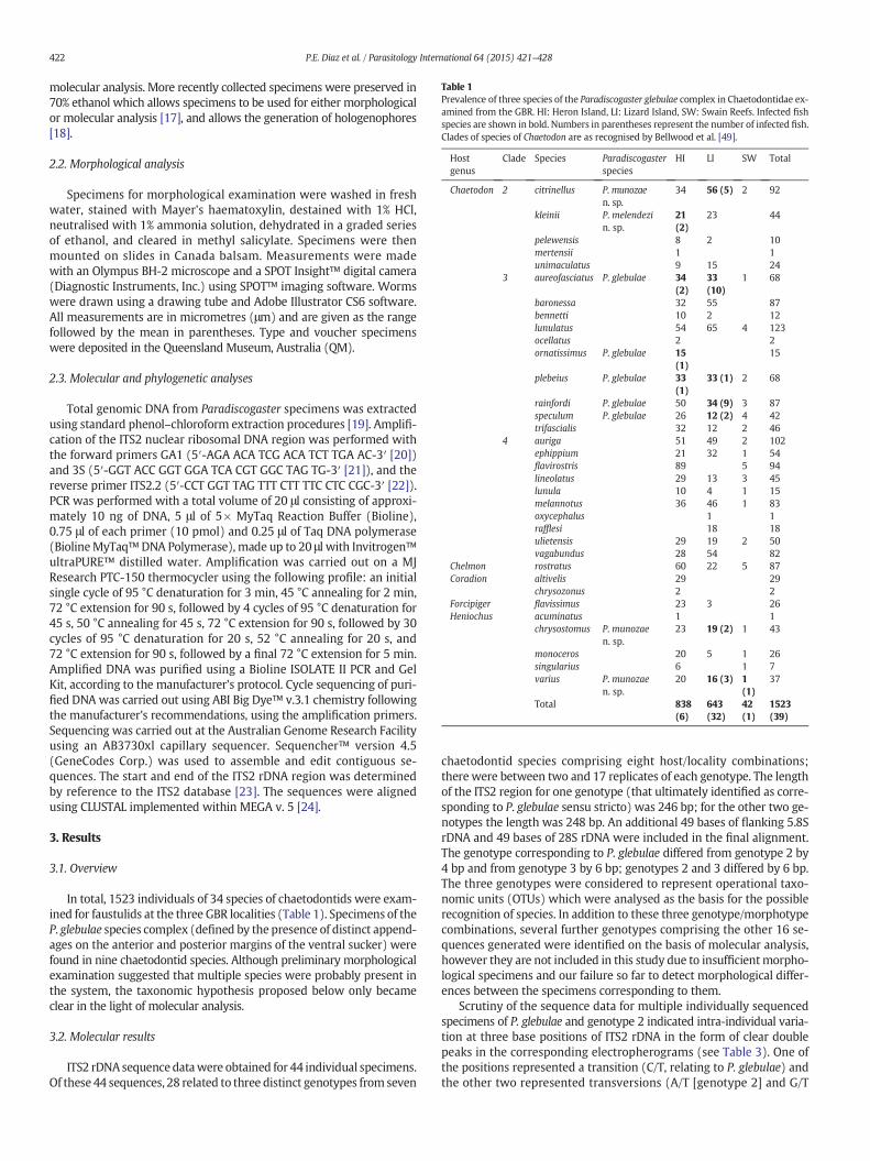
Table 1Prevalence of three species of the Paradiscogaster glebulae complex in Chaetodontidae ex-amined from the GBR. HI: Heron Island, LI: Lizard Island, SW: Swain Reefs. Infected fishspecies are shown in bold. Numbers in parentheses represent the number of infected fish.Clades of species of Chaetodon are as recognised by Bellwood et al. [49].
Hostgenus
Clade Species Paradiscogasterspecies
HI LI SW Total
Chaetodon 2 citrinellus P.munozaen. sp.
34 56 (5) 2 92
kleinii P. melendezin. sp.
21(2)
23 44
pelewensis 8 2 10mertensii 1 1unimaculatus 9 15 24
3 aureofasciatus P. glebulae 34(2)
33(10)
1 68
baronessa 32 55 87bennetti 10 2 12lunulatus 54 65 4 123ocellatus 2 2ornatissimus P. glebulae 15
(1)15
plebeius P. glebulae 33(1)
33 (1) 2 68
rainfordi P. glebulae 50 34 (9) 3 87speculum P. glebulae 26 12 (2) 4 42trifascialis 32 12 2 46
4 auriga 51 49 2 102ephippium 21 32 1 54flavirostris 89 5 94lineolatus 29 13 3 45lunula 10 4 1 15melannotus 36 46 1 83oxycephalus 1 1rafflesi 18 18ulietensis 29 19 2 50vagabundus 28 54 82
Chelmon rostratus 60 22 5 87Coradion altivelis 29 29
chrysozonus 2 2Forcipiger flavissimus 23 3 26Heniochus acuminatus 1 1
chrysostomus P. munozaen. sp.
23 19 (2) 1 43
monoceros 20 5 1 26singularius 6 1 7varius P. munozae
n. sp.20 16 (3) 1
(1)37
Total 838(6)
643(32)
42(1)
1523(39)
422 P.E. Diaz et al. / Parasitology International 64 (2015) 421–428
molecular analysis. More recently collected specimens were preserved in70% ethanol which allows specimens to be used for either morphologicalor molecular analysis [17], and allows the generation of hologenophores[18].
2.2. Morphological analysis
Specimens for morphological examination were washed in freshwater, stained with Mayer's haematoxylin, destained with 1% HCl,neutralised with 1% ammonia solution, dehydrated in a graded seriesof ethanol, and cleared in methyl salicylate. Specimens were thenmounted on slides in Canada balsam. Measurements were madewith an Olympus BH-2 microscope and a SPOT Insight™ digital camera(Diagnostic Instruments, Inc.) using SPOT™ imaging software. Wormswere drawn using a drawing tube and Adobe Illustrator CS6 software.All measurements are in micrometres (μm) and are given as the rangefollowed by the mean in parentheses. Type and voucher specimenswere deposited in the Queensland Museum, Australia (QM).
2.3. Molecular and phylogenetic analyses
Total genomic DNA from Paradiscogaster specimens was extractedusing standard phenol–chloroform extraction procedures [19]. Amplifi-cation of the ITS2 nuclear ribosomal DNA region was performed withthe forward primers GA1 (5′-AGA ACA TCG ACA TCT TGA AC-3′ [20])and 3S (5′-GGT ACC GGT GGA TCA CGT GGC TAG TG-3′ [21]), and thereverse primer ITS2.2 (5′-CCT GGT TAG TTT CTT TTC CTC CGC-3′ [22]).PCR was performed with a total volume of 20 μl consisting of approxi-mately 10 ng of DNA, 5 μl of 5× MyTaq Reaction Buffer (Bioline),0.75 μl of each primer (10 pmol) and 0.25 μl of Taq DNA polymerase(BiolineMyTaq™DNA Polymerase), made up to 20 μl with Invitrogen™ultraPURE™ distilled water. Amplification was carried out on a MJResearch PTC-150 thermocycler using the following profile: an initialsingle cycle of 95 °C denaturation for 3 min, 45 °C annealing for 2 min,72 °C extension for 90 s, followed by 4 cycles of 95 °C denaturation for45 s, 50 °C annealing for 45 s, 72 °C extension for 90 s, followed by 30cycles of 95 °C denaturation for 20 s, 52 °C annealing for 20 s, and72 °C extension for 90 s, followed by a final 72 °C extension for 5 min.Amplified DNA was purified using a Bioline ISOLATE II PCR and GelKit, according to the manufacturer's protocol. Cycle sequencing of puri-fied DNA was carried out using ABI Big Dye™ v.3.1 chemistry followingthe manufacturer's recommendations, using the amplification primers.Sequencing was carried out at the Australian Genome Research Facilityusing an AB3730xl capillary sequencer. Sequencher™ version 4.5(GeneCodes Corp.) was used to assemble and edit contiguous se-quences. The start and end of the ITS2 rDNA region was determinedby reference to the ITS2 database [23]. The sequences were alignedusing CLUSTAL implemented within MEGA v. 5 [24].
3. Results
3.1. Overview
In total, 1523 individuals of 34 species of chaetodontids were exam-ined for faustulids at the three GBR localities (Table 1). Specimens of theP. glebulae species complex (defined by the presence of distinct append-ages on the anterior and posterior margins of the ventral sucker) werefound in nine chaetodontid species. Although preliminary morphologicalexamination suggested that multiple species were probably present inthe system, the taxonomic hypothesis proposed below only becameclear in the light of molecular analysis.
3.2. Molecular results
ITS2 rDNA sequencedatawere obtained for 44 individual specimens.Of these 44 sequences, 28 related to three distinct genotypes from seven
chaetodontid species comprising eight host/locality combinations;there were between two and 17 replicates of each genotype. The lengthof the ITS2 region for one genotype (that ultimately identified as corre-sponding to P. glebulae sensu stricto) was 246 bp; for the other two ge-notypes the length was 248 bp. An additional 49 bases of flanking 5.8SrDNA and 49 bases of 28S rDNA were included in the final alignment.The genotype corresponding to P. glebulae differed from genotype 2 by4 bp and from genotype 3 by 6 bp; genotypes 2 and 3 differed by 6 bp.The three genotypes were considered to represent operational taxo-nomic units (OTUs) which were analysed as the basis for the possiblerecognition of species. In addition to these three genotype/morphotypecombinations, several further genotypes comprising the other 16 se-quences generated were identified on the basis of molecular analysis,however they are not included in this study due to insufficientmorpho-logical specimens and our failure so far to detect morphological differ-ences between the specimens corresponding to them.
Scrutiny of the sequence data for multiple individually sequencedspecimens of P. glebulae and genotype 2 indicated intra-individual varia-tion at three base positions of ITS2 rDNA in the form of clear doublepeaks in the corresponding electropherograms (see Table 3). One ofthe positions represented a transition (C/T, relating to P. glebulae) andthe other two represented transversions (A/T [genotype 2] and G/T
Table 2Species of the Paradiscogaster glebulae complex sequenced for this study. n: number ofreplicated sequences for each host species.
Species Host species n GenBank acc. #
P. glebulae C. aureofasciatus 5 KR827590, KR827591, KR827595C. plebeius 1 KR827596C. rainfordi 9 KR827592, KR827594C. speculum 2 KR827593
P. melendezi n. sp. C. kleinii 2 KR827597P. munozae n. sp. C. citrinellus 6 KR827599, KR827600
H. chrysostomus 3 KR827598
423P.E. Diaz et al. / Parasitology International 64 (2015) 421–428
[P. glebulae]). For both genotypes, where only a single peak was detectedfor the base position, it was always the same base.
3.3. Morphological recognition of species
Iterative examination of morphological specimens corresponding tothe three OTUs distinguished here by ITS2 rDNA sequence data allowedthe identification of clear morphological distinctions between them. Onthis basis, and as considered further in the discussion, each of the OTUsis considered to represent a distinct species. Nomaterial for sequencingwas available from the type-host for P. glebulae (C. ornatissimus), but, onthe basis of morphology and host distribution, one of the three geno-types is confidently identified as P. glebulae sensu stricto and the othertwo genotypes are described as new species. Below, the three taxa arecharacterised in terms of host range, geographical distribution, mor-phology and molecular data. Table 1 summarises the host distributionof the three putative species.
3.4. Morphology
Class Trematoda Rudolphi, 1808Subclass Digenea Carus, 1863Order Plagiorchiida La Rue, 1957Suborder Xiphidiata Olson, Cribb, Tkach, Bray & Littlewood, 2003Superfamily Microphalloidea Ward, 1901Family Faustulidae Poche, 1926Genus Paradiscogaster Yamaguti, 1934
3.5. P. glebulae Bray, Cribb & Barker 1994 (Figs. 1–4)
Description: (modified from Bray et al. [4]) measurements seeTable 4. Body fusiform, slightly pointed posteriorly; tegument spinose;spines reaching into anterior hindbody. Oral sucker subglobular, openingsubterminally. Ventral sucker with prominent and rounded anterior andposterior muscular appendages which each exceed 35% of total lengthof ventral sucker. Prepharynx short, distinct, always within posteriorcavity of oral sucker. Pharynx small, oval. Oesophagus long. Intestinalbifurcation in anterior half of forebody. Caeca shorter than oesophagus,saccular, often terminate anterior to appendage of ventral sucker.
Testes subglobular, diagonal to nearly opposite, entirely in hindbodyor sometimes partly dorsal to posterior part of ventral sucker. Cirrus-sacoval. Internal seminal vesicle bipartite. Par prostatica elongated, occupy-ing anterior half of cirrus-sac. Ejaculatory duct short. Cirrus small,
Table 3Distribution of intraindividual variation in ITS2 rDNA sequences for species of theParadiscogaster glebulae complex. Position is within the alignment of 248 bases of ITS2rDNA.
Position 66 81 146
P. glebulae (n = 15) T A GP. glebulae (n = 3) Y (C/T) A K (G/T)P. melendezi n. sp. (n = 2) C A GP. munozae n. sp. (n = 5) C A GP. munozae n. sp. (n = 3) C W (A/T) G
papilla-like. Genital atrium distinct. Genital pore median, just posteriorto intestinal bifurcation.
Ovary subglobular, pre-testicular, slightly sinistral, at level of ventralsucker, separated from testes by uterus. Seminal receptacle canalicular,rounded postero-dorsal to ovary. Laurer's canal opens dorsally at abouttesticular level, sometimes on slight protuberance. Uterus fills much ofhindbody, overlapping and often passing just posteriorly to testicularfield. Eggs tanned, operculate. Vitellarium present as small, symmetri-cally placed lateral fields of few closely packed follicles; fields extendbetween levels just posterior to, or just overlapping, posterior ends ofcaeca and overlap ventral sucker.
Excretory pore terminal. Excretory vesicle short, widens anteriorlyto form campanuliform or rounded chamber, reaches just to posterioredge of uterus or testis, whichever is most posterior.
Remarks: specimens reported here are consistent with P. glebulae asdescribed from the type-host, C. ornatissimus, by Bray et al. [4].
At the present two species of Paradiscogaster have been describedwith prominent projections on the anterior and posterior margin ofthe ventral sucker. These are P. glebulae and Paradiscogaster lobomyzonCribb, Anderson & Bray, 1999, the latter reported from a Kyphosidae [1].P. glebulae differs from P. lobomyzon in the position of the testes and vitel-line follicles, which are anterior to the anteriormargin of the ventral suck-er for P. lobomyzon. they are anterior to the anterior margin of the ventralsucker. In the light of the present findingswe re-examined the type-seriesof 63 specimens deposited in the QM by Bray et al. [4]. These specimens,all from Heron Island, were collected from Chaetodon ornatissimus (type-host) (2), Chaetodon aureofasciatusMacleay, 1878 (4), Chaetodon bennettiCuvier, 1831 (1), Chaetodon citrinellus Cuvier, 1831 (33), Chaetodonmertensii Cuvier, 1831 (8), Chaetodon rainfordi McCulloch, 1923 (3),Chaetodon speculum Cuvier, 1831 (1), Chaetodon unimaculatus Bloch,1787 (8) andHeniochus varius (Cuvier, 1829) (3). Although the specimenswere broadly consistent with P. glebulae sensu lato, only those from C.ornatissimus, C. aureofasciatus and C. rainfordiwere consistentwith the re-stricted concept of P. glebulae recognised here. Some of the remainingspecimens may prove difficult to identify to species (especially the singlespecimens from C. bennetti and C. speculum and the three from H. varius).The remaining specimens, including the good series of specimens from C.citrinellus, C.mertensii and C.unimaculatus, appear to relate to one ormorespecies not dealt with here. None of the specimens appear to relate tothe two new species proposed below. The restricted concept of P.glebulae is identified as occurring in C. ornatissimus, C. aureofasciatus,Chaetodon plebeius Cuvier, 1831, C. rainfordi and C. speculum.
3.6. Taxonomic summary
Type-host: Chaetodon ornatissimus Cuvier, 1831 (Chaetodontidae).Other hosts: Chaetodon aureofasciatus Macleay, 1878; C. plebeius
Cuvier, 1831; C. rainfordi McCulloch, 1923; C. speculum Cuvier, 1831(Chaetodontidae).
Type-locality: off Heron Island, Great Barrier Reef, Queensland,Australia (23° 26′ S, 151° 54′ E).
Other locality: off Lizard Island, Great Barrier Reef, Queensland,Australia (14° 40′ S, 145° 27′ E).
Site: intestine.Prevalence: see Table 1.New records: see Table 1.Voucher material: 30 voucher specimens deposited in the QM (QMG
234917-46).Molecular sequence data: ITS2 rDNA, 17 identical replicates, eight
submitted to GenBank (Table 2).
3.7. Paradiscogaster munozae n. sp. (Figs. 5–6)
Description: Measurements see Table 4. Body fusiform, distinctlypointed posteriorly; tegument spinose; spines reaching into anteriorhindbody. Oral sucker subglobular, opening subterminally. Ventral
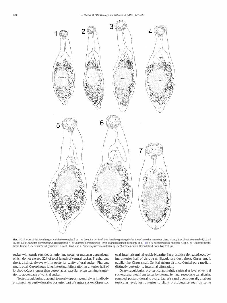
Figs. 1–7. Species of the Paradiscogaster glebulae complex from theGreat Barrier Reef. 1–4. Paradiscogaster glebulae. 1. ex Chaetodon speculum, Lizard Island; 2. ex Chaetodon rainfordi, LizardIsland; 3. ex Chaetodon aureofasciatus, Lizard Island; 4. ex Chaetodon ornatissimus, Heron Island (modified from Bray et al. [4]). 5–6. Paradiscogaster munozae n. sp. 5. ex Heniochus varius,Lizard Island; 6. ex Heniochus chrysostomus, Lizard Island. and 7. Paradiscogaster melendezi n. sp. ex Chaetodon kleinii, Heron Island. Scale-bar: 200 μm.
424 P.E. Diaz et al. / Parasitology International 64 (2015) 421–428
sucker with gently rounded anterior and posterior muscular appendageswhich do not exceed 22% of total length of ventral sucker. Prepharynxshort, distinct, always within posterior cavity of oral sucker. Pharynxsmall, oval. Oesophagus long. Intestinal bifurcation in anterior half offorebody. Caeca longer than oesophagus, saccular, often terminate ante-rior to appendage of ventral sucker.
Testes subglobular, diagonal to nearly opposite, entirely in hindbodyor sometimes partly dorsal to posterior part of ventral sucker. Cirrus-sac
oval. Internal seminal vesicle bipartite. Par prostatica elongated, occupy-ing anterior half of cirrus-sac. Ejaculatory duct short. Cirrus small,papilla-like. Cirrus small. Genital atrium distinct. Genital pore median,distinctly posterior to intestinal bifurcation.
Ovary subglobular, pre-testicular, slightly sinistral at level of ventralsucker, separated from testes by uterus. Seminal receptacle canalicular,rounded, postero-dorsal to ovary. Laurer's canal opens dorsally at abouttesticular level, just anterior to slight protuberance seen on some
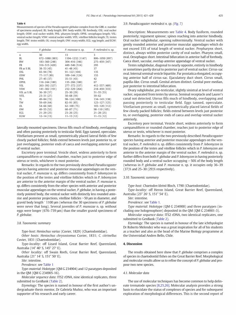
Table 4Measurements of species of the Paradiscogaster glebulae complex from theGBR. n: numberof specimens analysed; BL: body length; BW: body width; FB: forebody; OSL: oral suckerlength; OSW: oral sucker width; PHL: pharynx length; OPHL: oesophagus length; VSL:ventral sucker length; VSW: ventral suckerwidth; VITL: vitelline follicles length; TL: testeslength; TW: testes width; OL: ovary length; OW: ovarywidth; EGL: egg length; and EGW:egg width.
P. glebulae P. munozae n. sp. P. melendezi n. sp.
n 30 13 3BL 672–1088 (883) 1104–1232 (1147) 806–1050 (897)BW 183–360 (248) 304–416 (346) 275–459 (347)FB 316–515 (420) 449–546 (514) 295FB as % BL 38–53 (45) 41–48 (43) 37OSL 65–94 (78) 91–122 (104) 135OSW 77–117 (89) 109–144 (124) 155PHL 27–45 (37) 35–51 (43) 42OPHL 116–244 (180) 135–266 (180) 67VSL 263–392 (327) 277–372 (337) 412–557 (489)VSW 141–302 (191) 232–329 (264) 218–459 (333)VSL as % BL 36–39 (37) 25–30 (29) 51–55 (53)VITL 23–31 (27) 50–68 (59) 59–62TL 49–84 (72) 98–124 (108) 134–143 (138)TW 59–69 (64) 82–91 (85) 123–127 (125)OL 54–66 (60) 62–100 (75) 103–120 (112)OW 49–55 (52) 60–87 (71) 98–110 (105)EGL 21–29 (25) 23–26 (25) 21–28 (25)EGW 12–14 (13) 11–13 (12) 11–15 (12)
425P.E. Diaz et al. / Parasitology International 64 (2015) 421–428
laterally mounted specimens. Uterus fills much of hindbody, overlappingand often passing posteriorly to testicular field. Eggs tanned, operculate.Vitellarium present as small, symmetrically placed lateral fields of fewclosely packed follicles; fields extend between levels just posterior to, orjust overlapping, posterior ends of caeca and overlapping anterior partof ventral sucker.
Excretory pore terminal. Vesicle short, widens anteriorly to formcampanuliform or rounded chamber, reaches just to posterior edge ofuterus or testis, whichever is most posterior.
Remarks: In regards to the two previously described Paradiscogasterspecies having anterior and posterior muscular appendages on the ven-tral sucker, P. munozae n. sp. differs consistently from P. lobomyzon inthe position of the testes and vitelline follicles which in P. lobomyzonare anterior to the anterior margin of the ventral sucker. P. munozae n.sp. differs consistently from the other species with anterior and posteriormuscular appendages on the ventral sucker, P. glebulae, in having a poste-riorly pointed body, the ventral sucker with distinctly less rounded ante-rior and posterior projections, vitelline follicles N50 μm in diameter, andgravid body length ˃ 1100 μm (whereas the 30 specimens of P. glebulaewere never that long). Several juveniles of P. munozae n. sp. withouteggs were longer (676–739 μm) than the smaller gravid specimens ofP. glebulae.
3.8. Taxonomic summary
Type-host: Heniochus varius (Cuvier, 1829) (Chaetodontidae).Other hosts: Heniochus chrysostomus Cuvier, 1831; C. citrinellus
Cuvier, 1831 (Chaetodontidae).Type-locality: off Lizard Island, Great Barrier Reef, Queensland,
Australia (14° 40′ S, 145° 27′ E).Other locality: off Swain Reefs, Great Barrier Reef, Queensland,
Australia (21° 14′ S, 151° 50′ E).Site: intestine.Prevalence: see Table 1Type-material: Holotype (QMG234904) and 12 paratypes deposited
in the QM (QM G 234905-16).Molecular sequence data: ITS2 rDNA, nine identical replicates, three
submitted to GenBank (Table 2).Etymology: The species is named in honour of the first author's un-
dergraduate thesis mentor, Dr Gabriela Muñoz, who was an importantsupporter of his research and early career.
3.9. Paradiscogaster melendezi n. sp. (Fig. 7)
Description: Measurements see Table 4. Body fusiform, roundedposteriorly; tegument spinose; spines reaching into anterior hindbody.Oral sucker subglobular, opening subterminally. Ventral sucker withgently rounded anterior and posterior muscular appendages which donot exceed 33% of total length of ventral sucker. Prepharynx short,distinct, always within posterior cavity of oral sucker. Pharynx small,oval. Oesophagus short. Intestinal bifurcation in anterior half of forebody.Caeca short, saccular, overlap anterior appendage of ventral sucker.
Testes subglobular, diagonal to nearly opposite, entirely in hindbodyor sometimes partly dorsal to posterior part of ventral sucker. Cirrus-sacoval. Internal seminal vesicle bipartite. Par prostatica elongated, occupy-ing anterior half of cirrus-sac. Ejaculatory duct short. Cirrus small,papilla-like. Cirrus small. Genital atrium distinct. Genital pore median,just posterior to intestinal bifurcation.
Ovary subglobular, pre-testicular, slightly sinistral at level of ventralsucker, separated from testes by uterus. Seminal receptacle and Laurer'scanal not detected. Uterus fills much of hindbody, overlapping andpassing posteriorly to testicular field. Eggs tanned, operculate.Vitellarium present as small, symmetrically placed lateral fields offew closely packed follicles; fields extend between levels just posteriorto, or overlapping, posterior ends of caeca and overlap ventral suckeranteriorly.
Excretory pore terminal. Vesicle short, widens anteriorly to formcampanuliform or rounded chamber, reaches just to posterior edge ofuterus or testis, whichever is most posterior.
Remarks: In regards to the two previously described Paradiscogasterspecies having anterior and posterior muscular appendages on the ven-tral sucker, P. melendezi n. sp. differs consistently from P. lobomyzon inthe position of the testes and vitelline follicles which in P. lobomyzon areanterior to the anterior margin of the ventral sucker. P. melendezi n. sp.further differs fromboth P. glebulae and P. lobomyzon in havingposteriorlyrounded body and a ventral sucker occupying N 50% of the body length(whereas in P. glebulae and P. munozae n. sp. it occupies only 36–39(37)% and 25–30 (29)% respectively).
3.10. Taxonomic summary
Type-host: Chaetodon kleinii Bloch, 1790 (Chaetodontidae).Type-locality: off Heron Island, Great Barrier Reef, Queensland,
Australia (23° 26′ S, 151° 54′ E).Site: intestine.Prevalence: see Table 1.Type material: Holotype (QM G 234900) and three paratypes (in-
cluding one hologenophore) deposited in the QM (QM G 234901-3).Molecular sequence data: ITS2 rDNA, two identical replicates, one
submitted to GenBank (Table 2).Etymology: The species is named in honour of the late ichthyologist
Dr Roberto Melendez who was a great inspiration for all of his studentsas a teacher and also as the head of the Marine Biology programme atthe Universidad Andres Bello, Chile.
4. Discussion
The results obtained here show that P. glebulae comprises a complexof species in chaetodontid fishes on the Great Barrier Reef. Morphologicalandmolecular results allowus to refine the concept of P. glebulae andpro-pose two new species.
4.1. Molecular data
The use of molecular techniques has become common to help delin-eate trematode species [8,25,26]. Molecular analysis provides a strongbasis to elucidate the status of complexes of species and for subsequentexploration of morphological differences. This is the second report of
426 P.E. Diaz et al. / Parasitology International 64 (2015) 421–428
the use of sequences for the purposes of taxonomy in the Faustulidaefollowing the exploration of the status of Paradiscogaster flindersi Bray,Cribb & Barker, 1994 and P. oxleyi Diaz, Bray & Cribb, 2013 [5]. A recentreport of a species of Pseudobacciger, presently considered to belong inthe Faustulidae, incorporated sequence data but its analysis suggeststhat it is phylogenetically distinct from other faustulids [27]. The resultshere show variation of between four and six base pairs in this speciescomplex. It is common for many families of digeneans that small num-bers of replicated differences in the ITS2 rDNA region can distinguishspecies satisfactorily [28]. Some examples of this are a report of two toeight base differences between species of Transversotrema Witenberg,1944 [29], three between Phthinomita jonesi Nolan & Cribb, 2006 andPhthinomita hallae Nolan & Cribb, 2006 (Aporocotylidae) [15], one tofour bases between combinations of four species of Lobosorchis Miller& Cribb, 2005 [30] and two between two didymozoid species [20]. Inall of these cases, the small differences were supported bymorphologi-cal differences that allowed the conclusion that the genetic variationrepresents interspecific variation. In contrast, it is also possible to findreports of trematode species sharing the same host in which smalldifferences in the ITS2 rDNA region have been found where the mor-phology was indistinguishable and the authors interpreted the differ-ences as intra-specific variations [5,9,26]. Given the differences at thelevel of 4 to 6 bp, plus the evident differences in the morphology seenhere, it is concluded that the three genotypes represent clearly separatespecies.
A notable feature of this investigation was the detection of polymor-phic positions in the electropherograms corresponding to ITS2 rDNA se-quences representing individual specimens of P. glebulae and P. munozaen. sp. We interpret this sequence heterogeneity as evidence of intra-individual (or intra-genomic) variation (i.e. individuals possessing multi-ple ITS2 variants within their genome). Intra-individual sequence poly-morphism is relatively infrequently reported and perhaps not generallyanticipated because it is thought that rDNA typically undergoes ‘concert-ed evolution’ such that all paralogues (tandemly repeated copies) remainidentical [31–33]. Despite this, evidence of various forms of intra-individual variation in rDNA (ranging from single nucleotide differencesto variable numbers of repeated units) have been reported for beetles[34], cestodes [35], decapod crustaceans [36], foraminiferans [37], lice[38],molluscs [39,40],mosquitoes [41], nematodes [42], and turbellarians[43]. In most of these cases the variation was detected as a result of itbeing specifically sought by sequencing multiple clones derived from in-dividual animals.
Recent advances and developments in high-throughput DNA se-quencing and analyses also show that intra-individual polymorphismwithin rDNA regions is more common than previously expected.These recent ‘Next-generation’ technologies make it possible to rapidlysequence and analyse whole genomes (thus sequencing every rDNAcopy present in a given genome and providing an accurate estimate oftotal copy numbers present) allowing investigations into intra-individualvariation in rDNA regions (e.g. ITS1 and ITS2) that were previously im-practical [44,45]. For example, Weitemier et al. [45] used genome-wideanalyses to explore intra-individual variation in nuclear rDNA of 124 indi-viduals of the angiosperm genus Asclepias Linnaeus, 1753 and discoveredthat rDNA copies within individuals are not identical. Song et al. [44] alsofound that multiple ITS2 ‘variants’ existed within individuals across awide range of plant taxa.
In light of the above-mentioned studies and the polymorphismobserved here in our data, it is possible that intra-individual variation inrDNA copies is also common among trematodes. However, the only pre-vious explicit report of intra-individual variation in rDNA for trematodesof which we are aware, relates to natural hybrids of two African schisto-some species [46]. It is possible that the direct Sanger sequencing andelectropherogram editing methods, as reported here and in the vastmajority of trematode studies utilising rDNA, typically identifies the dom-inant ‘variant’ of each nucleotide position (in the form of a larger peakreading or base call in the electropherogram) so that intra-individual
variation is often not detected; perhaps smaller peaks in electrophero-grams are too readily dismissed as uninformative noise.
In terms of the interpretation of this data set, we think the intra-individual (and thus intra-specific) variationdetectedhere has no specialsignificance; probably it is of no greater significance than the low levels ofinterindividual variation that are sometimes reported. We see no evi-dence that the variation has arisen as the result of hybridisation. In eachof the three variable sites, one of the two bases recorded agrees withthat seen in the other species in the complex and the second base differs.Overall, the data are consistent with the observation of Elder & Turner[31] that “sequence similarity of repeating units [that] is greater withinthan among species”. The three species considered here remain reliablydistinct from each other on the basis of consistent differences in both se-quence sites and morphology.
4.2. Morphological taxonomy
Morphological differentiation of complexes of species may be prob-lematic [11,16]. Here, clear differenceswere observed in several body fea-tures of the three putative species. Thus, P.munozaen. sp. and P.melendezin. sp. differ from P. glebulae in their more robust bodies. These three taxawere then distinguishable on the basis of body size and the relative size ofthe ventral sucker. P. glebulae is much smaller than the other two speciesand has distinctively shaped anterior and posterior projections on theventral sucker.
4.3. Host-specificity
The species reported here show a variety of patterns of host-specificity. The depth of sampling reported makes it unlikely that fur-ther sampling will alter our understanding of the host-specificity ofthese species significantly. Only P.melendezi n. sp. was found in a singlehost species, C. kleinii Bloch, 1790. It is noticeable that C. kleinii andC. citrinellus have similar dietary components [47,48]. Given this, wecould predict that C. kleinii and C. citrinelluswould have a similar com-position of parasites species, however this is not the case; C. kleiniifrom Lizard Island did not have any Paradiscogaster species (Table 1)and C. kleinii from Heron Island had only P. melendezi n. sp. In contrast,C. citrinellus is infected by P.munozae n. sp. at Lizard Island and by threeother genotypes from the GBR that are not included in this study. Thisdistinction therefore suggests that host-specificity is driven by morethan the encounter component of the nature of the diet. The remainingtwo species both infected multiple chaetodontid hosts, but in two dis-tinct ways. The species of Chaetodon are presently recognised as fallinginto four primary clades [49,50]. P. glebulaewas found in five species ofChaetodon, all from clade 3, which are mainly specialised hard coralfeeders [49,50]. As a consequence of this, it can be predicted that themetacercariae of P. glebulae infect hard corals. However, of the ten spe-cies of clade 3 analysed in our study, only half were infected, despitesubstantial sampling of several of them. The basis of this distinctionmay relate to species-specific dietary specialisation. In contrast to theclade-specific distribution of P. glebulae, P. munozae n. sp. was foundin C. citrinellus and two species of Heniochus, H. chrysostomus Cuvier,1831 and H. varius. This pattern of specificity to relatively distantlyrelated chaetodontids is not explained by any known aspect of diet orbehaviour; all three species have broad diets not unlike those of numer-ous other chaetodontids. This unusual pattern of host-specificity has notbeen reported in other digeneans in Chaetodontidae and requires fur-ther exploration.
4.4. Biogeography
Most of the species of Chaetodontidae examined in this study are dis-tributed throughout the GBR and it might be expected that the distribu-tion of parasites would be the same as that of their hosts. P. glebulae andP. munozae n. sp. were found at two of the three localities, presenting
427P.E. Diaz et al. / Parasitology International 64 (2015) 421–428
similar distribution patterns to those of their hosts. However, P.melendezin. sp. was found only at Heron Island. The explanation may relate to thelife-cycles of the parasites, which are currently completely unknown.Potentially some of the intermediate hosts have narrow distributions.The presence of a parasite species depends on the presence of all thehosts in the life-cycle, local conditions for transmission, and the capacityof the parasites to colonise these places. The first intermediate host ofthe faustulids of chaetodontids remains entirely unknown. Otherfaustulids have been reported to infect bivalves [51,52] but a recentstudy [27] has cast doubt on the composition of the Faustulidae and it ispossible that species of Paradiscogaster and some related genera infectgastropods. Given the generally high host-specificity of trematodes fortheir first intermediate hosts [53], the differential distribution of interme-diate hosts considerably affects the distribution of Paradiscogaster species.It has also been established that larval coral reef fishes, the stage mostlikely to travel large distances in the life of most coral reef fishes, haveparasites quite different from those of adult fishes [54] implying thatthe mobile larvae are not important in the dispersal of the parasites ofthe adult fish. It thus seems likely that a differential dispersal capacity offish and parasites is important for the distribution of these parasites.
4.5. Further studies
In this study, three further genotypes that relate to forms resemblingP. glebulae are not reported, due partly to a lack of sufficient suitablemorphological specimens to allow exploration of distinctions amongthese three types. From the 44 ITS2 rDNA sequences generated, 16 notreported in this study represented three possibly distinct species ofwhich seven corresponded to Paradiscogaster molecular type 1, eightto Paradiscogastermolecular type 2 and one sequence to Paradiscogastermolecular type 3. These three genotypes require more sampling andfurther analysis to identify morphological differences between themand clarify if the recognition of further new species is necessary.
Acknowledgements
P.E. Diaz thanks Becas Chile of CONICYT Chile for granting a PhDscholarship. This work was supported by the Australian Research Counciland Australian Biological Resources Study funding to Cribb. We thankDerek Sun, Russell Yong and Matthew Nitschke, and Drs Rob Adlard,Marissa McNamara, Terry Miller and Matt Nolan for assistance with col-lection and dissection of fishes. We appreciate the support of staff of theHeron Island and Lizard Island Research Stations.
References
[1] T.H. Cribb, G.R. Anderson, R.A. Bray, Faustulid trematodes (Digenea) from marinefishes of Australia, Syst. Parasitol. 44 (1999) 119–138.
[2] S. Yamaguti, Studies on the Helminth Fauna of Japan. Part 21. Trematodes of Fishes,IV, Yamaguti, S, Kyöto, 1938.
[3] W.E. Martin, B. Hammerich, Paradiscogaster eniwetokensis n. sp. (Fellodistomidae:Trematoda) from Eniwetok, Marshall Islands, Pac. Sci. 24 (1970) 379–380.
[4] R.A. Bray, T.H. Cribb, S.C. Barker, Fellodistomidae and Lepocreadiidae (Platyhelminthes:Digenea) from chaetodontid fishes (Perciformes) from Heron Island, southern GreatBarrier Reef, Queensland, Australia, Invertebr. Taxon. 8 (1994) 545–581.
[5] P.E. Diaz, R.A. Bray, T.H. Cribb, Paradiscogaster flindersi and P. oxleyi n. sp. (Digenea:Faustulidae): overlapping host and geographical distributions in corallivorechaetodontid fishes in the tropical Indo-West Pacific, Syst. Parasitol. 86 (2013) 87–99.
[6] P.E. Diaz, T.H. Cribb, Paradiscogaster leichhardti sp. nov. (Digenea: Faustulidae) inChaetodontoplus meredithi (Perciformes: Pomacanthidae) from Heron Island, GreatBarrier Reef, Mem. Queensland Mus. 58 (2013) 49–53.
[7] I. Blasco-Costa, J.A. Balbuena, J.A. Raga, A. Kostadinova, P.D. Olson, Molecules andmorphology reveal cryptic variation among digeneans infecting sympatric mulletsin the Mediterranean, Parasitology 137 (2010) 287–302.
[8] J.A. Hunter, T.H. Cribb, A cryptic complex of species related to Transversotrema licinumManter, 1970 from fishes of the Indo-West Pacific, including descriptions of ten newspecies of TransversotremaWitenberg, 1944 (Digenea: Transversotrematidae), Zootaxa3176 (2012) 1–44.
[9] J.A. Hunter, E. Ingram, R.D. Adlard, R.A. Bray, T.H. Cribb, A cryptic complex ofTransversotrema species (Digenea: Transversotrematidae) on labroid, haemulid
and lethrinid fishes in the Indo-West Pacific Region, including the description ofthree new species, Zootaxa 2652 (2010) 17–32.
[10] T.L. Miller, R.D. Adlard, R.A. Bray, J.L. Justine, T.H. Cribb, Cryptic species of Euryakainan. g. (Digenea: Cryptogonimidae) from sympatric lutjanids in the Indo-West Pacific,Syst. Parasitol. 77 (2010) 185–204.
[11] M.K.A. McNamara, T.L. Miller, T.H. Cribb, Evidence for extensive cryptic speciation intrematodes of butterflyfishes (Chaetodontidae) of the tropical Indo-West Pacific,Int. J. Parasitol. 44 (2014) 37–48.
[12] S.S. Curran, V.V. Tkach, R.M. Overstreet, Molecular evidence for two cryptic speciesof Homalometron (Digenea: Apocreadiidae) in freshwater fishes of the southeasternUnited States, Comp. Parasitol. 80 (2013) 186–195.
[13] R. Rosas-Valdez, A. Choudhury, G.P.P. de Leon, Molecular prospecting for crypticspecies in Phyllodistomum lacustri (Platyhelminthes, Gorgoderidae), Zool. Scr. 40(2011) 296–305.
[14] M. Carreras-Aubets, A. Repulles-Albelda, A. Kostadinova, M. Carrasson, A new crypticspecies of Aponurus Looss, 1907 (Digenea: Lecithasteridae) from Mediterranean goat-fish (Teleostei: Mullidae), Syst. Parasitol. 79 (2011) 145–159.
[15] M.J. Nolan, T.H. Cribb, An exceptionally rich complex of Sanguinicolidae vonGraff, 1907(Platyhelminthes: Trematoda) from Siganidae, Labridae and Mullidae (Teleostei:Perciformes) from the Indo-West Pacific region, Zootaxa 1218 (2006) 1–80.
[16] R. Poulin, Uneven distribution of cryptic diversity among higher taxa of parasiticworms, Biol. Lett. 7 (2011) 241–244.
[17] T.H. Cribb, R.A. Bray, Gut wash, body soak, blender and heat-fixation: approaches tothe effective collection, fixation and preservation of trematodes of fishes, Syst.Parasitol. 76 (2010) 1–7.
[18] F. Pleijel, U. Jondelius, E. Norlinder, A. Nygren, B. Oxelman, C. Schander, P. Sundberg,M. Thollesson, Phylogenies without roots? A plea for the use of vouchers in molecularphylogenetic studies, Mol. Phylogenet. Evol. 48 (2008) 369–371.
[19] J. Sambrook, D. Russell, Molecular Cloning: A LaboratoryManual, 3rd ed. Cold SpringHarbor Laboratory Press, New York, 2001.
[20] G.R. Anderson, S.C. Barker, Inference of phylogeny and taxonomy within theDidymozoidae (Digenea) from the second internal transcribed spaces (ITS2) of ribo-somal DNA, Syst. Parasitol. 41 (1998) 87–94.
[21] J. Bowles,M. Hope,W.U. Tiu, X.S. Liu, D.P.McManus, Nuclear andmitochondrial geneticmarkers highly conserved betweenChinese and Philippine Schistosoma japonicum, ActaTrop. 55 (1993) 217–229.
[22] T.H. Cribb, G.R. Anderson, R.D. Adlard, R.A. Bray, A DNA-based demonstration of athree-host life-cycle for the Bivesiculidae (Platyhelminthes: Digenea), Int. J.Parasitol. 28 (1998) 1791–1795.
[23] C. Koetschan, T. Hackl, T. Müller, M. Wolf, F. Förster, J. Schultz, ITS2 database IV: in-teractive taxon sampling for internal transcribed spacer 2 based phylogenies, Mol.Phylogenet. Evol. 63 (2012) 585–588.
[24] K. Tamura, D. Peterson, N. Peterson, G. Stecher, M. Nei, S. Kumar, MEGA5: molecularevolutionary genetics analysis using maximum likelihood, evolutionary distance,and maximum parsimony methods, Mol. Biol. Evol. 28 (2011) 2731–2739.
[25] M.K.A. McNamara, R.D. Adlard, R.A. Bray, P. Sasal, T.H. Cribb, Monorchiids(Platyhelminthes: Digenea) of chaetodontid fishes (Perciformes): biogeographicalpatterns in the tropical Indo-West Pacific, Parasitol. Int. 61 (2012) 288–306.
[26] A.J. Downie, R.A. Bray, B.E. Jones, T.H. Cribb, Taxonomy, host-specificity and biogeog-raphy of Symmetrovesicula Yamaguti, 1938 (Digenea: Fellodistomidae) fromchaetodontids (Teleostei: Perciformes) in the tropical Indo-west Pacific region,Syst. Parasitol. 78 (2011) 1–18.
[27] D. Sun, R.A. Bray, R.Q. Yong, S.C. Cutmore, T.H. Cribb, Pseudobacciger cheneyae n. sp.(Digenea: Gymnophalloidea) from Weber's chromis (Chromis weberi Fowler &Bean) (Perciformes: Pomacentridae) at Lizard Island, Great Barrier Reef, Australia,Syst. Parasitol. 88 (2014) 141–152.
[28] M.J. Nolan, T.H. Cribb, The use and implications of ribosomal DNA sequencing for thediscrimination of digenean species, Adv. Parasitol. 60 (2005) 101–163.
[29] T.H. Cribb, R.D. Adlard, R.A. Bray, P. Sasal, S.C. Cutmore, Biogeography of tropicalIndo-West Pacific parasites: a cryptic species of Transversotrema and evidence forrarity of Transversotrematidae (Trematoda) in French Polynesia, Parasitol. Int. 63(2014) 285–294.
[30] T.L. Miller, A.J. Downie, T.H. Cribb, Morphological disparity despite genetic similari-ty; new species of Lobosorchis Miller & Cribb, 2005 (Digenea: Cryptogonimidae)from the Great Barrier Reef and the Maldives, Zootaxa 2009 (1992) 37–52.
[31] J.F. Elder, B.J. Turner, Concerted evolution of repetitiveDNA-sequences in eukaryotes, Q.Rev. Biol. 70 (1995) 297–320.
[32] D.M. Hillis, S.K. Davis, Evolution of ribosomal DNA — 50 million years of recordedhistory in the frog genus Rana, Evolution 40 (1986) 1275–1288.
[33] J.A.T. Morgan, D. Blair, Nuclear rDNA ITS sequence variation in the trematode genusEchinostoma: an aid to establishing relationships within the 37-collar-spine group,Parasitology 111 (1995) 609–615.
[34] A.P. Vogler, R. Desalle, Evolution and phylogenetic information content of the ITS-1region in the tiger beetle Cicindela dorsalis, Mol. Biol. Evol. 11 (1994) 393–405.
[35] I. Králová-Hromadová, J. Stefka, M. Spakulová, M. Orosová, M. Bombarová, V.Hanzelová, E. Bazsalovicsová, T. Scholz, Intra-individual internal transcribed spacer1 (ITS1) and ITS2 ribosomal sequence variation linked with multiple rDNA loci: acase of triploid Atractolytocestus huronensis, the monozoic cestode of commoncarp, Int. J. Parasitol. 40 (2010) 175–181.
[36] D.J. Harris, K.A. Crandall, Intragenomic variation within ITS1 and ITS2 of freshwatercrayfishes (Decapoda: Cambaridae): implications for phylogenetic and microsatellitestudies, Mol. Biol. Evol. 17 (2000) 284–291.
[37] L. Pillet, D. Fontaine, J. Pawlowski, Intra-genomic ribosomal RNA polymorphism andmorphological variation in Elphidium macellum suggests inter-specific hybridizationin foraminifera, PLoS One 7 (2) (2012) e32373, http://dx.doi.org/10.1371/journal.pone.0032373.
428 P.E. Diaz et al. / Parasitology International 64 (2015) 421–428
[38] N.P. Leo, S.C. Barker, Intragenomic variation in ITS2 rDNA in the louse of humans,Pediculus humanus: ITS2 is not a suitable marker for population studies in this spe-cies, Insect Mol. Biol. 11 (2002) 651–657.
[39] M.S. Hoy, R.J. Rodriguez, Intragenomic sequence variation at the ITS1–ITS2 regionand at the 18S and 28S nuclear ribosomal DNA genes of the New Zealand mudsnail, Potamopyrgus antipodarum (Hydrobiidae: Mollusca), J. Molluscan Stud. 79(2013) 205–217.
[40] R. Freire, A. Arias, J. Mendez, A. Insua, Sequence variation of the internal transcribedspacer (ITS) region of ribosomal DNA in Cerastoderma species (Bivalvia: Cardiidae),J. Molluscan Stud. 76 (2010) 77–86.
[41] J.E. Bower, R.D. Cooper, N.W. Beebe, Internal repetition and intraindividual variationin the rDNA ITS1 of the Anopheles punctulatus group (Diptera: Culicidae): multipleunits and rates of turnover, J. Mol. Evol. 68 (2009) 66–79.
[42] A. Hugall, J. Stanton, C.Moritz, Reticulate evolution and the origins of ribosomal internaltranscribed spacer diversity in apomictic Meloidogyne, Mol. Biol. Evol. 16 (1999)157–164.
[43] S. Carranza, G. Giribet, C. Ribera, J. Baguna, M. Riutort, Evidence that two types of 18SrDNA coexist in the genome of Dugesia (Schmidtea) mediterranea (Platyhelminthes,Turbellaria, Tricladida), Mol. Biol. Evol. 13 (1996) 824–832.
[44] J.Y. Song, L.C. Shi, D.Z. Li, Y.Z. Sun, Y.Y. Niu, Z.D. Chen, H.M. Luo, X.H. Pang, Z.Y. Sun, C.Liu, A.P. Lv, Y.P. Deng, Z. Larson-Rabin, M. Wilkinson, S.L. Chen, Extensive pyrose-quencing reveals frequent intra-genomic variations of internal transcribed spacerregions of nuclear ribosomal DNA, PloS One 7 (8) (2012) e43971, http://dx.doi.org/10.1371/journal.pone.0043971.
[45] K. Weitemier, S.C.K. Straub, M. Fishbein, A. Liston, Intragenomic polymorphismsamong high-copy loci: a genus-wide study of nuclear ribosomal DNA in Asclepias(Apocynaceae), PeerJ 3 (2015) e718, http://dx.doi.org/10.7717/peerj.718.
[46] H. Mone, S. Minguez, M. Ibikounle, J. Allienne, A. Massougbodji, G. Mouahid, Naturalinteractions between S. haematobium and S. guineensis in the Republic of Benin, Sci.World J. 2012 (2012) 793420.
[47] M.S. Pratchett, Dietary selection by coral-feeding butterflyfishes (Chaetodontidae)on the Great Barrier Reef, Australia, Raffles Bull. Zool. 14 (2007) 155–160.
[48] A.J. Cole, M.S. Pratchett, G.P. Jones, Diversity and functional importance of coral-feeding fishes on tropical coral reefs, Fish Fish. 9 (2008) 286–307.
[49] D.R. Bellwood, S. Klanten, P.F. Cowman, M.S. Pratchett, N. Konow, L. van Herwerden,Evolutionary history of the butterflyfishes (f: Chaetodontidae) and the rise of coralfeeding fishes, J. Evol. Biol. 23 (2010) 335–349.
[50] J.L. Fessler, M.W. Westneat, Molecular phylogenetics of the butterflyfishes(Chaetodontidae): taxonomy and biogeography of a global coral reef fish family,Mol. Phylogenet. Evol. 45 (2007) 50–68.
[51] L.G. Ben Abdallah, N.T. El Menif, E. Maamouri, The morphology and behaviour ofCercaria lata Lespes, 1857 (Digenea, Faustulidae) from the Mediterranean clamTapes decussata (L.), J. Helminthol. 83 (2009) 69–76.
[52] K.A. Hall, T.H. Cribb, S.C. Barker, V4 region of small subunit rDNA indicates polyphylyof the Fellodistomidae (Digenea) which is supported by morphology and life-cycledata, Syst. Parasitol. 43 (1999) 81–92.
[53] T.H. Cribb, R.A. Bray, D.T.J. Littlewood, The nature and evolution of the associationbetween digeneans, molluscs and fishes, Int. J. Parasitol. 31 (2001) 997–1011.
[54] T.H. Cribb, S. Pichelin, V. Dufour, R.A. Bray, C. Chauvet, E. Faliex, R. Galzin, C.M. Lo, A.Lo-Yat, S. Morand, M.C. Rigby, P. Sasal, Parasites of recruiting coral reef fish larvae inNew Caledonia, Int. J. Parasitol. 30 (2000) 1445–1451.
Related Documents


