Research paper A comparative study of chitosan and chitosan/cyclodextrin nanoparticles as potential carriers for the oral delivery of small peptides q Adriana Trapani a , Angela Lopedota a , Massimo Franco a , Nicola Cioffi b , Eliana Ieva b , Marcos Garcia-Fuentes c , Maria José Alonso c, * a Dept. of Pharmaceutical Chemistry, Bari University, Aldo Moro, Bari, Italy b Dept. of Chemistry, Bari University, Bari, Italy c Dept. of Pharmaceutical Technology, Santiago de Compostela University, Santiago de Compostela, Spain article info Article history: Received 26 June 2009 15 December 2009 Accepted in revised form 20 January 2010 Available online 25 January 2010 Keywords: Glutathione Chitosan Cyclodextrins Nanoparticles Oral administration abstract The aim of this study was to characterize new nanoparticles (NPs) containing chitosan (CS), or CS/cyclo- dextrin (CDs), and evaluate their potential for the oral delivery of the peptide glutathione (GSH). More precisely, NP formulations composed of CS, CS/a-CD and CS/sulphobutyl ether-b-cyclodextrin (SBE 7m - b-CD) were investigated for this application. CS/CD NPs showed particle sizes ranging from 200 to 500 nm. GSH was loaded more efficiently in CS/SBE 7m -b-CD NPs by forming a complex between the tri- peptide and the CD. X-ray Photoelectron Spectroscopy (XPS) analysis suggested that GSH is located in the core of CS/SBE 7m -b-CD NPs and that it is almost absent from the NP surface. Release studies performed in vitro at pH 1.2 and pH 6.8 showed that NP release properties can be modulated by selecting an appro- priate CD. Transport studies performed in the frog intestine model confirmed that both CS and CS/CD nanoparticles could induce permeabilization of the intestinal epithelia. However, CS/SBE 7m -b-CD NPs provided absorption-enhancing properties in all segments of the duodenum, whereas CS NPs effect was restricted to the first segment of the duodenum. From the data obtained, we believe that CS/CD nano- particles might represent an interesting technological platform for the oral administration of small peptides. Ó 2010 Elsevier B.V. All rights reserved. 1. Introduction Glutathione (c-glutamylcysteinylglycine, GSH) is the major thi- olated small peptide in mammalian cells. Its reducing and nucleo- philic properties render GSH a fundamental redox buffer that prevents the oxidative damage caused by free radicals from oxygen species circulating in the body. GSH is also speculated to influence gene expression in two ways: (i) via its reactive oxidant species (ROS) scavenging role and (ii) via protein glutathionylation [1]. The clinical value of this small peptide includes its use for the treatment of alcohol and drug poisoning, as well as for protection against cytotoxic chemotherapy and radiation trauma [2]. Cur- rently, GSH is only available on the market as parenteral dosage forms (Gluthion Ò ) because of its low and variable oral bioavailabil- ity [3]. This limited absorption has been mainly attributed to the chemical and enzymatic degradation of the peptide in the jejunum. More specifically, it is known that the thiol group of the cysteine moiety in GSH is susceptible to enzymatic (c-glutamyltranspepti- dase) and non-enzymatic pH-dependent oxidation [4], leading to the formation of inactive products. However, the oral route contin- ues to be the most desirable pathway for drug administration, par- ticularly for therapeutic agents in which several doses are necessary. To achieve oral delivery of GSH, drug carrier systems are re- quired to protect this drug from the gastrointestinal environment and from enzymatic degradation. Several nanoparticle (NP) proto- types from biodegradable polymers have been proposed for trans- mucosal drug delivery [5–7]. Among them, NPs based on the polysaccharide chitosan (CS) have shown particularly promising results due to their intrinsic properties including biocompatibility, mucoadhesion and ability to transiently open the tight junctions of the intestinal barrier [8–10]. CS NPs have already shown capacity to deliver orally hydrophilic macromolecules as confirmed both in cellular models and in animal experiments [11]. Recently, we re- ported the formation of a new NP carrier based on the combination of CS and CDs [12,13]. Originally, the inclusion of CDs in the NP structures was designed as a modification intended to improve 0939-6411/$ - see front matter Ó 2010 Elsevier B.V. All rights reserved. doi:10.1016/j.ejpb.2010.01.010 q This work is dedicated to the memory of Prof. Gaetano Liso, recently deceased, for his example of man and scientist. * Corresponding author. Department of Pharmacy and Pharmaceutical Technol- ogy, Faculty of Pharmacy, University of Santiago de Compostela, 15782 Santiago de Compostela, Spain. Tel.: +34 981 594 488; fax: +34 981 547 148. E-mail address: [email protected] (M.J. Alonso). European Journal of Pharmaceutics and Biopharmaceutics 75 (2010) 26–32 Contents lists available at ScienceDirect European Journal of Pharmaceutics and Biopharmaceutics journal homepage: www.elsevier.com/locate/ejpb 434

Welcome message from author
This document is posted to help you gain knowledge. Please leave a comment to let me know what you think about it! Share it to your friends and learn new things together.
Transcript
Research paper
A comparative study of chitosan and chitosan/cyclodextrin nanoparticlesas potential carriers for the oral delivery of small peptides q
Adriana Trapani a, Angela Lopedota a, Massimo Franco a, Nicola Cioffi b, Eliana Ieva b,Marcos Garcia-Fuentes c, Maria José Alonso c,*
a Dept. of Pharmaceutical Chemistry, Bari University, Aldo Moro, Bari, Italyb Dept. of Chemistry, Bari University, Bari, Italyc Dept. of Pharmaceutical Technology, Santiago de Compostela University, Santiago de Compostela, Spain
a r t i c l e i n f o
Article history:Received 26 June 200915 December 2009
Accepted in revised form 20 January 2010Available online 25 January 2010
Keywords:GlutathioneChitosanCyclodextrinsNanoparticlesOral administration
a b s t r a c t
The aim of this study was to characterize new nanoparticles (NPs) containing chitosan (CS), or CS/cyclo-dextrin (CDs), and evaluate their potential for the oral delivery of the peptide glutathione (GSH). Moreprecisely, NP formulations composed of CS, CS/a-CD and CS/sulphobutyl ether-b-cyclodextrin (SBE7m-b-CD) were investigated for this application. CS/CD NPs showed particle sizes ranging from 200 to500 nm. GSH was loaded more efficiently in CS/SBE7m-b-CD NPs by forming a complex between the tri-peptide and the CD. X-ray Photoelectron Spectroscopy (XPS) analysis suggested that GSH is located in thecore of CS/SBE7m-b-CD NPs and that it is almost absent from the NP surface. Release studies performedin vitro at pH 1.2 and pH 6.8 showed that NP release properties can be modulated by selecting an appro-priate CD. Transport studies performed in the frog intestine model confirmed that both CS and CS/CDnanoparticles could induce permeabilization of the intestinal epithelia. However, CS/SBE7m-b-CD NPsprovided absorption-enhancing properties in all segments of the duodenum, whereas CS NPs effectwas restricted to the first segment of the duodenum. From the data obtained, we believe that CS/CD nano-particles might represent an interesting technological platform for the oral administration of smallpeptides.
� 2010 Elsevier B.V. All rights reserved.
1. Introduction
Glutathione (c-glutamylcysteinylglycine, GSH) is the major thi-olated small peptide in mammalian cells. Its reducing and nucleo-philic properties render GSH a fundamental redox buffer thatprevents the oxidative damage caused by free radicals from oxygenspecies circulating in the body. GSH is also speculated to influencegene expression in two ways: (i) via its reactive oxidant species(ROS) scavenging role and (ii) via protein glutathionylation [1].The clinical value of this small peptide includes its use for thetreatment of alcohol and drug poisoning, as well as for protectionagainst cytotoxic chemotherapy and radiation trauma [2]. Cur-rently, GSH is only available on the market as parenteral dosageforms (Gluthion�) because of its low and variable oral bioavailabil-ity [3]. This limited absorption has been mainly attributed to the
chemical and enzymatic degradation of the peptide in the jejunum.More specifically, it is known that the thiol group of the cysteinemoiety in GSH is susceptible to enzymatic (c-glutamyltranspepti-dase) and non-enzymatic pH-dependent oxidation [4], leading tothe formation of inactive products. However, the oral route contin-ues to be the most desirable pathway for drug administration, par-ticularly for therapeutic agents in which several doses arenecessary.
To achieve oral delivery of GSH, drug carrier systems are re-quired to protect this drug from the gastrointestinal environmentand from enzymatic degradation. Several nanoparticle (NP) proto-types from biodegradable polymers have been proposed for trans-mucosal drug delivery [5–7]. Among them, NPs based on thepolysaccharide chitosan (CS) have shown particularly promisingresults due to their intrinsic properties including biocompatibility,mucoadhesion and ability to transiently open the tight junctions ofthe intestinal barrier [8–10]. CS NPs have already shown capacityto deliver orally hydrophilic macromolecules as confirmed bothin cellular models and in animal experiments [11]. Recently, we re-ported the formation of a new NP carrier based on the combinationof CS and CDs [12,13]. Originally, the inclusion of CDs in the NPstructures was designed as a modification intended to improve
0939-6411/$ - see front matter � 2010 Elsevier B.V. All rights reserved.doi:10.1016/j.ejpb.2010.01.010
q This work is dedicated to the memory of Prof. Gaetano Liso, recently deceased,for his example of man and scientist.
* Corresponding author. Department of Pharmacy and Pharmaceutical Technol-ogy, Faculty of Pharmacy, University of Santiago de Compostela, 15782 Santiago deCompostela, Spain. Tel.: +34 981 594 488; fax: +34 981 547 148.
E-mail address: [email protected] (M.J. Alonso).
European Journal of Pharmaceutics and Biopharmaceutics 75 (2010) 26–32
Contents lists available at ScienceDirect
European Journal of Pharmaceutics and Biopharmaceutics
journal homepage: www.elsevier .com/locate /e jpb
434
the capacity of these carriers to load poorly soluble drug [14].However, it has became evident that these NPs can also loadhydrophilic molecules with high efficiency [13] and enhance theirtransport across mucosal surfaces [15,16]. In the case of GSH, thispossibility is even more attractive, as previous studies performedby us have confirmed the possible inclusion of this molecule insidethe cavity of CDs and that this interaction prevents the degradationof this molecule by endopeptidases [17].
In summary, taking into account the increasing clinical value ofGSH, we have investigated the potential of CS and CS/CD NPs as po-tential carrier systems for the oral delivery of this small peptide.The resulting prototypes were extensively characterized with re-gard to their physicochemical properties, their capacity to loadand release GSH and the capacity to protect and promote the trans-port of GSH across the intestinal frog sac model.
2. Materials and methods
2.1. Materials
The following chemicals were obtained from commercialsources and used as received. Chitosan hydrochloride (UP CL 113,Mw = 110 kDa, deacetylation degree = 86% according to manufac-turer instructions) was purchased from Pronova Biopolymer (Nor-way). Alpha-cyclodextrin (a-CD, Mw = 972 Da) was kindly given byWacker-Chemie (Peschiera Borromeo, Italy). Glutathione (GSH),glycerol (over 99.5% purity) and Pentasodium tripolyphospate(TPP) were purchased from Sigma–Aldrich (Milan, Italy). Sulp-hobutyl ether-b-cyclodextrin sodium salt (SBE7m-b-CD,Mw = 2163 Da, average substitution degree = 6.40) was bought byCyDex, Inc. (USA). Both CDs were kept in a desiccator until to beused. Ultrapure water (Carlo Erba, Italy) was used throughout thestudy. All other chemicals were reagent grade or better.
Frog Ringer (FR) solution (pH = 6.0) was prepared as follows:NaH2PO4 H2O 5.0 mmol, K2HPO4 1.0 mmol, CaCl2 2H2O 1.0 mmoland the amount of NaCl required to reach 230 ± 10 mOsm/kg (Mi-cro-Osmometer Automatic Type 13 RS, Hermann Roebling Mess-technik, Berlin, Germany).
2.2. Nanoparticle preparation
CS or CS/CD-based NPs were prepared according to a modified io-nic gelation method [12,18]. (a) CS Nanoparticles: 1.5 ml of a GSHaqueous solution (0.10%w/v) was mixed with 1.5 ml of a CS solution(0.40%w/v). NPs were spontaneously formed upon addition of 1 mlof TPP aqueous solution (0.15%w/v) to 3 ml of the CS/GSH solutionunder magnetic stirring. (b) CS/CD NPs were prepared differentlydepending on the type of CD used. However, for both a-CD andSBE7m-b-CD, 1:1 M ratio stoichiometry CD:GSH complexes werepreviously prepared by co-incubation of the aqueous solutions ofboth compounds for 24 h at room temperature and under moderatestirring. For a-CD:GSH, an aqueous solution was prepared by adding23.15 mg of a-CD and 7.0 mg of GSH to 7 ml of distilled water. ForSBE7m-b-CD:GSH, an aqueous solution was prepared by adding31.5 mg of SBE7m-b-CD and 4.5 mg of GSH to 7 ml of distilled water.To prepare CS/a-CD NPs, 1.5 ml of the solution containing a-CD:GSHcomplexes were mixed with 1.5 ml of a CS solution (0.40%w/v). Theresulting solution was mixed with 1 ml of TPP aqueous solution(0.15%w/v) under magnetic stirring, leading to spontaneous NP pre-cipitation. To prepare CS/SBE7m-b-CD NPs, a 3 ml CS solution(0.20%w/v) was mixed with 1 ml of the SBE7m-b-CD:GSH solution,a process already reported to lead to NP formation [19]. For all for-mulations, the resulting NPs were isolated by centrifugation(16,000g, 45 min, Eppendorf 5415D, Eppendorf, Germany) andresuspended in ultrapure water by manual shaking.
2.3. Physicochemical and morphological characterization ofnanoparticles
Particle size and polydispersity index (PI) were determined indouble distilled water by photon correlation spectroscopy (PCS)using a Zetasizer NanoZS (ZEN 3600, Malvern, UK). The determina-tion of the f-potential was performed using laser Doppler ane-mometry (Zetasizer NanoZS, ZEN 3600, Malvern, UK) afterdilution with KCl 1 mM.
The morphological examination of the NP prototypes was per-formed by transmission electron microscopy (TEM) (CM12 Philips,Eindhoven, Netherlands). For sample preparation, NPs were resus-pended in water, stained with 2% (w/v) phosphotungstic acid,placed on copper grids with Formvar� films and dried overnightat room temperature.
2.4. HPLC analysis
High-performance liquid chromatography (HPLC) analyseswere performed with a Waters (Waters Corp., Milford, MA) Model600 pump equipped with a Waters 2996 photodiode array detector(set at the wavelength of 220 nm), a 20 ll loop injection autosam-pler (Waters 717 plus) and processed by Empower™ SoftwareBuild. For analysis, a reversed phase Synergy Hydro-RP(25 cm � 4.6 mm; 4 lm particles; Phenomenex, Torrance, CA) col-umn in conjunction with a precolumn C18 insert was eluted with1:99 (v:v) acetonitrile:0.025 M phosphate buffer (pH 2.7) in iso-cratic mode. The flow rate was maintained at 0.7 ml/min. Standardcalibration curves were prepared at 220 nm wavelength using0.025 M phosphate buffer (pH 2.7) as solvent. Calibration curve lin-earity (r2 > 0.999) was maintained over the range of concentrationstested (5.46 � 10�6 M–3.32 � 10�3 M). The retention times of GSHand its disulphide degradation product (GSSG) were 7.2 and18 min, respectively. Under these conditions, the quantificationlimit was determined to be 2 lg/ml for both GSH and GSSG.
2.4.1. Determination of GSH encapsulation efficiencyThe Encapsulation Efficiency (EE) of the tripeptide to the parti-
cles was calculated by an indirect method. CS or CS/CD NPs wereisolated from free GSH by centrifugation (16,000g, 45 min, Eppen-dorf 5415D, Eppendorf, Germany), and free GSH in the supernatantwas quantified by HPLC as described above. Experiments were per-formed in triplicate and the encapsulation efficiency was calcu-lated as follows:
%EE ¼ 100� ðtotal GSH� free GSHÞ=total GSH
2.4.2. Determination of SBE7m-b-CD content in NPsSBE7m-b-CD was quantified in the supernatants of unloaded and
GSH-loaded CS/SBE7m-b-CD NPs by spectrophotometric analysis ofthe fading of phenolphthalein alkaline solutions [20,21]. Briefly, a3 mM phenolphthalein stock solution in methanol was diluted1:100 in 0.05 M carbonate buffer (pH 10.5). Freeze-dried superna-tants of NPs were dissolved in 1000 ll of acetic acid (0.1%, v/v).400 ll of this mixture were added to 2.6 ml of diluted phenol-phthalein solution prepared as described above. The absorbanceat 553 nm of resulting solution was measured by Perkin ElmerLamba Bio 20 spectrophotometer. For quantification, the SBE7m-b-CD content was calculated by comparing the results to that ofa standard curve obtained by using standard CD solutions. Linear-ity was checked in the range from 0.10 mg/ml to 2.20 mg/ml.
2.5. XPS analysis
X-ray Photoelectron Spectroscopy (XPS) analysis was per-formed to explore the differences in chemical composition at
A. Trapani et al. / European Journal of Pharmaceutics and Biopharmaceutics 75 (2010) 26–32 27
435
various depths from the NP surface. CS/SBE7m-b-CD NPs loadedwith GSH were analyzed with a Thermo VG Theta Probe spectrom-eter equipped with a micro-spot monochromatized Al Ka source.Conventional XPS analyses provided information on the elementalcomposition and chemical speciation of the NP outer surface (5 nmthick), while ion-etching assisted Depth Profile XPS was used toquantify the in-depth distribution of GSH in the NPs. In particular,thiol and sulphonate chemical environments (resolved at the base-line in the S2p high-resolution spectra) were chosen as unambigu-ous labels for GSH and SBE7m-b-CDs, respectively.
2.6. In vitro release study
In vitro release of GSH from loaded CS and CS/CD NPs was car-ried out for 3 h in simulated gastric (pH 1.2) and intestinal (pH 6.8)medium without enzymes and performed according to USP XXVIrecommendations. Freshly prepared CS and CS/CD NPs were iso-lated in Eppendorf tubes with a 10 ll glycerol bed laid in the bot-tom of the tube, set to help on NP resuspension. In screw-cappedtest tubes, each formulation was resuspended in 0.4 ml of distilledwater and the resulting resuspended formulation was mixed with1 ml of release medium (simulated gastric or intestinal medium)and incubated at 37 �C under mechanical agitation (150 oscilla-tions/min). At appropriate time intervals, an aliquot (0.4 ml) waswithdrawn and centrifuged (16,000g, 40 min, Eppendorf 5415D,Eppendorf, Germany). The initial volume of release medium wasmaintained by refilling 0.4 ml of the same medium after each with-drawal. The supernatant was analyzed for GSH content by HPLC asdescribed above. GSH in solution was dissolved in the same mediaand analyzed at the same time points, as a control for GSH degra-dation. Each experiment was performed in triplicate.
2.7. Transport studies in the frog intestinal sac model
Animal care and handling throughout the experimental proce-dure was carried out in accordance with the European CommunityCouncil Directive of 24 November 1986 (86/609/EEC). Healthyfrogs of the species Rana esculenta (Animali da Laboratorio R. Ore-fice, Arzano, Italy) were housed in a cold room, in an aquarium, andwithout food. The aquarium was thoroughly washed once a week.The frogs were killed by decapitation and pithing of the spinal cord,and small intestines were quickly removed by laparotomy. The ex-cised piece of intestine was immediately washed with Frog Ringer(FR) solution, at room temperature, to remove the intestinalcontent.
Two intestinal segments of approximately 2.50 cm in lengthwere tied at one end with a silk suture and then used to assessthe permeability of this model epithelium to GSH, both in presenceor in the absence of two nanocarriers: blank CS and blank CS/SBE7m-b-CD NPs. Permeability studies for all GSH formulationswere performed both in the proximal and in the distal segmentof duodenum. The integrity of these intestinal membrane sacswas checked throughout the study by using phloridzin as internalstandard at the concentration of 160 � 10�6 M. The sacs were filledwith (i) free GSH in FR solution, (ii) free GSH added to resuspendedblank CS NPs, or (iii) free GSH added to blank resuspended CS/SBE7m-b-CD NPs. The volume of the donor compartment was inthe range 0.1–0.3 ml. The sacs were sealed and immersed in vials(2.70 cm width � 5.73 cm high) containing 1.0–2.0 ml of FR solu-tion. These vials were incubated in a water shaking bath (Iscomod. SBH Milan, Italy), at 26 ± 0.1 �C (100 rpm) for 5 h. At fixedtime points, the amount of GSH in the acceptor chamber was quan-tified by withdrawing a 0.2 ml sample from the vial and measuringGSH concentration by HPLC. The initial volume of the externalmedium was maintained by refilling the volume withdrawn withfresh FR.
The apparent permeability coefficient Papp (cm/s) was calcu-lated using the following equation:
Papp ¼ dQ=dtð1=A� 60� c0Þ
where dQ/dt is the permeability rate (mg/s), namely the amount ofGSH permeating the tissue in time t (min); A is the area of exposedfrog intestine tissue (around 2.5 cm2); c0 is the initial GSH concen-tration (mg/ml) inside the intestinal sac.
2.8. Statistics
Data from different experimental groups were compared by aone-way ANOVA with p < 0.05 (GraphPad Prism v.4.00, GraphPadSoftware, Inc., San Diego, CA). Tukey tests were used for post hoccontrast.
3. Results and discussion
In this work, we aimed at the design and characterization ofnew drug nanocarriers for the oral delivery of low molecularweight peptides. More specifically, in this work, we studied chito-san nanoparticles and chitosan/cyclodextrin NPs as potential carri-ers for oral delivery of GSH. The rationale behind the selection ofthese nanocarriers can be summarized as follows: (i) CS NPs pres-ent very good record as oral delivery systems of poorly permeabledrugs [9,22]. Recently, CS/CD NPs have also shown promising re-sults as delivery systems of hydrophilic macromolecules [15,16].(ii) GSH has already shown the capability to interact with a-CD[17], a characteristic that might increase the affinity of this drugfor the NP matrix. (iii) The formation of GSH:a-CD complexes sta-bilizes this molecule towards its degradation by endopeptidases[17].
In the following lines, we describe the preparation of CS and CS/CD NPs loaded with GSH, their physicochemical properties are pre-sented, and the capability to release GSH in simulated physiologi-cal fluids and promote GSH transport across model epithelia arealso reported.
3.1. Formation and characterization of CS and CS/CD NPs loaded withGSH
To exclude the possible formation of covalent linkages betweenCS and GSH, or the induction of degradative processes arising fromco-incubating these molecules, the chemical stability of GSH in anaqueous CS solution was checked by HPLC up to 24 h at room tem-perature. This study allowed us to exclude any chemical modifica-tion of GSH during this incubation process (data not shown).
CS and CS/CD NPs were prepared as described in Section 2.3. Inthe specific case of the CS/CD NPs system, two CDs with very differ-ent properties were selected: a-CD and SBE7m-b-CD. Overall, themethods for CS NP preparation were similar to those describedby Calvo et al. [18] and CS/CD NPs were formed by a method sim-ilar to that previously reported [12]. However, in this work,GSH:CD complexes were formed prior to NP preparation throughthe co-incubation of both molecules for 24 h.
Table 1 displays the physicochemical properties of the differentNPs prepared. It is well known that the size of CS NPs depends onthe concentration of CS, the concentration of TPP and the CS/TPPratio [23]. However, in this case, other factors affecting NP sizewere detected, namely, the nature and the amount of the CD incor-porated (Table 1). Indeed, NPs incorporating the neutral a-CD werebigger than the other formulations: 512 nm average particle sizeand moderately wide size distribution (PI = 0.40–0.60). On theother hand, the incorporation of the negatively charged CD,SBE7m-b-CD, resulted in NPs that were markedly smaller than the
28 A. Trapani et al. / European Journal of Pharmaceutics and Biopharmaceutics 75 (2010) 26–32
436
other formulations: 210 nm average particle size, very narrow par-ticle distribution (PI = 0.06–0.15). Overall, these results confirm thecapacity of neutral and anionic CDs to influence the process of CSassembly during NP formation, as it has been discussed elsewhere[12]. In all cases, positive zeta potential values were detected, sug-gesting that CS is mainly located on the surface of the particles.However, it is interesting to note that CS/a-CD NPs presentedslightly higher zeta potential values than the other formulations.The addition of GSH did not produce significant changes to thephysicochemical properties of the NPs, except for the PI of the CSand CS/SBE7m-b-CD NP formulation that increased moderately(Table 1).
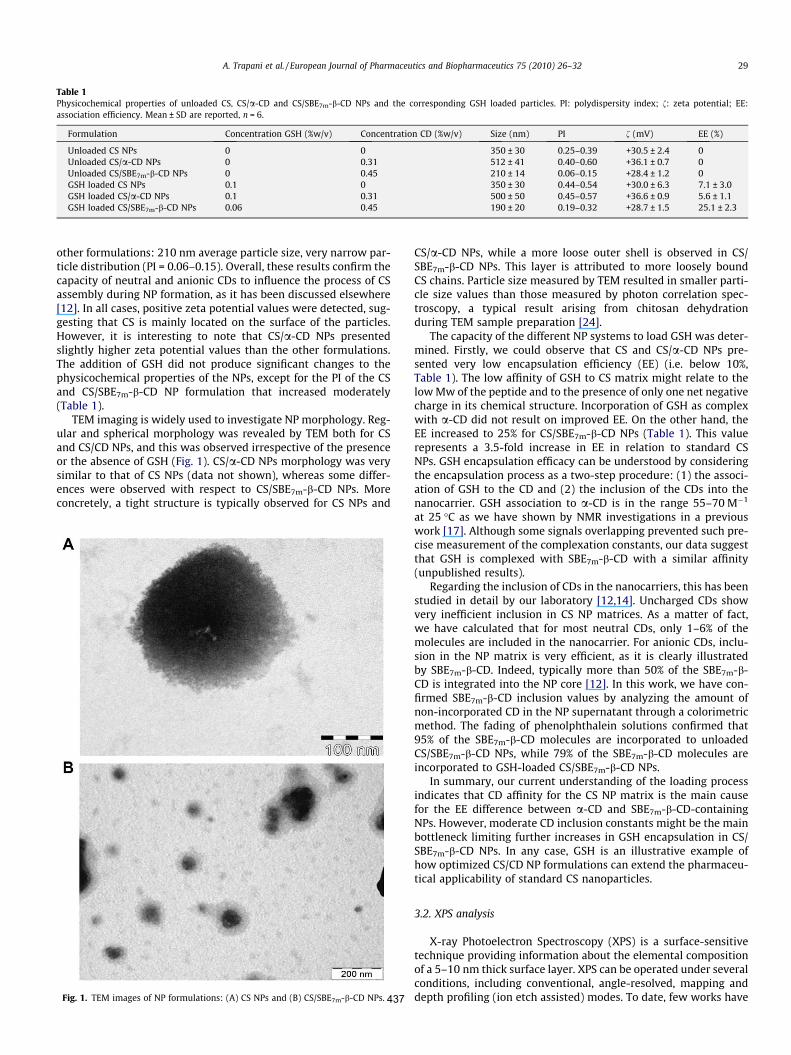
TEM imaging is widely used to investigate NP morphology. Reg-ular and spherical morphology was revealed by TEM both for CSand CS/CD NPs, and this was observed irrespective of the presenceor the absence of GSH (Fig. 1). CS/a-CD NPs morphology was verysimilar to that of CS NPs (data not shown), whereas some differ-ences were observed with respect to CS/SBE7m-b-CD NPs. Moreconcretely, a tight structure is typically observed for CS NPs and
CS/a-CD NPs, while a more loose outer shell is observed in CS/SBE7m-b-CD NPs. This layer is attributed to more loosely boundCS chains. Particle size measured by TEM resulted in smaller parti-cle size values than those measured by photon correlation spec-troscopy, a typical result arising from chitosan dehydrationduring TEM sample preparation [24].
The capacity of the different NP systems to load GSH was deter-mined. Firstly, we could observe that CS and CS/a-CD NPs pre-sented very low encapsulation efficiency (EE) (i.e. below 10%,Table 1). The low affinity of GSH to CS matrix might relate to thelow Mw of the peptide and to the presence of only one net negativecharge in its chemical structure. Incorporation of GSH as complexwith a-CD did not result on improved EE. On the other hand, theEE increased to 25% for CS/SBE7m-b-CD NPs (Table 1). This valuerepresents a 3.5-fold increase in EE in relation to standard CSNPs. GSH encapsulation efficacy can be understood by consideringthe encapsulation process as a two-step procedure: (1) the associ-ation of GSH to the CD and (2) the inclusion of the CDs into thenanocarrier. GSH association to a-CD is in the range 55–70 M�1
at 25 �C as we have shown by NMR investigations in a previouswork [17]. Although some signals overlapping prevented such pre-cise measurement of the complexation constants, our data suggestthat GSH is complexed with SBE7m-b-CD with a similar affinity(unpublished results).
Regarding the inclusion of CDs in the nanocarriers, this has beenstudied in detail by our laboratory [12,14]. Uncharged CDs showvery inefficient inclusion in CS NP matrices. As a matter of fact,we have calculated that for most neutral CDs, only 1–6% of themolecules are included in the nanocarrier. For anionic CDs, inclu-sion in the NP matrix is very efficient, as it is clearly illustratedby SBE7m-b-CD. Indeed, typically more than 50% of the SBE7m-b-CD is integrated into the NP core [12]. In this work, we have con-firmed SBE7m-b-CD inclusion values by analyzing the amount ofnon-incorporated CD in the NP supernatant through a colorimetricmethod. The fading of phenolphthalein solutions confirmed that95% of the SBE7m-b-CD molecules are incorporated to unloadedCS/SBE7m-b-CD NPs, while 79% of the SBE7m-b-CD molecules areincorporated to GSH-loaded CS/SBE7m-b-CD NPs.
In summary, our current understanding of the loading processindicates that CD affinity for the CS NP matrix is the main causefor the EE difference between a-CD and SBE7m-b-CD-containingNPs. However, moderate CD inclusion constants might be the mainbottleneck limiting further increases in GSH encapsulation in CS/SBE7m-b-CD NPs. In any case, GSH is an illustrative example ofhow optimized CS/CD NP formulations can extend the pharmaceu-tical applicability of standard CS nanoparticles.
3.2. XPS analysis
X-ray Photoelectron Spectroscopy (XPS) is a surface-sensitivetechnique providing information about the elemental compositionof a 5–10 nm thick surface layer. XPS can be operated under severalconditions, including conventional, angle-resolved, mapping anddepth profiling (ion etch assisted) modes. To date, few works have
Table 1Physicochemical properties of unloaded CS, CS/a-CD and CS/SBE7m-b-CD NPs and the corresponding GSH loaded particles. PI: polydispersity index; f: zeta potential; EE:association efficiency. Mean ± SD are reported, n = 6.
Formulation Concentration GSH (%w/v) Concentration CD (%w/v) Size (nm) PI f (mV) EE (%)
Unloaded CS NPs 0 0 350 ± 30 0.25–0.39 +30.5 ± 2.4 0Unloaded CS/a-CD NPs 0 0.31 512 ± 41 0.40–0.60 +36.1 ± 0.7 0Unloaded CS/SBE7m-b-CD NPs 0 0.45 210 ± 14 0.06–0.15 +28.4 ± 1.2 0GSH loaded CS NPs 0.1 0 350 ± 30 0.44–0.54 +30.0 ± 6.3 7.1 ± 3.0GSH loaded CS/a-CD NPs 0.1 0.31 500 ± 50 0.45–0.57 +36.6 ± 0.9 5.6 ± 1.1GSH loaded CS/SBE7m-b-CD NPs 0.06 0.45 190 ± 20 0.19–0.32 +28.7 ± 1.5 25.1 ± 2.3
Fig. 1. TEM images of NP formulations: (A) CS NPs and (B) CS/SBE7m-b-CD NPs.
A. Trapani et al. / European Journal of Pharmaceutics and Biopharmaceutics 75 (2010) 26–32 29
437
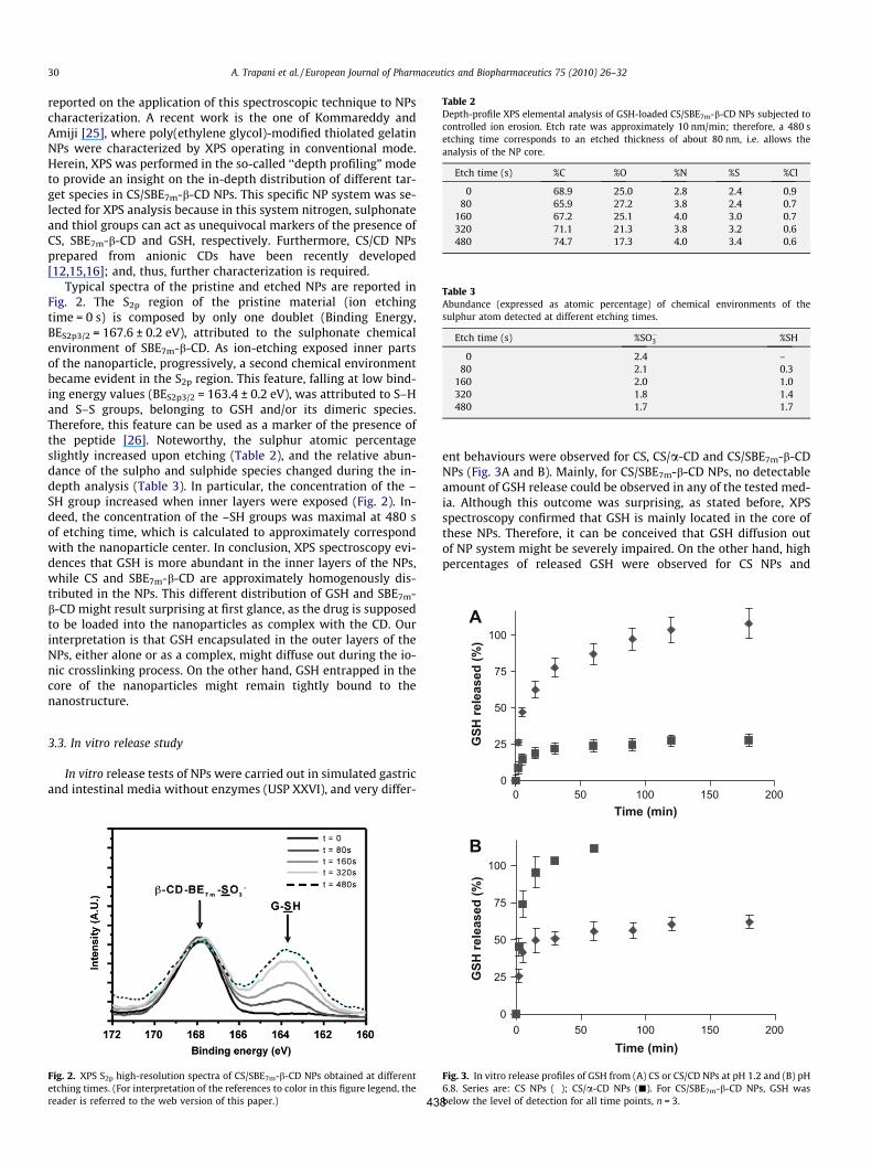
reported on the application of this spectroscopic technique to NPscharacterization. A recent work is the one of Kommareddy andAmiji [25], where poly(ethylene glycol)-modified thiolated gelatinNPs were characterized by XPS operating in conventional mode.Herein, XPS was performed in the so-called ‘‘depth profiling” modeto provide an insight on the in-depth distribution of different tar-get species in CS/SBE7m-b-CD NPs. This specific NP system was se-lected for XPS analysis because in this system nitrogen, sulphonateand thiol groups can act as unequivocal markers of the presence ofCS, SBE7m-b-CD and GSH, respectively. Furthermore, CS/CD NPsprepared from anionic CDs have been recently developed[12,15,16]; and, thus, further characterization is required.
Typical spectra of the pristine and etched NPs are reported inFig. 2. The S2p region of the pristine material (ion etchingtime = 0 s) is composed by only one doublet (Binding Energy,BES2p3/2 = 167.6 ± 0.2 eV), attributed to the sulphonate chemicalenvironment of SBE7m-b-CD. As ion-etching exposed inner partsof the nanoparticle, progressively, a second chemical environmentbecame evident in the S2p region. This feature, falling at low bind-ing energy values (BES2p3/2 = 163.4 ± 0.2 eV), was attributed to S–Hand S–S groups, belonging to GSH and/or its dimeric species.Therefore, this feature can be used as a marker of the presence ofthe peptide [26]. Noteworthy, the sulphur atomic percentageslightly increased upon etching (Table 2), and the relative abun-dance of the sulpho and sulphide species changed during the in-depth analysis (Table 3). In particular, the concentration of the –SH group increased when inner layers were exposed (Fig. 2). In-deed, the concentration of the –SH groups was maximal at 480 sof etching time, which is calculated to approximately correspondwith the nanoparticle center. In conclusion, XPS spectroscopy evi-dences that GSH is more abundant in the inner layers of the NPs,while CS and SBE7m-b-CD are approximately homogenously dis-tributed in the NPs. This different distribution of GSH and SBE7m-b-CD might result surprising at first glance, as the drug is supposedto be loaded into the nanoparticles as complex with the CD. Ourinterpretation is that GSH encapsulated in the outer layers of theNPs, either alone or as a complex, might diffuse out during the io-nic crosslinking process. On the other hand, GSH entrapped in thecore of the nanoparticles might remain tightly bound to thenanostructure.
3.3. In vitro release study
In vitro release tests of NPs were carried out in simulated gastricand intestinal media without enzymes (USP XXVI), and very differ-
ent behaviours were observed for CS, CS/a-CD and CS/SBE7m-b-CDNPs (Fig. 3A and B). Mainly, for CS/SBE7m-b-CD NPs, no detectableamount of GSH release could be observed in any of the tested med-ia. Although this outcome was surprising, as stated before, XPSspectroscopy confirmed that GSH is mainly located in the core ofthese NPs. Therefore, it can be conceived that GSH diffusion outof NP system might be severely impaired. On the other hand, highpercentages of released GSH were observed for CS NPs and
Fig. 2. XPS S2p high-resolution spectra of CS/SBE7m-b-CD NPs obtained at differentetching times. (For interpretation of the references to color in this figure legend, thereader is referred to the web version of this paper.)
Table 2Depth-profile XPS elemental analysis of GSH-loaded CS/SBE7m-b-CD NPs subjected tocontrolled ion erosion. Etch rate was approximately 10 nm/min; therefore, a 480 setching time corresponds to an etched thickness of about 80 nm, i.e. allows theanalysis of the NP core.
Etch time (s) %C %O %N %S %Cl
0 68.9 25.0 2.8 2.4 0.980 65.9 27.2 3.8 2.4 0.7
160 67.2 25.1 4.0 3.0 0.7320 71.1 21.3 3.8 3.2 0.6480 74.7 17.3 4.0 3.4 0.6
Table 3Abundance (expressed as atomic percentage) of chemical environments of thesulphur atom detected at different etching times.
Etch time (s) %SO�3 %SH
0 2.4 –80 2.1 0.3
160 2.0 1.0320 1.8 1.4480 1.7 1.7
0
25
50
75
100
0 50 100 150 200
0 50 100 150 200
Time (min)
GSH
rele
ased
(%)
0
25
50
75
100
Time (min)
GSH
rele
ased
(%)
A
B
Fig. 3. In vitro release profiles of GSH from (A) CS or CS/CD NPs at pH 1.2 and (B) pH6.8. Series are: CS NPs (�); CS/a-CD NPs (j). For CS/SBE7m-b-CD NPs, GSH wasbelow the level of detection for all time points, n = 3.
30 A. Trapani et al. / European Journal of Pharmaceutics and Biopharmaceutics 75 (2010) 26–32
438
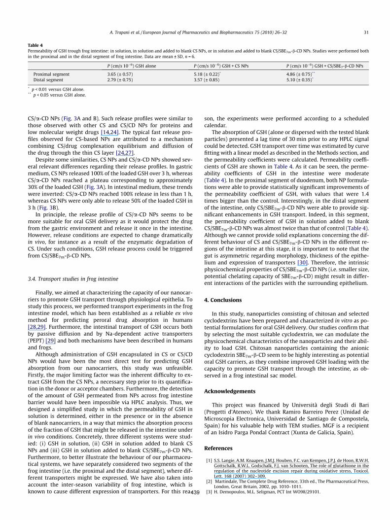
CS/a-CD NPs (Fig. 3A and B). Such release profiles were similar tothose observed with other CS and CS/CD NPs for proteins andlow molecular weight drugs [14,24]. The typical fast release pro-files observed for CS-based NPs are attributed to a mechanismcombining CS/drug complexation equilibrium and diffusion ofthe drug through the thin CS layer [24,27].
Despite some similarities, CS NPs and CS/a-CD NPs showed sev-eral relevant differences regarding their release profiles. In gastricmedium, CS NPs released 100% of the loaded GSH over 3 h, whereasCS/a-CD NPs reached a plateau corresponding to approximately30% of the loaded GSH (Fig. 3A). In intestinal medium, these trendswere inverted: CS/a-CD NPs reached 100% release in less than 1 h,whereas CS NPs were only able to release 50% of the loaded GSH in3 h (Fig. 3B).
In principle, the release profile of CS/a-CD NPs seems to bemore suitable for oral GSH delivery as it would protect the drugfrom the gastric environment and release it once in the intestine.However, release conditions are expected to change dramaticallyin vivo, for instance as a result of the enzymatic degradation ofCS. Under such conditions, GSH release process could be triggeredfrom CS/SBE7m-b-CD NPs.
3.4. Transport studies in frog intestine
Finally, we aimed at characterizing the capacity of our nanocar-riers to promote GSH transport through physiological epithelia. Tostudy this process, we performed transport experiments in the frogintestine model, which has been established as a reliable ex vivomethod for predicting peroral drug absorption in humans[28,29]. Furthermore, the intestinal transport of GSH occurs bothby passive diffusion and by Na-dependent active transporters(PEPT) [29] and both mechanisms have been described in humansand frogs.
Although administration of GSH encapsulated in CS or CS/CDNPs would have been the most direct test for predicting GSHabsorption from our nanocarriers, this study was unfeasible.Firstly, the major limiting factor was the inherent difficulty to ex-tract GSH from the CS NPs, a necessary step prior to its quantifica-tion in the donor or acceptor chambers. Furthermore, the detectionof the amount of GSH permeated from NPs across frog intestinebarrier would have been impossible via HPLC analysis. Thus, wedesigned a simplified study in which the permeability of GSH insolution is determined, either in the presence or in the absenceof blank nanocarriers, in a way that mimics the absorption processof the fraction of GSH that might be released in the intestine underin vivo conditions. Concretely, three different systems were stud-ied: (i) GSH in solution, (ii) GSH in solution added to blank CSNPs and (iii) GSH in solution added to blank CS/SBE7m-b-CD NPs.Furthermore, to better illustrate the behaviour of our pharmaceu-tical systems, we have separately considered two segments of thefrog intestine (i.e. the proximal and the distal segment), where dif-ferent transporters might be expressed. We have also taken intoaccount the inter-season variability of frog intestine, which isknown to cause different expression of transporters. For this rea-
son, the experiments were performed according to a scheduledcalendar.
The absorption of GSH (alone or dispersed with the tested blankparticles) presented a lag time of 30 min prior to any HPLC signalcould be detected. GSH transport over time was estimated by curvefitting with a linear model as described in the Methods section, andthe permeability coefficients were calculated. Permeability coeffi-cients of GSH are shown in Table 4. As it can be seen, the perme-ability coefficients of GSH in the intestine were moderate(Table 4). In the proximal segment of duodenum, both NP formula-tions were able to provide statistically significant improvements ofthe permeability coefficient of GSH, with values that were 1.4times bigger than the control. Interestingly, in the distal segmentof the intestine, only CS/SBE7m-b-CD NPs were able to provide sig-nificant enhancements in GSH transport. Indeed, in this segment,the permeability coefficient of GSH in solution added to blankCS/SBE7m-b-CD NPs was almost twice than that of control (Table 4).Although we cannot provide solid explanations concerning the dif-ferent behaviour of CS and CS/SBE7m-b-CD NPs in the different re-gions of the intestine at this stage, it is important to note that thegut is asymmetric regarding morphology, thickness of the epithe-lium and expression of transporters [30]. Therefore, the intrinsicphysicochemical properties of CS/SBE7m-b-CD NPs (i.e. smaller size,potential chelating capacity of SBE7m-b-CD) might result in differ-ent interactions of the particles with the surrounding epithelium.
4. Conclusions
In this study, nanoparticles consisting of chitosan and selectedcyclodextrins have been prepared and characterized in vitro as po-tential formulations for oral GSH delivery. Our studies confirm thatby selecting the most suitable cyclodextrin, we can modulate thephysicochemical characteristics of the nanoparticles and their abil-ity to load GSH. Chitosan nanoparticles containing the anioniccyclodextrin SBE7m-b-CD seem to be highly interesting as potentialoral GSH carriers, as they combine improved GSH loading with thecapacity to promote GSH transport through the intestine, as ob-served in a frog intestinal sac model.
Acknowledgements
This project was financed by Università degli Studi di Bari(Progetti d’Ateneo). We thank Ramiro Barreiro Perez (Unidad deMicroscopia Electronica, Universidad de Santiago de Compostela,Spain) for his valuable help with TEM studies. MGF is a recipientof an Isidro Parga Pondal Contract (Xunta de Galicia, Spain).
References
[1] S.S. Langie, A.M. Knaapen, J.M.J. Houben, F.C. van Kempen, J.P.J. de Hoon, R.W.H.Gottschalk, R.W.L. Godschalk, F.J. van Schooten, The role of glutathione in theregulation of the nucleotide excision repair during oxidative stress, Toxicol.Lett. 168 (2007) 302–309.
[2] Martindale, The Complete Drug Reference, 33th ed., The Pharmaceutical Press,London, Great Britain, 2002, pp. 1010–1011.
[3] H. Demopoulos, M.L. Seligman, PCT Int WO98/29101.
Table 4Permeability of GSH trough frog intestine: in solution, in solution and added to blank CS NPs, or in solution and added to blank CS/SBE7m-b-CD NPs. Studies were performed bothin the proximal and in the distal segment of frog intestine. Data are mean ± SD, n = 6.
P (cm/s 10�6) GSH alone P (cm/s 10�6) GSH + CS NPs P (cm/s 10�6) GSH + CS/SBE7-b-CD NPs
Proximal segment 3.65 (± 0.57) 5.18 (± 0.22)* 4.86 (± 0.75)**
Distal segment 2.79 (± 0.75) 3.57 (± 0.85) 5.10 (± 0.35)*
* p < 0.01 versus GSH alone.** p < 0.05 versus GSH alone.
A. Trapani et al. / European Journal of Pharmaceutics and Biopharmaceutics 75 (2010) 26–32 31
439
[4] E. Camera, M. Picardo, Analytical methods to investigate glutathione andrelated compounds in biological and pathological processes, J. Chromatogr. B781 (2002) 181–206.
[5] M. Tobio, A. Sanchez, A. Vila, C. Soriano, J.L. Evora, J.L. Vila-Jato, M.J. Alonso,Investigations on the role of PEG on the stability in digestive fluids and in vivofate of PEG–PLA nanoparticle following oral administration, Colloids Surf. B:Biointerf. 18 (2000) 315–32330.
[6] G. Sandri, M.C. Bonferoni, S. Rossi, F. Ferrari, S. Gibin, Y. Zambito, G. Di Colo, C.Caramella, Nanoparticles based on N-trimethylchitosan: evaluation ofabsorption properties using in vitro (Caco-2 cells) and ex vivo (excised ratjejunum) models, Eur. J. Pharm. Biopharm. 65 (2007) 68–77.
[7] Y.-H. Lin, F.-L. Min, C.-T. Chen, W.-C. Chang, S.-F. Peng, H.-F. Liang, H.-W. Sung,Preparation and characterization of nanoparticles shelled with chitosan fororal insulin delivery, Biomacromolecules 8 (2007) 146–152.
[8] R. Fernandez-Urrusuno, D. Romani, P. Calvo, J.L. Vila-J, M.J. Alonso,Development of freeze-dried formulation of insulin loaded chitosannanoparticles intended for nasal administration, STP Pharma Sci. 9 (5) (1999)429–436.
[9] Y. Pan, Y. Li, H. Zhao, J. Zheng, H. Xu, G. Wei, et al., Bioadhesive polysaccharidein protein delivery system: chitosan nanoparticles improve the intestinalabsorption of insulin in vivo, Int. J. Pharm. 249 (2002) 139–147.
[10] Y. Zhang, Y. Yang, K. Tang, X. Hu, G. Zou, Physicochemical characterization andantioxidant activity of quercetin-loaded chitosan nanoparticles, J. Appl. Polym.Sci. 107 (2008) 891–897.
[11] N. Csaba, M. Garcia-Fuentes, M.J. Alonso, The performance of nanocarriers fortransmucosal drug delivery, Expert. Opin. 3 (4) (2006) 463–478.
[12] A. Trapani, M. Garcia-Fuentes, M.J. Alonso, Novel drug nanocarriers combininghydrophilic cyclodextrins and chitosan, Nanotechnology 19 (18) (2008)185101/1–185101/10.
[13] A.H. Krauland, M.J. Alonso, Chitosan/cyclodextrin nanoparticles asmacromolecular drug delivery system, Int. J. Pharm. 340 (2007) 134–142.
[14] F. Maestrelli, M. Garcia-Fuentes, P. Mura, M.J. Alonso, A new drug nanocarriersystem consisting of chitosan and hydroxypropylcyclodextrin, Eur. J. Pharm.Biopharm. 63 (2) (2006) 79–86.
[15] D. Teijeiro-Osorio, C. Remuñan-Lopez, M.J. Alonso, Chitosan/cyclodextrinnanoparticles can efficiently transfect the airway epithelium in vitro, Eur. J.Pharm. Biopharm. 71 (2009) 257–263.
[16] D. Teijeiro-Osorio, C. Remuñan-Lopez, M.J. Alonso, New generation of hybridpoly/oligosaccharide nanoparticles as carriers for the nasal delivery ofmacromolecules, Biomacromolecules 10 (2) (2009) 243–249.
[17] M. Garcia-Fuentes, A. Trapani, M.J. Alonso, Protection of the peptideglutathione by complex formation with a-cyclodextrin: NMR spectroscopicanalysis and stability study, Eur. J. Pharm. Biopharm. 64 (2006) 146–153.
[18] P. Calvo, C. Remuñan-Lopez, J.L. Vila-J, M.J. Alonso, Novel hydrophilic chitosan–polyethylene oxide nanoparticles as protein carriers, J. Appl. Polym. Sci. 63(1997) 125–132.
[19] E. Ieva, A. Trapani, N. Cioffi, N. Ditaranto, A. Monopoli, L. Sabbatini, Analyticalcharacterization of chitosan nanoparticles for peptide drug deliveryapplications, Anal. Bioanal. Chem. 393 (2009) 207–215.
[20] A.M. Da Silveira, G. Ponchel, F. Puisieux, D. Duchene, Combinedpoly(isobutylcyanoacrilate) and cyclodextrins nanoparticles for enhancingthe encapsulation of lipophilic drugs, Pharm. Res. 15 (1998) 1051–1055.
[21] A. Lopedota, A. Trapani, A. Cutrignelli, L. Chiarantini, E. Pantucci, R. Curci, E.Manuali, G. Trapani, The use of Eudragit RS 100/cyclodextrin nanoparticles forthe transmucosal administration of glutathione, Eur. J. Pharm. Biopharm. 72(2009) 509–520.
[22] Z. Ma, T.M. Lim, L.-Y. Lim, Pharmacological activity of peroral chitosan–insulinnanoparticles in diabetic rats, Int. J. Pharm. 293 (2005) 271–280.
[23] K.A. Janes, P. Calvo, M.J. Alonso, Polysaccharide colloidal nanoparticles asdelivery systems for macromolecules, Adv. Drug Deliv. Rev. 47 (2001) 83–97.
[24] Y. Aktas, K. Andrieux, M.J. Alonso, P. Calvo, R.N. Gursoy, P. Couvreur, Y. Capan,Preparation and in vitro evaluation of chitosan nanoparticles containing acaspase inhibitor, Int. J. Pharm. 298 (2005) 378–383.
[25] S. Kommareddy, M. Amiji, Poly(ethylene glycol)-modified thiolated gelatinenanoparticles for glutathione-responsive intracellular DNA delivery,Nanomedicine 3 (2007) 32–42.
[26] K.J. Cantrell, S.B. Yabusaki, M.H. Engelhard, A.V. Mitroshkov, E.C. Thornton,Oxidation of H2S by iron oxides in unsaturated conditions, Environ. Sci.Technol. 37 (2003) 2192–2199.
[27] M. Hamidi, A. Azadi, P. Rafiei, Hydrogel nanoparticles in drug delivery, Adv.Drug Deliv. Rev. 60 (2008) 1638–1649.
[28] M. Franco, A. Lopedota, A. Trapani, A. Cutrignelli, D. Meleleo, S. Micelli, G.Trapani, Frog intestinal sac as an in vitro method for the assessment ofintestinal permeability in humans: application to carrier transported drugs,Int. J. Pharm. 352 (2008) 182–188.
[29] G. Trapani, M. Franco, A. Trapani, A. Lopedota, A. Latrofa, E. Gallucci, S. Micelli,G. Liso, Frog intestinal sac: a new in vitro method for the assessment ofintestinal permeability, J. Pharm. Sci. 93 (2004) 2909–2919.
[30] T.Z. Csaky, E. Gallucci, Seasonal variation in the active transporting ability andin the membrane ATPase activity of the frog intestinal epithelium, Biochim.Biophys. Acta 466 (1977) 521–525.
32 A. Trapani et al. / European Journal of Pharmaceutics and Biopharmaceutics 75 (2010) 26–32
440
Related Documents