RESEARCH ARTICLE Open Access A combined approach for comparative exoproteome analysis of Corynebacterium pseudotuberculosis Luis GC Pacheco 1,2,3 , Susan E Slade 4 , Núbia Seyffert 2 , Anderson R Santos 2 , Thiago LP Castro 2 , Wanderson M Silva 2 , Agenor V Santos 1 , Simone G Santos 5 , Luiz M Farias 5 , Maria AR Carvalho 5 , Adriano MC Pimenta 1 , Roberto Meyer 3 , Artur Silva 6 , James H Scrivens 4 , Sérgio C Oliveira 1 , Anderson Miyoshi 2 , Christopher G Dowson 4 , Vasco Azevedo 2* Abstract Background: Bacterial exported proteins represent key components of the host-pathogen interplay. Hence, we sought to implement a combined approach for characterizing the entire exoproteome of the pathogenic bacterium Corynebacterium pseudotuberculosis, the etiological agent of caseous lymphadenitis (CLA) in sheep and goats. Results: An optimized protocol of three-phase partitioning (TPP) was used to obtain the C. pseudotuberculosis exoproteins, and a newly introduced method of data-independent MS acquisition (LC-MS E ) was employed for protein identification and label-free quantification. Additionally, the recently developed tool SurfG+ was used for in silico prediction of sub-cellular localization of the identified proteins. In total, 93 different extracellular proteins of C. pseudotuberculosis were identified with high confidence by this strategy; 44 proteins were commonly identified in two different strains, isolated from distinct hosts, then composing a core C. pseudotuberculosis exoproteome. Analysis with the SurfG+ tool showed that more than 75% (70/93) of the identified proteins could be predicted as containing signals for active exportation. Moreover, evidence could be found for probable non-classical export of most of the remaining proteins. Conclusions: Comparative analyses of the exoproteomes of two C. pseudotuberculosis strains, in addition to comparison with other experimentally determined corynebacterial exoproteomes, were helpful to gain novel insights into the contribution of the exported proteins in the virulence of this bacterium. The results presented here compose the most comprehensive coverage of the exoproteome of a corynebacterial species so far. Background Corynebacterium pseudotuberculosis is a facultative intracellular pathogen that belongs to the so-called CMN ( Corynebacterium-Mycobacterium-Nocardia) group, a distinct subgroup of the Actinobacteria that also includes other highly important bacterial pathogens, such as Corynebacterium diphtheriae and Mycobacter- ium tuberculosis. The most distinctive feature of these Gram-positive bacteria is the unique composition of the cell envelope, characterized by the presence of long chain fatty acids, known as mycolic acids, on the surface of the cell [1,2]. The main recognizable disease caused by C. pseudotu- berculosis is caseous lymphadenitis (CLA) in sheep and goats, though this bacterium can also infect several other hosts, including humans [1,3]. Typical manifesta- tions of CLA in small ruminants include formation of abscesses in superficial and internal lymph nodes, and in visceral organs [3]. Despite the important economic losses caused by this disease to sheep and goat husban- dry worldwide, no effective treatment exists, and the efficacy of the currently available vaccines and diagnos- tic methods is still controversial [4]. The search for C. pseudotuberculosis molecular deter- minants that contribute to CLA pathogenesis lead to the * Correspondence: [email protected] 2 Department of General Biology, Instituto de Ciências Biológicas, Universidade Federal de Minas Gerais, Av. Antônio Carlos, Belo Horizonte, 31.270-901, Brazil Full list of author information is available at the end of the article Pacheco et al. BMC Microbiology 2011, 11:12 http://www.biomedcentral.com/1471-2180/11/12 © 2011 Pacheco et al; licensee BioMed Central Ltd. This is an Open Access article distributed under the terms of the Creative Commons Attribution License (http://creativecommons.org/licenses/by/2.0), which permits unrestricted use, distribution, and reproduction in any medium, provided the original work is properly cited.

Welcome message from author
This document is posted to help you gain knowledge. Please leave a comment to let me know what you think about it! Share it to your friends and learn new things together.
Transcript

RESEARCH ARTICLE Open Access
A combined approach for comparativeexoproteome analysis of CorynebacteriumpseudotuberculosisLuis GC Pacheco1,2,3, Susan E Slade4, Núbia Seyffert2, Anderson R Santos2, Thiago LP Castro2, Wanderson M Silva2,Agenor V Santos1, Simone G Santos5, Luiz M Farias5, Maria AR Carvalho5, Adriano MC Pimenta1, Roberto Meyer3,Artur Silva6, James H Scrivens4, Sérgio C Oliveira1, Anderson Miyoshi2, Christopher G Dowson4, Vasco Azevedo2*
Abstract
Background: Bacterial exported proteins represent key components of the host-pathogen interplay. Hence, wesought to implement a combined approach for characterizing the entire exoproteome of the pathogenicbacterium Corynebacterium pseudotuberculosis, the etiological agent of caseous lymphadenitis (CLA) in sheep andgoats.
Results: An optimized protocol of three-phase partitioning (TPP) was used to obtain the C. pseudotuberculosisexoproteins, and a newly introduced method of data-independent MS acquisition (LC-MSE) was employed forprotein identification and label-free quantification. Additionally, the recently developed tool SurfG+ was used for insilico prediction of sub-cellular localization of the identified proteins. In total, 93 different extracellular proteins ofC. pseudotuberculosis were identified with high confidence by this strategy; 44 proteins were commonly identifiedin two different strains, isolated from distinct hosts, then composing a core C. pseudotuberculosis exoproteome.Analysis with the SurfG+ tool showed that more than 75% (70/93) of the identified proteins could be predicted ascontaining signals for active exportation. Moreover, evidence could be found for probable non-classical export ofmost of the remaining proteins.
Conclusions: Comparative analyses of the exoproteomes of two C. pseudotuberculosis strains, in addition tocomparison with other experimentally determined corynebacterial exoproteomes, were helpful to gain novelinsights into the contribution of the exported proteins in the virulence of this bacterium. The results presentedhere compose the most comprehensive coverage of the exoproteome of a corynebacterial species so far.
BackgroundCorynebacterium pseudotuberculosis is a facultativeintracellular pathogen that belongs to the so-calledCMN (Corynebacterium-Mycobacterium-Nocardia)group, a distinct subgroup of the Actinobacteria thatalso includes other highly important bacterial pathogens,such as Corynebacterium diphtheriae and Mycobacter-ium tuberculosis. The most distinctive feature of theseGram-positive bacteria is the unique composition of thecell envelope, characterized by the presence of long
chain fatty acids, known as mycolic acids, on the surfaceof the cell [1,2].The main recognizable disease caused by C. pseudotu-
berculosis is caseous lymphadenitis (CLA) in sheep andgoats, though this bacterium can also infect severalother hosts, including humans [1,3]. Typical manifesta-tions of CLA in small ruminants include formation ofabscesses in superficial and internal lymph nodes, and invisceral organs [3]. Despite the important economiclosses caused by this disease to sheep and goat husban-dry worldwide, no effective treatment exists, and theefficacy of the currently available vaccines and diagnos-tic methods is still controversial [4].The search for C. pseudotuberculosis molecular deter-
minants that contribute to CLA pathogenesis lead to the
* Correspondence: [email protected] of General Biology, Instituto de Ciências Biológicas,Universidade Federal de Minas Gerais, Av. Antônio Carlos, Belo Horizonte,31.270-901, BrazilFull list of author information is available at the end of the article
Pacheco et al. BMC Microbiology 2011, 11:12http://www.biomedcentral.com/1471-2180/11/12
© 2011 Pacheco et al; licensee BioMed Central Ltd. This is an Open Access article distributed under the terms of the CreativeCommons Attribution License (http://creativecommons.org/licenses/by/2.0), which permits unrestricted use, distribution, andreproduction in any medium, provided the original work is properly cited.
recognition of two exported proteins as the major viru-lence-associated factors of this bacterium known to date:a secreted phospholipase D (PLD) [5]; and an ABC-typetransporter component of an iron uptake system (FagB)[6]. In fact, one might expect that the majority of thevirulence determinants of C. pseudotuberculosis would bepresent in the exoproteome, i.e. the entire set of bacterialproteins found in the extracellular milieu [7]. This isbecause exported proteins participate in essential steps ofthe host-pathogen interplay, including: (i) adhesion tohost cells; (ii) invasion; (iii) damage to host tissues;(iv) resistance to environmental stresses during infection;and (iv) subversion of the host’s immune responsemechanisms [8-10].In two previous attempts to characterize the C. pseudo-
tuberculosis exoproteome, our group optimized a proto-col of salting out of proteins using sulfate and butanol,known as three-phase partitioning (TPP), for isolation ofthe extracellular proteins of this bacterium [11], and gen-erated a library of C. pseudotuberculosis mutant strainspossessing transposon insertions in genes coding forprobable exported proteins [12]. In the former study, wewere able to determine the optimal conditions for obtain-ing the best recovery of immunoreactive extracellularproteins of C. pseudotuberculosis [11]. The second studyin turn, enabled us to identify various previously unchar-acterized C. pseudotuberculosis exported proteins, beingthat at least two of them are apparently involved in viru-lence [12]. Now, the very recent conclusion of the C.pseudotuberculosis Genome Project by our group, asso-ciated to the current availability of high-throughput pro-teomic technologies, permitted us to perform a muchmore comprehensive analysis of this bacterium’sexoproteome.In this study, we sought to implement a combined
approach for comparative exoproteome analysis of dif-ferent C. pseudotuberculosis strains. The strategyincluded: (i) the previously optimized TPP protocol forisolation of the extracellular proteins [11]; (ii) a newlyintroduced method of data-independent LC-MS acquisi-tion (LC-MSE) for protein identification and quantifica-tion [13,14]; and (iii) the recently developed tool SurfG+for in silico prediction of protein sub-cellular localiza-tion in Gram-positive bacteria [15]. We believe that theexperimental approach used is very suitable for profilingbacterial exoproteomes, as it shown to be easily applic-able to different strains with very good reproducibility.This is an advantage over what is commonly observedfor proteomic approaches based on two-dimensional(2D) gel electrophoresis, where there is more variability,but is apparently the method of choice for most of thebacterial exoproteome studies published recently[16-20]. Furthermore, the LC-MSE method provideshigh subproteome coverage, due to enhanced sensitivity,
and allows for label-free analysis of differentiallyexpressed proteins [14]; this latter possibility enables thedetection of variations in the exoproteomes of differentstrains that could be missed by simply profiling the exo-proteins, and meets the growing interest in performingphysiological proteomic studies of bacteria [21,22].We were able to identify 93 different C. pseudotuber-
culosis extracellular proteins with high confidence byanalyzing the exoproteomes of two strains isolated fromdifferent hosts that presented distinct virulence pheno-types under laboratory conditions [23,24]. Most of theidentified proteins were predicted in silico to have anextracytoplasmic localization. To the best of our knowl-edge, these results compose the largest inventory ofexperimentally confirmed exoproteins of a single cory-nebacterial species to date. Importantly, the comparativeexoproteome analyses permitted us to speculate on theprobable contributions of different C. pseudotuberculosisextracellular proteins to the virulence of this bacterium.
Results and DiscussionExoproteome analysis of CorynebacteriumpseudotuberculosisThe extracellular proteins of two C. pseudotuberculosisstrains, one isolated from a goat (strain 1002) the otherfrom a sheep (strain C231), cultivated in a chemically-defined medium, were extracted/concentrated by theTPP technique. The trypsinized protein samples werethen submitted to LC-MSE analysis.Seventy soluble extracellular proteins of the 1002
strain could be confidentially identified by this metho-dology, whereas the number of proteins identified in theexoproteome of the C231 strain was sixty-seven. Alto-gether, 93 different C. pseudotuberculosis exoproteinswere identified in this study (Figure 1). These findingsagree with the results of previous experiments by ourgroup, in which we have used a 2D-PAGE based strat-egy for a preliminary appraisal of the C. pseudotubercu-losis exoproteome (additional file 1). Eighty proteinspots, mostly concentrated in the pI range between 3.0and 6.0, could be reproducibly visible in the 2D gelsgenerated from TPP-extracted extracellular proteins ofthe 1002 strain (additional file 1). The fact that we havefound 70 proteins in the exoproteome of this strainwith high confidence when using the LC-MSE method(Figure 1) indicates that this novel methodology allowedus to identify virtually the complete set of extracellularproteins that are commonly observed in the gel basedmethodologies (additional file 1). Moreover, theexpected existence of protein isoforms among the eightyprotein spots observed in the 2D gels, and the identifica-tion by LC-MSE of many proteins out of the pI range3.0-6.0, suggests that the latter methodology is muchmore suitable for obtaining a comprehensive coverage of
Pacheco et al. BMC Microbiology 2011, 11:12http://www.biomedcentral.com/1471-2180/11/12
Page 2 of 14

the bacterial exoproteome. Noteworthy, is the use ofLC-MSE for exoproteome profiling which required(i) much less time and labor than the gel based proteo-mic strategy, and (ii) much less protein sample neces-sary for each experimental replicate, with only 0.5 μgper replicate used in the LC-MSE compared to 150 μgfor the 2D gels [refer to Patel et al. [25] for a compre-hensive comparison on these proteomic strategies].The performance of the combined methodology used
in the present study (TPP/LC-MSE) for mapping theC. pseudotuberculosis exoproteome was very similar forboth strains analyzed, as can be seen by the averagenumbers of peptides observed per protein in the twoproteomes (16.5 and 15.0) and by the average sequencecoverage of the proteins identified (37.5% and 35.0%)(Figure 1). Consistent with this, the majority of the pro-teins detected in each extracellular proteome wereshared by the goat and sheep isolates; this permitted usto define a core C. pseudotuberculosis exoproteomecomposed of 44 proteins out of the 93 different
extracellular proteins identified. Additional files 2, 3 and4 list all the proteins identified in the exoproteomes ofthe two C. pseudotuberculosis strains, along with mole-cular weights, isoelectric points, main orthologs, pre-dicted sub-cellular localizations, number of peptidesexperimentally observed, and sequence coverage.Searches of similarity against publicly available protein
databases using the Blast-p tool [26] showed that ortho-log proteins can be found in the pathogenic Corynebac-terium diphtheriae for most of the identifiedC. pseudotuberculosis exoproteins (additional files 2, 3and 4), as would be expected due to the close phyloge-netic relationship of these species [27]. Nevertheless, nosignificant orthologs could be found for six proteins ofthe C. pseudotuberculosis exoproteome, even when usingthe position-specific iterated BLAST (PSI-BLAST) algo-rithm [28], namely the proteins [GenBank:ADL09626],[GenBank:ADL21925], [GenBank:ADL11253], [GenBank:ADL20222], [GenBank:ADL09871], and [GenBank:ADL21537] (additional files 2, 3 and 4). With the
Figure 1 Analysis of the extracellular proteins of two different C. pseudotuberculosis strains allowed for identification of the core andvariant exoproteomes. TPP-extracted extracellular proteins of the strains 1002 and C231 of C. pseudotuberculosis were submitted to LC-MSE
analysis. The Venn-diagram shows the numbers of commonly identified and variant exoproteins between the strains. The number of replicates inwhich a given protein was observed, the average peptides identified per protein, and the average sequence coverage of the proteins in eachexoproteome studied, are shown as frequency distributions for comparison purposes.
Pacheco et al. BMC Microbiology 2011, 11:12http://www.biomedcentral.com/1471-2180/11/12
Page 3 of 14
exception of [GenBank:ADL11253], all these proteinswere predicted by different tools as being truly exportedproteins. This means they are the only five exoproteinsidentified in this study which are probably unique forC. pseudotuberculosis.
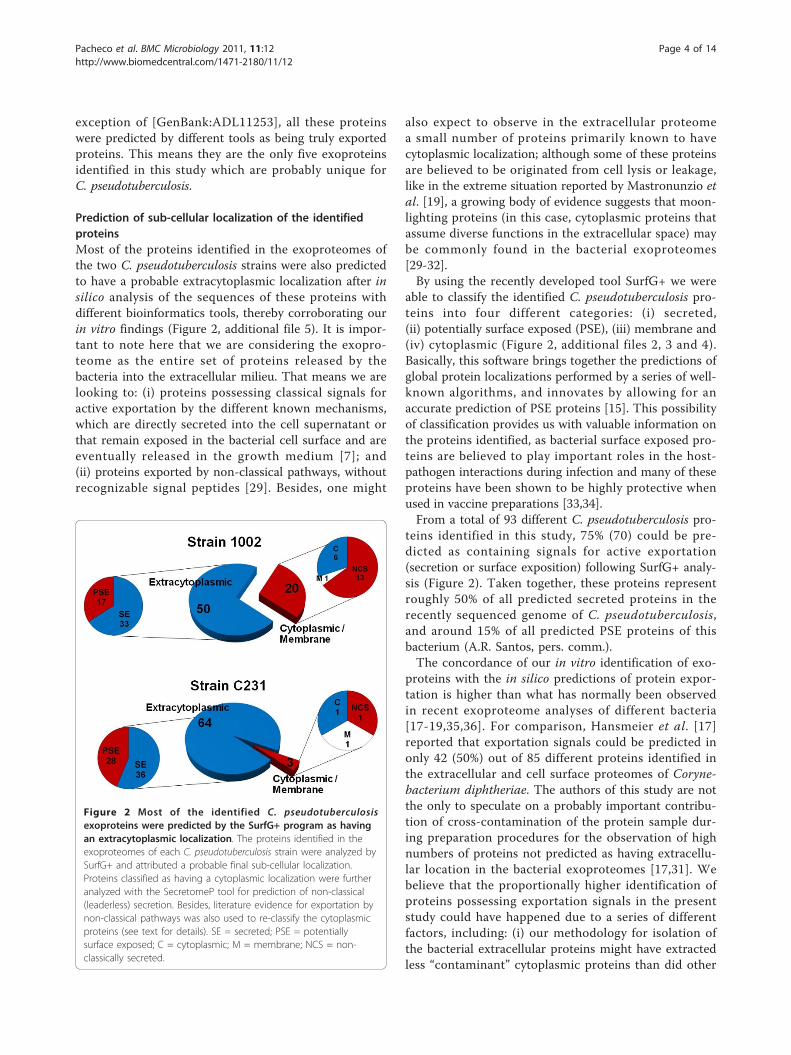
Prediction of sub-cellular localization of the identifiedproteinsMost of the proteins identified in the exoproteomes ofthe two C. pseudotuberculosis strains were also predictedto have a probable extracytoplasmic localization after insilico analysis of the sequences of these proteins withdifferent bioinformatics tools, thereby corroborating ourin vitro findings (Figure 2, additional file 5). It is impor-tant to note here that we are considering the exopro-teome as the entire set of proteins released by thebacteria into the extracellular milieu. That means we arelooking to: (i) proteins possessing classical signals foractive exportation by the different known mechanisms,which are directly secreted into the cell supernatant orthat remain exposed in the bacterial cell surface and areeventually released in the growth medium [7]; and(ii) proteins exported by non-classical pathways, withoutrecognizable signal peptides [29]. Besides, one might
also expect to observe in the extracellular proteomea small number of proteins primarily known to havecytoplasmic localization; although some of these proteinsare believed to be originated from cell lysis or leakage,like in the extreme situation reported by Mastronunzio etal. [19], a growing body of evidence suggests that moon-lighting proteins (in this case, cytoplasmic proteins thatassume diverse functions in the extracellular space) maybe commonly found in the bacterial exoproteomes[29-32].By using the recently developed tool SurfG+ we were
able to classify the identified C. pseudotuberculosis pro-teins into four different categories: (i) secreted,(ii) potentially surface exposed (PSE), (iii) membrane and(iv) cytoplasmic (Figure 2, additional files 2, 3 and 4).Basically, this software brings together the predictions ofglobal protein localizations performed by a series of well-known algorithms, and innovates by allowing for anaccurate prediction of PSE proteins [15]. This possibilityof classification provides us with valuable information onthe proteins identified, as bacterial surface exposed pro-teins are believed to play important roles in the host-pathogen interactions during infection and many of theseproteins have been shown to be highly protective whenused in vaccine preparations [33,34].From a total of 93 different C. pseudotuberculosis pro-
teins identified in this study, 75% (70) could be pre-dicted as containing signals for active exportation(secretion or surface exposition) following SurfG+ analy-sis (Figure 2). Taken together, these proteins representroughly 50% of all predicted secreted proteins in therecently sequenced genome of C. pseudotuberculosis,and around 15% of all predicted PSE proteins of thisbacterium (A.R. Santos, pers. comm.).The concordance of our in vitro identification of exo-
proteins with the in silico predictions of protein expor-tation is higher than what has normally been observedin recent exoproteome analyses of different bacteria[17-19,35,36]. For comparison, Hansmeier et al. [17]reported that exportation signals could be predicted inonly 42 (50%) out of 85 different proteins identified inthe extracellular and cell surface proteomes of Coryne-bacterium diphtheriae. The authors of this study are notthe only to speculate on a probably important contribu-tion of cross-contamination of the protein sample dur-ing preparation procedures for the observation of highnumbers of proteins not predicted as having extracellu-lar location in the bacterial exoproteomes [17,31]. Webelieve that the proportionally higher identification ofproteins possessing exportation signals in the presentstudy could have happened due to a series of differentfactors, including: (i) our methodology for isolation ofthe bacterial extracellular proteins might have extractedless “contaminant” cytoplasmic proteins than did other
Figure 2 Most of the identified C. pseudotuberculosisexoproteins were predicted by the SurfG+ program as havingan extracytoplasmic localization. The proteins identified in theexoproteomes of each C. pseudotuberculosis strain were analyzed bySurfG+ and attributed a probable final sub-cellular localization.Proteins classified as having a cytoplasmic localization were furtheranalyzed with the SecretomeP tool for prediction of non-classical(leaderless) secretion. Besides, literature evidence for exportation bynon-classical pathways was also used to re-classify the cytoplasmicproteins (see text for details). SE = secreted; PSE = potentiallysurface exposed; C = cytoplasmic; M = membrane; NCS = non-classically secreted.
Pacheco et al. BMC Microbiology 2011, 11:12http://www.biomedcentral.com/1471-2180/11/12
Page 4 of 14

methodologies reported in previous studies; (ii) thecombined strategy used by SurfG+ to predict proteinsub-cellular localization might have performed better inthe identification of exported proteins than happenedwith other strategies, sometimes based in only one pre-diction tool; (iii) the fact that we have included in thefinal exoproteome lists only proteins identified with highconfidence, in at least two experimental replicates,reduced significantly the possibilities of false-positiveidentifications that might account for some of the unex-pected proteins; and finally (iv) the lower proportion ofproteins primarily regarded as cytoplasmic might beactually a typical characteristic of the C. pseudotubercu-losis exoproteome.
Non-classically secreted proteinsIntriguingly, a much higher proportion (29.0%) of the exo-proteome of the 1002 strain of C. pseudotuberculosis wascomposed by proteins predicted by SurfG+ as not havingan extracytoplasmic location, when compared to only4.5% in the exoproteome of the strain C231 (Figure 2).The possibility of these proteins being non-classicallysecreted has been evaluated using the SecretomeP algo-rithm [29]. We have also reviewed the literature for evi-dence of other bacterial exoproteomes that could supportthe extracellular localization found for these proteins inour study.High SecP scores (above 0.5) could be predicted for 5
of the 19 proteins in the exoproteome of the 1002 strainconsidered by SurfG+ as having a cytoplasmic location(additional files 2 and 3); this could be an indicative thatthey are actually being secreted by non-classicalmechanisms [29]. Nonetheless, 2 of these 5 proteins([GenBank:ADL09626] and [GenBank:ADL20555]) werealso detected in the exoproteome of the C231 strain, inwhich they were predicted by SurfG+ as possessing anextracytoplasmic location (additional file 2). A compara-tive analysis of the sequences encoding these proteins inthe genomes of the two C. pseudotuberculosis strainsshowed that the disparate results were generated due tothe existence of nonsense mutations in the genomesequence of the 1002 strain, which impaired the identifi-cation of signal peptides for the two proteins at the timeof SurfG+ analysis (data not shown). We believe that itis unlikely that these differences represent true poly-morphisms, as the proteins were identified in the extra-cellular proteome, indicating the real existence ofexportation signals. This indeed demonstrates theobvious vulnerability of the prediction tools tothe proper annotation of the bacterial genomes. On theother hand, the assignment of high SecP scores to thesetwo proteins, even though they are not believed to besecreted by non-classical mechanisms, would be totallyexpected, as the SecretomeP is a predictor based on a
neural network trained to identify general features ofextracellular proteins; this means the prediction tool willattribute SecP scores higher than 0.5 to most of thesecreted proteins, regardless the route of export [29].We have found reports in the literature that strongly
support the extracellular localization observed for 8 ofthe 14 remaining proteins considered as non-secretoryby SurfG+ and SecretomeP in the exoproteome of the1002 strain, and without any detectable signal peptide(additional files 2 and 3, Figure 2). Among these pro-teins there are the elongation factors Tu and Ts [16,33,35,37-39]; the glycolytic enzymes triosephosphate iso-merase, phosphoglycerate kinase and phosphoglyceratemutase [16-20,37-40]; the chaperonin GroES [16-18,20,39]; a putative peptidyl prolyl cis trans isomerase[17,18,35,37,41]; and a hydroperoxide reductase enzyme[17,35,39].Proteins primarily regarded as cytoplasmic have con-
sistently been identified in the exoproteomes of differentbacterial species, and moonlighting roles in the extracel-lular environment have already been demonstrated forsome of them [31,32], including evasion of host’simmune system [42], adhesion to host cells [43,44], fold-ing of extracytoplasmic proteins [41,45], and interactionbetween microorganisms [40,46]. Noteworthy, specificevidences for active secretion of such cytoplasmic pro-teins have been demonstrated for only a few examplesto date, and demonstration of an extracellular functionis still missing for many of these proteins [30,31].
The variant exoproteome may account for differentialvirulence of the two C. pseudotuberculosis strainsA considerable number (49/93) of the extracellular pro-teins identified in this work was observed in only one ofthe two strains studied, then composing a variantexperimental C. pseudotuberculosis exoproteome (addi-tional files 3 and 4). Highly variant exoproteomes havealso been reported recently for other Gram+ bacterialpathogens [20,36,39,47-49], and such a variation may beconsidered an important factor leading to the observablephenotypic dissimilarities and ultimately to differentialvirulence of the various strains [50,51]. Hecker et al.[36] reported on how the composition of the exopro-teome can vary extremely within a single species, Sta-phylococcus aureus, being that only 7 out of 63identified extracellular proteins were found in all thetwenty-five clinical isolates studied.One of the most intriguing results in the present study
was the detection of the phospholipase D (PLD) proteinonly in the extracellular proteome of the strain C231(additional file 4). As the regulation of PLD expressionwas demonstrated to be complex and highly affectedby multiple environmental factors [52], we sought todetect this protein in the culture supernatant of the
Pacheco et al. BMC Microbiology 2011, 11:12http://www.biomedcentral.com/1471-2180/11/12
Page 5 of 14

C. pseudotuberculosis 1002 strain grown in a rich med-ium (brain-heart infusion broth) instead of only chemi-cally-defined medium (CDM), but these attempts werealso unfruitful (data not shown). Besides, we were notable to detect secretion of PLD following total exopro-teome analysis of the 1002 strain grown under specificstress generating conditions (Pacheco et al., unpub-lished). The results strongly indicate that this protein isactually not being secreted by the 1002 strain in culture.PLD is an exotoxin considered as the major viru-
lence factor of C. pseudotuberculosis [5,52]. It pos-sesses sphingomyelinase activity that contributes toendothelial permeability and then to spreading of thebacteria within the host [5]. Mutation of the pld genein C. pseudotuberculosis rendered strains no longercapable of causing caseous lymphadenitis (CLA) insheep and goats; the potential of these strains to beused as live attenuated vaccines was already evaluated[53-55]. Similarly, the strain 1002 of C. pseudotubercu-losis was already tested as a possible live attenuatedvaccine against CLA due to its natural low virulentstatus, and administration of this bacterium to goatsdid not cause lesions formation [23,56]. The molecularmechanisms leading to the low virulence of the 1002strain however remain undetermined so far. Webelieve that non-secretion of PLD might be one of themain factors responsible for the lowered virulence ofthe strain. Importantly, we currently cannot affirmthat the 1002 strain does not produce this proteinwhile infecting a mammalian host. Besides, this strainstill retains the capability of causing localizedabscesses and disease in susceptible mice (Pachecoet al., unpublished results).Other proteins believed to be associated with the
virulence of C. pseudotuberculosis were also identifiedexclusively in the exoproteome of the C231 strain,namely FagD and Cp40 (Table 1). The former proteinis a component of an iron uptake system, whose cod-ing sequences are clustered immediately downstreamof the pld gene in the C. pseudotuberculosis genome[6]. The latter protein is a secreted serine proteaseshown to be protective against CLA when used tovaccinate sheep [57].Strikingly, one variant protein of the C. pseudotuber-
culosis exoproteome, a conserved hypothetical exportedprotein with a cutinase domain [GenBank:ADL10384],has its coding sequence present in the genome of theC231 strain but absent from the genome of the 1002strain (additional file 6). The genomic structure of thegene’s surroundings is indicative of a region prone torecombination events, such as horizontal gene transfer[58]. In fact, it seems that gene gain and loss are fre-quent events leading to variations observed in the bac-terial exoproteomes [39,59].
Variation of the core exoproteome: differential expressionanalysis of the common proteins by LC-MSE
In addition to identifying qualitative variations in theexoproteomes of the two C. pseudotuberculosis strains,we were also able to detect relative differences inexpression of the proteins common to the two pro-teomes through label-free protein quantification by theLC-MSE method. Relative protein quantification by thismethod can be obtained with basis on the accurate pre-cursor ion mass and electrospray intensity data, acquiredduring the low energy scan step of the alternating scanmode of MS acquisition [14]. Importantly, this quantita-tive attribute of the technique opens up new possibilitiesof utilization, as grows the interest on the so-called phy-siological proteomics [21].Thirty-four out of 44 proteins commonly identified in
the exoproteomes of the strains 1002 and C231 ofC. pseudotuberculosis were considered by the PLGSquantification algorithm as having significantly variableexpression (score > 250; 95% CI) (Figure 3, additionalfiles 2 and 7). If we further filter these results for theproteins presenting differential expression higher than2-fold between the strains, we end up with only fourproteins up-regulated in the 1002 strain and sixteen inthe C231 strain (Figure 3).Among the group of proteins not presenting consider-able variations in expression between the two C. pseudo-tuberculosis strains, proteins probably participating inbasic bacterial physiological processes could be easilyidentified, as would be expected, including cell shapemaintenance and cell division (penicillin binding pro-tein, transglycosylases, peptidases, PGRP amidase) [60];and iron uptake and utilization (HmuT) [61] (Figure 3,additional file 2). In this sense, one might also speculatethat the hypothetical proteins identified as non variantin the two strains may have functions associated to thegeneral physiology of C. pseudotuberculosis, when grownin minimal medium.The most up-regulated proteins were observed in the
extracellular proteome of the C231 strain, including twocell envelope-associated proteins [62], namely the majorsecreted (mycoloyltransferase) protein PS1 (10-fold up-regulated), and the S-layer protein A (8-fold up-regula-tion) (Figure 3). This may be indicative of differences oncell envelope-related activities in the two C. pseudotu-berculosis strains, such as nutrient acquisition, proteinexport, adherence and interaction with the host [63].Dumas et al. [49] compared the exoproteomes of Lis-teria monocytogenes strains of different virulence groups,and found that altered expression (up- or down-regula-tion) of a protein related to the bacterial cell wall couldbe a marker of specific virulence phenotypes. Addition-ally, surface associated proteins have been shown toundergo phase and antigenic variation in some bacterial
Pacheco et al. BMC Microbiology 2011, 11:12http://www.biomedcentral.com/1471-2180/11/12
Page 6 of 14

pathogens, and ultimately affect the infectivity potentialof different strains [50].
Comparative analyses of corynebacterial exoproteomesRecent studies attempted to characterize the extracellu-lar proteomes of other pathogenic (C. diphtheriae andC. jeikeium) and non-pathogenic (C. glutamicum andC. efficiens) corynebacterial species [17,37,64,65]. Allthese studies used 2D-PAGE to resolve the extracellular
proteins of the different corynebacteria, and PMF byMALDI-TOF-MS was the method of choice in most ofthem for protein identification [17,37,64,65]. Figure 4shows the numbers of proteins identified in the exopro-teomes of all strains studied, in comparison to thenumbers obtained in the present study for C. pseudotu-berculosis. Despite one study with the strain R of C. glu-tamicum, which reports identification of only twosecreted proteins [65], all the corynebacterial strains hadsomehow similar numbers of extracellular proteins iden-tified, ranging from forty-seven in C. jeikeium K411 toseventy-four in C. diphtheriae C7s(-)tox-. Importantly,the fact that we have identified in this study 93 differentexoproteins of C. pseudotuberculosis, through the analy-sis of two different strains, means that our dataset repre-sents the most comprehensive exoproteome analysis ofa corynebacterial species so far.
Table 1 Formerly and newly identified‡ exported proteins that may be associated with the virulence phenotype ofCorynebacterium pseudotuberculosis strains
Protein Descriptiona GenBank Accession Identified in theexoproteome of
the strainb:
Orhologs found in otherCorynebacteriac:
References
1002 C231 Pathogenic Non-pathogenic
Phospholipase D (PLD) ADL09524.1 No Yes Yes No [54]
Iron siderophore binding protein (FagD) ADL09528.1 No Yes Yes Yes [6]
Serine proteinase precursor (CP40) ADL11339.1 No Yes No No [57]
Putative iron transport system binding (secreted) protein ADL10460.1 No Yes Yes No [12]
Glycerophosphoryl diester phosphodiesterase ADL11410.1 No Yes Yes No This work. [72]
Putative surface-anchored membrane protein ADL20074.1 Yes Yes Yes No This work.
Putative hydrolase (lysozyme-like) ADL20788.1 Yes Yes Yes No This work.
Putative secreted protein ADL21714.1 Yes Yes Yes No This work.
Putative sugar-binding secreted protein ADL09872.1 No Yes Yes No This work.‡ The inclusion criteria followed three main requisites: (i) experimental detection of the proteins in the exoproteomes of the pathogenic C. diphtheriae andC. jeikeium; (ii) non-detection of the proteins in the exoproteomes of the non-pathogenic C. glutamicum and C. efficiens; and (iii) in silico detection of orthologproteins in pathogenic, but not in non-pathogenic, corynebacteria through search of similarity against public protein repositories.a This protein list is not meant to be all-inclusive. Rather, it wants to give an overview of the exported proteins identified in this study for which it was possibleto speculate on a probable involvement in C. pseudotuberculosis virulence after comparative proteomic analyses.b Proteins identified in this study by TPP/LC-MSE.c Searches of similarity against publicly available protein databases using Blast-p.
Figure 3 Differential expression of the proteins composing thecore C. pseudotuberculosis exoproteome, evaluated by label-free relative quantification using LC-MSE. Results are shown asnatural log scale of the relative quantifications (1002:C231) for eachprotein. Only proteins that were given a variation score higher than250 by PLGS quantification algorithm are presented. Proteinsregulated more than 2-fold in each strain are indicated. Proteinidentification numbers correspond to additional files 2 and 7: TablesS1 and S4.
Figure 4 Comparative analysis of corynebacterial exoproteomes.Numbers of extracellular proteins identified in previous corynebacterialexoproteome analyses [17,37,69,70] in comparison to those identifiedin this study with the two strains of C. pseudotuberculosis.
Pacheco et al. BMC Microbiology 2011, 11:12http://www.biomedcentral.com/1471-2180/11/12
Page 7 of 14

Regardless the different methodologies employed tocharacterize the exoproteomes of the various corynebac-teria, we sought to identify extracellular proteins com-monly identified in most of the studies, taking thecatalogue of C. pseudotuberculosis exoproteins generatedin this work as the comparison dataset. Besides corro-borating our findings, the objective here was to identifyextracellular proteins that could be associated exclu-sively to pathogenic corynebacterial species.In total, 34 proteins identified in the exoproteome of
the strain 1002 of C. pseudotuberculosis were found tobe present in the experimentally determined extracellu-lar proteomes of other corynebacteria, whereas thenumber of common corynebacterial exoproteins in theC231 strain was 32 (Figure 5). Only 6 proteins wereconsistently identified in all the corynebacterial exopro-teomes, including pathogenic and non-pathogenicspecies: (i) S-layer protein A [62]; (ii) resuscitation-pro-moting factor RpfB [66]; (iii) cytochrome c oxidase
subunit II [67]; (iv) a putative esterase; (v) a NLP/P60family protein (putative cell wall-associated hydrolase)[68]; and (vi) a trehalose corynomycolyl transferase(Figure 5, additional file 8). Interestingly, three of thesesix proteins are predicted to be regulated by the sametranscription factor [GenBank:ADL09702], a member ofthe cAMP receptor protein (Crp) family of transcriptionregulators which are found controlling a diversity ofphysiological functions in various bacteria [69].Twelve proteins of the exoproteome of the 1002 strain
and fifteen of the C231 strain were also detected experi-mentally only in the exoproteomes of other pathogeniccorynebacteria, namely C. diphtheriae and C. jeikeium(Figure 5). Altogether, this represents 19 differentC. pseudotuberculosis proteins (additional file 8). Asearch of similarity using the sequences of these proteinsagainst publicly available databases, believed to containthe predicted proteomes of all corynebacteria with com-pletely sequenced genomes, showed that 6 of these
Figure 5 Distribution of orthologous proteins of the C. pseudotuberculosis experimental exoproteins throughout other experimentallyconfirmed corynebacterial exoproteomes. Pathogenic species: C. diphtheriae C7s(-)tox- and C. jeikeium K411 [17,69]; non-pathogenic species:C. glutamicum ATCC13032 and C. efficiens YS-314 [37,70]. Pie charts show Gene Ontology (GO) functional annotations for the 93 differentC. pseudotuberculosis exoproteins identified (24 commonly identified in pathogenic and non-pathogenic corynebacteria; 19 commonly identifiedonly in pathogenic corynebacteria; and 50 only identified in C. pseudotuberculosis). Annotations were obtained following analyses with theBlast2GO tool [84], used through the web application available at http://www.blast2go.org/start_blast2go.
Pacheco et al. BMC Microbiology 2011, 11:12http://www.biomedcentral.com/1471-2180/11/12
Page 8 of 14

19 proteins are apparently absent from non-pathogeniccorynebacterial species (Table 1). Moreover, 5 of theseproteins are predicted to be part of regulatory networksalready shown to be involved in virulence functions,including those regulated by the diphtheria toxin repres-sor (DtxR)-like protein [70] and the cAMP-binding tran-scription regulator GlxR [71].Two proteins presented orthologs highly distributed in
various bacterial pathogens: (i) a putative iron transportsystem binding (secreted) protein [GenBank:ADL10460];and (ii) a putative glycerophosphoryl diester phospho-diesterase [GenBank:ADL11410]. Interestingly, an ortho-log of this latter protein was included recently in a list ofseventeen proteins found to be very common in patho-genic bacteria and absent or very uncommon in non-pathogens, representing then probable virulence-asso-ciated factors [72]. In fact, reports in the literature can befound that associate orthologs of the two aforementionedproteins with virulence phenotypes [73,74]. Noteworthy,both proteins were detected in this study only in the exo-proteome of the C231 strain of C. pseudotuberculosis, themore virulent one.
ConclusionsThere seems to be a growing interest in profiling theexoproteomes of bacterial pathogens, due to the distin-guished roles played by exported proteins on host-pathogen interactions [10]. Classical proteomic profilingstrategies, normally involving two-dimensional (2D) gelelectrophoresis, have been extensively used for this pur-pose [16-20]. Nevertheless, the introduction of morehigh-throughput proteomic technologies brings newperspectives to the study of bacterial exoproteomes, as itmakes it easier to analyze multiple phenotypically dis-tinct strains, yielding better subproteome coverage withfewer concerns regarding technical sensitivity and repro-ducibility [75]. Besides, the currently available methodsfor label-free quantification of proteins [76] allow us tocompare the “dynamic behavior” of the exoproteomeacross different bacterial strains, and this in turn willhelp us to better identify alterations of the exoproteomethat may contribute to the various virulence phenotypes.By using a high-throughput proteomic strategy, based
on a recently introduced method of LC-MS acquisition(LC-MSE) [14], we were able to perform a very compre-hensive analysis of the exoproteome of an importantveterinary pathogen, Corynebacterium pseudotuberculo-sis. Comparative exoproteome analysis of two strainspresenting different virulence status allowed us to detectconsiderable variations of the core C. pseudotuberculosisextracellular proteome, and thereby the number of exo-proteins identified increased significantly. Most impor-tantly, it was helpful to gain new insights into theprobable participation of C. pseudotuberculosis exported
proteins, other than the well-known PLD and FagB, inthe virulence of this bacterium. Several novel targets forfuture work on C. pseudotuberculosis molecular deter-minants of virulence can be identified from the catalo-gue of exoproteins generated in this study. Interestingly,around 30% of the proteins identified were predicted bythe SurfG+ software [15] as being probably surfaceexposed in C. pseudotuberculosis. Such proteins mayrepresent promising new candidates for composinga CLA vaccine more effective than the ones currentlyavailable [4], as has been demonstrated for a series ofother bacterial pathogens [33,34]. Therefore, it will becritical to further study the role of this protein set invirulence and vaccine design.
MethodsBacterial strains and culture conditionsThe strains 1002 and C231 of Corynebacterium pseudo-tuberculosis were used in this study. Strain 1002 wasisolated from an infected goat in Brazil and has beenshown to be naturally low virulent [23,56]; strain C231was isolated from an infected sheep in Australia, and itshowed a more virulent phenotype [24]. Species confir-mation was performed by biochemical and molecularmethods for both strains, as described [77]. Completegenome sequences of the two strains were generated byGenome Networks in Brazil and Australia (RGMG/RPGP and CSIRO Livestock Industries), and made avail-able for this study (unpublished results).C. pseudotuberculosis strains were routinely maintained
in Brain Heart Infusion broth (BHI: Oxoid, Hampshire,UK) or in BHI 1.5% bacteriological agar plates, at 37°C.For proteomic studies, strains were grown in a chemicallydefined medium (CDM) previously optimized for C.pseudotuberculosis cultivation [78]. The composition ofthe CDM was as follows: autoclaved 0.067 M phosphatebuffer [Na2HPO4·7H2O (12.93 g/L), KH2PO4 (2.55 g/L),NH4Cl (1 g/L), MgSO4·7H2O (0.20 g/L), CaCl2 (0.02 g/L), and 0.05% (v/v) Tween 80]; 4% (v/v) MEM VitaminsSolution 100X (Invitrogen); 1% (v/v) MEM Amino AcidsSolution 50X (Invitrogen); 1% (v/v) MEM Non EssentialAmino Acids Solution 100X (Invitrogen); and 1.2% (w/v)filter-sterilized glucose.
Three-phase partitioningExtraction/concentration of the soluble supernatant pro-teins of C. pseudotuberculosis followed the TPP protocolpreviously optimized by our group [11], with minormodifications. Briefly, overnight cultures (ca. 24 hours)of the different C. pseudotuberculosis strains were inocu-lated (1:100) separately into 500 mL of pre-warmedfresh CDM and incubated at 37°C, with agitation at 100rpm, until reach the mid-exponential growth phase(OD540 nm = 0.4; LabSystems iEMS Absorbance Plate
Pacheco et al. BMC Microbiology 2011, 11:12http://www.biomedcentral.com/1471-2180/11/12
Page 9 of 14

Reader). At this point, cultures were centrifuged at roomtemperature (RT) for 20 min, 4000 rpm, and 400 mL ofeach supernatant was transferred into new sterile flaks.Following addition of 20 μL Protease Inhibitor CocktailP8465 (Sigma-Aldrich), supernatants were filteredthrough 0.22 μm filters; ammonium sulphate was addedto the samples at 30% (w/v) and the pH of the mixtureswere set to 4.0. Then, n-butanol was added to each sam-ple at an equal volume; samples were vigorously vor-texed and left to rest for 1 h at RT, until the mixturesseparated into three phases. The interfacial precipitatewas collected in 1.5 mL microtubes, and re-suspendedin 1 mL Tris 20 mM + 10 μL protease inhibitor. Finally,samples were submitted to diafiltration and bufferexchange with NH4HCO3 (100 mM), using 5 kDa cut-off spin columns (Millipore).
In-solution tryptic digestion of TPP-extracted proteinsProtein samples were resuspended in 1 mL of 0.1%Rapigest (Waters Corporation, Milford, MA) and con-centrated using a 5 kDa cut-off spin column. The solu-tion was heated at 80°C for 15 minutes, reduced withdithiothreitol, alkylated with iodoacetamide and digestedwith 1:50 (w/w) sequencing grade trypsin for 16 hours.RapiGest was hydrolysed by the addition of 2 μL of13 M trifluoroacetic acid, filtered using a 0.22 μm spincolumn and each sample was typically diluted to 1 μg/μL prior to a 1:1 dilution with a 100 fmol/μL glycogenphosphorylase B standard tryptic digest to give a finalprotein concentration of 500 ng/μL per sample and50 fmol/μL phosphorylase B.
LC-MS configurations for label-free analysis (LC-MSE)Nanoscale LC separations of tryptic peptides for qualita-tive and quantitative multiplexed LC-MS analysis wereperformed with a nanoACQUITY system (Waters Cor-poration) using a Symmetry C18 trapping column (180μm × 20 mm 5 μm) and a BEH C18 analytical column(75 μm × 250 mm 1.7 μm). The composition of solventA was 0.1% formic acid in water, and solvent B (0.1%formic acid in acetonitrile). Each sample (total digestedprotein 0.5 μg) was applied to the trapping column andflushed with 0.1% solvent B for 2 minutes at a flow rateof 15 μL/min. Sample elution was performed at a flowrate of 250 nL/min by increasing the organic solventconcentration from 3 to 40% B over 90 min. Three tech-nical replicate injections of the TPP-extracted 1002 sam-ple and four technical replicates of the TPP-extractedC231 sample were used for subsequent data analysis inthis study. These were from two biological cultures ofeach C. pseudotuberculosis stain.The precursor ion masses and associated fragment ion
spectra of the tryptic peptides were mass measured witha Q-ToF Ultima Global or Synapt HDMS mass
spectrometer (Waters Corporation) directly coupled tothe chromatographic system. The time-of-flight analy-zers of both mass spectrometers were externally cali-brated using the MS/MS spectrum from [Glu1]-Fibrinopeptide B (human - Sigma Aldrich, UK) obtainedfrom the doubly charged peptide ion at m/z 785.8426.The monoisotopic mass of the doubly charged speciesin MS mode was also used for post-acquisition data cor-rection. The latter was delivered at 500 fmol/μL to themass spectrometer via a NanoLockSpray interface usingthe auxiliary pump of a nanoACQUITY system at a flowrate of 500 nL/min, sampled every 60 seconds.Accurate mass data were collected in data indepen-
dent mode of acquisition by alternating the energyapplied to the collision cell/s between a low and ele-vated energy state (MSE). The spectral acquisition scanrate was typically 0.9 s with a 0.1 s interscan delay. Onthe Synapt HDMS instrument in the low energy MSmode, data were collected at constant trap and transfercollision energies (CE) of 3 eV and 1 eV respectively. Inelevated energy MS mode, the trap collision energy wasramped from 15 eV to 30 eV with the transfer collisionenergy at 10 eV. On the Ultima Global instrument alow energy of 6 eV was applied to the collision cell,increasing from 6 eV to 35 eV in elevated MS mode.
Data processing for label-free acquisitions (MSE)The LC-MSE data were processed using ProteinLynxGlobal Server v2.4 (Waters Corporation, Milford, MA)(see additional file 9). In brief, lockmass-corrected spec-tra are centroided, deisotoped, and charge-state-reducedto produce a single accurately mass measured monoiso-topic mass for each peptide and the associated fragmention. The initial correlation of a precursor and a potentialfragment ion is achieved by means of time alignment.The detection and correlation principles for data inde-pendent, alternate scanning LC-MSE data have beendescribed [14].
Database searchesAll data were searched using PLGS v2.4 against aCorynebacterium pseudotuberculosis database (NCBIGenome Project ID: 40687 and 40875), released inNovember 2009, to which the glycogen phosphorylase Band trypsin sequences had been appended. The databasewas randomised within PLGS generating a new concate-nated database consisting of the original sequences plusone additional sequence for each entry with identicalcomposition but randomly scrambled residues. Thisdatabase contained a total of 4314 entries. A fixed modi-fication of carbamidomethyl-C was specified, and vari-able modifications included were acetyl N-terminus,deamidation N, deamidation Q and oxidation M. Onemissed trypsin cleavage site was permitted.
Pacheco et al. BMC Microbiology 2011, 11:12http://www.biomedcentral.com/1471-2180/11/12
Page 10 of 14
For the MSE data, the time-based correlation appliedin data processing is followed by a further correlationprocess during the database search that is based on thephysicochemical properties of peptides when theyundergo collision induced fragmentation. The precursorand fragment ion tolerances were determined automati-cally. The initial protein identification criteria used bythe IdentityE algorithm within PLGS for a single repli-cate data file, required the detection of at least threefragment ions per peptide, seven fragment ions anda minimum of one peptide per protein.A process analogous to the Bayesian model described
by Nesvizhskii et al. [79] was used by PLGS to assignprobability values to scores of peptide and protein iden-tifications. Two automated mechanisms determinedpeptide and protein threshold identification criteria pro-viding a 95% identification confidence interval. A back-ground search is conducted by the search algorithmcreating a discriminating decoy identification distribu-tion. The determined peptide cut-off score, typically alog value of 6.25 for the expected 95% identificationprobability is automatically applied to the results.Further more stringent filtering was then applied to the
database search results from each sample to improve theconfidence in the protein observations and quantitativemeasurements. The results from each of the individualreplicate analyses from each sample were combined andproteins were removed that were observed in only one ofthe replicates. Using this additional and rigorous filterthe false discovery rate was further reduced to 0.2% forthis study, with an average of 16.5 peptides/protein and37.5% sequence coverage for the TPP-extracted 1002sample and 15 peptides/protein with 35% sequence cov-erage for the respective C231 sample. Proteins wereobserved on average in 2.81 technical replicates in the1002 sample where 3 replicate analyses were used and3.52 for the C231 sample in which 4 replicates wereincluded.
Protein quantification using label-free system (MSE)Relative quantitative analysis between samples was per-formed by comparing normalized peak area/intensity ofeach identified peptide [80]. For relative quantification,automatic normalization was applied to the data setwithin PLGS using the total peptide complement ofeach sample. The redundant, proteotypic quantitativemeasurements generated from the tryptic peptide identi-fications from each protein were used to determine anaverage, relative protein fold-change, with a confidenceinterval and a regulation probability. The confidentlyidentified peptides to protein ratios were automaticallyweighted based on their identification probability. Binarycomparisons were conducted to generate an averagenormalized intensity ratio for all matched proteins.
The entire data set of differentially expressed proteinswas further filtered by considering only the identifiedproteins that replicated in at least two technical repli-cates with a score > 250 and likelihood of regulationvalue greater than 0.95 for upregulation and lower than0.05 for downregulation as determined by the PLGSquantification algorithm.
In silico predictions of protein sub-cellular localizationPrediction of sub-cellular localization was performedinitially for the identified proteins by using the SurfG+program v1.0, run locally in a Linux environment, asdescribed [15] (see additional file 9). For prediction ofpotentially surface exposed (PSE) proteins, a cut-offvalue of 73 amino acids was calculated as the minimumdistance from the C. pseudotuberculosis outermostmembrane until the surface of the cell-wall, based onelectron microscopy of this bacterium’s cell envelope(data not shown).The programs TatP v1.0 and SecretomeP v2.0 were
used through the web applications available at http://www.cbs.dtu.dk/services/, for prediction of twin-argininepathway-linked signal peptides and non-classical (leader-less) secretion, respectively [29,81].
Comparative analyses of multiple corynebacterialexoproteomesA list of experimentally observed extracellular proteinsof pathogenic (C. diphtheriae and C. jeikeium) and non-pathogenic (C. glutamicum and C. efficiens) corynebac-teria was identified in previously published studies[17,37,64,65]. The amino acid sequences of these pro-teins were retrieved from public repositories of proteinsequences to create a local database. This database wasused in similarity searches with the Blast-p algorithm(E-value < 10-4) [26], taking the group of proteins iden-tified in the C. pseudotuberculosis exoproteome as theinput sequences. Additionally, transitivity clustering [82]was used to identify proteins (i) commonly detected inthe exoproteomes of pathogenic and non-pathogeniccorynebacteria, and proteins detected in exoproteomesof (ii) only pathogenic corynebacteria or (iii) onlyC. pseudotuberculosis. A more detailed description onthe transitivity clustering analysis can be found in thesupplementary material (additional file 9). The aminoacid sequences of the identified C. pseudotuberculosisexoproteins were also used in similarity searches againstpublic databases, namely NCBI nr and Swissprot.
Transcriptional regulation of the identified exoproteinsThe search for transcription factors that regulate expres-sion of the identified corynebacterial exoproteins wasperformed through the CoryneRegNet database, asdescribed previously [83].
Pacheco et al. BMC Microbiology 2011, 11:12http://www.biomedcentral.com/1471-2180/11/12
Page 11 of 14

Accession numbersThe sequences of all proteins identified in this work areaccessible through GenBank and correspond to the Cor-ynebacterium pseudotuberculosis Genome Projectsdeposited in NCBI (IDs: 40687 and 40875).
Additional material
Additional file 1: Figure S1. Comparison between the experimental(A) and virtual (B) 2-D gels of the exoproteome of the strain 1002of C. pseudotuberculosis. (A) 2D-gel with 150 μg of TPP extractedextracellular proteins of the 1002 strain. Proteins were separated in thefirst dimension by isoelectric focusing using strips of 3.0-5.6 NL pI range(GE Healthcare). Visualization was by Colloidal Coomassie staining. (B) Thevirtual 2D-gel was generated with the theoretical pI and MW values ofthe proteins identified by LC-MSE.
Additional file 2: Table S1. Proteins composing the core C.pseudotuberculosis exoproteome, identified by LC-MSE.
Additional file 3: Table S2. Variant exoproteome of the strain 1002of Corynebacterium pseudotuberculosis.
Additional file 4: Table S3. Variant exoproteome of the strain C231of Corynebacterium pseudotuberculosis.
Additional file 5: Figure S2. Predictions of LPXTG motif-containingproteins, lipoproteins and Tat-pathway associated signal peptidesin the exoproteomes of the strains 1002 and C231 of C.pseudotuberculosis.
Additional file 6: Figure S4. A conserved hypothetical exportedprotein present in the Genome of the strain C231 but absent fromthe strain 1002 of C. pseudotuberculosis. The two sequencedGenomes were aligned using the Artemis Comparison Tool (ACT). Thearrows point to tRNA genes.
Additional file 7: Table S4. Relative expression analysis of theextracellular proteins common to the strains 1002 and C231 ofCorynebacterium pseudotuberculosis.
Additional file 8: Figure S5. Distribution of orthologous proteins ofthe C. pseudotuberculosis experimental exoproteins throughoutother experimentally confirmed exoproteomes of pathogeniccorynebacteria, as determined through transitivity clusteringanalysis. The 19 C. pseudotuberculosis exoproteins only identified in theexoproteomes of other pathogenic corynebacteria are presented in thetable. Cp = C. pseudotuberculosis; Cd = C. diphtheriae; Cj = C. jeikeium.
Additional file 9: Supplementary information on the bioinformaticstools used in this study.
List of abbreviationsCDM: chemically defined medium; CLA: caseous lymphadenitis; LC-MS: liquidchromatography - mass spectrometry; NCS: non-classically secreted; PLD:phospholipase D; PLGS: ProteinLynx Global Server; PMF: peptide massfingerprinting; PSE: potentially surface exposed; RGMG: Minas Gerais GenomeNetwork; RPGP: Genome and Proteome Network of the State of Pará; TPP:Three-Phase Partitioning.
AcknowledgementsWe are thankful to the Minas Gerais Genome Network (RGMG) and to theGenome and Proteome Network of the State of Pará (RPGP). We thankDr. Robert Moore (CSIRO Livestock Industries) for providing the C231 strainof C. pseudotuberculosis.This work was supported by grants from the Funding Agencies CNPq (grantCNPq/MAPA/SDA) and FAPEMIG, in Brazil; and by The Medical ResearchFund and Advantage West Midlands, in the UK.
Author details1Department of Biochemistry and Immunology, Instituto de CiênciasBiológicas, Universidade Federal de Minas Gerais, Av. Antônio Carlos, BeloHorizonte, 31.270-901, Brazil. 2Department of General Biology, Instituto deCiências Biológicas, Universidade Federal de Minas Gerais, Av. AntônioCarlos, Belo Horizonte, 31.270-901, Brazil. 3Institute of Health Sciences,Universidade Federal da Bahia, Av. Reitor Miguel Calmon, Salvador, 40.110-902, Brazil. 4School of Life Sciences, University of Warwick, Gibbet Hill Road,Coventry, CV4 7AL, United Kingdom. 5Department of Microbiology, Institutode Ciências Biológicas, Universidade Federal de Minas Gerais, Av. AntônioCarlos, Belo Horizonte, 31.270-901, Brazil. 6Genome and Proteome Networkof the State of Pará, Universidade Federal do Pará, R. Augusto Corrêa, Belém,66.075-110, Brazil.
Authors’ contributionsLGCP, SES, LMF, MARC, AMCP, RM, AS, JHS, SCO, AM, CGD, and VAconceived the idea, participated in the design of the study, and criticallyread the manuscript. LGCP, SES, NS, TLPC, WMS, AGV, and SGS performedmicrobiological and/or proteomic experiments. LGCP, SES and ARSperformed bioinformatical analyses. LGCP and SES wrote the manuscript. Allauthors read and approved the final manuscript.
Competing interestsThe authors declare that they have no competing interests.
Received: 15 October 2010 Accepted: 17 January 2011Published: 17 January 2011
References1. Dorella FA, Pacheco LGC, Oliveira SC, Miyoshi A, Azevedo V:
Corynebacterium pseudotuberculosis: microbiology, biochemicalproperties, pathogenesis and molecular studies of virulence. Vet Res2006, 37:201-218.
2. Ventura M, Canchaya C, Tauch A, Chandra G, Fitzgerald GF, Chater KF, vanSinderen D: Genomics of Actinobacteria: tracing the evolutionary historyof an ancient phylum. Microbiol Mol Biol Rev 2007, 71:495-548.
3. Baird GJ, Fontaine MC: Corynebacterium pseudotuberculosis and its role inovine caseous lymphadenitis. J Comp Pathol 2007, 137:179-210.
4. Dorella FA, Pacheco LG, Seyffert N, Portela RW, Meyer R, Miyoshi A,Azevedo V: Antigens of Corynebacterium pseudotuberculosis andprospects for vaccine development. Expert Rev Vaccines 2009, 8:205-213.
5. Hodgson AL, Bird P, Nisbet IT: Cloning, nucleotide sequence, andexpression in Escherichia coli of the phospholipase D gene fromCorynebacterium pseudotuberculosis. J Bacteriol 1990, 172:1256-1261.
6. Billington SJ, Esmay PA, Songer JG, Jost BH: Identification and role invirulence of putative iron acquisition genes from Corynebacteriumpseudotuberculosis. FEMS Microbiol Lett 2002, 208:41-45.
7. Desvaux M, Hébraud M, Talon R, Henderson IR: Secretion and subcellularlocalizations of bacterial proteins: a semantic awareness issue. TrendsMicrobiol 2009, 17:139-145.
8. Bhavsar AP, Guttman JA, Finlay BB: Manipulation of host-cell pathways bybacterial pathogens. Nature 2007, 449:827-834.
9. Stavrinides J, McCann HC, Guttman DS: Host-pathogen interplay and theevolution of bacterial effectors. Cell Microbiol 2008, 10:285-292.
10. Sibbald MJJB, van Dij JML: Secretome Mapping in Gram-PositivePathogens. In Bacterial secreted protein: secretory mechanisms and role inpathogenesis Edited by: Karl Wooldridge 2009, 193-225.
11. Paule BJA, Meyer R, Moura-Costa LF, Bahia RC, Carminati R, Regis LF,Vale VLC, Freire SM, Nascimento I, Schaer R, Azevedo V: Three-phasepartitioning as an efficient method for extraction/concentration ofimmunoreactive excreted-secreted proteins of Corynebacteriumpseudotuberculosis. Protein Expr Purif 2004, 34:311-316.
12. Dorella FA, Estevam EM, Pacheco LGC, Guimarães CT, Lana UGP, Gomes EA,Barsante MM, Oliveira SC, Meyer R, Miyoshi A, Azevedo V: In vivoinsertional mutagenesis in Corynebacterium pseudotuberculosis: anefficient means to identify DNA sequences encoding exported proteins.Appl Environ Microbiol 2006, 72:7368-7372.
Pacheco et al. BMC Microbiology 2011, 11:12http://www.biomedcentral.com/1471-2180/11/12
Page 12 of 14

13. Silva JC, Gorenstein MV, Li G, Vissers JPC, Geromanos SJ: Absolutequantification of proteins by LCMSE a virtue of parallel MS acquisition.Mol Cell Proteomics 2006, 5:144-156.
14. Geromanos SJ, Vissers JPC, Silva JC, Dorschel CA, Li G, Gorenstein MV,Bateman RH, Langridge JI: The detection, correlation, and comparison ofpeptide precursor and product ions from data independent LC-MS withdata dependant LC-MS/MS. Proteomics 2009, 9:1683-1695.
15. Barinov A, Loux V, Hammani A, Nicolas P, Langella P, Ehrlich D, Maguin E,van de Guchte M: Prediction of surface exposed proteins in Streptococcuspyogenes, with a potential application to other Gram-positive bacteria.Proteomics 2009, 9:61-73.
16. Trost M, Wehmhöner D, Kärst U, Dieterich G, Wehland J, Jänsch L:Comparative proteome analysis of secretory proteins from pathogenicand nonpathogenic Listeria species. Proteomics 2005, 5:1544-1557.
17. Hansmeier N, Chao T, Kalinowski J, Pühler A, Tauch A: Mapping andcomprehensive analysis of the extracellular and cell surface proteome ofthe human pathogen Corynebacterium diphtheriae. Proteomics 2006,6:2465-2476.
18. Målen H, Berven FS, Fladmark KE, Wiker HG: Comprehensive analysis ofexported proteins from Mycobacterium tuberculosis H37Rv. Proteomics2007, 7:1702-1718.
19. Mastronunzio JE, Huang Y, Benson DR: Diminished exoproteome ofFrankia spp. in culture and symbiosis. Appl Environ Microbiol 2009,75:6721-6728.
20. Dumas E, Desvaux M, Chambon C, Hébraud M: Insight into the core andvariant exoproteomes of Listeria monocytogenes species by comparativesubproteomic analysis. Proteomics 2009, 9:3136-3155.
21. Hecker M, Reder A, Fuchs S, Pagels M, Engelmann S: Physiologicalproteomics and stress/starvation responses in Bacillus subtilis andStaphylococcus aureus. Res Microbiol 2009, 160:245-258.
22. Becher D, Hempel K, Sievers S, Zühlke D, Pané-Farré J, Otto A, Fuchs S,Albrecht D, Bernhardt J, Engelmann S, Völker U, van Dijl JM, Hecker M: Aproteomic view of an important human pathogen–towards thequantification of the entire Staphylococcus aureus proteome. PLoS One2009, 4:e8176.
23. Ribeiro OC, Silva JAH, Oliveira SC, Meyer R, Fernandes GB: Preliminaryresults on a living vaccince against caseous lymphadenitis. PesquisaAgropecuaria Brasileira 1991, 26:461-465.
24. Simmons CP, Dunstan SJ, Tachedjian M, Krywult J, Hodgson AL, Strugnell RA:Vaccine potential of attenuated mutants of Corynebacteriumpseudotuberculosis in sheep. Infect Immun 1998, 66:474-479.
25. Patel VJ, Thalassinos K, Slade SE, Connolly JB, Crombie A, Murrell JC,Scrivens JH: A comparison of labeling and label-free mass spectrometry-based proteomics approaches. J Proteome Res 2009, 8:3752-3759.
26. Altschul SF, Gish W, Miller W, Myers EW, Lipman DJ: Basic local alignmentsearch tool. J Mol Biol 1990, 215:403-410.
27. Khamis A, Raoult D, La Scola B: rpoB gene sequencing for identification ofCorynebacterium species. J Clin Microbiol 2004, 42:3925-3931.
28. Altschul SF, Madden TL, Schäffer AA, Zhang J, Zhang Z, Miller W,Lipman DJ: Gapped BLAST and PSI-BLAST: a new generation of proteindatabase search programs. Nucleic Acids Res 1997, 25:3389-3402.
29. Bendtsen JD, Kiemer L, Fausbøll A, Brunak S: Non-classical proteinsecretion in bacteria. BMC Microbiol 2005, 5:58.
30. Vanet A, Labigne A: Evidence for specific secretion rather than autolysisin the release of some Helicobacter pylori proteins. Infect Immun 1998,66:1023-1027.
31. Bendtsen JD, Wooldridge KG: Non-Classical Secretion. In Bacterial secretedproteins: secretory mechanisms and role in pathogenesis Edited by: KarlWooldridge 2009, 225-239.
32. Jeffery CJ: Moonlighting proteins–an update. Mol Biosyst 2009, 5:345-350.33. Rodríguez-Ortega MJ, Norais N, Bensi G, Liberatori S, Capo S, Mora M,
Scarselli M, Doro F, Ferrari G, Garaguso I, Maggi T, Neumann A, Covre A,Telford JL, Grandi G: Characterization and identification of vaccinecandidate proteins through analysis of the group A Streptococcussurface proteome. Nat Biotechnol 2006, 24:191-197.
34. Doro F, Liberatori S, Rodríguez-Ortega MJ, Rinaudo CD, Rosini R, Mora M,Scarselli M, Altindis E, D’Aurizio R, Stella M, Margarit I, Maione D, Telford JL,Norais N, Grandi G: Surfome analysis as a fast track to vaccine discovery:identification of a novel protective antigen for Group B Streptococcushypervirulent strain COH1. Mol Cell Proteomics 2009, 8:1728-1737.
35. Barbey C, Budin-Verneuil A, Cauchard S, Hartke A, Laugier C, Pichereau V,Petry S: Proteomic analysis and immunogenicity of secreted proteinsfrom Rhodococcus equi ATCC 33701. Vet Microbiol 2009, 135:334-345.
36. Hecker M, Becher D, Fuchs S, Engelmann S: A proteomic view of cellphysiology and virulence of Staphylococcus aureus. Int J Med Microbiol2010, 300:76-87.
37. Hansmeier N, Chao T, Pühler A, Tauch A, Kalinowski J: The cytosolic, cellsurface and extracellular proteomes of the biotechnologically importantsoil bacterium Corynebacterium efficiens YS-314 in comparison to thoseof Corynebacterium glutamicum ATCC 13032. Proteomics 2006, 6:233-250.
38. Schaumburg J, Diekmann O, Hagendorff P, Bergmann S, Rohde M,Hammerschmidt S, Jänsch L, Wehland J, Kärst U: The cell wallsubproteome of Listeria monocytogenes. Proteomics 2004, 4:2991-3006.
39. Sibbald MJJB, Ziebandt AK, Engelmann S, Hecker M, de Jong A,Harmsen HJM, Raangs GC, Stokroos I, Arends JP, Dubois JYF, van Dijl JM:Mapping the pathways to staphylococcal pathogenesis by comparativesecretomics. Microbiol Mol Biol Rev 2006, 70:755-788.
40. Furuya H, Ikeda R: Interaction of triosephosphate isomerase from the cellsurface of Staphylococcus aureus and alpha-(1->3)-mannooligosaccharides derived from glucuronoxylomannan ofCryptococcus neoformans. Microbiology 2009, 155:2707-2713.
41. Söderberg MA, Cianciotto NP: A Legionella pneumophila peptidyl-prolylcis-trans isomerase present in culture supernatants is necessary foroptimal growth at low temperatures. Appl Environ Microbiol 2008,74:1634-1638.
42. Kunert A, Losse J, Gruszin C, Hühn M, Kaendler K, Mikkat S, Volke D,Hoffmann R, Jokiranta TS, Seeberger H, Moellmann U, Hellwage J, Zipfel PF:Immune evasion of the human pathogen Pseudomonas aeruginosa:elongation factor Tuf is a factor H and plasminogen binding protein. JImmunol 2007, 179:2979-2988.
43. Tsugawa H, Ito H, Ohshima M, Okawa Y: Cell adherence-promoted activityof Plesiomonas shigelloides groEL. J Med Microbiol 2007, 56:23-29.
44. Feng Y, Pan X, Sun W, Wang C, Zhang H, Li X, Ma Y, Shao Z, Ge J, Zheng F,Gao GF, Tang J: Streptococcus suis enolase functions as a protectiveantigen displayed on the bacterial cell surface. J Infect Dis 2009,200:1583-1592.
45. Pissavin C, Hugouvieux-Cotte-Pattat N: Characterization of a periplasmicpeptidyl-prolyl cis-trans isomerase in Erwinia chrysanthemi. FEMSMicrobiol Lett 1997, 157:59-65.
46. Bergonzelli GE, Granato D, Pridmore RD, Marvin-Guy LF, Donnicola D,Corthésy-Theulaz IE: GroEL of Lactobacillus johnsonii La1 (NCC 533) is cellsurface associated: potential role in interactions with the host and thegastric pathogen Helicobacter pylori. Infect Immun 2006, 74:425-434.
47. He X, Zhuang Y, Zhang X, Li G: Comparative proteome analysis of culturesupernatant proteins of Mycobacterium tuberculosis H37Rv and H37Ra.Microbes Infect 2003, 5:851-856.
48. Sumby P, Whitney AR, Graviss EA, DeLeo FR, Musser JM: Genome-wideanalysis of group a streptococci reveals a mutation that modulatesglobal phenotype and disease specificity. PLoS Pathog 2006, 2:e5.
49. Dumas E, Meunier B, Berdagué J, Chambon C, Desvaux M, Hébraud M:Comparative analysis of extracellular and intracellular proteomes ofListeria monocytogenes strains reveals a correlation between proteinexpression and serovar. Appl Environ Microbiol 2008, 74:7399-7409.
50. van der Woude MW, Bäumler AJ: Phase and antigenic variation inbacteria. Clin Microbiol Rev 2004, 17:581-611, table of contents.
51. Behr MA, Sherman DR: Mycobacterial virulence and specialized secretion:same story, different ending. Nat Med 2007, 13:286-287.
52. McKean SC, Davies JK, Moore RJ: Expression of phospholipase D themajor virulence factor of Corynebacterium pseudotuberculosis, isregulated by multiple environmental factors and plays a role inmacrophage death. Microbiology 2007, 153:2203-2211.
53. Hodgson AL, Krywult J, Corner LA, Rothel JS, Radford AJ: Rationalattenuation of Corynebacterium pseudotuberculosis: potential cheesygland vaccine and live delivery vehicle. Infect Immun 1992, 60:2900-2905.
54. McNamara PJ, Bradley GA, Songer JG: Targeted mutagenesis of thephospholipase D gene results in decreased virulence of Corynebacteriumpseudotuberculosis. Mol Microbiol 1994, 12:921-930.
55. Moore RJ, Rothel L, Krywult J, Radford AJ, Lund K, Hodgson AL: Foreigngene expression in Corynebacterium pseudotuberculosis: development ofa live vaccine vector. Vaccine 1999, 18:487-497.
Pacheco et al. BMC Microbiology 2011, 11:12http://www.biomedcentral.com/1471-2180/11/12
Page 13 of 14

56. Meyer R, Carminati R, Bahia R, Vale V, Viegas S, Martinez T, Nascimento I,Schaer R, Silva J, Ribeiro M, Regis L, Paule B, Freire S: Evaluation of thegoats humoral immune response induced by the Corynebacteriumpseudotuberculosis lyophilized live vaccine. J Med Biol Sci 2002, 1:42-48.
57. Walker J, Jackson HJ, Eggleton DG, Meeusen EN, Wilson MJ, Brandon MR:Identification of a novel antigen from Corynebacteriumpseudotuberculosis that protects sheep against caseous lymphadenitis.Infect Immun 1994, 62:2562-2567.
58. Koonin EV, Makarova KS, Aravind L: Horizontal gene transfer inprokaryotes: quantification and classification. Annu Rev Microbiol 2001,55:709-742.
59. Nogueira T, Rankin DJ, Touchon M, Taddei F, Brown SP, Rocha EPC:Horizontal gene transfer of the secretome drives the evolution ofbacterial cooperation and virulence. Curr Biol 2009, 19:1683-1691.
60. Hett EC, Rubin EJ: Bacterial growth and cell division: a mycobacterialperspective. Microbiol Mol Biol Rev 2008, 72:126-56, table of contents.
61. Allen CE, Schmitt MP: HtaA is an iron-regulated hemin binding proteininvolved in the utilization of heme iron in Corynebacterium diphtheriae. JBacteriol 2009, 191:2638-2648.
62. Puech V, Chami M, Lemassu A, Lanéelle MA, Schiffler B, Gounon P, Bayan N,Benz R, Daffé M: Structure of the cell envelope of corynebacteria:importance of the non-covalently bound lipids in the formation of thecell wall permeability barrier and fracture plane. Microbiology 2001,147:1365-1382.
63. Jordan S, Hutchings MI, Mascher T: Cell envelope stress response inGram-positive bacteria. FEMS Microbiol Rev 2008, 32:107-146.
64. Hansmeier N, Chao T, Daschkey S, Müsken M, Kalinowski J, Pühler A,Tauch A: A comprehensive proteome map of the lipid-requiringnosocomial pathogen Corynebacterium jeikeium K411. Proteomics 2007,7:1076-1096.
65. Suzuki N, Watanabe K, Okibe N, Tsuchida Y, Inui M, Yukawa H:Identification of new secreted proteins and secretion of heterologousamylase by C. glutamicum. Appl Microbiol Biotechnol 2009, 82:491-500.
66. Hartmann M, Barsch A, Niehaus K, Pühler A, Tauch A, Kalinowski J: Theglycosylated cell surface protein Rpf2, containing a resuscitation-promoting factor motif, is involved in intercellular communication ofCorynebacterium glutamicum. Arch Microbiol 2004, 182:299-312.
67. Sakamoto J, Shibata T, Mine T, Miyahara R, Torigoe T, Noguchi S,Matsushita K, Sone N: Cytochrome c oxidase contains an extra chargedamino acid cluster in a new type of respiratory chain in the amino-acid-producing Gram-positive bacterium Corynebacterium glutamicum.Microbiology 2001, 147:2865-2871.
68. Tsuge Y, Ogino H, Teramoto H, Inui M, Yukawa H: Deletion of cgR_1596and cgR_2070, encoding NlpC/P60 proteins, causes a defect in cellseparation in Corynebacterium glutamicum R. J Bacteriol 2008,190:8204-8214.
69. Körner H, Sofia HJ, Zumft WG: Phylogeny of the bacterial superfamily ofCrp-Fnr transcription regulators: exploiting the metabolic spectrum bycontrolling alternative gene programs. FEMS Microbiol Rev 2003,27:559-592.
70. Oram D, Avdalovic A, Holmes R: Analysis of genes that encode DtxR-liketranscriptional regulators in pathogenic and saprophytic corynebacterialspecies. Infect Immun 2004, 72:1885-1895.
71. Kohl T, Baumbach J, Jungwirth B, Puhler A, Tauch A: The GlxR regulon ofthe amino acid producer Corynebacterium glutamicum: in silico and invitro detection of DNA binding sites of a global transcription regulator. JBiotechnol 2008, 135:340-350.
72. Stubben CJ, Duffield ML, Cooper IA, Ford DC, Gans JD, Karlyshev AV,Lingard B, Oyston PCF, de Rochefort A, Song J, Wren BW, Titball RW,Wolinsky M: Steps toward broad-spectrum therapeutics: discoveringvirulence-associated genes present in diverse human pathogens. BMCGenomics 2009, 10:501.
73. Janson H, Melhus A, Hermansson A, Forsgren A: Protein D theglycerophosphodiester phosphodiesterase from Haemophilus influenzaewith affinity for human immunoglobulin D influences virulence in a ratotitis model. Infect Immun 1994, 62:4848-4854.
74. Braun V: Iron uptake mechanisms and their regulation in pathogenicbacteria. Int J Med Microbiol 2001, 291:67-79.
75. Roe MR, Griffin TJ: Gel-free mass spectrometry-based high throughputproteomics: tools for studying biological response of proteins andproteomes. Proteomics 2006, 6:4678-4687.
76. Panchaud A, Affolter M, Moreillon P, Kussmann M: Experimental andcomputational approaches to quantitative proteomics: status quo andoutlook. J Proteomics 2008, 71:19-33.
77. Pacheco LGC, Pena RR, Castro TLP, Dorella FA, Bahia RC, Carminati R,Frota MNL, Oliveira SC, Meyer R, Alves FSF, Miyoshi A, Azevedo V: MultiplexPCR assay for identification of Corynebacterium pseudotuberculosis frompure cultures and for rapid detection of this pathogen in clinicalsamples. J Med Microbiol 2007, 56:480-486.
78. Moura-Costa LF, Paule BJA, Azevedo V, Freire SM, Nascimento I, Schaer R,Regis LF, Vale VLC, Matos DP, Bahia RC, Carminati R, Meyer R: Chemicallydefined synthetic medium for Corynebacterium pseudotuberculosisculture. Rev. Bras. Saúde e Produção Animal 2002, 3:1-9.
79. Nesvizhskii AI, Keller A, Kolker E, Aebersold R: A statistical model foridentifying proteins by tandem mass spectrometry. Anal Chem 2003,75:4646-4658.
80. Silva JC, Denny R, Dorschel CA, Gorenstein M, Kass IJ, Li G, McKenna T,Nold MJ, Richardson K, Young P, Geromanos S: Quantitative proteomicanalysis by accurate mass retention time pairs. Anal Chem 2005,77:2187-2200.
81. Bendtsen JD, Nielsen H, Widdick D, Palmer T, Brunak S: Prediction of twin-arginine signal peptides. BMC Bioinformatics 2005, 6:167.
82. Wittkop T, Emig D, Lange S, Rahmann S, Albrecht M, Morris JH, Böcker S,Stoye J, Baumbach J: Partitioning biological data with transitivityclustering. Nat Methods 2010, 7:419-420.
83. Baumbach J, Wittkop T, Kleindt CK, Tauch A: Integrated analysis andreconstruction of microbial transcriptional gene regulatory networksusing CoryneRegNet. Nat Protoc 2009, 4:992-1005.
84. Götz S, García-Gómez JM, Terol J, Williams TD, Nagaraj SH, Nueda MJ,Robles M, Talón M, Dopazo J, Conesa A: High-throughput functionalannotation and data mining with the Blast2GO suite. Nucleic Acids Res2008, 36:3420-3435.
doi:10.1186/1471-2180-11-12Cite this article as: Pacheco et al.: A combined approach forcomparative exoproteome analysis of Corynebacteriumpseudotuberculosis. BMC Microbiology 2011 11:12.
Submit your next manuscript to BioMed Centraland take full advantage of:
• Convenient online submission
• Thorough peer review
• No space constraints or color figure charges
• Immediate publication on acceptance
• Inclusion in PubMed, CAS, Scopus and Google Scholar
• Research which is freely available for redistribution
Submit your manuscript at www.biomedcentral.com/submit
Pacheco et al. BMC Microbiology 2011, 11:12http://www.biomedcentral.com/1471-2180/11/12
Page 14 of 14
Related Documents