Comparative Biochemistry and Physiology Part B 125 (2000) 279 – 289 A collectin-like protein from tunicates Sham V. Nair, Sarina Pearce, Peter L. Green, Deepika Mahajan, Rebecca A. Newton, David A. Raftos * Department of Biological Sciences, Macquarie Uni6ersity, North Ryde, NSW, 2109, Australia Received 6 August 1999; received in revised form 16 November 1999; accepted 18 November 1999 Abstract Collectins are a sub-family of C-type lectins from mammals and birds that are characterized by their collagen-like domains. The mammalian collectin, mannose binding lectin, has attracted considerable interest because it can activate complement components via a lectin-mediated complement pathway that is independent of immunoglobulins. In this study, we have identified a calcium-dependent lectin from the invertebrate (tunicate), Styela plicata, that bears substantial similarities to mammalian collectins. The tunicate lectin, which was isolated by carbohydrate affinity chromatography, has a reduced apparent molecular mass of 43 kDa. The 43 kDa reduced polypeptide appeared as dimers, trimers and hexamers when analyzed by non-reducing and two-dimensional sodium dodecyl sulfate-polyacrylamide gel electrophore- sis, while gel filtration suggested that the native form of the protein was a nonamer. Amino acid sequence and amino acid composition analysis revealed obvious similarities between the tunicate lectin and mammalian collectins, notably the inclusion of a collagenous domain and a short, cysteine bearing N-terminal domain. The identification of a collectin-like protein in an invertebrate such as S. plicata, which does not express immunoglobulin, indicates that lectin-mediated complement pathways may predate the origin of antibodies. © 2000 Elsevier Science Inc. All rights reserved. Keywords: Collectin; Lectin; Tunicate; Ascidian; Complement; Immunity; Evolution; Mannose-binding lectin www.elsevier.com/locate/cbpb 1. Introduction Collectins are small family of calcium-depen- dent (C-type) lectins that incorporate collagen-like domains (Holmskov et al., 1994; Hoppe and Reid, 1994; Epstein et al., 1996; Lu, 1997). Five distinct collectins have so far been identified in mammals and birds: mannose binding lectins (MBL), lung surfactant proteins A and D (sp-A and sp-D), conglutinins and serum collectin-43 (CL-43) (Holmskov et al., 1994). Collectin polypeptides range in apparent molecular mass from 24 to 48 kDa, and are characterized by the expression of four discrete functional domains (Lee et al., 1991; Holmskov et al., 1994). A short (7–25 amino acid) amino terminal ‘tail’ domain precedes a Abbre6iations: MBL, mannose binding lectin; sp-A, sp-D, lung surfactant proteins A and D; CL-43, bovine serum col- lectin-43; CRD, carbohydrate recognition domain; MASP, mannose binding lectin-associated serine protease; PBS, phos- phate buffered saline; BSA, bovine serum albumin; FSW, sterile filtered seawater; EDTA, ethylenediamine tetraacetic acid; galNAc, N-acetyl-D-galactosamine; gluNAc, N-acetyl-D- glucosamine; DTT, dithiothreitol; PVDF, polyvinyl difluoride; CAPS, cyclohexylaminopropane sulfonic acid; ODU, optical density units; SDS-PAGE, Sodium dodecyl sulfate-polyacry- lamide gel electrophoresis. * Corresponding author. Tel.: +61-2-9850-8402; fax: +61- 2-9850-8245. E-mail address: [email protected] (D.A. Raftos) 0305-0491/00/$ - see front matter © 2000 Elsevier Science Inc. All rights reserved. PII:S0305-0491(99)00180-7

Welcome message from author
This document is posted to help you gain knowledge. Please leave a comment to let me know what you think about it! Share it to your friends and learn new things together.
Transcript

Comparative Biochemistry and Physiology Part B 125 (2000) 279–289
A collectin-like protein from tunicates
Sham V. Nair, Sarina Pearce, Peter L. Green, Deepika Mahajan,Rebecca A. Newton, David A. Raftos *
Department of Biological Sciences, Macquarie Uni6ersity, North Ryde, NSW, 2109, Australia
Received 6 August 1999; received in revised form 16 November 1999; accepted 18 November 1999
Abstract
Collectins are a sub-family of C-type lectins from mammals and birds that are characterized by their collagen-likedomains. The mammalian collectin, mannose binding lectin, has attracted considerable interest because it can activatecomplement components via a lectin-mediated complement pathway that is independent of immunoglobulins. In thisstudy, we have identified a calcium-dependent lectin from the invertebrate (tunicate), Styela plicata, that bears substantialsimilarities to mammalian collectins. The tunicate lectin, which was isolated by carbohydrate affinity chromatography,has a reduced apparent molecular mass of 43 kDa. The 43 kDa reduced polypeptide appeared as dimers, trimers andhexamers when analyzed by non-reducing and two-dimensional sodium dodecyl sulfate-polyacrylamide gel electrophore-sis, while gel filtration suggested that the native form of the protein was a nonamer. Amino acid sequence and aminoacid composition analysis revealed obvious similarities between the tunicate lectin and mammalian collectins, notably theinclusion of a collagenous domain and a short, cysteine bearing N-terminal domain. The identification of a collectin-likeprotein in an invertebrate such as S. plicata, which does not express immunoglobulin, indicates that lectin-mediatedcomplement pathways may predate the origin of antibodies. © 2000 Elsevier Science Inc. All rights reserved.
Keywords: Collectin; Lectin; Tunicate; Ascidian; Complement; Immunity; Evolution; Mannose-binding lectin
www.elsevier.com/locate/cbpb
1. Introduction
Collectins are small family of calcium-depen-dent (C-type) lectins that incorporate collagen-likedomains (Holmskov et al., 1994; Hoppe and Reid,1994; Epstein et al., 1996; Lu, 1997). Five distinctcollectins have so far been identified in mammalsand birds: mannose binding lectins (MBL), lungsurfactant proteins A and D (sp-A and sp-D),conglutinins and serum collectin-43 (CL-43)(Holmskov et al., 1994). Collectin polypeptidesrange in apparent molecular mass from 24 to 48kDa, and are characterized by the expression offour discrete functional domains (Lee et al., 1991;Holmskov et al., 1994). A short (7–25 aminoacid) amino terminal ‘tail’ domain precedes a
Abbre6iations: MBL, mannose binding lectin; sp-A, sp-D,lung surfactant proteins A and D; CL-43, bovine serum col-lectin-43; CRD, carbohydrate recognition domain; MASP,mannose binding lectin-associated serine protease; PBS, phos-phate buffered saline; BSA, bovine serum albumin; FSW,sterile filtered seawater; EDTA, ethylenediamine tetraaceticacid; galNAc, N-acetyl-D-galactosamine; gluNAc, N-acetyl-D-glucosamine; DTT, dithiothreitol; PVDF, polyvinyl difluoride;CAPS, cyclohexylaminopropane sulfonic acid; ODU, opticaldensity units; SDS-PAGE, Sodium dodecyl sulfate-polyacry-lamide gel electrophoresis.
* Corresponding author. Tel.: +61-2-9850-8402; fax: +61-2-9850-8245.
E-mail address: [email protected] (D.A. Raftos)
0305-0491/00/$ - see front matter © 2000 Elsevier Science Inc. All rights reserved.PII: S 0 3 0 5 -0491 (99 )00180 -7

S.V. Nair et al. / Comparati6e Biochemistry and Physiology, Part B 125 (2000) 279–289280
collagen-like domain that is typified by its repeat-ing pattern of glycine-X-Y (Gly-X-Y) amino acidtriplets (where X and Y can be any amino acid,frequently proline or hydroxyproline). A distinctneck region appears between the collagenous do-main and the carboxy-terminal C-type carbohy-drate recognition domain (CRD) (Holmskov etal., 1994; Epstein et al., 1996).
The distinctive domain structure of collectins isresponsible for their characteristic patterns ofoligomerization. Collectin polypeptides tend toassociate into homotrimers, in which collagenousdomains form collagen-like triple helices (Lu etal., 1993; Crouch et al., 1994; Holmskov et al.,1994). These helices are stabilized by hydrophobicinteractions between leucines and valines in theneck region, as well as by interchain disulfidebonding between cysteine residues in the tail do-main (Crouch et al., 1994; Hoppe and Reid,1994). Most collectins also exhibit higher orderstructures (oligomers of collectin trimers) that areestablished by disulfide bonding between cysteinesin the tail domain or by non-covalent interactions(Strang et al., 1986; Lu et al., 1993; Holmskov etal., 1994; Laursen et al., 1995).
Considerable interest has focused on collectinsbecause of their ability to interact with compo-nents of the complement system (Lu, 1997). Com-plement is a major immunological effectormechanism in all classes of vertebrates (Doddsand Day, 1993). The initiation of proteolytic com-plement cascades has traditionally been associatedwith either antibody-based antigen recognition(the classical activation pathway) or spontaneoushydrolysis (the alternative activation pathway).However, it has recently been shown that comple-ment can also be activated by the collectin, MBL,that is secreted during acute phase responses toinfection (Holmskov et al., 1994; Epstein et al.,1996). MBL can recognize carbohydrates on thesurface of pathogens and then activate the centralcomplement component, C3, via a mannose bind-ing lectin-associated serine protease (MASP)(Matsushita and Fujita, 1992; Tan et al., 1996;Thiel et al., 1997). MASP can either activate C3directly or, more often, via the activation of theclassical pathway C3 convertase.
Studies of a number of invertebrate species,which do not express immunoglobulin antibodies,indicate that the lectin-mediated complementpathway evolved before antibody-based comple-ment activation mechanisms. Evolutionary ho-
mologs of C3 have been identified in both seaurchins (phylum Echinodermata) and tunicates(phylum Urochordata), which share common an-cestors with the vertebrates (Smith et al., 1996;Al-Sharif et al., 1998; Nonaka et al., 1998). Se-quence comparisons between invertebrate C3 ho-mologs and their vertebrate counterparts suggestthat the invertebrate C3 proteins have the capac-ity for proteolytic activation. It has also beenshown that tunicates express serine proteases thatare closely related to vertebrate MASPs (Ji et al.,1997), and complement Factor B homologs areevident in echinoderms (Smith et al., 1996). Thisconfirms that critical components of lectin-medi-ated complement activation (C3 and MASP) areexpressed in phylogenetically primitive deuteros-tomes. However, proteins analogous to MBL,which undertakes antigen recognition duringlectin-mediated complement activation, have notyet been identified among invertebrates.
In this study, we characterize a calcium-depen-dent lectin from the tunicate, Styela plicata, whichbears extensive physicochemical and amino acidsequence similarities with vertebrate collectins.
2. Experimental procedures
2.1. Tunicates
S. plicata were collected from boat mooringsand sharkproof netting in Sydney Harbor withthe permission of New South Wales FisheriesService (Research permit c 95067). The tunicateswere kept in 40 l aerated aquaria (15°C) untilrequired for study. Hemolymph was harvested onice from incisions in the buccal siphon. Exudinghemolymph was immediately centrifuged (2000×g, 7 min, 4°C) and filtered (0.45 mm; Costar,Pleasanton, CA) before the addition of proteaseinhibitor (2 mM Pefabloc; Boehringer Mannheim,North Ryde, NSW).
2.2. Biotinylation of hemolymph
Hemolymph was concentrated to a final totalprotein content of 1 mg/ml with Centriprep 10centrifugal concentrators (Amicon, Davers, MA).The concentrated hemolymph was then biotiny-lated with Immunopure NHS-LC-biotin (Pierce,Rockwood, IL) according to the manufacturer’sinstructions.
S.V. Nair et al. / Comparati6e Biochemistry and Physiology, Part B 125 (2000) 279–289 281
2.3. Carbohydrate binding assay
The ability of hemolymph components to binddefined carbohydrates was quantified using bovineserum albumin (BSA) neoglycoproteins (galacto-syl, fucosyl, lactosyl, maltosyl, N-acetyl glu-cosamide, N-acetyl glucosaminide, N-acetylgalactosamide and N-acetyl galactosaminide;Sigma Chemicals, St Louis, MO) in a microplatespectrophotometric assay. Neoglycoproteins (100mg/ml in 150mM phosphate buffered saline (PBS);pH 7.6) were bound to 96-well flat-bottomedRIA/EIA plates (Costar, Pleasanton, CA) byovernight incubation at room temperature. Theplates were then washed extensively with PBS andblocked for 2 h with 5% w/v BSA (Sigma) in PBS.After further washing, wells were incubated withbiotinylated hemolymph (20 mg/ml in sterilefiltered seawater (FSW)) for 2 h (room tempera-ture). Biotinylated hemolymph proteins that hadbound to the plate were identified by incubationwith streptavidin-alkaline phosphatase (1:5000 inPBS; Sigma) for 2 h followed by p-nitrophenylphosphate (Sigma). Color development wasquantified on a microplate reader at 405 nm. Insome experiments, biotinylated hemolymph wasincubated with potential inhibitors of binding (20mM ethylenediamine tetraacetic acid (EDTA), 50mM N-acetyl-D-galactosamine (galNAc), or 50mM N-acetyl-D-glucosamine (gluNAc)) for 15min before being added to plates. Fractions ofhemolymph from galNAc affinity columns (seelater) were tested for binding activity with orwithout the addition of 50 mM CaCl2 or 50 mMMgCl2. The statistical significance of differencesbetween binding activities were determined bytwo-tailed Student’s t-tests (Sokal and Rohlf,1981).
2.4. Affinity purification of lectins from tunicatehemolymph
GalNac binding specificities were investigatedfurther because previous studies have shown thatopsonic activity in S. plicata is directed againstgalactose residues (unpublished data). Threemilliliters of galNAc conjugated to 4% beadedagarose (Sigma Chemicals) were mixed with 50 mlnon-biotinylated hemolymph or 1 ml biotinylatedhemolymph overnight (4°C). The slurry was thendecanted into a polypropylene chromatographycolumn (BioRad, North Ryde, NSW) so that the
beads could be washed with 50–100 ml FSW.Bound proteins were eluted with 3 ml EDTA (50mM in 150 mM phosphate buffered saline; pH7.2). Then, 200 ml fractions were collected.
2.5. Gel filtration
Fractions from the galNAc affinity columnwere subjected to gel filtration with a SMARThigh-performance liquid chromatography (HPLC)gel filtration system (Pharmacia, Uppsala, Swe-den) using PBS containing 50 mM EDTA as theelution buffer. The column was calibrated withmolecular mass standards in a 24–2000 kDarange (Sigma). Fifty microliter fractions were col-lected and subjected to sodium dodecyl sulfate-polyacrylamide gel electrophoresis (SDS-PAGE)analysis.
2.6. Electrophoresis
Fractions from the galNAc affinity and gelfiltration columns were electrophoresed through10% SDS-PAGE gels by the method of Laemmli(1970). When required, samples were reduced bythe addition of 10 mg/ml w/v dithiothreitol (DTT)to sample buffers. Gels were silver stained usingthe method of Blum et al. (1987).
2.7. Two-dimensional electrophoresis
Affinity chromatography fractions ofhemolymph were electrophoresed through pre-cast 4–20% gradient gels (Ready Gels; BioRad)under non-reducing conditions. Lanes containingelectrophoresed proteins were then excised andreduced by soaking for 10 min in 0.12 M Tris–HCl (pH 6.8) containing 10% v/v glycerol, 2% w/vSDS and 0.5% w/v DTT. The reduced gel slicewas placed across the top of a 10% SDS-PAGEgel and electrophoresed before being silver stained(Blum et al., 1987).
2.8. Western blotting
Proteins were transferred from SDS-PAGE gelsto polyvinyl difluoride (PVDF) membranes (Bio-Rad) using cyclohexylaminopropane sulfonic acid(CAPS) transfer buffer (10 mM CAPS (pH 11),10% v/v methanol) according to the method ofAebersold et al. (1987). Blotted proteins werestained with Coomassie blue (0.5% w/v
S.V. Nair et al. / Comparati6e Biochemistry and Physiology, Part B 125 (2000) 279–289282
Coomassie blue, 50% v/v methanol). Individualbands were excised, and destained in 50% v/vmethanol before being subjected to either aminoacid composition analysis, tryptic digestion orN-terminal amino acid sequencing.
2.9. Amino acid composition analysis
The amino acid compositions of bands excisedfrom PVDF membranes were determined by Aus-tralian Proteome Analysis Facility (MacquarieUniversity, NSW). Amino acid compositions werecompared with data in the SwissProt data baseusing the AACOMPIDENT tool provided by theEXPASY Molecular Biology Server (http://ex-pasy.hcuge.ch/) (Wilkins et al., 1996a,b). Searcheswere key-worded for the term ‘lectin’ and molecu-lar mass tolerances were set at 43920 kDa. Thestatistical significance of differences in the aminoacid compositions of different proteins were deter-mined by x2 analysis (Moroney et al., 1951).
2.10. Tryptic digestion
Proteins cut from PVDF membranes were re-duced and alkylated before being digestedovernight (37°C) with 1 mg trypsin (sequencinggrade; Boehringer Mannheim, North Ryde,NSW) in 10 ml Tris–HCl (100 mM; pH 8.5)containing 1% acetyl glucoside. The digestedPVDF membrane slice was placed in tri-fluoroacetic acid (TFA) (2% v/v in isopropanol)and sonicated for 10 min. The membrane wasthen further extracted with acetonitrile. The elu-ates from the PVDF membrane and the superna-tant from the digest were pooled, dried in acentrifugal evaporator and resuspended in 0.5%TFA. The pooled samples were then subjectedto reverse-phase HPLC on a SMART HPLCsystem fitted with a Sephasil C8 column (Pharma-cia) to isolate individual peptides for sequencing.
2.11. Collagenase digestion
Fifty microliter aliquots of galNAc affinitychromatography fractions were digested for vari-ous periods in 50 ml collagenase (Sigma Chemi-cals; 7.17 U in 10 mM KCl, 15 mM MgCl2, 10mM CaCl2 and 10 mM Tris–HCl; pH 7.5). Colla-genase-treated samples were resuspended in SDS-PAGE reducing sample buffer and electro-phoresed through 12% SDS-PAGE gels.
2.12. Protein sequencing
Sequencing of PVDF immobilized proteins orHPLC fractions from tryptic digests was per-formed by the Macquarie University Center forAnalytical Biochemistry using an Edman Seque-nator (HP G1000 A Protein Sequencer; HewlettPackard, Palo Alto, CA) with routine 3.1 and 3.1PVDF chemistries.
3. Results
3.1. Carbohydrate binding acti6ity of hemolymph
The ability of biotinylated hemolymph to bindvarious neoglycoproteins is shown in Fig. 1. Sig-nificantly enhanced binding (PB0.05 relative tocontrol wells containing immobilized BSA withno carbohydrate) was evident when hemolymphwas incubated in wells containing galactosyl, fu-
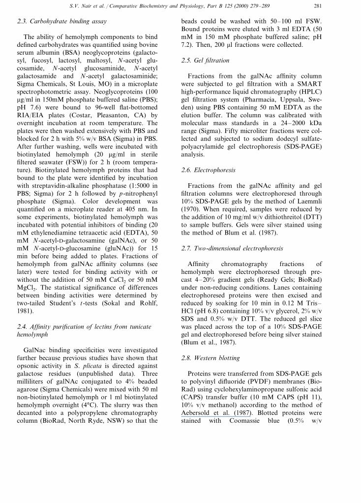
Fig. 1. Binding (absorbance as optical density units (ODU405))of biotinylated hemolymph to BSA or a range of BSA neogly-coproteins that were immobilized on EIA/RIA plates.Hemolymph was incubated with BSA or neoglycoproteins,either without potential inhibitors of binding or in the pres-ence of 20 mM EDTA, 50 mM gluNAc or 50 mM galNAc.Bars represent standard errors, n=6. The neoglycoproteinsused were galactosyl (BSA-galactose), fucosyl (BSA-fucosy-lamide), maltosyl (BSA-maltosyl), lactosyl (BSA-lactosyl), glu-amide(BSA-glucosamide),glu-aminide(BSA-p-aminophenyl-N-acetyl-b-D-glucosaminide), gal-amide (BSA-galactosamide), gal-aminide (BSA-p-aminophenyl-N-acetyl-b-D-galactosaminide).
S.V. Nair et al. / Comparati6e Biochemistry and Physiology, Part B 125 (2000) 279–289 283
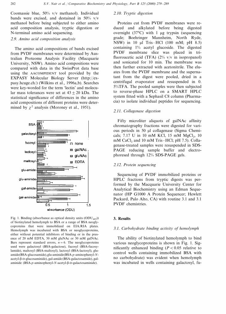
Fig. 2. Binding to immobilized BSA-N-acetyl galactosamide(absorbance as ODU405) of a fraction of biotinylatedhemolymph isolated by galNAc affinity chromatography. Thefraction was incubated with BSA-N-acetyl galactosamide with-out added divalent cations (no Ca2+ or Mg2+) or in thepresence of 50 mM Ca2+ or 50 mM Mg2+. Bars=standarderrors, n=6.
cosyl, N-acetyl glucosamide, N-acetyl glu-cosaminide, N-acetyl galactosamide and N-acetylgalactosaminide neoglycoproteins. No significantbinding (P\0.05 compared with BSA controls)was detected in wells containing lactosyl or malto-syl neoglycoproteins. In all cases, the binding ofhemolymph to neoglycoproteins was significantlyinhibited by pre-incubating hemolymph with 20mM EDTA.
Pre-incubation of hemolymph with soluble N-acetyl-D-galactosamine inhibited the ability ofhemolymph proteins to bind galactose, N-acetylgalactosamide and N-acetyl galactosaminide neo-glycoproteins, but did not significantly affectbinding to fucose, N-acetyl glucosamide, N-acetylglucosaminide neoglycoproteins (P\0.05). Con-versely, soluble N-acetyl-D-glucosamine inhibitedthe binding of hemolymph proteins to fucose,N-acetyl glucosamide and N-acetyl glu-cosaminide, but not to galactose, N-acetyl galac-tosamide and N-acetyl galactosaminide.
Fig. 2 shows that a fraction of biotinylatedhemolymph isolated by galNAc affinity chro-matography could not bind wells coated withN-acetyl galactosamide-BSA without the additionof divalent cations to quench the effects of theEDTA that was used to elute protein from theaffinity column. The N-acetyl galactosamide-BSAbinding activity of the fraction was restored byco-incubation with 50 mM Ca2+, but not 50 mMMg2+.
3.2. SDS-PAGE of galNAc affinity fractions
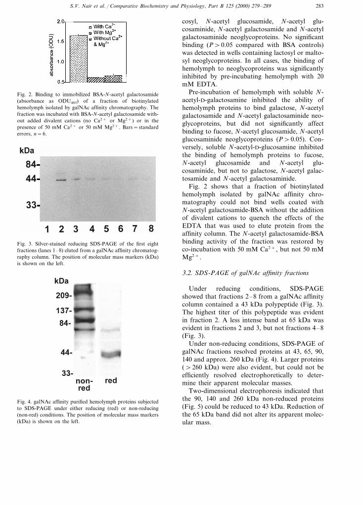
Under reducing conditions, SDS-PAGEshowed that fractions 2–8 from a galNAc affinitycolumn contained a 43 kDa polypeptide (Fig. 3).The highest titer of this polypeptide was evidentin fraction 2. A less intense band at 65 kDa wasevident in fractions 2 and 3, but not fractions 4–8(Fig. 3).
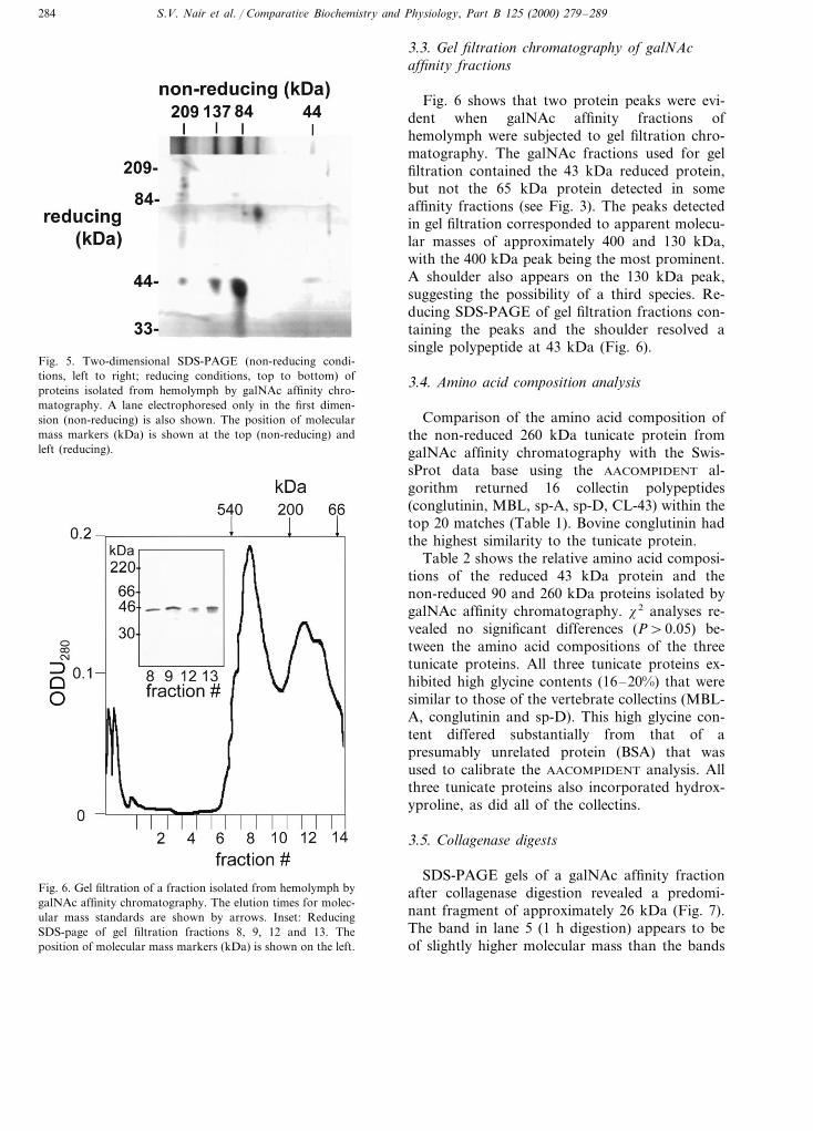
Under non-reducing conditions, SDS-PAGE ofgalNAc fractions resolved proteins at 43, 65, 90,140 and approx. 260 kDa (Fig. 4). Larger proteins(\260 kDa) were also evident, but could not beefficiently resolved electrophoretically to deter-mine their apparent molecular masses.
Two-dimensional electrophoresis indicated thatthe 90, 140 and 260 kDa non-reduced proteins(Fig. 5) could be reduced to 43 kDa. Reduction ofthe 65 kDa band did not alter its apparent molec-ular mass.
Fig. 3. Silver-stained reducing SDS-PAGE of the first eightfractions (lanes 1–8) eluted from a galNAc affinity chromatog-raphy column. The position of molecular mass markers (kDa)is shown on the left.
Fig. 4. galNAc affinity purified hemolymph proteins subjectedto SDS-PAGE under either reducing (red) or non-reducing(non-red) conditions. The position of molecular mass markers(kDa) is shown on the left.
S.V. Nair et al. / Comparati6e Biochemistry and Physiology, Part B 125 (2000) 279–289284
3.3. Gel filtration chromatography of galNAcaffinity fractions
Fig. 6 shows that two protein peaks were evi-dent when galNAc affinity fractions ofhemolymph were subjected to gel filtration chro-matography. The galNAc fractions used for gelfiltration contained the 43 kDa reduced protein,but not the 65 kDa protein detected in someaffinity fractions (see Fig. 3). The peaks detectedin gel filtration corresponded to apparent molecu-lar masses of approximately 400 and 130 kDa,with the 400 kDa peak being the most prominent.A shoulder also appears on the 130 kDa peak,suggesting the possibility of a third species. Re-ducing SDS-PAGE of gel filtration fractions con-taining the peaks and the shoulder resolved asingle polypeptide at 43 kDa (Fig. 6).
3.4. Amino acid composition analysis
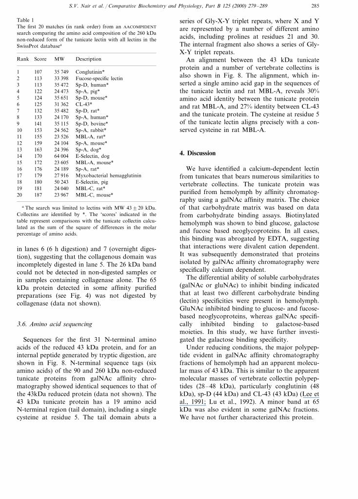
Comparison of the amino acid composition ofthe non-reduced 260 kDa tunicate protein fromgalNAc affinity chromatography with the Swis-sProt data base using the AACOMPIDENT al-gorithm returned 16 collectin polypeptides(conglutinin, MBL, sp-A, sp-D, CL-43) within thetop 20 matches (Table 1). Bovine conglutinin hadthe highest similarity to the tunicate protein.
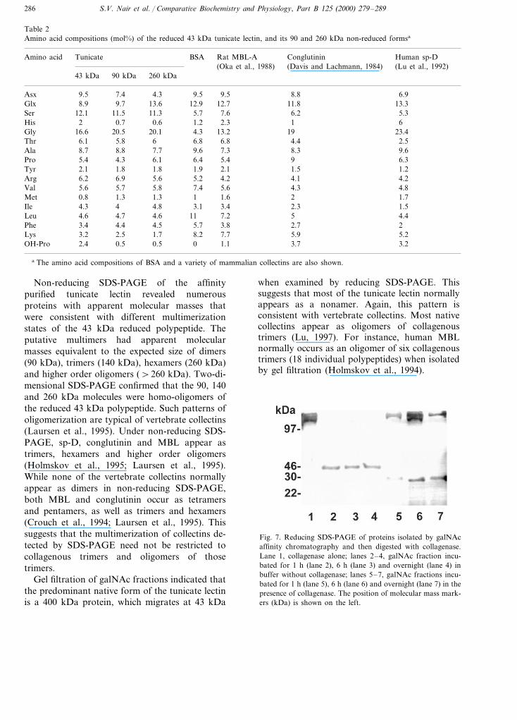
Table 2 shows the relative amino acid composi-tions of the reduced 43 kDa protein and thenon-reduced 90 and 260 kDa proteins isolated bygalNAc affinity chromatography. x2 analyses re-vealed no significant differences (P\0.05) be-tween the amino acid compositions of the threetunicate proteins. All three tunicate proteins ex-hibited high glycine contents (16–20%) that weresimilar to those of the vertebrate collectins (MBL-A, conglutinin and sp-D). This high glycine con-tent differed substantially from that of apresumably unrelated protein (BSA) that wasused to calibrate the AACOMPIDENT analysis. Allthree tunicate proteins also incorporated hydrox-yproline, as did all of the collectins.
3.5. Collagenase digests
SDS-PAGE gels of a galNAc affinity fractionafter collagenase digestion revealed a predomi-nant fragment of approximately 26 kDa (Fig. 7).The band in lane 5 (1 h digestion) appears to beof slightly higher molecular mass than the bands
Fig. 5. Two-dimensional SDS-PAGE (non-reducing condi-tions, left to right; reducing conditions, top to bottom) ofproteins isolated from hemolymph by galNAc affinity chro-matography. A lane electrophoresed only in the first dimen-sion (non-reducing) is also shown. The position of molecularmass markers (kDa) is shown at the top (non-reducing) andleft (reducing).
Fig. 6. Gel filtration of a fraction isolated from hemolymph bygalNAc affinity chromatography. The elution times for molec-ular mass standards are shown by arrows. Inset: ReducingSDS-page of gel filtration fractions 8, 9, 12 and 13. Theposition of molecular mass markers (kDa) is shown on the left.
S.V. Nair et al. / Comparati6e Biochemistry and Physiology, Part B 125 (2000) 279–289 285
Table 1The first 20 matches (in rank order) from an AACOMPIDENT
search comparing the amino acid composition of the 260 kDanon-reduced form of the tunicate lectin with all lectins in theSwissProt databasea
Rank Score MW Description
1 107 35 749 Conglutinin*1132 33 398 Fucose-specific lectin
3 113 35 472 Sp-D, human*4 122 24 473 Sp-A, pig*
1245 35 651 Sp-D, mouse*6 125 31 362 CL-43*
1327 35 482 Sp-D, rat*8 133 24 170 Sp-A, human*9 141 35 115 Sp-D, bovine*
15310 24 562 Sp-A, rabbit*15511 23 526 MBL-A, rat*15912 24 104 Sp-A, mouse*
13 163 24 396 Sp-A, dog*14 170 64 004 E-Selectin, dog
17215 23 605 MBL-A, mouse*176 24 189 Sp-A, rat*1617917 27 916 Myxobacterial hemagglutinin
18 180 50 243 E-Selectin, pig181 24 040 MBL-C, rat*19187 23 96720 MBL-C, mouse*
a The search was limited to lectins with MW 43920 kDa.Collectins are identified by *. The ‘scores’ indicated in thetable represent comparisons with the tunicate collectin calcu-lated as the sum of the square of differences in the molarpercentage of amino acids.
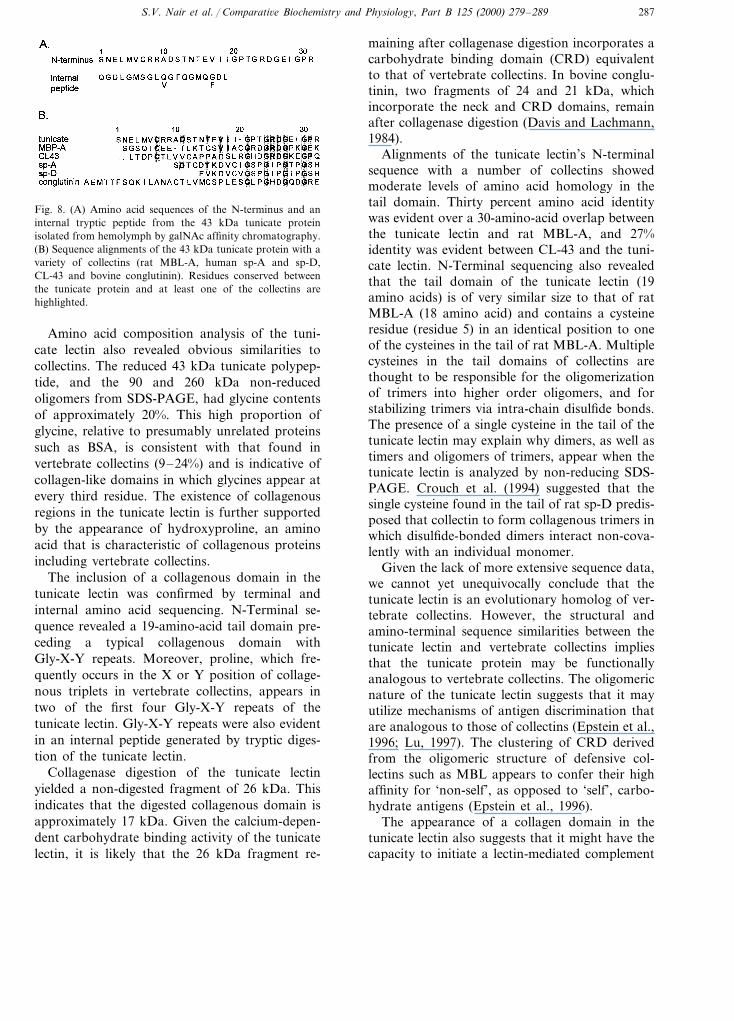
series of Gly-X-Y triplet repeats, where X and Yare represented by a number of different aminoacids, including prolines at residues 21 and 30.The internal fragment also shows a series of Gly-X-Y triplet repeats.
An alignment between the 43 kDa tunicateprotein and a number of vertebrate collectins isalso shown in Fig. 8. The alignment, which in-serted a single amino acid gap in the sequences ofthe tunicate lectin and rat MBL-A, reveals 30%amino acid identity between the tunicate proteinand rat MBL-A, and 27% identity between CL-43and the tunicate protein. The cysteine at residue 5of the tunicate lectin aligns precisely with a con-served cysteine in rat MBL-A.
4. Discussion
We have identified a calcium-dependent lectinfrom tunicates that bears numerous similarities tovertebrate collectins. The tunicate protein waspurified from hemolymph by affinity chromatog-raphy using a galNAc affinity matrix. The choiceof that carbohydrate matrix was based on datafrom carbohydrate binding assays. Biotinylatedhemolymph was shown to bind glucose, galactoseand fucose based neoglycoproteins. In all cases,this binding was abrogated by EDTA, suggestingthat interactions were divalent cation dependent.It was subsequently demonstrated that proteinsisolated by galNAc affinity chromatography werespecifically calcium dependent.
The differential ability of soluble carbohydrates(galNAc or gluNAc) to inhibit binding indicatedthat at least two different carbohydrate binding(lectin) specificities were present in hemolymph.GluNAc inhibited binding to glucose- and fucose-based neoglycoproteins, whereas galNAc specifi-cally inhibited binding to galactose-basedmoieties. In this study, we have further investi-gated the galactose binding specificity.
Under reducing conditions, the major polypep-tide evident in galNAc affinity chromatographyfractions of hemolymph had an apparent molecu-lar mass of 43 kDa. This is similar to the apparentmolecular masses of vertebrate collectin polypep-tides (28–48 kDa), particularly conglutinin (48kDa), sp-D (44 kDa) and CL-43 (43 kDa) (Lee etal., 1991; Lu et al., 1992). A minor band at 65kDa was also evident in some galNAc fractions.We have not further characterized this protein.
in lanes 6 (6 h digestion) and 7 (overnight diges-tion), suggesting that the collagenous domain wasincompletely digested in lane 5. The 26 kDa bandcould not be detected in non-digested samples orin samples containing collagenase alone. The 65kDa protein detected in some affinity purifiedpreparations (see Fig. 4) was not digested bycollagenase (data not shown).
3.6. Amino acid sequencing
Sequences for the first 31 N-terminal aminoacids of the reduced 43 kDa protein, and for aninternal peptide generated by tryptic digestion, areshown in Fig. 8. N-terminal sequence tags (sixamino acids) of the 90 and 260 kDa non-reducedtunicate proteins from galNAc affinity chro-matography showed identical sequences to that ofthe 43kDa reduced protein (data not shown). The43 kDa tunicate protein has a 19 amino acidN-terminal region (tail domain), including a singlecysteine at residue 5. The tail domain abuts a
S.V. Nair et al. / Comparati6e Biochemistry and Physiology, Part B 125 (2000) 279–289286
Table 2Amino acid compositions (mol%) of the reduced 43 kDa tunicate lectin, and its 90 and 260 kDa non-reduced formsa
Tunicate BSAAmino acid Rat MBL-A Human sp-DConglutinin(Lu et al., 1992)(Davis and Lachmann, 1984)(Oka et al., 1988)
43 kDa 90 kDa 260 kDa
9.5 7.4 4.3Asx 9.5 9.5 8.8 6.98.9 9.7 13.6Glx 12.9 12.7 11.8 13.3
12.1Ser 11.5 11.3 5.7 7.6 6.2 5.32 0.7 0.6His 1.2 2.3 1 6
16.6 20.5 20.1Gly 4.3 13.2 19 23.46.1 5.8 6Thr 6.8 6.8 4.4 2.5
Ala 8.7 8.8 7.7 9.6 7.3 8.3 9.65.4 4.3 6.1Pro 6.4 5.4 9 6.3
Tyr 2.1 1.8 1.8 1.9 2.1 1.5 1.26.2 6.9 5.6Arg 5.2 4.2 4.1 4.25.6 5.7 5.8Val 7.4 5.6 4.3 4.80.8 1.3 1.3Met 1 1.6 2 1.74.3 4 4.8Ile 3.1 3.4 2.3 1.5
Leu 4.6 4.7 4.6 11 7.2 5 4.43.4 4.4 4.5Phe 5.7 3.8 2.7 2
Lys 3.2 2.5 1.7 8.2 7.7 5.9 5.22.4 0.5 0.5 0 1.1OH-Pro 3.7 3.2
a The amino acid compositions of BSA and a variety of mammalian collectins are also shown.
Non-reducing SDS-PAGE of the affinitypurified tunicate lectin revealed numerousproteins with apparent molecular masses thatwere consistent with different multimerizationstates of the 43 kDa reduced polypeptide. Theputative multimers had apparent molecularmasses equivalent to the expected size of dimers(90 kDa), trimers (140 kDa), hexamers (260 kDa)and higher order oligomers (\260 kDa). Two-di-mensional SDS-PAGE confirmed that the 90, 140and 260 kDa molecules were homo-oligomers ofthe reduced 43 kDa polypeptide. Such patterns ofoligomerization are typical of vertebrate collectins(Laursen et al., 1995). Under non-reducing SDS-PAGE, sp-D, conglutinin and MBL appear astrimers, hexamers and higher order oligomers(Holmskov et al., 1995; Laursen et al., 1995).While none of the vertebrate collectins normallyappear as dimers in non-reducing SDS-PAGE,both MBL and conglutinin occur as tetramersand pentamers, as well as trimers and hexamers(Crouch et al., 1994; Laursen et al., 1995). Thissuggests that the multimerization of collectins de-tected by SDS-PAGE need not be restricted tocollagenous trimers and oligomers of thosetrimers.
Gel filtration of galNAc fractions indicated thatthe predominant native form of the tunicate lectinis a 400 kDa protein, which migrates at 43 kDa
when examined by reducing SDS-PAGE. Thissuggests that most of the tunicate lectin normallyappears as a nonamer. Again, this pattern isconsistent with vertebrate collectins. Most nativecollectins appear as oligomers of collagenoustrimers (Lu, 1997). For instance, human MBLnormally occurs as an oligomer of six collagenoustrimers (18 individual polypeptides) when isolatedby gel filtration (Holmskov et al., 1994).
Fig. 7. Reducing SDS-PAGE of proteins isolated by galNAcaffinity chromatography and then digested with collagenase.Lane 1, collagenase alone; lanes 2–4, galNAc fraction incu-bated for 1 h (lane 2), 6 h (lane 3) and overnight (lane 4) inbuffer without collagenase; lanes 5–7, galNAc fractions incu-bated for 1 h (lane 5), 6 h (lane 6) and overnight (lane 7) in thepresence of collagenase. The position of molecular mass mark-ers (kDa) is shown on the left.
S.V. Nair et al. / Comparati6e Biochemistry and Physiology, Part B 125 (2000) 279–289 287
Fig. 8. (A) Amino acid sequences of the N-terminus and aninternal tryptic peptide from the 43 kDa tunicate proteinisolated from hemolymph by galNAc affinity chromatography.(B) Sequence alignments of the 43 kDa tunicate protein with avariety of collectins (rat MBL-A, human sp-A and sp-D,CL-43 and bovine conglutinin). Residues conserved betweenthe tunicate protein and at least one of the collectins arehighlighted.
maining after collagenase digestion incorporates acarbohydrate binding domain (CRD) equivalentto that of vertebrate collectins. In bovine conglu-tinin, two fragments of 24 and 21 kDa, whichincorporate the neck and CRD domains, remainafter collagenase digestion (Davis and Lachmann,1984).
Alignments of the tunicate lectin’s N-terminalsequence with a number of collectins showedmoderate levels of amino acid homology in thetail domain. Thirty percent amino acid identitywas evident over a 30-amino-acid overlap betweenthe tunicate lectin and rat MBL-A, and 27%identity was evident between CL-43 and the tuni-cate lectin. N-Terminal sequencing also revealedthat the tail domain of the tunicate lectin (19amino acids) is of very similar size to that of ratMBL-A (18 amino acid) and contains a cysteineresidue (residue 5) in an identical position to oneof the cysteines in the tail of rat MBL-A. Multiplecysteines in the tail domains of collectins arethought to be responsible for the oligomerizationof trimers into higher order oligomers, and forstabilizing trimers via intra-chain disulfide bonds.The presence of a single cysteine in the tail of thetunicate lectin may explain why dimers, as well astimers and oligomers of trimers, appear when thetunicate lectin is analyzed by non-reducing SDS-PAGE. Crouch et al. (1994) suggested that thesingle cysteine found in the tail of rat sp-D predis-posed that collectin to form collagenous trimers inwhich disulfide-bonded dimers interact non-cova-lently with an individual monomer.
Given the lack of more extensive sequence data,we cannot yet unequivocally conclude that thetunicate lectin is an evolutionary homolog of ver-tebrate collectins. However, the structural andamino-terminal sequence similarities between thetunicate lectin and vertebrate collectins impliesthat the tunicate protein may be functionallyanalogous to vertebrate collectins. The oligomericnature of the tunicate lectin suggests that it mayutilize mechanisms of antigen discrimination thatare analogous to those of collectins (Epstein et al.,1996; Lu, 1997). The clustering of CRD derivedfrom the oligomeric structure of defensive col-lectins such as MBL appears to confer their highaffinity for ‘non-self’, as opposed to ‘self’, carbo-hydrate antigens (Epstein et al., 1996).
The appearance of a collagen domain in thetunicate lectin also suggests that it might have thecapacity to initiate a lectin-mediated complement
Amino acid composition analysis of the tuni-cate lectin also revealed obvious similarities tocollectins. The reduced 43 kDa tunicate polypep-tide, and the 90 and 260 kDa non-reducedoligomers from SDS-PAGE, had glycine contentsof approximately 20%. This high proportion ofglycine, relative to presumably unrelated proteinssuch as BSA, is consistent with that found invertebrate collectins (9–24%) and is indicative ofcollagen-like domains in which glycines appear atevery third residue. The existence of collagenousregions in the tunicate lectin is further supportedby the appearance of hydroxyproline, an aminoacid that is characteristic of collagenous proteinsincluding vertebrate collectins.
The inclusion of a collagenous domain in thetunicate lectin was confirmed by terminal andinternal amino acid sequencing. N-Terminal se-quence revealed a 19-amino-acid tail domain pre-ceding a typical collagenous domain withGly-X-Y repeats. Moreover, proline, which fre-quently occurs in the X or Y position of collage-nous triplets in vertebrate collectins, appears intwo of the first four Gly-X-Y repeats of thetunicate lectin. Gly-X-Y repeats were also evidentin an internal peptide generated by tryptic diges-tion of the tunicate lectin.
Collagenase digestion of the tunicate lectinyielded a non-digested fragment of 26 kDa. Thisindicates that the digested collagenous domain isapproximately 17 kDa. Given the calcium-depen-dent carbohydrate binding activity of the tunicatelectin, it is likely that the 26 kDa fragment re-

S.V. Nair et al. / Comparati6e Biochemistry and Physiology, Part B 125 (2000) 279–289288
pathway. Vertebrate MASPs, which proteolyti-cally activate C3, are known to specifically associ-ate with collagen domains of MBL (Matsushita etal., 1995). Moreover, MASP and C3 homologshave already been identified in tunicates (Ji et al.,1997; Nonaka et al., 1998). To confirm that acomplement pathway which can be activated bycollectin-like proteins exists in tunicates, we arecurrently testing the functional relationship be-tween the collagenous lectin identified in thisstudy and tunicate C3 homologs.
Acknowledgements
This study was supported by the AustralianResearch Council (grant c A97-942) and theMacquarie University Internal Research GrantsScheme. The research was facilitated by access tothe Australian Proteome Analysis Facility estab-lished under the Australian Government’s MajorNational Research Facilities Program.
References
Aebersold, R.H., Leavitt, J., Saavedra, R.A., Hood,L.E., Kent, S.B.H., 1987. Internal amino acid se-quence analysis of proteins separated by one or twodimensional gel electrophoresis after in situ diges-tion on nitrocellulose. Proc. Natl. Acad. Sci. USA84, 6970–6974.
Al-Sharif, W.Z., Sunyer, J.O., Lambris, J.D., Smith,L.C., 1998. Sea urchin coelomocytes specifically ex-press a homologue of the complement componentC3. J. Immunol. 160, 2983–2997.
Blum, H., Beier, H., Gross, H.J., 1987. Improved stain-ing of plant proteins, RNA and DNA in polyacry-lamide gels. Electrophoresis 8, 93–99.
Crouch, E., Persson, A., Chang, D., Heuser, J., 1994.Molecular structure of pulmonary surfactant proteinD (SP-D). J. Biol. Chem. 269, 17311–17319.
Davis, A.E., Lachmann, P.J., 1984. Bovine conglutininis a collagen-like protein. Biochemistry 23, 2139–2144.
Dodds, A.W., Day, A.J., 1993. In: Whaley, K., Loos,M., Weiler, J.M. (Eds.), Complement in Health andDisease. Kluwer, Boston, pp. 39–88.
Epstein, J., Eichbaum, Q., Sheriff, S., Ezekowitz,R.A.B., 1996. The collectins in innate immunity.Curr. Opin. Immunol. 8, 29–35.
Holmskov, U., Malhotra, R., Sim, R.B., Jensenius,J.C., 1994. Collectins: collagenous C-type lectins ofthe innate immune defense system. Immunol. Today15, 67–74.
Holmskov, U., Laursen, S.B., Malhotra, R., Wiede-mann, H., Timpl, R., Stuart, G.R., Tornoe, H.,Madsen, P.S., Reid, K.B.M., Jensenius, J.C., 1995.Comparative study of the structural and functionalproperties of a bovine plasma C-type lectin, col-lectin-43, with other collectins. Biochem. J. 305,889–896.
Hoppe, H.J., Reid, K.B.M., 1994. Collectins — solubleproteins containing collagenous regions and lectindomains — and their roles in innate immunity.Protein Sci. 3, 1143–1158.
Ji, X., Azumi, K., Sasaki, M., Nonaka, M., 1997.Ancient origin of the complement lectin pathwayrevealed by molecular cloning of mannan binding-protein-associated serine protease from a urochor-date, the Japanese ascidian, Halocynthia roretzi.Proc. Natl. Acad. Sci. USA 94, 6340–6345.
Laemmli, U.K., 1970. Cleavage of structural proteinsduring the assembly of the head of bacteriophageT4. Nature 227, 680–685.
Laursen, S.B., Hedemand, J.E., Thiel, S., Willis, A.C.,Skriver, E., Madsen, P.S., Jensenius, J.C., 1995.Collectin in a non-mammalian species: isolation andcharacterisation of mannan binding protein (MBP)from chicken serum. Glycobiology 5, 553–561.
Lee, Y.-M., Leiby, K.R., Allar, J., Paris, K., Lerch, B.,Okarma, T.B., 1991. Primary structure of bovineconglutinin, a member of the C-type animal lectinfamily. J. Biol. Chem. 266, 2715–2723.
Lu, J.H., 1997. Collectins — collectors of microorgan-isms for the innate immune system. Bioessays 19,509–518.
Lu, J., Willis, A.C., Reid, K.B.M., 1992. Purification,characterisation and cDNA cloning of human lungsurfactant protein D. Biochem. J. 284, 795–802.
Lu, J., Wiedeman, H., Holmskov, U., Thiel, S., Timpl,R., Reid, K.B.M., 1993. Structural similarity be-tween lung surfactant protein D and conglutinin.Eur. J. Biochem. 215, 793–799.
Matsushita, M., Fujita, T., 1992. Activation of theclassical complement pathway by mannose-bindingprotein in association with a novel C1s-like serineprotease. J. Exp. Med. 176, 1497–1502.
Matsushita, M., Ezekowitz, R.A.B., Fujita, T., 1995.The gly-54 asp allelic form of human mannose-bind-ing protein (BP) fails to bind MBL-associated serineprotease. Biochem. J. 311, 1021–1023.
Moroney, M.J., 1951. Facts from Figures. PenguinBooks, Reading.
Nonaka, M., Azumi, K., Ji, X., Namikawa-Yamada,C., Sasaki, M., Saiga, H., Dodds, A.W., Sekine, H.,Homma, M.K., Matsushita, M., Endo, Y., Fujita,T., 1998. Opsonic complement component C3 in thesolitary ascidian, Halocynthia roretzi. J. Immunol.162, 387–391.

S.V. Nair et al. / Comparati6e Biochemistry and Physiology, Part B 125 (2000) 279–289 289
Oka, S., Ikeda, K., Kawasaka, T., Yamashina, I., 1988.Isolation and characterisation of two distinct man-nan-binding proteins from rat serum. Arch.Biochem. Biophys. 260, 257–266.
Smith, L.C., Chang, L., Britten, R.J., Davidson, E.H.,1996. Sea urchin genes expressed in activated coelo-mocytes are identified by expressed sequence tags. J.Immunol. 156, 593–602.
Sokal, R.R., Rohlf, F.J., 1981. Biometry. W.H. Free-man, London.
Strang, C.J., Slayter, H.S., Lachmann, P.J., David,A.E., 1986. Ultrastructure and composition ofbovine conglutinin. Biochem. J. 234, 381–389.
Tan, S.M., Chung, M.C.M., Kon, O.L., Thiel, S., Lee,S.H., Lu, J., 1996. Improvement on the purificationof mannan-binding lectin and demonstration of itsCa2+-independent association with a C1s-like serineprotease. Biochem. J. 319, 329–332.
Thiel, S., Vorup-Jensen, T., Stover, C.M., Schwaeble,W., Laursen, S.B., Poulsen, K., Willis, A.C., Eggle-ton, P., Hansen, S., Holmskov, U., Reid, K.B.M.,Jensenius, J.C., 1997. A second serine protease asso-ciated with mannan-binding lectin that activatescomplement. Nature 386, 506–510.
Wilkins, M.R., Pasquali, C., Appel, R.D., Ou, K.,Golaz, O., Sanchez, J.C., Yan, J.X., Gooley, A.A.,Hughes, G., Humphery-Smith, I., Williams, K.L.,Hochstrasser, D.F., 1996a. From proteins to pro-teomes: large scale protein identification by two-di-mensional electrophoresis and amino acid analysis.Bio/Technology 14, 61–65.
Wilkins, M.R., Ou, K., Appel, R.D., Sanchez, J.C.,Yan, J.X., Golaz, O., Farnsworth, V., Cartier, P.,Hochstrasser, D.F., Williams, K.L., Gooley, A.A.,1996b. Rapid protein identification using N-terminal‘sequence tag’ and amino acid analysis. Biochem.Biophys. Res. Commun. 221, 609–613.
.
Related Documents