A chemokine-binding domain in the tumor necrosis factor receptor from variola (smallpox) virus Alı´Alejo* †‡ , M. Begon ˜ a Ruiz-Argu ¨ ello* ‡§ , Yin Ho*, Vincent P. Smith* ¶ , Margarida Saraiva* , and Antonio Alcami* † ** *Department of Medicine, University of Cambridge, Addenbrooke’s Hospital, Cambridge CB2 2QQ, United Kingdom; † Centro Nacional de Biotecnologı´a, Consejo Superior de Investigaciones Cientı´ficas, Campus de Cantoblanco, 28049 Madrid, Spain; and § Centro de Investigacio ´ n en Sanidad Animal, Instituto Nacional de Investigaciones Agrarias, Valdeolmos, 28130 Madrid, Spain Edited by Bernard Moss, National Institutes of Health, Bethesda, MD, and approved February 10, 2006 (received for review December 7, 2005) Variola virus (VaV) is the causative agent of smallpox, one of the most devastating diseases encountered by man, that was eradi- cated in 1980. The deliberate release of VaV would have cata- strophic consequences on global public health. However, the mechanisms that contribute to smallpox pathogenesis are poorly understood at the molecular level. The ability of viruses to evade the host defense mechanisms is an important determinant of viral pathogenesis. Here we show that the tumor necrosis factor recep- tor (TNFR) homologue CrmB encoded by VaV functions not only as a soluble decoy TNFR but also as a highly specific binding protein for several chemokines that mediate recruitment of immune cells to mucosal surfaces and the skin, sites of virus entry and viral replication at late stages of smallpox. CrmB binds chemokines through its C-terminal domain, which is unrelated to TNFRs, was named smallpox virus-encoded chemokine receptor (SECRET) do- main and uncovers a family of poxvirus chemokine inhibitors. An active SECRET domain was found in another viral TNFR (CrmD) and three secreted proteins encoded by orthopoxviruses. These find- ings identify a previously undescribed chemokine-binding and inhibitory domain unrelated to host chemokine receptors and a mechanism of immune modulation in VaV that may influence smallpox pathogenesis. immune evasion viral pathogenesis cytokine receptor poxvirus inflammation T he poxvirus variola virus (VaV) is the causative agent of smallpox, which was declared to be eradicated in 1980 as a result of the World Health Organization Smallpox Global Erad- ication Campaign, becoming the first and only viral disease eradicated by mass vaccination (1, 2). Thus, research on VaV terminated and the remaining smallpox samples were stored in two high security laboratories. The deliberate release of VaV would have catastrophic consequences on global public health considering that the majority of the human population has not been vaccinated or received a vaccination boost in recent years. Therefore, smallpox is considered one of the most dangerous threats as a biological weapon in bioterrorism, and there is an urgent need to define the mechanisms of smallpox pathogenesis (3). The available data suggest that the toxaemia reported in individuals suffering from severe smallpox may be immune- related. Considering recent advances in molecular pathogenesis with related poxviruses, it is likely that immune evasion strategies play a critical role as determinants of immunopathology and pathogenesis of smallpox (4, 5). Moreover, immune evasion mechanisms may modulate an immunopathological reaction responsible for adverse effects after smallpox vaccination (1). The primary function of the immune system is to protect the host from invading pathogens such as viruses. To survive in the immunocompetent host, viral mechanisms that target specific immune pathways have evolved. A unique immune evasion strategy used by large DNA viruses (poxviruses and herpesvi- ruses) is the production of secreted versions of host receptors or binding proteins that sequester cytokines and neutralize these regulatory molecules (4, 5). Viral TNF receptors (vTNFRs) encoded by poxviruses block the activity of this proinf lammatory cytokine and are examples of viral decoy receptors with se- quence similarity to the extracellular cytokine-binding domain of their cellular counterparts. Some poxviruses and herpesviruses encode secreted chemokine-binding proteins belonging to a second class of viral decoy receptors with unique structures unrelated to host receptors (6, 7) that bind with high affinity a broad range of chemokines, which are mediators of cell migra- tion (5, 8–14). The secreted 35-kDa protein that binds CC chemokines is the only viral chemokine-binding protein identi- fied in VaV and the vaccinia virus (VV) smallpox vaccine strains Lister and DryVax (Wyeth) (8–10). Four genes encoding vTNFRs have been described in poxvi- ruses and named cytokine response modifier B (CrmB), CrmC, CrmD, and CrmE, and their expression varies among viral species (Fig. 1; refs. 4 and 5; www.poxvirus.org). Cowpox virus (CPV), a rodent virus that infects other species sporadically, encodes all four vTNFRs (15–18). Ectromelia virus (EV) is a highly virulent mouse pathogen that causes mousepox, a disease similar to smallpox, and encodes CrmD only (19). VV Western Reserve and the strains used as smallpox vaccines Copenhagen, DryVax (Wyeth), and Tian-Tan, do not encode vTNFRs, but CrmC and CrmE are encoded by the vaccine strains Lister, USSR, and Evans (20, 21). VaV and monkeypox virus, which causes a smallpox-like disease in humans, encode CrmB only (22–26). The reasons for the variety of vTNFRs are not under- stood. In addition to the cysteine-rich domains (CRDs) charac- teristic of the ligand-binding region of cellular TNFRs, CrmB and CrmD have a C-terminal domain (CTD) unrelated to host proteins (Fig. 1). We have characterized the CrmB protein encoded by VaV and found that it functions not only as a decoy TNFR but it also interacts with chemokines through its CTD, which is unrelated to host proteins. This previously undescribed chemokine-binding domain uncovers a family of poxvirus-encoded secreted chemo- kine inhibitors with potential immunomodulatory activity. Results VaV CrmB Functions as a Secreted Decoy TNF Receptor (TNFR). CrmB is the only vTNFR predicted to be active in the VaV strains Conflict of interest statement: No conflicts declared. This paper was submitted directly (Track II) to the PNAS office. Abbreviations: CPV, cowpox virus; CRD, cysteine-rich domain; Crm, cytokine response modifier; CTD, C-terminal domain; EV, ectromelia virus; LT, lymphotoxin-; SCP, SECRET domain-containing protein; SECRET, smallpox virus-encoded chemokine receptor; SPR, surface plasmon resonance; TNFR, TNF receptor; VaV, variola virus; vTNFR, viral TNF receptor; VV, vaccinia virus. ‡ A. Alejo and M.B.R.-A. contributed equally to this work ¶ Present address: Solexa, Ltd., Chesterton Research Park, Little Chesterford, Essex CB10 1XL, United Kingdom. Present address: National Institute for Medical Research, The Ridgeway, Mill Hill, London NW7 1AA, United Kingdom. **To whom correspondence should be addressed. E-mail: [email protected]. © 2006 by The National Academy of Sciences of the USA www.pnas.orgcgidoi10.1073pnas.0510462103 PNAS April 11, 2006 vol. 103 no. 15 5995– 6000 MICROBIOLOGY

Welcome message from author
This document is posted to help you gain knowledge. Please leave a comment to let me know what you think about it! Share it to your friends and learn new things together.
Transcript
A chemokine-binding domain in the tumor necrosisfactor receptor from variola (smallpox) virusAlı Alejo*†‡, M. Begona Ruiz-Arguello*‡§, Yin Ho*, Vincent P. Smith*¶, Margarida Saraiva*�, and Antonio Alcami*†**
*Department of Medicine, University of Cambridge, Addenbrooke’s Hospital, Cambridge CB2 2QQ, United Kingdom; †Centro Nacional de Biotecnologıa,Consejo Superior de Investigaciones Cientıficas, Campus de Cantoblanco, 28049 Madrid, Spain; and §Centro de Investigacion en Sanidad Animal, InstitutoNacional de Investigaciones Agrarias, Valdeolmos, 28130 Madrid, Spain
Edited by Bernard Moss, National Institutes of Health, Bethesda, MD, and approved February 10, 2006 (received for review December 7, 2005)
Variola virus (VaV) is the causative agent of smallpox, one of themost devastating diseases encountered by man, that was eradi-cated in 1980. The deliberate release of VaV would have cata-strophic consequences on global public health. However, themechanisms that contribute to smallpox pathogenesis are poorlyunderstood at the molecular level. The ability of viruses to evadethe host defense mechanisms is an important determinant of viralpathogenesis. Here we show that the tumor necrosis factor recep-tor (TNFR) homologue CrmB encoded by VaV functions not only asa soluble decoy TNFR but also as a highly specific binding proteinfor several chemokines that mediate recruitment of immune cellsto mucosal surfaces and the skin, sites of virus entry and viralreplication at late stages of smallpox. CrmB binds chemokinesthrough its C-terminal domain, which is unrelated to TNFRs, wasnamed smallpox virus-encoded chemokine receptor (SECRET) do-main and uncovers a family of poxvirus chemokine inhibitors. Anactive SECRET domain was found in another viral TNFR (CrmD) andthree secreted proteins encoded by orthopoxviruses. These find-ings identify a previously undescribed chemokine-binding andinhibitory domain unrelated to host chemokine receptors and amechanism of immune modulation in VaV that may influencesmallpox pathogenesis.
immune evasion � viral pathogenesis � cytokine receptor � poxvirus �inflammation
The poxvirus variola virus (VaV) is the causative agent ofsmallpox, which was declared to be eradicated in 1980 as a
result of the World Health Organization Smallpox Global Erad-ication Campaign, becoming the first and only viral diseaseeradicated by mass vaccination (1, 2). Thus, research on VaVterminated and the remaining smallpox samples were stored intwo high security laboratories. The deliberate release of VaVwould have catastrophic consequences on global public healthconsidering that the majority of the human population has notbeen vaccinated or received a vaccination boost in recent years.Therefore, smallpox is considered one of the most dangerousthreats as a biological weapon in bioterrorism, and there is anurgent need to define the mechanisms of smallpox pathogenesis(3). The available data suggest that the toxaemia reported inindividuals suffering from severe smallpox may be immune-related. Considering recent advances in molecular pathogenesiswith related poxviruses, it is likely that immune evasion strategiesplay a critical role as determinants of immunopathology andpathogenesis of smallpox (4, 5). Moreover, immune evasionmechanisms may modulate an immunopathological reactionresponsible for adverse effects after smallpox vaccination (1).
The primary function of the immune system is to protect thehost from invading pathogens such as viruses. To survive in theimmunocompetent host, viral mechanisms that target specificimmune pathways have evolved. A unique immune evasionstrategy used by large DNA viruses (poxviruses and herpesvi-ruses) is the production of secreted versions of host receptors orbinding proteins that sequester cytokines and neutralize theseregulatory molecules (4, 5). Viral TNF receptors (vTNFRs)
encoded by poxviruses block the activity of this proinflammatorycytokine and are examples of viral decoy receptors with se-quence similarity to the extracellular cytokine-binding domain oftheir cellular counterparts. Some poxviruses and herpesvirusesencode secreted chemokine-binding proteins belonging to asecond class of viral decoy receptors with unique structuresunrelated to host receptors (6, 7) that bind with high affinity abroad range of chemokines, which are mediators of cell migra-tion (5, 8–14). The secreted 35-kDa protein that binds CCchemokines is the only viral chemokine-binding protein identi-fied in VaV and the vaccinia virus (VV) smallpox vaccine strainsLister and DryVax (Wyeth) (8–10).
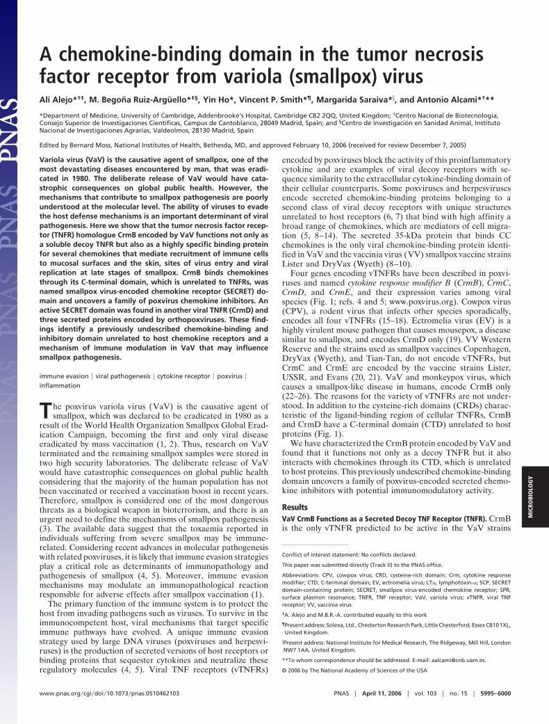
Four genes encoding vTNFRs have been described in poxvi-ruses and named cytokine response modifier B (CrmB), CrmC,CrmD, and CrmE, and their expression varies among viralspecies (Fig. 1; refs. 4 and 5; www.poxvirus.org). Cowpox virus(CPV), a rodent virus that infects other species sporadically,encodes all four vTNFRs (15–18). Ectromelia virus (EV) is ahighly virulent mouse pathogen that causes mousepox, a diseasesimilar to smallpox, and encodes CrmD only (19). VV WesternReserve and the strains used as smallpox vaccines Copenhagen,DryVax (Wyeth), and Tian-Tan, do not encode vTNFRs, butCrmC and CrmE are encoded by the vaccine strains Lister,USSR, and Evans (20, 21). VaV and monkeypox virus, whichcauses a smallpox-like disease in humans, encode CrmB only(22–26). The reasons for the variety of vTNFRs are not under-stood. In addition to the cysteine-rich domains (CRDs) charac-teristic of the ligand-binding region of cellular TNFRs, CrmBand CrmD have a C-terminal domain (CTD) unrelated to hostproteins (Fig. 1).
We have characterized the CrmB protein encoded by VaV andfound that it functions not only as a decoy TNFR but it alsointeracts with chemokines through its CTD, which is unrelatedto host proteins. This previously undescribed chemokine-bindingdomain uncovers a family of poxvirus-encoded secreted chemo-kine inhibitors with potential immunomodulatory activity.
ResultsVaV CrmB Functions as a Secreted Decoy TNF Receptor (TNFR). CrmBis the only vTNFR predicted to be active in the VaV strains
Conflict of interest statement: No conflicts declared.
This paper was submitted directly (Track II) to the PNAS office.
Abbreviations: CPV, cowpox virus; CRD, cysteine-rich domain; Crm, cytokine responsemodifier; CTD, C-terminal domain; EV, ectromelia virus; LT�, lymphotoxin-�; SCP, SECRETdomain-containing protein; SECRET, smallpox virus-encoded chemokine receptor; SPR,surface plasmon resonance; TNFR, TNF receptor; VaV, variola virus; vTNFR, viral TNFreceptor; VV, vaccinia virus.
‡A. Alejo and M.B.R.-A. contributed equally to this work
¶Present address: Solexa, Ltd., Chesterton Research Park, Little Chesterford, Essex CB10 1XL,United Kingdom.
�Present address: National Institute for Medical Research, The Ridgeway, Mill Hill, LondonNW7 1AA, United Kingdom.
**To whom correspondence should be addressed. E-mail: [email protected].
© 2006 by The National Academy of Sciences of the USA
www.pnas.org�cgi�doi�10.1073�pnas.0510462103 PNAS � April 11, 2006 � vol. 103 � no. 15 � 5995–6000
MIC
ROBI
OLO
GY
sequenced to date (refs. 22–24; www.poxvirus.org). To explorethe immunomodulatory activity of CrmB, we generated the VaVstrain Bangladesh-1975 CrmB gene (22) by site-directed mu-tagenesis from the CrmB gene encoded by camelpox virus, aclose relative of VaV (Fig. 8, which is published as supporting
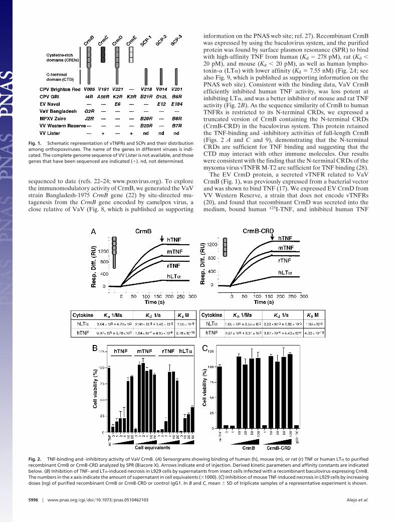
information on the PNAS web site; ref. 27). Recombinant CrmBwas expressed by using the baculovirus system, and the purifiedprotein was found by surface plasmon resonance (SPR) to bindwith high-affinity TNF from human (Kd � 278 pM), rat (Kd �20 pM), and mouse (Kd � 20 pM), as well as human lympho-toxin-� (LT�) with lower affinity (Kd � 7.55 nM) (Fig. 2A; seealso Fig. 9, which is published as supporting information on thePNAS web site). Consistent with the binding data, VaV CrmBefficiently inhibited human TNF activity, was less potent atinhibiting LT�, and was a better inhibitor of mouse and rat TNFactivity (Fig. 2B). As the sequence similarity of CrmB to humanTNFRs is restricted to its N-terminal CRDs, we expressed atruncated version of CrmB containing the N-terminal CRDs(CrmB-CRD) in the baculovirus system. This protein retainedthe TNF-binding and -inhibitory activities of full-length CrmB(Figs. 2 A and C and 9), demonstrating that the N-terminalCRDs are sufficient for TNF binding and suggesting that theCTD may interact with other immune molecules. Our resultswere consistent with the finding that the N-terminal CRDs of themyxoma virus vTNFR M-T2 are sufficient for TNF binding (28).
The EV CrmD protein, a secreted vTNFR related to VaVCrmB (Fig. 1), was previously expressed from a bacterial vectorand was shown to bind TNF (17). We expressed EV CrmD fromVV Western Reserve, a strain that does not encode vTNFRs(20), and found that recombinant CrmD was secreted into themedium, bound human 125I-TNF, and inhibited human TNF
Fig. 1. Schematic representation of vTNFRs and SCPs and their distributionamong orthopoxviruses. The name of the genes in different viruses is indi-cated. The complete genome sequence of VV Lister is not available, and thosegenes that have been sequenced are indicated (�). nd, not determined.
Fig. 2. TNF-binding and -inhibitory activity of VaV CrmB. (A) Sensorgrams showing binding of human (h), mouse (m), or rat (r) TNF or human LT� to purifiedrecombinant CrmB or CrmB-CRD analyzed by SPR (Biacore X). Arrows indicate end of injection. Derived kinetic parameters and affinity constants are indicatedbelow. (B) Inhibition of TNF- and LT�-induced necrosis in L929 cells by supernatants from insect cells infected with a recombinant baculovirus expressing CrmB.The numbers in the x axis indicate the amount of supernatant in cell equivalents (�1000). (C) Inhibition of mouse TNF-induced necrosis in L929 cells by increasingdoses (ng) of purified recombinant CrmB or CrmB-CRD or control IgG1. In B and C, mean � SD of triplicate samples of a representative experiment is shown.
5996 � www.pnas.org�cgi�doi�10.1073�pnas.0510462103 Alejo et al.
biological activity (Fig. 10, which is published as supportinginformation on the PNAS web site). In addition, purified re-combinant EV CrmD expressed in the baculovirus system boundhuman TNF by SPR and inhibited its activity (data not shown).To determine the region of CrmD required for TNF inhibitoryactivity, the N-terminal CRDs of CrmD (CrmD-CRD) wereexpressed in the VV system. Similar to full-length CrmD,CrmD-CRD-bound human TNF in soluble binding assays (datanot shown) and inhibited the biological activity of human TNF(Fig. 10). Thus, similar to VaV CrmB, EV CrmD inhibited TNFthrough its N-terminal CRDs.
Previously Undescribed Chemokine-Binding Activity Encoded by VaVCrmB. The data described above suggested an additional role ofthe CTD of the VaV CrmB protein. A screening with differentcytokines by SPR identified chemokines as ligands of VaVCrmB. This result led us to screen by SPR the potential bindingof purified CrmB to all 43 commercially available humanchemokines. VaV CrmB interacted with some chemokines withbinding affinities similar to that of TNF. The chemokines thatbest bound to VaV CrmB were as follows: CCL28, CCL25,CXCL12�, CXCL13, and CXCL14 (Fig. 3 and Table 1). Similarstudies by SPR with human and mouse chemokines showed thatrecombinant EV CrmD bound with high affinity the same set ofchemokines (Table 2, which is published as supporting informa-tion on the PNAS web site). Recombinant CrmB from CPVexpressed in the baculovirus system also bound these chemo-kines (data not shown).
We next addressed whether VaV CrmB has two independentbinding sites for TNF and chemokines (Fig. 4). In SPR exper-iments, TNF and CXCL12� were injected independently orsimultaneously, and the signal was compared with that obtainedwhen the cytokines were injected consecutively. The bindingsignal after the addition of TNF and CXCL12� together wasequivalent to the sum of that obtained with TNF or CXCL12�alone. Moreover, consecutive addition of TNF and CXCL12�, orCXCL12� and TNF, gave maximum binding, whereas two
successive injections of TNF or CXCL12� alone did not increasethe binding detected with either of them. Thus, saturation of theTNF-binding sites did not affect binding of the chemokine toCrmB and vice versa, supporting the presence of two indepen-dent sites for TNF and chemokines in CrmB. Similarly, com-petitive inhibition studies indicated distinct binding sites for TNFand chemokines in EV CrmD (data not shown).
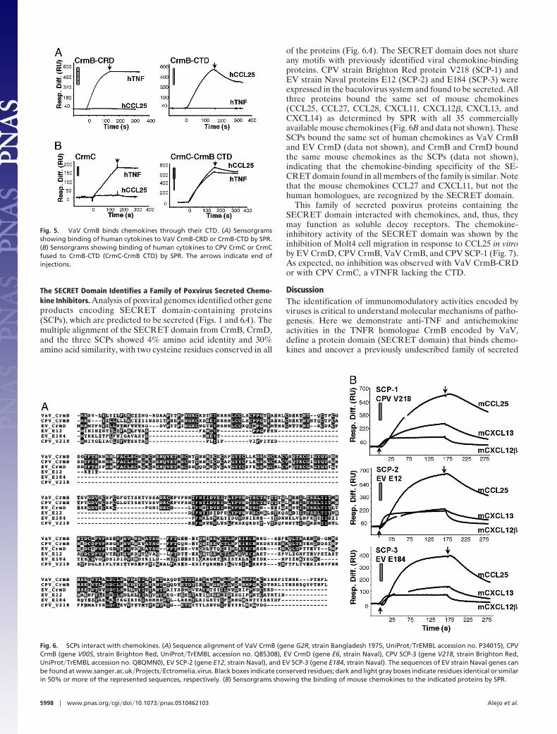
The CTD of VaV CrmB Represents a Previously Undescribed ProteinDomain That Interacts with Chemokines. The presence in CrmB ofa CTD unrelated to host TNFRs and dispensable for TNFbinding, together with the identification of independent bindingsites for TNF and chemokines in CrmB, suggested that thepreviously undescribed chemokine-binding activity of CrmBmay reside in the CTD. To test this hypothesis, we expressed theCTD of CrmB (CrmB-CTD) fused to the predicted N-terminalsignal peptide from CrmB in the baculovirus system. SecretedCrmB-CTD purified from culture supernatants bound chemo-kines but not TNF, whereas CrmB-CRD bound TNF but notchemokines (Fig. 5A), demonstrating that CTD is a previouslyundescribed chemokine-binding domain that we named small-pox virus-encoded chemokine receptor (SECRET) domain.Similarly, EV CrmD-CRD did not bind chemokines (data notshown). Moreover, purified vTNFRs CrmC and CrmE encodedby CPV, which lack the CTD, did not bind chemokines (Fig. 5Band data not shown). The ability of the SECRET domain toconfer chemokine-binding activity to vTNFRs was illustrated byexpression in the baculovirus system of CPV CrmC and CrmEfused to CrmB-CTD. The fusion proteins retained TNF-bindingactivity but, in contrast to CrmC and CrmE, they also boundchemokines (Fig. 5B and data not shown).
Fig. 3. VaV CrmB binds chemokines. Sensorgrams showing binding of theindicated human chemokines to purified recombinant VaV CrmB analyzed bySPR. Arrow indicates end of injection.
Table 1. Kinetic parameters and derived affinity constants of the binding of VaV CrmB to theindicated human chemokines
Chemokine Ka, 1/Ms Kd, 1/s KD, M
hCCL28 1.11 � 106 � 2.63 � 104 4.33 � 10�4 � 1.44 � 10�5 0.30 � 10�9
hCCL25 1.54 � 106 � 2.58 � 104 7.74 � 10�4 � 1.74 � 10�5 0.50 � 10�9
hCXCL12� 1.05 � 106 � 1.91 � 104 4.48 � 10�3 � 2.98 � 10�5 4.26 � 10�9
hCXCL13 3.63 � 105 � 1.47 � 104 2.16 � 10�3 � 3.46 � 10�5 5.95 � 10�9
hCXCL14 1.34 � 105 � 3.41 � 103 8.40 � 10�4 � 1.87 � 10�5 6.29 � 10�9
hXCL1 9.06 � 104 � 1.75 � 103 2.61 � 10�3 � 2.14 � 10�5 28.8 � 10�9
hCCL20 7.59 � 104 � 1.02 � 103 2.22 � 10�3 � 4.16 � 10�5 29.2 � 10�9
Fig. 4. VaV CrmB binds chemokines and TNF through different sites. Sen-sorgrams showing the binding of chemokines and TNF to purified recombi-nant VaV CrmB by SPR. The binding of human TNF or CXCL12� (CK) alone,simultaneous injections of both cytokines (TNF�CK), or two consecutive injec-tions of cytokines (TNF�TNF and CK�CK) are shown. In Lower Right, adiscontinuous line indicates an injection of TNF followed by an injection of CK.Arrow indicate start (1) and end (2) of injection.
Alejo et al. PNAS � April 11, 2006 � vol. 103 � no. 15 � 5997
MIC
ROBI
OLO
GY
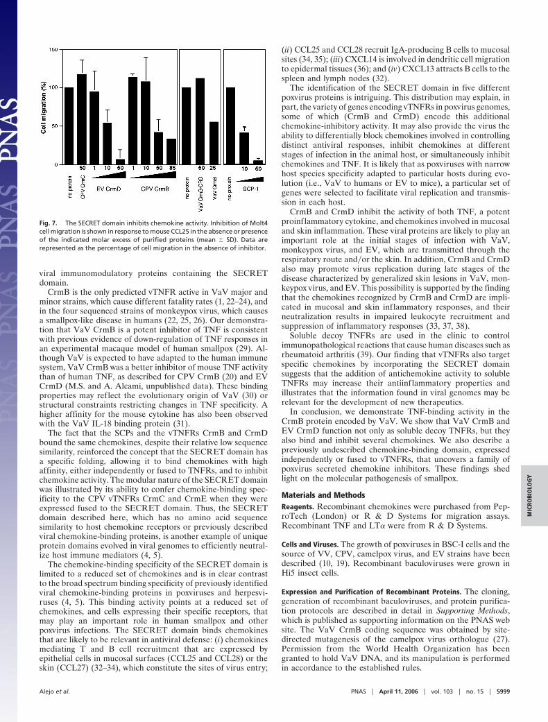
The SECRET Domain Identifies a Family of Poxvirus Secreted Chemo-kine Inhibitors. Analysis of poxviral genomes identified other geneproducts encoding SECRET domain-containing proteins(SCPs), which are predicted to be secreted (Figs. 1 and 6A). Themultiple alignment of the SECRET domain from CrmB, CrmD,and the three SCPs showed 4% amino acid identity and 30%amino acid similarity, with two cysteine residues conserved in all
of the proteins (Fig. 6A). The SECRET domain does not shareany motifs with previously identified viral chemokine-bindingproteins. CPV strain Brighton Red protein V218 (SCP-1) andEV strain Naval proteins E12 (SCP-2) and E184 (SCP-3) wereexpressed in the baculovirus system and found to be secreted. Allthree proteins bound the same set of mouse chemokines(CCL25, CCL27, CCL28, CXCL11, CXCL12�, CXCL13, andCXCL14) as determined by SPR with all 35 commerciallyavailable mouse chemokines (Fig. 6B and data not shown). TheseSCPs bound the same set of human chemokines as VaV CrmBand EV CrmD (data not shown), and CrmB and CrmD boundthe same mouse chemokines as the SCPs (data not shown),indicating that the chemokine-binding specificity of the SE-CRET domain found in all members of the family is similar. Notethat the mouse chemokines CCL27 and CXCL11, but not thehuman homologues, are recognized by the SECRET domain.
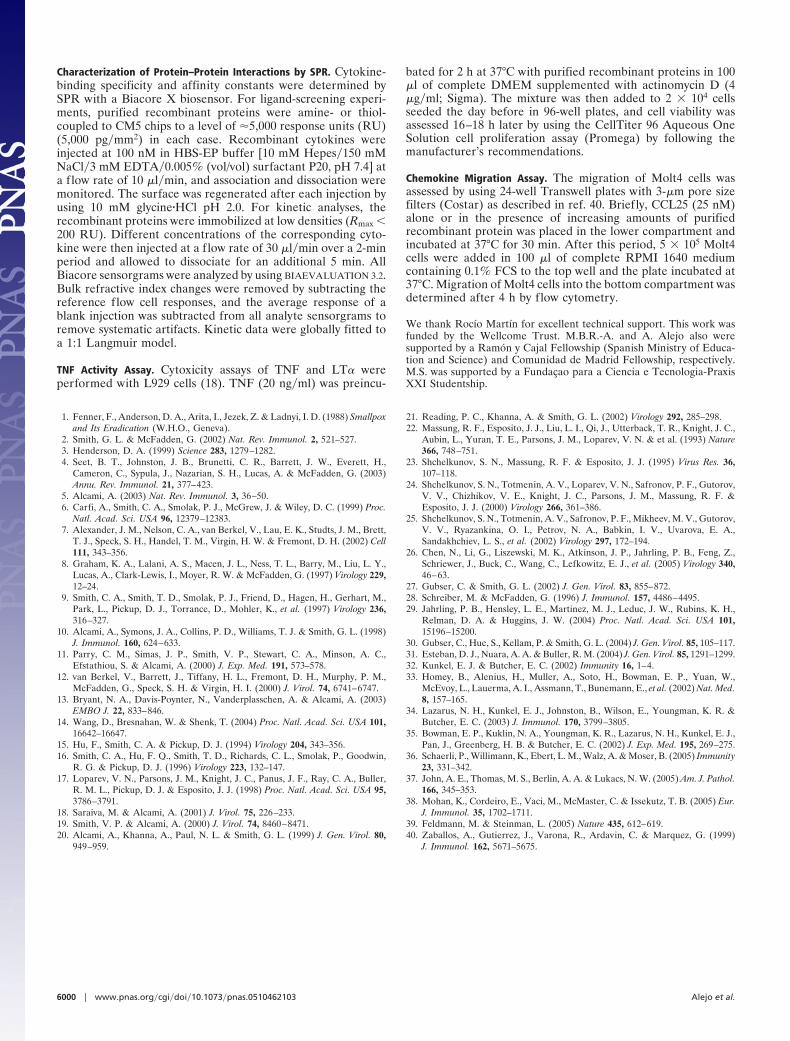
This family of secreted poxvirus proteins containing theSECRET domain interacted with chemokines, and, thus, theymay function as soluble decoy receptors. The chemokine-inhibitory activity of the SECRET domain was shown by theinhibition of Molt4 cell migration in response to CCL25 in vitroby EV CrmD, CPV CrmB, VaV CrmB, and CPV SCP-1 (Fig. 7).As expected, no inhibition was observed with VaV CrmB-CRDor with CPV CrmC, a vTNFR lacking the CTD.
DiscussionThe identification of immunomodulatory activities encoded byviruses is critical to understand molecular mechanisms of patho-genesis. Here we demonstrate anti-TNF and antichemokineactivities in the TNFR homologue CrmB encoded by VaV,define a protein domain (SECRET domain) that binds chemo-kines and uncover a previously undescribed family of secreted
Fig. 5. VaV CrmB binds chemokines through their CTD. (A) Sensorgramsshowing binding of human cytokines to VaV CrmB-CRD or CrmB-CTD by SPR.(B) Sensorgrams showing binding of human cytokines to CPV CrmC or CrmCfused to CrmB-CTD (CrmC-CrmB CTD) by SPR. The arrows indicate end ofinjections.
Fig. 6. SCPs interact with chemokines. (A) Sequence alignment of VaV CrmB (gene G2R, strain Bangladesh 1975, UniProt�TrEMBL accession no. P34015), CPVCrmB (gene V005, strain Brighton Red, UniProt�TrEMBL accession no. Q85308), EV CrmD (gene E6, strain Naval), CPV SCP-3 (gene V218, strain Brighton Red,UniProt�TrEMBL accession no. Q8QMN0), EV SCP-2 (gene E12, strain Naval), and EV SCP-3 (gene E184, strain Naval). The sequences of EV strain Naval genes canbe found at www.sanger.ac.uk�Projects�Ectromelia�virus. Black boxes indicate conserved residues; dark and light gray boxes indicate residues identical or similarin 50% or more of the represented sequences, respectively. (B) Sensorgrams showing the binding of mouse chemokines to the indicated proteins by SPR.
5998 � www.pnas.org�cgi�doi�10.1073�pnas.0510462103 Alejo et al.
viral immunomodulatory proteins containing the SECRETdomain.
CrmB is the only predicted vTNFR active in VaV major andminor strains, which cause different fatality rates (1, 22–24), andin the four sequenced strains of monkeypox virus, which causesa smallpox-like disease in humans (22, 25, 26). Our demonstra-tion that VaV CrmB is a potent inhibitor of TNF is consistentwith previous evidence of down-regulation of TNF responses inan experimental macaque model of human smallpox (29). Al-though VaV is expected to have adapted to the human immunesystem, VaV CrmB was a better inhibitor of mouse TNF activitythan of human TNF, as described for CPV CrmB (20) and EVCrmD (M.S. and A. Alcami, unpublished data). These bindingproperties may reflect the evolutionary origin of VaV (30) orstructural constraints restricting changes in TNF specificity. Ahigher affinity for the mouse cytokine has also been observedwith the VaV IL-18 binding protein (31).
The fact that the SCPs and the vTNFRs CrmB and CrmDbound the same chemokines, despite their relative low sequencesimilarity, reinforced the concept that the SECRET domain hasa specific folding, allowing it to bind chemokines with highaffinity, either independently or fused to TNFRs, and to inhibitchemokine activity. The modular nature of the SECRET domainwas illustrated by its ability to confer chemokine-binding spec-ificity to the CPV vTNFRs CrmC and CrmE when they wereexpressed fused to the SECRET domain. Thus, the SECRETdomain described here, which has no amino acid sequencesimilarity to host chemokine receptors or previously describedviral chemokine-binding proteins, is another example of uniqueprotein domains evolved in viral genomes to efficiently neutral-ize host immune mediators (4, 5).
The chemokine-binding specificity of the SECRET domain islimited to a reduced set of chemokines and is in clear contrastto the broad spectrum binding specificity of previously identifiedviral chemokine-binding proteins in poxviruses and herpesvi-ruses (4, 5). This binding activity points at a reduced set ofchemokines, and cells expressing their specific receptors, thatmay play an important role in human smallpox and otherpoxvirus infections. The SECRET domain binds chemokinesthat are likely to be relevant in antiviral defense: (i) chemokinesmediating T and B cell recruitment that are expressed byepithelial cells in mucosal surfaces (CCL25 and CCL28) or theskin (CCL27) (32–34), which constitute the sites of virus entry;
(ii) CCL25 and CCL28 recruit IgA-producing B cells to mucosalsites (34, 35); (iii) CXCL14 is involved in dendritic cell migrationto epidermal tissues (36); and (iv) CXCL13 attracts B cells to thespleen and lymph nodes (32).
The identification of the SECRET domain in five differentpoxvirus proteins is intriguing. This distribution may explain, inpart, the variety of genes encoding vTNFRs in poxvirus genomes,some of which (CrmB and CrmD) encode this additionalchemokine-inhibitory activity. It may also provide the virus theability to differentially block chemokines involved in controllingdistinct antiviral responses, inhibit chemokines at differentstages of infection in the animal host, or simultaneously inhibitchemokines and TNF. It is likely that as poxviruses with narrowhost species specificity adapted to particular hosts during evo-lution (i.e., VaV to humans or EV to mice), a particular set ofgenes were selected to facilitate viral replication and transmis-sion in each host.
CrmB and CrmD inhibit the activity of both TNF, a potentproinflammatory cytokine, and chemokines involved in mucosaland skin inflammation. These viral proteins are likely to play animportant role at the initial stages of infection with VaV,monkeypox virus, and EV, which are transmitted through therespiratory route and�or the skin. In addition, CrmB and CrmDalso may promote virus replication during late stages of thedisease characterized by generalized skin lesions in VaV, mon-keypox virus, and EV. This possibility is supported by the findingthat the chemokines recognized by CrmB and CrmD are impli-cated in mucosal and skin inflammatory responses, and theirneutralization results in impaired leukocyte recruitment andsuppression of inflammatory responses (33, 37, 38).
Soluble decoy TNFRs are used in the clinic to controlimmunopathological reactions that cause human diseases such asrheumatoid arthritis (39). Our finding that vTNFRs also targetspecific chemokines by incorporating the SECRET domainsuggests that the addition of antichemokine activity to solubleTNFRs may increase their antiinflammatory properties andillustrates that the information found in viral genomes may berelevant for the development of new therapeutics.
In conclusion, we demonstrate TNF-binding activity in theCrmB protein encoded by VaV. We show that VaV CrmB andEV CrmD function not only as soluble decoy TNFRs, but theyalso bind and inhibit several chemokines. We also describe apreviously undescribed chemokine-binding domain, expressedindependently or fused to vTNFRs, that uncovers a family ofpoxvirus secreted chemokine inhibitors. These findings shedlight on the molecular pathogenesis of smallpox.
Materials and MethodsReagents. Recombinant chemokines were purchased from Pep-roTech (London) or R & D Systems for migration assays.Recombinant TNF and LT� were from R & D Systems.
Cells and Viruses. The growth of poxviruses in BSC-I cells and thesource of VV, CPV, camelpox virus, and EV strains have beendescribed (10, 19). Recombinant baculoviruses were grown inHi5 insect cells.
Expression and Purification of Recombinant Proteins. The cloning,generation of recombinant baculoviruses, and protein purifica-tion protocols are described in detail in Supporting Methods,which is published as supporting information on the PNAS website. The VaV CrmB coding sequence was obtained by site-directed mutagenesis of the camelpox virus orthologue (27).Permission from the World Health Organization has beengranted to hold VaV DNA, and its manipulation is performedin accordance to the established rules.
Fig. 7. The SECRET domain inhibits chemokine activity. Inhibition of Molt4cell migration is shown in response to mouse CCL25 in the absence or presenceof the indicated molar excess of purified proteins (mean � SD). Data arerepresented as the percentage of cell migration in the absence of inhibitor.
Alejo et al. PNAS � April 11, 2006 � vol. 103 � no. 15 � 5999
MIC
ROBI
OLO
GY
Characterization of Protein–Protein Interactions by SPR. Cytokine-binding specificity and affinity constants were determined bySPR with a Biacore X biosensor. For ligand-screening experi-ments, purified recombinant proteins were amine- or thiol-coupled to CM5 chips to a level of �5,000 response units (RU)(5,000 pg�mm2) in each case. Recombinant cytokines wereinjected at 100 nM in HBS-EP buffer [10 mM Hepes�150 mMNaCl�3 mM EDTA�0.005% (vol/vol) surfactant P20, pH 7.4] ata flow rate of 10 �l�min, and association and dissociation weremonitored. The surface was regenerated after each injection byusing 10 mM glycine�HCl pH 2.0. For kinetic analyses, therecombinant proteins were immobilized at low densities (Rmax �200 RU). Different concentrations of the corresponding cyto-kine were then injected at a flow rate of 30 �l�min over a 2-minperiod and allowed to dissociate for an additional 5 min. AllBiacore sensorgrams were analyzed by using BIAEVALUATION 3.2.Bulk refractive index changes were removed by subtracting thereference flow cell responses, and the average response of ablank injection was subtracted from all analyte sensorgrams toremove systematic artifacts. Kinetic data were globally fitted toa 1:1 Langmuir model.
TNF Activity Assay. Cytoxicity assays of TNF and LT� wereperformed with L929 cells (18). TNF (20 ng�ml) was preincu-
bated for 2 h at 37°C with purified recombinant proteins in 100�l of complete DMEM supplemented with actinomycin D (4�g�ml; Sigma). The mixture was then added to 2 � 104 cellsseeded the day before in 96-well plates, and cell viability wasassessed 16–18 h later by using the CellTiter 96 Aqueous OneSolution cell proliferation assay (Promega) by following themanufacturer’s recommendations.
Chemokine Migration Assay. The migration of Molt4 cells wasassessed by using 24-well Transwell plates with 3-�m pore sizefilters (Costar) as described in ref. 40. Briefly, CCL25 (25 nM)alone or in the presence of increasing amounts of purifiedrecombinant protein was placed in the lower compartment andincubated at 37°C for 30 min. After this period, 5 � 105 Molt4cells were added in 100 �l of complete RPMI 1640 mediumcontaining 0.1% FCS to the top well and the plate incubated at37°C. Migration of Molt4 cells into the bottom compartment wasdetermined after 4 h by flow cytometry.
We thank Rocıo Martın for excellent technical support. This work wasfunded by the Wellcome Trust. M.B.R.-A. and A. Alejo also weresupported by a Ramon y Cajal Fellowship (Spanish Ministry of Educa-tion and Science) and Comunidad de Madrid Fellowship, respectively.M.S. was supported by a Fundacao para a Ciencia e Tecnologia-PraxisXXI Studentship.
1. Fenner, F., Anderson, D. A., Arita, I., Jezek, Z. & Ladnyi, I. D. (1988) Smallpoxand Its Eradication (W.H.O., Geneva).
2. Smith, G. L. & McFadden, G. (2002) Nat. Rev. Immunol. 2, 521–527.3. Henderson, D. A. (1999) Science 283, 1279–1282.4. Seet, B. T., Johnston, J. B., Brunetti, C. R., Barrett, J. W., Everett, H.,
Cameron, C., Sypula, J., Nazarian, S. H., Lucas, A. & McFadden, G. (2003)Annu. Rev. Immunol. 21, 377–423.
5. Alcami, A. (2003) Nat. Rev. Immunol. 3, 36–50.6. Carfi, A., Smith, C. A., Smolak, P. J., McGrew, J. & Wiley, D. C. (1999) Proc.
Natl. Acad. Sci. USA 96, 12379–12383.7. Alexander, J. M., Nelson, C. A., van Berkel, V., Lau, E. K., Studts, J. M., Brett,
T. J., Speck, S. H., Handel, T. M., Virgin, H. W. & Fremont, D. H. (2002) Cell111, 343–356.
8. Graham, K. A., Lalani, A. S., Macen, J. L., Ness, T. L., Barry, M., Liu, L. Y.,Lucas, A., Clark-Lewis, I., Moyer, R. W. & McFadden, G. (1997) Virology 229,12–24.
9. Smith, C. A., Smith, T. D., Smolak, P. J., Friend, D., Hagen, H., Gerhart, M.,Park, L., Pickup, D. J., Torrance, D., Mohler, K., et al. (1997) Virology 236,316–327.
10. Alcami, A., Symons, J. A., Collins, P. D., Williams, T. J. & Smith, G. L. (1998)J. Immunol. 160, 624–633.
11. Parry, C. M., Simas, J. P., Smith, V. P., Stewart, C. A., Minson, A. C.,Efstathiou, S. & Alcami, A. (2000) J. Exp. Med. 191, 573–578.
12. van Berkel, V., Barrett, J., Tiffany, H. L., Fremont, D. H., Murphy, P. M.,McFadden, G., Speck, S. H. & Virgin, H. I. (2000) J. Virol. 74, 6741–6747.
13. Bryant, N. A., Davis-Poynter, N., Vanderplasschen, A. & Alcami, A. (2003)EMBO J. 22, 833–846.
14. Wang, D., Bresnahan, W. & Shenk, T. (2004) Proc. Natl. Acad. Sci. USA 101,16642–16647.
15. Hu, F., Smith, C. A. & Pickup, D. J. (1994) Virology 204, 343–356.16. Smith, C. A., Hu, F. Q., Smith, T. D., Richards, C. L., Smolak, P., Goodwin,
R. G. & Pickup, D. J. (1996) Virology 223, 132–147.17. Loparev, V. N., Parsons, J. M., Knight, J. C., Panus, J. F., Ray, C. A., Buller,
R. M. L., Pickup, D. J. & Esposito, J. J. (1998) Proc. Natl. Acad. Sci. USA 95,3786–3791.
18. Saraiva, M. & Alcami, A. (2001) J. Virol. 75, 226–233.19. Smith, V. P. & Alcami, A. (2000) J. Virol. 74, 8460–8471.20. Alcami, A., Khanna, A., Paul, N. L. & Smith, G. L. (1999) J. Gen. Virol. 80,
949–959.
21. Reading, P. C., Khanna, A. & Smith, G. L. (2002) Virology 292, 285–298.22. Massung, R. F., Esposito, J. J., Liu, L. I., Qi, J., Utterback, T. R., Knight, J. C.,
Aubin, L., Yuran, T. E., Parsons, J. M., Loparev, V. N. & et al. (1993) Nature366, 748–751.
23. Shchelkunov, S. N., Massung, R. F. & Esposito, J. J. (1995) Virus Res. 36,107–118.
24. Shchelkunov, S. N., Totmenin, A. V., Loparev, V. N., Safronov, P. F., Gutorov,V. V., Chizhikov, V. E., Knight, J. C., Parsons, J. M., Massung, R. F. &Esposito, J. J. (2000) Virology 266, 361–386.
25. Shchelkunov, S. N., Totmenin, A. V., Safronov, P. F., Mikheev, M. V., Gutorov,V. V., Ryazankina, O. I., Petrov, N. A., Babkin, I. V., Uvarova, E. A.,Sandakhchiev, L. S., et al. (2002) Virology 297, 172–194.
26. Chen, N., Li, G., Liszewski, M. K., Atkinson, J. P., Jahrling, P. B., Feng, Z.,Schriewer, J., Buck, C., Wang, C., Lefkowitz, E. J., et al. (2005) Virology 340,46–63.
27. Gubser, C. & Smith, G. L. (2002) J. Gen. Virol. 83, 855–872.28. Schreiber, M. & McFadden, G. (1996) J. Immunol. 157, 4486–4495.29. Jahrling, P. B., Hensley, L. E., Martinez, M. J., Leduc, J. W., Rubins, K. H.,
Relman, D. A. & Huggins, J. W. (2004) Proc. Natl. Acad. Sci. USA 101,15196–15200.
30. Gubser, C., Hue, S., Kellam, P. & Smith, G. L. (2004) J. Gen. Virol. 85, 105–117.31. Esteban, D. J., Nuara, A. A. & Buller, R. M. (2004) J. Gen. Virol. 85, 1291–1299.32. Kunkel, E. J. & Butcher, E. C. (2002) Immunity 16, 1–4.33. Homey, B., Alenius, H., Muller, A., Soto, H., Bowman, E. P., Yuan, W.,
McEvoy, L., Lauerma, A. I., Assmann, T., Bunemann, E., et al. (2002) Nat. Med.8, 157–165.
34. Lazarus, N. H., Kunkel, E. J., Johnston, B., Wilson, E., Youngman, K. R. &Butcher, E. C. (2003) J. Immunol. 170, 3799–3805.
35. Bowman, E. P., Kuklin, N. A., Youngman, K. R., Lazarus, N. H., Kunkel, E. J.,Pan, J., Greenberg, H. B. & Butcher, E. C. (2002) J. Exp. Med. 195, 269–275.
36. Schaerli, P., Willimann, K., Ebert, L. M., Walz, A. & Moser, B. (2005) Immunity23, 331–342.
37. John, A. E., Thomas, M. S., Berlin, A. A. & Lukacs, N. W. (2005) Am. J. Pathol.166, 345–353.
38. Mohan, K., Cordeiro, E., Vaci, M., McMaster, C. & Issekutz, T. B. (2005) Eur.J. Immunol. 35, 1702–1711.
39. Feldmann, M. & Steinman, L. (2005) Nature 435, 612–619.40. Zaballos, A., Gutierrez, J., Varona, R., Ardavin, C. & Marquez, G. (1999)
J. Immunol. 162, 5671–5675.
6000 � www.pnas.org�cgi�doi�10.1073�pnas.0510462103 Alejo et al.
Related Documents