A Catastrophic Case of Thromboembolism: A Case Report Mahmoud H Abdelnaby 1* , Mohamed A Ahmed 2 , Abdallah M Almaghraby 3 , Ashraf A ElAmin 3 and Haitham A Badran 4 1 Department of Clinical and Experimental Internal Medicine, Medical Research Instute, University of Alexandria, Alexandria, Egypt 2 Department of Crical Care, University of Cairo, Giza, Egypt 3 Department of Cardiology, University of Alexandria, Alexandria, Egypt 4 Department of Cardiology, University of Ain Shams, El-Abaseya, Egypt * Corresponding author: Mahmoud H Abdelnaby, Cardiology and Angiology Unit, Department of Clinical and Experimental Internal Medicine, Medical Research Instute, University of Alexandria, Alexandria, Egypt, Tel: +201007573530; E-mail: [email protected] Received: 28 February 2018; Accepted: 20 March 2018; Published: 29 March 2018 Copyright: © 2018 Abdelnaby MH, et al. This is an open-access arcle distributed under the terms of the creave Commons aribuon License, which permits unrestricted use, distribuon and reproducon in any medium, provided the original author and source are credited. Citaon: Abdelnaby MH, Ahmed MA, Almaghraby MA, ElAmin AA, Badran HA (2018) A Catastrophic Case of Thromboembolism: A Case Report. J Heart Cardiovasc Res. Vol. 2 No. 1: 112. Abstract Pulmonary embolizaon (PE) carries a high mortality risk if not suspected and treated properly. Systemic embolizaon is quite common especially in cases of mobile leſt ventricular (LV) thrombi. We were encountered with a catastrophic case of thromboembolism with simultaneous massive PE and bilateral lower limb ischemia in a paent with biventricular systolic dysfuncon and mulple LV thrombi. Tissue plasminogen acvator (tPA) was given and the paents hemodynamics started to be stable. Unfortunately, he developed leſt lag, bilateral lower limb dry gangrene and subsequently he passed away despite all trials to resuscitate. Keywords: Pulmonary embolism; Lower limb ischemia; Tissue plasminogen acvator; Transthoracic echocardiography Introducon Pulmonary embolizaon (PE) carries a high mortality risk if not suspected and treated properly. Systemic embolizaon is quite common especially in cases of mobile leſt ventricular (LV) thrombi. Case Presentaon A 44 year-old-male paent with past medical history of deep vein thrombosis (DVT) and PE two months ago who wasn’t compliant on his medical therapy presented to our medical facility complaining of severe chest pain; bilateral lower limb pain and dyspnea. On clinical examinaon; the paent was severely distressed; hypoxic; tachycardic and in a shock state with cyanoc cold moled lower limbs and absent distal pulsaons beyond the femoral arteries with delayed capillary refilling. The paent was resuscitated in a cardiac care unit. Electrocardiogram (ECG) showed bigeminy with S1Q3T3 paern. Urgent bedside transthoracic echocardiography (TTE) revealed bi-ventricular systolic dysfuncon with mulple mobile LV thrombi and a huge thrombus at the main pulmonary artery extending to the right main pulmonary branch. There was no evidence of an intracardiac shunt by TTE. Urgent ultrasound (US) Doppler of both lower limbs’ arteries and veins showed bilateral thrombi at both femoral arteries with no distal flow and no DVT. The paent was diagnosed with acute massive PE associated with acute bilateral lower limb ischemia. A muldisciplinary approach was conducted including Cardiologists; intensivists; and vascular surgeons. Due to high-risk surgery and late presentaon of lower limb ischemia which had already progressed to dry gangrene; vascular surgery had no role for any urgent intervenons. The paent received tPA and vasopressors to support the hemodynamics. His hemodynamics stabilized and vasopressors were weaned but unfortunately with well-established ongoing dry gangrene of both lower limbs. Few hours later; the paent developed new dense leſt lag; urgent TTE revealed embolizaon of one of the LV thrombi. Urgent Computed tomography (CT) of the brain was unremarkable. Neurologists recommended not to start therapeuc ancoagulaon; to iniate acetylsalicylic acid and to do a follow-up CT of the brain aſter 24 hours. Within the next 12 hours; his hemodynamics deteriorated again; with high-grade fever reaching 39-40°C which was not responding to anpyrecs. Follow-up TTE revealed the same data. He didn’t respond to maximum doses of vasopressors. Later; the paent developed cardiac arrest with failure of all trials for resuscitaon (Figures 1-3). Case Report iMedPub Journals www.imedpub.com DOI: 10.21767/2576-1455.100014 Journal of Heart and Cardiovascular Research ISSN 2576-1455 Vol.2 No.1:2 2018 © Copyright iMedPub | This article is available from: http://www.imedpub.com/heart-and-cardiovascular-research/ 1

Welcome message from author
This document is posted to help you gain knowledge. Please leave a comment to let me know what you think about it! Share it to your friends and learn new things together.
Transcript

A Catastrophic Case of Thromboembolism: A Case ReportMahmoud H Abdelnaby1*, Mohamed A Ahmed2, Abdallah M Almaghraby3, Ashraf A ElAmin3 andHaitham A Badran4
1Department of Clinical and Experimental Internal Medicine, Medical Research Institute, University of Alexandria, Alexandria, Egypt2Department of Critical Care, University of Cairo, Giza, Egypt3Department of Cardiology, University of Alexandria, Alexandria, Egypt4Department of Cardiology, University of Ain Shams, El-Abaseya, Egypt*Corresponding author: Mahmoud H Abdelnaby, Cardiology and Angiology Unit, Department of Clinical and Experimental Internal Medicine,Medical Research Institute, University of Alexandria, Alexandria, Egypt, Tel: +201007573530; E-mail:[email protected]
Received: 28 February 2018; Accepted: 20 March 2018; Published: 29 March 2018
Copyright: © 2018 Abdelnaby MH, et al. This is an open-access article distributed under the terms of the creative Commons attributionLicense, which permits unrestricted use, distribution and reproduction in any medium, provided the original author and source are credited.
Citation: Abdelnaby MH, Ahmed MA, Almaghraby MA, ElAmin AA, Badran HA (2018) A Catastrophic Case of Thromboembolism: A Case Report.J Heart Cardiovasc Res. Vol. 2 No. 1: 112.
Abstract
Pulmonary embolization (PE) carries a high mortality riskif not suspected and treated properly. Systemicembolization is quite common especially in cases ofmobile left ventricular (LV) thrombi. We wereencountered with a catastrophic case ofthromboembolism with simultaneous massive PE andbilateral lower limb ischemia in a patient withbiventricular systolic dysfunction and multiple LV thrombi.Tissue plasminogen activator (tPA) was given and thepatient’s hemodynamics started to be stable.Unfortunately, he developed left lag, bilateral lower limbdry gangrene and subsequently he passed away despiteall trials to resuscitate.
Keywords: Pulmonary embolism; Lower limb ischemia;Tissue plasminogen activator; Transthoracicechocardiography
IntroductionPulmonary embolization (PE) carries a high mortality risk if
not suspected and treated properly. Systemic embolization isquite common especially in cases of mobile left ventricular (LV)thrombi.
Case PresentationA 44 year-old-male patient with past medical history of deep
vein thrombosis (DVT) and PE two months ago who wasn’tcompliant on his medical therapy presented to our medicalfacility complaining of severe chest pain; bilateral lower limbpain and dyspnea. On clinical examination; the patient was
severely distressed; hypoxic; tachycardic and in a shock statewith cyanotic cold mottled lower limbs and absent distalpulsations beyond the femoral arteries with delayed capillaryrefilling. The patient was resuscitated in a cardiac care unit.Electrocardiogram (ECG) showed bigeminy with S1Q3T3pattern. Urgent bedside transthoracic echocardiography (TTE)revealed bi-ventricular systolic dysfunction with multiplemobile LV thrombi and a huge thrombus at the mainpulmonary artery extending to the right main pulmonarybranch. There was no evidence of an intracardiac shunt by TTE.Urgent ultrasound (US) Doppler of both lower limbs’ arteriesand veins showed bilateral thrombi at both femoral arterieswith no distal flow and no DVT. The patient was diagnosedwith acute massive PE associated with acute bilateral lowerlimb ischemia. A multidisciplinary approach was conductedincluding Cardiologists; intensivists; and vascular surgeons.Due to high-risk surgery and late presentation of lower limbischemia which had already progressed to dry gangrene;vascular surgery had no role for any urgent interventions. Thepatient received tPA and vasopressors to support thehemodynamics. His hemodynamics stabilized and vasopressorswere weaned but unfortunately with well-established ongoingdry gangrene of both lower limbs. Few hours later; the patientdeveloped new dense left lag; urgent TTE revealedembolization of one of the LV thrombi. Urgent Computedtomography (CT) of the brain was unremarkable. Neurologistsrecommended not to start therapeutic anticoagulation; toinitiate acetylsalicylic acid and to do a follow-up CT of the brainafter 24 hours. Within the next 12 hours; his hemodynamicsdeteriorated again; with high-grade fever reaching 39-40°Cwhich was not responding to antipyretics. Follow-up TTErevealed the same data. He didn’t respond to maximum dosesof vasopressors. Later; the patient developed cardiac arrestwith failure of all trials for resuscitation (Figures 1-3).
Case Report
iMedPub Journalswww.imedpub.com
DOI: 10.21767/2576-1455.100014
Journal of Heart and Cardiovascular Research
ISSN 2576-1455Vol.2 No.1:2
2018
© Copyright iMedPub | This article is available from: http://www.imedpub.com/heart-and-cardiovascular-research/ 1
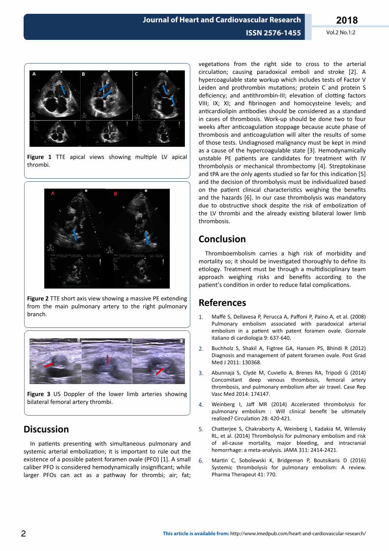
Figure 1 TTE apical views showing multiple LV apicalthrombi.
Figure 2 TTE short axis view showing a massive PE extendingfrom the main pulmonary artery to the right pulmonarybranch.
Figure 3 US Doppler of the lower limb arteries showingbilateral femoral artery thrombi.
DiscussionIn patients presenting with simultaneous pulmonary and
systemic arterial embolization; it is important to rule out theexistence of a possible patent foramen ovale (PFO) [1]. A smallcaliber PFO is considered hemodynamically insignificant; whilelarger PFOs can act as a pathway for thrombi; air; fat;
vegetations from the right side to cross to the arterialcirculation; causing paradoxical emboli and stroke [2]. Ahypercoagulable state workup which includes tests of Factor VLeiden and prothrombin mutations; protein C and protein Sdeficiency; and antithrombin-III; elevation of clotting factorsVIII; IX; XI; and fibrinogen and homocysteine levels; andanticardiolipin antibodies should be considered as a standardin cases of thrombosis. Work-up should be done two to fourweeks after anticoagulation stoppage because acute phase ofthrombosis and anticoagulation will alter the results of someof those tests. Undiagnosed malignancy must be kept in mindas a cause of the hypercoagulable state [3]. Hemodynamicallyunstable PE patients are candidates for treatment with IVthrombolysis or mechanical thrombectomy [4]. Streptokinaseand tPA are the only agents studied so far for this indication [5]and the decision of thrombolysis must be individualized basedon the patient clinical characteristics weighing the benefitsand the hazards [6]. In our case thrombolysis was mandatorydue to obstructive shock despite the risk of embolization ofthe LV thrombi and the already existing bilateral lower limbthrombosis.
ConclusionThromboembolism carries a high risk of morbidity and
mortality so; it should be investigated thoroughly to define itsetiology. Treatment must be through a multidisciplinary teamapproach weighing risks and benefits according to thepatient’s condition in order to reduce fatal complications.
References1. Maffè S, Dellavesa P, Perucca A, Paffoni P, Paino A, et al. (2008)
Pulmonary embolism associated with paradoxical arterialembolism in a patient with patent foramen ovale. Giornaleitaliano di cardiologia 9: 637-640.
2. Buchholz S, Shakil A, Figtree GA, Hansen PS, Bhindi R (2012)Diagnosis and management of patent foramen ovale. Post GradMed J 2011: 130368.
3. Abunnaja S, Clyde M, Cuviello A, Brenes RA, Tripodi G (2014)Concomitant deep venous thrombosis, femoral arterythrombosis, and pulmonary embolism after air travel. Case RepVasc Med 2014: 174147.
4. Weinberg I, Jaff MR (2014) Accelerated thrombolysis forpulmonary embolism : Will clinical benefit be ultimatelyrealized? Circulation 28: 420-421.
5. Chatterjee S, Chakraborty A, Weinberg I, Kadakia M, WilenskyRL, et al. (2014) Thrombolysis for pulmonary embolism and riskof all-cause mortality, major bleeding, and intracranialhemorrhage: a meta-analysis. JAMA 311: 2414-2421.
6. Martin C, Sobolewski K, Bridgeman P, Boutsikaris D (2016)Systemic thrombolysis for pulmonary embolism: A review.Pharma Therapeut 41: 770.
Journal of Heart and Cardiovascular Research
ISSN 2576-1455 Vol.2 No.1:2
2018
2 This article is available from: http://www.imedpub.com/heart-and-cardiovascular-research/
Related Documents


