Infection & Chemotherapy Received: March 2, 2016 Accepted: April 7, 2016 Published online: November 11, 2016 Corresponding Author : Sun Hee Lee, MD Division of Infectious Diseases, Department of Internal Medicine, Pusan National University Hospital, 179 Gudeok-ro, Seo-gu, Busan 49241, Korea Tel: +82-51-240-7673, Fax: +82-51-247-3213 E-mail: [email protected] This is an Open Access article distributed under the terms of the Creative Commons Attribution Non-Commercial License (http://creativecommons.org/licenses/by-nc/3.0) which permits unrestricted non-commercial use, distribution, and repro- duction in any medium, provided the original work is properly cited. Copyrights © 2016 by The Korean Society of Infectious Diseases | Korean Society for Chemotherapy www.icjournal.org A Case of Transverse Myelitis Caused by Varicella Zoster Virus in an Immunocompetent Older Patient Jeong Eun Lee, Shinwon Lee, Kye-Hyung Kim, Hee Ryeong Jang , Young Joo Park, Jin Suk Kang, Sung Yong Han, and Sun Hee Lee Deparment of Internal Medicine, Pusan National University School of Medicine, Medical Research Institute, Pusan National University Hospital, Busan, Korea Varicella zoster virus (VZV) is a human neurotropic alphaherpesvirus that causes chickenpox (varicella) in children. VZV re- activation may lead to neurological complications, including transverse myelitis. However, transverse myelitis caused by VZV reactivation is rare in immunocompetent patients. Herein, we report a case of transverse myelitis caused by VZV in an immu- nocompetent older patient, and confirmed this case by polymerase chain reaction. A 79-year-old woman visited our service with complaints of weakness in the right lower leg, generalized vesicular eruptions, and throbbing pain in the right flank for ten days. Spine MRI showed transverse myelitis in the thoracic spine at level T4–T11. The patient was treated with acyclovir and her neurological functions improved, except for sensory impairment below level T10. For older patients, early and aggressive antivi- ral treatment against VZV may be necessary even though these patients are immunocompetent. Key Words: Varicella zoster virus; Transverse myelitis; Immunocompetent; Older patients; Antiviral treatment https://doi.org/10.3947/ic.2016.48.4.334 Infect Chemother 2016;48(4):334-337 ISSN 2093-2340 (Print) · ISSN 2092-6448 (Online) Case Report Introduction Varicella-zoster virus (VZV) is a human herpesvirus that causes chickenpox and herpes zoster [1, 2]. The most com- mon neurologic complication of VZV reactivation is herpetic neuralgia, which is usually self-limited. However, VZV reacti- vation in immunocompromised patients can cause dissemi- nated infections and severe neurologic dysfunctions, includ- ing meningitis, neuropathy, myelitis, stroke, and encephalitis [3]. Transverse myelitis is an unusual complication caused by VZV reactivation in immunocompetent patients [4]. To date, few cases of transverse myelitis were reported in Korea and most of them were not confirmed microbiologically [5-9]. Herein we report a case of transverse myelitis caused by VZV in an immunocompetent older patient, and this case was con- firmed microbiologically by detection of VZV DNA in the ce- rebrospinal fluid (CSF) by polymerase chain reaction (PCR). Case Report A 79-year-old woman was admitted to our Institution with

Welcome message from author
This document is posted to help you gain knowledge. Please leave a comment to let me know what you think about it! Share it to your friends and learn new things together.
Transcript

Infection & Chemotherapy
Received: March 2, 2016 Accepted: April 7, 2016 Published online: November 11, 2016Corresponding Author : Sun Hee Lee, MDDivision of Infectious Diseases, Department of Internal Medicine, Pusan National University Hospital, 179 Gudeok-ro, Seo-gu, Busan 49241, KoreaTel: +82-51-240-7673, Fax: +82-51-247-3213E-mail: [email protected]
This is an Open Access article distributed under the terms of the Creative Commons Attribution Non-Commercial License (http://creativecommons.org/licenses/by-nc/3.0) which permits unrestricted non-commercial use, distribution, and repro-duction in any medium, provided the original work is properly cited.
Copyrights © 2016 by The Korean Society of Infectious Diseases | Korean Society for Chemotherapy
www.icjournal.org
A Case of Transverse Myelitis Caused by Varicella Zoster Virus in an Immunocompetent Older Patient Jeong Eun Lee, Shinwon Lee, Kye-Hyung Kim, Hee Ryeong Jang , Young Joo Park, Jin Suk Kang, Sung Yong Han, and Sun Hee LeeDeparment of Internal Medicine, Pusan National University School of Medicine, Medical Research Institute, Pusan National University Hospital, Busan, Korea
Varicella zoster virus (VZV) is a human neurotropic alphaherpesvirus that causes chickenpox (varicella) in children. VZV re-activation may lead to neurological complications, including transverse myelitis. However, transverse myelitis caused by VZV reactivation is rare in immunocompetent patients. Herein, we report a case of transverse myelitis caused by VZV in an immu-nocompetent older patient, and confirmed this case by polymerase chain reaction. A 79-year-old woman visited our service with complaints of weakness in the right lower leg, generalized vesicular eruptions, and throbbing pain in the right flank for ten days. Spine MRI showed transverse myelitis in the thoracic spine at level T4–T11. The patient was treated with acyclovir and her neurological functions improved, except for sensory impairment below level T10. For older patients, early and aggressive antivi-ral treatment against VZV may be necessary even though these patients are immunocompetent.
Key Words: Varicella zoster virus; Transverse myelitis; Immunocompetent; Older patients; Antiviral treatment
https://doi.org/10.3947/ic.2016.48.4.334
Infect Chemother 2016;48(4):334-337
ISSN 2093-2340 (Print) · ISSN 2092-6448 (Online)
Case Report
Introduction
Varicella-zoster virus (VZV) is a human herpesvirus that
causes chickenpox and herpes zoster [1, 2]. The most com-
mon neurologic complication of VZV reactivation is herpetic
neuralgia, which is usually self-limited. However, VZV reacti-
vation in immunocompromised patients can cause dissemi-
nated infections and severe neurologic dysfunctions, includ-
ing meningitis, neuropathy, myelitis, stroke, and encephalitis
[3]. Transverse myelitis is an unusual complication caused by
VZV reactivation in immunocompetent patients [4]. To date,
few cases of transverse myelitis were reported in Korea and
most of them were not confirmed microbiologically [5-9].
Herein we report a case of transverse myelitis caused by VZV
in an immunocompetent older patient, and this case was con-
firmed microbiologically by detection of VZV DNA in the ce-
rebrospinal fluid (CSF) by polymerase chain reaction (PCR).
Case Report
A 79-year-old woman was admitted to our Institution with
11-증례_IC-15-498.indd 1 2016-12-27 오후 4:56:58

https://doi.org/10.3947/ic.2016.48.4.334 • Infect Chemother 2016;48(4):334-337www.icjournal.org 335
weakness in the right lower leg and numbness in the lower
limbs for three days. Ten days before admission, the patient
felt a throbbing pain in the right flank, and the pain was not
relieved by analgesics. Three days before admission, the pa-
tient’s right leg was paralyzed, and she felt numbness in both
legs, lost the control of micturition, and presented with multi-
ple skin eruptions, which appeared on the right flank and
gradually spread to the trunk, face, and extremities.
Upon admission, blood pressure was 140/110 mmHg, the
heart rate was 66 beats per minute, and body temperature was
36.3°C. Multiple vesicles and pustules were observed on the
whole body (Fig. 1). The patient had never had contact with
chickenpox or herpes zoster patients. In addition, she had no
history of herpes zoster infection and vaccination. Neurologic
examination indicated that the strength of the proximal half
(grade 3/5) and the distal half (grade 4/5) of the right lower
limb decreased. Furthermore, the sense of pain and tempera-
ture below level T9 on the right side of the body and below T6
on the left side of the body decreased. Magnetic resonance
imaging (MRI) indicated diffuse hyperintensity of the spinal
cord at level T4–T11 on T2-weighting (Fig. 2A).
The results of blood tests were as follows: white blood cell
count, 7.09 × 10³/mL; hemoglobin, 13.3 g/dL; platelet count,
187 × 103/mL; C-reactive protein, 1.06 mg/dL; erythrocyte
sedimentation rate, 16 mm/hour; and positive serum IgG and
IgM antibodies against VZV.
CSF analysis indicated abnormal values for white blood cell
count (1076 cells/μL; 0% of neutrophils, 96% of lymphocytes,
and 4% of monocytes), and protein (442.5 mg/dL), and a neg-
ative result for bacterial culture. VZV DNA was detected by
PCR amplification in CSF. The PCR results for IgG and IgM of
herpes simplex virus-1 (HSV-1), herpes simplex virus-2 (HSV-
2), and cytomegalovirus (CMV) were negative in the CSF.
Intravenous acyclovir was initiated at 500 mg every 8 hours.
The dosage of acyclovir was adjusted to 250 mg every 8 hours
for three days after initiation of therapy because of deteriora-
tion of renal function. On admission day 3, the patient pre-
sented deterioration of consciousness and convulsions; there-
fore, brain MRI was performed. MRI indicated increased
signal intensity at the right temporo-occipital lobe and left
frontal lobe on T2 weighting, suggesting the occurrence of en-
cephalitis. The patient was maintained on antiviral and antie-
pileptic therapy for three weeks. After this period, her mental
status was recovered, and the function of both legs improved
with a rehabilitation program involving strength training. Re-
peat MRI showed partial improvement of myelopathy (Fig.
2B). However, sensory impairment below level T10 persisted
for four months after initiation of therapy.
Discussion
VZV is a human neurotropic alphaherpesvirus that causes
chickenpox (varicella) in children [1, 2]. After primary infec-
tion, the virus becomes latent in cranial nerve and sensory
root ganglia [2, 3]. However, VZV reactivation may occur with
advanced age or immunosuppression, particularly in cases of
cell-mediated immunosuppression [1-3].
VZV reactivation may cause neurological complications
such as chronic pain (postherpetic neuralgia), cranial nerve
palsy, zoster paresis, meningoencephalitis, cerebellitis, my-
elopathy, multiple ocular disorders, and stroke [2-4]. The most
A B
Figure 1. Development of multiple vesicles and pustules in the right flank (A) and their spread to the trunk (B).
11-증례_IC-15-498.indd 2 2016-12-27 오후 4:57:00

Lee JE, et al. • A case of VZV transverse myelitis www.icjournal.org336
common manifestation of VZV reactivation is herpes zoster.
Unvaccinated individuals aged 85 years or older have a 50%
risk of developing herpes zoster [10]. However, transverse my-
elitis is one of the rarest complications, particularly in immu-
nocompetent patients [1-3]. To date, five cases of VZV myelitis
have been reported in Korea; however, most of them were
clinically suspicious cases with consistent image findings [5-
9]. Only one microbiologically confirmed case of transverse
myelitis caused by VZV was reported approximately 20 years
ago [6]. Four other reported cases of VZV in Korea were diag-
nosed by classical imaging findings and clinical examination.
This report describes a microbiologically confirmed case of
transverse myelitis caused by VZV in an immunocompetent
older patient. Myelitis and encephalitis due to VZV reactiva-
tion are more common in immunocompromised patients [1].
In these patients, VZV myelitis may occur without typical skin
lesions and can occur far different level of skin lesion [4, 11]. By
contrast, in immunocompetent patients, VZV myelitis has a
typical presentation (dermatomal rashes followed by myelitis
at the corresponding level) and good outcomes [1, 4, 11]. How-
ever, our patient showed an atypical presentation, character-
ized by generalized and disseminated eruptions on the body.
The diagnosis of VZV myelitis can be challenging [12]. Older
patients may show a variety of neurologic symptoms from lo-
cal paralysis to severe neurologic dysfunction due to multiple
causes; therefore, thinking of several possibilities is critical
and various examinations are needed to differentiate the
causes. To date, no predictable markers of disease progression
are available to patients with VZV myelitis [4]. Therefore, clini-
cal suspicion and aggressive evaluation are crucial for the ear-
ly diagnosis of VZV myelitis [12].
The detection of VZV antibodies and VZV DNA in CSF are
confirmatory diagnostic tests [11-13]. However, Rosenfeld et
al. reported that patients showed clinical signs of severe VZV
myelitis, although the VZV antibody tests and PCR results for
VZV DNA were all negative [12]. Imaging studies are useful for
the diagnosis of VZV myelitis. MRI of VZV myelitis is likely to
show T2-hyperintensity in the spinal cord [12, 13]. Although
the standard treatment regimen for VZV myelitis is not yet es-
tablished, there is anecdotal evidence for treatment of VZV
myelitis with acyclovir [4, 12-15]. Moreover, there is little evi-
dence that early antiviral treatment reduces the risk of VZV
myelitis. Therefore, the early diagnosis and antiviral treatment
of VZV is essential to recovery from myelitis and minimize its
complications, and this treatment is crucial to prevent the de-
velopment of postherpetic neuralgia [14]. Our case and some
other cases reported previously also support the advantages
of early antiviral treatment for VZV myelitis. We did not use
corticosteroids because the additional benefit of the steroid
was not clear although the combination of high-dose acyclo-
vir and corticosteroids have shown a good prognosis in previ-
ous case reports [11].
In conclusion, this is the second confirmed case of VZV my-
elitis in immunocompetent patients in Korea. Even in immu-
nocompetent older patients, VZV myelitis may be severe and
involve atypical skin lesions. Therefore, early diagnosis and
aggressive antiviral treatment may be necessary.
Conflicts of InterestNo conflicts of interest.
ORCIDJeong Eun Lee http://orcid.org/0000-0003-3027-1381
Sun Hee Lee http://orcid.org/0000-0003-2093-3628
A B
Figure 2. Whole spine MRI (T2-weighted sagittal image) on admission (A) shows high-signal intensity from level T4 (upper arrow) to T11 (lower arrow). On admission day 36 (B), the extent of diffuse hyperintensity de-creased, with faint enhancement of the spinal cord from level T8 –T9 (upper arrow) to T9 (lower arrow).
11-증례_IC-15-498.indd 3 2016-12-27 오후 4:57:01
https://doi.org/10.3947/ic.2016.48.4.334 • Infect Chemother 2016;48(4):334-337www.icjournal.org 337
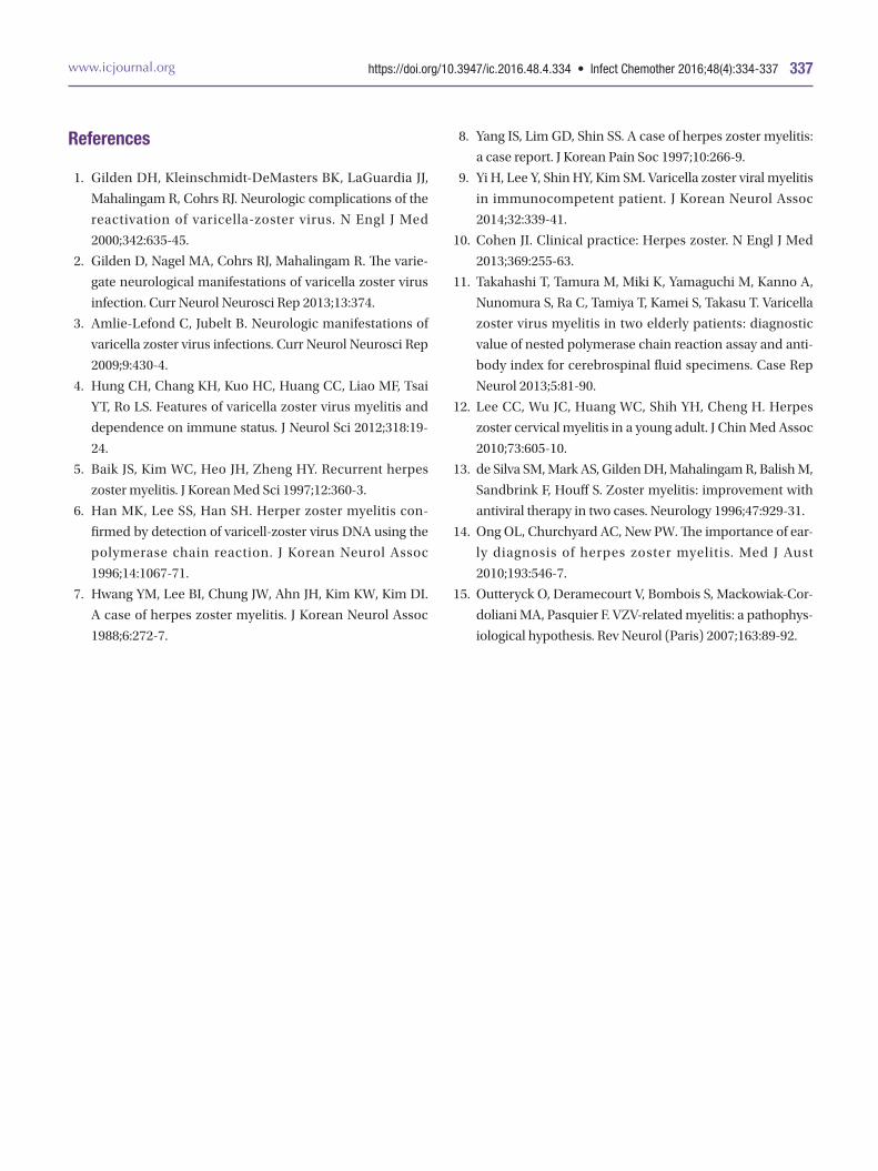
References
1. Gilden DH, Kleinschmidt-DeMasters BK, LaGuardia JJ,
Mahalingam R, Cohrs RJ. Neurologic complications of the
reactivation of varicella-zoster virus. N Engl J Med
2000;342:635-45.
2. Gilden D, Nagel MA, Cohrs RJ, Mahalingam R. The varie-
gate neurological manifestations of varicella zoster virus
infection. Curr Neurol Neurosci Rep 2013;13:374.
3. Amlie-Lefond C, Jubelt B. Neurologic manifestations of
varicella zoster virus infections. Curr Neurol Neurosci Rep
2009;9:430-4.
4. Hung CH, Chang KH, Kuo HC, Huang CC, Liao MF, Tsai
YT, Ro LS. Features of varicella zoster virus myelitis and
dependence on immune status. J Neurol Sci 2012;318:19-
24.
5. Baik JS, Kim WC, Heo JH, Zheng HY. Recurrent herpes
zoster myelitis. J Korean Med Sci 1997;12:360-3.
6. Han MK, Lee SS, Han SH. Herper zoster myelitis con-
firmed by detection of varicell-zoster virus DNA using the
polymerase chain reaction. J Korean Neurol Assoc
1996;14:1067-71.
7. Hwang YM, Lee BI, Chung JW, Ahn JH, Kim KW, Kim DI.
A case of herpes zoster myelitis. J Korean Neurol Assoc
1988;6:272-7.
8. Yang IS, Lim GD, Shin SS. A case of herpes zoster myelitis:
a case report. J Korean Pain Soc 1997;10:266-9.
9. Yi H, Lee Y, Shin HY, Kim SM. Varicella zoster viral myelitis
in immunocompetent patient. J Korean Neurol Assoc
2014;32:339-41.
10. Cohen JI. Clinical practice: Herpes zoster. N Engl J Med
2013;369:255-63.
11. Takahashi T, Tamura M, Miki K, Yamaguchi M, Kanno A,
Nunomura S, Ra C, Tamiya T, Kamei S, Takasu T. Varicella
zoster virus myelitis in two elderly patients: diagnostic
value of nested polymerase chain reaction assay and anti-
body index for cerebrospinal fluid specimens. Case Rep
Neurol 2013;5:81-90.
12. Lee CC, Wu JC, Huang WC, Shih YH, Cheng H. Herpes
zoster cervical myelitis in a young adult. J Chin Med Assoc
2010;73:605-10.
13. de Silva SM, Mark AS, Gilden DH, Mahalingam R, Balish M,
Sandbrink F, Houff S. Zoster myelitis: improvement with
antiviral therapy in two cases. Neurology 1996;47:929-31.
14. Ong OL, Churchyard AC, New PW. The importance of ear-
ly diagnosis of herpes zoster myelitis. Med J Aust
2010;193:546-7.
15. Outteryck O, Deramecourt V, Bombois S, Mackowiak-Cor-
doliani MA, Pasquier F. VZV-related myelitis: a pathophys-
iological hypothesis. Rev Neurol (Paris) 2007;163:89-92.
11-증례_IC-15-498.indd 4 2016-12-27 오후 4:57:01
Related Documents