1 doi: 10.2169/internalmedicine.5608-20 Intern Med Advance Publication http://internmed.jp 【 CASE REPORT 】 A Case of Glomerulonephritis Caused by Bartonella spp. Infective Endocarditis: The Difficulty and Importance of Differentiation from Anti-neutrophil Cytoplasmic Antibody-related Rapidly Progressive Glomerulonephritis Ayumi Yoshifuji 1 , Yuuka Hibino 1 , Motoaki Komatsu 1 , Seiichi Yasuda 1 , Koji Hosoya 1 , Emi Kobayashi 1 , Yuko Baba 2 , Shigemichi Hirose 3 , Akinori Hashiguchi 4 , Yoshihiko Kanno 5 and Munekazu Ryuzaki 1 Abstract: A 65-year-old man with valvular disorder presented to his physician because of widespread purpura in both lower extremities. Blood tests showed elevated serum creatinine levels and proteinase 3-anti-neutrophil cytoplasmic antibody (ANCA) with hematuria, suggesting ANCA-related rapidly progressive glomeru- lonephritis (RPGN). Although multiple blood cultures were negative, transthoracic echocardiography revealed warts in the valves, and a renal biopsy also showed findings of glomerular infiltration by mononuclear leuko- cytes and C3 deposition in the glomeruli, suggesting infection-related glomerulonephritis. Later, Bartonella antibody turned positive. Antimicrobial treatment improved the purpura and renal function without any recur- rence. ANCA-positive RPGN requires the exclusion of infective endocarditis, especially that induced by Bar- tonella spp. Key words: ANCA, rapidly progressive glomerulonephritis, Bartonella, infective endocarditis (Intern Med Advance Publication) (DOI: 10.2169/internalmedicine.5608-20) Introduction Infective endocarditis is a fatal systemic septic disease characterized by the formation of warts in the valvular or large-vessel endocardium that contain bacterial, fungal, viral, or other infectious microorganisms. It presents with a variety of clinical symptoms. At least three sets of blood cultures are recommended for the diagnosis of infective endocarditis before the administra- tion of antimicrobial agents. However, it has been reported that culture-negative infective endocarditis accounts for 12- 25% of total cases (1, 2). Patients with infective endocarditis caused by Bartonella spp. often have negative blood cul- tures (1). In addition, infective endocarditis presents with symptoms similar to vasculitis with anti-neutrophil cytoplas- mic antibodies (ANCAs) (3). When we treat cases of ANCA-positive infective endocarditis with renal impairment, it is essential to differentiate between infection-related glomerulonephritis (IRGN) due to infective endocarditis and ANCA-related nephritis to determine the appropriate treat- ment. We treated a case of ANCA-positive glomerulonephritis caused by Bartonella spp. infective endocarditis. An early renal biopsy helped to differentiate IRGN due to infective endocarditis from ANCA-associated nephritis with a favor- able prognosis following treatment with antibacterial agents. 1 Division of Nephrology, Department of Internal Medicine, Tokyo Saiseikai Central Hospital, Japan, 2 Department of Dermatology, Tokyo Saisei- kai Central Hospital, Japan, 3 Department of Pathology, Tokyo Saiseikai Central Hospital, Japan, 4 Department of Pathology, School of Medicine, Keio University, Japan and 5 Division of Nephrology, Department of Internal Medicine, Tokyo Medical University, Japan Received: June 14, 2020; Accepted: November 26, 2020; Advance Publication by J-STAGE: January 15, 2021 Correspondence to Dr. AyumiYoshifuji, [email protected]

Welcome message from author
This document is posted to help you gain knowledge. Please leave a comment to let me know what you think about it! Share it to your friends and learn new things together.
Transcript
1
doi: 10.2169/internalmedicine.5608-20
Intern Med Advance Publication
http://internmed.jp
【 CASE REPORT 】
A Case of Glomerulonephritis Caused by Bartonella spp.Infective Endocarditis: The Difficulty and Importance of
Differentiation from Anti-neutrophil CytoplasmicAntibody-related Rapidly Progressive Glomerulonephritis
Ayumi Yoshifuji 1, Yuuka Hibino 1, Motoaki Komatsu 1, Seiichi Yasuda 1, Koji Hosoya 1,
Emi Kobayashi 1, Yuko Baba 2, Shigemichi Hirose 3, Akinori Hashiguchi 4,
Yoshihiko Kanno 5 and Munekazu Ryuzaki 1
Abstract:A 65-year-old man with valvular disorder presented to his physician because of widespread purpura in
both lower extremities. Blood tests showed elevated serum creatinine levels and proteinase 3-anti-neutrophil
cytoplasmic antibody (ANCA) with hematuria, suggesting ANCA-related rapidly progressive glomeru-
lonephritis (RPGN). Although multiple blood cultures were negative, transthoracic echocardiography revealed
warts in the valves, and a renal biopsy also showed findings of glomerular infiltration by mononuclear leuko-
cytes and C3 deposition in the glomeruli, suggesting infection-related glomerulonephritis. Later, Bartonellaantibody turned positive. Antimicrobial treatment improved the purpura and renal function without any recur-
rence. ANCA-positive RPGN requires the exclusion of infective endocarditis, especially that induced by Bar-tonella spp.
Key words: ANCA, rapidly progressive glomerulonephritis, Bartonella, infective endocarditis
(Intern Med Advance Publication)(DOI: 10.2169/internalmedicine.5608-20)
Introduction
Infective endocarditis is a fatal systemic septic disease
characterized by the formation of warts in the valvular or
large-vessel endocardium that contain bacterial, fungal, viral,
or other infectious microorganisms. It presents with a variety
of clinical symptoms.
At least three sets of blood cultures are recommended for
the diagnosis of infective endocarditis before the administra-
tion of antimicrobial agents. However, it has been reported
that culture-negative infective endocarditis accounts for 12-
25% of total cases (1, 2). Patients with infective endocarditis
caused by Bartonella spp. often have negative blood cul-
tures (1). In addition, infective endocarditis presents with
symptoms similar to vasculitis with anti-neutrophil cytoplas-
mic antibodies (ANCAs) (3). When we treat cases of
ANCA-positive infective endocarditis with renal impairment,
it is essential to differentiate between infection-related
glomerulonephritis (IRGN) due to infective endocarditis and
ANCA-related nephritis to determine the appropriate treat-
ment.
We treated a case of ANCA-positive glomerulonephritis
caused by Bartonella spp. infective endocarditis. An early
renal biopsy helped to differentiate IRGN due to infective
endocarditis from ANCA-associated nephritis with a favor-
able prognosis following treatment with antibacterial agents.
1Division of Nephrology, Department of Internal Medicine, Tokyo Saiseikai Central Hospital, Japan, 2Department of Dermatology, Tokyo Saisei-
kai Central Hospital, Japan, 3Department of Pathology, Tokyo Saiseikai Central Hospital, Japan, 4Department of Pathology, School of Medicine,
Keio University, Japan and 5Division of Nephrology, Department of Internal Medicine, Tokyo Medical University, Japan
Received: June 14, 2020; Accepted: November 26, 2020; Advance Publication by J-STAGE: January 15, 2021
Correspondence to Dr. Ayumi Yoshifuji, [email protected]
Intern Med Advance Publication DOI: 10.2169/internalmedicine.5608-20
2
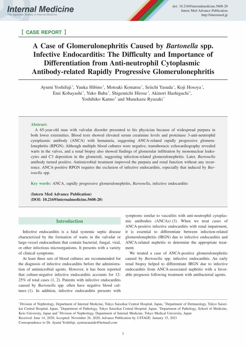
Figure 1. Cutaneous findings of the lower extremities. A) Right lower extremity, enlarged photo (before treatment). B) Right lower extremity, enlarged photo (after treatment). Red to purplish-red purpura a few millimeters in size were observed in both lower extremities on admission, but they disappeared after the antimicrobial treatment.
(A) Right lower extremity (before treatment) Enlarged photo (before treatment)
(B) Right lower extremity (a er treatment) Enlarged photo (a er treatment)
Case presentation
A 65-year-old man with a history of alcoholism and aor-
tic and mitral regurgitation who was living in a lodging
house without any contact with cats presented to his physi-
cian because of a fever (38.2 °C), weakness in both lower
extremities that had started 1 month before the appointment,
and purpura on the extensor surfaces of both lower extremi-
ties that started 1 week before the appointment. He was re-
ferred to our hospital because he was suspected of having
rapidly progressive glomerulonephritis based on elevated
creatinine levels (3.07 mg/dL) with hematuria, although
there were no abnormalities in the blood analysis, including
creatinine levels (0.85 mg/dL), or according to a urinalysis
performed 6 months earlier. A physical examination revealed
no apparent petechial hemorrhaging in his conjunctiva, Os-
ler’s nodes, or Janeway lesions. However, chest auscultation
revealed a systolic ejection murmur (Levine III/VI) at the
right margin of the second intercostal sternum. In addition,
the patient had pitting edema and numerous sites of purpura
several millimeters across on the bilateral lower extremities
(Fig. 1A). However, he lacked lymphadenopathy.
Blood and urine analyses showed elevated blood urea ni-
trogen levels (46 mg/dL) and serum creatinine levels (2.69
md/dL), occult hematuria, and proteinuria, which suggested
glomerulonephritis. They also showed a normal range of
leukocytes (7,500 cells/μL) but mildly elevated C-reactive
protein (CRP; 3.36 mg/dL) and normocytic hypochromic
anemia with 8.2 g/dL hemoglobin. We observed no de-
creases in complement, but we did observe elevated rheuma-
toid factor (2,880 IU/mL), proteinase 3 (PR3)-ANCA (186
U/mL), and myeloperoxidase (MPO)-ANCA (7.0 U/mL).
Three sets of blood cultures were all negative.
Transthoracic and transesophageal echocardiography re-
vealed warts on the aortic and mitral valves with moderate
to severe aortic regurgitation (Fig. 2). Magnetic resonance
imaging of the head showed a 6-mm cerebral aneurysm at
the bifurcation of the right middle cerebral artery (Fig. 3).
In addition, a skin biopsy of the purpura demonstrated lym-
phocyte and neutrophil infiltration around and within the ar-
teriole of the dermal reticular layer, consistent with vasculi-
tis (Fig. 4A and B). A renal biopsy performed one week af-
ter antimicrobial treatment showed a fibrous to fibrocellular
crescent in one glomerulus (Fig. 5A a-c). Glomerular infil-
tration by mononuclear leukocytes was observed, although
no capillary occlusion was identified. Neither mesangial ex-
pansion nor hypercellularity were observed. Immunofluores-
cent staining revealed IgM and complement component 3c
(C3) in the mesangial region (Fig. 5B), and electron micros-
copy revealed high-electron-density deposits in the mesan-
gial region (Fig. 5C). These findings were consistent with

Intern Med Advance Publication DOI: 10.2169/internalmedicine.5608-20
3
Figure 2. Transesophageal echocardiography findings. A) Day 6. Warts 10×6 mm in size were found at the aortic valve (total valve apex) and mitral valve. Moderate-to-severe aortic regurgitation and mitral regurgitation were also detected. B) Two months after treatment. The warts at the aortic valve (total valve apex) and mitral valve had remarkably regressed. Moderate aortic regurgitation and mitral regurgitation were still detected. C) One year after treatment. Regression was maintained without treatment. Moderate aortic regurgitation and mitral regurgitation were still detected. LV: left ventricle, Ao: aorta
wart
LV
Ao
LV: Le Ventricle, Ao: Aorta
(A) Day6 (B) 2 month a er the treatment (C) 1 year a er the treatment
LVAo LV
Ao
Figure 3. Head MRI. A 6-mm aneurysm was detected at the bifurcation of the right middle cerebral artery.
middle cerebral artery aneurysm
the resolution of IRGN.
The patient was diagnosed with infective endocarditis ac-
cording to the modified Duke’s criteria (4) (one major crite-
rion, warts by echocardiography; and four minor criteria,
predisposing cardiac disease, fever of 38.2 °C, cerebral
aneurysm, and positive for rheumatoid factor). He was then
treated with ampicillin/sulbactam (7 weeks) and gentamicin
(2 weeks) for blood culture-negative infective endocarditis.
After 7 weeks of antimicrobial treatment, his serum cre-
atinine levels decreased from 2.69 mg/dL (on admission) to
1.95 mg/dL, and his CRP level improved from 3.4 mg/dL
(on admission) to 0.5 mg/dL. In addition, the purpura that
had been observed at the time of admission disappeared
(Fig. 1B).
The antibodies against Bartonella spp. used to identify
the causative organism were markedly elevated to more than
1,024× the typical level for anti-Bartonella henselae IgG
and 512× for anti-Bartonella quintana IgG, which led to the
diagnosis of infective endocarditis caused by Bartonella spp.
After two weeks of antimicrobial treatment with ceftriaxone,
gentamicin, and doxycycline, the patient self-discharged
from the hospital and refused further treatment. Judging by
the numerous reports that infective endocarditis caused by
Bartonella spp. is difficult to treat with only antimicrobial
agents and based on his moderate to severe valve insuffi-
ciency, we proposed a valve replacement operation, which
the patient unfortunately refused. At the time of discharge,
his serum creatinine had decreased to 2.14 mg/dL, CRP to
0.87 mg/dL, and PR3-ANCA to 141 U/mL. Thereafter,
given the possibility of relapse, the patient continued to be
followed in our outpatient clinic. Two months after his dis-
charge, the warts had remarkably regressed (Fig. 2B). His
creatinine level had further decreased to 1.53 mg/dL, CRP
to 0.54 mg/dL, and PR3-ANCA to 93.6 U/mL, and urine
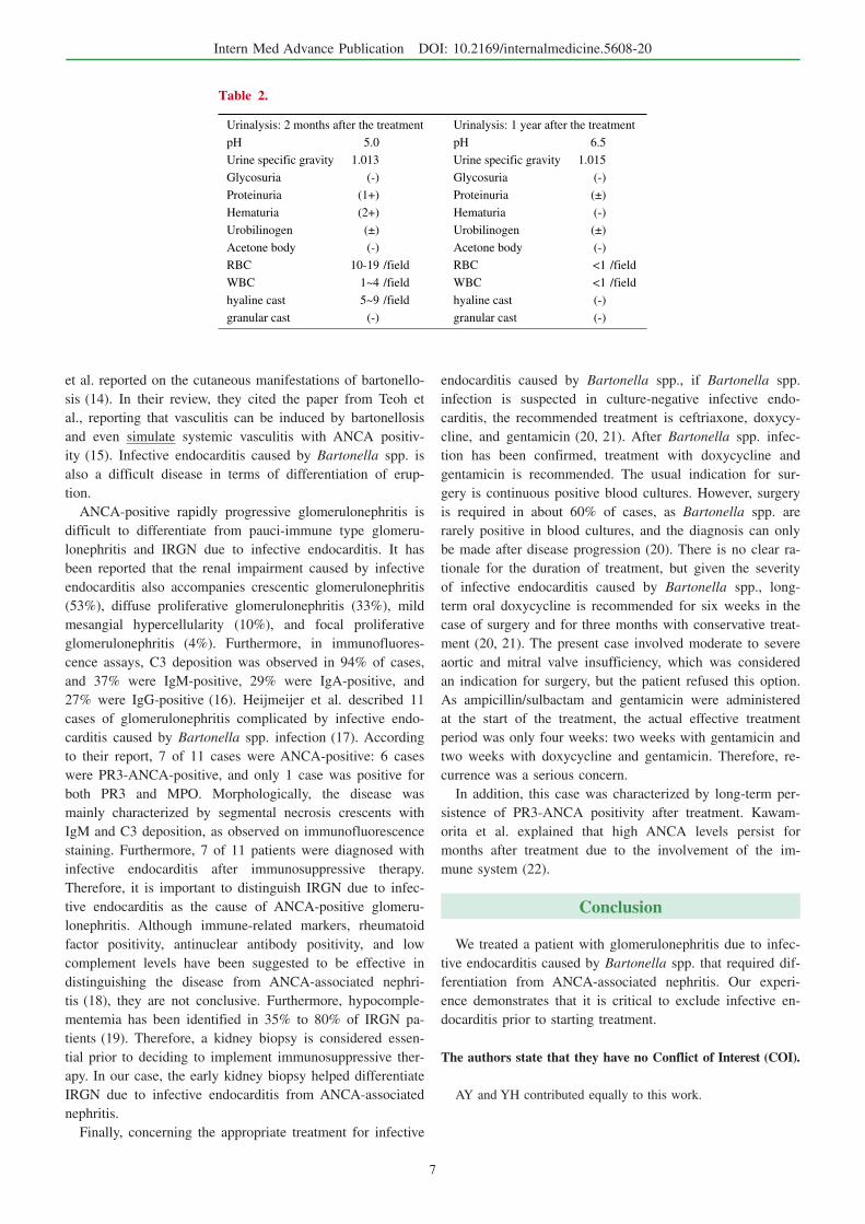
sediment showed improvement in hematuria (Table 2). Fur-
thermore, at 1 year later, his regression was maintained
without treatment (Fig. 2C), and his creatinine level had de-
creased to 1.09 mg/dL and PR3-ANCA to 28.6 U/mL with-
out any signs of relapse. His urine sediment showed no he-
maturia (Table 2).
Discussion
We experienced a case of ANCA-positive glomeru-
lonephritis caused by Bartonella spp. infective endocarditis.
An early renal biopsy helped to differentiate IRGN due to
infective endocarditis from ANCA-associated nephritis, with
the patient showing a favorable prognosis following treat-
ment with antibacterial agents.
Multiple blood cultures from the patient were negative. In
addition to prior administration of antimicrobials, difficulty
Intern Med Advance Publication DOI: 10.2169/internalmedicine.5608-20
4
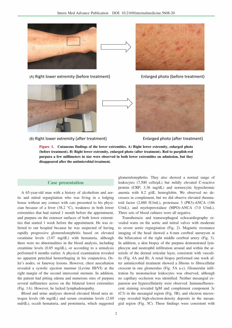
Figure 4. Skin biopsy findings. A) 100×. B) 400×. A skin biopsy of the purpura was performed on Day 8. Hematoxylin and Eosin staining revealed the infiltration of lymphocytes and neutrophils with nuclear dust from the dermal papillary layer to the dermal reticular layer, and red blood cell leakage was observed around the capillary lumen with swollen endothelial cells and arterioles where inflam-matory cells had infiltrated into the vessel wall. These findings were consistent with ANCA-associated vasculitis.
(A) × 100 (B) × 400
Figure 5. Kidney biopsy findings. A) Optical microscopy. a: PAS staining, b: PAS staining (left) and PAM staining (right) c: Masson-Trichrome staining (×100). Cortical:medulla=6:4. A total of 25 glomeruli, 8 global sclerosis, 1 fibrous crescent, tubular atrophy, and interstitial fibrosis, hyalinosis of small arteries, and erythrocytic casts were detected. B) Immunofluorescent assay findings. Positive IgM and C3c findings in the mesangial region were detected. C) Electron microscopy findings. High-electron-density deposits were found in the mesangial region (arrows).
A Op cal microscope
(a)PAS staining (b)PAS staining (le ), PAM staining (right) (c) Masson-Trichrome staining
B Immuno uorescent assay ndings C Electron microscopy

Intern Med Advance Publication DOI: 10.2169/internalmedicine.5608-20
5
Figure 6. Clinical course. The clinical course is shown along with the kind, amount, and duration of antimicrobial agents. Creatinine and CRP levels were used as indicators of the disease severity. GM: Gentamicin, SBT/ABPC: Sulbactam/Ampicillin, CTRX: Ceftriaxone, DOXY: Doxycycline
0
0.5
1
1.5
2
2.5
3
3.5
4
CRP
Cr
CRP
Crea nine
0 10 20 30 40 50 60 120 360 [day]
[mg/dl]PR3-ANCA186U/ml
PR3-ANCA141U/ml
PR3-ANCA93.6U/ml
PR3-ANCA28.6U/ml
SBT/ABPC 6g/day
GM40mg/day
GM40mg/day
CTRX2g/dayDOXY
200mg/day
Bartonella anP si ve
in detecting microbes can be caused by slow-growing patho-
gens, such as Haemophilus spp., Actinobacillus spp., Car-diobacterium hominis, Eikenella corrodens, and Kingellaspp.; intracellular parasites, such as Coxiella, Bartonella,
Brucella, Mycoplasma, Legionella, or Chlamydia spp.; and
pathogens that are difficult to culture, such as fungi and
acid-fast bacteria (5). In particular, Bartonella spp. infection
reportedly accounts for 28% of blood culture-negative endo-
carditis in Europe and the United States (1).
However, as few as 25% of Bartonella spp. are detected
in blood cultures. Detection requires a long incubation pe-
riod of at least 21 days in a special environment: culture in
5% CO2 in chocolate agar or heart infusion medium with
5% rabbit serum at 35-37 °C (6). Other known diagnostic
methods include serum antibody testing, serum polymerase
chain reaction (PCR) testing, PCR testing of heart valves,
and serum Western blotting, but these are not covered by in-
surance. In serological tests, anti-B. henselae and anti-B.quintana IgG levels are considered positive if they are
greater than 800× those in healthy individuals. However, the
differentiation of these two bacteria is difficult because the
antibodies show cross-reactivity (7). In contrast, serum PCR
testing reportedly has a sensitivity of 58% and a specificity
of 100%. PCR testing of heart valves has a sensitivity of >
95%; even after antimicrobial treatment, it still has a sensi-
tivity >60% (8). Serum Western blotting has a sensitivity of
97% for infective endocarditis caused by B. quintana and
100% for infective endocarditis caused by B. henselae (7).
In our case, we requested that research institutions perform
serum PCR and Western blotting tests, but they failed to de-
tect any bacteria, partly because the patient had been treated
with antimicrobials for four weeks prior to testing. Further-
more, we were unable to perform PCR on the heart valve
because the patient declined surgery. However, the serum
antibody test showed 1,024× higher levels of anti-B. hense-lae IgG than in healthy serum, which enabled us to diag-
nose infective endocarditis caused by this bacterium. Unfor-
tunately, we failed to freeze the serum before antimicrobial
administration because infective endocarditis caused by Bar-tonella spp. was not listed as a differential diagnosis at the
time of admission.
Infective endocarditis caused by Bartonella spp. typically
occurs in adults, with more than 70% of cases occurring in
men (9). Individuals are particularly susceptible if they re-
side in an unsanitary environment where lice are present, as
is often the case for homeless individuals, or if they have a
history of alcoholism or cardiac valvular disease (6, 8). Our
patient was considered at a high risk for Bartonella spp. in-
fection because he was living in temporary accommodations,
bathed once a month, and had a medical history of cardiac
valvular disease. Furthermore, although B. henselae is also
known to cause cat scratch disease, the patient had had no
contact with cats nor any symptoms of lymphadenopathy.
In our case, we detected strong PR3-ANCA positivity and
weak MPO-ANCA positivity. Among infective endocarditis
cases, the rate of ANCA positivity has been reported to
range from 18% to 33% (10). Langlois et al. reported that
63% of cases were positive for PR3-ANCA alone, 17%
were positive for MPO-ANCA alone, and 10% were positive
for both PR3- and MPO-ANCA (3). Streptococcus spp.
(38%), Bartonella spp. (18%), Staphylococcus spp. (12%),
and Enterococcus spp. (12%) are listed as the causative or-

Intern Med Advance Publication DOI: 10.2169/internalmedicine.5608-20
6
Table 1. Blood Analysis at Admission.
WBC 7,500 /μL PT 12.1 s
Neutrophil 86.0 % PT-INR 0.98
Eosinophil 0.0 % APTT 38.0 s
Basophil 3.0 % D-Dimer 3.6 μg/dL
Monocyte 7.0 %
Lymphocyte 4.0 % IgG 3,320 mg/dL
RBC 2.83×10⁴ /μL IgA 288 mg/dL
Hb 8.2 g/dL IgM 966 mg/dL
MCV 93 fL IgE 937 IU/mL
HCT 24.6 % C3 91 mg/dL
PLT 22.2×10⁴ /μL C4 70 mg/dL
ASO 32 IU/mL
TP 8.3 g/dL Antinuclear antibody <40 times
Alb 3.0 g/dL RF 2,880 IU/mL
T-Bil 0.1 mg/dL PR3-ANCA 186 U/mL
AST 21 IU/L *(<2.0 U/mL)
ALT 12 IU/L MPO-ANCA 7.0 U/mL
LDH 343 IU/L *(<3.5U /mL)
γGTP 29 IU/L Anti GBM antibody <2.0 U/mL
BUN 46 mg/dL Cryoglobulin (-)
Cre 2.69 mg/dL FT3 1.96 pg/mL
Na 137 mEq/L FT4 0.95 ng/mL
K 5.1 mEq/L TSH 7.35 μU/mL
Cl 104 mEq/L BNP 113.3 pg/mL
Ca 8.0 mg/dL
IP 3.9 mg/dL HBS-Ag (-)
TC 131 mg/dL HCV-Ab (-)
HDL-C 23 mg/dL HIV (-)
LDL-C 82 mg/dL
CRP 3.36 mg/dL T-SPOT (-)
Glu 126 mg/dL
HbA1c 5.6 Blood culture 3/3set negative
<Urineanalysis>
pH 5.5
Urine specific gravity 1.011
glycosuria (-)
proteinuria (2+)
hematuria (3+)
Urobilinogen (-)
Acetone body (-)
RBC 100< /field
WBC 1~4 /field
hyaline cast 10~19 /field
granular cast (-)
Urinary NAG 17.4 IU/L
Urinary β-2MG 23.89 mg/L
Urinary TP/Cr 1.12 g/gCr
*PR3 ANCA, MPO ANCA normal range
ganisms of infective endocarditis that is likely to be positive
for ANCA (3).
Conversely, it has been reported that approximately 60%
of infective endocarditis cases caused by Bartonella spp. are
positive for ANCA (11). Although the mechanism by which
infective endocarditis generates a positive ANCA result is
unclear, it has been speculated that the bacterium possesses
a peptide homologous to PR3 with autoantibodies produced
during the antibacterial immune response, or that hypometh-
ylated bacterial DNA acts as a ligand for toll-like receptor 9
and triggers ANCA production by B cells (12, 13).
Our patient did not have Osler’s nodes or Janeway le-
sions, but he did present with purpura in his lower extremi-
ties, which was reminiscent of ANCA-related vasculitis. Lin
Intern Med Advance Publication DOI: 10.2169/internalmedicine.5608-20
7
Table 2.
Urinalysis: 2 months after the treatment Urinalysis: 1 year after the treatment
pH 5.0 pH 6.5
Urine specific gravity 1.013 Urine specific gravity 1.015
Glycosuria (-) Glycosuria (-)
Proteinuria (1+) Proteinuria (±)
Hematuria (2+) Hematuria (-)
Urobilinogen (±) Urobilinogen (±)
Acetone body (-) Acetone body (-)
RBC 10-19 /field RBC <1 /field
WBC 1~4 /field WBC <1 /field
hyaline cast 5~9 /field hyaline cast (-)
granular cast (-) granular cast (-)
et al. reported on the cutaneous manifestations of bartonello-
sis (14). In their review, they cited the paper from Teoh et
al., reporting that vasculitis can be induced by bartonellosis
and even simulate systemic vasculitis with ANCA positiv-
ity (15). Infective endocarditis caused by Bartonella spp. is
also a difficult disease in terms of differentiation of erup-
tion.
ANCA-positive rapidly progressive glomerulonephritis is
difficult to differentiate from pauci-immune type glomeru-
lonephritis and IRGN due to infective endocarditis. It has
been reported that the renal impairment caused by infective
endocarditis also accompanies crescentic glomerulonephritis
(53%), diffuse proliferative glomerulonephritis (33%), mild
mesangial hypercellularity (10%), and focal proliferative
glomerulonephritis (4%). Furthermore, in immunofluores-
cence assays, C3 deposition was observed in 94% of cases,
and 37% were IgM-positive, 29% were IgA-positive, and
27% were IgG-positive (16). Heijmeijer et al. described 11
cases of glomerulonephritis complicated by infective endo-
carditis caused by Bartonella spp. infection (17). According
to their report, 7 of 11 cases were ANCA-positive: 6 cases
were PR3-ANCA-positive, and only 1 case was positive for
both PR3 and MPO. Morphologically, the disease was
mainly characterized by segmental necrosis crescents with
IgM and C3 deposition, as observed on immunofluorescence
staining. Furthermore, 7 of 11 patients were diagnosed with
infective endocarditis after immunosuppressive therapy.
Therefore, it is important to distinguish IRGN due to infec-
tive endocarditis as the cause of ANCA-positive glomeru-
lonephritis. Although immune-related markers, rheumatoid
factor positivity, antinuclear antibody positivity, and low
complement levels have been suggested to be effective in
distinguishing the disease from ANCA-associated nephri-
tis (18), they are not conclusive. Furthermore, hypocomple-
mentemia has been identified in 35% to 80% of IRGN pa-
tients (19). Therefore, a kidney biopsy is considered essen-
tial prior to deciding to implement immunosuppressive ther-
apy. In our case, the early kidney biopsy helped differentiate
IRGN due to infective endocarditis from ANCA-associated
nephritis.
Finally, concerning the appropriate treatment for infective
endocarditis caused by Bartonella spp., if Bartonella spp.
infection is suspected in culture-negative infective endo-
carditis, the recommended treatment is ceftriaxone, doxycy-
cline, and gentamicin (20, 21). After Bartonella spp. infec-
tion has been confirmed, treatment with doxycycline and
gentamicin is recommended. The usual indication for sur-
gery is continuous positive blood cultures. However, surgery
is required in about 60% of cases, as Bartonella spp. are
rarely positive in blood cultures, and the diagnosis can only
be made after disease progression (20). There is no clear ra-
tionale for the duration of treatment, but given the severity
of infective endocarditis caused by Bartonella spp., long-
term oral doxycycline is recommended for six weeks in the
case of surgery and for three months with conservative treat-
ment (20, 21). The present case involved moderate to severe
aortic and mitral valve insufficiency, which was considered
an indication for surgery, but the patient refused this option.
As ampicillin/sulbactam and gentamicin were administered
at the start of the treatment, the actual effective treatment
period was only four weeks: two weeks with gentamicin and
two weeks with doxycycline and gentamicin. Therefore, re-
currence was a serious concern.
In addition, this case was characterized by long-term per-
sistence of PR3-ANCA positivity after treatment. Kawam-
orita et al. explained that high ANCA levels persist for
months after treatment due to the involvement of the im-
mune system (22).
Conclusion
We treated a patient with glomerulonephritis due to infec-
tive endocarditis caused by Bartonella spp. that required dif-
ferentiation from ANCA-associated nephritis. Our experi-
ence demonstrates that it is critical to exclude infective en-
docarditis prior to starting treatment.
The authors state that they have no Conflict of Interest (COI).
AY and YH contributed equally to this work.

Intern Med Advance Publication DOI: 10.2169/internalmedicine.5608-20
8
References
1. Werner M, Andersson R, Olaison L, Hogevik H. A clinical study
of culture-negative endocarditis. Medicine (Baltimore) 82: 263-
273, 2003.
2. Cecchi E, Forno D, Imazio M, et al. Piemonte Infective Endo-
carditis Study Group. New trends in the epidemiological and clini-
cal features of infective endocarditis: results of a multicenter pro-
spective study. Italian Heart Journal 5: 249-256, 2004.
3. Langlois V, Lesourd A, Girszyn N, et al. Antineutrophil cytoplas-
mic antibodies associated with infective endocarditis. Medicine 95:
e2564, 2016.
4. Li JS, Sexton DJ, Mick N, et al. Proposed modifications to the
Duke criteria for the diagnosis of infective endocarditis. Clin In-
fect Dis 30: 633-638, 2000.
5. Khalighi MA, Nguyen S, Wiedeman JA, Palma Diaz MF. Bar-tonella endocarditis-associated glomerulonephritis: a case report
and review of the literature. American Journal of Kidney Disease
63: 1060-1065, 2014.
6. Larson AM, Dougherty MJ, Nowowiejski DJ, et al. Detection of
Bartonella (Rochalimaea) quintana by routine acridine orange
staining of broth blood cultures. Journal of Clinical Microbiology
32: 1492-1496, 1994.
7. Houpikian P, Raoult D. Western immunoblotting for Bartonellaendocarditis. Clinical and Diagnostic Laboratory Immunology 10:
95-102, 2003.
8. Fournier PE, Lelievre H, Eykyn SJ, et al. Epidemiologic and clini-
cal characteristics of Bartonella quintana and Bartonella henselaeendocarditis: a study of 48 patients. Medicine (Baltimore) 80: 245-
251, 2001.
9. Baorto E, Payne RM, Slater LN, et al. Culture-negative endocardi-
tis caused by Bartonella henselae. The Journal of Pediatrics 132:
1051-1054, 1998.
10. Mahr A, Batteux F, Tubiana S, et al. Prevalence of anti-neutrophil
cytoplasmic antibodies in infective endocarditis. Arthritis and
Rheumatology 66: 1672-1677, 2014.
11. Aslangul E, Goulvestre C, Mallat Z, et al. Human Bartonella in-
fective endocarditis is associated with high frequency of anti-
proteinase 3 antibodies. Journal of Rheumatology 41: 408-410,
2014.
12. Wagner J, Andrassy K, Ritz E. Is vasculitis in subacute bacterial
endocarditis associated with ANCA? Lancet 337: 799-800, 1991.
13. Matera G, Liberto MC, Quirino A, et al. Bartonella quintanalipopolysaccharide effects on leukocytes, CXC chemokines and
apoptosis: a study on the human whole blood and a rat model. In-
ternational Immunopharmacology 3: 853-864, 2003.
14. Lins KA, Drummond MR, Velho PENF. Cutaneous manifestations
of bartonellosis. An Bras Dermatol 94: 594-602, 2019.
15. Teoh LSG, Hart HH, Soh MC, et al. Bartonella henselae aortic
valve endocarditis mimicking systemic vasculitis. BMJ Case Rep
2010: bcr0420102945, 2010.
16. Boils CL, Nasr SH, Walker PD, Couser WG, Larsen CP. Update
on endocarditis-associated glomerulonephritis. Kidney Interna-
tional 87: 1241-1249, 2015.
17. Van Haare Heijmeijer S, Wilmes D, Aydin S, Clerckx C, Labriola
L. Necrotizing ANCA-Positive Glomerulonephritis Secondary to
Culture-Negative Endocarditis. Case Reports in Nephrology 2015:
649763, 2015.
18. Ghosh GC, Sharma B, Katageri B, Bhardwaj M. ANCA positivity
in a patient with infective endocarditis-associated glomerulonephri-
tis: a diagnostic dilemma. Yale Journal of Biology and Medicine
87: 373-377, 2014.
19. Nasr SH, Radhakrishnan J, D’Agati VD. Bacterial infection-related
glomerulonephritis in adults. Kidney Int 83: 792-803, 2013.
20. Raoult D, Fournier PE, Vandenesch F, et al. Outcome and treat-
ment of Bartonella endocarditis. Archives of Internal Medicine
163: 226-230, 2003.
21. Rolain JM, Brouqui P, Koehler JE, Maguina C, Dolan MJ, Raoult
D. Recommendations for treatment of human infections caused by
Bartonella species. Antimicrobial Agents and Chemotherapy 48:
1921-1933, 2004.
22. Kawamorita Y, Fujigaki Y, Imase A, et al. Successful treatment of
infectious endocarditis associated glomerulonephritis mimicking C
3 glomerulonephritis in a case with no previous cardiac disease.
Case Reports in Nephrology 2014: 569047, 2014.
The Internal Medicine is an Open Access journal distributed under the Creative
Commons Attribution-NonCommercial-NoDerivatives 4.0 International License. To
view the details of this license, please visit (https://creativecommons.org/licenses/
by-nc-nd/4.0/).
Ⓒ The Japanese Society of Internal Medicine
Intern Med Advance Publication
Related Documents