This article appeared in a journal published by Elsevier. The attached copy is furnished to the author for internal non-commercial research and education use, including for instruction at the authors institution and sharing with colleagues. Other uses, including reproduction and distribution, or selling or licensing copies, or posting to personal, institutional or third party websites are prohibited. In most cases authors are permitted to post their version of the article (e.g. in Word or Tex form) to their personal website or institutional repository. Authors requiring further information regarding Elsevier’s archiving and manuscript policies are encouraged to visit: http://www.elsevier.com/authorsrights

Welcome message from author
This document is posted to help you gain knowledge. Please leave a comment to let me know what you think about it! Share it to your friends and learn new things together.
Transcript

This article appeared in a journal published by Elsevier. The attachedcopy is furnished to the author for internal non-commercial researchand education use, including for instruction at the authors institution
and sharing with colleagues.
Other uses, including reproduction and distribution, or selling orlicensing copies, or posting to personal, institutional or third party
websites are prohibited.
In most cases authors are permitted to post their version of thearticle (e.g. in Word or Tex form) to their personal website orinstitutional repository. Authors requiring further information
regarding Elsevier’s archiving and manuscript policies areencouraged to visit:
http://www.elsevier.com/authorsrights

Author's personal copy
reports of practical oncology and radiotherapy 1 8 ( 2 0 1 3 ) 235–240
Available online at www.sciencedirect.com
jou rn al hom ep age: ht tp : / /www.e lsev ier .com/ locate / rpor
Original research article
A calibration method for patient specific IMRT QAusing a single therapy verification film
Arvind Kumar Shuklaa,∗, Arun S. Oinama, Sanjeev Kumarb, I.S. Sandhub,S.C. Sharmaa
a Department of Radiotherapy, Post Graduate Institute of Medical Education and Research, 160 012, Indiab Department of Applied Sciences, Chitkara University, Rajpura 140 401, India
a r t i c l e i n f o
Article history:
Received 4 November 2012
Received in revised form
1 March 2013
Accepted 15 April 2013
Keywords:
IMRT
EDR 2 film
Patient specific quality assurance
Sensitometric curve
a b s t r a c t
Aim: The aim of the present study is to develop and verify the single film calibration proce-
dure used in intensity-modulated radiation therapy (IMRT) quality assurance.
Background: Radiographic films have been regularly used in routine commissioning of
treatment modalities and verification of treatment planning system (TPS). The radiation
dosimetery based on radiographic films has ability to give absolute two-dimension dose dis-
tribution and prefer for the IMRT quality assurance. However, the single therapy verification
film gives a quick and significant reliable method for IMRT verification.
Materials and methods: A single extended dose rate (EDR 2) film was used to generate the
sensitometric curve of film optical density and radiation dose. EDR 2 film was exposed with
nine 6 cm × 6 cm fields of 6 MV photon beam obtained from a medical linear accelerator
at 5-cm depth in solid water phantom. The nine regions of single film were exposed with
radiation doses raging from 10 to 362 cGy. The actual dose measurements inside the field
regions were performed using 0.6 cm3 ionization chamber. The exposed film was processed
after irradiation using a VIDAR film scanner and the value of optical density was noted for
each region. Ten IMRT plans of head and neck carcinoma were used for verification using a
dynamic IMRT technique, and evaluated using the gamma index method with TPS calculated
dose distribution.
Results: Sensitometric curve has been generated using a single film exposed at nine field
region to check quantitative dose verifications of IMRT treatments. The radiation scattered
factor was observed to decrease exponentially with the increase in the distance from the
centre of each field region. The IMRT plans based on calibration curve were verified using
the gamma index method and found to be within acceptable criteria.
Conclusion: The single film method proved to be superior to the traditional calibration method
and produce fast daily film calibration for highly accurate IMRT verification.
© 2013 Greater Poland Cancer Centre. Published by Elsevier Urban & Partner Sp. z o.o. All
rights reserved.
∗ Corresponding author. Tel.: +91 9465447527; fax: +91 1722749338.E-mail address: [email protected] (A.K. Shukla).
1507-1367/$ – see front matter © 2013 Greater Poland Cancer Centre. Published by Elsevier Urban & Partner Sp. z o.o. All rights reserved.http://dx.doi.org/10.1016/j.rpor.2013.04.033
Author's personal copy
236 reports of practical oncology and radiotherapy 1 8 ( 2 0 1 3 ) 235–240
1. Background
Intensity-modulated radiotherapy (IMRT) is a highly con-formal treatment modality that requires precise doseverification. Due to the increased complexity of IMRT ascompared to conventional radiotherapy, various experimen-tal studies related to IMRT dosimetry have been performed.1–7
The interest in film dosimetry for IMRT quality assurance isdue to its ability to give precise two-dimensional absolutedose distributions having spatial resolution in the sub-millmetric range. The uses of radiographic films for IMRT doseverification require a quick and reliable method to generatean accurate dose response curve.8–11 This reliability dependson a number of contributing errors viz., variations of film man-ufacturers, day to day variation in processing conditions andenergy dependence of radiographic films. Errors due to film-to-film variation and geometrical conditions for film exposurecan affect the calibration curve while using multiple films fordifferent doses to generate a single sensitometric curve. Inthe case of multiple films, the error due to film storage, expo-sure conditions, film developer and scanner variation can bereduced by generating a calibration curve each day, rather thanrelying on old calibration curve. Such calibration techniquesare at best inefficient, consuming as many as 15 films to gen-erate a film sensitometric curve, and at worst unsuitable forexposure geometry.
On the other hand, the use of a single film can eliminateerrors due to film to film variation and scattering responsefor low energy photons. The advantages of the single filmcalibration are exposure simplicity, time saving and mini-mum use of radiographic resources with improved processorquality control. Potential limitations of using single films areover response of film due to low energy photons originat-ing from the penumbra region or edge of MLC treatmentsfields and significant scatter components resulting from allneighbouring fields. The over response of film with low energyphoton can be minimized by using scattering filters and theuse of high dose films.12,13 The use of scatter filtering cre-ates an additional unwanted component resulting from theCompton scattering of high energy photon, which can stillexpose the film. Response variations of a radiographic filmunder different exposure conditions are well known.14 Thehigh dose films are less sensitive to low energy photons andcontain less silver halide crystals as compared to low dosefilms. The reduced effective Z lowers the photoelectric atten-uation coefficient of a film; as a result the film responds tophotons in a manner similar to tissue. EDR 2 high dose radio-graphic film has been established an accurate 2D dosimeterfor IMRT QA, commissioning of treatment modalities and ver-ification of treatment planning system (TPS).15–18 The presentwork reports verification of the commission of patient specificintensity-modulated radiation therapy (IMRT) using a fast andefficient single film calibration method without any scatter fil-tering. We also investigate the contribution of scatter radiationto primary dose and scatter component on each field region.The present study is to introduce a fast calibration method tomeasure sensitometric curve using a single radiographic filmand its verification in patient specific intensity-modulatedradiation therapy (IMRT) quality assurance.
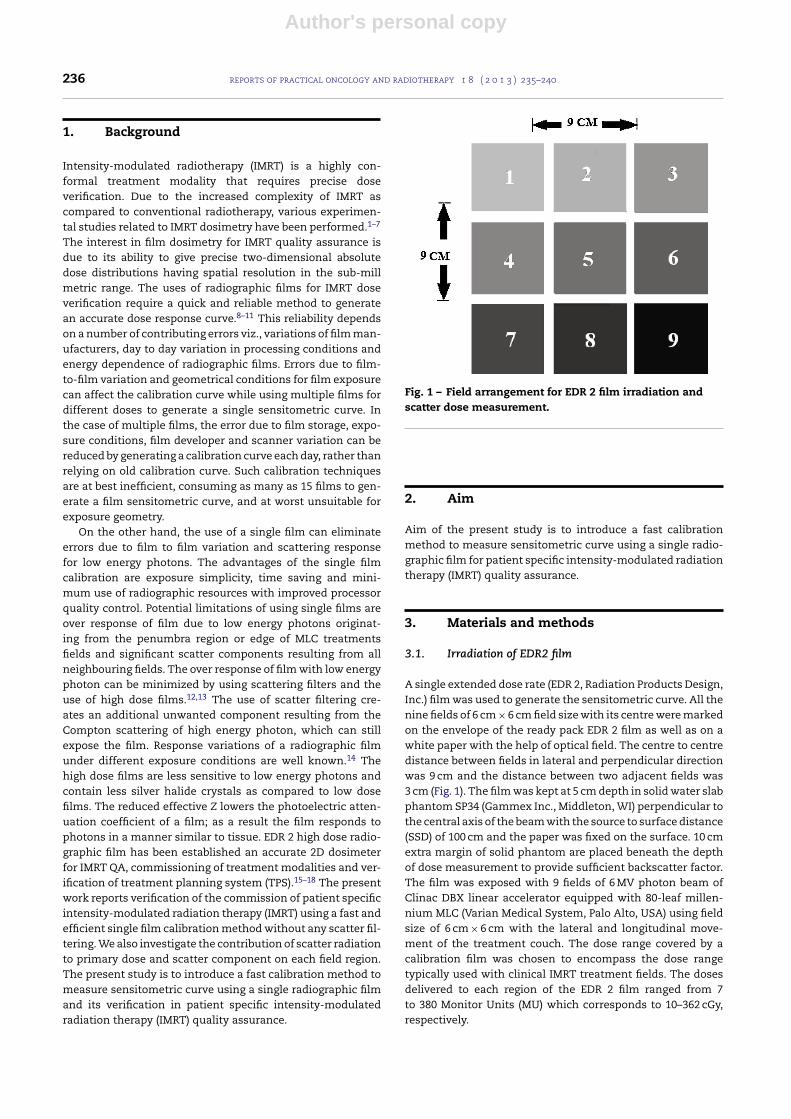
Fig. 1 – Field arrangement for EDR 2 film irradiation andscatter dose measurement.
2. Aim
Aim of the present study is to introduce a fast calibrationmethod to measure sensitometric curve using a single radio-graphic film for patient specific intensity-modulated radiationtherapy (IMRT) quality assurance.
3. Materials and methods
3.1. Irradiation of EDR2 film
A single extended dose rate (EDR 2, Radiation Products Design,Inc.) film was used to generate the sensitometric curve. All thenine fields of 6 cm × 6 cm field size with its centre were markedon the envelope of the ready pack EDR 2 film as well as on awhite paper with the help of optical field. The centre to centredistance between fields in lateral and perpendicular directionwas 9 cm and the distance between two adjacent fields was3 cm (Fig. 1). The film was kept at 5 cm depth in solid water slabphantom SP34 (Gammex Inc., Middleton, WI) perpendicular tothe central axis of the beam with the source to surface distance(SSD) of 100 cm and the paper was fixed on the surface. 10 cmextra margin of solid phantom are placed beneath the depthof dose measurement to provide sufficient backscatter factor.The film was exposed with 9 fields of 6 MV photon beam ofClinac DBX linear accelerator equipped with 80-leaf millen-nium MLC (Varian Medical System, Palo Alto, USA) using fieldsize of 6 cm × 6 cm with the lateral and longitudinal move-ment of the treatment couch. The dose range covered by acalibration film was chosen to encompass the dose rangetypically used with clinical IMRT treatment fields. The dosesdelivered to each region of the EDR 2 film ranged from 7to 380 Monitor Units (MU) which corresponds to 10–362 cGy,respectively.

Author's personal copy
reports of practical oncology and radiotherapy 1 8 ( 2 0 1 3 ) 235–240 237
Table 1 – Ion chamber measured data at each field position with all nine fields irradiated.
Field positions MUs delivered Dose/MU (cGy) Dprimary (cGy) Dscatter (cGy) Dtotal (cGy) Optical density (OD)
1 7 0.9350 6.55 3.852 10.40 0.1902 12 0.0085 11.22 5.187 16.41 0.2203 60 0.0031 56.10 4.707 60.81 0.4294 120 0.0085 112.20 6.428 118.63 0.7785 170 0.0035 158.95 8.535 167.48 1.0526 230 0.0013 215.05 7.180 222.23 1.3987 280 0.0031 261.80 6.092 267.89 1.6808 340 0.0013 317.90 8.412 326.31 1.9639 380 0.0008 355.30 6.691 361.99 2.143
3.2. Scattered dose measurement
The primary and scattered absorbed dose measurementswere performed using a 0.6 cm3 thimble ionization cham-ber (PTW, Freigburg, Germany). The ion chamber was placedin a predrilled cavity at the midpoint of a 2 cm thick solidwater slab. The dose measurements were performed withan additional 4 cm water slab produce buildup and suffi-cient attenuation. The field arrangement was kept identicalas used for film irradiation. Daily machine output fluctua-tion was recorded based on measurements at the centralaxis of a 10 cm × 10 cm field with an ion chamber at 10 cmdepth.
The primary and scattered doses at the centre of eachindividual field along the lateral and perpendicular directionsfrom field number 1 (No. 1) were measured with ionizationchamber. The radiation exposure due to accumulated scat-tered doses plus the primary radiation dose for each field werecalculated using the relation
Di = Dpi +N∑
j=1
Fij · Dj (1)
where Di is the dose at ith field contributed by both primaryradiation beam and scattered radiation from other fields andFij is the scattered factor on ith Field from jth field. Dj is theprimary radiation dose delivered by jth field.
3.3. Calibration of film
The duly developed film was scanned at 300 dpi resolution inVIDAR film scanner (VXR-16 Dosimetry PRO plus, Vidar Sys-tems Corp., Herndon) for the measurement of optical density(OD). The value of OD obtained from each exposed regionwas plotted against the effective doses contributed by primaryradiation dose delivered from 7 to 380 MUs plus accumulatedscattered dose from different 9 fields to generate the sensito-metric curve.
The sensitometric curve was repeated for four dates andfound at a very good agreement with the mean standard devi-ation of 0.48%. The generated sensitometric curve was usedfor IMRT patient specific QA to verify the measured and treat-ment planning system (TPS) calculated dose distribution ofthe IMRT fields.
3.4. Verification of IMRT
Ten IMRT plans for the treatment of head and neck carcinomausing dynamic IMRT technique of 7 fields of 6 MV photon beamwere considered in this study. Three-dimensional treatmentplanning system Eclipse version 8.6 (Varian Medical SystemsInc., Palo Alto, CA) with inverse plan optimization was usedin making the treatment plans. The optimal fluence profilescalculated by inverse plan optimization produce MLC motionpatterns with the help of a computerized program to controlthe leaf motion. The dose calculation was done using pencilbeam convolution (PBC) algorithm incorporated in the 3D-TPS. The patient specific hybrid IMRT verification plan wascreated in a solid water slab phantom using the same radia-tion fluence of each plan. The verification plan was executedon linear accelerator using 4 dimensional treatment console(4DTC) version 8.6 (Varian Medical Systems Inc., Palo Alto, CA).All the systems were networked through ARIA (Varian Medi-cal Systems Inc., Palo Alto, CA) networking system. Gamma(�) evaluation method was used to compare the planned dosedistribution in TPS and delivered dose distribution.15 Gammamethod is useful in measuring distance to agreement (DTA)in a high gradient region, and sensitive to dose differencesbetween calculated and delivered plan. The value of � calcu-lated by OmniPro I’mRT software was used for plan acceptancecriteria of dose difference of 3% and distance to dose agree-ment (DTA) of 3 mm. The plan was accepted only if more than95% pixels had the value of � ≤ 1 in planned active area.
4. Results and discussion
The irradiation of nine square field regions of single EDR 2 filmwas performed in less than 10 min. Childress et al.8 reportedthe single film calibration method using two field step andshoot MLC treatment. However, the MLC driven step and shoottechnique leads to a significant leakage factor and scatter radi-ation component from MLC. Later on, Kulasekere et al.9 usedthe same method to irradiate eight field patterns on a sin-gle film using jaws plus MLC motion. The use of jaws plusMLC reduces the scatter and transmission component over theMLC driven technique. In our work, the same technique wasused to irradiate nine square fields of 6 cm × 6 cm on a sin-gle film with X and Y jaws of medical linear accelerator. Themethod of irradiation with jaws only completely eliminatesthe MLC radiation leakage factor. The earlier measurementswere performed by irradiating the field at the off axis regions

Author's personal copy
238 reports of practical oncology and radiotherapy 1 8 ( 2 0 1 3 ) 235–240
0 10 20 30
1E-3
0.01
0.1
1
Distance(cm)
rotcaf
rett
acS
90o( )
45o
63o
0o( )
27o
Fig. 2 – Variation of scatter factor with different angles fromfield region 1 (0◦ and 90◦ positions represent the paralleland perpendicular positioning of cylindrical ion-chamberalong the direction of dose measurement respectively).
with respect to the central axis of the beam profile. However, inthe present method the film exposure was done on the centralaxis of the field with the use of lateral and longitudinal move-ment of the treatment couch. The present method is foundto be less error prone and significantly reduces the error dueto field geometry. Table 1 shows the ion chamber measureddata at each field position with all nine fields irradiated to
400300200100
0.6
1.2
1.8
2.4
Op
tica
l d
en
sity (
OD
)
Dose (cGy)
Day 1
Day 2
Day 3
Day 4
Fitted curve day 1
Fig. 3 – Sensitometric curves generated on four dates, usingEDR2 film. The line is third order polynomial fit of Day 1data.
calculate the absorbed dose contributed from the correspond-ing monitor units (MU) delivered to each field. The value ofoptical density corresponding to each region was also given inTable 1.
The contributions of scatter doses in each field regionfrom the other irradiated field regions were estimatedusing the scatter factors shown in Fig. 2. Fig. 2 showsthe variation of scattered doses received by each field
Fig. 4 – Plan verification for a typical IMRT plan using for EDR 2 film.

Author's personal copy
reports of practical oncology and radiotherapy 1 8 ( 2 0 1 3 ) 235–240 239
region from the primary irradiated field region 1 in lateral,perpendicular and other directions. There scattering factordecreases exponentially as a function of distance from thefield centre due to the decrease of side scattered radiationfrom the field edge. It has been noticed that the value ofscatter factor is approximately the same along parallel andperpendicular directions for all regions at the same distancesfrom the primary irradiated region. The value of parallel posi-tioning of ion-chamber is lower than that of perpendicularpositioning, the differences ranging from 0.0057 to 0.00071 asa function of distance from the centre of primary radiationexposure using 6 cm × 6 cm. This is due to the orientation ofchamber and a little effect produced by upper and lower jawsscattering. It was noticed that in the case of radiation expo-sure to region 1, the amount of scattered radiation dose atregions 2 and 4, regions 7 and 3, and regions 8 and 6 were thesame, respectively. The same results were found for the otherprimary field regions.
The measured OD values of the calibrated EDR2 film wereplotted against dose values to obtain sensitometric curve(Fig. 3). The third order fitted polynomial equation for the sen-sitometric curve is
Dose (cGy) = bo + b1x + b2x2 + b3x3 (2)
where bo = 0.1607, b1 = 0.00424, b2 = 8.74748 × 10⁻⁶,b3 = −1.63416 × 10⁻⁸ and x is value of optical density. Thecalibration curve generated from the film data analysis wasused by OmniPro I’mRT software to compare the measureddose from film with the corresponding treatment planningdata. The patient-specific IMRT QA has been performed toensure the accuracy of treatment planning. For this, tenIMRT plans with 6 MV photon beam were evaluated using thegamma index method with TPS calculated dose distributionto find an average of 97.86% pixel population and rangingfrom 95.53 to 99.57% passed for � < 1 with standard deviationof 1.57%. Fig. 4a and b shows the coronal dose distributionfor film and TPS calculated, respectively. Fig. 4c shows doseprofiles along the X direction, where red and green profilesare film measured and TPS calculated, respectively, andFig. 4d shows the gamma analysis of 3% delta dose and 3 mmdistance to agreement (DTA).
5. Conclusion
This method eliminates the scattering and leakage contribu-tion through MLC and provides a solution to reduce manyfilm errors while using a single film and multiple fields forthe creation of calibration curve each day, rather than relyingon old calibrations or attempting to scale a standard curve.This method minimizes errors due to film storage, exposureconditions, film developer, and scanner variations. The dailyproduction of a sensitometric curve requires a quick way toaccurately generate different known exposures on film so thatlabour costs can be minimized. The high reproducibility, lowerror, quick delivery time and ease of use of the calibrationmethod show that the single film calibration method is supe-rior to previous procedures. It allows fast daily calibrationsof films for highly accurate IMRT verification. It is further
recommended that EDR2 film can be used clinically due to itsnear tissue equivalent response to low-energy photons.
Conflict of interest
No conflict of interest.
Financial disclosure
The funding sources had no involvement for the conduct ofthe research and/or preparation of the article, analysis andinterpretation of data.
r e f e r e n c e s
1. Wang X, Spirou S, Losasso T, Stein J, Chui CS, Mohan R.Dosimetric verification of intensity-modulated fields. MedPhys 1996;23:317–27.
2. Low DA, Gerber RL, Mutic S, Purdy JA. Phantoms for IMRTdose distribution measurement and treatment verification.Int J Radiat Oncol Biol Phys 1998;40:1231–5.
3. Xing L, Curran B, Hill R, et al. Dosimetric verification of acommercial inverse planning system. Phys Med Biol1999;44:463–78.
4. Martens C, Wagter CD, Neve WD. The value of pin point ionchamber for characterization of small field segments used inintensity modulated radiotherapy. Phys Med Biol2000;45:2519–30.
5. Dong L, Antolak J, Salehpour M, et al. Patient specific pointdose measurements for IMRT monitoring unit verification. IntJ Radiat Oncol Biol Phys 2003;56:856–77.
6. Malicki J. The importance of accurate treatment planning,delivery and dose verification. Rep Pract Oncol Radiother2012;17:63–5.
7. Winiecki J, Zurawski Z, Drzewiecka B, Slosarek K.Anatomy-corresponding method of IMRT verification. RepPract Oncol Radiother 2011;16:1–9.
8. Childress NL, Dong L, Rosen II. Rapid radiographic filmcalibration for IMRT verification using automated MLC fields.Med Phys 2002;29:2384–90.
9. Kulasekere R, Jean M, Moran JM, Fraass BA, Roberson PL.Accuracy of rapid radiographic film calibration forintensity-modulated radiation therapy verification. J Appl ClinMed Phys 2006;7:86–95.
10. Shi C, Papanikolaou N, Yan Y, Weng X, Jiang H. Analysis ofthe sources of uncertainty for EDR 2 film-based IMRT qualityassurance. J Appl Clin Med Phys 2006;7:1–8.
11. Danciu C, Proimos BS, Rosenwald JC, Mijnheer BJ. Variation ofsensitometric curves of radiographic films in high energyphoton beams. Med Phys 2001;28:966–74.
12. Ju SG, Ahn YC, Huh SJ, Yeo IJ. Film dosimetry for intensitymodulated radiotherapy: dosimetric evaluation. Med Phys2002;29:351–5.
13. Burch SE, Kearfott KJ, Trueblood JH, Sheils WC, Yeo JL, WangCK. A new approach to film dosimetry for high energy photonbeams: lateral scatter filtering. Med Phys 1997;24:775–83.
14. Palm A, Kirov AS, Losasso T. Predicting energy response ofradiographic films in a 6 MV X-ray beam using Monte Carlocalculated fluence spectra and absorbed dose. Med Phys2004;31:3168–78.
15. Esthappan J, Mutic S, Harms WB, Dempsey JF, Low DA.Dosimetry of therapeutic photon beams using an extendeddose range film. Med Phys 2002;29:2438–45.

Author's personal copy
240 reports of practical oncology and radiotherapy 1 8 ( 2 0 1 3 ) 235–240
16. Olch AJ. Dosimetric performance of an enhanced dose rangeradiographic film for intensity-modulate radiation therapyquality assurance. Med Phys 2002;29:2159–68.
17. Winiecki J, Morgas T, Majewska K, Drzewiecka B. The gammaevaluation method as a routine QA procedure of IMRT. RepPract Oncol Radiother 2009;14:162–8.
18. Chełminski K, Rostkowska J, Kania M, Bulski W,Gwiazdowska B. Measurement of the sensitometric curves ofKodak EDR2 and X-Omat V films using enhanced dynamicwedges and dynamic multileaf collimators. Rep Pract OncolRadiother 2005;10(6):293–300.
Related Documents