48 Bmchlmzca et Bmphyswa Acta, 659 (1981) 48--61 Elsewer/North-Holland Bmmedlcal Press BBA 69255 A CALCIUM-ACTIVATED PROTEASE POSSIBLY INVOLVED IN MYOFIBRILLAR PROTEIN TURNOVER ISOLATION OF A LOW-CALCIUM-REQUIRING FORM OF THE PROTEASE WILLIAM R DAYTON a, JUDITH V SCHOLLMEYER b ROBERT A LEPLEY a and LISA R. CORTES a a Department of Animal Science, Unwerszty of Minnesota, St Paul and b Departments of Anatomy and Laboratory Medzcme and Pathology, Unwerszty of Mznnesota, Mmneapohs, MN55108 (USA) (Received October 28th, 1980) Key words Protease, Ca ~+ actwatmn, Protein turnover, Ca 2+ requirement, (Myofzbrd) Summary Two forms of a calcmm-actlvated neutral protease were isolated and purified from porcine skeletal muscle. The two forms of the protease differ markedly in their reqmrement for calcmm with the low-calclum-requmng form showing one-half maximal activation at 45 ~zM calcmm while the hlgh-calclum-requmng form shows one-half maximal activation at 0.74 mM calcium. Additionally, they chromatograph differently on DEAE-cellulose, exhibit different mobflltles In electrophoresls m a nondenaturlng buffer, are affected differently by certmn divalent cations, and have slightly different pH dependencies. Despite these dif- ferences, the purified forms of the calcium-activated protease co-chromato- graph in gel permeation chromatography, have identical banding patterns on sodium dodecyl sulfate (SDS)-polyacrylamlde gels, cross-react with an antibody directed against the 80 000-dalton subunlt of the calcmm-actlvated protease we orlgmally purified from skeletal muscle (Dayton, W.R., Goll, D.E., Zeece, M.G., Robson, R.M. and Revflle, W.J. (1976) Biochemistry 15, 2150--2158), and have Identical effects on the ultrastructure of myoflbrfls. The high-calcium- reqmrmg protease purified m this study is very lnkely ldentmal to the calcmm- activated protease we originally purified from skeletal muscle. The properties of the low-calclum-requmng form of the protease suggest that it is the form of the enzyme that is active in VlVO. Abbrevlahon IgG, lmmunoglobulm G 0005--2744/81/0000--0000/$02 50 © Elsevmr/North-Holland Blomedmal Press

Welcome message from author
This document is posted to help you gain knowledge. Please leave a comment to let me know what you think about it! Share it to your friends and learn new things together.
Transcript
48
Bmchlmzca et Bmphyswa Acta, 659 (1981) 48--61 Elsewer/North-Holland Bmmedlcal Press
BBA 69255
A CALCIUM-ACTIVATED PROTEASE POSSIBLY INVOLVED IN MYOFIBRILLAR PROTEIN TURNOVER
ISOLATION OF A LOW-CALCIUM-REQUIRING FORM OF THE PROTEASE
WILLIAM R DAYTON a, JUDITH V SCHOLLMEYER b ROBERT A LEPLEY a and LISA R. CORTES a
a Department o f Animal Science, Unwerszty o f Minnesota, St Paul and b Departments o f Anatomy and Laboratory Medzcme and Pathology, Unwerszty o f Mznnesota, Mmneapohs, MN55108 ( U S A )
(Received October 28th, 1980)
Key words Protease, Ca ~+ actwatmn, Protein turnover, Ca 2+ requirement, (Myofzbrd)
Summary
Two forms of a calcmm-actlvated neutral protease were isolated and purified from porcine skeletal muscle. The two forms of the protease differ markedly in their reqmrement for calcmm with the low-calclum-requmng form showing one-half maximal activation at 45 ~zM calcmm while the hlgh-calclum-requmng form shows one-half maximal activation at 0.74 mM calcium. Additionally, they chromatograph differently on DEAE-cellulose, exhibit different mobflltles In electrophoresls m a nondenaturlng buffer, are affected differently by certmn divalent cations, and have slightly different pH dependencies. Despite these dif- ferences, the purified forms of the calcium-activated protease co-chromato- graph in gel permeation chromatography, have identical banding patterns on sodium dodecyl sulfate (SDS)-polyacrylamlde gels, cross-react with an antibody directed against the 80 000-dalton subunlt of the calcmm-actlvated protease we orlgmally purified from skeletal muscle (Dayton, W.R., Goll, D.E., Zeece, M.G., Robson, R.M. and Revflle, W.J. (1976) Biochemistry 15, 2150--2158), and have Identical effects on the ultrastructure of myoflbrfls. The high-calcium- reqmrmg protease purified m this study is very lnkely ldentmal to the calcmm- activated protease we originally purified from skeletal muscle. The properties of the low-calclum-requmng form of the protease suggest that it is the form of the enzyme that is active in VlVO.
Abbrevlahon IgG, lmmunoglobulm G
0005--2744/81/0000--0000/$02 50 © Elsevmr/North-Holland Blomedmal Press
49
Introduchon
Calcmm-actlvated neutral proteolytm actlv:ty has been :solated from many tissues mcludmg bram [1], skeletal muscle [2], card:ac muscle [3,4], smooth muscle [5,6] and hen oviduct [7]. In addlhon, calcmm-actlvated proteases have been punfmd from mammal:an skeletal muscle [8,9] and chmken skeletal muscle [ 10]. Even so, the function of the calcmm-actlvated protease(s) m the cell is not well understood. Add:honaUy, there is disagreement con- cernmg the subumt compos: t lon of the enzyme from skeletal muscle [9,11].
Purffmd calcmm-actlvated protease from skeletal muscle causes partial degradahon of myoflbrils, attacking the Z-disk and hydrolyzmg troponm-T, t roponm-I, C-protein and t ropomyosm [12]. It is therefore possible that the calcmm-activated protease may be revolved m degradation of myoflbrillar protems m muscle [12] and also m non-muscle tissue. This theory :s supported by reports that the amount of calcium-activated protease isolated from rapidly atrophying muscle tissue (where myof:bril degradatmn :s proceeding rap:dly) :s several-fold greater than the amount :solated from normal muscle tissue [13]. Additmnally, lmmunocytochemlcal locallzatmn of the calcmm-actlvated protease has shown that the protease is located m the myofibrfl [15,16] . Our studms [16] using a monospecffm ant ibody d~rected against the 30 000-dalton subumt of the calcmm-actlvated protease to localize the enzyme have shown that the protease is located specffmaUy m the Z-disk of the myof ibn l and also m or near the sarcolemma. Locahzatmn of the calcmm-actlvated protease m the Z-d:sk, a myoflbnl lar structure shown to be extremely suscept:ble to calcmm- activated protease-catalyzed hydrolys:s, strongly supports the theory that the protease plays a role m myofibri l degradation.
A major problem m understandmg the physmlogmal role of the calcmm- act:vated protease(s) has been the fact that m m vitro assays the protease(s) reqmres the presence of 1--2 mM Ca 2÷ m order to express maximum activity [2,8,9]. This is h:gher than the levels of free calcmm normally thought to be present m muscle cells [17]. Consequently, a recent report that cardmc muscle contmns both the calcmm-actlvated protease reqmrmg mflhmolar levels of calcmm and a calcmm-dependent proteolytm act lwty detectable m the presence of physmlogmally obtmnable calcium concentratmns [3] was of extreme interest. Although the two calcmm-activated proteolytm actlvitms detected m cardmc muscle [3] are sh-nilar m some propertms, it was not clear whether or no t they were related smce neither protease was punfmd. We report here the purffmatmn of both the low-calcmm and high-calcmm-requmng proteases. The purffmd proteases are lmmunologmally cross-reactive and have ldentmal bandmg patterns on SDS-polyacrylamlde gels. Consequently, :t appears that the two calcmm-actlvated proteolytm actlvltms present m porcine skeletal muscle are due to two forms of the same basra calcmm-act:vated proteolytm enzyme. One form requires high calcmm for act :wty while the other form :s act:ve at much lower calcmm concentratmns approaching the free Ca 2÷ concentratmns attmned m muscle cells durmg contractmn. AddltmnaUy, :t appears that the hlgh-calcmm-requmng form is ldentmal to the calcium-activated protease we originally punfmd from porcme skeletal muscle [8].
50
Matenals and Methods
Isolatton of calcmm-actwated proteolytzc actzmty from muscle tzssue Extraction of the calcmm-actlvated proteolytm act lwty from porcme skeletal muscle was done essentially as described previously [8], except that the extrac- tion buffer was 50 mM Trls-acetate, pH 8.0/4 mM EDTA. Both the low- and hlgh-calcmm-requmng proteolytm actlvltms and an inhibitor of the calcmm- activated protease(s) precipitated between 30 and 40% (NH4)2804 saturation from the 15 000 × g supernatant of the whole muscle homogenate The pre- cipitate was dissolved m 20 mM Trls-acetate, pH 7.5/1 mM EDTA/10 mM 2-mercaptoethanol and dialyzed agamst this buffer for 12 h This solution was then clarlfmd at 105 000 × g for 1 h and added to DEAE-cellulose that had been equlhbrated agamst dialysis buffer. After allowing time for the protein to brad to the DEAE-cellulose, the cellulose was allowed to precipitate and the supernatant was decanted. The cellulose was subsequently washed twine with 3 vol {v/v) dialysis buffer and was then packed into a 4 X 85 cm column Protem concentratmns were determmed using the bmret method [19] or the method of Lowry et al. [20].
Chromatographic procedures. Sepharose 6B (Pharmacm Free Chemmals, Inc.) and Ultrogel (LKB Instruments, Inc.) were used for gel-permeatmn chromatography. Ion-exchange chromatography was done usmg DEAE-cellu- lose (Whatman DE-52). Column dlmensmns, protein loads, flow rates and buffer systems for individual columns are given m the appropriate figure legend.
Electrophorettc procedures Analytmal slab-gel electrophoresls m the pres- ence of SDS was done accordmg to the procedure of Laemmh [21]. Gels were stained m 0.1% Coomassm blue/50% methanol/7% glacial acetm acid solutmn and were destamed electrophoretmally.
Preparative poly~crylamlde slab-gel electrophoresls m non-denaturing buffer was done accordmg to the procedure of Maurer [22] as descnbed prevmusly [8], except that a dual vertmal slab-gel electrophoresls cell (Bm-Rad Labora- tories) and an LKB 2103 power supply were used. 1 mg protem (either hlgh- calcmm-requtrmg or low-calcmm-requtring protease that m each case had been partmlly purffmd chromatographmally) was loaded onto a slab gel and elec- trophoresed at 14 mA/gel while the gel was bemg cooled to 4°C by cold water passmg through the electrophoresls cell At the completmn of the run, the polyacrylamlde contammg each individual protem band was removed and dis- rupted mechanmally to form a slurry whmh was then placed m a separate cyhn- drmal gel tube containing a 3.5% polyacrylamlde plug Electrophoresls for 8 h at 4°C and 2 mA/gel was suffmmnt to move the protein out of the slurry through the plug and into a dmlysls tubing which was fi t ted over the end of the gel tube The protem obtamed m this way was then dmlyzed overmght against 1 mM KHCO3/5 mM 2-mercaptoethanol/5 mM EDTA. Each sample was then assayed for the calcmm-actlvated proteolytm act lwty for homogenei ty usmg SDS-polyacrylamlde slab-gel electrophoresls.
Purffmatmn of the 80 000-dalton subumt of the calcmm-actlvated protease was done as described above usmg preparative SDS-polyacrylamlde slab-gel electrophorems [21].
51
Protease assays. Calcmm-actlvated proteolytm activity was assayed usmg either casein or myof lbn ls as the substrate. The details of these assays have been descnbed previously [8]. Calcmm dependence and pH dependence of the calcmm-actlvated proteolytm enzyme were determined using casein or myo- fibrils as a substrate. The methodology for these assays has been reported m detml previously [ 9].
Immunological procedures The hlgh-calcmm-requmng protease whmh we originally molated from skeletal muscle was punfmd to near homogenei ty usmg chromatographm procedures described prevmusly [8]. To prepare highly pun- fred 80 000-dalton subumt for use m ant ibody productmn, chromatographi- cally punfmd calcmm-actlvated protease was further punfmd usmg SDS-poly- acrylamide slab-gel electrophoresm as described prewously m tins sectmn. The homogenei ty of the electrophoretmaUy-punfmd 80 000-dalton subumt prepara- tmn was conftrmed usmg analytmal SDS-polyacrylamlde gel electrophoresls (Fig. 6). Approx. 75 ~g of the electrophoretmally-punfmd 80 000-dalton sub- umt was emulsffmd m an equal volume of Freund's complete adjuvant and rejected directly into the pophteal lymph node of a rabbit. This procedure was repeated 21 days after the first injection except that the antigen was emulsffmd m mcomple te adjuvant. Rabbits injected m thin way produced antisera with a high tlter dtrected against the 80 000-dalton subumt. This antmera was frozen m hquld nitrogen and stored at --60°C until needed. The IgG fractmn was precipitated from the antmera at 50% (NH4)2SO4 saturation and further pun- fred usmg DEAE-cellulose column chromatography [23,24] .
Double dfffusmn was carned ou t m 1% agarose (Indubolse A45, L'Industrm Bmlogique Francmse 5. A. Gennevflhers, France)contmnmg 0.01% Merthmlate (Eh Lilly) and borate-buffered salme, pH 8.5 [25]. The lmmunodfffusmn plates were allowed to mcubate m a momt chamber for 24 h at room temperature. The plates were then rinsed m three changes of 0.15 M NaC1 each day for three days, rinsed m distilled water for 2 h with one change and dried by placing a pmce of wet filter paper over the plate for at least 15 h [26]. Dned plates were stmned for 30 mm with 0.25% Coomassm brflhant blue m a 5 . 1 . 4 mLxture of methanol/acetm acid/water [27]. The plates were destmned m the same solu- tmn wi thout the stam.
Calcmm-actwated protease treatment of myofzbnls Myoflbrfls prepared from porcine skeletal muscle according to the procedure of Goll et al. [18] were treated with punfmd calcmm-actlvated protease as described prewously [12]. Control and treated myoflbrils were then prepared for electron micro- scopy using a procedure described m detail elsewhere [13]. Specific detmls of mdlwdual calcmm-actlvated protease treatments are given m the appropriate figure legend.
Results
Chromatographic purification of calcmm-actwated proteolyt~c actwlty The 4 × 85 cm DEAE-cellulose column to whmh the crude calcmm-actlvated
protease had been bound (Materials and Methods) was eluted with a lmear KC1 gradmnt (Fig. 1). Two peaks of calcmm-actlvated proteolytm act lwty elute f rom the column as the gradient is developed. One peak elutes a 70 mM KC1
52
S,o ,oo | " l Psokn =
8 ' ' ~ , o2oL ~ ' ~ ' ,oo _ -14o
// / , o I 0 Peek I / • 200 2 0 J~
/ / ,% / / / ., \ ', /
/ ; t *--m / 3-' :' .,oo
--~T--. ./A" , 0 20 40 60 80 I00 120 140 160 I( ,00 -J 0
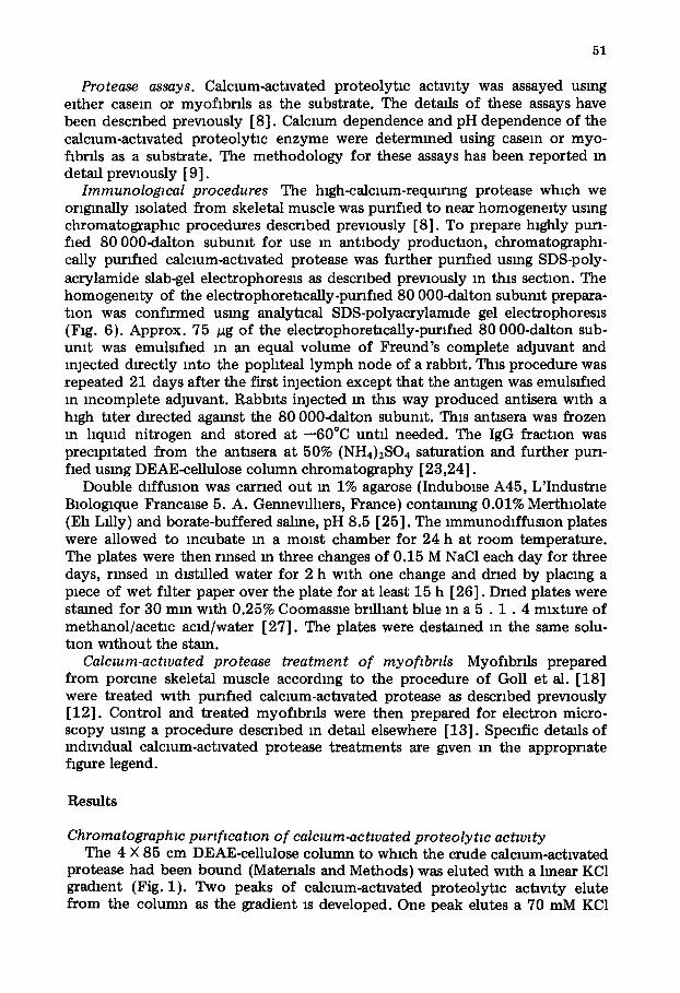
Fraction Number Fig 1 E lu t Ion p r o f d e o f c a l c i u m - a c t i v a t e d P ro tease ac tawty f r o m a 4 0 × 85 c m DEAE-ce l lu lose c o l u m n
P ~ o r to p o u r i n g the c o l u m n , the DEAE-ce l lu lose was maxed w i t h 31 059 m g o f t he p r o t e i n f r a c t m n
sa l t ing o u t b e t w e e n 30 a n d 40% ( N H 4 ) 2 S O 4 s a t u r a t i o n f r o m the 1 5 0 0 0 X g s u p e r n a t a n t o f t he w h o l e musc l e h o m o g e n a t e The b o u n d p r o t e i n was e lu t ed a t 60 m l / h w i t h a 2 0 0 0 ml c o n t i n u o u s g r a d i e n t f~om
10 to 4 0 0 m M KC1 m 20 m M Tr l s -ace ta te , p H 7 5/1 m M E D T A / 1 0 m M 2 - m e r c a p t o e t h a n o l 20-ml f rac- t i ons were co l lec ted . . . . . . , KC1 c o n c e n t x a t i o n m the e luan t , , Ca2+-act lva ted p r o t e o l y t m act iv- i ty (ad jus ted a b s o r b a n c e u m t s ) d e t e r n u n e d us ing case in as a subs t r a t e , - - a b s o r b a n e e at 280 n m Inchvldual t ubes m p e a k I and p e a k I I were p o o l e d as m d m a t e d and sa l ted o u t b e t w e e n 0 and 45% ( N H 4 ) 2 S O 4 sa tu~at lon to give the p e a k I and the p e a k II c a l c m m - a c t l v a t e d p r o t e a s e f r a c t i o n s
(peak I protease) and another elutes at 220 mM KC1 (peak II protease). Although both the peak I and peak II proteases are activated by calcium, the peak I protease reqmres substantially lower levels of calcium than does the
E
~ o ~
o~
02
30 40 50 60 70 80 90 Froct)on Number
,60 ,/o
6 0
40
,26 o
oo
80 u
g p
D. "o
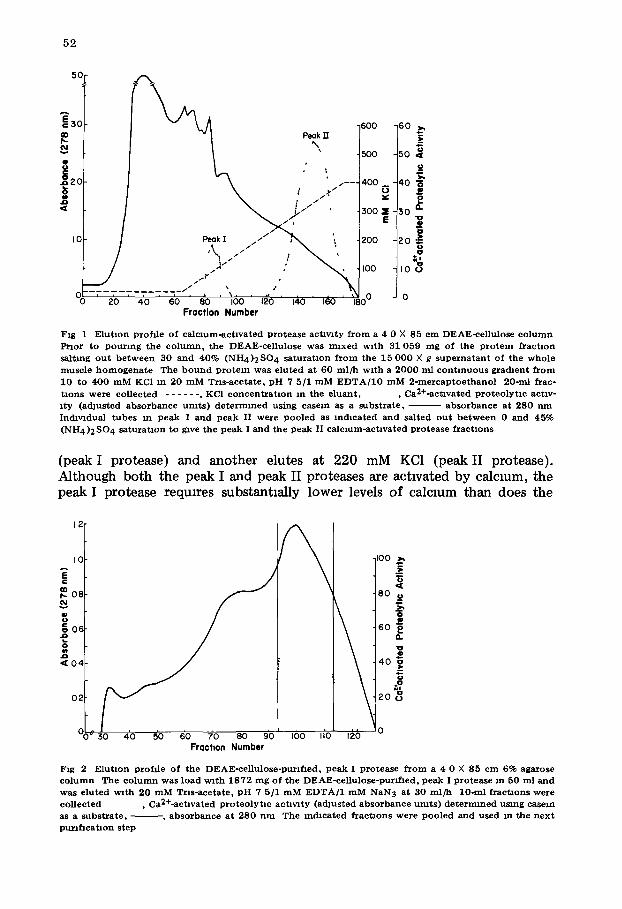
Fig 2 E lu t lon p rof i l e o f t he DEAE-ce l in lose-pur l f~ed , p e a k I p ro t ea se f r o m a 4 0 X 85 c m 6% agarose c o l u m n The c o l u m n was load w i t h 1 8 7 2 m g o f the DEAE-ce l lu lose -pur l f l ed , p e a k I p r o t e a s e m 50 m l and was e lu t ed wi th 20 m M T n s - a c e t a t e , p H 7 5/1 m M E D T A / 1 m M N a N 3 at 30 m l / h 10-ml f r a c t i o n s were co l lec ted , Ca2+oact4vated p r o t e o l y t l e act lvzty ( ad jus t ed a b s o r b a n c e u m t s ) d e t e r m i n e d u s m g case in as a s u b s t r a t e , - - - , a b s o r b a n c e a t 280 n m The mchca t ed f r a c t i o n s were p o o l e d a n d used m the n e x t pu~lf lca t lon s tep
53
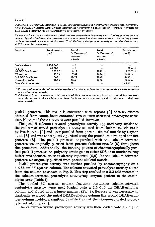
T A B L E I
S U M M A R Y O F T O T A L P R O T E I N Y I E L D , S P E C I F I C C A L C I U M - A C T I V A T E D P R O T E A S E A C T I V I T Y
A N D T O T A L C A L C I U M - A C T I V A T E D P R O T E A S E A C T I V I T Y A T E A C H S T E P IN P U R I F I C A T I O N O F
T H E P E A K I P R O T E A S E F R O M P O R C I N E S K E L E T A L M U S C L E
F igu re s are f o r a t y p m a l c a l c m m - a c t i v a t e d p r o t e a s e p r e p a r a t i o n b e g i n n i n g v~ th 12 0 0 0 g p o r c i n e ske le ta l
m u s c l e Spec i f i c Ca2+-actavated p r o t e a s e ac i awty is e x p r e s s e d as a b s o r b a n c e u m t s a t 2 7 8 n m / m g p r o t e i n per h as m e a s u r e d m the c a s e m o l y t l c assay To ta l Ca2+-actavated protease actawty IS t o t a l a b s o r b a n c e u m t s a t 278 n m m the case in assay
F r a c t i o n T o t a l p r o t e i n Spec i f i c T o t a l P u r l f m a t i o n
( rag) Ca2+-actavated Ca2+-ac t lva ted ( - fo ld)
p r o t e a s e p r o t e a s e
a c t l w t y actavl ty
Crude e x t r a c t 1 727 6 4 5 - - * - - - -
P 3 0 - 4 0 31 059 - - * - - 55 6 ** 1st DEAE-ce l l u lo se 1 8 7 2 5 3 12 5 8 4 2 2 922 7 **
6% aga rose 772 4 7 32 5 6 5 4 2 2 1 6 8 3 2 n d D EA E- ceH u l o se 2 4 8 15 72 3 8 9 9 4 6 6 7 1
Ul t roge l A c A - 3 4 156 4 20 0 3 1 2 8 5 9 0 0 8 Prep e l e c t r o p h o r e m s - - 50 - - 14 802
* P resence of an m l ~ b i t o r o f t he c a l c i u m - a c t i v a t e d p r o t e a s e m these fTactaons p r e v e n t s a c c u r a t e m e a s u r e - m e n t o f p r o t e a s e ac t awty
** Ca lcu l a t ed f r o m r e d u c t a o n m to t a l p r o t e i n o f these steps ( a s s u m i n g to t a l r e c o v e r y o f t h e p r o t e a s e ) s ince the p r e s e n c e of an m l ~ b l t o r m these fTactlons p r e v e n t s m e a s u r e m e n t o f c a l c m m - a c t l v a t e d p ro - tease a c t l w t y
peak II protease. This result is consistent with reports [3] that an extract obtained from canine heart contained two calcium-activated proteolytm actlv- 1tins. Neither of these actlwtles were purified, however.
The peak II calcmm-actlvated proteolyt lc act lwty appeared very similar to the calcium-activated proteolyt lc activity isolated from skeletal muscle t~ssue by Busch et al. [2] and later punfmd from porcme skeletal muscle by Dayton et al. [8] and was consequently punfmd using the procedure developed for this protease [8]. The peak II protease co-punfmd with the calcium-activated protease we originally purified from porcine skeleton muscle [8] throughout this procedure. Additionally, the banding pattern of chromatographically-purl- fled peak II protease on polyacrylamlde gels m either SDS or in nondenaturmg buffer was ~dentmal to that already reported [8,9] for the calcmm-actlvated protease we orlgmally purified from porcine skeletal muscle.
Peak I proteolyt lc activity was further purified by chromatography on a 4 × 85 cm 6% agarose column. The calcmm-actlvated proteolyt lc activity eluted from the column as shown m Fig. 2. This step resulted m a 2.3-fold mcrease in the calcium-activated proteolyt lc actlvlty/mg enzyme protein in the casem- olytm assay (Table I).
The pooled 6% agarose column fractions containing calcmm-actlvated proteolytm achwty were next loaded onto a 2.5 × 40 cm DEAE-cellulose column and eluted with a linear gradient (Fig. 3). Because it was necessary to drastmally overload the mltlal DEAE-cellulose column this second DEAE-cellu- lose column yielded a significant purlflcatmn of the calcium-activated proteo- lytlc activity (Table I).
The calcium-activated proteolyt ic act lwty was then loaded onto a 2.5 .X 85
54
3c
IO
o
/ /
~ j / ~ . / s
"3o 4o so 6o 7'o 8'o Fraction Number
/ L " / / / / - - - - / / /
\ ~'o ido
2O
15 I 00 E
...? 5 8
0 0
F ig 3 E l u t m n prof i le o f the 6% a g a r o s e - p u n f l e d , p e a k I p ro t ea se f r o m a 2 5 X 40 cm DEAE-ce l lu lose
c o l u m n The c o l u m n was loaded wi th 772 4 m g 6% agarose pu r i f i ed , p e a k I p ro t ea se m 4 0 0 ml and e lu t ed at 30 m l / h w i t h a 1 0 0 0 ml c o n t i n u o u s gra(hent f r o m 0 to 2 0 0 m M KC1 m 20 m M Tr l s -ace ta te , pH 7 5/1 m M
E D T A / 1 0 m M 2 - m e r c a p t o e t h a n o l 10-ml f r a c t m n s were co l lec ted . . . . . . , KC1 c o n c e n t r a t m n m e luan t , , Ca2+-act lvated p r o t e a s e ac t iv i ty (adJusted a b s o r b a n c e u m t s ) d e t e r m i n e d u s ing case in as a sub-
s t r a t e , ~ , a b s o r b a n c e at 280 n m The m d l c a t e d f r a c t m n s , c o n t a i n i n g p ro t ea se ac t~wty, were Poo led , sa l ted o u t b e t w e e n 0 a n d 45% ( N H 4)2 SO4 s a t u r a t m n and used m the n e x t p u r f f l c a t m n s tep
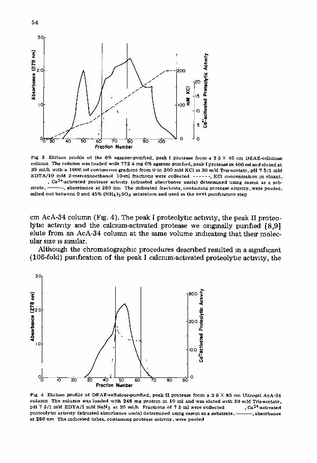
cm AcA-34 column (Fig. 4). The peak I proteolytm actwlty, the peak II proteo- lytm actwlty and the calcmm-actlvated protease we ongmally purified [8,9] elute from an AcA-34 column at the same volume mdmatmg that their molec- ular size is s~nllar.
Although the chromatographic procedures described resulted in a significant (106-fold) purification of the peak I calcmm-actwated proteolytlc activity, the
30
I0
io io 8o 9o o
i '
, Ix'i,., 30 40 5'0 60 70
Fraction Number
300 "~
o 200
Q. "0 Q
ioo
8
Fig 4 E lu t lon prof i le o f DE AE -ce l l u lo se -pun f l ed , p e a k II p ro t ea se f r o m a 2 5 × 85 c m Ul t roge l A c A - 3 4
c o l a m n The c o l u m n was l o a d e d w i t h 248 m g P ro t e in m 10 ml and was e lu ted w i t h 20 m M Tr i s -ace ta te , p H 7 5/1 m M E D T A / 1 m M N a N 3 at 20 m l / h F r a c t i o n s of 7 5 ml w e r e co l lec ted • Ca2+-act lvated p r o t e o l y t m a c t l w t y (adJus ted a b s o r b a n c e u m t s ) d e t e r n n n e d us ing case in as a s u b s t r a t e , - - , a b s o r b a n c e at 2 8 0 n m The m c h c a t e d tubes , c o n t a l m n g p ro t ea se a c t l w t y , were p o o l e d
55
pH 7.5 gels
a b
Dodecyl Sulfate gels
c d
~ -80 K
' - - - - - - - 3 0 K
i
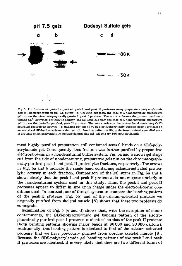
Fig 5 Pux l f l ca t lon o f p a r t m l l y pu~lf led p e a k I a n d p e a k II p r o t e a s e s u s ing P r e p a r a t i v e p o l y a c r y l a m i d e s lab-gel e l e c t r o p h o r e s l s m p H 7 .5 b u f f e r (a) Gel s t r ip c u t f r o m the edge o f a n o n d e n a t u r m g , p r e p a r a t i v e gel r u n o n t h e c h r o m a t o g r a p h m a l l y - p u n f l e d , p e a k I p r o t e a s e The a r r o w i n d i c a t e s t h e P r o t e i n b a n d c o n - taLmng Ca2+-ac t lva ted p r o t e o l y t l c a e t l w t y Co) Gel s t r ip c u t f r o m the edge o f a n o n d e n a t t t r l n g , p r e p a r a t i v e gel r u n o n t h e p a r t m l l y p u r t f l e d , p e a k II p r o t e a s e The a r r o w i n d i c a t e s t he p r o t e i n b a n d e o n t a l m n g Ca 2+o a c t i v a t e d p r o t e o l y t l c ac t i v i t y (e) B a n d i n g p a t t e r n o f 3 0 / z g e l e c t r o p h o r e t i c a l l y - p u n f m d p e a k I p r o t e a s e o n a n a n a l y t i c a l S D S - p o l y a c r y l a m l d e s lab gel (d) Banc tmg p a t t e r n o f 3 0 # g e l e c t r o p h o r e t m a l l y - p u r z f i e d p e a k II p r o t e a s e o n a n a n a l y t i c a l S D S - p o l y a c r y l a m l d e s lab gel All gels a re 1 0 % p o l y a e r y l a m l d e
most h~ghly purified preparation still contained several bands on a SDS-poly- acrylamlde gel. Consequently, this fractmn was further punfmd by preparatwe electrophoresls m a nondenatunng buffer system. Fig. 5a and b shows gel strips cut from the side of nondenaturlng, preparatwe gels run on the chromatograph- mally-punfmd peak I and peak II proteolytm fractions, respectwely. The arrows m Fig. 5a and b mdmate the smgle band contmnmg calcmm-actwated proteo- lytm actwlty m each fraction. Comparison of the gel strips m Fig. 5a and b shows clearly that the peak I and peak II proteases do not migrate similarly m the nondenaturmg system used m this study. Thus, the peak I and peak II proteases appear to differ in SLZe or m charge under the electrophoretm con- dltlons used. In contrast, use of this gel system to compare the banding pattern of the peak II protease (Fig. 5b) and of the calcmm-actwated protease we originally punfmd from skeletal muscle [8] shows that these two proteases do co-m~grate.
Examination of Fig. 5 (c and d) shows that, with the exceptmn of minor contammants, the SDS-polyacrylamlde gel bandmg pattern of the electro- phoretmaUy-punfmd peak I protease is ldentmal to that of the peak II protease (both banding patterns showing major bands at 80 000 and 30 000 daltons). Addltmnally, thLs banding pattern is ldentmal to that of the calcmm-actwated protease that we have prewously punfmd from porcine skeletal muscle [8]. Because the SDS-polyacrylamlde gel banding patterns of the peak I and peak II proteases are ldentmal, it is very hkely that they are two different forms of
56
8OK--
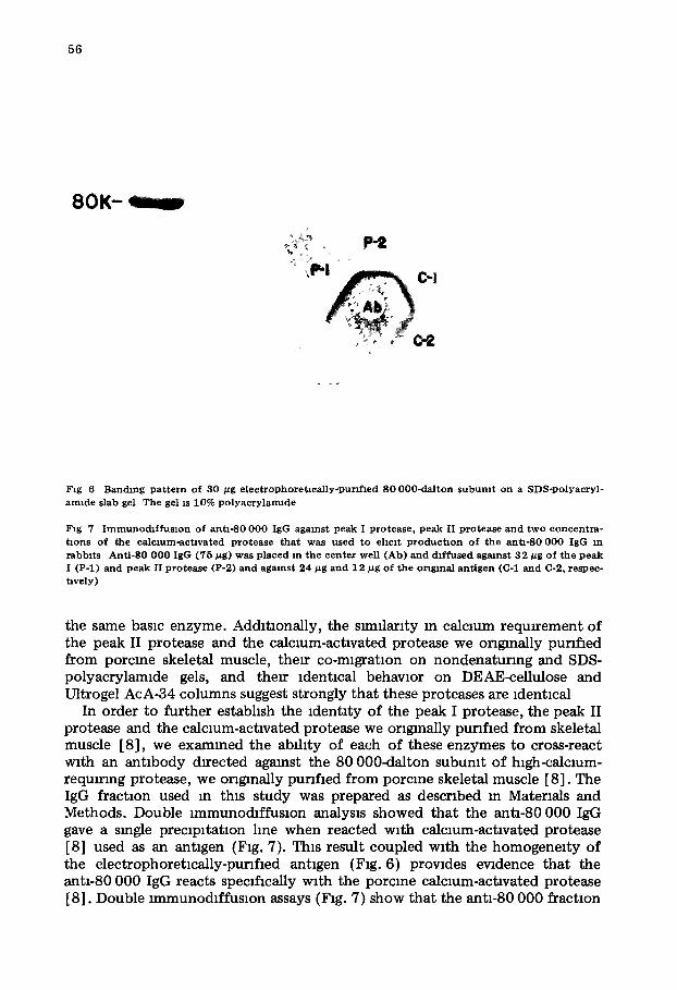
Fig 6 Banchng p a t t e r n o f 3 0 ~tg e l e c t r o p h o r e t l c a n y o p u r f f l e d 8 0 0 0 0 - d a l t o n s u b u m t o n a S D S - p o l y a c r y l - a m l d e s lab gel The gel is 10% p o l y a e r y l a n n d e
Fig 7 I m m u n o c h f f u s l o n o f a n t i - 8 0 0 0 0 IgG a g a i n s t p e a k I p r o t e a s e , p e a k II p r o t e a s e a n d t w o c o n c e n t r a - t i o n s o f the c a l c m m - a c t a v a t e d p r o t e a s e t h a t was u s e d t o e h m t p r o d u c t m n o f t h e a n t i - 8 0 0 0 0 IgG m r a b b i t s A n t i - S 0 0 0 0 IgG ( 7 5 #g) was p l a c e d xn t h e c e n t e r well ( A b ) a n d d i f f u s e d a g a i n s t 3 2 ~tg o f t h e p e a k I ( P - l ) a n d p e a k II p r o t e a s e (P-2) a n d a g a i n s t 2 4 #g a n d 1 2 #g o f t h e o r ig ina l a n t i g e n (C-1 a n d C-2, respec- tavely)
the same basra enzyme. Additionally, the snmlarlty m calcmm requtrement of the peak II protease and the calcmm-actxvated protease we originally punfmd from porcme skeletal muscle, thetr co-migration on nondenaturmg and SDS- polyacrylamlde gels, and their ldentmal behavior on DEAE-ceUulose and Ultrogel AcA-34 columns suggest strongly that these proteases are ldentmal
In order to further estabhsh the identity of the peak I protease, the peak II protease and the calcmm-actlvated protease we originally punfmd from skeletal muscle [8], we exammed the ability of each of these enzymes to cross-react with an ant ibody directed agmnst the 80 000-dalton subumt of hlgh-calcmm- requmng protease, we originally punfmd from porcine skeletal muscle [8]. The IgG fraction used m this s tudy was prepared as described m Materials and Methods. Double lmmunodfffusmn analysis showed that the ant1-80 000 IgG gave a single preclpltatmn hne when reacted with calcmm-actlvated protease [8] used as an antigen (Fig. 7). This result coupled with the homogenei ty of the electrophoretlcally-punfmd antigen (Fig. 6) prowdes ewdence that the ant1-80 000 IgG reacts specffmally with the porcme calcmm-actlvated protease [8]. Double lmmunodfffusmn assays (Fig. 7) show that the ant1-80 000 fractmn
57
also reacts with both the purified peak I and peak II proteases to form a smgle precipltln lme. In addition, It is clear (Fig. 7) that when the peak I protease, the peak II protease and the calcium-activated protease we ongmally purified from skeletal muscle are placed In adjacent wells and diffused agmnst the anti-80 000 IgG, preclpltm lines form a single continuous line with no spur formation. Thus, all three calcium-activated proteases that we have isolated and purified from porcine skeletal muscle share common antigenic determinants and are consequent ly related, if not identical, molecules. Consequently, we beheve that the peak I and peak II proteases are simply two forms of the same protease and that the peak II protease is identical to the calcium-activated protease we have previously purified [8].
Calclum requirement of purified peak I and peak H proteases The concentrat ion of calcium required to activate the peak I and the peak II
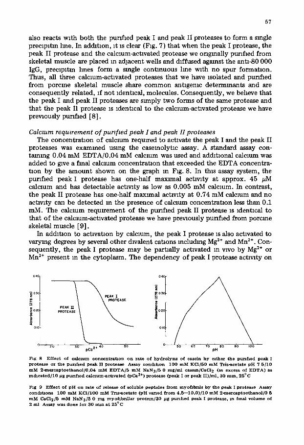
proteases was examined using the casemolytic assay. A standard assay con- taming 0.04 mM EDTA/0.04 mM calcium was used and additional calcium was added to give a final calcmm concentrat ion that exceeded the EDTA concentra- tion by the amount shown on the graph In Fig. 8. In this assay system, the purified peak I protease has one-half maxnnal activity at approx. 45 pM calcium and has detectable activity as low as 0.005 mM calcium. In contrast, the peak II protease has one-half maximal activity at 0.74 mM calcium and no activity can be detected m the presence of calcium concentrat ion less than 0.1 mM. The calcium reqmrement of the purlfmd peak II protease is identical to that of the calcium-activated protease we have previously purifmd from porcine skeletal muscle [9].
In addition to activation by calcium, the peak I protease is also activated to varying degrees by several other divalent catmns lncludmg Mg 2÷ and Mn 2+. Con- sequently, the peak I protease may be partially activated in wvo by Mg :÷ or Mn 2÷ present in the cytoplasm. The dependency of peak I protease activity on
0 4C 0 4C
\ \ -
e_
~ 03C 03C PEAK I
PEAK n ~
OIC OIC
- - z z'o 30 pCae ÷ 40 5'0 50 so "to pH eo 90 i00
Fzg 8 E f f e c t o f ca l czum c o n c e n t r a t i o n o n rate o f hyd ro lysLs o f c a se in b y e l ther the purl f led peak I p r o t e a s e o r t h e p u r l f t e d p e a k II p r o t e a s e A s s a y c o n d l t l o n 1 0 0 m M K C I / 5 0 m M Tr i s - ace t a t e p H 7 5 / 1 0 m M 2 - m e r c a p t o e t h a n o l / 0 . 0 4 m M E D T A / 5 m M NAN3/5 0 m g / m l c a s e m / C a C l 2 (In excess o f E D T A ) as m d t c a t e d / 1 0 ~g p u n h e d c a l c l u m - a c t l v a t e d (pCa 2+) p r o t e a s e ( p e a k I o r p e a k I I ) / m l , 3 0 m m , 2 5 ° C
Fig 9 E f f e c t o f p H o n rate o f re lease o f so luble p e p t l d e s f r o m m y o f l b n ] s b y t h e p e a k I p r o t e a s e A s s a y cond~t4ons 1 0 0 m M K C I / 1 0 0 m M Tr l s - ace t a t e ( p H v a n e d f r o m 4 . 5 - - 1 0 . 0 ) / 1 0 m M 2 - m e r c a p t o e t h a n o l / 0 5 m M CAC12/5 m M NAN3/5 0 m g myof lb r f l l a~ p r o t e l n / 3 0 /~g p u n f l e d p e a k I p r o t e a s e , m f ina l v o l u m e o f 2 m l A s s a y w a s d o n e f o r 3 0 m m a t 2 5 ° C
58
Mg 2÷ concentratmn is very Snnllar to its dependency on calcmm concentratmn, with maxnnum protease activity occurring at 0.5 mM Mg 2÷. The maxnnum activity obtained in the presence of Mg 2÷, however, is only about 34% of that obtmned m the presence of calcmm. Similarly, the peak I proteolyt lc act lwty is activated by Mn 2÷ with maximum activity (72% of that obtmned using calcmm as an activator) occurrmg at 0.5 mM.
pH dependency of peak I and peak H proteolyt~c actwtty The pH dependency of the purified peak II protease was found to be iden-
tical to that of the calcmm-actlvated protease we have prevmusly punfled from porcine skeletal muscle [9] Maximum proteolyt lc activity agmnst a myofsbrll substrate was observed between pH 7.5 and 8.0, with very httle activity below pH 6.0 or above pH 8.5. The purified peak I protease also exhibited maximum proteolytlc activity between pH 7.5 and 8.0 (Fig. 9). The peak I protease, how- ever, retmns slgmfmant activity as low as pH 5.5 and as high as pH 8.5. Thus, the purified peak I protease appears to have a broader pH dependence than does the peak II protease.
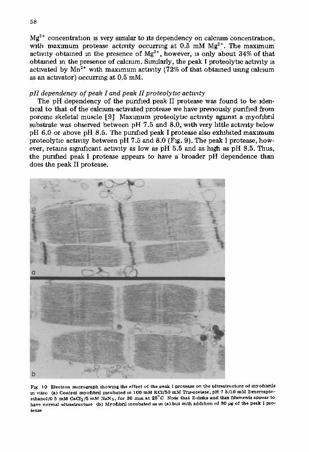
Fig 10 E lec t ron m l c r o g r a p h showing the e f fec t of the peak I p ro tease on the u l t r a s t ruc tuze of myof lbr f l s m v i t ro (a) Cont ro l myof lb r f l i n c u b a t e d m 100 m M KCI /50 m M Trls -ace ta te , p H 7 5 /10 m M 2 -mercap to - e t han o l / 0 5 m M CAC12/5 m M NAN3, for 30 m m at 25°C Note t h a t z-disks and th in f i l aments appea r to have n o r m a l u l t r a s t ruc tu re (b) Myoflbrf l i n c u b a t e d as m (a) b u t wi th adcht lon of 30/~g of the peak I pro-
tease
59
Effect of peak I and of peak H proteases on the ultrastructure of myoftbrds zn vztro
The effect of the purified peak I or the purffmd peak II protease on the ultrastructure of pumfmd porcine skeletal muscle myoflbrlls is ldentmal (Fig. 10). Additionally, the effect of these proteases on myoflbri l ultrastructure is ldentmal to that of the calcmm-achvated protease whmh we have prevaously pttrlfmd [9]. Treatment of myofxbrfls with either the purffmd peak I or peak II protease m the presence of the approprmte amount of calcmm, results in com- plete removal of the Z<hsk (Fig. 10). Thm filaments present m protease-treated myof lbnls appear to be disoriented compared to thin flllaments m non-treated myoflbrfls. This dlsorlentatmn may simply be due to the loss of the Z<hsk as an a t tachment point or it may be due to the effect of the protease on the thin filaments themselves.
Discussion
Based on the preceding data, we believe that the calcmm-actlvated proteo- lytm actlvatms that we and others [3] have isolated from skeletal muscle are the result of two forms of the same proteolyt lc enzyme. One form of the enzyme (responsible for the proteolytm activity m peak I from the DEAE~ellulose column) is activated by calcium concentratmns as low as 5 pM. The other form (responsible for the proteolytm activity m peak II from the DEAE-cellulose column and for the calcmm-actlvated proteolytm activity we omganally purifmd from porcine skeletal muscle) requxres calcium concentrations of at least 0.1--0.2 mM for actlvahon. The molecular modification responmble for the dif- ferent calcium requtrements exhibited by the two forms of the calcium- activated protease are not clear from our present data. Our studies do show that the modlfmatlon does no t revolve any major changes m molecular size, subumt molecular weight or compomtion, or immunologacal cross-reactlwty. The low~alcmm-requtrmg form of the protease appears to be less negatively charged than the hlgh-calclum-requmng form. This conclusion is based on the fact that the low-calclum-requtrmg form of the protease elutes from a DEAE- cellulose column at lower KC1 concentrat ion and has a lower mobil i ty m non- denaturing polyacrylamlde gel electrophoresls than the hlgh-calclum-requtrmg form of the protease. Our current data thus suggests that conversion from the low-calclum-requirmg form of the protease to the hlgh-calcmm-requmng form may mvolve addition of a relatively small, highly-charged, const i tuent to the low calclum-requmng protease. It is thus possible that the low-calcium- requtrmg form of the protease may be converted to the hlgh-calcmm-requirmg form by phosphorylatmn. We are currently investigating this possibility.
The htgh-calcium-requmng form of the calcmm-actlvated protease [8,9] has been shown to partially degrade myoflbrfls [12], and to hydrolyze fflamin and mtermedmte (100 A) fxlaments [28,29] . In addition, t reatment of cultured cells with the purified calcium-activated protease results m release of the cell- surface glycoprotem flbronectln, ultrastructural damage to the cell membrane and depolymemzatlon of mlcrofilaments which form part of the cytoskeleton of the cell (unpublished data). Even though the protease has been shown to have the above effects on specific cellular components , a major problem m
60
ascribmg a physlologmal role to the calcmm-actlvated proteolytm enzyme m skeletal muscle and m other t:ssues has been the mactlv:ty of the enzyme m m vitro assays done at physlologmal calcmm concentrations. Therefore, emstence of a form of the calcmm-actlvated protease that is act:ve at calcmm concentra- tions approaching those thought to be present m the cell ~s extremely nnpor- rant m understanding the cond:tlons necessary to activate the protease m v:vo. Moreover, m addltmn to actlvatmn by calcmm, it appears that the low- calcium-requmng form of the enzyme is partially activated by other divalent ions found m the cytoplasm of the cell (e.g., Mg :÷ and Mn:÷). It thus appears that the low-calcmm-requmng protease can be activated under the cond:tmns ex:stmg m muscle cells and :s therefore the form of the protease that is active I n VlVO.
Our recent :mmunocytochemmal locahzatmn of the calcmm-actlvated protease :n Z-disk of the myof:brfl [16] supports the theory that th:s protease plays a role m myoflbr:l disassembly and degradatmn, smce the Z-disk is a myohbrfllar structure that is extremely susceptible to calcmm-actlvated protease-catalyzed hydrolys:s. Based on these fmdmgs it seems almost certain that the enzyme is mvolved m degradatmn of at least this myof:brfllar struc- ture. Presence of the calcmm-act:vated protease m the Z-disk m normal muscle t:ssue makes it :mperatlve that the proteolytm actlwty of the enzyme be strmgently regulated, m order to prevent uncontrolled and complete degrada- tmn of Z-disks It is poss:ble that the actlv:ty of the protease is controlled, at least m part, by conversmn of the low-calcmm-requmng form of the enzyme to the h:gh-calcmm-requmng form, thereby rendering :t reactive at the free calcmm concentratmns normally found :n muscle cells. Conversely, act:vatmn of the protease might revolve conversmn of the hlgh-calcmm-requmng form of the protease to the low-calcmm-requmng form Add:tmnally, an endogenous mhlb:tor of the calcmm-act:vated protease has been isolated from several t:ssues, including"skeletal muscle [30,31], and It :s possible that this mh:bltor may also help to control the actlwty of the protease. Our studms (unpubhshed data) have shown that the inhibitor mhlb:ts both the hlgh-calcmm-requmng and the low-calcmm-requmng forms of the protease.
In summary, we have Isolated and purffmd a form of the calcmm-act:vated muscle protease [8,9] that is active at physmloglcal calcmm concentratmns The effect of the protease on myoflbrfllar ultrastructure is :dentmal to that reported for the calcmm-actlvated protease we prewously purffmd from porcine skeletal muscle [8,9,12]. Emstence of the low-calcmm-requmng form of the protease prowdes an explanatmn for how the calcmm-actlvated protease may be activated at the relatively low free calcmm concentratmns thought to be present m muscle cells and m other types of cells. Because the calcmm-act:- vated protease is present m muscle cells m structures that are affected early m the progressmn of many myopath:es (Le., the sarcolemma and Z-disk) [15,16], we beheve the enzyme may play a s:gmfmant role m degradatmn of these struc- tures. Smce mterconversmn of the low- and hlgh-calcmm-requmng forms of the calcmm-actlvated protease (assuming such mterconversmn occurs) may play an Important role m determining the level of protease activity m wvo and hence the rate of muscle protein degradatmn, we are currently studying the mechanism by which this mterconversmn might occur.
61
Acknowledgements
We would hke to thank L.C. for expert technical assistance. This work was supported by grants from the Muscular Dystrophy Assocmtlon of America, the Mmnesota Leukemia Task Force and the Minnesota Agricultural Experiment Station. This IS Scientific Journal Series Paper No. J 11598, Minnesota Agrmul- tural Experiment Station, Umverslty of Minnesota, St. Paul, MN 55108, U.S.A
References
1 G u z o f f , G ( 1 9 6 4 ) J Blol C h e m 2 3 9 , 1 4 9 - - 1 5 5 2 B u s c h , W A , S t r o m e r , H . H , Gol l , D E a n d S u z u k i , A ( 1 9 7 2 ) J Cell Blol 5 2 , 3 6 7 - - 3 8 1 3 Mel lg ren , R L (198D) F E B S L e t t 1 0 9 , 1 2 9 - - 1 3 3 4 D a y t o n , W R a n d S c h o l l m e y e r , J . V ( 1 9 8 0 ) J Mol Cell . Carchol 1 2 , 5 3 8 - - 5 5 0 5 S c h o l l m e y e r J V a n d D a y t o n , W R ( 1 9 7 7 ) J Cell B1ol 7 5 , 3 1 8 a 6 P u c a , G A , N o l a , E , Sma , V a n d B r e s c l a m , F ( 1 9 7 7 ) J Biol . C h e m 2 5 2 , 1 3 5 8 - - 1 3 7 0 7 V e d e c k l s , W V , F r e e m a n , M R . , S c h r a d e r , W T a n d O ' M a l l e y , B N ( 1 9 8 0 ) B i o c h e n n s t r y 1 9 ,
3 3 5 - - 3 4 3 8 D a y t o n , W R , Go l l , D E , Zeece , M G , R o b s o n , R M a n d Revf l le , W J ( 1 9 7 6 ) B i o c h e m i s t r y 1 5 ,
2 1 5 0 - - 2 1 5 8 9 D a y t o n , W.R , Revl l le , W . J , GoB, D E a n d S t r o m e r , M H ( 1 9 7 6 ) B i o c h e m i s t r y 1 5 , 2 1 5 9 - - 2 1 6 7
1 0 I s i n u r a , S , M u r o f u s h l , H , S u z u k t , K . a n d I m a h o r L H ( 1 9 7 8 ) J B l o c h e m ( T o k y o ) 8 4 , 2 2 5 - - 2 3 0 11 A z a n z a , J L , R a y m o n d , J , R o b i n , J M , C o t t m , P a n d D u c a s t m n g , A ( 1 9 7 9 ) B l o c h e m J . 1 8 3 ,
3 3 9 - - 3 4 7 1 2 D a y t o n , W R . Gol l , D E , S t r o m e r , M H , Revl l le , W . J , Zeece , M G a n d R o b s o n , R ~ I ( 1 9 7 5 ) In Co ld
S p n n g H a r b o r C o n f e r e n c e o n Cell P r o h f e r a h o n . P r o t e a s e s a n d B io log ica l C o n t r o l ( R e i c h , E , R l f k m , D B a n d S h a w , E , e d s ), p p 5 5 1 - - 5 7 7 , T h e C o l d S p n n g H a r b o r L a b o r a t o r y , C o l d S p r m g H a r b o r , N Y
1 3 D a y t o n , W R , S c h o H m e y e r , J V , C h a n , A C a n d Al len , C E. ( 1 9 7 9 ) BlochLm B l o p h y s A c t a 5 8 4 , 2 1 6 - - 2 3 0
1 4 Kay , N C a n d P e a r s o n , C.M ( 1 9 7 6 ) C h n C h l m . A c t a 7 3 , 2 9 3 - - 2 9 7 15 I s h i u r a , S , S u g l t a , H , N o n a k a , I a n d Imahor l0 K . ( 1 9 8 0 ) J . B l o c h e m 8 7 , 3 4 3 - - 3 4 6 1 6 D a y t o n , W . R a n d S c h o l l m e y e r , J V . ( 1 9 8 0 ) J Cell Blol 8 7 , 2 6 7 a 17 J o b s i s , F F a n d O ' C o n n e r , M.J ( 1 9 6 6 ) B l o c h e m B l o p h y s Res C o m m u n . 2 5 , 2 4 6 - - 2 5 2 18 Gol l , D .E , Y o u n g , R B. a n d S t r o m e r , M H ( 1 9 7 4 ) P r o c e e d i n g s 2 7 t h A n n u a l R e c i p r o c a l Mea t C o n -
f e r e n c e , Na t4ona l L i v e s t o c k a n d M e a t B o a r d , C h i c a g o , IL , p 2 5 0 19 G o r n a l l , A . G , Ba rdawKl , C J a n d Dav id , M M ( 1 9 4 9 ) J Blol C h e m 1 1 7 , 7 5 1 - - 7 6 0 2 0 L o w r y , O H , R o s e b r o u g h , N J , Farr0 A L a n d R a n d a l l , R . J ( 1 9 5 1 ) J Blol C h e m 1 9 3 , 2 6 5 21 L a e m m h , U K ( 1 9 7 0 ) N a t u r e ( L o n d o n ) 2 2 7 , 6 8 0 - - 6 8 5 2 2 Mauxer , H R ( 1 9 7 1 ) Dmc E l e c t r o p h o r e s m a n d R e l a t e d T e c h n i q u e s o f P o l y a c r y l a n n d e Gel E l e c t r o -
p h o r e s l s Wa l t e r de G r u y t e r , N e w Y o r k , N Y 2 3 C a m p b e l l , D H . , G a r v e y , J S , C r e m e r , N E a n d S u s s d o r f , D H ( 1 9 7 0 ) In M e t h o d s m I m m u n o l o g y ,
2 n d e d n . p p 1 8 3 - - 2 3 4 , W A B e n ] a n n n , Inc . , N e w Y o r k , N Y 2 4 N lco l , J C. a n d D e u t s c h , H F ( 1 9 4 8 ) J M C h e m Soc 70 , 8 0 - - 8 3 2 5 G a r v e y , J S , C r e m e r , N E a n d S u s s d o r f , D E eds ( 1 9 7 7 ) M e t h o d s in I m m u n o l o g y , p 5 2 3 , W A Ben-
] a Inm, Inc 26 Wflharns , C A a n d Chase , M W e d s ( 1 9 7 7 ) M e t h o d s m I m m u n o l o g y a n d I m m u n o c h e r m s t r y , p 1 4 6 ,
A c a d e m i c Press 27 F u j l w a r a , K a n d P o l l a r d , T D ( 1 9 7 6 ) J Cell Biol 7 1 , 8 4 8 - - 8 7 5 2 8 B e c h t e l , P~I. ( 1 9 7 9 ) J . Biol . C h e m . 2 5 4 , 1 7 5 5 - - 1 7 5 8 2 9 S c h o l l m e y e r , J V a n d D a y t o n , W . R ( 1 9 8 0 ) N a t u r e , m the p ress 3 0 O k l t a m , A , Gol l , D .E , S t r o m e r , M . H . a n d R o b s o n , R M. ( 1 9 7 6 ) F e d P r o c 3 5 , 1 7 4 6 31 W a x m a n , L a n d I ~ e b s , E G ( 1 9 7 8 ) J . Biol . C h e m 2 5 3 , 5 8 8 8 - - 5 8 9 1
Related Documents