A calcific pelvic mass in a woman with chronic spinal pain: a case of mature cystic teratoma Martha A. Kaeser DC a , Jennifer K. McDonald DO b , Norman W. Kettner DC c,⁎ a Chiropractor, Radiology Resident, Logan College of Chiropractic, Chesterfield, MO 63006 b Physician, Private Practice, Chesterfield, MO 63017 c Chair, Department of Radiology, Logan College of Chiropractic, Chesterfield, MO 63006 Received 28 December 2010; received in revised form 16 May 2011; accepted 21 June 2011 Key indexing terms: Chiropractic; Teratoma; Ovarian neoplasms; Laparotomy; Laparoscopy Abstract Objective: The purpose of this case is to describe findings of a mature cystic teratoma and to further provide differential diagnoses for ovarian pelvic masses and calcifications. Clinical Features: A 27-year-old woman presented to a chiropractic teaching clinic with a chief complaint of chronic multilevel spinal pain. During a full spine radiographic examination, radiopaque densities were incidentally identified in the pelvic bowl visualized through a gonad shield. Follow-up pelvic radiography revealed several radiopacities of uniform density localized in the pelvic bowl. Intervention/Outcomes: Medical (gynecological) consultation led to ultrasonography of the pelvis that revealed a mature cystic teratoma. The patient underwent complete excision of the mass through a laparotomy procedure. The patient continued to receive chiropractic treatment of her original cervical and lumbar spine complaints, further suggesting that the pelvic mass was not a source of her musculoskeletal complaints. Conclusion: This case demonstrates the detection and proper referral of a patient with a calcific mass. The presence of a pelvic mass, suspected of arising from the ovary, requires additional diagnostic imaging and careful clinical correlation. © 2011 National University of Health Sciences. Introduction Calcifications located within the pelvic bowl are frequent findings on radiography. Differential diagnos- tic considerations (listed in alphabetical order) are included in Fig 1. 1 Characteristic plain film findings of cystic teratomas include teeth, bone, and/or fat, with 40% of these masses solely confirmed by plain film radiography. 2 Mature cystic teratomas are classified as ovarian teratomas. Zagame et al 3 divide ovarian teratomas into 2 groups, mature (99% of all ovarian teratomas) and immature (1%), depending on their characteristics. Mature cystic teratomas of the ovary www.journalchiromed.com ⁎ Corresponding author. 1851 Schoettler Rd, PO Box 1065 Chesterfield, MO 63006-1065. Tel.: +1 636 227 1830; fax: +1 636 207 2429. E-mail address: [email protected] (N. W. Kettner). 1556-3707/$ – see front matter © 2011 National University of Health Sciences. doi:10.1016/j.jcm.2011.06.004 Journal of Chiropractic Medicine (2011) 10, 327–332

Welcome message from author
This document is posted to help you gain knowledge. Please leave a comment to let me know what you think about it! Share it to your friends and learn new things together.
Transcript
www.journalchiromed.com
Journal of Chiropractic Medicine (2011) 10, 327–332
A calcific pelvic mass in a woman with chronic spinalpain: a case of mature cystic teratomaMartha A. Kaeser DC a, Jennifer K. McDonald DO b, Norman W. Kettner DC c,⁎
a Chiropractor, Radiology Resident, Logan College of Chiropractic, Chesterfield, MO 63006b Physician, Private Practice, Chesterfield, MO 63017c Chair, Department of Radiology, Logan College of Chiropractic, Chesterfield, MO 63006
Received 28 December 2010; received in revised form 16 May 2011; accepted 21 June 2011
Key indexing terms: Abstract
C6
1d
Chiropractic;Teratoma;Ovarian neoplasms;Laparotomy;Laparoscopy
Objective: The purpose of this case is to describe findings of a mature cystic teratoma and tofurther provide differential diagnoses for ovarian pelvic masses and calcifications.Clinical Features: A 27-year-old woman presented to a chiropractic teaching clinic with a chiefcomplaint of chronic multilevel spinal pain. During a full spine radiographic examination,radiopaque densities were incidentally identified in the pelvic bowl visualized through a gonadshield. Follow-up pelvic radiography revealed several radiopacities of uniform densitylocalized in the pelvic bowl.Intervention/Outcomes: Medical (gynecological) consultation led to ultrasonography of thepelvis that revealed a mature cystic teratoma. The patient underwent complete excision of themass through a laparotomy procedure. The patient continued to receive chiropractic treatmentof her original cervical and lumbar spine complaints, further suggesting that the pelvic mass wasnot a source of her musculoskeletal complaints.Conclusion: This case demonstrates the detection and proper referral of a patient with a calcificmass. The presence of a pelvic mass, suspected of arising from the ovary, requires additionaldiagnostic imaging and careful clinical correlation.
⁎ Corresponding author. 1851hesterfield, MO 63006-1065. Tel36 207 2429.E-mail address: norman.kettner@
556-3707/$ – see front matter © 2oi:10.1016/j.jcm.2011.06.004
© 2011 National University of Health Sciences.
Introduction
Calcifications located within the pelvic bowl arefrequent findings on radiography. Differential diagnos-
Schoettler Rd, PO Box 1065.: +1 636 227 1830; fax: +1
logan.edu (N. W. Kettner).
011 National University of H
ealth Stic considerations (listed in alphabetical order) areincluded in Fig 1.1 Characteristic plain film findings ofcystic teratomas include teeth, bone, and/or fat, with40% of these masses solely confirmed by plain filmradiography.2 Mature cystic teratomas are classified asovarian teratomas. Zagame et al 3 divide ovarianteratomas into 2 groups, mature (99% of all ovarianteratomas) and immature (1%), depending on theircharacteristics. Mature cystic teratomas of the ovary
ciences.
Appendiceal calculus (fecalith)
Dermoid cyst
Foreign material
Leiomyoma or leiomyosarcoma of uterus
Lithopedion
Lymph node
Pregnancy
Urinary tract calculus
Fallopian tube calcification
Vascular (arteries, phleboliths)
Fig 1. Pelvic or lower quadrant calcification (in women).1
328 M. A. Kaeser et al.
make up 10% to 25% of ovarian neoplasms.4 Maturecystic teratomas, the most common germ cell neo-plasm, are collections of well-differentiated derivationsfrom at least 2 of 3 germ cell layers: ectoderm (skin,brain), mesoderm (muscle, fat), and endoderm (mucin-ous or ciliated epithelium). 5 The components of thecyst may include hair, sebum, and teeth. 6 Matureteratomas are principally cystic and lined by epitheli-um.7 Because of the predominance of skin elements,mature cystic teratomas are also referred to as dermoidcysts. 4 Mature cystic teratomas, in their pure form, arealways benign, although they may rarely undergomalignant transformation. The prevalence of maturecystic teratomas undergoing malignant transformationis 1% to 2%, with a subsequent poor prognosis due tothe delay in identifying the transformation beforemetastatic spread. Malignant degeneration, most com-monly to squamous cell carcinoma, tends to occur inpostmenopausal women.4
Mature cystic teratomas often occur in youngerpatients, with an average age of 30 years. Theseteratomas are usually asymptomatic8 and grow slowly,averaging 1.8 mm of growth per year. Mature cysticteratomas are typically evident within the pelvic bowlon radiographic examinations because of their fat andcalcific density. Given that most of these tumors areasymptomatic, 5,8 they are often incidental findings onlumbar or pelvic radiography.8 Treatment of maturecystic teratomas depends on the size, with nonsurgicalmanagement for smaller tumors, less than 6 cm, orcystectomy when larger. 5
Immature teratomas contain tissues derived fromthe 3 germ cell layers; but unlike mature teratomas,they demonstrate malignant behavior. Histologically,immature teratomas have components of immatureor embryonic tissues. In comparison to matureteratomas, immature teratomas are characteristically
larger, 14 to 25 cm, as compared with matureteratomas averaging 7 cm. In addition, immatureteratomas have a prominent solid component and aless prominent cystic component, with a less-definedcapsule in comparison to a mature cystic teratoma. 5
Radiography is unable to differentiate the mature fromthe immature teratoma. This limitation demands theuse of additional diagnostic imaging techniques suchas sonography, computed tomography (CT), ormagnetic resonance imaging (MRI).
Pelvic masses in the female may invoke a range ofdifferential diagnoses from benign to malignant. To thebest of our knowledge, this is the first reporteddiscussion of a benign cystic teratoma in the contem-porary chiropractic peer-reviewed literature. Thesemasses/pelvic calcifications are often incidental find-ings on lumbar or pelvic radiographic examinations,yet it should be acknowledged that incidental findingsdo not always equate as being inconsequential.Therefore, referral to an obstetrician/gynecologist iswarranted when a pelvic mass in a female is identifiedor suspected. The purpose of this case is to describefindings of a mature cystic teratoma and to furtherprovide differential diagnoses for ovarian pelvicmasses and calcifications.
Case report
A 27-year-old multiparous woman presented to achiropractic clinic with chronic multilevel spinal paindescribed as neck stiffness/pain, pain between theshoulders, low back pain, and painful joints, mostnotably the left knee. At presentation, the patient washeight/weight proportional and alert to person, place,and time. Her blood pressure was 90/60 mm Hg;respiration, 14 breaths per minute; and pulse, 84.Physical examination demonstrated bilateral subocci-pital and lower cervical spine tenderness, hypertonicityof the trapezius muscles, and left sacroiliac jointtenderness and restriction with motion palpation. Allactive ranges of motion and sensory test results werewithin reference ranges. Result of Patrick's Flexion,Abduction, External Rotation, Extension test waspositive bilaterally with pain in the hip joints. Acomplete blood count and urinalysis revealed noabnormal values. She reported a spinal curvature onher health history. On postural examination, a low leftshoulder, low right ilium, and minimally posterior rightscapula were noted. Full spine radiography wasperformed to assess for the extent of the scoliosis.
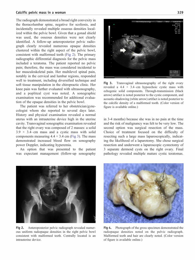
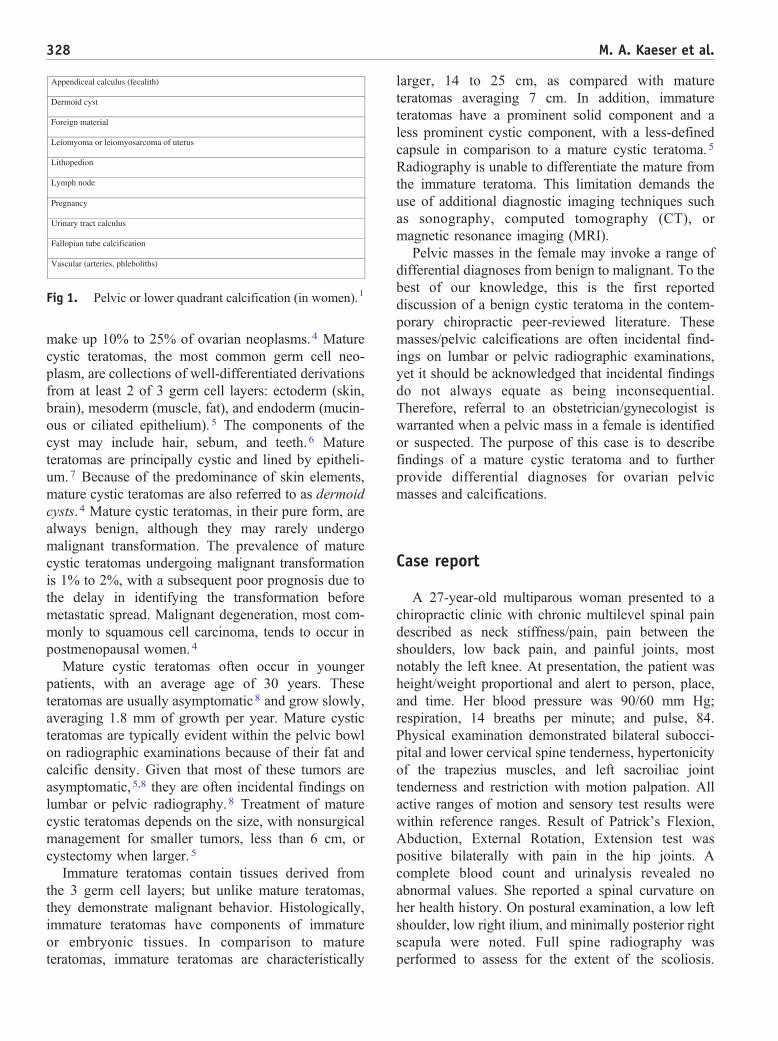
Fig 3. Transvaginal ultrasonography of the right ovaryrevealed a 4.4 × 3.4–cm hypoechoic cystic mass withechogenic solid components. Through-transmission (blackarrow) artifact is noted posterior to the cystic component, andacoustic-shadowing (white arrow) artifact is noted posterior tothe calcific density of a malformed tooth. (Color version offigure is available online.)
329Calcific pelvic mass in a woman
The radiograph demonstrated a broad right convexity inthe thoracolumbar spine, negative for scoliosis, andincidentally revealed multiple osseous densities local-ized within the pelvic bowl. Given that a gonad shieldwas used, the osseous densities were not clearlyidentified. A follow-up anteroposterior pelvic radio-graph clearly revealed numerous opaque densitiesclustered within the right aspect of the pelvic bowl,consistent with malformed teeth (Fig 2). The primaryradiographic differential diagnosis for the pelvic massincluded a teratoma. The patient reported no pelvicpain; therefore, the mass was considered incidental toher musculoskeletal pain. Her multilevel spinal pain,notably in the cervical and lumbar regions, respondedwell to treatment, including diversified technique andsoft tissue manipulation in the chiropractic clinic. Herknee pain was further evaluated with ultrasonography,and a popliteal cyst was noted. A sonographicexamination was recommended for additional evalua-tion of the opaque densities in the pelvic bowl.
The patient was referred to her obstetrician/gyne-cologist whom she reported to several days later.History and physical examination revealed a normaluterus with an intrauterine device high in the uterinecavity. Transvaginal sonographic examination revealedthat the right ovary was composed of 2 masses: a solid3.9 × 3.4–cm mass and a cystic mass with solidcomponents measuring 4.4 × 3.4 cm (Fig 3). The massdemonstrated increased blood flow on sonographypower Doppler, indicating hyperemia.
An option that was presented to the patientwas expectant management (follow-up sonography
Fig 2. Anteroposterior pelvic radiograph revealed numer-ous uniform radiopaque densities in the right pelvic bowconsistent with malformed teeth. Centrally located is anintrauterine device.
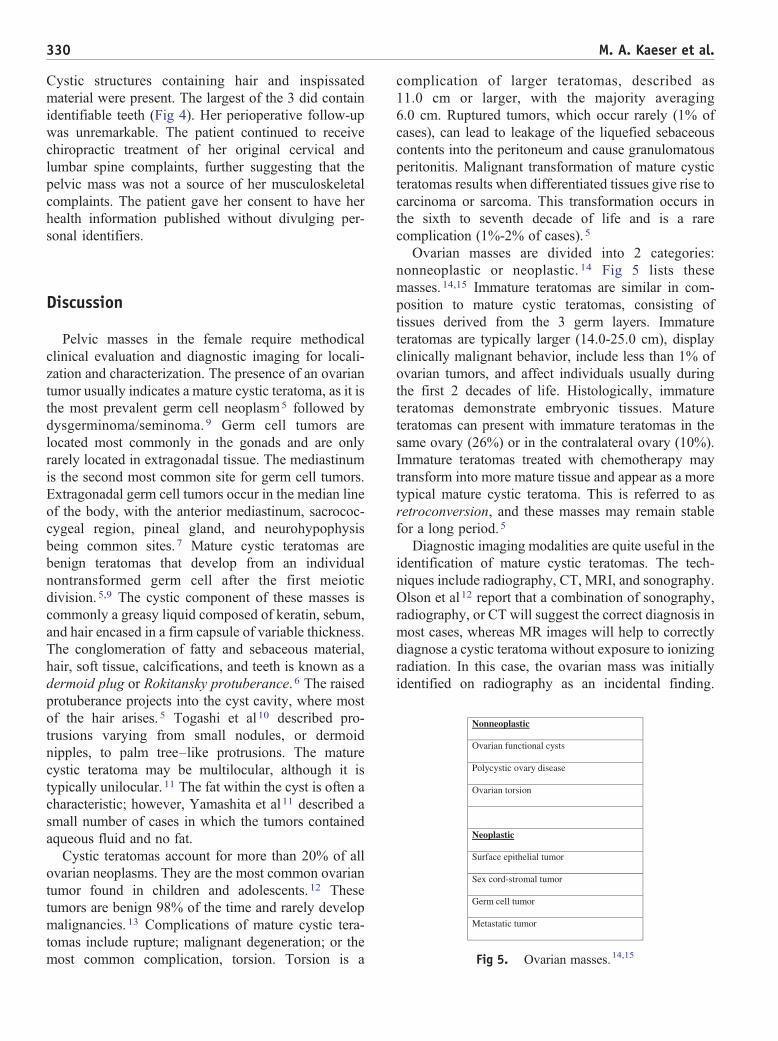
Fig 4. Photograph of the gross specimen demonstrated theradiopaque densities noted on the pelvic radiographMalformed teeth and hair are clearly noted. (Color versionof figure is available online.)
l
in 3-4 months) because she was in no pain at the timeand the risk of malignancy was felt to be very low. Thesecond option was surgical resection of the mass.Choice of treatment focused on the difficulty ofresecting such a large mass laparoscopically, indicat-ing the likelihood of a laparotomy. She chose surgicalresection and underwent a laparoscopic cystectomy of3 separate dermoid cysts on the right ovary. Finalpathology revealed multiple mature cystic teratomas.
.
330 M. A. Kaeser et al.
Cystic structures containing hair and inspissatedmaterial were present. The largest of the 3 did containidentifiable teeth (Fig 4). Her perioperative follow-upwas unremarkable. The patient continued to receivechiropractic treatment of her original cervical andlumbar spine complaints, further suggesting that thepelvic mass was not a source of her musculoskeletalcomplaints. The patient gave her consent to have herhealth information published without divulging per-sonal identifiers.
Nonneoplastic
Ovarian functional cysts
Polycystic ovary disease
Ovarian torsion
Neoplastic
Surface epithelial tumor
Sex cord-stromal tumor
Germ cell tumor
Metastatic tumor
Fig 5. Ovarian masses.14,15
Discussion
Pelvic masses in the female require methodicalclinical evaluation and diagnostic imaging for locali-zation and characterization. The presence of an ovariantumor usually indicates a mature cystic teratoma, as it isthe most prevalent germ cell neoplasm5 followed bydysgerminoma/seminoma. 9 Germ cell tumors arelocated most commonly in the gonads and are onlyrarely located in extragonadal tissue. The mediastinumis the second most common site for germ cell tumors.Extragonadal germ cell tumors occur in the median lineof the body, with the anterior mediastinum, sacrococ-cygeal region, pineal gland, and neurohypophysisbeing common sites. 7 Mature cystic teratomas arebenign teratomas that develop from an individualnontransformed germ cell after the first meioticdivision.5,9 The cystic component of these masses iscommonly a greasy liquid composed of keratin, sebum,and hair encased in a firm capsule of variable thickness.The conglomeration of fatty and sebaceous material,hair, soft tissue, calcifications, and teeth is known as adermoid plug or Rokitansky protuberance. 6 The raisedprotuberance projects into the cyst cavity, where mostof the hair arises. 5 Togashi et al10 described pro-trusions varying from small nodules, or dermoidnipples, to palm tree–like protrusions. The maturecystic teratoma may be multilocular, although it istypically unilocular. 11 The fat within the cyst is often acharacteristic; however, Yamashita et al11 described asmall number of cases in which the tumors containedaqueous fluid and no fat.
Cystic teratomas account for more than 20% of allovarian neoplasms. They are the most common ovariantumor found in children and adolescents. 12 Thesetumors are benign 98% of the time and rarely developmalignancies. 13 Complications of mature cystic tera-tomas include rupture; malignant degeneration; or themost common complication, torsion. Torsion is a
complication of larger teratomas, described as11.0 cm or larger, with the majority averaging6.0 cm. Ruptured tumors, which occur rarely (1% ofcases), can lead to leakage of the liquefied sebaceouscontents into the peritoneum and cause granulomatousperitonitis. Malignant transformation of mature cysticteratomas results when differentiated tissues give rise tocarcinoma or sarcoma. This transformation occurs inthe sixth to seventh decade of life and is a rarecomplication (1%-2% of cases). 5
Ovarian masses are divided into 2 categories:nonneoplastic or neoplastic. 14 Fig 5 lists thesemasses. 14,15 Immature teratomas are similar in com-position to mature cystic teratomas, consisting oftissues derived from the 3 germ layers. Immatureteratomas are typically larger (14.0-25.0 cm), displayclinically malignant behavior, include less than 1% ofovarian tumors, and affect individuals usually duringthe first 2 decades of life. Histologically, immatureteratomas demonstrate embryonic tissues. Matureteratomas can present with immature teratomas in thesame ovary (26%) or in the contralateral ovary (10%).Immature teratomas treated with chemotherapy maytransform into more mature tissue and appear as a moretypical mature cystic teratoma. This is referred to asretroconversion, and these masses may remain stablefor a long period.5
Diagnostic imaging modalities are quite useful in theidentification of mature cystic teratomas. The tech-niques include radiography, CT, MRI, and sonography.Olson et al12 report that a combination of sonography,radiography, or CT will suggest the correct diagnosis inmost cases, whereas MR images will help to correctlydiagnose a cystic teratoma without exposure to ionizingradiation. In this case, the ovarian mass was initiallyidentified on radiography as an incidental finding.
331Calcific pelvic mass in a woman
Osseous densities were noted within the pelvic bowlconsistent with a mature cystic teratoma.
Radiography may be the imaging modality thatinitially identifies calcifications within a pelvic mass.Mature teratomas are commonly detected incidentallyduring routine pelvic imaging studies. 8 Computedtomographic diagnosis of a benign cystic teratoma ismore specific than sonography or radiography. Char-acteristic findings on CT include a fat-containing masswith Hounsfield units greater than normal fat becauseof the mixture of fat, hair, debris, and fluid. Also notedare calcifications, including teeth and bone, in theRokitansky protuberance. 2 Sheth et al 16 note that CTcan precisely display the internal architecture of themass because of its capacity to discriminate betweentissues of subtle attenuations. Fragments of bone orteeth that are clear on CT are not readily depicted onsonography. This may be explained by noting that thecalcified elements may be within the adjacent hair orcyst wall or may merge with adjacent bowel. 16
Magnetic resonance imaging is useful in thediagnosis of the benign cystic teratoma given that thefatty component of the mass is isointense in relation tosubcutaneous fat. A fat-water interface and a typicalpattern of layering or floating debris are commonpresentations noted on MRI.12 Torsion is suspectedwhen the uterus is deviated to the twisted side withengorged blood vessels. A high–signal intensity rim onT1-weighted imaging is noted. A low–signal intensitymass (torsion knot) with thick straight blood vesselsencircling creates a complete absence of enhance-ment. 5 Malignant transformation is depicted on MRIby a sebaceous lipid component of the mass with aheterogeneous solid component projecting into the cystor extending transmurally into adjacent organs. 5
Sonography is used to diagnose most mature cysticteratomas. Sonographic diagnosis can be complicatedbecause of the variety of appearances5 and theirheterogeneous internal structure. 16 Three manifesta-tions of mature cystic teratomas are most common andinclude a cystic lesion with a densely echogenicprotuberance; a diffusely or partially echogenic masswith echogenicity demonstrated by sound attenuationcaused by the sebaceous material and hair within thecyst cavity; and a third manifestation consisting ofmultiple thin, echogenic bands due to hair within thecyst. 5 Fat demonstrated on CT as low attenuating andhomogeneous, representing pure sebum (liquid at bodytemperature), will have an anechoic appearancesonographically. Fat within the dermoid plug will beconsiderably echogenic because of the association withsoft tissue and hair. A combination of hair and sebum
will demonstrate a highly echogenic signal given thelarge acoustic impedance differences and variabletissue interfaces. 16 Outwater et al 5 reported that thecombination of hair mixed with the cyst fluid gave adiffuse echogenicity sonographically. A highly echo-genic focus with posterior shadowing produced by anectopic tooth or bone fragment is a specific feature of amature cystic teratoma. 6
Three treatment options are available for benigncystic teratomas. These include laparotomy, laparos-copy, and no treatment. Laparotomy is a treatmentoption that uses a Pfannenstiel or subumbilical midlineincision. 17 An abdominal incision is performed toallow adequate intraoperative exposure. 18 In a 1996article written by Yuen et al, 17 it was reported thatlaparoscopy was increasing in popularity in themanagement of benign ovarian masses. Benefits oflaparoscopy include reductions in operative morbidity,hospital stay, and recovery time. Worley et al19 notedother benefits including faster recovery due todecreased analgesic requirements and a decreasedtime for return of bowel function. Complications oflaparoscopy include tumor spillage during the opera-tion in cases of a malignant ovarian tumor beingmisdiagnosed17; vascular, bowel, and genitourinaryinjuries; incisional hernias; port-site metastases; andgas embolism.19
Limitations
Limitations for this case include no long-termfollow-up after surgical excision. Therefore, it isunknown as to whether any related complications/conditions exist currently for the patient. As previouslymentioned, the mature cystic teratoma was an inciden-tal finding and likely not related to the patient's clinicalpresentation of multilevel spinal pain.
Conclusion
Radiography frequently results in the detection ofincidental findings, the significance of which may bequite variable. The presence of a pelvic mass, suspectedof arising from the ovary, requires additional diagnosticimaging and careful clinical correlation. This casereport described an adult female patient who initiallypresented to a chiropractic teaching clinic with a chiefcomplaint of chronic neck and low back pain. A benignmature cystic teratoma was incidentally discovered at
332 M. A. Kaeser et al.
full spine radiography. Treatment consisted of surgicalexcision, and there was an uneventful recovery.
Funding sources and potential conflictsof interest
No funding sources or conflicts of interest werereported for this study.
References
1. Reeder MM. Reeder and Felson's gamuts in radiology,comprehensive lists of roentgen differential diagnosis. 4th ed.New York: Springer; 2003.
2. Friedman AC, Pyatt RS, Hartman DS, Downey EF, Olson WB.CT of benign cystic teratomas. AJR Am J Roentgenol 1982;138(4):659-65.
3. Zagame L, Pautier P, Duvillard P, Castaigne D, Patte C,Lhomme C. Growing teratoma syndrome after ovarian germcell tumors. Obstet Gynecol 2006;108(3 Pt 1):509-14.4.
4. Kido A, Togashi K, Konishi I, Kataoka ML, Koyama T,Ueda H, et al. Dermoid cysts of the ovary with malignanttransformation: MR appearance. AJR Am J Roentgenol 1999;172(2):445-9.
5. Outwater EK, Siegelman ES, Hunt JL. Ovarian teratomas:tumor types and imaging characteristics. Radiographics 2001;21(2):475-90.
6. Hertzberg BS, Kliewer MA. Sonography of benign cysticteratoma of the ovary: pitfalls in diagnosis. AJR Am JRoentgenol 1996;167(5):1127-33.
7. Ueno T, Tanaka YO, Nagata M, Tsunoda H, Anno I, IshikawaS, et al. Spectrum of germ cell tumors: from head to toe.Radiographics 2004;24(2):387-404.
8. Kim M, Kim NY, Lee DY, Yoon BK, Choi DS. Clinicalcharacteristics of ovarian teratoma: age-focused retrospective
analysis of 580 cases. Forthcoming: American Journal ofObstetrics and Gynecology; 2011.
9. Ulbright TM. Germ cell tumors of the gonads: a selectivereview emphasizing problems in differential diagnosis, newlyappreciated, and controversial issues. Mod Pathol 2005;18(Suppl 2):S61-79.
10. Togashi K, Nishimura K, Itoh K, Fujisawa I, Sago T, MinamiS, et al. Ovarian cystic teratomas: MR imaging. Radiology1987;162(3):669-73.
11. Yamashita Y, Hatanaka Y, Torashima M, Takahashi M,Miyazaki K, Okamura H. Mature cystic teratomas of theovary without fat in the cystic cavity: MR features in 12 cases.AJR Am J Roentgenol 1994;163(3):613-6.
12. Olson MC, Posniak HV, Tempany CM, Dudiak CM. MRimaging of the female pelvic region. Radiographics 1992;12(3):445-65.
13. Buy JN, Ghossain MA, Moss AA, Bazot M, Doucet M, HugolD, et al. Cystic teratoma of the ovary: CT detection. Radiology1989;171(3):697-701.
14. Sutton CL, McKinney CD, Jones JE, Gay SB. Ovarian massesrevisited: radiologic and pathologic correlation. Radiographics1992;12(5):853-77.
15. Jeong YY, Outwater EK, Kang HK. Imaging evaluation ofovarian masses. Radiographics 2000;20(5):1445-70.15.
16. Sheth S, Fishman EK, Buck JL, Hamper UM, Sanders RC.The variable sonographic appearances of ovarian teratomas:correlation with CT. AJR Am J Roentgenol 1988;151(2):331-4.
17. Yuen PM, Yu KM, Yip SK, Lau WC, Rogers MS, Chang A. Arandomized prospective study of laparoscopy and laparotomyin the management of benign ovarian masses. Am J ObstetGynecol 1997;177(1):109-14.
18. Ghanbari Z, Baratali BH, Foroughifar T, Pesikhani MD, ShariatM. Pfannenstiel versus Maylard incision for gynecologicsurgery: a randomized, double-blind controlled trial. Taiwan JObstet Gynecol 2009;48(2):120-3.
19. Worley MJ, Slomovitz BM, Ramirez PT. Complications oflaparoscopy in benign and oncologic gynecological surgery.Rev Obstet Gynecol 2009;2(3):169-75.
Related Documents


