Journal of Physiology (1997), 505.1, pp.13-22 A C-terminal peptide of the GIRKI subunit directly blocks the G protein-activated K+ channel (GIRK) expressed in Xenopu8 oocytes Tudor Luchian, Nathan Dascal *, Carmen Dessauer t, Dieter Platzer, Norman Davidson t, Henry A. Lester t and Wolfgang Schreibmayer § Department for Medical Physics and Biophysics, University of Graz, Harrachgasse 21/4, A -8010 Graz, Austria, *Department of Physiology and Pharmacology, Sackler School of Medicine, Tel Aviv University, Ramat Aviv, Israel, t Department of Pharmacology, South Western University, Dallas, TX and t California Institute of Technology, Division of Biology 156-29, Pasadena, CA, USA 1. In order to find out the functional roles of cytosolic regions of a G protein-activated, inwardly rectifying potassium channel subunit we studied block of GIRK channels, expressed in Xenopus laevis oocytes, by synthetic peptides in isolated inside-out membrane patches. 2. A peptide (DS6) derived from the very end of the C-terminus of GIRK1 reversibly blocked GIRK activity with IC50 values of 7 9 + 2'0 or 3 5 + 0' 5,ug ml-1 (corresponding to 3-7 + 09 or 1-7 + 0-2 #smol F1) for GIRK1/GIRK5 or GIRK1/GIRK4 channels, respectively. 3. Dose dependency studies of GIRK activation by purified fy subunits of the G protein (G8y) showed that DS6 block of GIRK channels is not the result of competition of the peptide with functional GIRK channels for the available G,Y. 4. Burst duration of GIRK channels was reduced, whereas long closed times between bursts were markedly increased, accounting for the channel block observed. 5. Block by the DS6 peptide was slightly voltage dependent, being stronger at more negative potentials. 6. These data support the hypothesis that the distal part of the carboxy-terminus of GIRK1 is a part of the intrinsic gate that keeps GIRK channels closed in the absence of G8y. The inwardly rectifying K+ channels of the GIRK (Kir3) family are important in the regulation of excitability in heart and brain. Opening of cardiac GIRK channels (KACh) by acetylcholine (ACh), via muscarinic m2 receptors that activate a pertussis toxin (PTX)-sensitive G protein of the G1/Go family mediates the negative chronotropic effect of the vagus on heartbeat (reviewed by Kurachi, 1995). Similar K+ channels are activated via PTX-sensitive G proteins by serotonin (5-HTIA), a-and #s-opioid, GABAB, dopamine and some other receptors in the brain, where they are believed to act as important mediators of inhibitory neurotransmission (reviewed by North, 1989; Hille, 1992; Wickman & Clapham, 1995; Dascal, 1997). GIRKs are members of the superfamily of inwardly rectifying K+ channels (Kir; for review see Doupnik, Davidson & Lester, 1995) characterized by two trans- membrane a-helical domains Ml and M2, a pore (P) region similar to that of voltage-dependent channels, and cytoplasmic N- and C-terminal regions. Four such proteins (subunits) form a functional channel. GIRK channels in the heart are heterotetramers of two subunits, GIRK1/KGA and GIRK4/CIR. Additional GIRK subunits, GIRK2 and GIRK3, are found in the brain where possibly they form heteromeric channels with GIRKI. GIRKI alone cannot form functional channels, but its expression in Xenopus oocytes gives functional channels due to association with the GIRK5/XIR of the endogenous oocyte (see Dascal 1997 for review). In addition to their physiological importance, GIRKs present the best studied case of ion channels directly gated by § To whom correspondence should be addressed. 6898 13 ) by guest on February 25, 2013 jp.physoc.org Downloaded from J Physiol (

Welcome message from author
This document is posted to help you gain knowledge. Please leave a comment to let me know what you think about it! Share it to your friends and learn new things together.
Transcript

Journal of Physiology (1997), 505.1, pp.13-22
A C-terminal peptide of the GIRKI subunit directly blocksthe G protein-activated K+ channel (GIRK) expressed in
Xenopu8 oocytes
Tudor Luchian, Nathan Dascal *, Carmen Dessauer t, Dieter Platzer,Norman Davidson t, Henry A. Lester t and Wolfgang Schreibmayer §
Department for Medical Physics and Biophysics, University of Graz, Harrachgasse 21/4,A -8010 Graz, Austria, *Department of Physiology and Pharmacology, Sackler School ofMedicine, Tel Aviv University, Ramat Aviv, Israel, t Department of Pharmacology, South
Western University, Dallas, TX and t California Institute of Technology,Division of Biology 156-29, Pasadena, CA, USA
1. In order to find out the functional roles of cytosolic regions of a G protein-activated,inwardly rectifying potassium channel subunit we studied block of GIRK channels,expressed in Xenopus laevis oocytes, by synthetic peptides in isolated inside-out membranepatches.
2. A peptide (DS6) derived from the very end of the C-terminus of GIRK1 reversibly blockedGIRK activity with IC50 values of 7 9 + 2'0 or 3 5 + 0' 5,ug ml-1 (corresponding to3-7 + 09 or 1-7 + 0-2 #smol F1) for GIRK1/GIRK5 or GIRK1/GIRK4 channels,respectively.
3. Dose dependency studies of GIRK activation by purified fy subunits of the G protein (G8y)showed that DS6 block of GIRK channels is not the result of competition of the peptide withfunctional GIRK channels for the available G,Y.
4. Burst duration of GIRK channels was reduced, whereas long closed times between burstswere markedly increased, accounting for the channel block observed.
5. Block by the DS6 peptide was slightly voltage dependent, being stronger at more negativepotentials.
6. These data support the hypothesis that the distal part of the carboxy-terminus of GIRK1 isa part of the intrinsic gate that keeps GIRK channels closed in the absence of G8y.
The inwardly rectifying K+ channels of the GIRK (Kir3)family are important in the regulation of excitability inheart and brain. Opening of cardiac GIRK channels (KACh)by acetylcholine (ACh), via muscarinic m2 receptors thatactivate a pertussis toxin (PTX)-sensitive G protein of theG1/Go family mediates the negative chronotropic effect ofthe vagus on heartbeat (reviewed by Kurachi, 1995).Similar K+ channels are activated via PTX-sensitiveG proteins by serotonin (5-HTIA), a-and #s-opioid, GABAB,dopamine and some other receptors in the brain, where theyare believed to act as important mediators of inhibitoryneurotransmission (reviewed by North, 1989; Hille, 1992;Wickman & Clapham, 1995; Dascal, 1997).
GIRKs are members of the superfamily of inwardlyrectifying K+ channels (Kir; for review see Doupnik,
Davidson & Lester, 1995) characterized by two trans-membrane a-helical domains Ml and M2, a pore (P) regionsimilar to that of voltage-dependent channels, andcytoplasmic N- and C-terminal regions. Four such proteins(subunits) form a functional channel. GIRK channels in theheart are heterotetramers of two subunits, GIRK1/KGAand GIRK4/CIR. Additional GIRK subunits, GIRK2 andGIRK3, are found in the brain where possibly they formheteromeric channels with GIRKI. GIRKI alone cannotform functional channels, but its expression in Xenopusoocytes gives functional channels due to association with theGIRK5/XIR of the endogenous oocyte (see Dascal 1997 forreview).
In addition to their physiological importance, GIRKs presentthe best studied case of ion channels directly gated by
§ To whom correspondence should be addressed.
6898 13
) by guest on February 25, 2013jp.physoc.orgDownloaded from J Physiol (

4T Luchian and others
heterotrimeric G proteins. Membrane-delimited modulationhas been described for other ion channels, notably voltage-operated Ca2P channels (Wickman & Clapham, 1995), buttheir direct interaction with G protein subunits is less wellestablished. Once activated by the fly subunits of theG proteins (Gf6y) GIRKs show intrinsic gating such as slowvoltage-dependent relaxations and, like all other channels ofthe Kir superfamily, display inward rectification which ismainly due to blockade by intracellular Mg2+ andpolyamines (see Dascal 1997 for review).
While GIRKs are activated by binding of the fly subunits(reviewed by Kurachi, 1995; Wickman & Clapham, 1995),the molecular mechanism of gating of GIRKs by Gfl,remains obscure. The cytosolic C- and N-terminal regions ofthe GIRK1, and probably of the other GIRK subunits, playa role in G,y-dependent activation, since Gay binds directlyto a part of the C-terminal cytoplasmic region of GIRK1and GIRK4 and to (a part of) the N-terminus (as reviewedby Dascal, 1997). It is unclear how the channels aretransformed by binding with G,8y from a basal state with alow activity to a state with a high probability of opening.Because treatment with trypsin from the cytosolic sideconstitutively activates GIRK in a manner similar to thatproduced by ACh, it has been proposed (Kirsch & Brown,1989) that a cytoplasmic segment may physically block thechannel, and that binding of the G protein displaces thisblocking gate. The proposed mechanism resembles theprinciple underlying fast inactivation of the voltage-dependent Shaker type potassium channels, which areblocked by a part of the N-terminal domain (Hoshi, Zagotta& Aldrich, 1990), although the biophysical and moleculardetails may differ significantly. The hypothetical blockinggate may reside at the C-terminus of GIRK subunits. Thus,Pessia, Bond, Kavanaugh & Adelman (1995) proposed, onthe basis of work with chimeric inwardly rectifyingchannels, that the pore can be occluded by reversibleinteractions with C-terminal residues of GIRKI. We haveproposed a similar hypothesis on the basis of the observationthat co-expression in the oocytes of a membrane-attachedC-terminal part of GIRKI, GIRK1183501, strongly blocksthe agonist-evoked GIRK1/GIRK5 activity and, to someextent, the activity of an inward rectifier that is not gatedby G proteins, ROMK1 (Dascal et al. 1995).
In order to elucidate the possible role of putative cytoplasmicregions of the channel in G protein-dependent and intrinsicgating of the channel, we designed synthetic peptides derivedfrom the GIRKI sequence. The peptides were tested inisolated inside-out patches from membranes of Xenopuslaevis oocytes expressing GIRKI/GIRK5 (in oocytesinjected with GIRK1 RNA) and GIRK1/GIRK4 channels,for their ability to modify gating behaviour. In this studywe present the results obtained with one of the peptides,homologous to the very C-terminus of the GIRKI subunit(DS6) which proved to be a potent blocker of channel activity.Part of this study has been presented in abstract form(Luchian, Dascal, Davidson, Lester & Schreibmayer, 1996).
METHODSSolutionsThe composition of physiological and buffering solutions was asfollows. ND96 (mM): NaCl, 96; KCl, 2; CaCl2, 1; MgCl2, 1; Hepes,5; titrated with NaOH to pH 7-8. hK (mM): KCl, 96; NaCl, 2;MgCl2, 1; CaCl2, 1; Hepes, 5; titrated with NaOH to pH 7-8.PG200Ca (mM): glutamate, 180; KCl, 37-5; CaCl2, 1; MgCl2, 1;Hepes, 10; titrated with NaOH to pH 7*5. CHAPS buffer (mM):3-((3-cholamidopropyl)-dimethylammonio)-1-propane sulphonate(CHAPS), 11-4; NaCl, 50; DIT, 3; Hepes, 20; titrated with NaOHto pH 8. Bathing solution (BS) (mM): KCl, 140; NaCl, 10; EGTA, 1;MgCl2, 4; NaATP, 1; Hepes, 10; titrated with KOH to pH 7.5.Pipette solution (PS) (mM): KCl, 150; MgCl2, 1; CaCl2, 1; GdCl3,0 05; Hepes, 10; titrated with KOH to pH 7.5. PS was filteredthrough a 0-2 ,um filter before use.
Oocyte preparation and RNA injectionXenopus laevis frogs were anaesthetized in 0 15% (w/v) procainemethanesulphonate and dissected as previously described (Dascal &Lotan, 1992) by opening a small incision in the abdomen, whichwas sutured after removal of the desired amount of oocytes fromthe ovary. After recovery from anaesthesia, the animal wasreturned to the tank and allowed to rest for at least 4 weeks beforethe next surgery. The oocytes were defolliculated using collagenase,prepared and injected as previously described (Dascal & Lotan,1992). Plasmids containing the GIRK1 (Dascal et al. 1993) or theGIRK4 (Ashford, Bond, Blair & Adelman, 1995) sequences werelinearized with XhoI (GIRKI and GIRK4), cRNA was transcribedas described in Dascal & Lotan (1992) and stored at -70 °C untiluse. Amounts of cRNA injected per oocyte were as follows (ng):GIRKI, 1; GIRK4, 0 05-5; IRK1, 0 5. Oocytes were kept in anincubator at 19 °C and used 3-7 days after injection for electro-physiological recordings.
ElectrophysiologyTwo-electrode voltage clamp experiments. Oocytes were placedinto a -500 #1 recording chamber, which allowed superfusion withdifferent solutions and exchange of the buffering solution within lessthan 5 s. Subsequently oocytes were impaled with agarose cushionelectrodes (Schreibmayer, Lester & Dascal, 1994), the holdingpotential was set to -80 mV and the current was recorded using aGeneclamp500 amplifier connected via a TL-125 A/D and D/Aconverter (Axon Instruments) to an IBM-compatible computer. Thecurrent was low-pass filtered with a 4-pole Bessel filter at 100 Hz,digitized at 10 Hz and traces stored on hard disk for subsequentanalysis. The basal inwardly rectifying potassium current wasmeasured as the current induced by the exchange of bathing mediumfrom ND96 to hK at -80 mV, whereas the ACh-evoked current wasinduced by a subsequent addition of 10/FM ACh to the hK medium.
Patch clamp experiments. For patch clamp experiments, theoocytes were placed for 3-6 min into PG200Ca medium forshrinking and the vitelline layer was removed with fine forceps.The devitellinized oocyte was placed into a recording chamber witha volume of 500 #1 filled with BS. The pipettes were filled with PSsolution. Inside-out patches were produced by a brief air-exposureof the pipette tip. Patch currents were recorded at -80 mV androom temperature (19 °C) using an Axopatch-ID amplifier,equipped with the IHS integrating headstage (Axon Instruments)connected via a TL-125 A/D and D/A converter to an IBM-compatible computer. Current traces were low-pass filtered at1 kHz, using a 4-pole Bessel filter, digitized at 5 kHz, and stored onhard disk for subsequent analysis. Data analysis was performedusing pCLAMP 6.0 software (Axon Instruments). Closed stateslonger than 4-5 times the duration of the intermediate closed time,
14 J Phy8iol.505.1
) by guest on February 25, 2013jp.physoc.orgDownloaded from J Physiol (

denoted as 7c2 in the text, were regarded as long-lived closures,separating individual bursts; this resulted in a minimum oferroneously counted closed times between bursts and within bursts.Peptides were applied by diluting an appropriate amount in 50 1dlbathing solution and adding it directly to the recording chamber.Alternatively the recording chamber was perfused with a bathingsolution containing the appropriate amount of the peptide bygravitation. Dose-response curves for DS6 inhibition of channelactivity were fitted by Sigmaplot 2.0 software (Jandel Scientific,Erkrath, Germany), according to the following equation:
100
1 + 1ologaD86])-logECw/h
where %I represents percentage inhibition of the channel open
probability (PO) at a certain concentration of DS6 relative to PO inthe absence of the peptide, EC50 represents the DS6 concentrationresulting in half-maximal block and h represents the Hillcoefficient. The gating parameters of the GIRK1/GIRK5 channel(Table 2) were calculated for patches containing between one andthree functional channels, as judged from the maximum number ofoverlapping openings for a period of at least tO min.
StatisticsParameter sets obtained under different experimental conditionswere tested for significant differences using Student's t test. Thecorrelation between membrane potential and percentage inhibitionof P. by DS6 was tested using correlation analysis (Kreiszig, 1979).
15
MaterialsPeptides were synthesized at the California Institute of TechnologyBiopolymer Synthesis Center. The sequences of the peptides were:DS6, KTRMEGNLPAKLRKMNSD; DS4, KHGNLGSETSRYLSDLFTT (the lysine residue at the N-terminus inthese two peptides is not contained in the GIRK1 sequence and hasbeen added in order to enable coupling of the peptide to a carrierprotein, bovine serum albumin, and raising of antibodies, whichhave been used for another study (see Dascal et al. 1995); DS1,KKKRQRFVDKNGRCNVQH. Recombinant G,,1y2 waspurified as described (Kozasa & Gilman, 1995) and stored inCHAPS buffer at -70 °C until use. Aliquots of Gfly solutions werethawed at 30 °C and stored on ice for up to 5 h before application tothe bathing solution. Chemicals were reagent grade throughout,except chemicals for molecular biology, which were obtained fromBoehringer (Mannheim, Germany). Guanosine 5'-O(3-thiotriphosphate) (GTP-y-S) was stored in 2 l#I aliquots of 0 1 M at-20 °C and used no later than 2 h after thawing. The cDNA of theIRKI channels was kindly supplied by Dr L. Y. Jan (University ofCalifornia at San Francisco, USA) and the cDNA of the m2 receptorby Dr E. Peralta (Harvard University, Cambridge, MA, USA).
RESULTSIn the initial series of experiments we examined the effectof the DS6 peptide corresponding to GIRKI amino acidresidues 482-498, i.e. of length 17, and omitting the three
B
A
a
0-05Cell attached
'' r 1 r'r 1" r - I -' 1 -o I - P 1 | IPI
100 jM GTP-y-S
b
2-5 pA100 ,g ml-' DS6 100 ms
c_
Washout
d --pi I
0X00.
C
c0
0)
0
h-
GTP-y-S
100(2) * (2)
(6)
50- (3)
(3)n-_
1 10 100DS6 (ug ml')
1000
Figure 1. Block of GIRK1/GIRK5 channels by DS6 peptide applied from the cytosolic sideA, the activity of an inside-out patch containing 3 GIRK channels is shown at 2 different time scales inpairs of traces recorded at -80 mV (upper trace is shown at a compressed timescale. The segment fromwhich the high resolution trace was taken is marked with an asterisk). a, basal activity of channels; b, afteractivation of channels with 100 /SM GTP-y-S; c, channel activity after the addition of 100 #g ml-, DS6;d, activity after washout of the peptide. B, statistics of washout experiments. The hatched bars representthe open probability of 1 channel for cell-attached (ca.), GTP-y-S-activated, DS6-blocked and recoveredchannel activity. Error bars are +S.E.M. values. C, dose-response curve for block of open probability of thechannel by the DS6 peptide. The numbers of individual experiments for each DS6 concentration are shownin parentheses. The curve represents the best fit to the data points as explained in Methods.
J Physiol.505.1 Peptide block of GIRK channels
00I([DS61) = 100 -
) by guest on February 25, 2013jp.physoc.orgDownloaded from J Physiol (

6T Luchian and others
100 /M GTP-y-SI- -.-
H If RI "' "9TI TI-FFW
100 ,ug ml' DS410ic s7 p
lriIqFNr _n-_
aI
amino acids at the C-terminal (with the addition of a lysineat the N-terminus of the peptide) on GIRKI/GIRK5channels. Channel activity was quantified as NP0, i.e. thetotal open probability of all the channels in the patch. When100 /SM GTP-y-S was added to the bath solution facing thecytosolic side of the excised inside-out patch, a markedincrease in channel activity could be observed, which reachedsteady state after about 15-20 min (see Fig. lAa and b).The channel activity displayed a characteristic burstingbehaviour (Sakmann, Noma & Trautwein, 1983; Ivanova-Nikolova & Breitwieser, 1997; see Figs 2 and 4). After NPoappeared to have reached a constant value, DS6 at aconcentration of 100 ,ug ml-' (corresponding to 48 ,umol 1-')was added to the bath. This resulted in inhibition of channelactivity (Fig. lAc) by 83-4 + 3-1 % (mean + S.E.M.; n = 10).As shown in Fig. lAd, this inhibition was reversible uponwashout of the peptide from the bath solution (threepatches; see Fig. 1B for statistics of washout experiments).
Figure 2. Application of the DS4 control peptide to amembrane patch containing GIRK1/GIRK5 channelsThannel activity at-80 mV is shown at two different time scalesbefore (upper two traces) and after addition of 100 jug ml-' DS4.rhe asterisk denotes the sequence that is shown at high timeresolution.
Cumulative dose-response curves for peptide block (Fig. 1 C)revealed an IC50 value of 7-9 + 2'0 ,ug ml-' (correspondingto 3-75 + 0 95 /smol 1-') with a Hill slope of 1 2 + 0 3(n = 6). Application of 10 or 100 jug ml-' DS6 to excisedmembrane patches expressing the IRK1 channel (Kubo,Baldwin, Jan & Jan, 1993) did not lead to a noticeablechange in channel activity (two patches; data not shown).Figure 2 demonstrates that application of another peptide(DS4) derived from the N-terminal part adjacent to the firstputative transmembrane-spanning domain (GIRK1 aminoacid residues 57-74, with the addition of an N-terminallysine), at a concentration of 100 jug ml-' (corresponding to47 ,umol F-) was ineffective (a change in NPo of 3f2 + 4.3%,n= 3). Another N-terminal peptide, DS1 (amino acidresidues 40-57) also did not block, in fact slightly enhanced,the channel opening at 100 jug ml-' (corresponding to45 ,umol F-; 49-4 + 20 1 %; n = 3 ; data not shown).
100.uM GDP-/I-S
1 nM Ggy
10 nM Gpy_-
100 ,M GDP-f-S
10 jig ml' DS61_flM_G_-D1 nm Gafy + DS6
.- IT ''I-- 'Il
10 nMGfy + DS6
T T T ,..- PO
J3 pA
100 nM Gay 5s 100 nM G,jy + DS6
Gj8l.2 (nM)
Figure 3. Dose-response traces for G,, activation of GIRKI/GIRK5 channels in the absenceand presence of 10 j#g ml' DS6 peptideOriginal current traces recorded at -80 mV from an inside-out patch are shown. The bathing solutioncontained 100 /M GDP-fl-S and the indicated concentrations of G6y. Left column, control. Middle column,current traces recorded in the same experimental protocol, but 10 jug ml' DS6 added before G,Y. Right,open probability vs. free concentration of G,,, in the presence (0) and absence (0) of DS6 (10 ,ug ml'). Meanvalues from 4 different patches are shown +S.E.M. The continuous lines represent the best fit through theoriginal data points.
16 J Phy8iol.505.1
) by guest on February 25, 2013jp.physoc.orgDownloaded from J Physiol (

J Physiol.505.1 Peptide block of GIRK channels
The block of GIRK by the DS6 peptide could be due tointeraction of the peptide with GflY, resulting in competitionbetween the channel and the peptide for the available freeG,6y; in such a case, addition of excess G,6y should overcomethe inhibition. Competitive inhibition by G,6y-bindingpeptides has already been described (Koch, Inglese, Stone &Lefkowitz, 1993; Reuveny et al. 1994; Nair et al. 1995).Another possibility is that DS6 is part of the blocking gateof the channel which directly interacts with an acceptor siteon the channel molecule; in this case, no competitionbetween DS6 and G,,y is expected. To distinguish betweenthese possibilities, we examined the dose dependence ofGIRK activation by purified G,,ly2 subunit heterodimer(termed Gay in the following) in the presence or absence of aroughly half-maximal concentration of the DS6 peptide.Representative single channel traces from such competitionexperiments are shown in Fig. 3 (left and middle). It isalready obvious from the original traces that the maximumopen probability of the channel is considerably reduced when10 /sg ml-' DS6 is present in the bathing solution. Increasing
A100 juM GTP-y-S
100 jiM GTP-y-S + 10 ug ml1' DS6
10 s
72 pA
DControl 10 jg ml1' DS6
250T1r1=0-7 ms 50 rol = 0-4 ms0 o
. ms 02=3.7ms6
0 15 0 15to (ms) to (ms)
ric1= 05 ms rT1= 0-4 msa) 250- rC2 = 1 5 ms 25 rC2= 1 3 ms
o i TO73 = 19-9 ms Tc3 = 39-9 ms0
0 25 0 25tc (Ms) tc (Ms)
0100
CD0
6Z O+ 0
0 100 0 100
tb (inS) tb (inS)
Table 1. Effect of DS6 peptide on activation of theGIRK1/GIRK5 channel by G protein flY2 subunits
Control DS6 (10 jug ml-')
Po(max) 0'052 + 0 01 0.019 + 0.00*EC50 4-75 + 2 61 14-27 + 4A44h 1P21 + 0-62 0-83 + 0-11n 4 4
Mean values of maximum open probability (Po(max)), half-maximaleffective concentration of Gfly (EC50) and Hill coefficient (h) aregiven +S.E.M. * Statistically significant difference at the P < 0 05level. n, number of individual patches.
the free G,Y concentration up to 100 nM was unable torestore the normal channel activity (Fig. 3, right). Table 1summarizes the dose-response experiments and shows thatthe only statistically significant change caused by 10 sg ml'DS6 was a -2-5-fold decrease in maximal PO (Po(max)).
B100 juM GTP-y-S
100 ms7 2 pA
C
100jum GTP-y-S + 10 9u ml-' DS6
_.q --I I
. ~~ ~ ~ ~ . A. . . _ _ W
100 ms
2 pA
Figure 4. Single channel analysis of DS6 block
Original single channel traces recorded at a holding potential of -80 mV are shown in A at a compressedtime scale. Single channel traces at a higher time resolution without the peptide and in the presence of10 #g ml-' DS6 are shown in B and C, respectively. D, representative open (top), closed (middle) and burst(bottom) duration histograms from single channel patches under control conditions (left column) and in thepresence of 10 jug ml-' DS6 (right column).
17
) by guest on February 25, 2013jp.physoc.orgDownloaded from J Physiol (
18
Table 2. Time constants of single channel recordings
100 /M GTP-y-S100 ,uM GTP-y-S + 10 jug ml-' DS6
Open timesrOI (ms) 0-52 + 0-06 0 45 + 0 07fo 1 0-88 + 0-02 0-72 + 0-15TO2 (ms) 5X15 + 0 54 3-55 + 0 49fo2 0-12+0-03 0-28+0-16
Closed timesTc( (ms) 0 39 + 0'08 0'40 + 0'04fc, 0-88 + 0-03 0'92 + 0-03Tc2 (ms) 1P78 + 0 30 2-81 + 0'65.C2 0-10 + 003 0-068 + 003Tc3 (ms) 26-4 + 2-0 39-8 + 6.5**fc3 0 003 + 0-001 0-002 + 0-001
Burst timesTb (ms) 32-4 + 3-3 15'6 + 2.9***
Tol and T02 denote open time constants; Tc, to Tc3' closed timeconstants and Tb, burst time duration, respectively. f is the fractionspent by the channel in each open or closed state. Meanvalues +S.E.M. are given. ** P < 0-01; *** P< 0-001. Data werederived from 5 individual patches.
In order to elucidate further the molecular mechanism ofDS6 interaction with GIRK, DS6 block was studied inmembrane patches containing only between one and threefunctional GIRKI/GIRK5 channels. Channel activation
J Physiol. 505.1
was achieved by addition of 100 /lM GTP-y-S to the bathingsolution; then DS6 at 10 /sg ml-' was added (see Fig. 4A-Cfor original records). Single channel analysis revealed thatthe open state of the activated channel displays at least twokinetically different states, one with a very short lifetime(Tol < 1 ms) and another with a considerably longer lifetime,with a T02 of about 5 ms (Fig. 4D and Table 2). Closed-timehistograms (Fig. 4D, middle) could be fitted by a three-exponential decay, with one long-lasting time constantrepresenting closures between bursts (Tc3, -.26 ms) and twoshorter closed states (TCr and TC2 of about 0 4 and 1-8 ms,respectively) which occur within bursts. In most cases, anadditional population of very long (hundreds of milliseconds)interburst intervals could be observed, but they were tooinfrequent to be included in the routine fitting procedure.Table 2 summarizes the results of five experiments andshows that the main effects of 10 jug ml-' of the DS6 peptidewere (i) a significant increase in the duration of interburstintervals and (ii) a decrease in burst duration (both timeconstants were changed about 2-fold). The gatingparameters within the bursts were marginally affected if atall. Single channel slope conductance (measured between-140 and -40 mV) was linear and unaffected by applicationof the peptide (32-9 + 1-2 pS before and 34-6 + 0 9 pS afterDS6 addition, n = 4; data not shown).
In order to investigate whether the receptor site for the(charged) DS6 molecule is located within the transmembraneelectrical field, block of GIRKI/GIRK5 channels by DS6
B
30 ,ug ml-' DS6
IF7FI
PI
C
"- I '''' 111II .-I0
C
0
-60 mV
cD4)
0)
C4 pA 4)
0)2 sa
2s X.
005 4 ... .
ooo- a, ~~~~~~~~..........
0.00-140 -60
Vm (mV)
100
50 .........
0-140
Vm (mV)-60
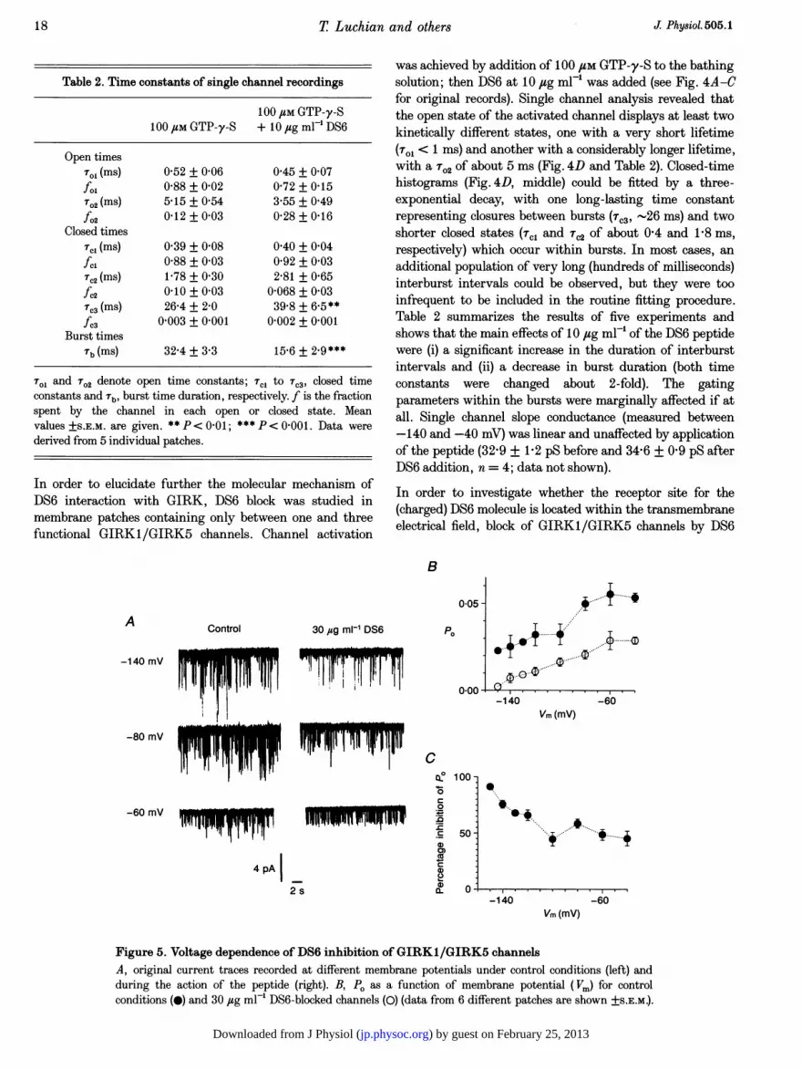
Figure 5. Voltage dependence of DS6 inhibition of GIRK1/GIRK5 channelsA, original current traces recorded at different membrane potentials under control conditions (left) andduring the action of the peptide (right). B, PO as a function of membrane potential (Vm) for controlconditions (0) and 30 jug ml-1 DS6-blocked channels (0) (data from 6 different patches are shown +S.E.M.).
T Luchian and others
ControlA
-140 mV
-80 mV 1 71
) by guest on February 25, 2013jp.physoc.orgDownloaded from J Physiol (

was measured at different potentials (see Fig. 5A for originalrecordings). As can be seen from Fig. 5B, P. exerts intrinsicvoltage dependence and hence the percentage inhibition ofP0 was calculated for each potential tested (Fig. 5C). As can
be seen from Fig. 5C, the inhibitory effect of DS6 on PO wasstronger at more negative potentials. This dependency of POon transmembrane potentials was tested using correlationanalysis and the correlation coefficient was found to be -0-70with a 95% confidence interval ranging between -0f42 and-0-86. The error probability for no correlation betweenmembrane potential (Em) and PO was found to be P< 0-0001.
Since the G protein-activated potassium channel from atrium(KACh) is a hetero-oligomer of GIRKI and GIRK4 subunits(Krapivinsky, Gordon, Wickman, Velimirovic, Krapivinsky& Clapham, 1995), we tested whether GIRK1/GIRK4channels are blocked by DS6. cRNAs encoding GIRK1 andGIRK4 were injected into the oocytes at different ratios;cRNA encoding the m2 muscarinic receptors was also co-
injected in order to be able to induce channel activation uponextracellular application of ACh. Oocytes were screened usingthe two-electrode voltage clamp method for macroscopicwhole-cell currents resulting from activation of GIRK.Figure 6A confirms that hetero-oligomeric proteins are
formed between the GIRKI and GIRK4 subunits: GIRK4
cRNA, which at this concentration was not able to inducesignificant currents in X. laevis oocytes, greatly potentiatedIACh in a dose-dependent manner, when constant amountsof GIRKI cRNA were co-injected (Fig. 6B). Subsequentlyexperiments were performed with oocytes injected withGIRK1/GIRK4 cRNAs at a ratio of 1: 10 (500 pg and 5 ng,
respectively). As can be seen from our isolated patchrecordings, shown in Fig. 6A, DS6 blocks hetero-oligomericGIRK1/GIRK4 channel activity with a potency similar toGIRK1/GIRK5 channels, with a maximal inhibition of91-9 + 54% (n=4) at 100,ug ml-' DS6. Dose-responsecurves showed that the IC50 and Hill coefficient values are
within the same order of magnitude, though not identical toGIRKI/GIRK5 channels (IC50, 3-49 + 053 ,ug ml-'(corresponding to 1P67 + 0-25 flmol F'); Hill coefficient,0 75 + 0-08; n = 4 individual patches; see Fig. 6C).
DISCUSSIONOur results demonstrate clearly that the DS6 peptidederived from the extreme C-terminal end of the GIRKIsubunit is a potent and reversible blocker of GIRK channelsof GIRKI/GIRK5 and GIRK1/GIRK4 composition. Blockof GIRK channels in excised patches by fusion proteins or a
B 6000-
A100 juM GTP-y-S
100 uM GTP-y-S
.Il -r l p 1 )T
100 uM GTP-y-S + 100 /g ml-' DS6
1r0 2Fr-Ir gIl- 100 r1mvl 1DS6
100 /Lm GTP-y-S + 100jg9 Ml- DS6
<3000-
1-
0-5/0*5
GIRK1/GIRK4 (ng)
C
1C
c0
.r_
CN
ax
0)
CdCDa)
CD)
5 s
1 07
)0 - (4),,,,, .
/(4)(4) (4) 4
(4) )
50(4) ,
1 10 100 101DS6 (g9 ml-')
Figure 6. Effect of the D86 peptide on GIRK1/GIRK4 channelsA, selected traces from inside-out patches containing a few hetero-oligomeric GIRKt/GIRK4 channelsbefore (upper 2 traces) and after addition of 100 ,g ml-' DS6 peptide. B, effect of increasing amounts ofcRNA encoding the GIRK4 subunit on basal (O) and ACh-induced (8) (cf Methods) recorded from oocytesinjected with constant amounts of RNAs of GIRK1 and the muscarinic m2 receptor. C, dose-responsecurve for DS6 block of GIRK1/GIRK4 channels. The continuous line represents best fit to the data. Thedashed line corresponds to the dose-response curve for GIRK1/GIRK5 channels. Numbers in parenthesesrepresent the number of individual patches.
J Physiot.505.1 Peptide block of GIRK channels
c
00
) by guest on February 25, 2013jp.physoc.orgDownloaded from J Physiol (
0T Luchian and others
peptide comprising parts of the GfYl-binding segment of/l-adrenergic receptor kinase has been observed by Reuvenyet al. (1994) and Nair et al. (1995). Although directcompetition experiments of these peptides with Gay havenot been performed, a variety of indirect evidencesuggested that the mechanism of block in this case was
sequestration of free G,6Y by the peptide (Nair et al. 1995), ina way similar to the GDP-bound G protein a-subunit blockof GIRK channels (Wickman et al. 1994).
As in frog atrial cells (Ivanova-Nikolova & Breitwieser,1997), the GIRK channels expressed in Xenopus oocytesdisplay a complex gating behaviour characterized by burstsof activity with variable interburst intervals. Because of theextreme complexity of gating our present analysis issomewhat preliminary and more single channel experimentswill be needed in order to understand how GY alters thekinetics. However, the present level of analysis alreadyprovides several insights into the mechanism of GIRKactivation and its block by DS6. A direct block by DS6 issupported by two independent observations. First, thecompetition experiments (Fig. 3) demonstrated that DS6blocks the channel activity by reducing the maximal open
probability rather than by competing with G,,Y. Theblocking effect of DS6 could not be overcome by increasingthe concentration of free G,6Y to 100 nm, a concentrationabout 20- to 40-fold higher than the apparent affinity for
G,,y in this preparation (see Table 1 and Schreibmayer et al.1996). Although there was an apparent shift in the EC50 for
G',Y in the presence of DS6, it was not statisticallysignificant (Table 1) and could result from the ambiguities inmeasuring PO at very low levels of channel activity, as was
the case in the presence of the peptide. Second, burstduration is most probably independent of GIY concentration(Ivanova-Nikolova & Breitwieser, 1997), whereas DS6significantly shortens the bursts; therefore the changes ingating kinetics caused by DS6 are incompatible with a
competition mechanism in which DS6 effectively reduces thefree concentration of G,lY. The shallow, but negative,correlation between membrane potential and blockingefficiency of DS6 excludes the hypothesis that the positivelycharged peptide crosses significant amounts of electricaldistance across the membrane when diffusing to its receptorsite on the channel molecule. Hence the peptide does not go
into the pore of the channel, and blocking is more
compatible with the idea of a large inhibitory gate at thechannel mouth that occludes it at rest. The present results fitinto the framework of the hypothesis proposed in theintroduction, that activation of the GIRK channel by G,6yinvolves the removal of a blocking gate that normally blocksthe channel; a hallmark of this removal is the appearance ofbursts of channel openings. This hypothesis further impliesthat the constitutively active Kir channels, such as Kirl (Hoet al. 1993) or Kir2 (Kubo et al. 1993) lack a blocking gate(cf. Pessia et al. 1995). The intraburst closed-opentransitions in Kir channels probably reflect an intrinsicgating process, because they persist in the absence of thepresumptive blocking gate (after trypsin treatment; Kirsch
& Brown, 1989) or Mg2+ and polyamine cations (Ficker,Taglialatella, Wilbe, Henley & Brown, 1994; Yamada &Kurachi, 1995). The fact that DS6 does not appreciably alterthe parameters of intraburst transitions suggests that DS6does not interfere with the intrinsic gating machinery of theactivated channel. (This does not preclude a degree ofinteraction between the blocking gate and permeant orblocking ions.) The only apparent change in the intraburstgating parameters - a reduction in the intraburst open timeconstant, To2 - is mild and statistically insignificant,implying that DS6 does not act as a fast channel blocker.Furthermore, any possible contribution of the apparentchange in To2 to the overall reduction in PO caused by DS6 iscounterbalanced by the increase in the proportion of theseopenings (Table 2). Thus, our results conform to a model inwhich DS6 mimics the blocking gate by binding to anacceptor site and blocking the ion flow through the channel,while the interaction with Gy remains unaltered. Indeed,the main effects of DS6 on single GIRK1 channels were (i) amarked and significant reduction in burst duration and (ii) aprolongation of silent periods between bursts. The greatlyprolonged interburst intervals probably indicate permanentoccupancy of the channel at a time scale of many tens ofmilliseconds, as expected for a high-affinity blocking gate,whereas the reduced burst duration reveals that the peptidemay interact with the active channel, i.e. with the open orclosed state(s) within a burst. We note parenthetically that,under the conditions used here, in the long (interburst)closed state the acceptor site may be inaccessible to theexogenously added DS6 for most of the time, since it isoccupied by the channel's own blocking gate. Assuming thatdurations of prolonged interburst intervals reflect singleunblocking events and reduced burst durations reflectblocking events, we estimate a calculated time constant forinterburst intervals of 38-1 ms (see Appendix), whichcorresponds fairly well with the 39-8 ms measured in oursingle channel experiments. (Note, however, that the dataset has been derived from patches containing 1-3 functionalchannels.)
Our data further support the notion (Dascal et al. 1995;Pessia et al. 1995) that the structural determinants of theputative blocking gate are situated at the end of theC-terminus of GIRKI. The segment constituting theblocking gate may not be limited to the last twenty aminoacid residues; preliminary results indicate that a moreproximally situated peptide, DD7 (amino acid residues439-464 of GIRK1), also efficiently blocks the channel. Sofar, our data indicate that the N-terminus, at least beyondamino acid residue 40, is not involved in channel block, aswitnessed by the inability of DS1 (residues 40-57) and DS4(residues 57-74) to block the channel. At present we cannotexclude the possibility that the beginning of the N-terminusmay be involved in the formation of the blocking gate.GIRKI may not be the only subunit contributing to theformation of the blocking gate, since channels formed byGIRK2 or GIRK4 alone are G, -gated (Krapivinsky,Krapivinsky, Wickman & Clapham, 1995; Kofuji, Davidson
20 J Physiol.505.1
) by guest on February 25, 2013jp.physoc.orgDownloaded from J Physiol (
Peptide block of GIRK channels
& Lester, 1995). The C-terminus of GIRK1 is > 80 aminoacids longer than that of other members of the Kir3 family,and its distal part is practically unique. However, we foundthat there is a limited homology between GIRKI (rat orhuman), GIRK2 (mouse), GIRK4 (rat or human) andGIRK5 (Xenopus) within the last twenty amino acidstretch, the consensus being ExxxPxxL (residues 485-492of rat GIRKI). It is tempting to speculate that these aminoacids participate in the formation of the blocking gate.
The straightforward interpretation of our principal result,i.e. that D86 is a blocking peptide that interacts with thechannel and not with GY,, is that the C-terminal segment ofGIRK1 is part of a blocking segment, and that Ga,8 bindselsewhere in GIRKI. Our results show that the DS6 peptideadded exogenously can block the channel, but we consider itlikely that the entire blocking gate includes other regions ofGIRK. This is supported by our observation that peptideDD7 (residues 439-464 of GIRKI) also blocks the channel.A more elaborate model (Cohen et al. 1996) suggests thatthe channel may normally be blocked by a complex formedby the N-terminus, the heterotrimeric G protein bound to it(Huang et al. 1995), and the C-terminal part of the blockinggate that includes DS6 and possibly additional stretches.Activation of the G protein by the receptor may causedissociation of Ga and G,6y and translocation of G,,y to thecentral portion of the C-terminus comprising the G.y-binding site (Slesinger et al. 1995; Huang et al. 1995;Kunkel & Peralta, 1995), followed by a conformationalchange resulting in channel opening. In such a model, thedistal C-terminal sequence may serve as an anchor for thewhole blocking complex. To provide further insights into themechanistic details of a particular model as envisionedabove, experiments involving C-terminal deletions ofGIRK1 clones would be of tremendous significance.Unfortunately, attempts made to study gating properties oftwo C-terminal deletions (namely with the last 40 and withthe last 160 amino acids deleted) were unsuccessful sincenone of the deleted constructs (neither expressed alone norco-expressed with GIRK4) were able to be expressed (datanot shown).
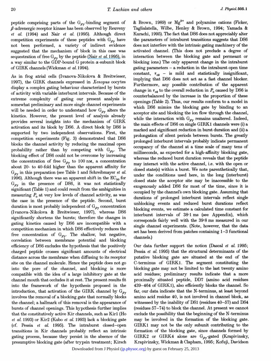
APPENDIXAssuming that the prolonged interburst interval indeedrepresents unblocking of the channel by the peptide andthat shortening of burst duration represents channelblocking events, the kinetics can be treated by using theNeher-Steinbach model (Neher & Steinbach, 1978)according to the scheme below:
C3 A + DS6 7A*DS6, (Al)aZ k0ff
where C3 denotes the long-lived, inactive state of thechannel in between bursts, A denotes all active states withina burst and A*DS6 denotes the blocked channel states. a,y,Bkic and kff represent the rate constants corresponding to
inactive . active interconversions and the drug blocking/unblocking processes, respectively. According to the model,the unblocked time constant (ru) is given as:
TU= (a + kon [DS6])-t, (A2)
wherea = Tb1 and rU = Tb*. (A3)
Tb and Tb* correspond to time constants of burst durationwithout and in the presence of DS6, respectively. [DS6]stands for the free concentration of the peptide, which was4-75 ,umol F-1 in our single channel experiments (molecularweight of DS6 was 2107-44). By solving eqn (A2) for kon andinserting the appropriate values for Tb and Tb*, we end upwith a value of kon = 7 00 s-'umol-' 1. From this, k0ff canbe calculated since:
koff = ion KD (A3)
and KD = 3X75 usmol F1 (as measured in our dose-responseexperiments), yielding an actual value of k0ff = 26-3 st.Since the prolonged interburst intervals (TC3 in our case) areregarded as representing the dwell time of the peptide onthe channel blocker, Tc3 can be calculated according to:
(A4)Tb = Tc3 =-kof f-
yielding a calculated value of r,3 = 3841 ms.
ASHFORD, W. L. J., BOND, C. T., BLAIR, T. A. & ADELMAN, J. P.(1995). Cloning and functional expression of a rat-heart K-ATPchannel. Nature 378, 792.
COHEN, N. A., SHA, Q., MAKHINA, E. N., LOPATIN, A. N., LINDER,M. E., SNYDER, S. H. & NIcHoLs, C. G. (1996). Inhibition of aninward rectifier potassium channel (Kir2.3) by G protein frysubunits. Journal of Biological Chemistry 271, 32301-32306.
DASCAL, N. (1997). Signalling via the G protein-activated K+channels. Cell Signal (in the Press).
DASCAL, N., DOUPNIK, C. A., IVANINA, T., BAUSCH, S., WANG, W.,LIN, C., GARVEY, J., CHAVKIN, C., LESTER, H. A. & DAVIDSON, N.(1995). Inhibition of function in Xenopus oocytes of the G protein-activated atrial K channel (GIRK1) by over-expression of amembrane attached form of the C-terminal tail. Proceedings of theNational Academy of Sciences of the USA 92, 6758-6762.
DAScAL, N. & LOTAN, I. (1992). Expression of exogenous ion channelsand neurotransmitter receptors in RNA-injected Xenopus oocytes.In Methods in Molecular Neurobiology 13, ed. LoNGSTAFF, A. &REVERT, P., pp. 205-225. Humana Press, Totowa, NJ, USA.
DASCAL, N., SCHREIBMAYER, W., LiM, N. F., WANG, W., CHAVKIN, C.,DIMAGNO, L., LABARCA, C., KIEFFER, B. L., GAVERIAUX-RUFF, C.,TROLLINGER, D., LESTER, H. A. & DAVIDsON, N. (1993). AtrialG protein-activated K channel: expression cloning and molecularproperties. Proceedings of the National Academy of Sciences of theUSA 90,10235-10239.
DOUPNIK, C. A., DAVIDSON, N. & LESTER, H. A. (1995). The inwardrectifier potassium channel family. Current Opinions in Neurobiology5, 268-277.
FICKER, E., TAGLIALATELLA, M., WILBE, B. A., HENLEY, C. M. &BROWN, A. M. (1994). Spermine and spermidine as gating moleculesfor inward rectifier K+ channels. Science 266, 1068-1073.
J. Physiol.505.1
) by guest on February 25, 2013jp.physoc.orgDownloaded from J Physiol (
22 T Luchia
HILLE, B. (1992). G protein-coupled mechanisms and nervoussignialing. Neuroni 9, 187-195.
Ho, K., NICHOLS, C. G., LEDERE, W. J., LYTTON, J., VASSILEV, P. M.,KANAZIRSKA, M. V & HEBERT, S. C. (1993). Cloning and expressionof' an inwardly rectifying ATP-regulated potassium channel. Nature362, 31-38.
HOSHI, T., ZAGOTTA, W. N. & ALDRICH R. W. (1990). Biophysical andmolecular mechanisms of Shaker potassium channel inactivation.Science 230, 533-538.
HUANG, C. L., SLESINGER, P. A., CASEY, P. J., JAN, Y. N. & JAN, L. Y.(1995). Evidence that direct binding of Gay to the GIRKIG protein-gated inwardly rectifying K+ channel is important forchannel activation. Neuron 15, 1133-1143.
IVANOVA-NIKOLOVA, T. T. & BREITWIESER, G. E. (1997). Effectorcontribution to Gay-mediated signaling is revealed by muscarinicpotassium channel gating. Journal of General Physiology 109,245-253.
KIRSCH, G. E. & BROWN, A. M. (1989). Trypsin activation of atrialimiuscarinic K+ channels. A merican Journal of Physiology 257,H334-338.
KOCH, X. J., INGLESE, J., STONE, XV. C. & LEFKOWITZ, R. J. (1993).The binding-site for the fy-subunits of heterotrimeric G-proteins onthe ,-adrenergic receptor kinase. Journal of Biological Chemistry268, 8256-8260.
KOFUJI, P., DAVIDSON, N. & LESTER, H. A. (1995). Evidence thatneuronal G-protein-gated inwardly rectifying K+ channels areactivated by GY subunits and function as heteromultimers.Proceedings of the National Academy of Sciences of the lTSA 92,6542-6546.
KOZASA, T. & GILMAN, A. G. (1995) Purification of recombinantG proteins from Sf9 cells by hexahistidine tagging of associatedsubunits - Characterization of a125 and inhibition of adenylyl cyclaseby az. Journal of Biological Chemistry 270, 1734-1741.
KRAPIVINSKY, G., GORDON, E. A., MWICKMAN, K., VELINIIROVIC, B.,KRAPIVINSKY, L. & CLAPHAM, D. E. (1995). The G-protein-gatedK+ channel IKACh is a heteromultimer of two inwardly rectifyingK+ -channel proteins. Nature 374, 135-141.
KRAIPIVINSKY, G., KRAPIVINSKY, L., WTICKMAN, K. & CLAPHAM, D. E(1995). G,ay binds directly to the G protein-gated K+ channel,'KAChI Jourtnal of Biological Chemistry 270, 29059-29062.
KREISZIG, E. (1979). Statistische Methoden und ihre Anwendung, 7thedn. Vandenhoeck und Ruprecht, G6ttingen.
KUBO, Y., BALDWIN, T. J., JAN, Y. N. & JAN, L. Y. (1993). Primarystructure and functional expression of a mouse inward rectifierpotassium channel. Nature 362, 127-133.
KUNKEL, M. T. & PERALTA, E. G. (1995). Identification of domainsconferring G protein regulation on inward rectifiei potassiumchannels. Cell 83, 443-449.
!n amnd others J Physiol. 505.1
NORTH, A. R. (1989). Drug receptors and the inhibition of nerve cells.British Journal of Pharmacology 98, 13-28
PESSIA, M., BOND, C. T., KAVANAUGH, M. P & ADELMAN, J. P. (1995).Contributions of the C-terminal domain to gating properties ofinward rectifier potassium channels. Neuron 14, 1039-1045.
REUVENY, E., SLESINGER, P., INGLESE, J., MORALES, J. M., INIGUES-LLUHI, J. A., LEFKOWITZ, R. J., BOURNE, H. R., JAN Y. N. & JANL. Y. (1994). Activation of the cloned muscarinic potassium channelby G protein fly subunits. Nature 370, 143-146.
SAKMANN, B., NOMA, A. & TRAUTWEIN, W. (1983). Acetylcholineactivation of single muscarinic K+ channels in isolated pacemakercells of the mammalian heart. Nature 303, 250-253.
SCHREIBMAYER, W., DESSAUER, C. W., VOROBIOV, D., GILMAN, A. G.,LESTER, H. A., DAVIDSON, N. & DASCAL, N. (1996). Inhibition of aninwardly rectifying K+ channel by G protein a subunits. Nature380, 624-627.
SCHREIBMAYER, W., LESTER, H. A. & DASCAL, N. (1994). Voltageclamping of Xenopus laevis oocytes utilizing agarose-cushionelectrodes. Pfluigers Archiv 426, 453-458.
SLESINGER, P. A., REUVENI, E., JAN, Y. N. & JAN, L. Y. (1995).Identification of structural elements involved in G protein gating ofthe GIRKI potassium channel. Neuron 15, 1145-1156.
WICKMAN, K. & CLAPHAM, D. E. (1995). Ion channel regulation byG proteins. Physiological Reviews 75, 865-885.
XVICKMAN, K. D., INIGUEZ-LLUHI, J. A., DAVENPORT, P. D., TAussIa,R., KRAPIVINSKY, G. B., LINDER, M. E., GILMAN, A. G. &CLAPHAM, D. E. (1994). Recombinant G-protein fly-subunitsactivate the muscarinic-gated atrial potassium channel. Nature 368,255-257.
YAMADA, M. & KURACHI, Y. (1995). Spermine gates inward-rectifyingmuscarinic but not ATP-sensitive K+ channels in rabbit atrialmyocytes. Intracellular substance-mediated mechanism of inwardrectification. Jourtnal of Biological Chemistry 270, 9289-9294.
AcknowledgementsWe thank Gerda Breitwieser for sharing a preprint during thepreparation of this manuscript. The authors greatly acknowledgefinancial support by the Human Frontiers Science Project (HFSPRG-379/94), the Austrian Research Foundation (Special ResearchCentre 'Biomembranes and Atherosclerosis', SFBO07/08; W.S.) andthe Israel-USA Binational Science Foundation (N.D. and H.A.L.).
Author's email addressW. Schreibmayer: [email protected]
Received 8 AMay 1997; accepted 21 July 1997.
KURACHI, Y. (1995). G protein regulation of cardiac muscarinicpotassium channel. A merican Journal of Physiology 38, C821-830.
LUCiHIAN, T., DASCAL, N., DAVIDSON, N., LESTER, H. A. &SCHREIBMAYER, WV. (1996). Single channel analysis of block of theC-protein activated potassium channel from rat atrium(KGA/GIRK1) by the C-terminal peptide. Biophysical Journal 70,A31 0 (Abstract).
NAIR, L. A., INGLESE, J., STOFFEL, R., KoCH, W. J., LEFKOWITZ,R. J., KWATRA, M. Al. & GRANT A. 0. (1995). Cardiac muscarinicpotassium channel activity is attenuated by inhibitors of GO,Circulation Research 76, 832-838.
NEHER, E. & STEINBACH, J. H. (1978). Local anaesthetics transientlyblock currents through single acetylcholine-receptor channels.Journlal of Physiology 277, 153-176.
) by guest on February 25, 2013jp.physoc.orgDownloaded from J Physiol (
Related Documents