Welcome message from author
This document is posted to help you gain knowledge. Please leave a comment to let me know what you think about it! Share it to your friends and learn new things together.
Transcript


A COMPREHENSIVE APPROACH TOTREATMENT WITH AND WITHOUT
ENDOSSEOUS IMPLANTS
Neil L. Starr, DDS, PC
The incorporation of one single-tooth implant and
crown, together with a series of all-ceramic crowns or
veneers for the adjacent natural teeth, creates a great
challenge for the clinician and dental ceramist.
Endosseous implant placement requires careful
staging in accordance with the healing time frames
associated with tissue maturation.
[Au: Please approve the selection of this
principle for the opening page or select a
different one of your choice.]

IPPHILOSOPHY & BACKGROUND
Editor’s note: In this closing chapter, Dr Starr synthesizes the treatment-planning principles
relevant to a partially edentulous case. This article appeared in the Seattle Study Club Journal
over a decade ago, and yet the concepts are timeless. Dr Starr’s chapter convincingly demon-
strates that once understood, the principles of treatment planning transcend the cases pre-
sented in this book and readily become part of the clinician’s everyday armamentarium.
INTRODUCTION
Our ultimate therapeutic goal as dentists is to achieve maximum health, masticatory function,
speech, esthetics, and comfort for our patients. Generally, treatment can be divided into three
levels: (1) emergency care for relief of pain or sudden dysfunction; (2) removal of the causative
factors of the disease processes; and (3) removal of the effects of the disease or traumatic insult.
Level 1, emergency treatment, must be accomplished before any other level of therapy is insti-
tuted (Table 17-1).
The purpose of level 2 is to control inflammation. A basic tenet of periodontal therapy is the
mechanical debridement of all accretions adherent to the clinical crowns and roots of teeth or
restorative materials, both supragingivally and subgingivally. This is accomplished by scaling,
root planing, and curettage procedures in concert with plaque-control instruction. For dental
caries, it is evident that early placement of restorations prevents the need for more extensive
intervention later.
The focus of this article is level 3: attempting to correct alterations in form and function.
2
17 A COMPREHENSIVE APPROACH TO TREATMENT WITH AND WITHOUT ENDOSSEOUS IMPLANTS • Starr
TABLE 17-1 EMERGENCY DENTAL TREATMENTS
Problem Treatment category Treatment
Deep caries crowns Sedative restorations “Direct fillings” or temporary fillings
Symptomatic teeth/abscess Endodontic Drainage, antibiotics, conventional or surgical treatment
Occlusal trauma or Occlusal therapy Selective adjustment, appliance myofascial pain syndrome therapy, anti-inflammatory
medication
Large circumscribing periodontal/ Extraction of hopeless teeth Interim fixed or removable periapical lesions restoration as necessary
Broken appliances Prosthetic repair Re-establish masticatory function and esthetics

3
DIAGNOSTIC EVALUATION
DDIAGNOSTIC EVALUATION
Diagnosis, treatment planning, and treatment sequencing continue to be difficult and trouble-
some areas for dentists and dental specialists in the therapeutic approach to the partially eden-
tulous patient. A comprehensive dental-periodontal examination must be performed first.
This will ensure that all members of the treating team have addressed their problem areas and
have collated their respective treatments into the overall therapeutic scheme. The clinical eval-
uation consists of caries, periodontal, endodontic, orthodontic, orthognathic, occlusal/
temporomandibular joint (TMJ), and systemic examinations (Box 17-1). To facilitate this diag-
nostic evaluation, a full-mouth series of periapical radiographs of teeth and residual ridges
must be taken. A panoramic radiograph, possibly a cephalometric radiograph, and a dental
computed axial tomography (CAT) scan are suggested to help assess the bone quality and den-
sity and thereby supplement conventional dental radiography. CAT scan technology is often
enhanced today by the use of barium-impregnated surgical templates2 or with gutta percha
markers to more precisely analyze all available bone sites. Mounted study casts should also be
made. In most situations it is suggested that two sets of original casts be taken: one to be pre-
served diagnostically and the other to be worked on therapeutically.
After clinical examination, radiographic imaging, and study casts, the next level of diagnosis
can begin. For a case in which the needs are largely restorative, such as veneering or crowning one
BOX 17-1 DIAGNOSTIC EVALUATION
Caries• Supragingival• Subgingival• Insufficient clinical crown height
Endodontic considerations• Symptomatic teeth• Separated instruments• Dystrophic calcifications• Fractured roots• Apical and lateral zones of osseous destruction• Status of existing posts-cores
Esthetics• Smile analysis• Lip line analysis• Gingival topography assessment• Incisal plane assessment
Malocclusion• Loss of occlusal vertical dimension
Missing teeth• Without replacement• With delayed replacement
Occlusal trauma• Primary: bruxism, clenching, retrograde wear• Secondary• TMJ considerations
Orthodontics• Tooth shift or collapse
Periodontal disease1
• Degree of bone loss• Topography of alveolar defect (potential
impact of bone loss on adjacent teeth)• Classification of periodontal biotypes
Size and shape of residual deformed bony ridgeareas• The degree of resorption will influence the
surgical and restorative ventures
Medical status• Systemic disorders• Psychological concerns
Traumatic injury• Clinical crown deformity• Soft and hard tissue deformities• Facial deformity
Developmental/acquired deformities• Cleft palate, cleft lip• Amelogenesis Imperfecta, other deformities
Systemic influences• Systemic diseases (eg, diabetes, cirrhosis)• Osteoporosis, osteopenia, osteomalacia• Liver or kidney dysfunction• Anticonvulsants• Antidepressants• Vitamin D deficiency• Parathyroid hormone• Aging; estrogen deficiency• Gastrointestinal problems• Psychiatric/psychological considerations

4
17 A COMPREHENSIVE APPROACH TO TREATMENT WITH AND WITHOUT ENDOSSEOUS IMPLANTS • Starr
E
or more teeth, gingival esthetic guidelines may be a large component of the treatment planning.
To establish a diagnosis in more compromised situations, it is important to ascertain the
patient’s tooth loss history. A variety of etiologic factors may have been responsible for tooth loss:
caries, subsequent endodontic complications, traumatic injuries to teeth (and/or alveolus), peri-
odontal disease (acute or refractory), trauma from occlusion, or iatrogenesis.
Many teeth may serve as strong viable abutments. However, teeth substantially affected by
periodontal disease, caries, or endodontic problems must be identified early because they may
have minimal value as abutments for either individual crowns or splinted restorations. These
teeth may also represent a serious periodontal liability to adjacent teeth or bony ridges.
ESTHETIC TREATMENT APPROACH
Esthetics and osseointegration were developing on parallel paths during the mid-1980s to early
1990s. Each emphasized the importance of the integrated team approach to achieve the ulti-
mate periodontal and restorative result. Preserving the soft tissue architecture, and in particu-
lar the papillae, was a major concern. We know that maintaining the papilla between two teeth
is somewhat predictable,3 but between a tooth and an adjacent implant it is less predictable.4,5
Concern for the loss or reduction in height of the papilla between two adjacent implants has
created a new esthetic issue.3 Therefore, the concepts of selective extraction of teeth and sock-
et preservation and augmentation at the time of tooth extraction appear to be invaluable in the
restoration of form, function, and esthetics.6–11
Principle 1Esthetics plays a major role in our diagnostic and therapeutic endeavors. Howev-
er, long-term clinical assessments have shown that its real value will play out opti-
mally when it is achieved in concert with all of the functional needs of the dentition.
Tarnow12 and colleagues observed that in healthy mouths the gingival
papilla filled the space between the teeth 100% of the time when the distance
from the contact point of adjacent teeth to the crest of bone was 5 mm or
less. When the distance was 6 mm, the papilla did not fill the space com-
pletely in approximately 50% of the patients, and when it was 7 mm or more,
it did not fill the space in about 75% of the cases. The pronounced scalloped
periodontal biotype (because of its triangular-shaped tooth) usually has a
distance between 6 to 7 mm. Under normal conditions, this is the tissue type
that usually has some interproximal recession with the formation of “black
triangles.” Further clinical insults to the soft tissue, such as tooth prepara-
tion, excessively rapid orthodontic tooth movement, tooth extraction, scal-
ing, root planing, and injudicious retraction of soft tissue may increase the
gingival recession, thus further compromising the esthetic result (Fig 17-1).
The extraction of an anterior tooth usually results in resorption of bone
on the facial and interproximal surface. In addition, a decrease in the faci-
olingual dimension of the interproximal areas is not uncommon. These
findings are obvious in the scalloped type of periodontium and even more
Fig 17-1b All-ceramic crowns for maxillary anterior teeth,respecting the gingiva and harmonizing with the gingivaltopography.
Fig 17-1a Maxillary anterior teeth with facial margins ofcrowns exposed and failing composite restorations.

5
Fig 17-2c All-ceramic veneers on master stonecast.
Fig 17-2d Preoperative smile profile. Fig 17-2f Definitive ceramic veneers with smileprofile.
Fig 17-2e Definitive ceramic veneers for themaxillary teeth, with restored occlusal functionand improved dental and facial esthetics.
obvious in the pronounced scalloped type.13 This can create an esthetic dilemma for both the
patient and the dentist. Complicating the matter is that the root morphology of the anterior teeth
is usually more tapered, both faciolingually and mesiodistally, than those found in the flat type of
periodontium.14–16 The end result of extracting an anterior tooth with a scalloped type of periodon-
tium is (1) greater loss of interproximal hard and soft tissues; (2) a more palatal positioning of the
interproximal papillae; and (3) a wider mesiodistal dimension between the adjacent teeth (because
of the taper of their roots). The outcome is a large noticeable black triangle, which is often treated
by closing the space with a wider crown, with laminate17 placed on the adjacent teeth, or with the
use of pink porcelain to simulate the lost gingiva. Often these options are not satisfactory.
When a patient’s needs are primarily restoratively focused, such as veneering or crowning
one or more teeth, gingival esthetic guidelines (Figs 17-2 and 17-3) will be a significant compo-
nent of the overall effort.
Fig 17-2a Edge-to-edge maxillary incisor rela-tion, with crossbite at the mandibular right lat-eral incisor, canine, and first premolar,demonstrating marked incisal wear. The den-toskeletal Class III arrangement (with thin lipform) exaggerates the flat facial profile.
Fig 17-2b Following a diagnostic compositemock-up directed at creating anterior guidance,building out the teeth to enhance the facial pro-file, and improving the incisal edge relation tothe lower lip, the maxillary teeth were preparedfor ceramic veneers. The incisal edges of themandibular teeth were reshaped by odontoplas-ty to create the proper overbite-overjet relation.
Fig 17-3a Mottled enamel with marked discol-oration and recurrent caries.
Fig 17-3c Smile view with provisional acrylicrestorations, which create both gingival and incisalbalance with the patient’s lips and facial form.
Fig 17-3b Provisional acrylic restorations torestore form, function, and esthetics to theinvolved maxillary teeth.

6
The addition of bone and soft tissue at or after tooth extraction, or of tooth lengthening by
restorative and/or surgical measures to achieve esthetic outcomes, requires even greater inter-
disciplinary planning (Box 17-2).
Fig 17-4a Frontal view of preoperative worndentition.
Fig 17-4c Composite mock-up of lip line smile.Fig 17-4b Frontal view of composite mock-up.
To properly address the esthetic requirements of the patient, it is necessary to envision the
desired outcome before performing the procedure.18
Esthetics is fundamentally about tooth form, and it is most predictably realized with the
assistance of an intraoral diagnostic mock-up to improve incisal form, lip line esthetics, and
gingival topography. (Fig 17-4) The outcome is the development of an intraoral esthetic blue-
print. This results in dentist verification, improved laboratory communication, and patient
affirmation. Molds of the improved intraoral anatomic form of the teeth should be poured in
stone and then enhanced further in the dental laboratory with the application of wax. Silicone
impressions are fabricated by the laboratory, then returned to the clinician to be used to verify
proper tooth reduction.18
Principle 2The incorporation of one single-tooth implant and crown, together with a series of all-ceramiccrowns or veneers for the adjacent natural teeth, creates a great challenge for the clinician anddental ceramist.6 Endosseous implant placement requires careful staging in accordance withthe healing time frames associated with tissue maturation.
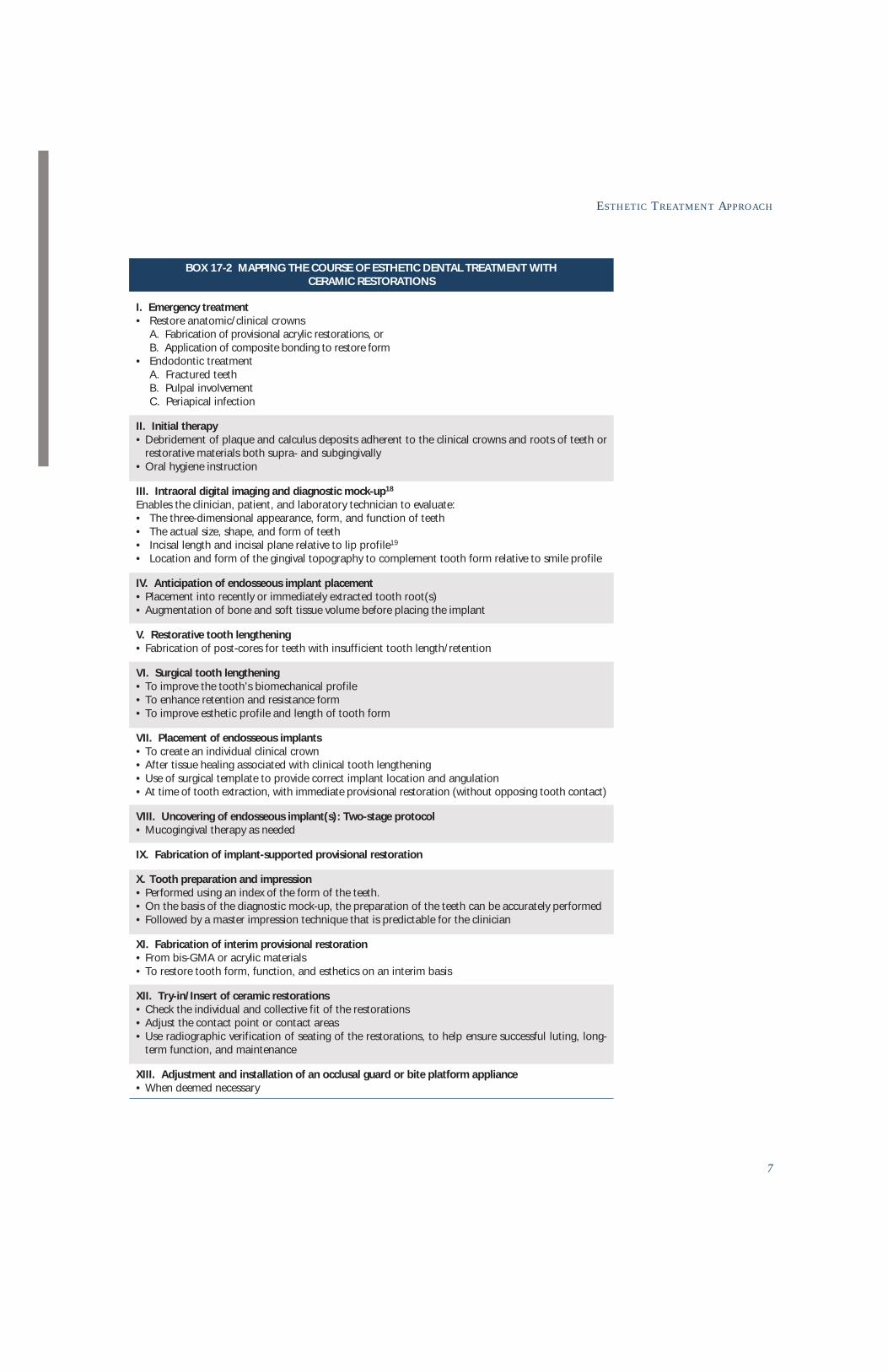
BOX 17-2 MAPPING THE COURSE OF ESTHETIC DENTAL TREATMENT WITH CERAMIC RESTORATIONS
7
ESTHETIC TREATMENT APPROACH
I. Emergency treatment• Restore anatomic/clinical crowns
A. Fabrication of provisional acrylic restorations, orB. Application of composite bonding to restore form
• Endodontic treatmentA. Fractured teethB. Pulpal involvementC. Periapical infection
II. Initial therapy• Debridement of plaque and calculus deposits adherent to the clinical crowns and roots of teeth or
restorative materials both supra- and subgingivally• Oral hygiene instruction
III. Intraoral digital imaging and diagnostic mock-up18
Enables the clinician, patient, and laboratory technician to evaluate:• The three-dimensional appearance, form, and function of teeth• The actual size, shape, and form of teeth• Incisal length and incisal plane relative to lip profile19
• Location and form of the gingival topography to complement tooth form relative to smile profile
IV. Anticipation of endosseous implant placement• Placement into recently or immediately extracted tooth root(s)• Augmentation of bone and soft tissue volume before placing the implant
V. Restorative tooth lengthening• Fabrication of post-cores for teeth with insufficient tooth length/retention
VI. Surgical tooth lengthening• To improve the tooth’s biomechanical profile• To enhance retention and resistance form• To improve esthetic profile and length of tooth form
VII. Placement of endosseous implants• To create an individual clinical crown• After tissue healing associated with clinical tooth lengthening • Use of surgical template to provide correct implant location and angulation• At time of tooth extraction, with immediate provisional restoration (without opposing tooth contact)
VIII. Uncovering of endosseous implant(s): Two-stage protocol• Mucogingival therapy as needed
IX. Fabrication of implant-supported provisional restoration
X. Tooth preparation and impression• Performed using an index of the form of the teeth.• On the basis of the diagnostic mock-up, the preparation of the teeth can be accurately performed• Followed by a master impression technique that is predictable for the clinician
XI. Fabrication of interim provisional restoration• From bis-GMA or acrylic materials• To restore tooth form, function, and esthetics on an interim basis
XII. Try-in/Insert of ceramic restorations• Check the individual and collective fit of the restorations• Adjust the contact point or contact areas• Use radiographic verification of seating of the restorations, to help ensure successful luting, long-
term function, and maintenance
XIII. Adjustment and installation of an occlusal guard or bite platform appliance• When deemed necessary

8
17 A COMPREHENSIVE APPROACH TO TREATMENT WITH AND WITHOUT ENDOSSEOUS IMPLANTS • Starr
Dental therapeutics without implantsWhen sophisticated dental therapy will be managed without the use of endosseous implants, the
approach to treatment can be subdivided into periodontal, orthodontic/orthognathic, occlusal,
and restorative phases. These phases are interdependent even if one initially takes precedence over
another, or if two or more of the phases are concurrent (Box 17-3).
BOX 17-3 MAPPING THE COURSE OF DENTAL THERAPY—WITHOUT IMPLANTS
I. Emergency treatments (Table 17-I)
II. Scaling, root planing, curettage, oral hygiene instruction• Closed or open flap debridement
A. Mechanical debridement of calcareous plaque deposits adherent to clinical crowns and roots ofteeth or restorative materials both supra- and subgingivally
B. Removal of all chronic granulation tissue
III. Operative dentistry• Restoration and conservative control of dental caries
IV. Orthodontic treatment (partial or full)• Level and align teeth• Erupt fractured or impacted teeth• Extrude teeth to level infrabony defects and/or augment the bone and soft tissue topography• Support orthognathic correction
V. Fabrication of interim provisional restoration• Guidelines
A. Replace missing and/or recently extracted teethB. Maintain or improve inter- and intra-arch harmonyC. Assess adequacy of tooth reductionD. Determine the clinical crown profilesE. Develop therapeutic occlusal schemeF. Control occlusal forces and assess function
VI. Periodontal surgery• Osseous therapy
A. Regeneration/augmentation1. Regeneration of attachment apparatus of teeth2. Regeneration and augmentation of ridge deformities
B. Ostectomy/osteoplasty1. Improve alveolar topography2. Achieve minimal sulcus depth
C. Mucogingival therapy1. Enhance the gingival complex around teeth and implants2. Grafting procedures (eg, subepithelial connective tissue grafts, allogeneic dermal grafts, etc)
VII. Re-evaluation• Establish prognosis of the remaining teeth
A. Function and estheticsB. OcclusionC. PhoneticsD. Mucogingival considerationsE. Emergence profiles
VIII. Prosthetic phase• Fixed prosthesis• Fixed-removable prostheses• Fabrication of an occlusal appliance after installation of the final prosthesis, when deemed necessary
IX. Maintenance

9
AHEAD
The objective of periodontal therapy is alveolar repair and restoration of normal anatomic
form (gingival health). Treatment is directed at decreasing the inflammatory response by
improving the osseous topography and the relationship of the overlying soft tissue, to decrease
probing depth. Amsterdam20,21 has noted that this is most predictably accomplished for teeth
of normal anatomic root lengths with probing depths not exceeding 4 to 7 mm [Au: Changeto 7 mm, which already exceeds 4 mm?] measured from the cementoenamel junction (CEJ)
(Fig 17-5). The advantage of this osseous surgical approach is an increase in the clinical crown
length, and hence a final crown design with sufficient biomechanical retention-resistance.
Figs 17-5a to 17-5c Radiographic evidence of subgingival calculus accumulations and inconsistent bony margins. Resulting increase in crown-to-root ratiosof maxillary anterior group of teeth.
Fig 17-5d Poorly adapted composite veneers formaxillary anterior teeth, with marked gingivalinflammation and generalized probing depth inthe 5- to 6-mm range.
Figs 17-5e and 17-5f Splinted ceramo-gold-metal restorations, after healing from apically positionedmucoperiosteal flap surgery to eliminate the periodontal disease and create normal topographic form,with minimal probing depth throughout. (Periodontal therapy by Dr Garry Miller. [Au: Please providecity and state.])
a b c

10
17 A COMPREHENSIVE APPROACH TO TREATMENT WITH AND WITHOUT ENDOSSEOUS IMPLANTS • Starr
Experimental and clinical research over the last decade has shifted the focus of periodontics
toward increased use of guided tissue membrane techniques. These approaches seek to regen-
erate desired attachment apparatus circumscribing the periodontally compromised root.
Although there is some clinical unpredictability associated with this therapeutic approach, it
represents a step in the direction of augmentation/regeneration versus resection.22 This new
era of regeneration therapeutics constitutes a positive shift in treatment strategy.22
Secondary occlusal traumatism, represented by moderate-to-severe loss of alveolar support
and significant clinical mobility, results in a need to splint two or more teeth to recreate collec-
tive stability and functionality. This was and continues to be predictably achieved with the use
of partial- or full-coverage crowns.
Where bite collapse has occurred, the restoration is more difficult. In this case, a provision-
al restoration is used to replace the extracted teeth, to restore lost occlusal vertical dimension,
and to establish or re-establish anterior guidance, allowing for disarticulation of posterior
teeth during excursive movements (Fig 17-6).
When teeth require subgingival preparation in conjunction with full-coverage restorations, it
is important to evaluate the mucogingival environment and determine the value of recreating or
enhancing the masticatory mucosa. Autogenous gingival grafts, subepithelial connective tissue
grafts, and repositioning of an existing gingival complex are commonly used approaches.
The orthodontic phase seeks to improve tooth alignment, erupt fractured or impacted teeth,
or facilitate the extrusion of teeth with infrabony defects.23 If the gingival zone on the facial,
lingual, or palatal surface is deficient, it is prudent to consider a mucogingival procedure
before tooth movement. This minimizes the concern about recession if the tooth must assume
a position not directly over its basal support.
When indicated, orthodontic intervention generally precedes the provisional restorative
phase. If the protocol is reversed, the clinical team may be required to make significant addi-
tional repairs and recementations. Cement washout places teeth at greater risk of developing
caries, and the patient may require a new provisional restoration before the impression phase
of therapy.
After the provisional restorations are fabricated and tooth stability is achieved, the restora-
tions can be removed to allow better access to the surgical field for correction of any residual
hard and soft tissue inconsistencies. After the tissue has matured and the prognosis is estab-
lished for all remaining teeth on both an individual and collective basis, subgingival prepara-
tion can be finalized and the provisional restorations relined, followed by completion of the
fixed or fixed-removable prosthesis.

11
Fig 17-6a Pretreatment view of 55-year-old male withmissing teeth (partial denture replacement), severe peri-odontitis, and complete bite collapse.
Figs 17-6c to 17-6e Pretreatment radiographic and clinical views showing periodontal and periapical pathology, a Class III mal-occlusion, and primary and secondary occlusal traumatism.
Fig 17-6b Posttreatment: Metal-ceramic fixed splintedrestorations.
Figs 17-6f to 17-6h Provisional acrylic restorations (before periodontal surgical correction). Immediate replacement of extracted teeth. Lat-eral excursive movement demonstrating re-establishment of anterior guidance.
Figs 17-6i to 17-6k Definitive metal-ceramic fixed restorations. Prosthesis continues to function at 12 years follow-up. (Periodontal therapyby Dr M. Stiglitz, Washington, DC.)
c
d e
f
g h
k
i
j

12
17 A COMPREHENSIVE APPROACH TO TREATMENT WITH AND WITHOUT ENDOSSEOUS IMPLANTS • Starr
Dental therapeutics with implants
Based on longitudinal studies of the viability24–31 of endosseous implantology, implants used
as an integral part of periodontal prosthesis now offer the patient and dentist a more stable
and predictable restoration. Their relative immobility and load-bearing capacity, when secured
in a qualitatively adequate bony housing, may allow for fabrication of a fixed prosthesis resist-
ant to displacement. The damage pattern of primary and secondary occlusal trauma attendant
many teeth may be reversed. Even teeth that appear to have a hopeless prognosis may be able
to assume a useful prosthetic role (Box 17-4).
In periodontal disease, as with dental caries, we may find permanent scars that complicate ther-
apy more than the active disease process itself. In many circumstances, anatomic deformities such
as an altered residual ridge form32,33 or close proximity of the sinus wall34,35 represent propagating
factors that require additional surgical correction. These difficulties may necessitate a staged
approach of augmentation, regeneration32,36 or onlay grafting first,33 followed by a second surgical
phase of implant placement and healing.
During the past decade, implant restorations were reasonably acceptable from an esthetic per-
spective. However, in cases of advanced periodontal disease, the resorption of alveolar bone creates
a significant challenge to achieve an esthetic, functional restoration. Placing implants in resorbed
bone often results in long, unesthetic teeth with an adverse crown-to-implant ratio. Implant posi-
tioning is critical from faciolingual, mesiodistal, and incisoapical perspectives. It was quickly deter-
mined that the type of periodontium, whether thick-flat or thin-scalloped, significantly affects the
esthetic outcome. The thin-scalloped type, with its friable gingival and osseous morphology, often
results in tissue recession, ultimately exposing metal at the gingival crown margins.
From a diagnostic standpoint, the dental team must try to anticipate the size and shape of
the deformity that would be created by removing the involved teeth. These judgments will play
heavily in the restorative design.
From a periodontal-prosthetic perspective, we know that many severely compromised teeth can
still offer the patient short-term function. For this reason, the restorative dentist may strategically
retain some of these teeth to facilitate an interim fixed provisional prosthesis rather than rely on a
removable design.36 This decreases the risk of prematurely loading the implant body, and induc-
ing micromotion during initial stages of interfacial bony healing.37 A well designed and construct-
ed interim provisional restoration is most important now that osseointegration technology38–46
and osseous regenerative technology47,48 have significantly changed the sequencing and length-
ened the timing of prosthetic treatment for the partially edentulous case (see Box 17-3). The weak
remaining teeth may be removed at a later stage in favor of additional endosseous implant sup-
port, as dictated by the biomechanical needs of the final restoration.
Periodontal surgical therapy is performed for all teeth that have a favorable prognosis.
Either regenerative approaches23 or pocket reduction49–51 and clinical crown exposure proce-
dures should be rendered before endosseous implant placement.
When orthodontic treatment is involved, it may require early tooth positioning to create
adequate space prior to implant placement. In some situations, such as a flared maxillary ante-
rior segment with few or no posterior teeth, implants may be placed first and employed as the
anchorage mechanism to retract and align the remaining teeth. In these situations, we are lim-
ited only by the treatment-planning creativity of the dental team. One must not minimize the
value of orthodontic mechanotherapy to move teeth through a healthy bony environment.
This can reduce and modify the size and shape of angular osseous defects, often through erup-
tion or extrusion.52–54 The improvement in hard and soft tissue topography allows the newly
regenerated bone to successfully receive endosseous implants.
Forced eruption of hopeless teeth is used to alter the soft and hard tissues before placing
implants. In addition, orthodontic extrusion is used to re-create lost interproximal papillae.

13
ESTHETIC TREATMENT APPROACH
BOX 17-4 MAPPING THE COURSE OF DENTAL THERAPY—WITHOUT IMPLANTS
I. Emergency treatments (Table 17-I)
II. Scaling, root planing, curettage, oral hygiene instruction• Closed- or open-flap procedures
A. Mechanical debridement of calcareous plaque deposits adherent to clinical crowns and roots ofteeth or restorative materials both supra- and subgingivally
B. Removal of all chronic granulation tissue
III. Operative dentistry• Conservative control of dental caries
IV. Orthodontic treatment (partial or full)• Level and align teeth to improve position• Erupt fractured or impacted teeth to rebuild/reposition bony complex• Extrude teeth to correct infrabony defects and augment soft and hard tissue topography• Support orthognathic correction.
V. Fabrication of interim provisional restoration• Guidelines
A. Allow for extraction of hopeless teethB. Maintain or re-establish inter- and intra-arch harmonyC. Assess adequacy of tooth reductionD. Determine the clinical crown profilesE. Develop therapeutic occlusal arrangementF. Control occlusal forces and assess functionG. Allow for fabrication of “diagnostic template” with markers for radiographic analysis
VI. Periodontal surgery• Osseous therapy
A. Regeneration/augmentation1. Regeneration of the attachment apparatus of teeth2. Bone augmentation of deformed alveolar ridges
B. Ostectomy/osteoplasty1. Improve bone morphology2. Reduce pocket depth
• Mucogingival therapyA. Enhance the gingival complex around teeth and implantsB. Grafting procedures (eg, subepithelial connective tissue grafts, allogeneic dermal grafts, etc)
VII. Bone grafting, sinus bone augmentation procedures• Dictated by the need to most ideally locate and place implants
VIII. Fabrication of surgical template• Guide implant placement (based on clinical and radiographic interpretation)
IX. Implant placement• In existing alveolar bone sites or healed extraction sites• In bone augmentation sites (eg, sinus, alveolar ridges)• In fresh extraction sites
X. Interim maintenance to facilitate healing• For surgical sites, control the exposure of occlusive membranes and/or loose cover screws• For interim provisional prosthesis
A. Repair broken acrylic jointsB. Replace soft reline materials
• Maintain and monitor transitional implants
XI. Transitional implant-assisted/-supported restoration• Preservation or augmentation of gingival complex• Placement of transepithelial healing components for second-stage implants• Allow for soft tissue maturation• Selection of implant abutments• Conversion of existing provisional to implant-assisted/-supported restoration• Fabrication of new implant-/implant- and tooth-assisted provisional restoration

14
17 A COMPREHENSIVE APPROACH TO TREATMENT WITH AND WITHOUT ENDOSSEOUS IMPLANTS • Starr
OOUTCOME-BASED PLANNING:INTERIM PROVISIONAL RESTORATIONS
The interim restoration may be designed in several different ways. One approach is to modify
an existing denture or splint, reline the crowns on selected natural teeth, and convert other
crowns to pontics as necessary. With a dearth of strong, well-distributed natural teeth, the
existing rigid metal framework can better resist normal occlusal forces and help prevent pros-
thesis fracture.
The removable interim prosthesis is the least desirable measure for preserving masticatory
function. Unfortunately, it must be used when the support provided by the remaining teeth is
too compromised and the number and distribution of teeth is insufficient to allow for the use
of a fixed prosthesis. In this situation it is imperative that the restorative dentist inspect the
edentulous areas at regular clinical intervals and replace the soft liner material of the denture
base when it becomes hard or brittle, or elicits a pressure ulceration in the soft tissue.
Currently, there are “temporary” or transitional dental implant systems that preclude the use of
the removable appliance. They allow the clinician to use a fixed “mini-implant” or conventional
endosseous implant–supported restoration throughout the plase of implant osteointegration.55–60
Ideally, a new fixed provisional restoration, with or without a rigid metal reinforcement, should
be made from a diagnostic wax-up, incorporating all of the esthetic, functional, and phonetic char-
acteristics being considered in the case. Any pre-existing limitations should be removed. This will
serve as a blueprint of the final prosthetic outcome. It can be used as a guide and will allow the den-
tal team to plan the case construction from the desired end point in reverse order (Fig 17-7).
In advanced periodontal disease, the maxilla generally resorbs apically and palatally; there-
fore, the mandible appears to be much larger than the maxilla. When all of the maxillary teeth
are eventually lost and the edentulous cast is mounted on an articulator, it appears as if the
patient has a prognathic relationship. However, this is not a true prognathic arch profile, but
rather a result of the bone resorption of the maxilla. And if the patient desires implants and a
fixed restoration, this case61 becomes a surgical and restorative challenge. The clinician(s) must
know before the implants are placed how this occlusal disparity will be corrected in the defin-
BOX 17-4 MAPPING THE COURSE OF DENTAL THERAPY—WITHOUT IMPLANTS (CONT)
XII. Re-evaluation• Stability of implants and remaining teeth• Occlusal vertical dimension• Esthetics• Phonetics• Proper emergence profiles of crowns for teeth and implants
XIII. Prosthetic phase• Implant-assisted
A. Fixed prosthesisB. Fixed-removable prosthesis
• Implant-supportedA. Fixed prosthesisB. Fixed-removable prosthesis
• Fabrication of occlusal appliance after final prosthesis is inserted if necessary
XIV. Maintenance

15
Fig 17-7a Pretreatment Class II, division 1 malocclusion with fail-ing crown and bridgework [Au: splint?]—a result of caries, post-core failures, and periodontitis.
Figs 17-7b to 17-7d Pretreatment radiographs.
Fig 17-7e Fabrication of acrylic provisional restorations. Note themarked anterior platform created to provide both centric holdingarea and necessary anterior guidance. (Socket preservation by DrKarl A. Rose [Au: Please provide city and state].)
Fig 17-7f to 17-7h Radiographs of tooth preparations after fabrication of provisional restorations.
Fig 17-7j Final ceramo-gold implant- and tooth-sup-ported restoration.
Fig 17-7i Final radiographic appearanceof completed maxillary restoration.
Fig 17-7k Final radiographic appear-ance of completed maxillary restoration.
b c d
f g h

16
17 A COMPREHENSIVE APPROACH TO TREATMENT WITH AND WITHOUT ENDOSSEOUS IMPLANTS • Starr
D
itive prosthesis. The volume of available bone is significant to the long-term survival of
implants in this situation because of the exaggerated anterior-posterior discrepancy.
It is wise and judicious to fabricate a temporary appliance simulating the final restoration
before surgical procedures. This is essential when the clinician is contemplating a change in the
occlusal vertical dimension. This alteration will change the faciopalatal relationships of the
mandible to the maxilla.
The lip line esthetic diagnosis, as well as the lip support, will influence the decision to fabri-
cate a fixed or removable prosthesis. A simple and effective way to make a reasonable esthetic
appraisal of the final prosthesis is to evaluate the appearance of the patient’s existing prosthe-
sis. Assuming that it is acceptable to the patient and to the dentist, it is wise to duplicate the
existing prosthesis and evaluate the patient’s profile. If the appearance is the same as the orig-
inal restoration, it can be assumed that the teeth are supporting the lip. In this situation, it is
likely that an acceptable fixed restoration can be made. Conversely, if the lip “collapses in,” the
final prosthesis will likely require some form of labial support, often necessitating a removable
prosthesis. A fixed restoration would likely be unsatisfactory.
DIAGNOSTIC AND SURGICAL TEMPLATES
Like the surgical template, a diagnostic template with radiographic markers can be fabricated
to help both surgeon and restorative dentist in analyzing available bone sites via panoramic or
CT scan radiography prior to the surgical phase.62
The surgical template, a guide to surgical implant placement, is fabricated from either a
diagnostic wax-up or, preferably, a stone cast of the functioning provisional restoration.
After the provisional restoration is placed intraorally, impressions are taken of both the
prosthesis and the underlying edentulous ridges and tooth preparations. Stone casts are made
and an acrylic shell of the restoration is cured on a cast of the remaining prepared and/or
unprepared teeth. Access locations and axial alignments are carefully planned with the surgeon
and are carved into the acrylic form to anticipate all future implant placements.
While a lingual or palatal approach is commonly used to design the surgical guide, a facial
approach may also be considered. This will provide the surgeon with an accurate visualization
of the ideal implant sites, the desired path of abutment emergence, and the axis relation to the
final prosthesis.
The ability to perform surgical procedures demands excellent access, which is provided by
temporary removal of the provisional interim restoration. The surgeon will then orient the sur-
gical template by securing it to the prepared and/or unprepared teeth and penetrate into the
bone so as to bring about proper positioning of the implants.
Significant progress in biotechnology, radiology, and computer technology have allowed for
accurate diagnosis and treatment planning. This has recently resulted in the construction of
three-dimensional bone models, stereolithography63 (Fig 17-8), and navigational surgery to
position endosseous implants with greater precision.
Principle 3The trends that have brought dentistry to its current level of esthetic sophistication require theclinician to predict the outcome before implants are placed. If the esthetic evaluation is inaccu-rate, the final result will be less than desirable to the patient and the dentist.

17
Figs 17-8c and 17-8d Simulation of endosseous implant placement into four anterior maxillary sites, as determined by evidence of boneon the CT scan images.
Fig 17-8a Premaxilla after Le Fort I osteotomy. Soft tis-sue graft increased ridge height by 7 mm.
Fig 17-8b Diagnostic template using gutta percha mark-ers and barium sulfate to locate endosseous implants inanterior maxillary region.
Fig 17-8e Surgical template with titanium cylinders tolocate the implant sites with surgical precision.
Fig 17-8f Surgical endosseous implant placement basedon CT scan technology and stereolithography.
Fig 17-8g Provisional acrylic implant-supported transitional restoration.

18
17 A COMPREHENSIVE APPROACH TO TREATMENT WITH AND WITHOUT ENDOSSEOUS IMPLANTS • Starr
SSURGICAL CONSIDERATIONS
The well-designed treatment plan may require one of a host of scenarios to deal with the installa-
tion of endosseous implants into bony sites that either still house teeth, or have ridge deformities.
Where implant placement is anticipated, the most common approach is to extract teeth at
the time of provisional restoration. Full maturation of the bony socket may then take anywhere
from 3 to 6 months. The newly formed bone in these recent extraction sites has proven to be
an excellent reservoir of pluripotential cells to promote successful osseointegration.
Another treatment approach may considerably shorten the duration of
treatment. Here the effort is made to place the implant at the time of tooth
extraction (Fig 17-9). The implant should be submerged several millimeters
below the bony crest to reduce the risk of dehiscence formation. In these situ-
ations, the ability to achieve primary flap closure will decrease the risk of post-
operative complications, especially if a cell-occlusive membrane is indicated.
In an effort to more precisely determine the quality of the bony housing for
possible immediate implant placement and to minimize the overall maturation
phase, the teeth may be sectioned horizontally at their gingival margins or at the
height of the alveolus.64 The pulp should be extirpated, the canal medicated and
sealed, and provisional restorations fabricated, leaving these tooth roots for the
surgeon to extract at the time of implant placement. This approach avoids inter-
ference with early socket healing and precludes the risk of additional crestal
bone resorption of the healing socket.65 The surgeon will decide whether to
extract and immediately place an implant into the socket. In some cases, the sur-
geon may prefer to extract the tooth, place a bone graft and membrane, and
allow the area to heal for 3 to 4 months before placing an implant.
When an edentulous ridge has a modest defect and the site has been
planned for implant placement, the surgeon may elect to position the
implants at an angle that corresponds to the ideal final restoration. Any pos-
sible fenestration over the implant may be corrected by placement of a phys-
ical membrane barrier (based on the principles of guided bone
regeneration).66 Here adequate space must be achieved to promote complete
reformation of the bone complex.
Dental implant placement in the atrophic or deformed alveolar ridge can be
a surgical challenge. Alveolar augmentation is currently accomplished with guided bone regenera-
tion techniques,47,67,68 sinus bone augmentation,69 bone grafting,70–72 and alveolar distraction
osteogenesis.73 Two or more surgical interventions are frequently required to correct a major ridge
deformity. First, the ridge must be reconstructed to a more normal anatomic shape and size,62 fol-
lowed by implant placement and soft tissue augmentation (Fig 17-10).74,75
Fig 17-9b Second-stage uncovering of implants. Notebone formation to crest of both sites. (Surgery by Dr G.Miller, Washington, DC.)
Fig 17-9a Extraction of the maxillary lateral incisorand first premolar with immediate implant placement.

19
Figs 17-10a and 17-10b Views of block bone graft with fixation in the anterior maxilla. (Surgery by Dr Jeffrey Posnick[Au: Please provide city and state].)
Fig 17-10e Definitive ceramic-gold implant- and tooth-supportedrestoration.
Fig 17-10c Preparation of block graft site to place endosseousimplants. First, the surgical template is installed. Measurementstaken relative to the template will locate the implants vertically in theblock-grafted bone relative to the cementoenamel junction of theadjacent teeth. Implants were placed to the proper mesiodistal, buc-copalatal, and vertical positions. (Surgery by Dr Garry Miller, Wash-ington, DC.)
Fig 17-10d Soft tissue topography demonstrating modest rise andfall of gingival topography. [Au: Is edit okay?]
Fig 17-10f Radiographic images of definitive maxillaryrestoration.
a b

20
Depending on the extent of the original ridge deformity, the surgical bone augmentation pro-
cedure can be relatively successful at restoring the bony contour to the following levels: Class I, 1
to 2 mm apical to the CEJ level of the adjacent teeth; Class II, 3 to 4 mm apical to the CEJ level of
the adjacent teeth; Class III, 5 mm or more apical to the CEJ level of the adjacent teeth.62
In reconstructing the deformed ridge to a Class I bone level, a normal overlaying soft tissue
profile will often be created. For the Class II bone level, where there is still some horizontal and
vertical depression, soft tissue augmentation by means of connective tissue grafts,74,75 autoge-
nous grafts (free or pedicle), or repositioning of the gingival complex, may mask the bony defi-
ciency and create a normal topographic appearance.62
For the Class III level, prosthetic materials are frequently required to restore the hard tissue
and soft tissue deformities and simulate the Class I reconstructed profile, which otherwise may
be compromised in both height and width (Fig 17-11).62
After the bony ridge has been reconstructed and endosseous implants placed, a sufficient
healing period must be observed to ensure a satisfactory “take.” Bone remodeling adjacent to
implant fixtures occurs over a period of at least a year, leading to a more mature bone (lamel-
lar compacta) within which the implant can better tolerate the forces of occlusion.77
Fig 17-11a Preoperative cast of residual ridge extending from formertooth site #17 - #26 inclusive, the result of removal of a squamouscell carcinoma.
Fig 17-11b Implant-supported fixed, ceramic-gold restorationsreplacing the anatomic crown, root, and gingiva—the Misch FPIII109classification for fixed implant prostheses. [Au: Please provide refer-ence.] (Implant placement by Dr Karl A. Rose, [Au: Please providecity and state.)
Principle 4The essential criteria for alveolar ridge reconstruction for successful implant placement are as fol-lows: (1) appropriate quantity of horizontal and vertical bone and adequate quality of bone; (2)sufficient keratinized tissue overlying the bony crest; and (3) adequate distance betweenimplants.3

T
21
Figs 17-12d to 17-12f Provisional restoration secured with set screw retention.
Fig 17-12a Maxillary dentition of 24-year-oldmale with partial anodontia.
Fig 17-12b Master cast of implant locationswith temporary crown cylinders.
Fig 17-12c Fabrication of heat-processed provi-sional acrylic restorations.
TRANSITIONAL IMPLANT-ASSISTEDRESTORATIONS
It is extremely important to coordinate the schedules of the surgeon and restorative dentist to
begin the process of restoring the implants after an established healing period.
The surgeon will perform a small gingival punch procedure or a more extensive mucope-
riosteal flap procedure, repositioning the gingival complex around the implants. A transepithe-
lial healing component is then fastened to each implant body. After soft tissue and periosteal
maturation, a high- or low-profile transepithelial abutment may be selected and a provisional
restoration can be made to restore form and function (see Fig 17-4). For one-stage implants,
the restorative dentist may begin the provisional restoration process directly.
When multiple implants are exposed and angulation concerns are anticipated, it is valuable
to make an impression that records the orientation of the fixture heads after early soft tissue
healing. A new provisional restoration may be fabricated in the laboratory (Fig 17-12) with tita-
nium temporary cylinders that are designed to mate directly with the implant body or to a
selection of available abutment heads.78
d e f

22
17 A COMPREHENSIVE APPROACH TO TREATMENT WITH AND WITHOUT ENDOSSEOUS IMPLANTS • Starr
F
Frequently at the second stage, the existing interim provisional prosthesis must be modified
by shortening the undersurface of the pontics to provide room for the healing components.
Later, these components are removed; abutments of proper height are screwed into position,
and temporary crown cylinders are seated, shortened to contact the opposing occlusion, and
incorporated into the existing provisional restoration.
If there is any doubt as to the feasibility of accomplishing functional and esthetic alignment
of the implant abutment, temporary crown cylinders can be secured directly to the implant
body. It is noteworthy that these metal cylinders are available from most implant manufactur-
ers. They ensure intimate fit to the titanium abutment or implant head and allow the clinician
to start developing the anticipated contours. Of course, should the form need to be modified,
the acrylic itself offers ample opportunity without jeopardizing any accuracy of fit.
A transepithelial collar of minimal height, shallow sulcular depth, and a circumscribed border
of bound-down keratinized tissue are essential ingredients in allowing for conventional plaque-
control measures.
Chiche et al79 have pointed out that as a result of “surgical and anatomic limitations,”
implant placement may not correspond to the initial expectation set at the presurgical
phase, and over-contouring the final restoration could create esthetic and functional liabil-
ities. The path of emergence of the fastening screw through the prosthesis may compromise
part of the facial or occlusal morphology, especially if it passes through a primary centric
occlusal contact, an interproximal embrasure [or the facial veneer.] Even minor discrepan-
cies between an implant and crown axis may result in eccentric screws, since such deviations
are magnified at the level of the occlusion.
Here the transitional prosthesis is invaluable in diagnosing these prosthetic limitations. With
this early awareness, we can better anticipate and plan for the fabrication of an auxiliary sub-
structure to facilitate the prosthetic result in the dental laboratory.
Some have conjectured that the implant-assisted provisional restoration may provide a
shock-dampening effect that may be beneficial during the first year of bone maturation adja-
cent to the implants. Of possibly greater value is the role of the provisional restoration in estab-
lishing the esthetic, phonetic, and functional needs for the final prosthetic design.
At this stage, a radiographic and clinical evaluation of the stability of the implant fixtures is
made. The weak teeth that were held strategically to support the interim prosthesis are extract-
ed at this time.37 Some of the natural teeth may be removed in favor of additional implants or
retained as indirect retainers in situations where fewer implants are used in the overall support
of the prosthesis. If a new transitional prosthesis has recently been fabricated, there may have
been a change in the occlusal vertical dimension or the esthetic form, both of which would
require further modification. Additionally, it may be necessary to consider mucogingival treat-
ment to enhance the complex of masticatory mucosa around selected teeth or implants.79
FINAL PROSTHETIC PHASE OF TREATMENT
When the final prognosis for all teeth and implants has been established, the restorative dentist
can employ crown and bridge techniques to construct the fixed or fixed-removable prosthesis. The
dentist may proceed with final impressions of the natural teeth, relate them to the proper position

23
of the implants or abutments, and fabricate a master mold. To initiate the laboratory procedures,
the case is carefully mounted on an appropriate articulator by a series of occlusal registrations.
On the master cast, a soft tissue marginal profile should be constructed around each natural
tooth die and implant analogue to simulate the gingival condition in the oral cavity. This allows for
predictable abutment head selection based on height, angulation, and emergence profile.
Technical choices are now made concerning case design, case construction, the use of tele-
scopic copings on retained natural teeth, or the use of precision dovetail slide attachments to
interlock sections of teeth and implants, when indicated.
The primary substructure is fabricated and tried in, the fit of the copings is tested individually
then collectively soldered, and the definitive metal-ceramic restoration is completed (Fig 17-13).
Today it is possible for the computer to be used as a complementary technique or an alter-
native to conventional impressions. Photogrammetry with digitized images, and laser/optical
scanners can support computer-aided design/computer-assisted manufacturing (CAD/CAM)
and computer-milling techniques in the fabrication of titanium-implant frameworks and
ceramic and zirconium-oxide implant frameworks.80
Upon delivery of the final case, a strong cement is used to secure the telescopic copings on the
remaining teeth. The implant-assisted dental reconstruction is then seated with a temporary
cement to create a hermetic seal at the interface of the abutment and superstructure. Retention
and resistance to displacement are provided by securing the prosthesis with set screws.81 Using
carefully machined/milled abutments, the practitioner may choose to cement the prosthesis in lieu
of screw retention.82
Fig 17-13b Ceramic-gold implant crowns and their relation to theimplant abutments.
Fig 17-13c Definitive gold implant abutments replacing maxillary leftlateral incisor, canine, and first premolar.
Fig 17-13d The implant crowns have been created with a ceramicroot form to address the loss of residual ridge height. The definitiveprosthetic replacement of both the anatomic crown and anatomicroot is classified by Misch as the FPII109 prosthesis. [Au: Please pro-vide reference.]
Fig 17-13a Definitive gold implant abutments replacing the maxillaryright premolars.

L
24
17 A COMPREHENSIVE APPROACH TO TREATMENT WITH AND WITHOUT ENDOSSEOUS IMPLANTS • Starr
A
LONG-TERM PROFESSIONAL MAINTENANCE
Implant prostheses and their supporting components and adjacent tooth-supported prosthe-
ses are carefully monitored on a hygiene-recall maintenance program, alternating visitations
between the surgeon and the restorative dentist. Any tissue changes or prosthetic mechanical
problems83–85 can thereby be detected early and addressed accordingly. Although conventional
periodontal indices such as Plaque Index, sulcus bleeding, and probing depth are not directly
related to the success or failure of implant osseointegration, they may be appropriate for assess-
ing and monitoring the health of the peri-implant tissues. Periapical and panoramic radi-
ographs are taken at 12- to 18-month intervals to ascertain any changes that may take place in
the osseous configuration around implants or natural teeth.
Mechanical failures (such as breakage of porcelain, solder joints, components, and implants)
may occur long after the placement of the prosthesis, sometimes between 5 and 10 years.86 The cli-
nician who deals with these types of prostheses must be committed to servicing them in the future.
According to Wiskott and colleagues,85 fatigue failure is a “result of the development of microscop-
ic cracks in areas of stress concentration.” Continual loadings result in the cracks fusing to an ever-
growing fissure that insidiously weakens the restoration. Eventually, catastrophic failure results
from a final loading cycle that exceeds the mechanical capacity of the remaining sound portion of
the material.85 Based on the occlusal indicators, there may be great value in fitting the patient’s
dentition with an occlusal appliance as part of the long-term preservation of the prosthesis.
ACKNOWLEDGMENTS
Most of this chapter was originally published in the inaugural issue of the Seattle Study Club
Journal 1995;1: 21-34. It has been updated for use in this textbook. Portions of this chapter are
adapted from Weisgold AS, Starr NL. Restoration of the periodontally compromised dentition.
In: Periodontics: Medicine, Surgery, and Implants, Rose LF, Mealey BL, Genco RJ, Cohen DW
(eds). Philadelphia: Elsevier, 2004: chapter 27, 677–717. Used with permission.
I would like to express my thanks to Dr Arnold Weisgold for allowing me to reformat and
reprint this article from our original articles: Starr NL, Weisgold AS. Implant prosthodontics–An
adjunct in periodontal prosthesis, Part I and Part II. Alpha Omegan Scientific 1992:85(4)29–40.
I also thank the Alpha Omegan and its editor for the right to republish these articles in their new
form.
I would like to give a special thanks to my dear, trusted dental support team: Petra Nikolow, the
best dental assistant in the world; Jose Lara, a master technician whose quality is second to none;
Sylvie Rupple-Bozilov, our gifted and super-knowledgeable dental administrator; Cathrine Dagdag,
our graphic and technical information expert; Joan Meyer, the scheduling coordinator; and Som-
chay Moukdarath, an excellent dental hygienist possessing a strong combination of clinical and
people skills.
An additional expression of appreciation goes to Sylvie Rupple-Bozilov for her technical
assistance in the preparation of this manuscript.

25
REFERENCES
RREFERENCES
1. Armitage GC. Development of a classification system of periodontal disease and conditions. Ann Periodontol
1999;4:1–6.
2. Israelson H, Plemons JM, Watkins P, Sony C. Barium-coated surgical stents and computer-assisted tomography
in the preoperative assessment of dental implant patients. Int J Periodontics Restorative Dent 1992;12:53–61.
3. Tarnow D, Cho SC, Wallace SS. The effect of inter-implant distance on the height of inter-implant bone crest.
J Periodontol 2000;4:546–549.
4. Choquet V, Hermans M, Adrianssens P, Daelemans P, Tarnow DP, Malevez C. Clinical and radiographic evalu-
ation of the papilla level adjacent to single-tooth dental implants. A retrospective study in the maxillary anteri-
or region. J Periodontol 2001;72:1364–1371.
5. Salama H, Salama M, Garber D, Adar P. The interproximal height of bone. A guidepost to predictable esthetic
strategies and soft tissue contours in anterior tooth replacement. Pract Periodontics Aesthet Dent 1998;
10:1131–1141.
6. Garber D. The esthetic dental implant: Letting the restoration be the guide. J Am Dent Assoc 1995;126:319–325.
7. Kois JC. Predictable single tooth peri-implant esthetics: Five diagnostic keys. Compend Contin Educ Dent
2001;22:199–206.
8. Salama H, Salama M, Garber D, Adar P. Developing optimal peri-implant papillae within the esthetic zone:
Guided soft tissue augmentation. J Esthet Dent 1995;7:125–129.
9. Phillips K, Kois JC. Esthetic peri-implant site development: The restorative connection. Dent Clin North Am
1998;42:57–70.
10. Jansen C, Weisgold AS. Presurgical treatment planning for the anterior single tooth implant restoration. Com-
pend Contin Educ Dent 1995;16:746–761.
11. Weisgold AS, Arnoux J-P, Lu J. The single tooth anterior implant: A word of caution, Part I. J Esthet Dent
1998;9:225–233.
12. Tarnow DP, Magner AW, Fletcher P. The effect of the distance from the contact point to the crest of bone on
the presence or absence of the interproximal dental papilla. J Periodontol 1992;63:935–996.
13. Becker W, Ochsenbein C, Tibbetts L, Becker BE. Alveolar bone anatomic profiles as measured from dry skulls.
Clinical ramifications. J Clin Periodontol 1997;24:727–731.
14. Ochsenbein C, Ross S. A concept of osseous surgery and its clinical applications. In: Ward HL, Chas C (eds). A
Periodontal Point of View. Springfield, IL: Charles C. Thomas, 1973.
15. Weisgold AS. Contours of the full crown restoration. Alpha Omegan 1977;10:77–89.
16. Olsson M, Lindhe J. Periodontal characteristics in individuals with varying forms of the upper central incisors.
J Clin Periodontol 1991;18:78–82.
17. Magne P, Douglas WH. Additive contour of porcelain veneers: A key element in enamel preservation, adhesion,
and esthetics for aging dentition. J Adhesive Dent 1999;1:81–92.
18. Gürel G. Predictable, precise, and repeatable tooth preparation for porcelain laminate veneers. Pract Proced Aes-
thet Dent 2003;15:17–24.
19. Pinault A, Chiche GJ. Esthetics of Anterior Fixed Prosthodontics. Chicago: Quintessence, 1994.
20. Amsterdam M. Periodontal prosthesis: Twenty-five years in retrospect. Alpha Omegan 1974;7:8–52.
21. Amsterdam M, Abrams L. Periodontal prosthesis. In: Goldman HM, Cohen DW (eds). Periodontal Therapy. St
Louis: Mosby, 1973: chapter 34, 977–1013.
22. Nyman S, Lindhe J, Karring T. Reattachment—new attachment. In: Lindhe J (ed). Textbook of Clinical Peri-
odontology. Copenhagen: Munksgaard, 1989:450–476.
23. Marks MH. Tooth movement in periodontal therapy. In: Goldman HM, Cohen DW (eds). Periodontal Therapy.
St Louis: Mosby, 1973:533–537.
24. Adell R, Lekholm U, Rockler B, Brånemark PI. A 15-year study of osseointegrated implants in the treatment of
the edentulous jaw. Int J Oral Surg 1981;10:387–416.
25. Lekholm U, Zarb GA (eds). Tissue-Integrated Prostheses: Osseointegration in Clinical Dentistry. Chicago: Quin-
tessence, 1985:199–209.
26. Albrektsson T, Zarb GA, Worthington P, Eriksson AR. The long-term efficacy of currently used dental implants:
A review and proposed criteria of success. Int J Oral Maxillofac Implants 1986;1:11–25.
27. Zarb GA, Schmitt A. The longitudinal clinical effectiveness of osseointegrated dental implants. The Toronto
Study. Part I: Surgical results. J Prosthet Dent 1989;64:451–457.

26
17 A COMPREHENSIVE APPROACH TO TREATMENT WITH AND WITHOUT ENDOSSEOUS IMPLANTS • Starr
28. Zarb GA, Schmitt A. The longitudinal clinical effectiveness of osseointegrated dental implants. The Toronto
Study. Part II: Prosthetic results. J Prosthet Dent 1989;64:53–61.
29. Zarb GA, Schmitt A. The longitudinal clinical effectiveness of osseointegrated dental implants. The Toronto
Study. Part III: Problems and complications encountered. J Prosthet Dent 1989;64:185–194.
30. Friberg B, Jemt T, Lekholm U. Early failures in 4,641 consecutively placed Brånemark dental implants: A study from
stage I surgery to the connection of the completed prostheses. Int J Oral Maxillofac Implants 1991;6:142–146.
31. Lekholm U, Adell R, Lindhe J, et al. Marginal tissue reactions at osseointegrated titanium fixtures. (II) A cross-
section retrospective study. Int J Oral Maxillofac Surg 1986;15:53–61.
32. Nevins M, Mellonig JT. Enhancement of the damaged edentulous ridge to receive dental implants: A combina-
tion of allograft and the Gore-Tex membrane. Int J Periodontics Restorative Dent 1992;12:97–111.
33. Misch CM, Misch CE, Resnick RR, Ismail YH. Reconstruction of maxillary alveolar defects with mandibular
symphysis grafts for dental implants: A preliminary procedural report. Int J Oral Maxillofac Implants
1992;3:360–366.
34. Hirsch JM, Ericsson I. Maxillary sinus augmentation using mandibular bone grafts and simultaneous installa-
tion of implants. A surgical technique. Clin Oral Implants Res 1991;2:91–96.
35. Hurzeler MB, Kirsch A, Ackermann K-L, Quinones CR. Reconstruction of the severely resorbed maxilla with
dental implants in the augmented maxillary sinus: A 5-year clinical investigation. Int J Oral Maxillofac Implants
1996;11:466–475.
36. Amsterdam M, Fox I. Provisional splinting—principles and techniques. Dent Clin North Am 1959;March:73.
37. Langer B, Sullivan DY. Osseointegration: Its impact on the interrelationship of periodontics and restorative
dentistry. Part III. Int J Periodontics Restorative Dent 1989;4:241–261.
38. Ericsson I, Lekholm U, Brånemark PI, Lindhe J, Glantz PO, Nyman S. A clinical evaluation of fixed bridge restora-
tions supported by the combination of teeth and osseointegrated implants. J Clin Periodontol 1986;13:307–312.
39. Ericsson I, Glantz PO, Brånemark PI. Use of implants in restorative therapy in patients with reduced periodon-
tal tissue support. Quintessence Int 1988;19:801–807.
40. Van Steenberge D. A retrospective multicenter evaluation of the survival rate of osseointegrated fixtures sup-
porting fixed partial prostheses in the treatment of partial edentulism. J Prosthet Dent 1989;61:217–223.
41. Van Steenberge D, Lekholm U, Bolender C, et al. The applicability of osseointegrated oral implants in the reha-
bilitation of partial edentulism: A prospective multicenter study of 558 fixtures. Int J Oral Maxillofac Implants
1990;5:272–281.
42. Jemt T, Lekholm U, Adell R. Osseointegrated implants in the treatment of partially edentulous patients: A pre-
liminary study on 876 consecutively placed fixtures. Int J Oral Maxillofac Implants 1989;4:211–217.
43. Klinge B. Implants in relation to natural teeth. J Clin Periodontol 1991;18:482–487.
44. Astrand P, Borg K, Gunne J, Olsson M. Combination of natural teeth and osseointegrated implants as prosthe-
sis abutments: A 2-year longitudinal study. Int J Oral Maxillofac Implants 1991;6:305–312.
45. Gunne J, Astrand P, Ahlen K, Borg K, Olsson M. Implants in partially edentulous patients: A longitudinal study
of bridges supported by both implants and natural teeth. Clin Oral Implants Res 1992;2:49–56.
46. Pylant T, Triplett RG, Brunwold MA. A retrospective evaluation of endosseous titanium implants in the partial-
ly edentulous patient. Int J Oral Maxillofac Implants 1992;7:195-202.
47. Buser D, Bragger U, Lang NP, Nyman S. Regeneration and enlargement of jaw bone using guided tissue regen-
eration. Clin Oral Implants Res 1990;1:22–32.
48. Jovanovic SA, Nevins M. Bone-formation utilizing titanium-reinforced barrier membranes. Int J Periodontics
Restorative Dent 1995;15:1.
49. Schluger S. Osseous resection: A basic principle in periodontal surgery. Oral Surg Oral Med Oral Pathol 1949;2:316.
50. Oschsenbein C. Osseous resection in periodontal therapy. J Periodontol 1958;29:15.
51. Friedman N. Periodontal osseous surgery, osteoplasty, osteoectomy. J Periodontol 1955; 26:257.
52. Salama H, Salama M. The role of orthodontic extrusive remodeling in the enhancement of soft and hard tissue
profiles prior to implant placement: A systemic approach to the management of extraction site defects. Int J
Periodontics Restorative Dent 1993;13:312–333.
53. Ingber JS. Forced eruption, I. A method of treating isolated one and two wall infrabony osseous defects—ration-
ale and case report. J Periodontol 1974;45:199–206.
54. Ingber JS. Forced eruption, II. A method of treating isolated one and two wall infrabony osseous defects: Ration-
ale and case report. J Periodontol 1976;47:203–216.
55. Schnitman P, Wohrle P, Rubenstein J. Immediate fixed interim prosthesis supported by two stage threaded
implants: Methodology and results. J Oral Implantol 1990;16:96–105.

27
REFERENCES
56. Wong KM, Youdelis RA, Heindl H. Aesthetic tooth replacement using osseointegrated implants: Pontics and
immediate implant site development. Pract Periodontics Aesthet Dent 2003;15:45–47.
57. Proussaefs P. Histologic evaluation of an immediately loaded titanium provisional implant retrieved after func-
tioning for 18 months: A clinical report. J Prosthet Dent 2003;89:331–334.
58. Froum S, Emtiaz S, Bloom M, Scolnick J, Tarnow D. The use of transitional implants for immediate fixed tem-
porary prosthesis in cases of implant restorations. Pract Periodontics Aesthet Dent 1998;10:737–746.
59. Bohsali K, Simon H, Kan J, Redd M. Modular transitional implants to support the interim maxillary overden-
ture. Compend Contin Educ Dent 1999;20:975–978, 980, 982–983.
60. Zubery Y, Bichacho N, Moses O, Tal H. Immediate loading of modular transitional implants: A histologic and
histomorphometric study in dogs. Int J Periodontics Restorative Dent 1999;19:343–353.
61. Zitman N, Marinello G. Treatment plan for restoring the edentulous maxilla with implant-supported restora-
tions: Removable overdenture versus fixed partial denture design. J Prosthet Dent 1999;82:188–196.
62. Starr NL, Miller GM. Implant placement in the vertically enhanced ridge. Compend Contin Educ Dent
2001;22:13–22.
63. Sarment DP, Alp S, Khalaf K, Christopher E. Stereolithographic surgical templates for placement of dental
implants in complex cases. Int J Periodontics Restorative Dent 2003;23:287–295.
64. Langer B. Spontaneous in situ gingival augmentation. Int J Periodontics Restorative Dent 1994;14:525–535.
65. Lazzara RJ. Immediate implant placement into extraction sites: Surgical and restorative advantages. Int J Peri-
odontics Restorative Dent 1989;9:333–343.
66. Shanaman RH. The use of guided tissue regeneration to facilitate ideal prosthetic placement of implants. Int J
Periodontics Restorative Dent 1992;12:257–265.
67. Simion M, Trisi P, Piattelli A. Vertical ridge augmentation using a membrane technique associated with osseoin-
tegrated implants. Int J Periodontics Restorative Dent 1994;14:496–511.
68. Buser D, Dula K, Hirt HP, et al. Localized ridge augmentation using guided bone regeneration. In: Buser D, Dahlin
C, Schenk RK (eds). Guided Bone Regeneration in Implant Dentistry. Chicago: Quintessence, 1994:189–233.
69. Froum SJ, Tarnow DP, Wallace SS, Rohrer MD, Cho SC. Sinus floor elevation using anorganic bovine bone matrix
(Osteograf/N) with and without autogenous bone: A clinical, histologic, radiographic, and histomorphometric
analysis—Part 2 of an ongoing prospective study. Int J Periodontics Restorative Dent 1998;18:528–543.
70. Collins TA, Brown GK, Johnson N, et al. Team management of atrophic edentulism with autogenous inlay,
veneer, and split grafts and endosseous implants: Case reports. Quintessence Int 1995;26:79–93.
71. Keller EE, Tolman DE, Eckert S. Surgical-prosthodontic reconstruction of advanced maxillary bone compro-
mise with autogenous onlay block bone grafts and osseointegrated endosseous implants: A 12-year study of 32
consecutive patients. Int J Oral Maxillofac Implants 1999;14:197–209.
72. Keller EE, Van Roekel NB, Desjardins RP, et al. Prosthetic-surgical reconstruction of the severely resorbed max-
illa with iliac bone grafting and tissue-integrated prostheses. Int J Oral Maxillofac Implants 1987;3:155–165.
73. Chin M, Toth BA. Distraction osteogenesis in maxillofacial surgery using internal devices: Review of five cases.
J Oral Maxillofac Surg 1996;54:45–53.
74. Siebert JS. Reconstruction of deformed, partially edentulous ridges, using full thickness onlay grafts. Part I.
Technique and wound healing. Compend Contin Educ Dent 1983;4:437–453.
75. Seibert JS, Louis JV. Soft-tissue ridge augmentation utilizing a combination onlay-interpositional graft proce-
dure: A case report. Int J Periodontics Restorative Dent 1996;16:310–321.
76. Roberts WE, Turley PK, Brizniak N, et al. Bone physiology and metabolism. Cal Dent Assoc J 1987;15:54–61.
77. Binon P. Provisionalization in implant prosthodontics for partially edentulous arch. Dent Implantol Update
1995;6:57–61.
78. Chiche G, Weaver C, Pinault A, Elliot R. Auxiliary substructure for screw-retained prostheses. Int J Prosthodon-
tics 1989;5:407–412.
79. Krekeler G, Schilli W, Diemer J. Should the exit of the artificial abutment tooth be positioned in the region of
the attached gingiva? Int J Oral Surg 1985;14:504–508.
80. Lang LA, Hoffensburger M, Wang R-F, Razzoog ME, Lang BR. The universal acceptance of a CAD/CAM created
abutment by six implant systems. The Nobel Biocare/University of Michigan website Center for Excellence website.
Available at: http://www.umich.edu/~nbumictr/Restorative-procera/custom_abutment/abstracts.html#Universal.
Accessed [Au: Please provide date of access.]
81. Rangert B, Gunne J, Sullivan DY. Mechanical aspects of a Brånemark implant connected to a natural tooth: An
in vitro study. Int J Oral Maxillofac Implants 1991;6:177–186.

82. Hebel K, Gajjar R. Cement-retained versus screw-retained implant restorations: Achieving optimal occlusion
and esthetics in implant dentistry. J Prosthet Dent 1997;77:28–35.
83. Van Steenberge D. Periodontal aspects of osseointegrated oral implants ad modum Brånemark. Dent Clin
North Am 1988;32:355–370.
84. Lekholm U, Ericsson I, Adell R, Slots J. The condition of soft tissues at tooth and fixture abutments supporting
fixed bridges: A microbiological and histological study. J Clin Periodontol 1986;13:558–562.
85. Baumgarten H, Chiche G. Diagnosis and evaluation of complications and failures associated with osseointe-
grated implants. Compendium 1995;16:814–823.
86. Wiskott H, Nicholls J, Belser U. Stress fatigue: Basic principles and prosthodontics implications. Int J Prostho-
dontics 1995;8:105–116.
28
17 A COMPREHENSIVE APPROACH TO TREATMENT WITH AND WITHOUT ENDOSSEOUS IMPLANTS • Starr
Related Documents