Journal of Biotechnology 135 (2008) 377–384 Contents lists available at ScienceDirect Journal of Biotechnology journal homepage: www.elsevier.com/locate/jbiotec A biosensor for all d-amino acids using evolved d-amino acid oxidase Elena Rosini, Gianluca Molla, Carlo Rossetti, Mirella S. Pilone, Loredano Pollegioni ∗ , Silvia Sacchi Dipartimento di Biotecnologie e Scienze Molecolari, Università degli Studi dell’Insubria, via J.H. Dunant 3, 21100 Varese, Italy article info Article history: Received 14 January 2008 Received in revised form 21 May 2008 Accepted 2 June 2008 Keywords: RgDAAO d-Amino acids Biosensor Food quality Directed evolution Flavoprotein abstract Determination of the d-amino acid content in foods and in biological samples is a very important task. In order to achieve this goal we developed a biosensor employing the flavoenzyme d-amino acid oxidase from the yeast Rhodotorula gracilis. To produce a device in which the d-amino acid composition does not alter the results, both the wild-type and a number of mutants obtained by rational design and directed evolution approaches were used. An analysis of d-amino acid oxidase mutants activity on d-amino acid mixtures containing various ratios of neutral, acidic, and basic substrates identified the Amberzyme- immobilized T60A/Q144R/K152E and M213G mutants as the best choice: their response shows an only limited dependence on the solution composition when at least 20% of the d-amino acid is made up of d- alanine (standard error is ∼5–9%). This is the first report, to our knowledge, demonstrating that the entire d-amino acid content can be determined by using a screen-printed electrode amperometric biosensor, with a detection limit of 0.25 mM and a mean response time of 10–15 min. The d-amino acid assay based on R. gracilis DAAO-biosensor is inexpensive, simple to perform, and rapid: the d-amino acid concentration of a variety of biological samples can be investigated using this assay. © 2008 Elsevier B.V. All rights reserved. 1. Introduction In recent years it has become an important goal to determine and quantify d-amino acids in biological samples. The occurrence of d-amino acids in food is normally associated with a decrease in protein digestibility, thus affecting the bio-availability of essen- tial amino acids and ultimately impairing the nutritional value of food (Friedman, 1999). Both the concentration of d-amino acids and the d/(d + l) ratio have been proposed as indexes for assess- ing food quality (and identifying food adulteration) and as reliable molecular markers of ripening (Friedman, 1999; Marchelli et al., 1997). The d-amino acid concentration in food often depends on the original d-amino acid content of the raw material: d-Ala, d-Glu, d-Asp, d-Lys, d-Ser, d-Pro, d-Phe, d-Arg and d-Leu have been iden- tified as natural components of various foods (Friedman, 1999). Anyway, d-amino acid content can also be related to bacterial con- tamination. The bacterial cell wall consists of peptidoglycans and polysaccharides cross-linked with peptides in which d-Ala is the central molecule and d-Glu is also present; therefore, the d-Ala and d-Glu content can be used to measure bacterial contamina- tion (Gandolfi et al., 1992). Significant amounts of free d-Ala were found in juices affected by bacterial growth and the content of free ∗ Corresponding author. Tel.: +39 0332 421506; fax: +39 0332 421500. E-mail address: [email protected] (L. Pollegioni). d-Ala in milk samples increases significantly when milk is stored at 4 ◦ C for 1 month. Several d-amino acids have also been detected in fermented products, where they can arise from “natural” pro- cesses (d-Ala, d-Gln, and d-Asp are abundant in cheese, yoghurt, wine, vinegar, etc.) (Friedman, 1999; Marchelli et al., 2006). For example, Parmigiano Reggiano and Grana Padano cheeses can be distinguished from each other by the relative amounts of d-Ala, d- Asp, and d-Glu, and the racemization degree of l- and d-isomers can be used to estimate the sample age (Marchelli et al., 1997, 2006). In addition to these specific cases, all the d-isomers of amino acids can be found in foods as a consequence of adulteration, such as in the addition of hydrolyzed proteins to mask low nutrient content, or of technological processes (e.g., significant amounts of d-Asp are present in various kinds of baby formula as a result of the racemiza- tion processes of the l-isomer after spray drying and sterilization) (Payan et al., 1985; Marchelli et al., 2006). Diet then exposes mam- mals to the d-amino acids present in food impairing the nutritional quality of food and arising safety concerns (e.g., exposition to high doses of d-Ser was reported to give nephrotoxic effects in rats) (Pollegioni et al., 2007). For these reasons, the determination of the overall d-amino acid content constitutes an appropriated analytical parameter. Generally, efficient enantiomeric separations and analyses using high-performance liquid chromatography, gas chromatography, or capillary electrophoresis are performed to determine the d-amino acid content in food, beverages, and biological samples (Friedman, 1999; Payan et al., 1985; Warnke and Armstrong, 2006). Although 0168-1656/$ – see front matter © 2008 Elsevier B.V. All rights reserved. doi:10.1016/j.jbiotec.2008.06.001

Welcome message from author
This document is posted to help you gain knowledge. Please leave a comment to let me know what you think about it! Share it to your friends and learn new things together.
Transcript

A
E
aoitfaim1
odtAtpcatf
0d
Journal of Biotechnology 135 (2008) 377–384
Contents lists available at ScienceDirect
Journal of Biotechnology
journa l homepage: www.e lsev ier .com/ locate / jb io tec
biosensor for all d-amino acids using evolved d-amino acid oxidase
lena Rosini, Gianluca Molla, Carlo Rossetti, Mirella S. Pilone, Loredano Pollegioni ∗, Silvia Sacchivia J.H
inoal wea gracwilde useus ra/K152e solu∼5–9
be d.25 msor isample
Dipartimento di Biotecnologie e Scienze Molecolari, Università degli Studi dell’Insubria,
a r t i c l e i n f o
Article history:Received 14 January 2008Received in revised form 21 May 2008Accepted 2 June 2008
Keywords:RgDAAOd-Amino acidsBiosensorFood qualityDirected evolutionFlavoprotein
a b s t r a c t
Determination of the d-amIn order to achieve this gofrom the yeast Rhodotorulalter the results, both theevolution approaches wermixtures containing varioimmobilized T60A/Q144Rlimited dependence on thalanine (standard error isd-amino acid content canwith a detection limit of 0on R. gracilis DAAO-biosenof a variety of biological s
1. Introduction
In recent years it has become an important goal to determine
nd quantify d-amino acids in biological samples. The occurrencef d-amino acids in food is normally associated with a decreasen protein digestibility, thus affecting the bio-availability of essen-ial amino acids and ultimately impairing the nutritional value ofood (Friedman, 1999). Both the concentration of d-amino acidsnd the d/(d+ l) ratio have been proposed as indexes for assess-ng food quality (and identifying food adulteration) and as reliableolecular markers of ripening (Friedman, 1999; Marchelli et al.,997).
The d-amino acid concentration in food often depends on theriginal d-amino acid content of the raw material: d-Ala, d-Glu,-Asp, d-Lys, d-Ser, d-Pro, d-Phe, d-Arg and d-Leu have been iden-ified as natural components of various foods (Friedman, 1999).nyway, d-amino acid content can also be related to bacterial con-
amination. The bacterial cell wall consists of peptidoglycans andolysaccharides cross-linked with peptides in which d-Ala is theentral molecule and d-Glu is also present; therefore, the d-Aland d-Glu content can be used to measure bacterial contamina-ion (Gandolfi et al., 1992). Significant amounts of free d-Ala wereound in juices affected by bacterial growth and the content of free
∗ Corresponding author. Tel.: +39 0332 421506; fax: +39 0332 421500.E-mail address: [email protected] (L. Pollegioni).
168-1656/$ – see front matter © 2008 Elsevier B.V. All rights reserved.oi:10.1016/j.jbiotec.2008.06.001
. Dunant 3, 21100 Varese, Italy
acid content in foods and in biological samples is a very important task.developed a biosensor employing the flavoenzyme d-amino acid oxidaseilis. To produce a device in which the d-amino acid composition does not
-type and a number of mutants obtained by rational design and directedd. An analysis of d-amino acid oxidase mutants activity on d-amino acidtios of neutral, acidic, and basic substrates identified the Amberzyme-E and M213G mutants as the best choice: their response shows an onlytion composition when at least 20% of the d-amino acid is made up of d-
%). This is the first report, to our knowledge, demonstrating that the entireetermined by using a screen-printed electrode amperometric biosensor,M and a mean response time of 10–15 min. The d-amino acid assay basedinexpensive, simple to perform, and rapid: thed-amino acid concentrations can be investigated using this assay.
© 2008 Elsevier B.V. All rights reserved.
d-Ala in milk samples increases significantly when milk is storedat 4 ◦C for 1 month. Several d-amino acids have also been detectedin fermented products, where they can arise from “natural” pro-cesses (d-Ala, d-Gln, and d-Asp are abundant in cheese, yoghurt,wine, vinegar, etc.) (Friedman, 1999; Marchelli et al., 2006). Forexample, Parmigiano Reggiano and Grana Padano cheeses can be
distinguished from each other by the relative amounts of d-Ala, d-Asp, andd-Glu, and the racemization degree of l- andd-isomers canbe used to estimate the sample age (Marchelli et al., 1997, 2006).In addition to these specific cases, all the d-isomers of amino acidscan be found in foods as a consequence of adulteration, such as inthe addition of hydrolyzed proteins to mask low nutrient content,or of technological processes (e.g., significant amounts of d-Asp arepresent in various kinds of baby formula as a result of the racemiza-tion processes of the l-isomer after spray drying and sterilization)(Payan et al., 1985; Marchelli et al., 2006). Diet then exposes mam-mals to the d-amino acids present in food impairing the nutritionalquality of food and arising safety concerns (e.g., exposition to highdoses of d-Ser was reported to give nephrotoxic effects in rats)(Pollegioni et al., 2007). For these reasons, the determination of theoveralld-amino acid content constitutes an appropriated analyticalparameter.Generally, efficient enantiomeric separations and analyses usinghigh-performance liquid chromatography, gas chromatography, orcapillary electrophoresis are performed to determine the d-aminoacid content in food, beverages, and biological samples (Friedman,1999; Payan et al., 1985; Warnke and Armstrong, 2006). Although
otechn
378 E. Rosini et al. / Journal of Bithese methods are very sensitive, reliable, and reproducible, theyare time-consuming, highly specialized, and expensive and they areoften too slow for the food industry (Friedman, 1999). For routineanalysis and quality control measurements simple and easily appli-cable analytical methods are needed: biosensors can often satisfythese requirements.d-Amino acid oxidases (EC 1.4.3.3, DAAO) are FAD-containing
flavoenzymes that oxidize the d-isomers of amino acids with abso-lute stereospecificity according to the general reaction (Pollegioniet al., 2007):
d-Amino acid + O2 + H2O → �-ketoacid + H2O2 + NH3 (1)
In past years, many research groups have proposed enzymesensors that specifically detect d-amino acids by determining thehydrogen peroxide content produced by the reaction catalyzed byDAAO from pig kidney; for a review see Pollegioni et al. (2008).However, application of these sensors was limited by the low spe-cific activity of the mammalian enzyme and the inactivity on acidicd-amino acids (Pollegioni et al., 2007). The first problem was over-come by using DAAO from the yeast Rhodotorula gracilis (RgDAAO)(Sacchi et al., 1998), which exhibits a very high turnover number, abroad substrate specificity, and a tight binding with the FAD cofac-tor (Pollegioni et al., 2007). The second one was recently solvedby using two different protein-engineering approaches with whichRgDAAO variants could be isolated with an enlarged substratespecificity (and with a remarkably improved catalytic efficiency onthe acidic and the basic substrates, as well as on unnatural d-aminoacids) (Sacchi et al., 2002, 2004; Caligiuri et al., 2006).
The aim of the present work is to apply RgDAAO mutantsobtained both by rational design and directed evolution approachesin a pilot biosensor to rapidly determine the total content of d-amino acids in biological samples. We report on the set-up of asimple “one-shot” (disposable) device characterized by low costand ease of use, properties that represent major advantages of ourenzymatic biosensor.
2. Materials and methods
2.1. Expression and purification of enzymes
Recombinant wild-type, T60A/Q144R/K152E, Q144R (Sacchi etal., 2004), M213R (Sacchi et al., 2002), and M213G (Caligiuri etal., 2006) mutants of RgDAAO were expressed in BL21(DE3)pLysSE. coli cells using the pT7-HisDAAO expression vector and then
purified by Hitrap Chelating chromatography (GE Healthcare) (allof them contain an His-tag at the N-terminal end). Moreover,recombinant Y238R, Q339E, and M213E RgDAAO mutants (Boselliet al., 2007) were prepared as previously reported for the wild-type protein (Molla et al., 1998). The protein concentration of thepurified enzymes was estimated using the extinction coefficient(12.6 mM−1 cm−1 for wild-type DAAO at 455 nm) (Molla et al.,1998).2.2. Activity assay and kinetic measurements
DAAO activity was assayed with an oxygen electrode at air sat-uration (0.253 mM O2) and 25 ◦C, using 28 mM d-Ala as substratein 75 mM sodium pyrophosphate buffer, pH 8.5 (Molla et al., 1998).One DAAO unit is defined as the amount of enzyme that converts1 �mol of d-amino acid per minute at 25 ◦C. The initial reactionrates at different substrate concentrations were used to calculatethe kinetic parameters employing the KaleidaGraph software (Syn-ergy Software). The substrate specificity was investigated by meansof the same polarographic assay, employing different concentra-tions of various d-amino acids as substrate.
ology 135 (2008) 377–384
In order to identify the most suitable DAAO mutants to quan-tify the total content of d-amino acids, we measured the initial rateof oxygen consumption using solutions of d-amino acids (10 mMfinal concentration) containing different ratios of d-Ala (0–10 mM),d-Glu (0–8 mM), d-Lys (0–8 mM), d-Gln (0–3.3 mM), and d-Met(0–2 mM) and 0.05 units of each RgDAAO mutant enzyme. The theo-retical activity values on these amino acid mixtures were calculatedusing the multi-substrate Michaelis–Menten Eq. (2):
Vtot = Vmax,d-Ala · [d-Ala][d-Ala]+Km,d-Ala
+Vmax,d-Glu · [d-Glu][d-Glu] + Km,d-Glu
+ Vmax,d-Lys · [d-Lys][d-Lys] + Km,d-Lys
+Vmax,d-Gln · [d-Gln][d-Gln] + Km,d-Gln
+ Vmax,d-Met · [d-Met][d-Met] + Km,d-Met
(2)
and the apparent kinetic parameters determined for these RgDAAOenzyme variants for each d-amino acid.
In order to identify competitive inhibition effects due to thepresence of alternative substrates (e.g., A and B) in a complicatedsituation in which the products are identical for the assay methodused (as in this case), we calculated the theoretical activity valuesby using the steady-state equation proposed by (Segel, 1993):
Vtot = Vmax,A · [A]/Km,A + Vmax,B · [B]/Km,B
1 + [A]/Km,A + 1 + [B]/Km,Bor
Vtot = Vmax,A · [A]Km,A · (1 + [B]/Km,B) + [A]
+ Vmax,B · [B]Km,B · (1 + [A]/Km,A) + [B]
(3)
Thus, one enzyme that accepts two substrates will give results sim-ilar to two enzymes each of which is specific for one substrate but iscompetitively inhibited by the other’s substrate. The Vtot observedin the presence of any given mixture of A and B will be the same asthe V observed with A or B alone at the same total specific concen-tration only if A and B are equally acceptable substrates (i.e., sameVmax and Km parameters).
2.3. Enzyme immobilization
Purified RgDAAO mutants were covalently immobilized on anAmberzyme Oxirane support (Rohm and Haas Advanced Bio-sciences) by a coupling procedure involving protein-free aminogroups. The dry matrix (2.5 g) was washed at room temperaturefor 15 min with 400 mM potassium phosphate, pH 4.6; the sus-pension was dried, 250 units of enzyme were added, and then itwas incubated under constant stirring for 7 h at room tempera-
ture. The matrix was then packed into a column and washed with100 mM potassium phosphate, pH 8.0. The immobilized enzymeactivity was determined by a polarographic method as describedabove using a weighted amount of matrix suspended (100 mg ml−1)in 100 mM potassium phosphate buffer, pH 8.0.2.4. Biosensor: electrochemical cells and amperometricprocedures
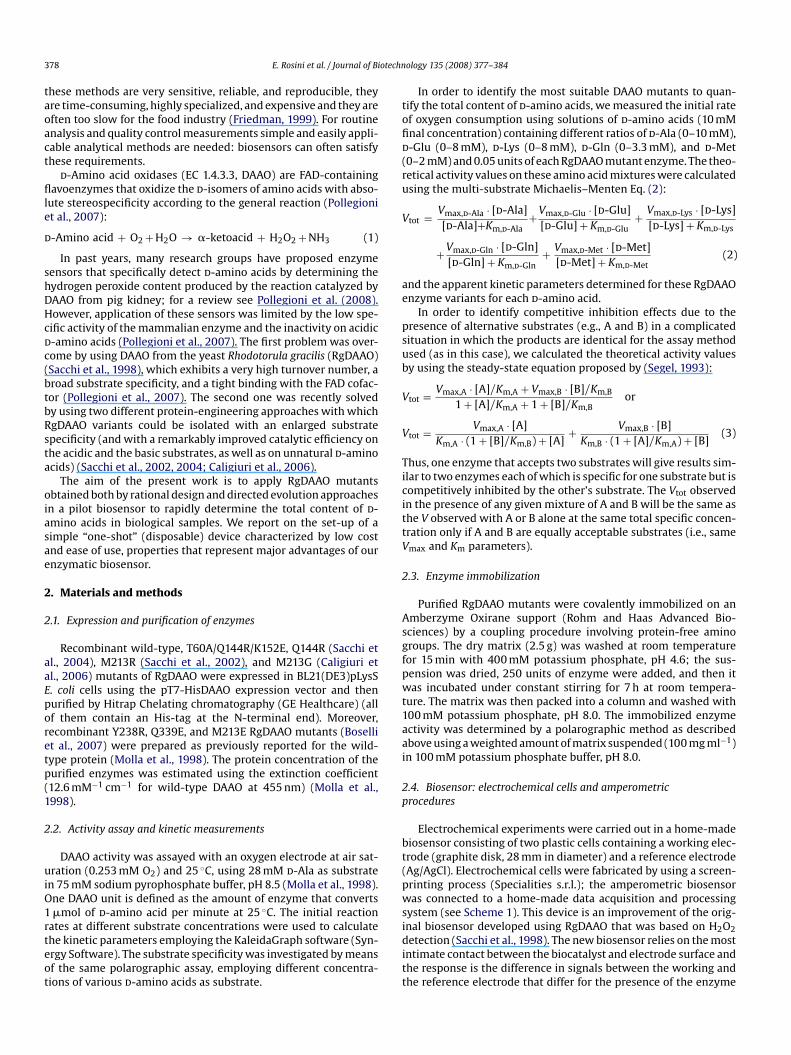
Electrochemical experiments were carried out in a home-madebiosensor consisting of two plastic cells containing a working elec-trode (graphite disk, 28 mm in diameter) and a reference electrode(Ag/AgCl). Electrochemical cells were fabricated by using a screen-printing process (Specialities s.r.l.); the amperometric biosensorwas connected to a home-made data acquisition and processingsystem (see Scheme 1). This device is an improvement of the orig-inal biosensor developed using RgDAAO that was based on H2O2detection (Sacchi et al., 1998). The new biosensor relies on the mostintimate contact between the biocatalyst and electrode surface andthe response is the difference in signals between the working andthe reference electrode that differ for the presence of the enzyme
E. Rosini et al. / Journal of Biotechn
Scheme 1. Schematic representation of the biosensor for amperometric determi-nation of d-amino acids. The output is the difference in signal between the workingcell (containing the enzyme) and the reference cell.
in the previous. To determine the optimal electrode potential wemeasured the signal given by 1 mM d-Ala at increasing appliedpotentials (from 150 to 800 mV). A value of 250 mV was chosen asthe measuring potential since the optimal amplitude of the signalwas coupled to a low operating potential that reduced interference(in particular that from the electrochemically active species presentin many food matrices).
Amperometric measurements were carried out in the electro-chemical cells containing 3 ml of d-amino acid solution in 100 mMpotassium phosphate buffer, pH 7.0, at room temperature. Then, asmall amount of different immobilized RgDAAO variants (0.5–4.0units) was added in the working cell and the current was recorded.A calibration curve was obtained using a standard solution of d-Ala(in the 0.25–5 mM final concentration range). To detect d-aminoacids from complex samples (such as cheese samples, see below),RgDAAO was added when the difference in signal between theworking and the reference electrode reached a stable value, i.e.after few minutes from sample addition. The effect of the sub-
strate composition on the amperometric response was evaluatedusing different immobilized RgDAAO variants and d-amino acidsolutions (0.5 mM final concentration) containing different ratiosof d-Ala (0–0.5 mM), d-Glu (0–0.4 mM), d-Lys (0–0.4 mM), d-Gln(0–0.16 mM), and d-Met (0–0.1 mM).Data are expressed as mean ± standard deviation (S.D.) exceptfor the mean values on different d-amino acid mixtures, where weused mean ± S.E. of the mean (S.E.M.). Statistical tests were per-formed using Kaleidagraph (Synergy Software).
2.5. Biosensor: test on real samples
Finely grated Grana Padano cheese samples were suspendedin 100 mM potassium phosphate buffer, pH 7.0 at a final con-centration of 0.1 g ml−1. The resulting suspension was sonicatedfor 30 min and centrifuged at 3200 × g for 30 min. The aqueousphase was newly centrifuged at 27,000 × g for 1 h and the result-ing supernatant was used for the biosensor analyses. For sake ofcomparison, the amount of d-amino acids in cheese samples wasalso determined spectrophotometrically following their enzymatic
ology 135 (2008) 377–384 379
conversion into the corresponding �-keto acids and reaction with2,4-dinitrophenylhydrazine (Caldinelli et al., 2006).
3. Results
3.1. Selection of RgDAAO variants active on all d-amino acids
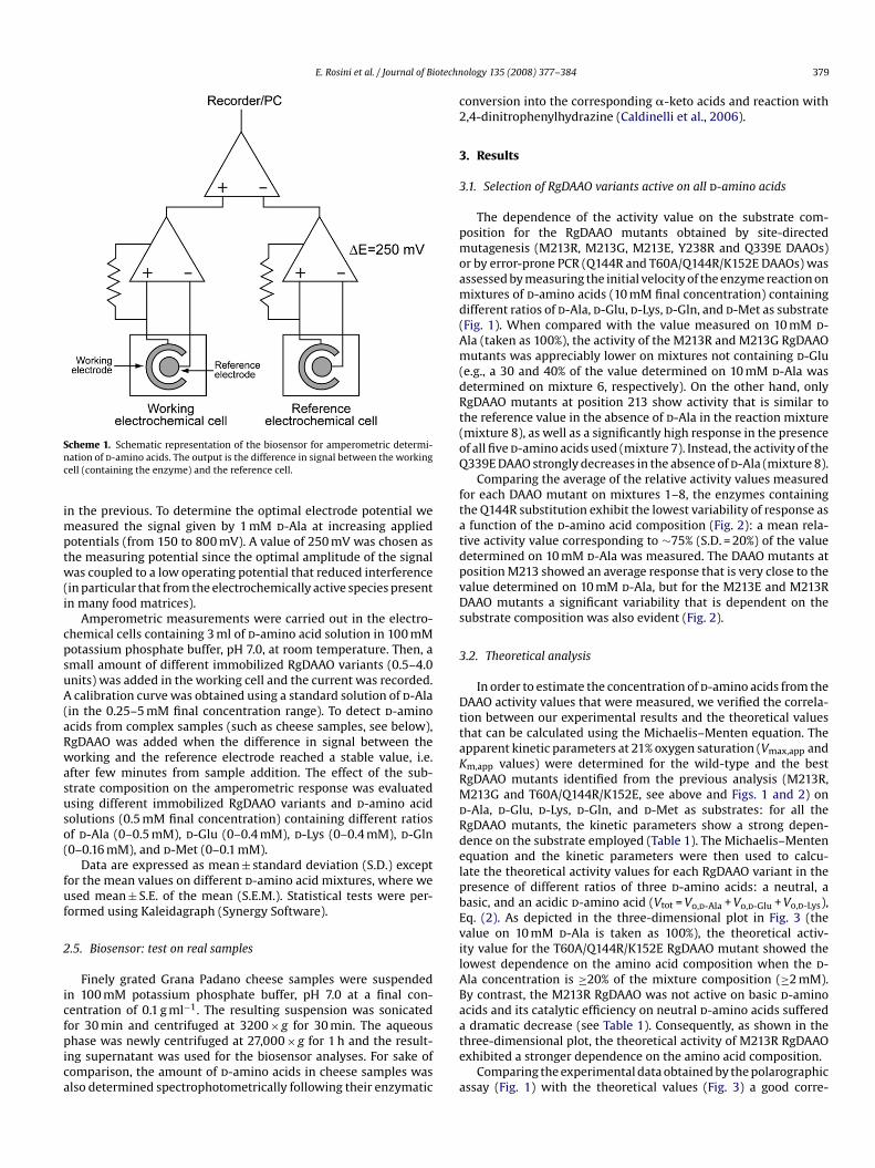
The dependence of the activity value on the substrate com-position for the RgDAAO mutants obtained by site-directedmutagenesis (M213R, M213G, M213E, Y238R and Q339E DAAOs)or by error-prone PCR (Q144R and T60A/Q144R/K152E DAAOs) wasassessed by measuring the initial velocity of the enzyme reaction onmixtures of d-amino acids (10 mM final concentration) containingdifferent ratios of d-Ala, d-Glu, d-Lys, d-Gln, and d-Met as substrate(Fig. 1). When compared with the value measured on 10 mM d-Ala (taken as 100%), the activity of the M213R and M213G RgDAAOmutants was appreciably lower on mixtures not containing d-Glu(e.g., a 30 and 40% of the value determined on 10 mM d-Ala wasdetermined on mixture 6, respectively). On the other hand, onlyRgDAAO mutants at position 213 show activity that is similar tothe reference value in the absence of d-Ala in the reaction mixture(mixture 8), as well as a significantly high response in the presenceof all fived-amino acids used (mixture 7). Instead, the activity of theQ339E DAAO strongly decreases in the absence ofd-Ala (mixture 8).
Comparing the average of the relative activity values measuredfor each DAAO mutant on mixtures 1–8, the enzymes containingthe Q144R substitution exhibit the lowest variability of response asa function of the d-amino acid composition (Fig. 2): a mean rela-tive activity value corresponding to ∼75% (S.D. = 20%) of the valuedetermined on 10 mM d-Ala was measured. The DAAO mutants atposition M213 showed an average response that is very close to thevalue determined on 10 mM d-Ala, but for the M213E and M213RDAAO mutants a significant variability that is dependent on thesubstrate composition was also evident (Fig. 2).
3.2. Theoretical analysis
In order to estimate the concentration ofd-amino acids from theDAAO activity values that were measured, we verified the correla-tion between our experimental results and the theoretical valuesthat can be calculated using the Michaelis–Menten equation. Theapparent kinetic parameters at 21% oxygen saturation (Vmax,app andKm,app values) were determined for the wild-type and the best
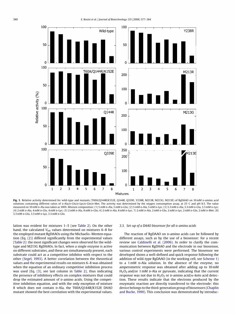
RgDAAO mutants identified from the previous analysis (M213R,M213G and T60A/Q144R/K152E, see above and Figs. 1 and 2) ond-Ala, d-Glu, d-Lys, d-Gln, and d-Met as substrates: for all theRgDAAO mutants, the kinetic parameters show a strong depen-dence on the substrate employed (Table 1). The Michaelis–Mentenequation and the kinetic parameters were then used to calcu-late the theoretical activity values for each RgDAAO variant in thepresence of different ratios of three d-amino acids: a neutral, abasic, and an acidic d-amino acid (Vtot = Vo,d-Ala + Vo,d-Glu + Vo,d-Lys),Eq. (2). As depicted in the three-dimensional plot in Fig. 3 (thevalue on 10 mM d-Ala is taken as 100%), the theoretical activ-ity value for the T60A/Q144R/K152E RgDAAO mutant showed thelowest dependence on the amino acid composition when the d-Ala concentration is ≥20% of the mixture composition (≥2 mM).By contrast, the M213R RgDAAO was not active on basic d-aminoacids and its catalytic efficiency on neutral d-amino acids suffereda dramatic decrease (see Table 1). Consequently, as shown in thethree-dimensional plot, the theoretical activity of M213R RgDAAOexhibited a stronger dependence on the amino acid composition.Comparing the experimental data obtained by the polarographicassay (Fig. 1) with the theoretical values (Fig. 3) a good corre-
380 E. Rosini et al. / Journal of Biotechnology 135 (2008) 377–384
2E, Q1ity wa
Fig. 1. Relative activity determined for wild-type and mutants (T60A/Q144R/K15solutions containing different ratios of d-Ala/d-Glu/d-Lys/d-Gln/d-Met. The activ
measured on 10 mM d-Ala was taken as 100%. Mixture composition: (1) 5 mM d-Ala, 5 mM(4) 2 mM d-Ala, 4 mM d-Glu, 4 mM d-Lys; (5) 2 mM d-Ala, 8 mM d-Glu; 6) 2 mM d-Ala, 83.3 mM d-Glu, 3.3 mM d-Lys, 3.3 mM d-Gln.lation was evident for mixtures 1–5 (see Table 2). On the otherhand, the calculated Vtot values determined on mixtures 6–8 forthe employed mutant RgDAAOs using the Michaelis–Menten equa-tion (Eq. (2)) differed significantly from the experimental values(Table 2): the most significant changes were observed for the wild-type and M213G RgDAAOs. In fact, when a single enzyme is activeon different substrates, and these are simultaneously present, eachsubstrate could act as a competitive inhibitor with respect to theother (Segel, 1993). A better correlation between the theoreticalvalues and the experimental results on mixtures 6–8 was obtainedwhen the equation of an ordinary competitive inhibition processwas used (Eq. (3), see last column in Table 2), thus indicatingthe presence of inhibitory effects on complex mixtures that coulddrop the estimated amount of d-amino acids. Using the compet-itive inhibition equation, and with the only exception of mixture8 which does not contain d-Ala, the T60A/Q144R/K152E DAAOmutant showed the best correlation with the experimental values.
44R, Q339E, Y238R, M213R, M213G, M213E) of RgDAAO on 10 mM d-amino acids determined by the oxygen consumption assay, at 25 ◦C and pH 8.5. The value
d-Glu; (2) 5 mM d-Ala, 5 mM d-Lys; (3) 3.3 mM d-Ala, 3.3 mM d-Glu, 3.3 mM d-Lys;mM d-Lys; 7) 2 mM d-Ala, 2 mM d-Glu, 2 mM d-Lys, 2 mM d-Gln, 2 mM d-Met; (8)
3.3. Set-up of a DAAO biosensor for all d-amino acids
The reaction of RgDAAO on d-amino acids can be followed bydifferent assays, such as by the use of a biosensor: for a recentreview see Caldinelli et al. (2006). In order to clarify the com-munication between RgDAAO and the electrode in our biosensor,various control experiments were performed. The biosensor wedeveloped shows a well-defined and quick response following theaddition of wild-type RgDAAO (in the working cell, see Scheme 1)to a 1 mM d-Ala solution. In the absence of the enzyme, noamperometric response was obtained after adding up to 10 mMH2O2 and/or 1 mM d-Ala or pyruvate, indicating that the currentresponse was not due to H2O2 or d-amino acid/�-keto acid detec-tion. These results indicate that the electrons produced by theenzymatic reaction are directly transferred to the electrode: thisdevice belongs to the third-generation group of biosensors (Chaplinand Bucke, 1990). This conclusion was demonstrated by introduc-

E. Rosini et al. / Journal of Biotechn
Fig. 2. Average of the activity values determined for wild-type and mutants ofRgDAAO on the d-amino acid solutions used in Fig. 1. Bars indicate mean ± S.E.M.(n = 24) for the determination on the eight different d-amino acids mixtures as inFig. 1.
Fig. 3. Three-dimensional plots showing the theoretical dependence of the activity valuecomposition of a 10 mM d-amino acid solution made up of d-Ala, d-Glu and d-Lys. The consubtracting the d-Ala and d-Glu values from the final concentration (not shown).
ology 135 (2008) 377–384 381
ing the enzyme in the working cell separately from the solution bya dialysis membrane, which allows to small molecules (such as thesubstrate/product) to freely diffuse but prevents a direct contactbetween the electrode and the enzyme. In this case, and althoughthe spectrophotometric analytical method ford-amino acids detec-tion showed that >80% of substrate (1 mM d-Ala) was converted
after 10 min, no amperometric signal was recorded.Concerning the set up of the biosensor, a higher response wasobserved in a static solution than under stirring using the free formof the enzyme, with a coefficient of variation (CV = S.D./mean, n = 6)corresponding to 28%. Analogously, the enzyme immobilized onthe Amberzyme Oxirane support also showed a higher ampero-metric response in still cells compared to that in a stirred solution.At increasing speed of stirring a decrease of the amperometricresponse was observed (at 300 rpm only 10% of the value mea-sured under static conditions is recorded) in concert to an increaseof signal noise. We propose that it is due to the lower amount ofenzyme that can efficiently contact the electrode surface for directelectron transfer. Importantly, a comparatively higher response wasrecorded with the immobilized RgDAAO than when using the freeenzyme form (up to 1.5-fold), which was also coupled with goodrepeatability (CV ≤ 15%, n = 6). A further main advantage to useRgDAAO in the immobilized form is given by the decrease of appar-ent Km values because of partitioning effects. This effect is moreevident for d-amino acids that are not good substrates for DAAO,e.g., Km for d-Pro is 21.5 and 5.6 mM and Km for d-Val is 18.9 and1.3 mM for free and immobilized RgDAAO, respectively (Pilone et al.,
determined for wild-type, M213R, M213G and T60A/Q144R/K152E RgDAAOs on thecentration of this latter component in the assay solution is the value obtained after

382 E. Rosini et al. / Journal of Biotechn
Tab
le1
Subs
trat
esp
ecifi
city
ofd
iffe
ren
tR
gDA
AO
s
d-A
lad-G
lud-L
ysd-G
lnd-M
et
Vm
ax,a
pp
(U/m
g)K
m,a
pp
(mM
)V
max
,ap
p/
Km
,ap
p
Vm
ax,a
pp
(U/m
g)K
m,a
pp
(mM
)V
max
,ap
p/
Km
,ap
p
Vm
ax,a
pp
(U/m
g)K
m,a
pp
(mM
)V
max
,ap
p/
Km
,ap
p
Vm
ax,a
pp
(U/m
g)K
m,a
pp
(mM
)V
max
,ap
p/
Km
,ap
p
Vm
ax,a
pp
(U/m
g)K
m,a
pp
(mM
)V
max
,ap
p/
Km
,ap
p
Wil
d-t
ype
98±
5.0
0.9
±0.
110
81.
7±
0.1
76±
140.
0216
±0.
514
.6±
1.4
1.1
82±
0.9
5.4
±0.
215
162
±6.
00.
6±
0.09
270
M21
3R16
.6±
0.7
18.5
±2.
00.
920
±0.
421
.1±
1.2
1.0
b.d
.26
±0.
45.
4±
0.3
4.8
24.7
±0.
31.
2±
0.06
21M
213G
19.2
±0.
34.
9±
0.5
2.3
2.0
±0.
045.
1±
0.5
0.4
6.9
±0.
38.
0±
1.0
0.9
67.7
±2.
72.
5±
0.4
2770
.8±
2.6
0.8
±0.
189
T60A
/Q14
4R/
K15
2E92
±2.
10.
6±
0.06
153
2.4
±0.
210
1±
140.
0221
.4±
0.5
16.6
±1.
01.
393
±1.
86.
0±
0.4
1610
0±
1.0
0.3
±0.
0133
5
The
app
aren
tki
net
icp
aram
eter
sw
ere
det
erm
ined
for
wil
d-t
ype,
M21
3R,M
213G
,an
dT6
0A/Q
144R
/K15
2ER
gDA
AO
mu
tan
tson
fived-a
min
oac
ids
assu
bstr
ate
usi
ng
the
oxyg
enas
say
(at
25◦ C
,pH
8.5)
.b.d
.=be
low
det
ecti
on.
ology 135 (2008) 377–384
1992). Importantly, this effect also confers a lower dependence ofthe enzymatic activity on substrate composition. Altogether, theseresults suggest that the use of RgDAAO in the immobilized formand in a static cell represents the best set-up for the biosensor.
Under optimized working conditions, we calibrated the biosen-sor containing 2.5 units of immobilized wild-type RgDAAO,measuring the amperometric response following the addition ofd-Ala standard solutions (0.25–5.0 mM range). The current val-ues detected after 10 min were plotted as a function of substrateconcentration and fitted by means of a classic Michaelis–Mentenequation (not shown). The detection limit is 0.25 mM d-Ala (sig-nal/noise = 3). The best RgDAAO mutants, as identified by the kineticmeasurements on different mixtures of amino acids (see above),were then used in the immobilized form in the biosensor to mea-sure 0.5 mM solutions of d-amino acids containing different ratiosofd-Ala,d-Glu,d-Lys,d-Gln, andd-Met. The highest response, com-pared to the one on 0.5 mM d-Ala taken as 100%, was measured forthe T60A/Q144R/K152E and M213G RgDAAO mutants (Fig. 4). This
and the low dependence of the response on the substrate com-position (S.E.M. = 5% and 9% for M213G and T60A/Q144R/K152E,respectively) identify the latter immobilized RgDAAOs as the bestchoice for such an application.3.4. d-Amino acid measurements on real samples
d-Amino acids can act as indicators of both microbial contami-nation and ripening processes (see Section 1). The performance ofour biosensor as a means of monitoring the entire d-amino acidcontent in foods, such as Grana Padano cheese, was assessed dur-ing this study. An increase in signal response proportional to theamount of sample is observed when testing such cheese samples(Fig. 5). By means of the calibration curve an average content of6.1 ± 0.5 mMd-amino acids was determined, corresponding to ∼6.7d-amino acid per cheese sample (mg g−1). Interestingly, this valueis in good agreement with the amount estimated by the alter-native 2,4-dinitrophenylhydrazine method (6.0 ± 0.3 mg g−1) andwith data in literature based on chromatographic determinations(Marchelli et al., 2006). In order to verify the specificity of the assay,
Fig. 4. Average of the amperometric response of the enzymatic biosensors producedusing immobilized wild-type and mutants of RgDAAO obtained on eight different0.5 mM mixtures of d-amino acids (these solutions were prepared by 20-fold dilu-tion of the ones reported in Fig. 1 and Table 2). Data are expressed as mean ± S.E.M.(n = 24).
E. Rosini et al. / Journal of Biotechnology 135 (2008) 377–384 383
Table 2Comparison of the experimental and theoretical activity values of wild-type, M213R, M213G, and T60A/Q144R/K152E mutants of RgDAAO on differentd-amino acids mixtures
Mixture Enzyme Experimental value Theoretical value
Michaelis–Menten equation competitive inhibition equation
(U/mg protein) (U/mg protein) (%) (U/mg protein) (%)
1 Wild-type 83.5 83.2 100 82.2 95M213R 6.9 7.4 107 6.1 88M213G 10.2 10.7 105 7.2 71T60A/Q144R/K152E 68.6 82.3 120 81.7 119
2 Wild-type 76.3 87.1 114 79.5 104M213R 4.2 3.5 83 3.5 83M213G 8.3 12.4 149 9.0 108T60A/Q144R/K152E 70.9 87.1 123 80.2 113
3 Wild-type 62.0 80.0 129 73.5 118M213R 4.8 5.2 108 4.6 96M213G 8.4 10.5 125 6.3 75T60A/Q144R/K152E 67.2 81.5 121 75.8 113
4 Wild-type 56.8 71.1 125 62.6 110M213R 4.0 4.8 120 4.3 108M213G 5.7 8.7 153 4.8 84T60A/Q144R/K152E 63.6 75.0 118 67.6 106
5 Wild-type 59.6 67.7 114 65.5 110M213R 6.0 7.1 118 6.3 105M213G 8.0 6.8 85 3.7 46T60A/Q144R/K152E 66.3 71.0
6 Wild-type 52.3 73.3M213R 2.0 1.6M213G 4.3 9.0T60A/Q144R/K152E 52.8 77.7
7 Wild-type 61.8 216.3M213R 13.9 25.8M213G 15.3 82.1T60A/Q144R/K152E 66.5 183.3
8 Wild-type 18.4 34.1M213R 10.3 12.6M213G 11.8 43.3T60A/Q144R/K152E 20.8 36.6
10 mM d-Ala Wild-type 93.8 89.9M213R 6.8 5.8M213G 10.8 12.9T60A/Q144R/K152E 78.4 86.8
The activity values were measured on mixtures 1–8 (see legend of Fig. 1) and on 10-mM d(Eq. (2)) and the competitive inhibition equation in a mixture of alternative substrates (Eq
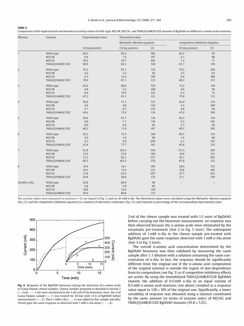
Fig. 5. Response of the RgDAAO biosensor during the detection of d-amino acidsin Grana Padano cheese samples. Cheese samples prepared as detailed in Section 2(—, 2 ml; - - -, 1 ml) were introduced in the 3 ml-cell of the biosensor. Inset: the 2 mlGrana Padano sample (—, 1) was treated for 20 min with 1.5 U of RgDAAO beforemeasurement (–·–, 2). Then 1 mM d-Ala (. . ., 3) was added to this sample and after10 min gave the same response as observed with 1 mM d-Ala alone (– – –, 4).
107 69.5 105
140 60.1 11580 1.6 80
209 6.1 142147 65.8 125
350 111.5 180186 16.8 121537 45.1 295276 87.8 132
185 28.6 155122 10.8 105367 27.7 235176 31.1 150
96 – –85
120111
-Ala. The theoretical values were calculated using the Michaelis–Menten equation. (3)) and reported as percentage of the corresponding experimental value.
2 ml of the cheese sample was treated with 1.5 units of RgDAAObefore carrying out the biosensor measurement: no response wasthen observed because the d-amino acids were eliminated by theenzymatic pre-treatment (line 2 in Fig. 5 inset). The subsequentaddition of 1 mM d-Ala to the cheese sample pre-treated withRgDAAO gave the same response observed with 1 mM d-Ala alone(line 3 in Fig. 5 inset).
The overall d-amino acid concentration determined by theRgDAAO biosensor was then validated by measuring the samesample after 1:1 dilution with a solution containing the same con-centration of d-Ala. In fact, the response should be significantlydifferent from the original one if the d-amino acid compositionof the original solution is outside the region of non-dependencefrom its composition (see Fig. 3) or if competitive inhibitory effectsare active. By using the immobilized T60A/Q144R/K152E RgDAAOmutant, the addition of 0.5 mM d-Ala to an equal volume of0.5 mM d-amino acid mixtures (see above) resulted in a responsevalue equal to 128 ± 10% of the original one. Significantly, a lowervariability of response was obtained using a mixture constitutedby the same amount (in terms of enzyme units) of M213G andT60A/Q144R/K152E RgDAAO mutants (91.8 ± 7.2%).

otechn
384 E. Rosini et al. / Journal of Bi4. Discussion
The presence of d-amino acids in foods is promoted by harshtechnological processes (e.g., high temperature or extreme pH val-ues), or can be the consequence of adulteration of food or arisefrom bacterial cell walls. For this reason, quality control is becom-ing more and more important both for the industry (as a cost factor)and for consumer protection. Therefore, if d-Ala can be consideredan indicator of milk contamination (Payan et al., 1985) the presenceof other d-amino acids can be specifically related to food process-ing (e.g., yoghurt contains d-Asp and d-Glu in addition to d-Ala)(Marchelli et al., 1997). On the other hand, the presence of all d-amino acids can be the consequence of adulteration as well as oftechnological food processing.
For routine food analysis and quality control simple and eas-ily applicable analytical methods are needed: biosensors can oftensatisfy these requirements. Up to now, practically all the DAAO-based biosensors reported use the enzyme from pig kidney andare therefore characterized by low stability, low turnover number,and inactivity on acidic d-amino acids. A further major draw-back of these systems is the different sensitivity to differentd-amino acids as substrates (Pollegioni et al., 2007), which impliesthat it is not possible to determine the total d-amino acid con-tent. As an example, a pig kidney DAAO-based screen-printedamperometric biosensor showed a response varying from 55%to 166% (with d-Arg and d-Val, respectively) of the value deter-mined on d-Phe and was fully inactive on d-Pro (Sarkar et al.,1999).
The biosensor based on a third-generation electrode andRgDAAO mutants responds to all (neutral, acidic and basic)d-aminoacids. The M213G and the T60A/Q144R/K152E variants (better ifused simultaneously) represent the best RgDAAO mutants for thisapplication. These enzyme mutants show the lowest variabilityof response as a function of the d-amino acid composition, inparticular when the d-Ala concentration is ≥20% of the mixturecomposition and when the enzyme is used in the immobilizedform. In addition, the T60A/Q144R/K152E RgDAAO exhibits the
best correlation between theoretical activity values on variousd-amino acid mixtures and the experimental ones. Due to the addi-tive nature of the individual amino acid responses this device canbe used to analyze solutions containing both single amino acidspecies and complexd-amino acid mixtures. The protocol for deter-mining d-amino acids based on RgDAAO mutants is simple, rapid(∼10–15 min), and reliable. The easy, fast, and inexpensive produc-tion procedure (the overall cost is ∼0.5 D /electrode) only requiresa single screen-printed electrode for each amperometric analysis(“one-shot” disposable device), with no need to regenerate the elec-trode surface. Because of the high specific activity of RgDAAO, lessenzyme is needed (2.5 units = 20 �g) than used in previous biosen-sors based on mammalian DAAO (≥30 �g) (Váradi et al., 1999). TheRgDAAO-based device was successfully used to monitor the totald-amino acid content in a cheese sample: the amount of d-aminoacids determined in Grana Padano cheese samples is in fairly goodagreement with the value reported in the literature (Marchelli etal., 2006). Interestingly, such a final d-amino acid concentrationis constituted by 17% d-Ala, 29% d-Asp, and 54% d-Glu, indicatingthat the RgDAAO-based biosensor is a powerful analytical tool evenon complex matrices. In conclusion, this electrochemical methodology 135 (2008) 377–384
compares favorably with standard methods (gas chromatography,HPLC or CE techniques) for overall d-amino acid determination.
Acknowledgments
This work was supported by grants from FAR to LP and GM andfrom Fondazione Cariplo to LP.
References
Boselli, A., Piubelli, L., Molla, G., Pilone, M.S., Pollegioni, L., Sacchi, S., 2007. Investi-gating the role of active site residues of Rhodotorula gracilisd-amino acid oxidaseon its substrate specificity. Biochimie 89, 360–368.
Caldinelli, L., Motteran, L., Sacchi, S., Piubelli, L., Boselli, A., Mothet, J.P., Pollegioni,L., Pilone, M.S., 2006. Detection of d-amino acids by d-amino acid oxidase. In:Konno, R., Brückner, H., D’Aniello, A., Fisher, G., Fujii, N., Homma, H. (Eds.), d-Amino Acids: A New Frontier in Amino Acid and Protein Research. Nova SciencePublishers, pp. 135–148 (Chapter 2.16).
Caligiuri, A., D’Arrigo, P., Rosini, E., Tessaro, D., Molla, G., Servi, S., Pollegioni, L., 2006.Enzymatic conversion of unnatural amino acids by yeast d-amino acid oxidase.Adv. Synth. Catal. 348, 2183–2190.
Chaplin, M., Bucke, C., 1990. Enzyme Technology. Cambridge University Press.Friedman, M., 1999. Chemistry, nutrition, and microbiology ofd-amino acids. J. Agric.
Food Chem. 47, 3457–3479.Gandolfi, I., Palla, G., Delprato, I., De Nisco, F., Marchelli, R., Salvatori, C., 1992. d-
Amino acids in milk as related to heat treatments and bacterial activity. J. FoodSci. 57, 377–379.
Marchelli, R., Palla, G., Dossena, A., Galaverna, G., Corradini, R., Clementi, S., 1997. d-Ammino acidi: Marker molecolari di stagionatura e di tipicità per il Parmigiano-Reggiano e il Grana Padano. Scienza Tecnica Lattiero-Casearia 48, 21–32.
Marchelli, R., Galaverna, G., Dossena, A., Palla, G., Bobbio, A., Santaguida, S., Grozeva,K., Corradini, R., Sforza, S., 2006. d-Amino acids in food. In: Konno, R., Brück-ner, H., D’Aniello, A., Fisher, G., Fujii, N., Homma, H. (Eds.), d-Amino Acids: ANew Frontier in Amino Acid and Protein Research. Nova Science Publishers, pp.299–315 (Chapter 4.2).
Molla, G., Vegezzi, C., Pilone, M.S., Pollegioni, L., 1998. Overexpression in Escherichiacoli of a recombinant chimeric Rhodotorula gracilis d-amino acid oxidase. ProteinExp. Purif. 14, 289–294.
Payan, I.L., Cadilla-Perezrios, R., Fischer, G.H., Man, E.H., 1985. Analysis of prob-lems encountered in the determination of amino acid enantiomeric ratios bygas chromatography. Anal. Biochem. 149, 484–491.
Pilone, M.S., Pollegioni, L., Butò, S., 1992. Stability and kinetic properties of immobi-lized Rhodotorula gracilis d-amino acid oxidase. Biotechnol. Appl. Biochem. 16,252–262.
Pollegioni, L., Piubelli, L., Sacchi, S., Pilone, M.S., Molla, G., 2007. Physiological func-
tions of d-amino acid oxidases: from yeast to humans. Cell. Mol. Life Sci. 64,1373–1394.Pollegioni, L., Molla, G., Sacchi, S., Rosini, E., Verga, R., Pilone, M.S., 2008. Prop-erties and applications of microbial d-amino acid oxidases: current state andperspectives. Appl. Microbiol. Biotechnol. 78, 1–16.
Sacchi, S., Pollegioni, L., Pilone, M.S., Rossetti, C., 1998. Determination of d-aminoacids using a d-amino acid oxidase biosensor with spectrophotometric andpotentiometric detection. Biotechnol. Tech. 12, 149–153.
Sacchi, S., Lorenzi, S., Molla, G., Pilone, M.S., Rossetti, C., Pollegioni, L., 2002. Engi-neering the substrate specificity of d-amino-acid oxidase. J. Biol. Chem. 277,27510–27516.
Sacchi, S., Rosini, E., Molla, G., Pilone, M.S., Pollegioni, L., 2004. Modulating d-aminoacid oxidase substrate specificity: production of an enzyme for analytical deter-mination of all d-amino acids by directed evolution. Protein Eng. Des. Sel. 17,517–525.
Sarkar, P., Tothill, I.E., Setford, S.J., Turner, A.P.F., 1999. Screen-printed amperometricbiosensors for the rapid measurement of l- and d-amino acids. Analyst 124,865–870.
Segel, I.H., 1993. Enzyme Kinetics Behavior and Analysis of Rapid Equilibrium andSteady-State Enzyme Systems. Wiley Classics Library Edition.
Váradi, M., Adányi, N., Szabó, E.E., Trummer, N., 1999. Determination of the ratio ofd- and l-amino acids in brewing by an immobilised amino acid oxidase enzymereactor coupled to amperometric detection. Biosens. Bioelectron. 14, 335–340.
Warnke, M., Armstrong, D.W., 2006.d-Amino acid determination in foods, beverages,and biological samples. In: Konno, R., Brückner, H., D’Aniello, A., Fisher, G., Fujii,N., Homma, H. (Eds.), d-Amino Acids: A New Frontier in Amino Acid and ProteinResearch. Nova Science Publishers, pp. 317–336 (Chapter 4.3).
Related Documents