1 A Basic Approach to Common Upper Extremity Mononeuropathies and Brachial Plexopathies Paul E. Barkhaus, MD Aiesha Ahmed, MD Kerry H. Levin, MD Zachary Simmons, MD AANEM 60th Annual Meeting San Antonio, Texas Copyright © October 2013 American Association of Neuromuscular & Electrodiagnostic Medicine 2621 Superior Drive NW Rochester, MN 55901 Printed by Johnson Printing Company, Inc.

Welcome message from author
This document is posted to help you gain knowledge. Please leave a comment to let me know what you think about it! Share it to your friends and learn new things together.
Transcript

11
A Basic Approach to Common Upper Extremity Mononeuropathies and Brachial Plexopathies
Paul E. Barkhaus, MDAiesha Ahmed, MDKerry H. Levin, MD
Zachary Simmons, MD
AANEM 60th Annual MeetingSan Antonio, Texas
Copyright © October 2013American Association of Neuromuscular
& Electrodiagnostic Medicine2621 Superior Drive NW
Rochester, MN 55901
Printed by Johnson Printing Company, Inc.

22
Please be aware that some of the medical devices or pharmaceuticals discussed in this handout may not be cleared by the FDA or cleared by the FDA for

33
Table of Contents
Faculty 5
The Median Nerve: A Schematic Approach With Clinical Neurophsyiological Pitfalls and Myths 7Paul E. Barkhaus, MD
Ulnar Nerve 17Aiesha Ahmed, MD
The Radial Nerve 23Kerry H. Levin, MD
Brachial Plexopathies: The Basics 27Zachary Simmons, MD
CME Questions 33
Authors/faculty have nothing to disclose
Chair: Zachary Simmons, MD
A Basic Approach to Common Upper Extremity Mononeuropathies and Brachial Plexopathies

Objectives - Participants will acquire skills to (1) Identify and distinguish the anatomical and clinical features of upper extremity mononeuropathies and brachial plexopathies, (2) design and perform EDX studies for assessment of median, ulnar, and radial neuropathies and brachial plexopathies, and
Target Audience:Neurologists, physical medicine and rehabilitation and other physicians interested in neuromuscular and electrodiagnostic medicineHealth care professionals involved in the diagnosis and management of patients with neuromuscular diseasesResearchers who are actively involved in the neuromuscular and/or electrodiagnostic research
Accreditation Statement - The AANEM is accredited by the Accreditation Council for Continuing Medical Education (ACCME) to provide continuing
CME Credit - AMA PRA Category 1 Credits™AMA PRA Category 1 Credits™
CEUs Credit -
Objectives
David B. Shuster, MDDayton, OH
Zachary Simmons, MDHershey, PA
Jeffrey A. Strommen, MDRochester, MN
T. Darrell Thomas, MDKnoxville, TN
Vincent Tranchitella, MD, ChairYork, PA
Thomas Bohr, MD, FRCPCLoma Linda, CA
Jasvinder P. Chawla, MBBS, MD, MBAAtlanta, GA
Robert W. Irwin, MDMiami, FL
Shawn Jorgensen, MDQueensbury, NY
A. Atruro Leis, MDJackson, MS
Maxim Moradian, MDNew Orleans, LA
2012-2013 Program Committee
2012-2013 AANEM PresidentPeter A. Grant, MD
Medford, OR

55
Paul E. Barkhaus, MDProfessor of Neurology and Physical Medicine & Rehabilitation, Medical College of Wisconsin
as well as a fellowship in clinical neuromuscular diseases at the
He is also head of the Neuromuscular Disease and Autonomic Section in the Department of Neurology and director of the ALS
motor nerve conductions, motor unit estimation as well as multi-
Aiesha Ahmed, MDProgram Director, Clinical Neurophysiology and Neuromuscular Medicine FellowshipsAssistant Professor of NeurologyPennsylvania State University Hershey Medical CenterHershey, PA
College in Karachi, Pakistan, and an internship in internal
and a clinical neurophysiology fellowship and neuromuscular
Ahmed is the program director of Clinical Neurophysiology and Neuromuscular medicine fellowships and an assistant professor
Kerry H. Levin, MDChairman, Department of NeurologyDirector, Neuromuscular CenterCleveland ClinicCleveland, OH
Zachary Simmons, MDProfessor of NeurologyDirector, Neuromuscular Program and EMG LaboratoryPennsylvania State University Hershey Medical CenterHershey, PA
Iowa and in neuromuscular diseases and electromyography at the
include studies of quality of life, the development of evidence-based practice protocols, the use of brain-computer interfaces,
Association of Neuromuscular and Electrodiagnostic Medicine
co-chair of the Program Committee and serves on the AANEM
Faculty
A Basic Approach to Common Upper Extremity Mononeuropathies and Brachial Plexopathies

66
7
A BASIC APPROACH TO COMMON UPPER EXTREMITY MONONEUROPATHIES AND BRACHIAL PLEXOPATHIES
7
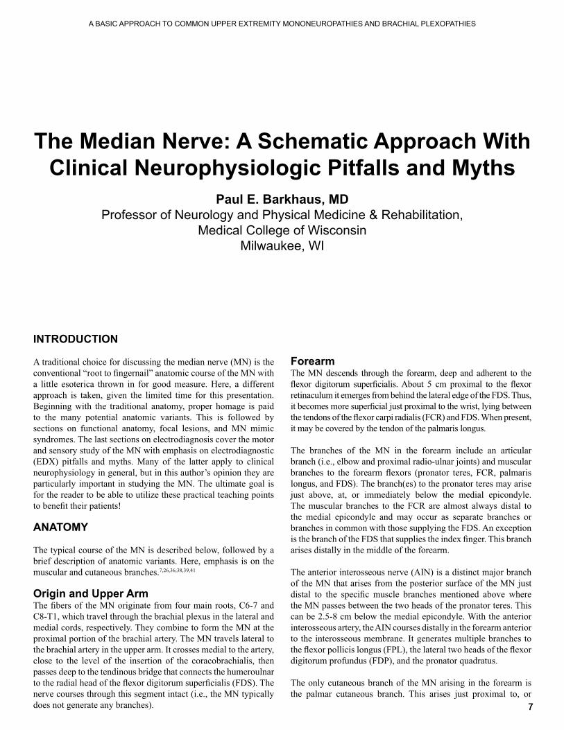
The Median Nerve: A Schematic Approach With Clinical Neurophysiologic Pitfalls and Myths
Paul E. Barkhaus, MDProfessor of Neurology and Physical Medicine & Rehabilitation,
Medical College of Wisconsin
INTRODUCTION
A traditional choice for discussing the median nerve (MN) is the
sections on functional anatomy, focal lesions, and MN mimic
and sensory study of the MN with emphasis on electrodiagnostic
ANATOMY
The typical course of the MN is described below, followed by a
Origin and Upper Arm
close to the level of the insertion of the coracobrachialis, then passes deep to the tendinous bridge that connects the humeroulnar
ForearmThe MN descends through the forearm, deep and adherent to the
The branches of the MN in the forearm include an articular
The muscular branches to the FCR are almost always distal to the medial epicondyle and may occur as separate branches or
interosseous artery, the AIN courses distally in the forearm anterior
The only cutaneous branch of the MN arising in the forearm is

anatomic point that allows differentiation of MN lesions distal
Wrist and HandThe MN then courses lateral to the tendons of the FDS in the
The MN terminates distal to the carpal tunnel in one main
typically lateral, underlying the palmar aponeurosis, and usually
terminal sensory branches are highly variable in number and
Anatomic VariantsIt is important to appreciate when reading about anatomy or anatomic variants whether the studies are anatomic, physiologic,
In the upper arm, communicating branches between the
from the musculocutaneous nerve to the MN have included
the more common proximal communications in the brachial
In the forearm, the MN may be penetrated by the ulnar artery
heads of the pronator teres rather than passing between its
Regarding the forearm communicating branch between the ),
foreseen the legacy they would create among EDX physicians when they made their original anatomic observations in
They could not tell whether these communications were
they contain, which are predominantly, if not exclusively,
arises distally in the middle of the forearm may sometimes
The thenar muscular branch may occasionally arise more
The thenar muscular branch may have a terminal branch that
between the deep branch of the ulnar nerve and the distal
There may be congenital absence of the intrinsic hand
alone, or may also involve the FPL, and be either unilateral
Figure 1. Varying patterns of Martin–Gruber anastomosis.
THE MEDIAN NERVE: A SCHEMATIC APPROACH WITH CLINICAL NEUROPHYSIOLOGIC PITFALLS AND MYTHS
9
A BASIC APPROACH TO COMMON UPPER EXTREMITY MONONEUROPATHIES AND BRACHIAL PLEXOPATHIES
9
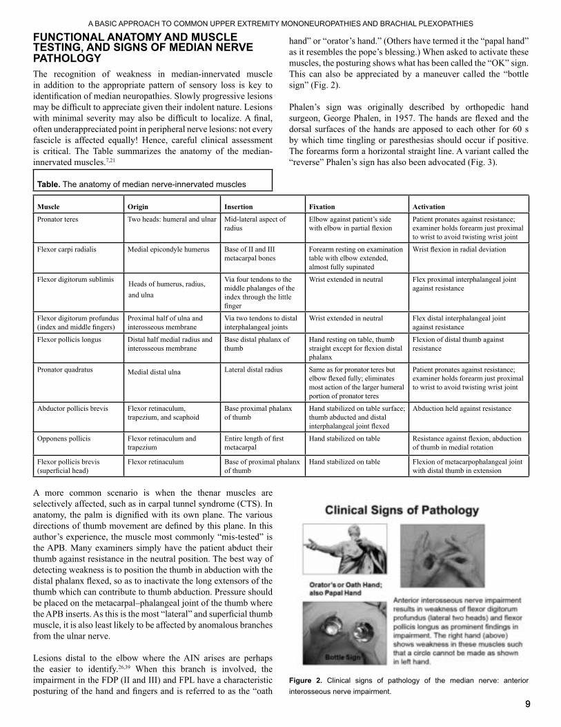
FUNCTIONAL ANATOMY AND MUSCLE TESTING, AND SIGNS OF MEDIAN NERVE PATHOLOGYThe recognition of weakness in median-innervated muscle in addition to the appropriate pattern of sensory loss is key to
often underappreciated point in peripheral nerve lesions: not every
Table.
Muscle Origin Insertion Fixation Activation
Pronator teres Two heads: humeral and ulnar Mid-lateral aspect of radius
Patient pronates against resistance;
Flexor carpi radialis Medial epicondyle humerusmetacarpal bones
Forearm resting on examination table with elbow extended, almost fully supinated
Flexor digitorum sublimis Heads of humerus, radius, and ulna
Via four tendons to the middle phalanges of the index through the little
against resistance
Flexor digitorum profundus Proximal half of ulna and interosseous membrane
Via two tendons to distal against resistance
Flexor pollicis longus Distal half medial radius and interosseous membrane thumb
Hand resting on table, thumb
phalanx
Flexion of distal thumb against resistance
Pronator quadratus Medial distal ulna Lateral distal radius Same as for pronator teres but
most action of the larger humeral portion of pronator teres
Patient pronates against resistance;
Abductor pollicis brevis Flexor retinaculum, of thumb thumb abducted and distal
Abduction held against resistance
Flexor retinaculum and metacarpal of thumb in medial rotation
Flexor pollicis brevis Flexor retinaculumof thumb with distal thumb in extension
A more common scenario is when the thenar muscles are
detecting weakness is to position the thumb in abduction with the
muscle, it is also least likely to be affected by anomalous branches
Lesions distal to the elbow where the AIN arises are perhaps
impairment in the FDP (II and III) and FPL have a characteristic
This can also be appreciated by a maneuver called the “bottle
Figure 2. Clinical signs of pathology of the median nerve: anterior interosseous nerve impairment.

1010
volar wrist at approximately the level of the distal wrist crease, then distally through the carpal tunnel, one may elicit paresthesias by
author prefers to initially percuss the lateral and medial wrists for
should be asked if the sensation is somewhat similar to when they
COMMON AND UNCOMMON FOCAL MEDIAN NERVE LESIONS
A number of other rare causes of median entrapment and
A band of muscle originating between the subscapularis and latissimus dorsi muscles can compress the MN as it courses
Proximal median neuropathy of the upper arm : This is rare and usually due to a focal lesion such as a Schwannoma or
may be pseudo-aneurysm after angiography where the
Ligament of Struthers : The medial supracondylar ridge
about 1-2% of the population there may be a spur or spicule
fairly prominent and give rise to a ligamentous process that
This canal may contain the medial branch of the brachial
of entrapment resulting in pain and dysfunction of some or
Pronator teres syndrome : This is another rare syndrome resulting from compression of the MN as it goes deep at the antecubital fossa between the two heads of the
Anterior interosseous syndrome : Aside from trauma,
of the upper arm may also show this pattern of pure motor
that it may be selectively involved as part of the syndrome
CTS is a focal distal median neuropathy and undoubtedly It arises from
compression of the distal MN where it travels under the
is beyond the scope of this review and the reader is referred
In addition, other structural abnormalities can produce CTS, such as aberrant muscle tissue in the carpal tunnel, leprosy,
Figure 3. Clinical signs of pathology of the median nerve: the Tinel and Phalen signs.
Figure 4.
THE MEDIAN NERVE: A SCHEMATIC APPROACH WITH CLINICAL NEUROPHYSIOLOGIC PITFALLS AND MYTHS
11
A BASIC APPROACH TO COMMON UPPER EXTREMITY MONONEUROPATHIES AND BRACHIAL PLEXOPATHIES
median neuropathy at the wrist due to chronic compression so as not to bias the referring physician from looking into
The advent of ultrasound has given an 11
Acute CTS is far less common and may represent a
seen a case of bilateral acute CTS secondary to an individual who was electrocuted while holding a metal ladder that
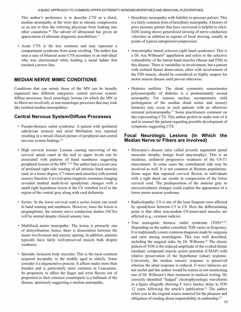
MEDIAN NERVE MIMIC CONDITIONS
Conditions that can mimic those of the MN can be broadly
diffuse processes, focal neurologic lesions (in which the MN or
Central Nervous System/Diffuse Processes
Pseudo-thoracic outlet syndrome: A patient with ipsilateral
resulting in a mixed clinical picture of peripheral and central
High cervical lesions: Lesions causing narrowing of the cervical spinal canal in the mid to upper levels can be associated with patterns of hand numbness suggesting
The author had a recent case of profound right side wasting of all intrinsic hand muscles
revealed marked multi-level spondylotic changes with a
Syrinx: In the lower cervical cord a syrinx lesion can result
preganglionic, the sensory nerve conduction studies (NCSs)
Multifocal motor neuropathy: The lesion is primarily one of demyelination; hence, there is dissociation between the
typically have fairly well-preserved muscle bulk despite
Sporadic inclusion body myositis: This is the most common
proportion to their extensor counterparts is a hallmark of the
Hereditary neuropathy with liability to pressure palsies: This
velocities in addition to regions of focal slowing, usually at
Amyotrophic lateral sclerosis (split hand syndrome): This is appellation and refers to the selective
vulnerability of the lateral hand muscles (thenar and FDI) in
with isolated thenar denervation, often with involvement of the FDI muscle, should be considered as highly suspect for
Diabetes mellitus: The distal symmetric sensorimotor polyneuropathy of diabetes is a predominantly axonal
prolongation of the median distal motor and sensory latencies may occur in such patients with an otherwise
Some practitioners comment on
and to counsel the patient regarding possible development of
Focal Neurologic Lesions (In Which the Median Nerve or Fibers are Involved)
muscular atrophy, benign focal amyotrophy): This is an
with a tight dural sac results in compression of the lower
microcirculatory changes could explain the appearance of a
:
It is traditionally a more common diagnosis made by surgeons
The classic
(median) compound muscle action potential (CMAP) with
Conversely, the median sensory response is preserved
not useful and this author would be remiss in not mentioning
The author refers you to the original source material for the pleasure and

1212
considered the culprit, advanced imaging has shown that any of the three components of the thoracic outlet region may be
13 A recent case seen by the
pain in the shoulder, rapidly followed by weakness in the
have been described, such as when the AIN is selectively
Non-Neurologic Processes
supracondylar fracture of the humerus where the brachial
Arthritis: Severe arthritis may result in thenar atrophy because of lack of movement of the thumb with consequent
of the distal extensor tendons or a positive Finkelstein
is usually an occupational syndrome resulting in secondary
ELECTRODIAGNOSTIC EVALUATION OF THE MEDIAN NERVE8,14,23
The median motor and sensory NCSs are among the most commonly performed (but not necessarily performed well) in
some investigators also record from the lateral lumbrical muscle
To ensure quality in the laboratory, the following steps should be observed: (1) ensure the area to be studied is adequately warmed; (2) when starting to stimulate, look at the muscle being studied for a response (twitch) to the stimulus; and (3) move the active recording electrode (E1) to ensure a location yielding the highest amplitude,
Figure 5. Electrodiagnostic testing of the median nerve: motor conduction montage.
The most common median sensory NCSs performed are
Although the latter has some motor contribution
The protocol is to make sure that the segment of sensory nerve to be studied is warmed, that the montage is correct with respect to E1
of the stimulator is moved so as to reduce stimulation artifact
Figure 6. Electrodiagnostic testing of the median nerve: sensory conduction montage.
THE MEDIAN NERVE: A SCHEMATIC APPROACH WITH CLINICAL NEUROPHYSIOLOGIC PITFALLS AND MYTHS
13
A BASIC APPROACH TO COMMON UPPER EXTREMITY MONONEUROPATHIES AND BRACHIAL PLEXOPATHIES
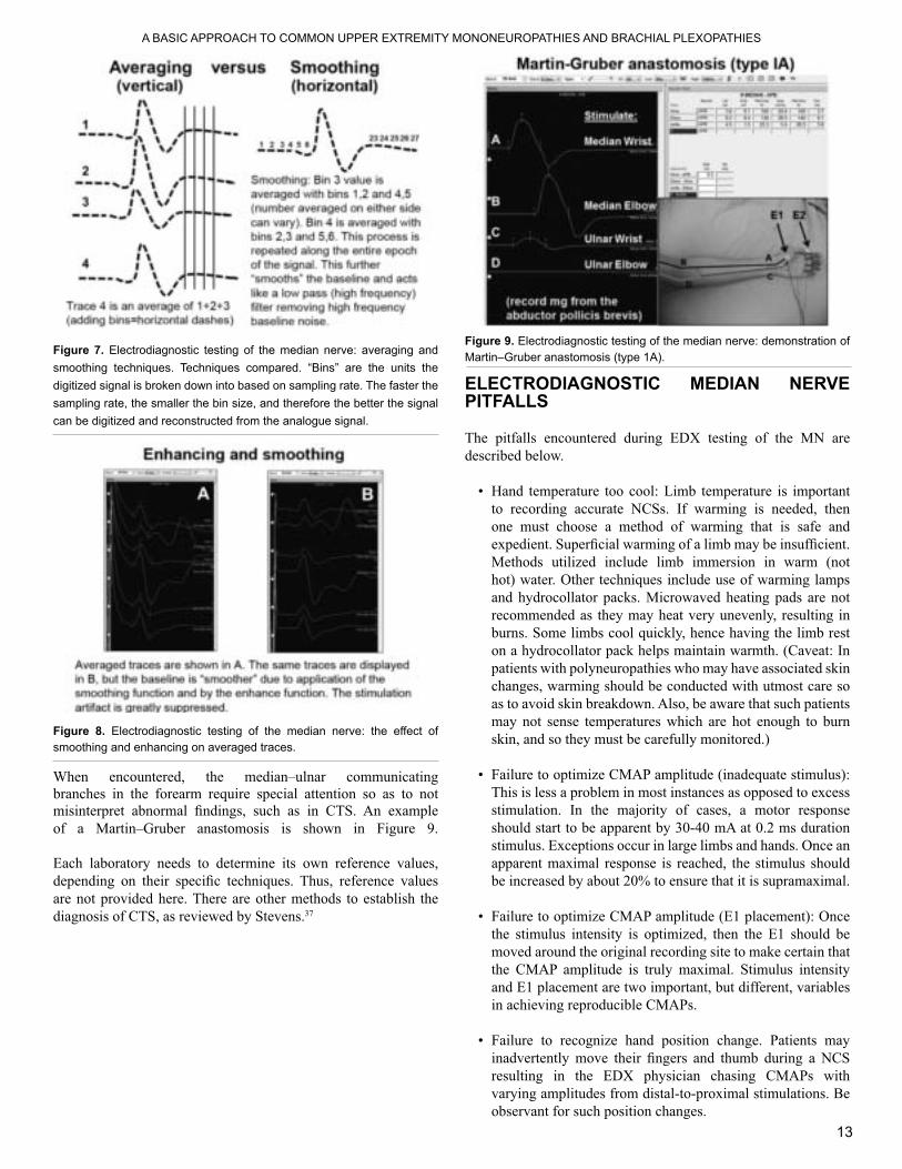
Figure 7. Electrodiagnostic testing of the median nerve: averaging and
sampling rate, the smaller the bin size, and therefore the better the signal can be digitized and reconstructed from the analogue signal.
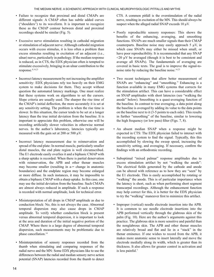
Figure 8. Electrodiagnostic testing of the median nerve: the effect of smoothing and enhancing on averaged traces.
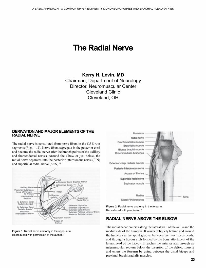
branches in the forearm require special attention so as to not
Each laboratory needs to determine its own reference values,
ELECTRODIAGNOSTIC MEDIAN NERVE PITFALLS
The pitfalls encountered during EDX testing of the MN are
Hand temperature too cool: Limb temperature is important
one must choose a method of warming that is safe and
recommended as they may heat very unevenly, resulting in
patients with polyneuropathies who may have associated skin changes, warming should be conducted with utmost care so
may not sense temperatures which are hot enough to burn
This is less a problem in most instances as opposed to excess
apparent maximal response is reached, the stimulus should
moved around the original recording site to make certain that
and E1 placement are two important, but different, variables
resulting in the EDX physician chasing CMAPs with
Figure 9. Electrodiagnostic testing of the median nerve: demonstration of
different signals: A CMAP often has subtle added curves
these as the CMAP contour between distal and proximal
Excessive nerve stimulation resulting in cathodal migration
occurs with excess stimulus, it is less often a problem than
is reduced, as in CTS, the EDX physician often is tempted to stimulate excessively, bringing in an ulnar contribution to the
important to appreciate this problem, otherwise one will be
Incorrect latency measurement due to reinnervation and spread of the end plate: In normal muscle, particularly smaller
The E1 electrode easily overlies it and a biphasic CMAP with
may become smaller (resulting in a-= change in anatomic boundaries) and the endplate region may become enlarged
Misinterpretation of all drops in CMAP amplitude as due to
temporal dispersion may also cause drops in CMAP
versus abnormal temporal dispersion, it is important to look at the area and duration of the initial negative portion of the
dispersion, such measurements may be problematic due to
Misinterpretation of sensory responses recorded from the thumb when stimulating and comparing responses of the radial nerve and the MN: Some EDX physicians like to use the differences between the radial and median sensory nerve action potential (SNAP) latencies recorded from the thumb to detect
Poorly reproducible sensory responses: This shows the
which case SNAPs may either be missed when small, or
Two recent techniques that allow better measurement of
on SNAP amplitudes with short latencies (see “walking the
the baseline is averaged by adding its value to the data points
An absent median SNAP when a response might be expected in CTS: The EDX physician failed to interact with
accomplished by slowing the sweep speed, increasing the
the latency is short, such as when performing short segment
may help correct for this, it is better for the EDX physician
It is common to see needle electrode insertions into the
makes more anatomic sense to insert laterally and move the electrode medially along its width, which is greater than its
THE MEDIAN NERVE: A SCHEMATIC APPROACH WITH CLINICAL NEUROPHYSIOLOGIC PITFALLS AND MYTHS

15
A BASIC APPROACH TO COMMON UPPER EXTREMITY MONONEUROPATHIES AND BRACHIAL PLEXOPATHIES
ELECTRODIAGNOSTIC MEDIAN NERVE MYTHS
A number of myths regarding the EDX assessment of the MN are
Paul Hoffmann, simultaneously described the same sign
The E2 varies in its contribution to the CMAP depending on
the result of volume conduction from other (non-median)
that the surface response is accurate, especially if no visible
Slowed distal median motor and sensory response in a
Diabetic patients with distal symmetric polyneuropathy often
further from the recording electrodes, the amplitude drops more precipitously than the analogous CMAPs recorded at
A slowed forearm median motor conduction velocity in an
polyneuropathy) means there is compression proximal to
in CTS may show mild slowing for at least two different
to-severe, then it is likely that the faster conducting motor axons have been compromised to a greater degree than the
are axonal changes proximal to the point of compression that
The basic concept is that in a lower cervical radiculopathy, there is
whether one should do a needle electrode examination in
favor of doing a needle electrode examination would be the ability to ascertain the presence of motor axonal involvement
Figure 10. Proper needle electrode examination of the abductor pollicis

16
REFERENCES
study to reveal current expert views on mechanisms underlying dual nerve
THE MEDIAN NERVE: A SCHEMATIC APPROACH WITH CLINICAL NEUROPHYSIOLOGIC PITFALLS AND MYTHS

17
A BASIC APPROACH TO COMMON UPPER EXTREMITY MONONEUROPATHIES AND BRACHIAL PLEXOPATHIES
INTRODUCTION
assessment of the ulnar nerve is important for physicians and technologists to accurately identify ulnar neuropathy as well as
ANATOMY
1 During its descent through the medial arm, the ulnar nerve does not give off any branches until it reaches the
formed by the medial epicondyle and olecranon process of the ulna bone, and it passes deep to the humeroulnar aponeurotic
2
branch provides sensation over the hypothenar area of the hand 2
canal, also known as the ulnar canal, is a small anatomical space
with the ulnar nerve through this space on their way into the
opponens digiti minimi, abductor digiti minimi [ADM], and
2
CLINICAL AND ELECTRODIAGNOSTIC FEATURES OF ULNAR NERVE DYSFUNCTION
Ulnar Neuropathy at the Elbow
common causes of ulnar neuropathy at the elbow are listed in 1
Typical symptoms include numbness and tingling in the distribution of the ulnar nerve along with elbow pain that radiates
3
3 An early sign may be inability to adduct the little
17
Ulnar Nerve
Aiesha Ahmed, MDProgram Director, Clinical Neurophysiology and Neuromuscular Medicine Fellowships
Assistant Professor of NeurologyPennsylvania State University Hershey Medical Center
Hershey, PA

In severe
Various provocative maneuvers have been described that may
manual pressure over the cubital tunnel has been reported to have
Table 1. Common causes of ulnar neuropathy1
At the elbow
Recent elbow trauma without fractureHabitual leaning on elbow
Diabetes mellitusHereditary neuropathy with liability to pressure palsiesRheumatoid arthritisIatrogenic
Malpositioning during surgeryNerve infarction during transposition
At the wrist
Frequent pressure on hypothenar eminence
laborers)HUA=humeroulnar aponeurotic arcade
Differential DiagnosisThe differential diagnosis of ulnar neuropathy at the elbow includes the following:
Lower trunk brachial plexopathy
Medial cord brachial plexopathy
Electrodiagnostic Testing
As with all nerve conduction studies (NCSs), limb temperatures should be maintained within the reference range (>32°C) and
Ulnar sensory study:is performed using ring electrodes to record from the little
1 The ulnar
1
electrode will be dependent on the normative values used in each
Figure 1.
Dorsal ulnar cutaneous sensory study:
1 The nerve is stimulated
1 Since this nerve arises proximal to the wrist, it may be involved in ulnar neuropathies at the elbow,
Figure 2. Dorsal ulnar cutaneous sensory study.
Ulnar motor study:
has been shown to be more sensitive than testing with the elbow
The ulnar motor NCS can be recorded from either the ADM
1 The ulnar nerve is then stimulated at up to four sites: (1)
ULNAR NERVE

19
A BASIC APPROACH TO COMMON UPPER EXTREMITY MONONEUROPATHIES AND BRACHIAL PLEXOPATHIES
19
Axilla (A): which lies in the proximal axilla, medial to the biceps 1 The measurement across the
Figure 3. Ulnar motor nerve conduction study, recording from the abductor digiti minimi muscle, stimulating at the wrist.
Figure 4. Ulnar motor conduction study, recording from the abductor digiti minimi muscle, stimulating below the elbow.
In certain cases, inching across the elbow can be performed to The setup is identical to
1 The most convincing abnormality would be a change in latency and/or
Figure 5. Ulnar motor nerve conduction study, recording from the abductor digiti minimi muscle, stimulating above the elbow.
Figure 6.muscle.
Evaluation of the ulnar nerve with NCSs should include the
while recording from the FDI muscle; (2) ulnar motor NCS using inching techniques across the elbow segment; (3) mixed-nerve
ulnar cutaneous sensory responses between the affected and
cutaneous sensory response between affected and asymptomatic
cases the lesion is at the elbow; however, lesions at the wrist or more proximal locations (brachial plexus or root) should be

2020
Table 2. Electrodiagnostic evaluation of ulnar neuropathy at the elbow1
A. Nerve conduction studies (Figs. 1-6)
and above elbow sites, while recording from the abductor digiti minimi muscle
sites while recording from the abductor pollicis brevis muscle
recording from the thumbB. Needle electromyography
carpi ulnaris)
additional needle examination should include:
indicis proprius)
Management
The treatment of patients with ulnar neuropathy at the elbow
management should include avoidance of pressure on the elbow
3
Ulnar Neuropathy at the Wrist
Entrapment of the ulnar nerve at the wrist is rare relative to
Patients may present with sensory and/or motor involvement
sensory loss, paresthesias, or pain in the region supplied by the
limited to the muscles of the hand with sparing of the proximal
weakness with atrophy or fasciculations of the intrinsic hand
Table 3. Clinical syndromes produced by ulnar nerve compression within the canal of Guyon1,9
A. Combined motor and sensory syndrome (type 1)A lesion at the proximal portion of the canal may involve both
hypothenar and dorsomedial surfaces of the hand should be
B. Pure sensory syndrome (type 2)Clinically, there is loss of sensation over the palmar surface of
C. Pure motor syndromesLesion affecting the deep palmar and hypothenar motor branches (type 3)This lesion affects the motor trunk proximal to the takeoff
This type of lesion spares the muscles of the hypothenar
Lesion affecting only the distal deep palmar motor branch
Differential Diagnosis
The differential diagnosis of ulnar neuropathy at the wrist includes the following:
Lower trunk brachial plexopathy
Medial cord brachial plexopathy
Early motor neuron disease1
Electrodiagnostic Testing
of ulnar to median nerve conduction across the wrist due to the fact that the second lumbrical muscle (median innervated)
ULNAR NERVE

21
A BASIC APPROACH TO COMMON UPPER EXTREMITY MONONEUROPATHIES AND BRACHIAL PLEXOPATHIES
21
stimulated supramaximally at their usual wrist locations using 1
Table 4. Electrodiagnostic evaluation of ulnar neuropathy at the wrist1
A. Nerve conduction studies (Figs. 1-6)
and above elbow sites, while recording from the abductor digiti minimi muscle
while recording from the dorsolateral hand
study
B. Needle electromyography
interosseous)
minimi)
additional needle examination should include:
indicis proprius)
Management
Magnetic resonance imaging (MRI) may be useful in detecting
11
Table 5. 10
A. Combined motor and sensory syndrome (type 1)
B. Pure sensory syndrome (type 2)
C. Pure motor syndromes
Lesion affecting the deep palmar and hypothenar motor branches (type 3)
Lesion affecting the deep palmar motor branch only
is decreased with prolonged distal latency when recording
and second lumbricals, and thenar muscles with sparing
Lesion affecting only the distal deep palmar motor
is decreased with prolonged distal latency when recording
adductor pollicis muscles with sparing of the hypothenar
EMG=electromyography
Figure 7. Lumbrical–interosseous motor study. Top: Stimulating the median nerve recording at the 2nd lumbrical muscle. Bottom: Stimulating the ulnar nerve recording at the interosseous muscle.

2222
REFERENCES
American Association of Neuromuscular & Electrodiagnostic
ULNAR NERVE
23
A BASIC APPROACH TO COMMON UPPER EXTREMITY MONONEUROPATHIES AND BRACHIAL PLEXOPATHIES
23
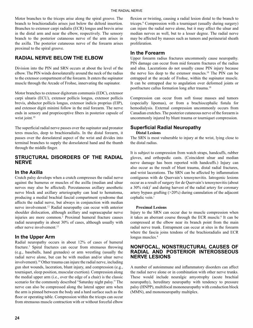
DERIVATION AND MAJOR ELEMENTS OF THE RADIAL NERVE
and become the radial nerve after the branch points of the axillary
radial nerve separates into the posterior interosseous nerve (PIN)
Figure 1. Radial nerve anatomy in the upper arm.Reproduced with permission of the author.10
RADIAL NERVE ABOVE THE ELBOW
The radial nerve courses along the lateral wall of the axilla and the
the humerus in the spiral groove, between the two triceps heads,
intermuscular septum below the insertion of the deltoid muscle and enters the forearm by going between the distal biceps and
The Radial Nerve
Kerry H. Levin, MDChairman, Department of Neurology
Director, Neuromuscular CenterCleveland ClinicCleveland, OH
Figure 2. Radial nerve anatomy in the forearm.Reproduced with permission.5
branch to the posterior cutaneous nerve of the arm arises in
RADIAL NERVE BELOW THE ELBOW
Division into the PIN and SRN occurs at about the level of the
Motor branches to extensor digitorum communis (EDC), extensor
brevis, abductor pollicis longus, extensor indicis proprius (EIP),
passes over the dorsolateral aspect of the wrist and divides into terminal branches to supply the dorsolateral hand and the thumb
STRUCTURAL DISORDERS OF THE RADIAL NERVE
In the AxillaCrutch palsy develops when a crutch compresses the radial nerve against the humerus or muscles of the axilla (median and ulnar
nerve block and axillary arteriography can lead to hematoma, producing a medial brachial fascial compartment syndrome that
12 Radial neuropathy can occur with anterior shoulder dislocation, although axillary and suprascapular nerve
2 Proximal humeral fracture causes
13
In the Upper ArmRadial neuropathy occurs in about 12% of cases of humeral
3 Spiral fractures can occur from strenuous throwing
radial nerve alone, but can be with median and/or ulnar nerve
nerve can also be compressed along the lateral upper arm when the arm is pinned between the body and a hard surface such as the
from strenuous muscle contraction with or without forceful elbow
Compression with a tourniquet (usually during surgery)
may be affected by masses such as tumors and perineurial sheath
In the Forearm
PIN damage can occur from mid forearm fractures of the radius
The PIN can be
Compression can occur from soft tissue masses and tumors
Distal Lesions
also occur as the result of blunt trauma, distal radial fractures,
and during harvest of the radial artery for coronary
1
Proximal Lesions
It can be compressed at the elbow near its branch point from the main
NONFOCAL, NONSTRUCTURAL CAUSES OF RADIAL AND POSTERIOR INTEROSSEOUS NERVE LESIONS
These would include neuralgic amyotrophy (acute brachial neuropathy), hereditary neuropathy with tendency to pressure palsy (HNPP), multifocal mononeuropathy with conduction block
THE RADIAL NERVE

25
A BASIC APPROACH TO COMMON UPPER EXTREMITY MONONEUROPATHIES AND BRACHIAL PLEXOPATHIES
25
CLINICAL FINDINGS IN RADIAL NEUROPATHIES
Lesions above the spiral groove produce main trunk radial neuropathy manifested by weakness of all radial-innervated muscles, including the triceps, as well as sensory loss in the SRN
11
leads to weakness limited to the extension of the index through
DIFFERENTIAL DIAGNOSIS
especially when there is prominent involvement of the triceps
PIN lesion, especially when there is prominent involvement of
be mistaken for a radial neuropathy, especially in the setting of shoulder pain that can mask the assessment of true weakness in the deltoid (involvement in the axillary nerve distribution) and
ELECTRODIAGNOSIS
Workup Nerve Conduction Studies
The radial motor nerve conduction study (NCS) is performed by recording over the EIP and/or EDC, stimulating at the forearm, elbow, below the spiral groove, above spiral groove, and axilla, with appropriate comparison studies on the opposite side
sensory NCS is performed by recording over the SRN at the snuffbox, stimulating above the wrist, with comparison on the
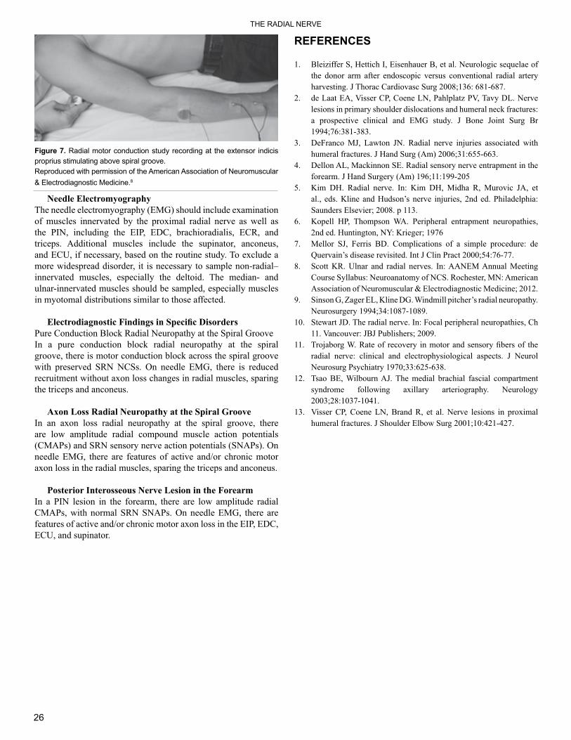
Figure 3. Radial sensory conduction study. The active recording electrode
can be palpated over the extensor pollicis longus tendon. The inactive or
placed over the dorsum of the wrist between the stimulating and recording electrodes. The nerve is stimulated along the dorsal edge of the radius, 10 cm from the active recording electrode, with the cathode distal to the anode.Reproduced with permission of the American Association of Neuromuscular & Electrodiagnostic Medicine.
Figure 4. Radial motor conduction study recording from the extensor indicis proprius and stimulating at the forearm. The recording electrodes are placed over the extensor indicis muscle with the active electrode over the belly of the muscle and the reference electrode at least 3 cm distal to the active electrode, over the tendon. The ground is placed between the recording electrodes and the stimulation site on the forearm. The nerve is stimulated distally over the lateral forearm, with the cathode distal to the anode. There
elbow, lateral to the biceps tendon, beneath the brachioradialis muscle. It can be stimulated more proximally over the lateral aspect of the arm at the
compound muscle action potential because of activation of other muscles innervated by the radial nerve.Reproduced with permission of the American Association of Neuromuscular & Electrodiagnostic Medicine.
Figure 5. Radial motor conduction study recording from the extensor indicis proprius stimulating at elbow.Reproduced with permission of the American Association of Neuromuscular & Electrodiagnostic Medicine.
Figure 6. Radial motor conduction study recording from the extensor indicis proprius stimulating below spiral groove.Reproduced with permission of the American Association of Neuromuscular & Electrodiagnostic Medicine.
26
Figure 7. Radial motor conduction study recording at the extensor indicis proprius stimulating above spiral groove.Reproduced with permission of the American Association of Neuromuscular & Electrodiagnostic Medicine.
Needle Electromyography
of muscles innervated by the proximal radial nerve as well as the PIN, including the EIP, EDC, brachioradialis, ECR, and
ulnar-innervated muscles should be sampled, especially muscles
In a pure conduction block radial neuropathy at the spiral groove, there is motor conduction block across the spiral groove
recruitment without axon loss changes in radial muscles, sparing
Axon Loss Radial Neuropathy at the Spiral GrooveIn an axon loss radial neuropathy at the spiral groove, there are low amplitude radial compound muscle action potentials
Posterior Interosseous Nerve Lesion in the ForearmIn a PIN lesion in the forearm, there are low amplitude radial
features of active and/or chronic motor axon loss in the EIP, EDC,
REFERENCES
the donor arm after endoscopic versus conventional radial artery
lesions in primary shoulder dislocations and humeral neck fractures:
THE RADIAL NERVE

27
A BASIC APPROACH TO COMMON UPPER EXTREMITY MONONEUROPATHIES AND BRACHIAL PLEXOPATHIES
27
INTRODUCTION
Patients commonly are referred to the neuromuscular specialist or electrodiagnostic (EDX) physician because of weakness, pain,
goal of this brief review is to provide information which will aid
ANATOMY OF THE BRACHIAL PLEXUS AND ITS MAJOR BRANCHES
muscles proximally and then behind the clavicle and pectoral muscles more distally as it courses from the neck into the shoulder
to all muscles of the upper extremities and shoulder girdle, the brachial plexus supplies upper extremity cutaneous sensation
Roots
ventral (motor) rootlets which then merge to form a short spinal
the roots of the brachial plexus, and they are located immediately
Figure 1. The brachial plexus: the components shown are the roots,
cases, all the nerve contributions to the brachial plexus are shifted
At times, the plexus may be expanded, with contributions from
Brachial Plexopathies: The Basics
Zachary Simmons, MDProfessor of Neurology
Director, Neuromuscular Program and EMG LaboratoryPennsylvania State University Hershey Medical Center
Hershey, PA

Figure 2.
EDX physicians should bear in mind that the cervical paraspinal muscles are innervated by the posterior primary rami, and therefore they can also be considered to have their innervation
Figure 3. Details of the anatomy at a cervical spinal cord level. The dorsal and ventral rootlets combine to form a spinal nerve, which then divides into anterior and posterior primary rami.
Trunks
off the upper trunk to provide innervation to the supraspinatus
notch of the scapula, an area covered by the transverse scapular ligament, and supplies motor branches to the supraspinatus
scapular spine (bounded by the scapula spine medially and the spinoglenoid ligament [inferior transverse scapular ligament]
(2) The nerve to the subclavius is a minor branch of the upper trunk which cannot be tested easily by physical examination or
DivisionsEach of the three trunks divides into an anterior and a posterior
Cords
divisions of the upper and middle trunks form the lateral cord, whereas the anterior division of the lower trunk continues as
BRACHIAL PLEXOPATHIES: THE BASICS

29
A BASIC APPROACH TO COMMON UPPER EXTREMITY MONONEUROPATHIES AND BRACHIAL PLEXOPATHIES
29
Branches of the Lateral Cord
brachialis muscles, and it gives rise to the lateral antebrachial cutaneous nerve (lateral cutaneous nerve of the forearm), which provides cutaneous sensation to the lateral forearm from wrist to
Branches of the Medial Cord
pectoralis minor muscle and the inferior portions of the pectoralis
cutaneous nerve of the arm) provides cutaneous sensation to the
cutaneous nerve (medial cutaneous nerve of the forearm) provides cutaneous sensation to the medial forearm between the wrist
forearm and hand muscles, and it provides cutaneous sensation
Branch of Both the Lateral and Medial Cords
Branches of the Posterior Cord
and innervates the lower portion of the subscapularis muscle
between the upper and lower subscapular nerves, derives from
supplies the teres minor muscle, then terminates by innervating
cutaneous sensation to the arm, forearm, and hand as the posterior cutaneous nerve of the arm, lower lateral cutaneous nerve of the
CAUSES OF BRACHIAL PLEXOPATHY
A listing of the most common causes of brachial plexopathy is
Table 1. Common causes of brachial plexopathy
TractionFall from a height, particularly onto shoulderTrauma in which the arm is pulled down, damaging the upper plexusTrauma in which the arm is pulled up, damaging the lower plexus
Motor vehicle accidents and other trauma
Surgery, particularly during median sternotomyCompression
Supraclavicular from pack strapsInfraclavicular from crutchesHematoma, aneurysm, and arteriovenous malformations
Ischemia
Radiation-inducedThoracic outlet syndrome
Iatrogenic
Traction
including a fall onto the shoulder from a height, traction to a limb
1 Traction
This most commonly occurs after chest surgeries due to stretch
or medial cord usually are involved, with the expected clinical
to traction on the neck by the clinician during passage in the 2 However, it now appears that some of these develop
prenatally or are due to propulsive forces over which the birth 1,3
plexus involvement are most common, although about 23% of
Neoplastic and Radiation-Induced Brachial PlexopathyRadiation-induced brachial plexopathy is most commonly a delayed syndrome, occurring from a few weeks to many years
The EDX physician is most often called upon to distinguish a radiation-
Radiation-induced plexopathy is less likely to be painful and
fasciculation potentials are more likely to be present in radiation-
3030
Tumors at
lower portion of the plexus, but metastases from other types of
Thoracic Outlet Syndrome
True neurogenic
usually are preferentially affected, resulting in greater atrophy of Sensory loss parallels that
Neuralgic Amyotrophy (Parsonage–Turner Syndrome)Also termed immune brachial plexus neuropathy, neuralgic amyotrophy is most commonly sporadic, although it may be
and then described in detail with respect 11 it is now well
12,13
Most commonly, the initial symptom is pain of abrupt onset,
generally begins to improve in 2-3 weeks, in association with
brachial plexus in a patchy fashion, for example affecting one or more trunks or single peripheral nerves, most commonly the long
and reports of this syndrome following a variety of conditions
reported, suggesting that a variety of events can trigger an EDX studies
may reveal a pattern of brachial plexus involvement not readily
CLINICAL PRESENTATIONS OF BRACHIAL PLEXOPATHY
Upper Trunk PlexopathyIn upper trunk plexopathy, weakness will be seen in muscles
rotation), deltoid (arm abduction), biceps, and brachioradialis
upper trunk and may be partially affected, such as the pronator
lateral upper arm in the distribution of the axillary nerve, in the
and radial sensory branches), and in the distribution of the lateral
Middle Trunk PlexopathyIsolated lesions of the middle trunk are rare, and they usually
cutaneous nerve of the forearm and in the hand over the middle
Lower Trunk Plexopathy
muscles such as the extensor indicis, extensor digitorum, and extensor carpi ulnaris, resulting in weakness of grip due to
abnormalities occur in the medial arm, medial forearm, medial
Lateral Cord Plexopathy
Posterior Cord Plexopathy
of the axillary nerve, posterior cutaneous nerve of the arm, and
Medial Cord Plexopathy
lower trunk lesion, except for preservation of radial-innervated
weakness of grip due to weakness of hand muscles, and to
same distribution as for lower trunk lesions: medial arm, medial
BRACHIAL PLEXOPATHIES: THE BASICS

31
A BASIC APPROACH TO COMMON UPPER EXTREMITY MONONEUROPATHIES AND BRACHIAL PLEXOPATHIES
31
Pan-Plexopathy
the entire upper extremity except for remaining function of
ELECTRODIAGNOSIS OF BRACHIAL PLEXOPATHY
brachial plexopathies generally are complex, involving the study
EDX evaluation of brachial plexopathies are provided in Tables
Upper Trunk PlexopathySensory studies will reveal abnormalities in the lateral antebrachial cutaneous, median sensory (particularly to the thumb), and radial
the median and ulnar motor nerves are normal, but studies of the suprascapular, axillary, and musculocutaneous nerves, if
to demonstrate abnormalities in the supraspinatus, infraspinatus,
the root level will be spared, including the cervical paraspinal,
Table 2. Guidelines for sensory nerve conduction studies in brachial plexopathy
Radiculopathy = lesion proximal to the dorsal root
because the sensory nerve is intact from the level of its cell body (the dorsal root ganglion) to the level of
because of axon loss from the level of the cell body to
Medial and lateral antebrachial cutaneous nerve conduction studies are particularly useful for distinguishing plexopathy
A sensory nerve action potential amplitude on the symptomatic side which is less than half of that on the asymptomatic side is considered to be abnormal, even if the absolute value of the amplitude falls within the normal
Table 3. Guidelines for the needle examination in brachial plexopathy
Presence or absence of axonal continuity often is of
Search carefully for voluntary motor unit action
Keep in mind those muscles which are innervated at the
Those muscles will be abnormal in some radiculopathies,
Anatomic variations occur, and studies may not be “black
Middle Trunk PlexopathyThe median sensory response is expected to be abnormal
innervated at the root level will be spared, such as the cervical
Lower Trunk PlexopathyIn a lower trunk plexopathy, several sensory studies will be abnormal, including the medial antebrachial cutaneous, the
abnormal, with the degree of abnormality being determined by
compound muscle action potential (CMAP) amplitudes, mildly to moderately prolonged distal latencies, and mildly to moderately
the radial-innervated muscles, the extensor indicis is a particularly
Lateral Cord PlexopathyIn a lateral cord plexopathy, abnormalities are expected in the lateral antebrachial cutaneous nerve and the median sensory
examination reveals abnormalities in the biceps brachii and
carpi radialis), with sparing of the more distal median-innervated
32
Posterior Cord Plexopathy
abnormal, with the degree of abnormality being determined by the
amplitudes, mildly to moderately prolonged distal latencies, and
abnormalities in all radial-innervated muscles and in the deltoid,
Medial Cord PlexopathyEDX testing in a medial cord plexopathy is expected to produce
expected on testing of the medial antebrachial cutaneous, the
abnormal, with the degree of abnormality being determined by the
amplitudes, mildly to moderately prolonged distal latencies,
and median nerves, including all ulnar-innervated muscles and
Pan-Plexopathy
Median, ulnar, and radial sensory responses are abnormal, as
studies may be abnormal, as may the suprascapular, axillary, and musculocutaneous studies, with the degree of abnormality being
reveal low CMAP amplitudes, mildly to moderately prolonged
abnormalities are expected in all muscles of the upper extremity and shoulder girdle except for those innervated directly at the root
REFERENCESBRACHIAL PLEXOPATHIES: THE BASICS

33
A BASIC APPROACH TO COMMON UPPER EXTREMITY MONONEUROPATHIES AND BRACHIAL PLEXOPATHIES
A Basic Approach to Common Upper Extremity Mononeuropathies and Brachial Plexopathies
CME Questions:
the following except:
lesion cannot be at the:
altered by all of the following except:
disruption of neural elements passing through all of the following except:
suggestive of an ulnar nerve entrapment at the elbow include all of the following except:
Pain at the elbow which radiates along the medial
Loss of sensation over the web space between the
compression at the wrist from a more proximal lesion would be:
chance of artifactual slowing of conduction velocity, include all of the following except:
Achieving supramaximal stimulation levels at the
Performing nerve conduction studies with the elbow
Performing an additional ulnar motor study recording
Performing a dorsal ulnar cutaneous sensory study (and
information would support the diagnosis of a focal lesion at
A decrease in compound muscle action potential
A change in compound muscle action potential (CMAP)
include all of the following except:Sensory impairment over the hypothenar area of the
Sensory impairment over the palmar little and medial
innervation to a territory usually served by which of the following nerves:
components of the brachial plexus are:
CME QUESTIONS
Related Documents