A bacterial genome in flux: the twelve linear and nine circular extrachromosomal DNAs in an infectious isolate of the Lyme disease spirochete Borrelia burgdorferi Sherwood Casjens, 1 * Nanette Palmer, 1 Rene ´ van Vugt, 1 Wai Mun Huang, 1 Brian Stevenson, 2 Patricia Rosa, 3 Raju Lathigra, 4 Granger Sutton, 5 Jeremy Peterson, 5 Robert J. Dodson, 5 Daniel Haft, 5 Erin Hickey, 5 Michelle Gwinn, 5 Owen White 5 and Claire M. Fraser 5 1 Division of Molecular Biology and Genetics, Department of Oncological Sciences, University of Utah Medical School, Salt Lake City, UT 84132, USA. 2 Department of Microbiology and Immunology, University of Kentucky College of Medicine, Lexington, KY 40536, USA. 3 Laboratory of Human Bacterial Pathogenesis, Rocky Mountain Laboratory, NIAID, NIH, Hamilton, MT 59840, USA. 4 MedImmune Inc., 35 West Watkins Mill Road, Gaithersburg, MD 20878, USA. 5 The Institute for Genomic Research, 9712 Medical Center Drive, Rockville, MD 20850, USA. Summary We have determined that Borrelia burgdorferi strain B31 MI carries 21 extrachromosomal DNA elements, the largest number known for any bacterium. Among these are 12 linear and nine circular plasmids, whose sequences total 610 694 bp. We report here the nucleotide sequence of three linear and seven circular plasmids (comprising 290 546 bp) in this infectious isolate. This completes the genome sequencing project for this organism; its genome size is 1 521 419 bp (plus about 2000 bp of undetermined telomeric sequences). Analysis of the sequence implies that there has been extensive and sometimes rather recent DNA rearrange- ment among a number of the linear plasmids. Many of these events appear to have been mediated by recom- binational processes that formed duplications. These many regions of similarity are reflected in the fact that most plasmid genes are members of one of the genome’s 161 paralogous gene families; 107 of these gene families, which vary in size from two to 41 members, contain at least one plasmid gene. These rearrangements appear to have contributed to a sur- prisingly large number of apparently non-functional pseudogenes, a very unusual feature for a prokaryotic genome. The presence of these damaged genes sug- gests that some of the plasmids may be in a period of rapid evolution. The sequence predicts 535 plasmid genes $300 bp in length that may be intact and 167 apparently mutationally damaged and/or unexpres- sed genes (pseudogenes). The large majority, over 90%, of genes on these plasmids have no convincing similarity to genes outside Borrelia, suggesting that they perform specialized functions. Introduction Spirochetes of the genus Borrelia are unique among bacteria in that they have linear chromosomes and carry a large number of linear and circular plasmids. Their linear chromosomes range from 900 to 920 kbp in length (Baril et al ., 1989; Ferdows and Barbour, 1989; Davidson et al ., 1992; Casjens and Huang, 1993; Ojaimi et al ., 1994; Casjens et al ., 1995; Fraser et al ., 1997) [the known range of bacterial chromosome sizes is 580–9300 kbp (Casjens, 1998)]. Barbour and co-workers originally found that Borrelia isolates carry multiple linear extrachromosomal elements (Plasterk et al ., 1985; Barbour and Garon, 1987; Barbour, 1988), and all natural isolates that have been examined since then have non-identical, but similar, complements of such DNAs (Simpson et al ., 1990a,b; Stalhammar-Carlemalm et al ., 1990; Hughes et al ., 1992; Sadziene et al ., 1993a; Samuels et al ., 1993; Busch et al ., 1995; Casjens et al ., 1995, 1997a; Xu and Johnson, 1995; Marconi et al ., 1996a). In the few cases that have been examined, the individual Borrelia extrachromosomal DNAs are present in approximately the same numbers of molecules per cell as the chromosome (Hinnebusch and Barbour, 1992; Kitten and Barbour, 1992; Casjens and Huang, 1993), although a small circular plasmid of isolate Ip90 appears to have a higher copy number (Dunn et al ., 1994). The linear DNAs have covalently closed hairpin telomeres (Barbour and Garon, 1987; Hinnebusch et al ., 1990; Hinnebusch and Barbour, 1991; Casjens et al ., 1997b; Fraser et al ., 1997). Most of the plasmids can be lost and are not required for propagation of the bacteria in culture, but loss of infectivity in mice often parallels plasmid Molecular Microbiology (2000) 35(3), 490–516 Q 2000 Blackwell Science Ltd Received 19 May, 1999; revised 27 September, 1999; accepted 4 October, 1999. *For correspondence. E-mail sherwood.casjens@hci. utah.edu; Tel. (1) 801 581 5980; Fax (1) 801 581 3607.

Welcome message from author
This document is posted to help you gain knowledge. Please leave a comment to let me know what you think about it! Share it to your friends and learn new things together.
Transcript

A bacterial genome in ¯ux: the twelve linear and ninecircular extrachromosomal DNAs in an infectious isolateof the Lyme disease spirochete Borrelia burgdorferi
Sherwood Casjens,1* Nanette Palmer,1
Rene van Vugt,1 Wai Mun Huang,1 Brian Stevenson,2
Patricia Rosa,3 Raju Lathigra,4 Granger Sutton,5
Jeremy Peterson,5 Robert J. Dodson,5 Daniel Haft,5
Erin Hickey,5 Michelle Gwinn,5 Owen White5 and
Claire M. Fraser5
1Division of Molecular Biology and Genetics,
Department of Oncological Sciences, University of Utah
Medical School, Salt Lake City, UT 84132, USA.2Department of Microbiology and Immunology, University
of Kentucky College of Medicine, Lexington, KY 40536,
USA.3Laboratory of Human Bacterial Pathogenesis,
Rocky Mountain Laboratory, NIAID, NIH, Hamilton, MT
59840, USA.4MedImmune Inc., 35 West Watkins Mill Road,
Gaithersburg, MD 20878, USA.5The Institute for Genomic Research, 9712 Medical
Center Drive, Rockville, MD 20850, USA.
Summary
We have determined that Borrelia burgdorferi strain
B31 MI carries 21 extrachromosomal DNA elements,
the largest number known for any bacterium. Among
these are 12 linear and nine circular plasmids,
whose sequences total 610 694 bp. We report here the
nucleotide sequence of three linear and seven circular
plasmids (comprising 290 546 bp) in this infectious
isolate. This completes the genome sequencing project
for this organism; its genome size is 1 521 419 bp (plus
about 2000 bp of undetermined telomeric sequences).
Analysis of the sequence implies that there has been
extensive and sometimes rather recent DNA rearrange-
ment among a number of the linear plasmids. Many of
these events appear to have been mediated by recom-
binational processes that formed duplications. These
many regions of similarity are re¯ected in the fact
that most plasmid genes are members of one of the
genome's 161 paralogous gene families; 107 of these
gene families, which vary in size from two to 41
members, contain at least one plasmid gene. These
rearrangements appear to have contributed to a sur-
prisingly large number of apparently non-functional
pseudogenes, a very unusual feature for a prokaryotic
genome. The presence of these damaged genes sug-
gests that some of the plasmids may be in a period of
rapid evolution. The sequence predicts 535 plasmid
genes $300 bp in length that may be intact and 167
apparently mutationally damaged and/or unexpres-
sed genes (pseudogenes). The large majority, over
90%, of genes on these plasmids have no convincing
similarity to genes outside Borrelia, suggesting that
they perform specialized functions.
Introduction
Spirochetes of the genus Borrelia are unique among
bacteria in that they have linear chromosomes and carry
a large number of linear and circular plasmids. Their linear
chromosomes range from 900 to 920 kbp in length (Baril
et al., 1989; Ferdows and Barbour, 1989; Davidson et al.,
1992; Casjens and Huang, 1993; Ojaimi et al., 1994;
Casjens et al., 1995; Fraser et al., 1997) [the known
range of bacterial chromosome sizes is 580±9300 kbp
(Casjens, 1998)]. Barbour and co-workers originally found
that Borrelia isolates carry multiple linear extrachromosomal
elements (Plasterk et al., 1985; Barbour and Garon, 1987;
Barbour, 1988), and all natural isolates that have been
examined since then have non-identical, but similar,
complements of such DNAs (Simpson et al., 1990a,b;
Stalhammar-Carlemalm et al., 1990; Hughes et al., 1992;
Sadziene et al., 1993a; Samuels et al., 1993; Busch et al.,
1995; Casjens et al., 1995, 1997a; Xu and Johnson,
1995; Marconi et al., 1996a). In the few cases that have
been examined, the individual Borrelia extrachromosomal
DNAs are present in approximately the same numbers of
molecules per cell as the chromosome (Hinnebusch and
Barbour, 1992; Kitten and Barbour, 1992; Casjens and
Huang, 1993), although a small circular plasmid of isolate
Ip90 appears to have a higher copy number (Dunn et al.,
1994). The linear DNAs have covalently closed hairpin
telomeres (Barbour and Garon, 1987; Hinnebusch et al.,
1990; Hinnebusch and Barbour, 1991; Casjens et al.,
1997b; Fraser et al., 1997). Most of the plasmids can be
lost and are not required for propagation of the bacteria in
culture, but loss of infectivity in mice often parallels plasmid
Molecular Microbiology (2000) 35(3), 490±516
Q 2000 Blackwell Science Ltd
Received 19 May, 1999; revised 27 September, 1999; accepted 4October, 1999. *For correspondence. E-mail [email protected]; Tel. (�1) 801 581 5980; Fax (�1) 801 581 3607.
loss (for example Barbour, 1988; Hyde and Johnson,
1988; Schwan et al., 1988; Norris et al., 1992; Sadziene
et al., 1993b; Persing et al., 1994; Xu et al., 1996; Zhang
et al., 1997; but see Casjens et al., 1997a). For brevity,
we will refer to the Borrelia extrachromosomal DNA ele-
ments as `plasmids', even though some of them may be
universally present and are probably essential in nature
(see Marconi et al., 1996a; Casjens et al., 1997b; Tilly
et al., 1997), carry genes that may be metabolically impor-
tant (Margolis et al., 1994) or have never been lost in cul-
ture. Perhaps some would more correctly be referred to as
`mini-chromosomes' (Barbour, 1993).
Borreliae were found to be the aetiological agent of
Lyme disease in the USA in 1982 (Burgdorfer et al.,
1982; Steere et al., 1983). Lyme disease is currently the
most prevalent tick-borne disease in the USA (Walker,
1998) and is known to be caused by at least three different
named bacterial species, Borrelia burgdorferi, Borrelia
garinii and Borrelia afzelii, in North America and Europe.
These are members of a cluster of very closely related
species that also currently includes Borrelia andersonii,
Borrelia japonica, Borrelia valaisiana, Borrelia lusitanie,
Borrelia turdae, Borrelia tanukii, Borrelia bissettii sp. nov.
and several other as yet unnamed types (see, for example,
Casjens et al., 1995; Fukunaga et al., 1996; Le Fleche
et al., 1997; Wang et al., 1997a; 1998; Postic et al.,
1998). Together, this cluster of bacteria is referred to
as the Lyme agent group or Borrelia burgdorferi (sensu
lato ).
The B. burgdorferi isolate characterized in this report,
strain B31 culture MI (Casjens et al., 1997a; Fraser et al.,
1997), was isolated from an Ixodes scapularis tick on
Shelter Island, NY, in 1982 (Burgdorfer et al., 1982; John-
son et al., 1984). In our ®rst report on the project to
sequence completely the B. burgdorferi genome (Fraser
et al., 1997), we showed that the random DNA clone
sequencing strategy gave contiguous sequence blocks
that unambiguously assembled into the large chromosome,
nine linear plasmids and two circular plasmids. At that time
there were approximately 300 kbp of sequence data that
could not be assembled unambiguously. We have since
re®ned the TIGR ASSEMBLER software and now report the
nucleotide sequences of seven additional circular and
three additional linear plasmids, which completes the
sequence of the genome of B. burgdorferi strain B31.
Results and discussion
Sequence determination of 10 additional B. burgdorferi
B31 plasmids
Sequence assembly. In the B. burgdorferi isolate B31
MI genome sequencing project described by Fraser et al.
(1997), the initial assembly of the whole-genome random
nucleotide sequence data resulted in contiguous blocks
(contigs) of nucleotide sequence that correspond to the
chromosome, two circular plasmids and nine linear plas-
mids. The remaining sequence data assembled ambigu-
ously. In order to determine the nucleotide sequence of
the remainder of the genome, the TIGR SEQUENCE ASSEMBLER
computer program was modi®ed (see Experimental proce-
dures ). After this modi®cation, the previously unassembled
raw sequence assembled uniquely into an additional seven
circular and three linear contigs, corresponding to the
following plasmids: cp32-1, cp32-3, cp32-4, cp32-6, cp32-7,
cp32-8, cp32-9, lp5, lp21 and lp56 (named `cp' for circu-
lar and `lp' for linear plasmids and according to their
approximate size in kbp. Previously utilized names were
not changed when the actual length did not correspond
precisely to those numbers). Plasmids lp5, lp21, cp32-8
and cp32-9 did not have previously used names, although
each had been previously observed: lp5 (B. Stevenson,
unpublished); lp21 (Barbour, 1988; P. Rosa and S. Casjens,
unpublished); cp32-8 and cp32-9 (Casjens et al., 1997a).
Casjens et al. (1997a) have described two additional circular
plasmids, cp32-2 and cp32-5, in other cultures of isolate B31
that are not present in B31 culture MI. These 10 new plasmid
DNA sequences, along with those previously published in
Fraser et al. (1997), account for all of the random
sequence generated by this genome sequencing project.
Because of the dif®culties encountered in the sequence
assembly process, it was necessary to con®rm the accu-
racy of the assembly of the plasmid sequences. Restric-
tion maps of six plasmids from strain B31 MI have been
described in Casjens et al. (1997a) and Tilly et al. (1997),
and, in this study, we determined the restriction maps of
13 of the remaining 15 plasmids (344 total sites mapped
and correctly predicted on 19 plasmids; N. Palmer, R.
van Vugt and S. Casjens, unpublished). Only the cp9 and
lp17 assemblies were not con®rmed in this way because:
(i) they assembled unambiguously, even with the original
less stringent TIGR ASSEMBLER; (ii) Barbour et al. (1996) pre-
viously reported the complete sequence of B31 lp17; and
(iii) Dunn et al. (1994) previously reported the very similar
sequence of a cp9-like plasmid from a related isolate.
Assembly of the sequences of the cp32s and the closely
related portion of lp56 were particularly dif®cult. Nonethe-
less, they are likely to be correct because all of their
restriction maps are predicted perfectly by the nucleotide
sequences, which were assembled without knowledge of
the restriction maps, and all of the 19 blocks of sequence
that had been previously mapped to individual cp32s
(Zuckert and Meyer, 1996; Casjens et al., 1997a; Steven-
son et al., 1998a) are present in the correct cp32 at the
experimentally determined location. [We note that the
pOMB25 sequence that was attributed without mapping
data to cp32-1 (Zuckert and Meyer, 1996) is actually in
the cp32-3 sequence.] Assembly of the lp21 sequence
had a special problem in that it contains a long tract of
Q 2000 Blackwell Science Ltd, Molecular Microbiology, 35, 490±516
Borrelia plasmids 491
176 (plus one partial) tandem copies of a 63 bp sequence
(11 004 bp total). There are no unique, unrelated sequ-
ences interspersed among the 63 bp repeats, but not all
of the repeats are identical (as is indicated experimentally
by the small number of Tsp509I sites within the tract, see
below). This non-identity made assembly from random
sequencing runs possible, and experimental determination
of the repeat tract length con®rmed the predicted tract
length (see below). Thus, the sequences of all 21 of the
B31 plasmids are strongly supported either by physical
maps that are correctly predicted by the sequence or by
independent sequence determinations.
Sequence accuracy and changes during growth in
culture. In general, our sequence agrees with all pre-
viously published nucleotide sequences from the strain
B31 plasmids. We will discuss only a few long sections
that have been previously sequenced. Our 16 823 bp
sequence of lp17 has 26 nucleotide differences (at 23
locations, all unambiguous with multiple runs in each
direction in our data) from the previously published com-
plete sequence of this plasmid by Barbour et al. (1996).
Thirteen of these are frameshift differences, one of which
lengthens orfH (our BBD11) of Barbour et al. (1996). The
reported lp28-1 8574 bp sequence of the silent vlsE
cassette region (BBF32) (Zhang et al., 1997) has one
difference from that reported here in the leftmost cassette
and 14 differences in the ,300 bp of known sequence
between the cassettes and the vlsE expression site (13 in
one 35 bp region!). An unknown mechanism rapidly
moves sequences to the vlsE expression site from the
silent cassettes when the bacteria are in a mouse, and it
is of interest to note that our B31 culture was passed
through a mouse independently from that of Zhang et al.
(1997) so that the extreme similarity of the cassette
regions in the two sequences indicates that this move-
ment is essentially unidirectional in that it does not rapidly
exchange sequences among the silent `genes' or from the
expression site to the cassettes (see also Zhang and
Norris, 1998). We reanalysed our previously reported
16 810 bp of sequence from cp32-1, cp32-3 and cp32-4
from high-passage B31 [clones e-1, e-2 and their parent
high-passage culture (Stevenson et al., 1996; Casjens
et al., 1997a)] and found 11 substantiated differences
from the B31 MI sequence reported here. In each of these
11 instances, as well as in the lp28-1 sequence (J. Zhang
and S. Norris, personal communication), the data are
unambiguous; there are multiple sequencing runs in
agreement from each source. Thus, the cp32 differences
between the B31 high-passage and MI (low passage)
cultures appear to be mutational changes that have
accumulated during long-term growth of several thousand
generations in culture [most are missense changes, but
frameshifts truncate genes BBP38 (erpB ) and BBR38 in
the high-passage culture]. Curiously, six of these differ-
ences are in one 31 bp region in gene BBP36 of cp32-1.
This and the group of differences in lp28-1 suggest that
such changes can be made in clusters; in the cp32-1
cluster, most of the changes that occurred in BBP36
during propagation do not appear to be simply derived by
recombination from paralogous sequences because none
of the seven BBP36 paralogues (see below) in B31 MI
contains all these changes.
The `complete' B. burgdorferi genome nucleotide
sequence. Sequence remains unknown for a few nucleo-
tides at the tips of the linear plasmid telomeres because the
DNA library used for sequencing did not contain cloned
terminal fragments (Fraser et al., 1997). Each of the
six B31 telomere sequences that have previously been
reported uniquely overlap one terminus among our library-
generated linear plasmid and chromosome sequences;
these terminal sequences show that the following numbers
of bp are missing from the cognate ends of our random
library-derived sequences: lp17 left end, 29 bp; lp17 right
end, 78 bp; lp28-1 right end, ,1300 bp; lp56 right end,
25 bp [this sequence, called TL49, was reported to be an
lp54 telomeric sequence at a time when the existence of
lp56 was not known (Hinnebusch et al., 1990)]; chromo-
some left end, 106 bp; chromosome right end, 72 bp
(Fraser et al., 1997). As between 25 and 106 bp were
missing from ®ve of these telomeres, we suspect that,
unless an unclonable region is positioned within 1±2 kbp of
a telomere, on average less than 100 terminal bp are likely
to be missing from the sequences determined in this project.
At one telomere, the right end of lp28-1, a short unclonable
region apparently kept the terminal 1300 bp from being
present in our library (Zhang et al., 1997; J. Zhang and
S. Norris, personal communication). Our measurements of
whole plasmid sizes and terminal restriction fragment sizes
supports the idea that unsequenced regions at most
plasmid telomeres are <1 kbp; in the case where we
analysed terminal fragment lengths most accurately, both
terminal fragments of lp5 extend #150 bp beyond the
ends of the nucleotide sequence (data not shown).
We conclude that at the 20 unsequenced telomeres a
total of 2000 bp or less of telomeric sequences and few, if
any, protein coding regions are likely to be missing from
the sequence of the B. burgdorferi B31 genome. The com-
plete genome thus includes the 910 725 bp chromosome,
249 330 bp in nine circular plasmids and 361 364 bp in 12
linear plasmids for a total genome size of 1521 419 bp
(plus # 2000 bp of unsequenced linear plasmid termini).
Twenty-two replicons in one bacterium?
Although the B31 MI culture whose genome was sequenced
had not been grown from a single bacterium, there is no
Q 2000 Blackwell Science Ltd, Molecular Microbiology, 35, 490±516
492 S. Casjens et al.
evidence for macrorestriction fragment length hetero-
geneity in its genome (R. van Vugt and S. Casjens, unpub-
lished), and we have found that nearly all of the 21 plasmids
found in B31 MI can coexist in an individual bacterium.
First, we found that 25 clones derived from B31 MI had
linear plasmid patterns in CHEF (contour-clamped homo-
geneous electric ®eld) electrophoresis gels that were
indistinguishable from the uncloned parent whose DNA
was sequenced (data not shown). In addition, we exam-
ined the parallel clonal culture B31 4a in detail using
DNA probes speci®c for each plasmid in Southern ana-
lyses. After the isolation of clone 4a from a solid agar
colony, passage through a BALB/c mouse for 4 weeks
and re-isolation from the mouse (see Casjens et al.,
1997a), it carried all of the plasmids whose sequences
are known except for cp9, lp5, lp28-3 and lp28-4 (data
not shown). The plasmids missing in clone 4a may well
have been lost during the cloning/mouse passage proce-
dure because Borrelia strains have often been found to
lose one or more plasmids during laboratory propagation
and cloning procedures (for example Barbour, 1988;
Schwan et al., 1988; Persing et al., 1994; Norris et al.,
1995; Xu et al., 1996).
In addition to the chromosome and 21 plasmids in B31
MI, two additional cp32 relatives, cp32-2 and cp32-5,
have been reported to be present in other subcultures of
the original B. burgdorferi B31 isolate (cp32-5 is present
in clone 4a above) (Stevenson et al., 1996; Zuckert and
Meyer, 1996; Casjens et al., 1997a). Because B31 MI is
infectious in mice, cp32-2 and cp32-5 must not be required
for this process. It is not known whether cp32-2 and cp32-
5 are absent from culture MI because they were lost during
propagation of an originally clonal isolate or whether the
original isolate was a mixture of closely related bacteria
carrying slightly different plasmid complements (Casjens
et al., 1997a; Stevenson et al., 1998a).
This analysis proves that at least 17 of B31 MI's 21 plas-
mids are present in the only clonal B31 subculture that has
been completely analysed, and it is probable that as many
as 23 plasmids existed in the original B31 isolate. Clearly,
the existence of so many replicons in one bacterium raises
issues concerning replication speci®city, compatibility and
segregation that remain to be addressed.
Features of the B. burgdorferi plasmid nucleotide
sequences
Nucleotide distribution. The overall G�C contents of
the B31 plasmids vary from 20.7% to 31.6% (cf. 28.6% in
the long chromosome; Table 1). Plots of G�C content by
position show a few notable features: (i) as has been
previously noted by Zhang et al. (1997), the vlsE gene
and its related pseudogene cassettes (BBF32) have a
G�C content of about 50%, which is strikingly higher than
the remainder of the plasmid where the local G�C content
is mostly between 25% and 20%; (ii) the middle 15 kbp of
lp28-2 has a relatively high G�C content of about 35%;
(iii) the very low G�C content of lp21 is as a result largely
of the ,18.5% G�C content of the long 63 bp repeat tract;
(iv) the G�C content of lp17 is very low at 23%; and (v) in
lp56, the cp32-like sequence (see below) is about 29%
G�C, whereas the remainder is mostly between 21% and
25% G�C. These variations from uniformity could be
indications of recent arrival of the lp28-1 and lp28-2 higher
G�C regions by horizontal transfer (Lawrence and Och-
man, 1997). In addition, it may be that the very low values
for the parts of lp17, lp28-1 and lp56 mentioned above are
so low because they no longer encode functional proteins
and are largely in a state of mutational decay (see below).
It has been proposed that genomes have different G�C
contents because of inherent species-speci®c direction-
ality of mutation and/or repair systems (Sueoka, 1993),
and one might imagine that Borrelia, whose chromosome
is 28.6% G�C, is approaching its lower `limit' in that most
new changes towards even lower G�C values would be
selected against. However, when selection for function is
lifted in a particular region (indicated here by the presence
of pseudogenes), G�C content there may continue to drift
to even lower values. GC skew [(GÿC)/(G �C)] analysis
of the plasmids (data not shown) shows that a number of
the plasmids, especially lp54, lp28-2 and the cp32s, show
a signi®cant skew sign change adjacent to the `partition
gene cluster' (see below), providing a weak indication of
possible divergent replication and hence an origin in those
regions (McLean et al., 1998 and references therein).
However, gene orientation may contribute signi®cantly to
GC skew on these DNAs.
Direct tandem repeats. Tracts of short, tandemly
repeated sequences are not abundant or well understood
in bacteria. However, in known cases, they often occur in
association with `contingency genes' because the hyper-
mutability of such sequences, due to changes in the
number of repeat units during slipped-strand replication
and/or recombination, can lead to switching between on
and off expression states (phase variation) of the associ-
ated genes at either a transcriptional or translational level
(Moxon et al., 1994; Saunders et al., 1998).
By far the most extensive short sequence repeat in the
B. burgdorferi B31 genome is the 11 kbp tract of 63 bp
repeats in lp21. Each repeat has stop codons in all six
frames. There are about one and a half copies of this repeat
between 1630 and 1780 bp on lp28-3, where gene BBH05
terminates within the repeat, and less well-conserved par-
tial copies about 200 bp from the right ends of lp28-4 and
lp36 where they do not overlap predicted open reading
frames. No other matches to the 63 bp unit were found in
the current sequence data base. Its function is unknown,
Q 2000 Blackwell Science Ltd, Molecular Microbiology, 35, 490±516
Borrelia plasmids 493

Q2000
Bla
ckw
ell
Scie
nce
Ltd
,M
ole
cula
rM
icro
bio
logy,
35,
490
±516
Table 1. The 22 B. burgdorferi B31 replicons.
Fraction FractionReplicon Geometry Size (bp) G�C (%) R sitesa Codingb (%) Genesc > 300 bp Genesc # 300 bp Pseudogenesd pseudogenese pseudogenesf LPg
Chromosomeh Linear 910 725 28.6 32 93 (93) 769 73 1 0.001 0.001 35/38/16cp9 Circular 9386 23.9 NDl 75 (75) 9 2 0 0.00 0.00 1/1/0cp26 Circular 26 498 26.5 16 88 (88) 26 3 0 0.00 0.00 7/7/1cp32-1 Circular 30 750 29.4 35 92 (92) 40 2 0 0.00 0.00 4/4/1cp32-3 Circular 30 223 28.9 30 92 (92) 40 4 0 0.00 0.10 2/2/1cp32-4 Circular 30 299 29.3 29 92 (86) 37 2 4 0.09 0.10 2(1)/2(1)/1cp32-6 Circular 29 838 29.3 21 92 (92) 39 2 0 0.00 0.00 3/3/1cp32-7 Circular 30 800 29.1 19 93 (93) 40 2 0 0.00 0.00 3/3/1cp32-8 Circular 30 885 29.1 12 92 (92) 40 3 0 0.00 0.00 2/3/1cp32-9 Circular 30 651 29.3 9 92 (71) 31 2 9 0.21 0.23 3/3/1lp5i Linear 5228 23.8 7 73 (47) 3 0 4 0.57 0.57 0/0/0lp17i Linear 16 928k 23.1 ND 64 (45) 9 10 9 0.32 0.50 2/3/0lp21i Linear 18 901 20.7 13 32 (21) 5 1 6 0.50 0.55 0/0/0lp25i Linear 24 177 23.4 8 66 (47) 10 15 13 0.34 0.57 4/4/0lp28-1i Linear 28 250k 32.3 12 79 (25) 10 3 36.m 0.73 0.78 1(2)/1(2)/1lp28-2 Linear 29 766 31.6 20 92 (85) 29 2 3 0.09 0.09 2/3/0lp28-3i Linear 28 601 25.0 8 66 (46) 12 11 17 0.41 0.57 3/4/1lp28-4i Linear 27 323 24.5 12 62 (51) 16 13 12 0.29 0.43 6(1)/8(1)/0lp36j Linear 36 849 26.9 17 76 (54) 21 18 11 0.22 0.34 6(1)/9(1)/2lp38i Linear 38 829 26.1 20 67 (49) 21 11 15 0.32 0.42 3(1)/4(1)/1lp54 Linear 53 541 28.2 23 82 (81) 53 21 2 0.03 0.04 19/21/2lp56 (cp32)j Linear 30 349 29.0 14 93 (85) 36 2 4 0.10 0.10 2/2/2lp56 (other)i,j Linear 22 622k 25.0 9 78 (22) 8 6 22 0.61 0.76 3(1)/3(1)/0
Pseudogene plasmid i total 247 708 25.6 106 67 (42) 116 88 145 0.42 0.56 28(6)/36(6)/5Other plasmid total 362 986 28.9 238 90 (86) 419 47 22 0.04 0.05 50(1)/54(1)/12All plasmid total 610 694 27.6 344 81 (68) 535 135 167 0.20 0.24 78(7)/90(7)/17
a. The number of experimentally determined restriction site locations. These were all correctly predicted by the sequence. In all plasmids, the restriction sites were scattered across the full length ofthe plasmid. Six apparent discrepancies between the published cp32-1, -3, -4 and -6 maps (made with B31 e-1 and B31 clone p4 DNAs; Casjens et al., 1997a) were resolved by additional mappingexperiments. In each case, our reported sequence was verified in strain B31 MI DNA. The confirmed results are as follows: cp32-1, Sac II site at 15.0 kbp and Sac I at 17.6 kbp; cp32-3, Sac II at15.0 kbp; cp32-4, Sac II at 22.5 kbp and there is no Pvu II site at 31 kbp; cp32-6, AlwNI at 13.6 kbp.b. Per cent of plasmid occupied by putative genes plus pseudogenes; putative intact genes alone in parentheses.c. Predicted potentially intact genes which have no substantially larger paralogues (the 61 `questionable' genes discussed in the text are not included). This is a best estimate of genes that are likelyto be functional, however the functionality of most Borrelia genes is unknown so there are many uncertainties. In the 10 plasmids noted in footnote i, the fraction of # 300 bp genes is high, and theyare not tightly packed with neighbouring genes, so it seems likely that many of these may not be real genes (see text).d. DNA regions with sequence similarity to a Borrelia gene, but which do not appear to contain a complete open reading frame (see text).e. Pseudogene fraction of all gene-like entities: number of pseudogenes/(number of all non-pseudogenes� number of pseudogenes).f. Pseudogene fraction if genes # 300 bp are ignored: number of pseudogenes/(number of non-pseudogenes > 300 bp� number of pseudogenes).g. Number of predicted lipoprotein-encoding genes (pseudogenes in parentheses): genes whose products contain the `stringent' [L,A,V,I,F,T,M]±[L,A,V,I,F,S]±X±[G,A,S,N]±C lipidation consen-sus/potential lipoprotein genes from our analysis (see text)/genes just below our lipidation prediction cut-off.h. Does not include the rightmost 7.2 kbp because this, unlike the `constant portion' of the chromosome (genes BB0001 to BB0843), has a plasmid-like character in that it contains mostly pseu-dogenes. About 40% of the chromosomal `# 300 bp genes' are homologues of similar small genes with known function in other bacteria and, unlike the plasmid `#300 bp genes', they usually areclosely packed with neighbouring genes.i. The 10 plasmids or parts thereof that contain $ 22% pseudogenes in column 10 (lp5, lp17, lp21, lp25, lp28-1, lp28-3, lp28-4, lp36, lp38 and the non-cp32-like portion of lp56).j. For demonstration purposes, we have separated the cp32-like and non-cp32-like parts of the linear plasmid lp56 (see text).k. These plasmid sizes include the known terminal sequences that were not determined in this study; Barbour et al. (1996) reported the terminal 29 bp left end and 78 bp right end for lp17; Zhanget al. (1997) reported an additional 1227 bp that lie beyond (about) 100 bp of unclonable DNA (J. Zhang and S. Norris, personal communication) at the right end of our lp28-1 sequence which is26 921 bp in length. Hinnebusch et al. (1990) reported a plasmid telomere sequence that corresponds to the right terminal 25 bp of lp56. Short regions remain unsequenced at all the other plasmidtelomeres (see text).l. ND, not determined.m. Includes 15 silent vlsE gene cassettes (Zhang et al., 1997).
494
S.
Casje
ns
et
al.

but to our knowledge it is the largest such repeat tract
to have been found in a prokaryote. In the reported
sequence, there are 34 distinct repeat types; 27 of these
types (128 total repeat units) are 63 bp long and seven
types (48 units) are 61 bp long. The maximum number of
adjacent identical units is three, and there are two large
exact repeats within the tract, suggesting possible recent
duplications; units 2±19 are identical to units 129±146,
and units 20±30 are identical to units 31±41. In order to
experimentally con®rm and characterize this repeat tract
further, we used Southern analyses to measure the
sizes of restriction fragments that contain the repeats.
Electrophoresis gels of B31 MI DNA cleaved with MseI
(which cleaves at TTAA, and so cleaves the 71.8% A�T
Borrelia DNA extremely frequently), DraI, AseI, HindIII,
EcoRI, StuI, BsrGI, XbaI and Eco O109I (all of which
are predicted not to cleave within the repeat tract) gave
single DNA bands that hybridize to a 63 bp repeat
DNA probe. Calculations from the resulting data gave an
experimentally determined repeat tract length value of
11.9 6 1.0 kbp. In addition, Tsp509I is predicted to cleave
the repeat region twice and gave three repeat-containing
bands close to the expected sizes. We conclude that the
assembly of this repeat region is likely to be accurate.
Several other smaller tandem repeat tracts (7±12 repeats
with repeat unit sizes of 21, 17, 7 and 11 bp lie on plasmids
lp17, lp38, lp38 and lp54 respectively) do not appear to be
within genes and their functions are also unknown. The
lp38 17 bp repeat (just 58 of the ospD gene BBJ09) and
the lp17 21 bp repeat have also been sequenced from
B31 derivatives with different propagation histories (Norris
et al., 1992; Barbour et al., 1996) and in each case the
number of repeats was the same as our determination,
suggesting that the repeat numbers are not in extremely
rapid ¯uctuation even during passage through a mouse.
Marconi et al. (1994) found that the number of the lp38
17 bp repeats varied from 1 to 12 in other B. burgdorferi
(sensu stricto) isolates, suggesting a longer-term insta-
bility in repeat number in that case. In addition, a number
of predicted plasmid genes include tandem direct repeats.
The paralogous family 80 genes (the bdr genes; see below)
carry within them 6±18 copies of related 33 and/or 54 bp
repeats (the latter include the 33 bp repeat with 21 addi-
tional bp; Porcella et al., 1996; Zuckert and Meyer, 1996;
Zuckert et al., 1999). These repeats are variable in number
among the genes within the paralogous family and often
contain imprecise or fragmented repeats, but in all cases
the repeats are translated in the same frame so that the
proteins have related amino acid repeats. The N- and
C-terminal non-repeated parts of these proteins are not
uniform within the family, but are present as several
types. In addition, BBI16, a putative lipoprotein, has 21.5
repeats of a nine codon unit, and BBQ47 (erpX ) has ®ve
repeats of a ®ve-codon unit; neither of these repeats are
found in other members of their paralogous families. In
all these intragenic repeats, the numbers of nucleotides
in the repeat units are multiples of three, so that changes
in repeat number would not cause early termination of
translation and so are unlikely to be involved in phase var-
iation of their expression, although variation of the proteins'
properties is possible. The function of all these proteins
and their repeats remains unknown.
Inverted repeats. On the cp32 plasmids, two of the
largest intergenic gaps bracket the BBP30±BBP34 gene
cluster and each of its paralogues. These gaps contain
,180 bp inverted repeats (which contain smaller inverted
repeats) that were previously noted for cp32-1 and lp56
by Zuckert and Meyer (1996); similar inverted repeats
surround the related BBC01±BBC03 cluster on cp9 (see
also plasmid cp8.3 in Dunn et al., 1994). The function of
these repeats is unknown, but each of them contains an
ATG with an associated GGAG possible ribosome
binding site that appears to be the most likely translation
start for the paralogous family 161 and 165 genes (for
example BBP29 and BBP35 respectively) that extend
outward from the inverted repeats (see Supplementary
materials in the Experimental procedures ). As a result,
these divergent genes all have very similar upstream
regulatory regions (for co-ordinate regulation?), and the
N-terminal 15±17 amino acids are predicted to be nearly
identical in all members of these two protein families.
Finally, all the B. burgdorferi telomeres that have been
studied have very similar ,25 bp sequences at their
tips, and so constitute inverted repeats at the two ends
of the linear replicons (Hinnebusch and Barbour, 1991;
Casjens et al., 1997b; Fraser et al., 1997; Zhang et al.,
1997; reviewed by Casjens, 1999). Because so little is
known about Borrelia molecular biology, we have not ana-
lysed the plasmid sequences for smaller inverted repeats
that might indicate regulatory protein binding sites.
Overall evolutionary relationships among the
B. burgdorferi plasmids
One of the most striking observations concerning the
sequences of the plasmids in B. burgdorferi B31 is their
high degree of apparent genetic redundancy. Several pre-
vious studies have shown the presence of multiple, similar
copies of various sequences in this species (Simpson
et al., 1990a,b; Stalhammar-Carlemalm et al., 1990; Hin-
nebusch and Barbour, 1991; Marconi et al., 1996b;
Porcella et al., 1996; Stevenson et al., 1996; Zuckert
and Meyer, 1996; Casjens et al., 1997a; Misonne et al.,
1997; Carlyon et al., 1998). We ®nd that all of these
previously identi®ed `repeated' sequences and many addi-
tional sequence similarities lie on the plasmids. We dis-
cuss some of these relationships below.
Q 2000 Blackwell Science Ltd, Molecular Microbiology, 35, 490±516
Borrelia plasmids 495
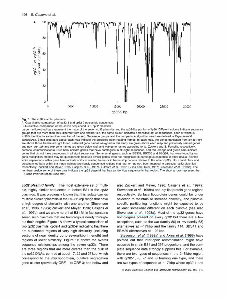
cp32 plasmid family. The most extensive set of multi-
ple, highly similar sequences in isolate B31 is the cp32
plasmids. It was previously known that this isolate carries
multiple circular plasmids in the 29±32 kbp range that have
a high degree of similarity with one another (Stevenson
et al., 1996, 1998a; Zuckert and Meyer, 1996; Casjens et
al., 1997a), and we show here that B31 MI in fact contains
seven such plasmids that are homologous nearly through-
out their lengths. Figure 1A shows a typical comparison of
two cp32 plasmids, cp32-1 and cp32-9, indicating that there
are substantial regions of very high similarity (including
sections of near identity up to several kbp in length) and
regions of lower similarity. Figure 1B shows the overall
sequence relationships among the seven cp32s. There
are three regions that are more diverse than the bulk of
the cp32 DNAs, centred at about 17, 22 and 27 kbp, which
correspond to the mlp lipoprotein, putative segregation
gene cluster (previously ORF-1 to ORF-3; see below and
also Zuckert and Meyer, 1996; Casjens et al., 1997a;
Stevenson et al., 1998a) and erp lipoprotein gene regions
respectively. Surface lipoprotein genes could be under
selection to maintain or increase diversity, and plasmid-
speci®c partitioning functions might be expected to be
at least somewhat different on each plasmid (see also
Stevenson et al., 1998a). Most of the cp32 genes have
homologues present on every cp32 but there are a few
exceptions, such as the bdr (family 80) or rev (family 63)
alternatives at ,17 kbp and the family 114, BBS41 and
BBM39 alternatives at ,28 kbp.
Stevenson et al. (1998a) and Akins et al. (1999) have
pointed out that inter-cp32 recombination might have
occurred in strain B31 and 297 progenitors, and the com-
plete sequence data strongly supports this. For example,
there are two types of sequences in the 2±5 kbp region,
with cp32-1, -3, -7 and -8 forming one type, and there
are two types of sequence at ,17 kbp where cp32-1 and
Q 2000 Blackwell Science Ltd, Molecular Microbiology, 35, 490±516
Fig. 1. The cp32 circular plasmids.A. Quantitative comparison of cp32-1 and cp32-9 nucleotide sequences.B. Qualitative comparison of the seven sequenced B31 cp32 plasmids.Large multicoloured bars represent the maps of the seven cp32 plasmids and the cp32-like portion of lp56. Different colours indicate sequencegroups that are more than 10% different from one another (i.e. the same colour indicates a transitive set of sequences, each of which is$ 90% identical to some other member of the set). Sequence groups and the comparison algorithm used are de®ned in Experimentalprocedures. Small solid bars above each map indicate the predicted open reading frames. In each map, the genes translated from left to rightare above those translated right to left; selected gene names assigned in this study are given above each map and previously named genesand new erp, bdr and mlp gene names are given below (bdr and mlp gene names according to W. Zuckert and S. Porcella, respectively,personal communications). Blue bars indicate genes that have paralogues in all eight sequences, and red, orange and green bars indicategenes that do not have paralogues in all eight sequences. Some small genes, such as BBS32, BBO35 and BBQ36, that were found by ourgene recognition method may be questionable because similar genes were not recognized in paralogous sequence in other cp32s. Slantedwhite separations within gene bars indicate shifts in reading frame or in frame stop codons relative to the other cp32s. Horizontal black andcrosshatched bars within the maps indicate previously sequenced regions that had, or had not, been mapped to particular cp32 plasmidsrespectively (Zuckert and Meyer, 1996; Casjens et al., 1997a; Gilmore et al., 1997; Guina and Oliver, 1997; Stevenson et al., 1998a). Thenumbers beside some of these bars indicate the cp32 plasmid that has an identical sequence in that region. The short arrows represent the,180 bp inverted repeat (see text).
496 S. Casjens et al.

cp32-6 have a rev gene and the others have a bdr gene
(Fig. 2B). Recombination is the simplest way to imagine
generating situations such as this in which all four possible
combinations of `alleles' are present in the cp32s: A±B in
cp32-1; A±b in cp32-3, cp32-7 and cp32-8; a±B in cp32-
6; and a±b cp32-4, cp32-9 and lp56 (upper- and lower-
case letters represent the two types of sequence in
each of the two regions). Given their extensive similarity,
it would be surprising if recombination did not occur
among the cp32s, although it is perhaps remarkable that
no such recombination has been observed in the labora-
tory (El Hage et al., 1999; R. van Vugt and S. Casjens,
unpublished).
The high variability of the Erp and Mlp lipoproteins has
contributed to speculation that they might be involved in
presenting different surface antigenicities to the host (Por-
cella et al., 1996; Stevenson et al., 1996, 1998a; Casjens
et al., 1997a). It is thus possible that the cp32s are `only'
complex mechanisms for disseminating and controlling
the expression of these and perhaps other cp32 genes
that encode possible host interaction proteins, such
as the Rev lipoprotein or BlyAB haemolysin (Guina and
Oliver, 1997; Gilmore and Mbow, 1998). We have pre-
viously speculated that these plasmids could be pro-
phages because: (i) bacteriophage-like particles have
been produced by several B. burgdorferi strains (Hayes
et al., 1983; Neubert et al., 1993; Schaller and Neubert,
1994); (ii) sequence relationships among the cp32s are
reminiscent of temperate bacteriophage families (Casjens
et al., 1992, 1997a); and (iii) prophages often express
genes that affect bacteria±host interactions (Cheetham
and Katz, 1995). Our ®nding here that cp32-1 gene
BBP42 and its family 145 paralogues are similar to a
putative Streptococcus thermophilus phage fO1205 mor-
phogenetic gene (Stanley et al., 1997) lends additional
credence to this hypothesis.
Similarity between lp56 and the cp32s. The linear
plasmid lp56 contains within it an essentially intact copy of
a cp32-like plasmid. This region of lp56 is not identical to
Q 2000 Blackwell Science Ltd, Molecular Microbiology, 35, 490±516
Fig. 1. Continued.
Borrelia plasmids 497
any of the seven circular cp32 plasmids, but represents
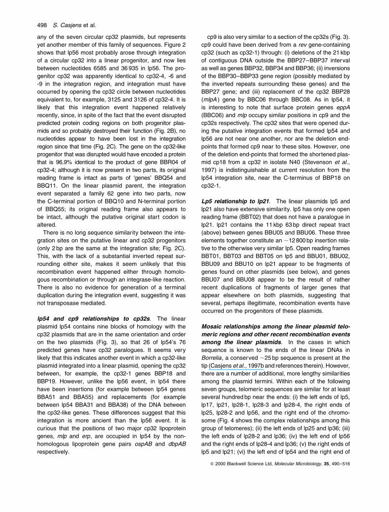
yet another member of this family of sequences. Figure 2
shows that lp56 most probably arose through integration
of a circular cp32 into a linear progenitor, and now lies
between nucleotides 6585 and 36 935 in lp56. The pro-
genitor cp32 was apparently identical to cp32-4, -6 and
-9 in the integration region, and integration must have
occurred by opening the cp32 circle between nucleotides
equivalent to, for example, 3125 and 3126 of cp32-4. It is
likely that this integration event happened relatively
recently, since, in spite of the fact that the event disrupted
predicted protein coding regions on both progenitor plas-
mids and so probably destroyed their function (Fig. 2B), no
nucleotides appear to have been lost in the integration
region since that time (Fig. 2C). The gene on the cp32-like
progenitor that was disrupted would have encoded a protein
that is 96.9% identical to the product of gene BBR04 of
cp32-4; although it is now present in two parts, its original
reading frame is intact as parts of `genes' BBQ54 and
BBQ11. On the linear plasmid parent, the integration
event separated a family 62 gene into two parts, now
the C-terminal portion of BBQ10 and N-terminal portion
of BBQ55; its original reading frame also appears to
be intact, although the putative original start codon is
altered.
There is no long sequence similarity between the inte-
gration sites on the putative linear and cp32 progenitors
(only 2 bp are the same at the integration site; Fig. 2C).
This, with the lack of a substantial inverted repeat sur-
rounding either site, makes it seem unlikely that this
recombination event happened either through homolo-
gous recombination or through an integrase-like reaction.
There is also no evidence for generation of a terminal
duplication during the integration event, suggesting it was
not transposase mediated.
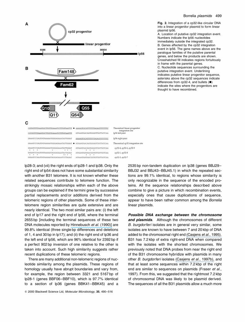
lp54 and cp9 relationships to cp32s. The linear
plasmid lp54 contains nine blocks of homology with the
cp32 plasmids that are in the same orientation and order
on the two plasmids (Fig. 3), so that 26 of lp54's 76
predicted genes have cp32 paralogues. It seems very
likely that this indicates another event in which a cp32-like
plasmid integrated into a linear plasmid, opening the cp32
between, for example, the cp32-1 genes BBP18 and
BBP19. However, unlike the lp56 event, in lp54 there
have been insertions (for example between lp54 genes
BBA51 and BBA55) and replacements (for example
between lp54 BBA31 and BBA38) of the DNA between
the cp32-like genes. These differences suggest that this
integration is more ancient than the lp56 event. It is
curious that the positions of two major cp32 lipoprotein
genes, mlp and erp, are occupied in lp54 by the non-
homologous lipoprotein gene pairs ospAB and dbpAB
respectively.
cp9 is also very similar to a section of the cp32s (Fig. 3).
cp9 could have been derived from a rev gene-containing
cp32 (such as cp32-1) through: (i) deletions of the 21 kbp
of contiguous DNA outside the BBP27±BBP37 interval
as well as genes BBP32, BBP34 and BBP36; (ii) inversions
of the BBP30±BBP33 gene region (possibly mediated by
the inverted repeats surrounding these genes) and the
BBP27 gene; and (iii) replacement of the cp32 BBP28
(mlpA ) gene by BBC06 through BBC08. As in lp54, it
is interesting to note that surface protein genes eppA
(BBC06) and mlp occupy similar positions in cp9 and the
cp32s respectively. The cp32 sites that were opened dur-
ing the putative integration events that formed lp54 and
lp56 are not near one another, nor are the deletion end-
points that formed cp9 near to these sites. However, one
of the deletion end-points that formed the shortened plas-
mid cp18 from a cp32 in isolate N40 (Stevenson et al.,
1997) is indistinguishable at current resolution from the
lp54 integration site, near the C-terminus of BBP18 on
cp32-1.
Lp5 relationship to lp21. The linear plasmids lp5 and
lp21 also have extensive similarity. lp5 has only one open
reading frame (BBT02) that does not have a paralogue in
lp21. lp21 contains the 11 kbp 63 bp direct repeat tract
(above) between genes BBU05 and BBU06. These three
elements together constitute an ,12 800 bp insertion rela-
tive to the otherwise very similar lp5. Open reading frames
BBT01, BBT03 and BBT05 on lp5 and BBU01, BBU02,
BBU09 and BBU10 on lp21 appear to be fragments of
genes found on other plasmids (see below), and genes
BBU07 and BBU08 appear to be the result of rather
recent duplications of fragments of larger genes that
appear elsewhere on both plasmids, suggesting that
several, perhaps illegitimate, recombination events have
occurred on the progenitors of these plasmids.
Mosaic relationships among the linear plasmid telo-
meric regions and other recent recombination events
among the linear plasmids. In the cases in which
sequence is known to the ends of the linear DNAs in
Borrelia, a conserved ,25 bp sequence is present at the
tip (Casjens et al., 1997b and references therein). However,
there are a number of additional, more lengthy similarities
among the plasmid termini. Within each of the following
seven groups, telomeric sequences are similar for at least
several hundred bp near the ends: (i) the left ends of lp5,
lp17, lp21, lp28-1, lp28-3 and lp28-4, the right ends of
lp25, lp28-2 and lp56, and the right end of the chromo-
some (Fig. 4 shows the complex relationships among this
group of telomeres); (ii) the left ends of lp25 and lp36; (iii)
the left ends of lp28-2 and lp36; (iv) the left end of lp56
and the right ends of lp28-4 and lp36; (v) the right ends of
lp5 and lp21; (vi) the left end of lp54 and the right end of
Q 2000 Blackwell Science Ltd, Molecular Microbiology, 35, 490±516
498 S. Casjens et al.
lp28-3; and (vii) the right ends of lp28-1 and lp38. Only the
right end of lp54 does not have some substantial similarity
with another B31 telomere. It is not known whether these
related sequences contribute to telomere function. The
strikingly mosaic relationships within each of the above
groups can be explained if the termini grew by successive
partial replacements and/or additions derived from the
telomeric regions of other plasmids. Some of these inter-
telomere region similarities are quite extensive and are
nearly identical. The two most similar pairs are: (i) the left
end of lp17 and the right end of lp56, where the terminal
2655 bp [including the terminal sequences of these two
DNA molecules reported by Hinnebusch et al. (1990)] are
99.8% identical (three single bp differences and deletions
of 1, 4 and 30 bp in lp17); and (ii) the right end of lp36 and
the left end of lp56, which are 96% identical for 2392 bp if
a perfect 902 bp inversion of one relative to the other is
taken into account. Such high similarity suggests rather
recent duplications of these telomeric regions.
There are many additional non-telomeric regions of nuc-
leotide similarity among the plasmids. These regions of
homology usually have abrupt boundaries and vary from,
for example, the region between 3321 and 5167 bp of
lp28-1 (genes BBF06±BBF10), which is 97.7% identical
to a section of lp36 (genes BBK41±BBK45) and a
2535 bp non-tandem duplication on lp38 (genes BBJ29±
BBJ32 and BBJ43±BBJ45.1) in which the repeated sec-
tions are 99.1% identical, to regions whose similarity is
only recognizable in the sequence of the encoded pro-
teins. All the sequence relationships described above
combine to give a picture in which recombination events,
especially ones that cause duplications of sequence,
appear to have been rather common among the Borrelia
linear plasmids.
Possible DNA exchange between the chromosome
and plasmids. Although the chromosomes of different
B. burgdorferi isolates are in general very similar, some
isolates are known to have between 7 and 20 kbp of DNA
added to the chromosomal right end (Casjens et al., 1995).
B31 has 7.2 kbp of extra right-end DNA when compared
with the isolates with the shortest chromosomes. We
previously noted that DNA probes from near the right end
of the B31 chromosome hybridize with plasmids in many
other B. burgdorferi isolates (Casjens et al., 1997b), and
that at least some sequences within 7.2 kbp of the right
end are similar to sequences on plasmids (Fraser et al.,
1997). From this, we suggested that the rightmost 7.2 kbp
of chromosomal DNA was likely to be plasmid derived.
The sequences of all the B31 plasmids allow a much more
Q 2000 Blackwell Science Ltd, Molecular Microbiology, 35, 490±516
Fig. 2. Integration of a cp32-like circular DNAinto a linear progenitor plasmid to form linearplasmid lp56.A. Location of putative cp32 integration event.Numbers indicate the lp56 nucleotidesimmediately outside the integrated cp32.B. Genes affected by the cp32 integrationevent in lp56. The gene names above are theparalogue families of the putative parentalgenes, and below the products are shown.Crosshatched ®ll indicates regions fortuitouslyin frame with the parental genes.C. Nucleotide sequences surrounding theputative integration event. Underliningindicates putative linear progenitor sequence,asterisks above the cp32 sequences indicatedifferences from cp32-4, and bullets (X)indicate the sites where the progenitors arethought to have recombined.
Borrelia plasmids 499
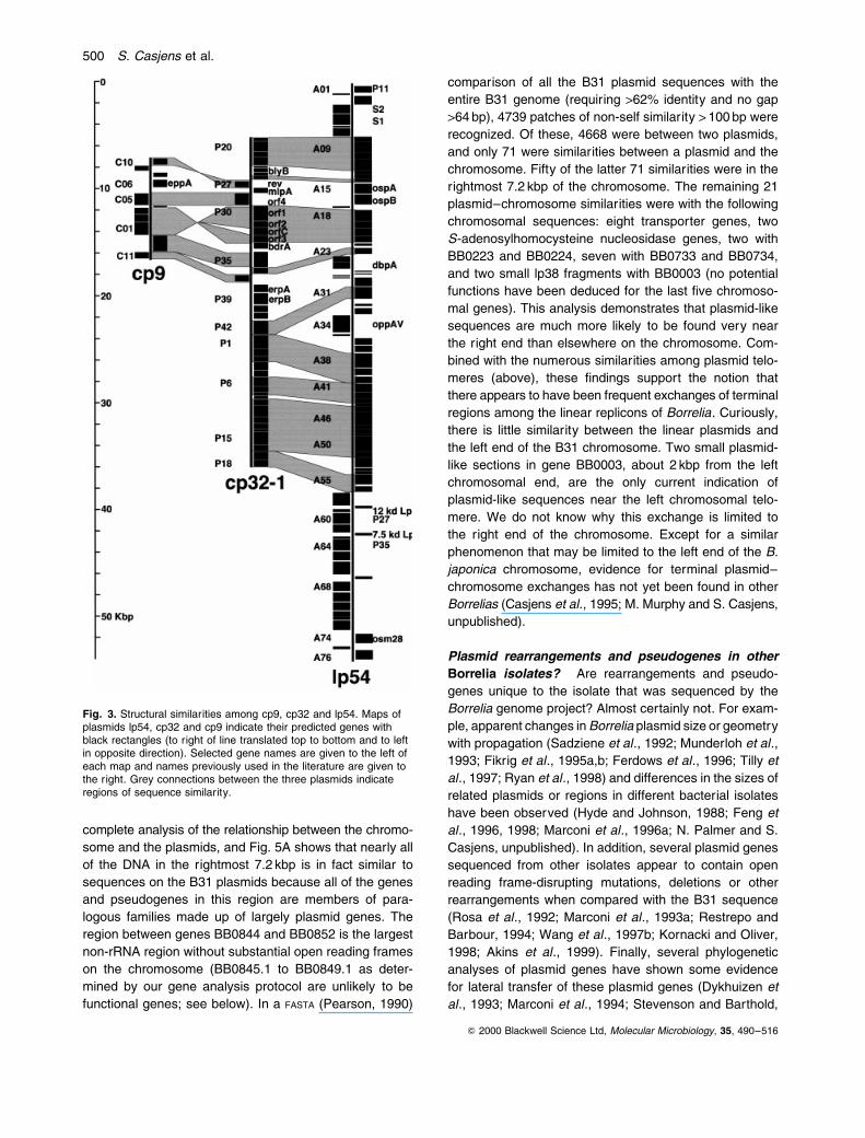
complete analysis of the relationship between the chromo-
some and the plasmids, and Fig. 5A shows that nearly all
of the DNA in the rightmost 7.2 kbp is in fact similar to
sequences on the B31 plasmids because all of the genes
and pseudogenes in this region are members of para-
logous families made up of largely plasmid genes. The
region between genes BB0844 and BB0852 is the largest
non-rRNA region without substantial open reading frames
on the chromosome (BB0845.1 to BB0849.1 as deter-
mined by our gene analysis protocol are unlikely to be
functional genes; see below). In a FASTA (Pearson, 1990)
comparison of all the B31 plasmid sequences with the
entire B31 genome (requiring >62% identity and no gap
>64 bp), 4739 patches of non-self similarity > 100 bp were
recognized. Of these, 4668 were between two plasmids,
and only 71 were similarities between a plasmid and the
chromosome. Fifty of the latter 71 similarities were in the
rightmost 7.2 kbp of the chromosome. The remaining 21
plasmid±chromosome similarities were with the following
chromosomal sequences: eight transporter genes, two
S-adenosylhomocysteine nucleosidase genes, two with
BB0223 and BB0224, seven with BB0733 and BB0734,
and two small lp38 fragments with BB0003 (no potential
functions have been deduced for the last ®ve chromoso-
mal genes). This analysis demonstrates that plasmid-like
sequences are much more likely to be found very near
the right end than elsewhere on the chromosome. Com-
bined with the numerous similarities among plasmid telo-
meres (above), these ®ndings support the notion that
there appears to have been frequent exchanges of terminal
regions among the linear replicons of Borrelia. Curiously,
there is little similarity between the linear plasmids and
the left end of the B31 chromosome. Two small plasmid-
like sections in gene BB0003, about 2 kbp from the left
chromosomal end, are the only current indication of
plasmid-like sequences near the left chromosomal telo-
mere. We do not know why this exchange is limited to
the right end of the chromosome. Except for a similar
phenomenon that may be limited to the left end of the B.
japonica chromosome, evidence for terminal plasmid±
chromosome exchanges has not yet been found in other
Borrelias (Casjens et al., 1995; M. Murphy and S. Casjens,
unpublished).
Plasmid rearrangements and pseudogenes in other
Borrelia isolates?. Are rearrangements and pseudo-
genes unique to the isolate that was sequenced by the
Borrelia genome project? Almost certainly not. For exam-
ple, apparent changes in Borrelia plasmid size or geometry
with propagation (Sadziene et al., 1992; Munderloh et al.,
1993; Fikrig et al., 1995a,b; Ferdows et al., 1996; Tilly et
al., 1997; Ryan et al., 1998) and differences in the sizes of
related plasmids or regions in different bacterial isolates
have been observed (Hyde and Johnson, 1988; Feng et
al., 1996, 1998; Marconi et al., 1996a; N. Palmer and S.
Casjens, unpublished). In addition, several plasmid genes
sequenced from other isolates appear to contain open
reading frame-disrupting mutations, deletions or other
rearrangements when compared with the B31 sequence
(Rosa et al., 1992; Marconi et al., 1993a; Restrepo and
Barbour, 1994; Wang et al., 1997b; Kornacki and Oliver,
1998; Akins et al., 1999). Finally, several phylogenetic
analyses of plasmid genes have shown some evidence
for lateral transfer of these plasmid genes (Dykhuizen et
al., 1993; Marconi et al., 1994; Stevenson and Barthold,
Q 2000 Blackwell Science Ltd, Molecular Microbiology, 35, 490±516
Fig. 3. Structural similarities among cp9, cp32 and lp54. Maps ofplasmids lp54, cp32 and cp9 indicate their predicted genes withblack rectangles (to right of line translated top to bottom and to leftin opposite direction). Selected gene names are given to the left ofeach map and names previously used in the literature are given tothe right. Grey connections between the three plasmids indicateregions of sequence similarity.
500 S. Casjens et al.

1994; Jauris-Heipke et al., 1995; Livey et al., 1995; Will et
al., 1995), so it seems likely that such rearrangements
and pseudogenes are not unique to B31 plasmids.
The open reading frames of the B. burgdorferi plasmids
Some plasmids carry numerous pseudogenes. There
are several very unusual aspects of the protein coding
potential of the B31 plasmids. Unlike the `constant portion'
of the chromosome (genes BB0001 to BB0843), a number
of the B31 plasmids have: (i) an apparent protein coding
density that is <70%, a value that is substantially less
than the B. burgdorferi chromosome or other bacterial
genomes; (ii) a surprisingly large fraction of small open
reading frames (#100 codons); and (iii) a large number of
predicted `genes' that are truncated or have damaged
reading frames relative to other members of their para-
logous gene families. For ease of discussion below, we
de®ne a `pseudogene' as any region of DNA that is similar
in sequence to a paralogous Borrelia predicted gene or to
a gene from another organism, but which is obviously
truncated and/or does not have full open reading frames
relative to its homologues. These mostly appear to be
mutationally damaged genes that include frameshift
changes, in frame stop codons, and fused or truncated
genes. We suspect that most of them may not currently
encode functional polypeptides. However, in any given
instance, we cannot rule out in vivo synthesis or even a
biologically important function of a protein `fragment'.
We initially identi®ed 731 putative (non-pseudo)genes
and 167 pseudogenes on the 21 plasmids, and their
names and locations are shown in Fig. 6. Putative genes
were identi®ed according to Salzberg et al. (1998), and
pseudogenes not found as truncated members of paralo-
gous gene families by that procedure were identi®ed by
DNA similarities; see Experimental procedures. Among
the 731 potential genes, 61 are probable false identi®ca-
tions because they lie inside another gene or pseudogene,
or because they are very small and were not identi®ed in
paralogous sequence elsewhere in the genome (these
`questionable genes' are ignored in the remainder of this
discussion, but are shown in Fig. 6 and are noted in the
complete predicted gene list in Supplementary infor-
mation ; see Experimental procedures ). Thus, our current
best estimate is that there are 670 potentially functional
genes and 167 pseudogenes on the B31 plasmids
(Table 1).
Ten of the B31 plasmids (lp5, lp17, lp21, lp25, lp28-1,
lp28-3, lp28-4, lp36, lp38 and the non-cp32-like portion
of lp56) contain 87% of the pseudogenes and have a total
non-pseudogene protein coding capacity of only 41%,
and a very large fraction (43%) of these predicted genes
Q 2000 Blackwell Science Ltd, Molecular Microbiology, 35, 490±516
Fig. 4. Some sequence relationships amongthe telomeres of the strain B31 linearreplicons. Eight linear plasmid ends and theright chromosomal end are shown with theirtelomeres on the left. Similarly colouredblocks indicate blocks of similar sequence(> 65% identity), and thinner black linesindicate sequences that have no paraloguewithin the regions shown. Each colourboundary indicates a sequence breakcompared with one of the other telomeres inthe ®gure.
Borrelia plasmids 501
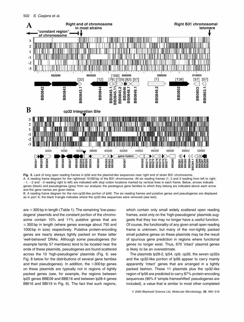
are # 300 bp in length (Table 1). The remaining `low-pseu-
dogene' plasmids and the constant portion of the chromo-
some contain 10% and 11% putative genes that are
# 300 bp in length (where genes average about 750 and
1000 bp in size) respectively. Putative protein-encoding
genes are nearly always tightly packed on these latter
`well-behaved' DNAs. Although some pseudogenes (for
example family 57 members) tend to be located near the
ends of these plasmids, pseudogenes are found scattered
across the 10 `high-pseudogene' plasmids (Fig. 6; see
Fig. 8 below for the distributions of several gene families
and their pseudogenes). In addition, the #300 bp genes
on these plasmids are typically not in regions of tightly
packed genes (see, for example, the regions between
lp25 genes BBE09 and BBE16 and between lp28-4 genes
BBI16 and BBI19 in Fig. 6). The fact that such regions,
which contain only small widely scattered open reading
frames, exist only on the `high-pseudogene' plasmids sug-
gests that they too may no longer have a useful function.
Of course, the functionality of any given small open reading
frame is unknown, but many of the non-tightly packed
small putative genes on these plasmids may be the result
of spurious gene prediction in regions where functional
genes no longer exist. Thus, 670 `intact' plasmid genes
is likely to be an overestimate.
The plasmids lp28-2, lp54, cp9, cp26, the seven cp32s
and the cp32-like portion of lp56 appear to carry mainly
apparently `intact' genes that are arranged in a tightly
packed fashion. These 11 plasmids plus the cp32-like
region of lp56 are predicted to carry 87% protein-encoding
sequences (90% if `simple frameshifted' pseudogenes are
included), a value that is similar to most other completed
Q 2000 Blackwell Science Ltd, Molecular Microbiology, 35, 490±516
Fig. 5. Lack of long open reading frames in lp56 and the plasmid-like sequences near right end of strain B31 chromosome.A. A reading frame diagram for the rightmost 10 000 bp of the B31 chromosome. All six reading frames (1, 2 and 3 reading from left to right;ÿ1, ÿ2 and ÿ3 reading right to left) are indicated with stop codon locations marked by vertical lines in each frame. Below, arrows indicategenes (black) and pseudogenes (grey) from our analysis; the paralogous gene families to which they belong are indicated above each arrowand the gene names are given below.B. A reading frame diagram for the non-cp32-like portion of lp56. The six reading frames and putative genes and pseudogenes are displayedas in part A; the black triangle indicates where the cp32-like sequences were removed (see text).
502 S. Casjens et al.

Q 2000 Blackwell Science Ltd, Molecular Microbiology, 35, 490±516
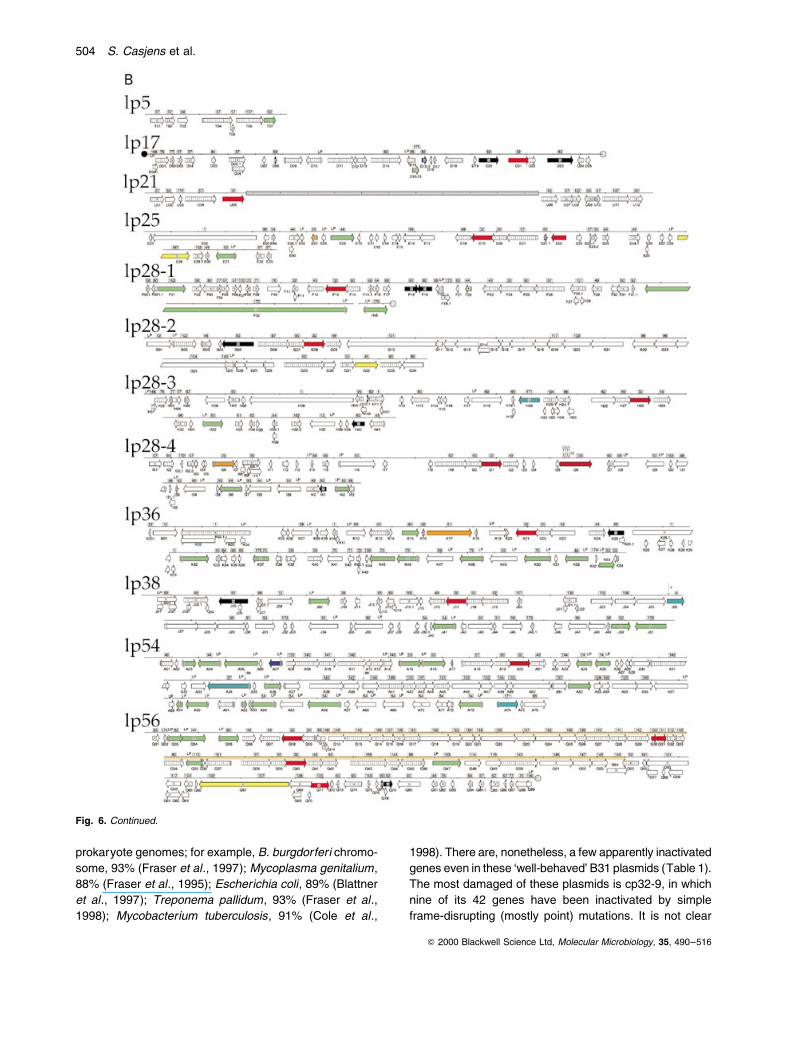
Fig. 6. Linear representations of the B. burgdorferi B31 MI extrachromosomal DNAs.A. The nine B31 circular plasmids.B. The 12 B31 linear plasmids.The locations of the predicted protein coding regions are colour-coded by biological role, and arrows represent the direction of transcription foreach predicted coding region. Pseudogenes, de®ned as in the text, are indicated by asterisks. Numbers associated with `GES' represent thenumber of membrane-spanning domains according to the Goldman, Engelman and Steitz scale as calculated by TOPPRED (Claros and vonHeijne, 1994); only proteins with ®ve or more such domains are shown. Members of paralogous gene families are indicated by numbers inboxes above each map (overlapping genes are only so indicated once). Putative transporter proteins are indicated by an arrow and thepossible substrate as follows: aa, amino acid or oligopeptide; glu, glucose;?, unknown. LP indicates the predicted protein meets our criteria forpotential N-terminal lipidation (see text).
Borrelia plasmids 503
prokaryote genomes; for example, B. burgdorferi chromo-
some, 93% (Fraser et al., 1997); Mycoplasma genitalium,
88% (Fraser et al., 1995); Escherichia coli, 89% (Blattner
et al., 1997); Treponema pallidum, 93% (Fraser et al.,
1998); Mycobacterium tuberculosis, 91% (Cole et al.,
1998). There are, nonetheless, a few apparently inactivated
genes even in these `well-behaved' B31 plasmids (Table 1).
The most damaged of these plasmids is cp32-9, in which
nine of its 42 genes have been inactivated by simple
frame-disrupting (mostly point) mutations. It is not clear
Q 2000 Blackwell Science Ltd, Molecular Microbiology, 35, 490±516
Fig. 6. Continued.
504 S. Casjens et al.
why cp32-9 contains so many mutations of this type; per-
haps all or parts of it have become super¯uous and have
begun to decay. The other `well-behaved' plasmids carry
a small number of more dramatic rearrangements, e.g.
the apparent insertion of the 58 portion of a BBP29 (family
161) homologue into a precursor erp-like (family 162) gene
to create two new genes on cp32-4, a severely truncated
erpH gene (BBR40) and an in frame fusion between the
N-terminus of the family 161 member and the C-terminus
of the precursor erp gene to form gene BBR41. Although
BBR41 may well be expressed because it carries the puta-
tive translation start signal of the family 161 gene, it seems
unlikely that this fusion protein is functional as the family
161 portion is severely truncated and the erp-like portion
has lost its lipidation signal. In a recent analysis of the
erp genes of strain 297, Akins et al. (1999) did not ®nd
a fusion gene analogous to BBR41, suggesting it may
have arisen recently. It is not clear why some plasmids
should carry so many pseudogenes and others have few
or none; perhaps those with many have undergone recent
rearrangements events that may have damaged genes
directly and/or made various regions redundant.
The nature of the plasmid pseudogenes. The least-
damaged pseudogenes contain only one or a few simple
frameshifts relative to their homologues. We ®nd a number
of such apparently lightly damaged genes in the B31
plasmids that contain only one or a few in frame stop
codons and/or frameshifts, e.g. BBG05 in lp28-2; BBQ04,
BBQ16 and BBQ51 in lp56; BBR02 and BBR35 in cp32-4;
BBN05, BBN06, BBN13, BBN16, BBN19, BBN21, BBN22,
BBN29 and BBN37 in cp32-9.
Most of the pseudogenes are much more badly damaged.
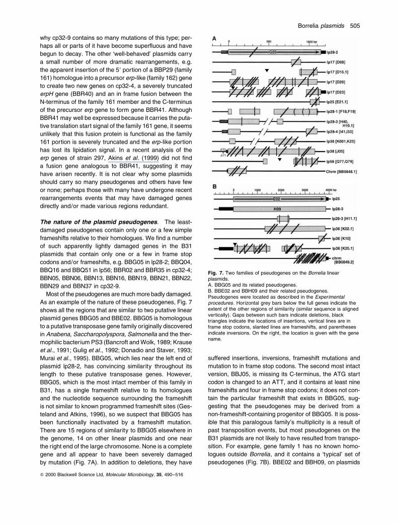
As an example of the nature of these pseudogenes, Fig. 7
shows all the regions that are similar to two putative linear
plasmid genes BBG05 and BBE02. BBG05 is homologous
to a putative transposase gene family originally discovered
in Anabena, Saccharopolyspora, Salmonella and the ther-
mophilic bacterium PS3 (Bancroft and Wolk, 1989; Krause
et al., 1991; Gulig et al., 1992; Donadio and Staver, 1993;
Murai et al., 1995). BBG05, which lies near the left end of
plasmid lp28-2, has convincing similarity throughout its
length to these putative transposase genes. However,
BBG05, which is the most intact member of this family in
B31, has a single frameshift relative to its homologues
and the nucleotide sequence surrounding the frameshift
is not similar to known programmed frameshift sites (Ges-
teland and Atkins, 1996), so we suspect that BBG05 has
been functionally inactivated by a frameshift mutation.
There are 15 regions of similarity to BBG05 elsewhere in
the genome, 14 on other linear plasmids and one near
the right end of the large chromosome. None is a complete
gene and all appear to have been severely damaged
by mutation (Fig. 7A). In addition to deletions, they have
suffered insertions, inversions, frameshift mutations and
mutation to in frame stop codons. The second most intact
version, BBJ05, is missing its C-terminus, the ATG start
codon is changed to an ATT, and it contains at least nine
frameshifts and four in frame stop codons; it does not con-
tain the particular frameshift that exists in BBG05, sug-
gesting that the pseudogenes may be derived from a
non-frameshift-containing progenitor of BBG05. It is poss-
ible that this paralogous family's multiplicity is a result of
past transposition events, but most pseudogenes on the
B31 plasmids are not likely to have resulted from transpo-
sition. For example, gene family 1 has no known homo-
logues outside Borrelia, and it contains a `typical' set of
pseudogenes (Fig. 7B). BBE02 and BBH09, on plasmids
Q 2000 Blackwell Science Ltd, Molecular Microbiology, 35, 490±516
Fig. 7. Two families of pseudogenes on the Borrelia linearplasmids.A. BBG05 and its related pseudogenes.B. BBE02 and BBH09 and their related pseudogenes.Pseudogenes were located as described in the Experimentalprocedures. Horizontal grey bars below the full genes indicate theextent of the other regions of similarity (similar sequence is alignedvertically). Gaps between such bars indicate deletions, blacktriangles indicate the locations of insertions, vertical lines are inframe stop codons, slanted lines are frameshifts, and parenthesesindicate inversions. On the right, the location is given with the genename.
Borrelia plasmids 505
lp25 and lp28-3, are thought to be intact as they are large
and have very similar open reading frames. There are four
badly damaged paralogues elsewhere on the linear plas-
mids, and one, near the right end of the large chromo-
some, that has suffered two deletions, an insertion, 12
frameshifts, and one in frame stop codon in about 1300 bp
of remaining DNA.
Some regions of the plasmids appear to be particularly
rich in pseudogenes. The non-cp32-like portion of lp56
contains one of the highest fractions of pseudogenes
among the B31 plasmids (Table 1). Of the 36 genes and
pseudogenes there, seven are short putative genes
#300 bp long that have no homologues, and 22 appear
to be pseudogenes (most of them severely damaged).
Figure 5B shows the nearly complete lack of substantial
open reading frames in long sections of this DNA. Interest-
ingly, the largest lp56 open reading frame BBQ67 is a
bipartite gene in which the N-terminal 80% is convincingly
similar to full-length adenine DNA methyltransferase
genes (best match Helicobacter pylori HP1354) and the
C-terminal 20% is very similar to BBG02, a putative lipo-
protein-encoding gene of unknown function on lp28-2
(but whose lipidation consensus was removed by the pos-
tulated BBQ67 fusion). Thus, even the larger genes on the
B31 plasmids may have been recently altered by DNA
rearrangements. Also noteworthy is a section of lp56
DNA that is similar to the BBI26±BBI34 region of lp28-4.
All of the lp28-4-like pseudogenes in this region of lp56
have accumulated serious mutational damage, and a
transposase BBG05 pseudogene between BBQ75 and
BBQ80 suggests that transposition may have contributed
to the damage. Curiously, only three of the lp56 cp32-like
progenitor's 41 genes are damaged; the gene broken by
the insertion event and two that contain a small number
of frameshift mutations. It is tempting to speculate that,
after the cp32-like plasmid integrated into lp56's linear pro-
genitor, many of the linear plasmid's genes became super-
¯uous. In addition, plasmid-like sequences in the rightmost
7.2 kbp of the chromosome also appear to be largely
decaying (Fig. 5A).
Relatively few pseudogenes have been found in other
bacteria, and these have been rare exceptions when com-
pared with the number of functional genes. Genes with one
or a few frameshifts, in frame stop codons or inactivated
control regions have been found in a few anecdotal cases
(for example Hall et al., 1983; Morris et al., 1995; Fsihi et
al., 1996; Lai et al., 1996). The number of such genes in
the completely sequenced bacterial genomes is low, e.g.
only 1.3% of the genes (23 out of 1758) in the complete
genome of Haemophilus in¯uenzae contain substantiated
in frame stop codons or frameshifts, and similar values
of 0.9%, 0.6% and 1.4% are found for the chromosomes
of T. pallidum, M. genitalium and H. pylori respectively
(some of these may be in the initial stages of evolutionary
inactivation whereas others could be phase variable
genes in the `off' state). Only one pseudogene, BB119
which contains a single simple frameshift, has been iden-
ti®ed among the 843 genes of the `constant portion' of the
Borrelia chromosome (Fraser et al., 1997). These values
may be underestimates because the status of genes of
unknown function that have no homologues cannot pre-
sently be assessed, but as related genomes are
sequenced other instances of damaged currently hypothe-
tical genes may become recognizable (the comparison of
two H. pylori isolates has allowed recognition of a few
additional apparently damaged genes; Alm et al., 1999).
Truncated gene fragments have been observed even
less frequently in bacteria, although a few have been
reported especially in association with defective pro-
phages (see, for example, Xiang et al., 1994; Skamrov
et al., 1995; Casjens, 1998). Smith et al. (1997) have
reported a substantial number of damaged genes in the
chromosome of Mycobacterium leprae (3.7% pseudo-
genes in a 1.5 Mbp region), and Andersson et al. (1998)
observed that 12 of the 846 genes in the complete genome
sequence of Rickettsia prowazekii appear to be mutation-
ally damaged and, in addition, ®nd that only 75.4% of its
genome appears to encode proteins. All of these pseudo-
gene frequencies are very much less than those found on
some of the Borrelia linear plasmids (over 50% in lp5, lp21,
lp28-1 and the non-cp32-like portion of lp56). Andersson
et al. (1998) interpreted pseudogenes in bacteria to be
the remnants of genes that are no longer useful but have
not yet been completely eliminated from the genome.
This is a reasonable hypothesis, but at present we do
not know why the Borrelia plasmids should have so
many damaged but not yet eliminated genes.
Highly paralogous nature of plasmid genes
As has been mentioned above, the genes and pseudo-
genes on the B31 plasmids form a large number of paralo-
gous gene families. The complete genome contains 161
families of paralogous genes, 107 of which contain at
least one plasmid-borne member. Family sizes range
from 41 members (including pseudogenes) in family 57 to
families with only two members; 83% of the $300-bp-long
plasmid non-pseudogenes are members of such families.
The very high fraction of genes with plasmid-borne paralo-
gues may re¯ect some as yet unknown advantage in car-
rying multiple similar but slightly different copies of these
genes. The family membership of each plasmid gene is
indicated in Fig. 6, and the members of each gene family
can be found in the Supplementary information section
(see Experimental procedures ).
Predicted functions of plasmid open reading frames
Each open reading frame on the plasmids was compared
Q 2000 Blackwell Science Ltd, Molecular Microbiology, 35, 490±516
506 S. Casjens et al.
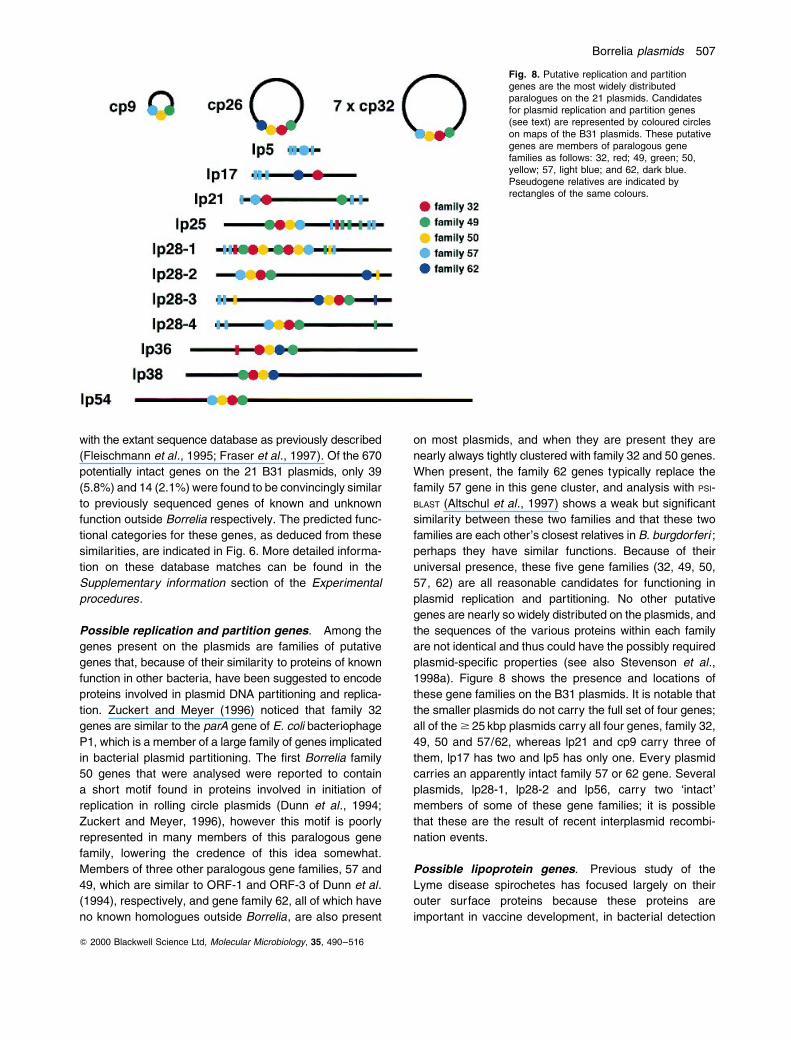
with the extant sequence database as previously described
(Fleischmann et al., 1995; Fraser et al., 1997). Of the 670
potentially intact genes on the 21 B31 plasmids, only 39
(5.8%) and 14 (2.1%) were found to be convincingly similar
to previously sequenced genes of known and unknown
function outside Borrelia respectively. The predicted func-
tional categories for these genes, as deduced from these
similarities, are indicated in Fig. 6. More detailed informa-
tion on these database matches can be found in the
Supplementary information section of the Experimental
procedures.
Possible replication and partition genes. Among the
genes present on the plasmids are families of putative
genes that, because of their similarity to proteins of known
function in other bacteria, have been suggested to encode
proteins involved in plasmid DNA partitioning and replica-
tion. Zuckert and Meyer (1996) noticed that family 32
genes are similar to the parA gene of E. coli bacteriophage
P1, which is a member of a large family of genes implicated
in bacterial plasmid partitioning. The ®rst Borrelia family
50 genes that were analysed were reported to contain
a short motif found in proteins involved in initiation of
replication in rolling circle plasmids (Dunn et al., 1994;
Zuckert and Meyer, 1996), however this motif is poorly
represented in many members of this paralogous gene
family, lowering the credence of this idea somewhat.
Members of three other paralogous gene families, 57 and
49, which are similar to ORF-1 and ORF-3 of Dunn et al.
(1994), respectively, and gene family 62, all of which have
no known homologues outside Borrelia, are also present
on most plasmids, and when they are present they are
nearly always tightly clustered with family 32 and 50 genes.
When present, the family 62 genes typically replace the
family 57 gene in this gene cluster, and analysis with PSI-
BLAST (Altschul et al., 1997) shows a weak but signi®cant
similarity between these two families and that these two
families are each other's closest relatives in B. burgdorferi ;
perhaps they have similar functions. Because of their
universal presence, these ®ve gene families (32, 49, 50,
57, 62) are all reasonable candidates for functioning in
plasmid replication and partitioning. No other putative
genes are nearly so widely distributed on the plasmids, and
the sequences of the various proteins within each family
are not identical and thus could have the possibly required
plasmid-speci®c properties (see also Stevenson et al.,
1998a). Figure 8 shows the presence and locations of
these gene families on the B31 plasmids. It is notable that
the smaller plasmids do not carry the full set of four genes;
all of the $ 25 kbp plasmids carry all four genes, family 32,
49, 50 and 57/62, whereas lp21 and cp9 carry three of
them, lp17 has two and lp5 has only one. Every plasmid
carries an apparently intact family 57 or 62 gene. Several
plasmids, lp28-1, lp28-2 and lp56, carry two `intact'
members of some of these gene families; it is possible
that these are the result of recent interplasmid recombi-
nation events.
Possible lipoprotein genes. Previous study of the
Lyme disease spirochetes has focused largely on their
outer surface proteins because these proteins are
important in vaccine development, in bacterial detection
Q 2000 Blackwell Science Ltd, Molecular Microbiology, 35, 490±516
Fig. 8. Putative replication and partitiongenes are the most widely distributedparalogues on the 21 plasmids. Candidatesfor plasmid replication and partition genes(see text) are represented by coloured circleson maps of the B31 plasmids. These putativegenes are members of paralogous genefamilies as follows: 32, red; 49, green; 50,yellow; 57, light blue; and 62, dark blue.Pseudogene relatives are indicated byrectangles of the same colours.
Borrelia plasmids 507

and in the interaction of these bacteria with their
arthropod and vertebrate hosts. Most of the outer surface
proteins that have been characterized are lipoproteins
(Rosa, 1997). We previously noted that there were 105
genes on the chromosome and the 11 previously published
plasmids that encode proteins that contain a type II signal
sequence in which a positively charged N-terminal region
is followed by a hydrophobic signal sequence and the
lipidation consensus [L,A,V,I,F,T,M]±[L,A,V,I,F,S]±X±
[G,A,S,N]±C (Fraser et al., 1997). In an effort to develop
more quantitative criteria for identifying potential Borrelia
lipoprotein genes, this lipidation consensus sequence
(which was deduced from data on other bacterial species;
Sutcliffe and Russell, 1995), information from the Borrelia
lipoprotein literature, and the assumption that proteins with
membership in paralogous families with bona ®de lipopro-
tein members and only conservative changes from the
above consensus are likely to be lipoproteins were used to
perform a hidden Markov model (HMM) analysis to ®nd all
of the B31 genes that might encode lipoproteins (see
Experimental procedures ). Some of the predicted proteins
that ®t these criteria include the following (mostly con-
servative) additions to the above lipidation consensus: G, N
and S in position ÿ1; M and T in position ÿ2; and L, T and I
in position ÿ4 (for example BBA68, BBA69, BBI36, BBI38,
BBI39 and BBJ41 which all have a T in position ÿ2 and an I
in position ÿ4 and are members of a gene family that
includes proteins that contain the more stringent consen-
sus). These slightly expanded criteria suggest that there
may be as many as 91 plasmid lipoprotein-encoding genes
[ignoring eight pseudogenes and a questionable gene
(BBI32) that meet the criteria] and 41 apparently intact
chromosomal lipoprotein genes. The genes that encode
these proteins fall into 27 of the B31 paralogous families,
nine of which have only lipoprotein members. Most of these
predicted lipoproteins are likely to be surface proteins
because nearly all of the previously characterized plasmid-
encoded putative lipoproteins are surface localized,
although periplasmic proteins may also be lipidated in
Borrelia (Bono et al., 1998; Kornacki and Oliver, 1998).
This may still be an underestimate of the lipoprotein-coding
potential of Borrelia because there are 32 additional
genes (15 chromosomal, 17 plasmid) that appear to have
a properly placed Cys and a reasonable HMM score, but
whose characteristics did not quite meet our criteria
(including the known surface protein gene BBK32 and
genes such as BBK45, BB0329 and BB0330 that belong to
paralogous families which contain other lipoprotein genes).
These are included in a complete list of predicted lipo-
protein genes in the Supplementary information section.
Borrelia appears to have an especially large fraction of
its genome devoted to lipoprotein production. Putative
lipoprotein genes represent about 4.9% of the chromo-
somal genes, a value somewhat higher than but similar
to other completely sequenced bacterial genomes such
as H. pylori (1.3%; Tomb et al., 1997) or T. pallidum
(2.1%; Fraser et al., 1998). The putative lipoprotein genes
on the B. burgdorferi plasmids represent 14.5% of the
`intact' plasmid genes (17% if the `near cut-off' genes
above are included). This high fraction of genes that
encode potentially outer surface proteins correlates
with the observation in other bacterial parasites that pro-
teins involved in interaction with the host are often plas-
mid encoded.
Each of the seven cp32 plasmids, and the cp32 inserted
into lp56, carry two types of putative lipoprotein genes, an
mlp-type gene (family 113) (characterized in isolate 297
by Porcella et al., 1996) and one or two adjacent erp
genes (families 162, 163 and 164; Lam et al., 1994; Ste-
venson et al., 1996, 1998a,b; Casjens et al., 1997a).
The ®ve new erp genes discovered here on plasmids
cp32-4, cp32-8 and cp32-9 are named erpY, erpN/O and
erpP/Q respectively (see Fig. 1B). The ErpY protein is
rather similar to ErpL, the ErpP and Q proteins are quite
similar to Erps A and B, respectively, and Erps N and O
are identical to Erps A and B respectively. cp32-1 and
cp32-6 each encode an additional putative lipoprotein
(family 63/Rev; Gilmore and Mbow, 1998) that are identi-
cal to one another.
Other possible functions for plasmid genes. In
addition to the putative partitioning proteins and lipopro-
teins discussed above, other plasmid-encoded functions
that have been characterized are a porin on lp54 (BBA74;
Skare et al., 1996), a ®bronectin binding protein on lp36
(BBK32; Probert and Johnson, 1998) and two haemolysin
genes blyA and blyB on a cp32 (Guina and Oliver, 1997;
we ®nd the bly genes they characterized on cp32-9 ±
BBN23 and BBN24 ± but each of the cp32s and lp56
carries paralogues of both of these genes). Other putative
functions convincingly predicted by similarity to genes of
known function in other organisms for intact genes on the
plasmids are two genes for GMP synthesis on cp26
(BBB17, BBB18; Margolis et al., 1994; Zhou et al., 1997),
four genes for sugar transport on cp26 (BBB04, BBB05,
BBB06 and BBB29; Fraser et al., 1997), a nicotinamidase
on lp25 (BBE22; Fraser et al., 1997), a DNA helicase on
lp28-2 (BBG32; Fraser et al., 1997), a 58-methylthioade-
nosine/S-adenosylhomocysteine nucleosidase and a multi-
drug transporter on lp28-4 (BBI06, BBI16; Fraser et al.,
1997; Cornell and Riscoe, 1998), ABC transporter com-
ponent genes on cp26, lp38 and lp54 (BBB16, BBJ26 and
BBA34; Fraser et al., 1997; Bono et al., 1998), an adenine
deaminase on lp36 (BBK17; Fraser et al., 1997), and a
possible DNA methylase on lp56 (BBQ67). (BBB01 and
BBA76 have weak similarities to acyl phosphatase of E.
coli and thymidylate synthase-complementing protein of
Dictyostelium discoideum respectively.) In addition, only
Q 2000 Blackwell Science Ltd, Molecular Microbiology, 35, 490±516
508 S. Casjens et al.
BBG02, BBK13 and members of paralogous families 94,
137 and 145 have convincing homologues among hypo-
thetical genes of unknown function from another bacteria.
B31-like plasmids present in other Lyme Borrelia
natural isolates
All natural isolates of B. burgdorferi (sensu stricto) and
related species that have been examined carry numerous
linear plasmids in the 5±56 kbp size range as well as multi-
ple circular plasmids. Do all isolates carry a set of extra-
chromosomal elements that are similar to those found in
this study of isolate B31, or does each bacterium carry a
complement of plasmids chosen from a much larger
menu? And how similar are similar-sized plasmids in differ-
ent isolates?
Linear plasmids. B. burgdorferi isolates Sh-2-82 and
CA-11.2A have lp54s that have gene orders and restric-
tion maps that are, at relatively low resolution, similar to
lp54 of B31 (Marconi et al., 1996a; Fraser et al., 1997; R.
van Vugt and S. Casjens, unpublished). These ®ndings,
combined with the observations that BBA15 and BBA16
(ospA and ospB ) are almost universally present in B.
burgdorferi isolates and when examined are always on a
50±55 kbp linear plasmid, strongly suggest that a plasmid
similar to B31 lp54 is present in almost all B. burgdorferi
bacteria in nature. (Out of hundreds of isolates analysed,
less than 10 have been found that may lack the ospA/B-
containing lp54 plasmid, and those few could, in theory,
have lost it during isolation or only lost the genes used as
probes, e.g. Samuels et al., 1993; Casjens et al., 1995;
Anderson et al., 1996; Guttman et al., 1996; Mathiesen
et al., 1997.) However, Feng et al. (1998) reported that,
unlike B31, in isolate N40 the ospA and dbpA genes are
on different linear plasmids, suggesting that this plasmid
may not be completely constant among different isolates.
Isolated B31 lp17 DNA hybridized only to a similar-sized
linear plasmid in three of four isolates previously examined
(Hinnebusch and Barbour, 1991). A vlsE gene similar to
that on B31 lp28-1 was found on an ,20 kbp linear plasmid
in isolate 297 (Kawabata et al., 1998), and two different
sequences cloned from a 29 kbp linear plasmid of B. burg-
dorferi isolate 297 hybridize to a 28 kbp plasmid in B31
and in 12 out of 16 other isolates examined (Xu and John-
son, 1995). Finally, an ospD gene probe from B31 lp38
hybridizes with a similar-sized plasmid in all cases in
which the ospD gene was present (18 of 24 isolates exam-
ined; Norris et al., 1992; Marconi et al., 1994). In a prelimin-
ary systematic study, we have found that hybridization
targets for multiple, unique probes from each of the 12
B31 linear plasmids are present in at least some members
(usually more than half) of a panel of 15 North American B.
burgdorferi (sensu stricto) isolates, and when present
almost always reside on a linear plasmid similar in size
to the cognate B31 plasmid. In addition, multiple probes
from any particular plasmid of B31 usually hybridize to a
single plasmid in most other isolates (N. Palmer and
S. Casjens, unpublished).
Circular plasmids. Tilly et al. (1997) showed that the
B31 cp26 has a restriction map that is very similar to
cp26s of three other independent geographically diverse
B. burgdorferi isolates. In addition, cp26 gene BBB19
(ospC ) has been found in all isolates examined (Marconi et
al., 1993b; Theisen et al., 1993; Stevenson and Barthold,
1994; Fukunaga and Hamase, 1995; Jauris-Heipke et al.,
1995; Livey et al., 1995; Wilske et al., 1995, 1996) and,
when studied, it was found to be present on a 26±28 kbp
circular plasmid, so B31 cp26-like DNA elements appear
to be universally present in B. burgdorferi.
Circular plasmids approximately 9 kbp in size have been
found in several Lyme agent isolates (Hyde and Johnson,
1988; Simpson et al., 1990a; Stalhammar-Carlemalm
et al., 1990; Champion et al., 1994; Dunn et al., 1994).
The relationships among most of these are not known,
but two, cp8.3 in B. afzelii isolate Ip21 (Dunn et al.,
1994) and cp9 in B31 (Fraser et al., 1997), have been
completely sequenced. Their structures are suf®ciently
similar (both have BBC08 homologues and inversions of
the BBC01±BBC03 regions relative to the cp32s; see
Fig. 3) that it seems very likely that they are derived
from the same small progenitor, rather than having inde-
pendently arisen from a cp32-like plasmid. There are
only two major differences: cp9 putative lipoprotein (rev
paralogue) gene BBC10 is missing in cp8.3, and in cp9
BBC06 (eppA) and BBC07 replace part of cp8.3.
Multiple related ,30 kbp plasmid sequences (very prob-
ably circular cp32 homologues) are present in all B. burg-
dorferi isolates that have been examined (Simpson et al.,
1990b; Marconi et al., 1996b; Porcella et al., 1996; Theisen,
1996; Zuckert and Meyer, 1996; Casjens et al., 1997a;
Akins et al., 1999). Porcella et al. (1996) and Akins et al.
(1999) found seven different homologues of cp32-borne
paralogue family 113 genes in isolate 297, and here we
characterize seven cp32 plasmids and the related lp56
in isolate B31, and other B31 cultures have been observed
to contain two additional cp32s (Casjens et al., 1997a). It
is not known whether this is an indication that all natural
isolates will carry 7±10 speci®c cp32 plasmids, or whether
each will carry different numbers of a large variety of cp32
family plasmids. The cp32 sequences presented here will
allow this question to be addressed in the future.
Unrelated extrachromosomal DNAs in other B. burg-
dorferi isolates? Has the genome project identi®ed all
or most of the types of extrachromosomal elements
present in B. burgdorferi in nature? There are no reports
Q 2000 Blackwell Science Ltd, Molecular Microbiology, 35, 490±516
Borrelia plasmids 509
of B. burgdorferi linear plasmids whose sizes are not close
to members of the B31 complement of linear plasmids or
of plasmid-borne genes that are not present in the B31
plasmid sequences. It is, however, noteworthy that closely
related species B. andersonii, B. afzelii, B. garinii, B.
valaisiana, B. hermsii and B. turicatae have been
reported to carry linear plasmids in the 90±180 kbp
range, which are not known to be related to the known
B31 plasmids (Casjens et al., 1995; Busch et al., 1996;
Marconi et al., 1996a), so it is quite possible that additional
linear plasmid types will be found in B. burgdorferi in the
future. Circular plasmid distributions have been surveyed
much less thoroughly. Although a uniquely sized 18 kbp
circular plasmid, cp18, was found in B. burgdorferi N40, it
appears to be a cp32-like plasmid with a single 14 kbp
deletion (Stevenson et al., 1997). Larger 50±70 kbp circu-
lar plasmids have been reported in B. garinii, B. bissettii
and B. burgdorferi isolates (Simpson et al., 1990a;
Casjens et al., 1995; Carlyon et al., 1998).
All these ®ndings combine to strongly indicate that a
very substantial overlap exists among the plasmid types
carried by various natural B. burgdorferi (sensu stricto)
isolates and that a signi®cant fraction of the plasmid types
present in this species in nature are likely to have been
characterized in this study. The plasmid sequences
reported here will provide a necessary knowledge base
for deciphering similarities and differences in the plasmid
complements carried by other Lyme agent isolates and
perhaps even the non-Lyme agent Borrelias.
Conclusions
The nucleotide sequences of the 21 plasmids in B. burg-
dorferi isolate B31 are now known. A surprisingly small
fraction (8%) of their putative genes have similarity to
genes in other genera, and these similarities are not to
known bacterial virulence genes. As many parasitic and
pathogenic bacteria carry host interaction genes on plas-
mids, this suggests that B. burgdorferi interacts with its
hosts in basically different ways than the more well-studied
bacterial pathogens. This is perhaps not surprising because
the spirochetes are only very distantly related to those pro-
teobacteria and Gram-positive bacteria. The complete B.
burgdorferi genome sequence will serve as a critical
resource in the unravelling of the molecular pathogenesis
of Lyme disease.
The most unusual aspects of the Borrelia genome are
the presence of: (i) linear replicons; (ii) more than 20 repli-
cons in a single bacterium; (iii) large tracts of directly
repeated short DNA sequences; (iv) a substantial fraction
of plasmid DNA that appears to be in a state of evolution-
ary decay; and (v) evidence for numerous and sometimes
recent exchanges of DNA sequences among the plas-
mids. These rearrangements probably contributed to the
presence of a large number of pseudogenes on some of
the linear plasmids. It appears that B. burgdorferi B31 is
in the throes of a rapid evolutionary spurt in terms of the
physical arrangements of the linear plasmids. Whether
this process is `®nished' (i.e. among the many rearrange-
ments are ones that gave selective advantage) but not
yet ameliorated (messy `loose ends' not yet made physi-
cally tidy) or is a continuing process in all extant Lyme
agent lineages is not yet known; however, Restrepo and
Barbour (1994) noted several pseudogenes on a B. herm-
sii plasmid that suggest a possible independent pseudo-
gene generation there. Given the presumed ongoing
evolutionary sparring between B. burgdorferi and the
defence mechanisms of its hosts, it is tempting to specu-
late that the rearrangements might be the product of a rela-
tively new, as yet untidy, diversity generation mechanism.
No potential mechanism for the plasmid DNA rearrange-
ments is decipherable from our current knowledge, how-
ever the events appear to include non-homologous
recombination (for example integration of a cp32-like plas-
mid into lp56 and the 902 bp inversion on lp56 relative
to lp36). Zhang et al. (1997) have described, on plasmid
lp28-1, a contiguous set of 15 silent gene-like cassettes
(`gene' BBF32) whose sequence information can be
moved into an expression site, the vlsE gene on the
same plasmid, to generate diversity in the VlsE outer sur-
face protein. This probably homologous recombination
appears to be quite active under some circumstances,
and its enzymatic machinery could possibly be respons-
ible for some of the other rearrangements. In other bac-
teria, dispersed paralogous gene fragments may serve
as silent cassettes for a `dispersed cassette' diversity gen-
eration mechanism (for example, the pilin gene in Neisseria
gonorrheoae ; Koomey, 1994). However, this seems a
rather unlikely function for most pseudogenes described
here because the many frameshifts and in frame stop
codons contained within them would block expression if
they were moved into an expression site.
Perhaps the simplest scenario for generation of the cur-
rent situation is: (i) that many DNA sequences have been
rapidly transferred among the plasmids, at least sometimes
through non-homologous and duplicative rearrangements;
and (ii) that many of the duplicated and/or truncated genes
thus generated were no longer under selection for func-
tion and so have begun to decay through random muta-
tional events. This model, however, gives rise to several
unanswered questions as follows. If illegitimate recombina-
tion is in fact frequent among the linear plasmids, how do
they maintain the apparently rather constant plasmid sizes
as has been observed in comparisons of multiple indepen-
dent isolates? Is plasmid spread fast compared with
the rate of rearrangements? Is there a mechanism for
maintaining plasmid sizes? Why have only certain plas-
mids and only the extreme right end of the chromosome
Q 2000 Blackwell Science Ltd, Molecular Microbiology, 35, 490±516
510 S. Casjens et al.

participated in the rearrangements? Is there an underlying
advantage to allowing such apparently disorderly DNA
rearrangements? These questions are not easily answered
at present, but study of plasmids from other isolates and
more knowledge of the biology of Borrelia should lead to
a better understanding.
Experimental procedures
Sequence determination and DNA methodology
The whole-genome random nucleotide sequencing method-ologies that were used are described by Fraser et al. (1997)and references therein. A summary of the features of theimproved TIGR ASSEMBLER program, which was used to assemblethe sequences described here, can be found in the Supple-mentary information deposited at the Molecular Microbiologyweb site (see below). Southern analysis and restriction mapconstruction were performed as described by Casjens andHuang (1993) and Casjens et al. (1997a).
Reading frame analysis
Potential protein coding genes were initially identi®ed usingGLIMMER (Salzberg et al., 1998). To ®nd additional pseudo-genes, a modi®ed version of FASTA (Pearson, 1990) wasused to ®nd nucleotide sequence similarities between plasmidgenes and regions where no open reading frames were initi-ally found. A set of nucleotide sequences containing 330 plas-mid genes (including all unique genes and at least the longestmember of each paralogous family with a plasmid-bornemember) was used to probe a set of target sequences thatcontained the 79 longest plasmid `intergenic regions' (asde®ned by the original gene search when only genes$300 bp were considered) for sequence similarities. FASTA
parameters were tuned so that about 600 matches werereturned. Lowering of the stringency of this search resultedin additional matches that were nearly always short (<20 bp)stretches of very high similarity in otherwise not convincinglysimilar sequence, so we believe that most regions of similarity$100 bp were found. Each of the resulting matches, as well asall truncated members of paralogous gene families were eval-uated manually, and matrix comparison plots of the tworegions (by DNA STRIDER; Douglas, 1994) were used to deter-mine whether the match was part of a longer region of similar-ity. Each resulting patch of similarity between one gene in theprobe set and a region of the target set were considered to bepseudogenes. It is often dif®cult to determine precisely wheresimilarity ends in such pseudogenes, so their boundaries areless precise than putative gene ends.
Searches for similarities between putative plasmid-encoded proteins and putative proteins in the extant sequencedata bases were performed with BLAST (Altschul et al., 1997)as previously described (Fleischmann et al., 1995; Fraser etal., 1997). Possible B31 encoded lipoproteins were identi®edby generating a preliminary list by rules derived from otherbacterial species (Sutcliffe and Russell, 1995), and using thisalignment in a hidden Markov model analysis of the N-terminal region of all predicted B31 proteins was constructed
using the HMMER 1.8.4 package (S. Eddy, personal communi-cation; Eddy, 1998).
Sequence comparisons
A `sequence type' in Fig. 1B is de®ned to be a set of sequ-ences where a path of $90% identity matches can be tracedfrom any member to any other member (perhaps throughother members), but in which any two members do not haveto be $90% identical to each other (transitive closure). Nogroup member is $90% identical to any non-group member.This transitive closure was applied to a set of pairwise com-parison data as follows. First, a multiple sequence alignmentof the seven cp32s and the cp32 sequence in lp56 was per-formed with a modi®ed FASTA that provided a common struc-ture and co-ordinate system. Each of the 28 pairwisecomparisons in this structure was analysed for per cent iden-tity for window lengths of 25, 50, 75. . .750 bp. Each 25 bp win-dow was then marked as a potential member of a $90%identical transitive closure set if any of the windows spanningthat 25 bp was $90% identical. Next, in each of the pairwisecomparisons, all $150 bp regions that were bounded by<90% identical 25 bp windows and whose set of overlapping100 bp windows were all <90% was marked as <90% identicalregions. If a $90% region that was spanned by a window (ofany of the 25±750 bp sizes) that was <90% identical and if the$90% region was <150 bp, it was marked as <90% identical.This procedure smoothes over some small features and effec-tively, at the pairwise level, shows features that are $150 bp.In this way, the $90% identity transitive closure sets shown inFig. 2B were deduced for each of the 25 bp windows in eachcp32 sequence.
Accession numbers and annotation
The nucleotide sequences have been deposited with Gene-Bank under the following accession numbers: cp32-1,AE001575; cp32-3, AE001576; cp32-4, AE001577; cp32-6,AE001578; cp32-7, AE001579; cp32-8, AE001580; cp32-9,AE001581; lp5, AE001583; lp21, AE001582; lp56, AE001584.There are 14 ambiguous nucleotides in the 21 B31 plasmids(see Supplementary information ); these are positions that weinterpret to be genuinely heterogeneous in the population ofDNA clones that was sequenced.
Supplementary information
Supplementary information has been deposited on theMolecular Microbiology web site (http:/ /www.blackwell-science. com/mmi). It contains: (i) a list of all of the BorreliaB31 plasmid genes and annotates them according to location,data base hits, predicted pseudogene status, previous namesand references, etc.; (ii) a cross-referenced table of para-logous gene families; (iii) an explanation of our lipoprotein pre-diction analysis and annotation of potential plasmid-encodedlipoproteins; (iv) a list of reasons why each of the 167 putativepseudogenes on the plasmids is thought to be a pseudogene;(v) an analysis of the tandemly repeated sequences on theplasmids and the cp32 inverted repeats; (vi) locations of the14 ambiguous nucleotides in the 21 strain B31 plasmids; and
Q 2000 Blackwell Science Ltd, Molecular Microbiology, 35, 490±516
Borrelia plasmids 511

(vii) methodological information on our sequence assemblytechniques. In addition, the (searchable and downloadable)B. burgdorferi B31 nucleotide sequences, gene list with pre-dicted gene functions, as well as paralogue and homologuealignments are available at the TIGR Borrelia web site (http:/ /www.tigr.org/tdb/mdb/bbdb/bbdb.html).
Note added in proof
Additional circumstantial evidence for cp32 plasmids beingprophage DNAs has been provided by Eggers and Samuels(1999) J Bacteriol 181: 7308±7313, who found cp32 DNAwithin the capsids of bacteriophage-like particles releasedfrom B. burgdorferi strain CA-11.2A.
Acknowledgements
We thank the members of the TIGR sequencing group for excel-lent technical assistance, Jeff Lawrence and David Ussery forhelp with GC skew analyses, and Tom Schwan for mousepassage and demonstration of the infectivity of B. burgdorfericlone 4a. This work was supported by a grant from the G.Harold and Leila Y. Mathers Charitable Foundation.
References
Akins, D.R., Caimano, M.J., Yang, X., Cerna, F., Norgard,M.V., and Radolf, J.D. (1999) Molecular and evolutionaryanalysis of Borrelia burgdorferi 297 circular plasmid-encoded lipoproteins with OspE- and OspF-like leader pep-tides. Infect Immun 67: 1526±1532.
Alm, A., Ling, L., Moir, D., King, B., Brown, E., Doig, P., et al.(1999) Genomic-sequence comparison of two unrelatedisolates of the human gastric pathogen Helicobacter pylori.Nature 397: 176±180.
Altschul, S.F., Madden, T.L., Schaffer, A.A., Zhang, J., Zhang,Z., Miller, W., et al. (1997) Gapped BLAST and PSI-BLAST:a new generation of protein database search programs.Nucleic Acids Res 25: 3389±3402.
Anderson, J.F., Flavell, R.A., Magnarelli, L.A., Barthold,S.W., Kantor, F.S., Wallich, R., et al. (1996) Novel Borreliaburgdorferi isolates from Ixodes scapularis and Ixodes denta-tus ticks feeding on humans. J Clin Microbiol 34: 524±529.
Andersson, S.G., Zomorodipour, A., Andersson, J.O., Sicher-itz-Ponten, T., Alsmark, U.C., Podowski, R.M., et al. (1998)The genome sequence of Rickettsia prowazekii and theorigin of mitochondria. Nature 396: 133±140.
Bancroft, I., and Wolk, C.P. (1989) Characterization of aninsertion sequence (IS891) of novel structure from thecyanobacterium Anabaena sp. strain M-131. J Bacteriol171: 5949±5954.
Barbour, A.G. (1988) Plasmid analysis of Borrelia burgdorferi,the Lyme disease agent. J Clin Microbiol 26: 475±478.
Barbour, A.G. (1993) Linear DNA of Borrelia species andantigenic variation. Trends Microbiol 1: 236±239.
Barbour, A.G., and Garon, C.F. (1987) Linear plasmids of thebacterium Borrelia burgdorferi have covalently closed ends.Science 237: 409±411.
Barbour, A.G., Carter, C.J., Bundoc, V., and Hinnebusch, J.(1996) The nucleotide sequence of a linear plasmid of
Borrelia burgdorferi reveals similarities to those of circularplasmids of other prokaryotes. J Bacteriol 178: 6635±6639.
Baril, C., Richaud, C., Baranton, G., and Saint Girons, I.S.(1989) Linear chromosome of Borrelia burgdorferi. ResMicrobiol 140: 507±516.
Blattner, F.R., Plunkett, G., Bloch, C.A., Perna, N.T., Burland,V., Riley, M., et al. (1997) The complete genome sequenceof Escherichia coli K-12. Science 277: 1453±1474.
Bono, J.L., Tilly, K., Stevenson, B., Hogan, D., and Rosa, P.(1998) Oligopeptide permease in Borrelia burgdorferi :putative peptide-binding components encoded by both chro-mosomal and plasmid loci. Microbiology 144: 1033±1044.
Burgdorfer, W., Barbour, A.G., Hayes, S.F., Benach, J.L.,Grunwaldt, E., and Davis, J.P. (1982) Lyme disease ± atick-borne spirochetosis? Science 216: 1317±1319.
Busch, U., Teufel, C.H., Boehmer, R., Wilske, B., and Preac-Mursic, V. (1995) Molecular characterization of Borreliaburgdorferi sensu lato strains by pulsed-®eld gel electro-phoresis. Electrophoresis 16: 744±747.
Busch, U., Hizo-Teufel, C., Boehmer, R., Fingerle, V.,Nitschko, H., Wilske, B., et al. (1996) Three species ofBorrelia burgdorferi sensu lato (B. burgdorferi sensu stricto,B afzelii, and B. garinii ) identi®ed from cerebrospinal ¯uidisolates by pulsed-®eld gel electrophoresis and PCR.J Clin Microbiol 34: 1072±1078.
Carlyon, J.A., LaVoie, C., Sung, S.Y., and Marconi, R.T.(1998) Analysis of the organization of multicopy linear-and circular-plasmid-carried open reading frames in Borreliaburgdorferi sensu lato isolates. Infect Immun 66: 1149±1158.
Casjens, S. (1998) The diverse and dynamic structure of bac-terial genomes. Annu Rev Genet 32: 339±377.
Casjens, S. (1999) Evolution of the linear DNA replicons ofthe Borrelia spirochetes. Curr Opin Microbiol 2: 529±534.
Casjens, S., Hatfull, G., and Hendrix, R. (1992) Evolution ofdsDNA tailed-bacteriophage genomes. Semin Virol 3:383±397.
Casjens, S., and Huang, W.M. (1993) Linear chromosomalphysical and genetic map of Borrelia burgdorferi, theLyme disease agent. Mol Microbiol 8: 967±980.
Casjens, S., Delange, M., Ley, 3rd, H.L., Rosa, P., and Huang,W.M. (1995) Linear chromosomes of Lyme disease agentspirochetes: genetic diversity and conservation of geneorder. J Bacteriol 177: 2769±2780.
Casjens, S., van Vugt, R., Tilly, K., Rosa, P.A., and Stevenson,B. (1997a) Homology throughout the multiple 32-kilobasecircular plasmids present in Lyme disease spirochetes.J Bacteriol 179: 217±227.
Casjens, S., Murphy, M., DeLange, M., Sampson, L., vanVugt, R., and Huang, W.M. (1997b) Telomeres of the linearchromosomes of Lyme disease spirochaetes: nucleotidesequence and possible exchange with linear plasmid telo-meres. Mol Microbiol 26: 581±596.
Champion, C.I., Blanco, D.R., Skare, J.T., Haake, D.A.,Giladi, M., Foley, D., et al. (1994) A 9.0-kilobase-pair circu-lar plasmid of Borrelia burgdorferi encodes an exportedprotein: evidence for expression only during infection.Infect Immun 62: 2653±2661.
Cheetham, B.F., and Katz, M.E. (1995) A role for bacterio-phages in the evolution and transfer of bacterial virulencedeterminants. Mol Microbiol 18: 201±208.
Q 2000 Blackwell Science Ltd, Molecular Microbiology, 35, 490±516
512 S. Casjens et al.
Claros, M.G., and von Heijne, G. (1994) TOPPRED II: an improvedsoftware for membrane protein structure predictions. Com-put Appl Biosci 10: 685±686.
Cole, S.T., Brosch, R., Parkhill, J., Garnier, T., Churcher, C.,Harris, D., et al. (1998) Deciphering the biology of Myco-bacterium tuberculosis from the complete genomesequence. Nature 393: 537±544.
Cornell, K.A., and Riscoe, M.K. (1998) Cloning and expressionof Escherichia coli 58-methylthioadenosine/S-adenosyl-homocysteine nucleosidase: identi®cation of the pfs geneproduct. Biochim Biophys Acta 1396: 8±14.
Davidson, B.E., MacDougall, J., and Saint Girons, I. (1992)Physical map of the linear chromosome of the bacteriumBorrelia burgdorferi 212, a causative agent of Lyme disease,and localization of rRNA genes. J Bacteriol 174: 3766±3774.
Donadio, S., and Staver, M.J. (1993) IS1136, an insertionelement in the erythromycin gene cluster of Saccharopoly-spora erythraea. Gene 126: 147±151.
Douglas, S.E. (1994) DNA STRIDER. A Macintosh program forhandling protein and nucleic acid sequences. MethodsMol Biol 25: 181±194.
Dunn, J.J., Buchstein, S.R., Butler, L.L., Fisenne, S., Polin,D.S., Lade, B.N., et al. (1994) Complete nucleotide sequ-ence of a circular plasmid from the Lyme disease spiro-chete, Borrelia burgdorferi. J Bacteriol 176: 2706±2717.
Dykhuizen, D.E., Polin, D.S., Dunn, J.J., Wilske, B., Preac-Mursic, V., Dattwyler, R.J., et al. (1993) Borrelia burg-dorferi is clonal: implications for taxonomy and vaccinedevelopment. Proc Natl Acad Sci USA 90: 10163±10167.
Eddy, S.R. (1998) Pro®le hidden Markov models. Bioinfor-matics 14: 755±763.
El Hage, N., Lieto, L.D. and Stevenson, B. (1999) Stability oferp loci during Borrelia burgdorferi infection: recombinationis not required for chronic infection of immunocompetentmice. Infect Immun 67: 3146±3150.
Feng, S., Das, S., Barthold, S.W., and Fikrig, E. (1996)Characterization of two genes, p11 and p5, on the Borreliaburgdorferi 49-kilo base linear plasmid. Biochim BiophysActa 1307: 270±272.
Feng, S., Hodzic, E., Stevenson, B., and Barthold, S.W.(1998) Humoral immunity to Borrelia burgdorferi N40decorin binding proteins during infection of laboratorymice. Infect Immun 66: 2827±2835.
Ferdows, M.S., and Barbour, A.G. (1989) Megabase-sizedlinear DNA in the bacterium Borrelia burgdorferi, theLyme disease agent. Proc Natl Acad Sci USA 86: 5969±5973.
Ferdows, M.S., Serwer, P., Griess, G.A., Norris, S.J., andBarbour, A.G. (1996) Conversion of a linear to a circularplasmid in the relapsing fever agent Borrelia hermsii.J Bacteriol 178: 793±800.
Fikrig, E., Liu, B., Fu, L.L., Das, S., Smallwood, J.I., Flavell,R.A., et al. (1995a) An ospA frame shift, identi®ed fromDNA in Lyme arthritis synovial ¯uid, results in an outer sur-face protein A that does not bind protective antibodies.J Immunol 155: 5700±5704.
Fikrig, E., Tao, H., Barthold, S.W., and Flavell, R.A. (1995b)Selection of variant Borrelia burgdorferi isolates from miceimmunized with outer surface protein A or B. Infect Immun63: 1658±1662.
Fleischmann, R.D., Adams, M.D., White, O., Clayton, R.A.,
Kirkness, E.F., Kerlavage, A.R., et al. (1995) Whole-genomerandom sequencing and assembly of Haemophilus in¯uen-zae Rd. Science 269: 496±512.
Fraser, C.M., Gocayne, J.D., White, O., Adams, M.D., Clayton,R.A., Fleischmann, R.D., et al. (1995) The minimal genecomplement of Mycoplasma genitalium. Science 270:397±403.
Fraser, C.M., Casjens, S., Huang, W.M., Sutton, G.G.,Clayton, R., Lathigra, R., et al. (1997) Genomic sequenceof a Lyme disease spirochaete, Borrelia burgdorferi. Nature390: 580±586.
Fraser, C.M., Norris, S.J., Weinstock, G.M., White, O., Sutton,G.G., Dodson, R., et al. (1998) Complete genome sequenceof Treponema pallidum, the syphilis spirochete. Science281: 375±388.
Fsihi, H., De Rossi, E., Salazar, L., Cantoni, R., Labo, M.,Riccardi, G., et al. (1996) Gene arrangement and organiza-tion in a approximately 76 kb fragment encompassing theoriC region of the chromosome of Mycobacterium leprae.Microbiology 142: 3147±3161.
Fukunaga, M., and Hamase, A. (1995) Outer surface proteinC gene sequence analysis of Borrelia burgdorferi sensulato isolates from Japan. J Clin Microbiol 33: 2415±2420.
Fukunaga, M., Hamase, A., Okada, K., and Nakao, M. (1996)Borrelia tanukii sp. nov. & Borrelia turdae sp. nov. foundfrom ixodid ticks in Japan: rapid species identi®cation by16S rRNA gene-targeted PCR analysis. Microbiol Immunol40: 877±881.
Gesteland, R.F., and Atkins, J.F. (1996) Recoding: dynamicreprogramming of translation. Annu Rev Biochem 65:741±768.
Gilmore, Jr, R.D., and Mbow, M.L. (1998) A monoclonal anti-body generated by antigen inoculation via tick bite is reac-tive to the Borrelia burgdorferi Rev protein, a member ofthe 2.9 gene family locus. Infect Immun 66: 980±986.
Gilmore, Jr, R.D., Kappel, K.J., and Johnson, B.J. (1997)Molecular characterization of a 35-kilodalton protein ofBorrelia burgdorferi, an antigen of diagnostic importancein early Lyme disease. J Clin Microbiol 35: 86±91.
Guina, T., and Oliver, D.B. (1997) Cloning and analysis of aBorrelia burgdorferi membrane-interactive protein exhibit-ing haemolytic activity. Mol Microbiol 24: 1201±1213.
Gulig, P.A., Caldwell, A.L., and Chiodo, V.A. (1992) Identi®-cation, genetic analysis and DNA sequence of a 7.8 kb viru-lence region of the Salmonella typhimurium virulenceplasmid. Mol Microbiol 6: 1395±1411.
Guttman, D.S., Wang, P.W., Wang, I.N., Bosler, E.M., Luft,B.J., and Dykhuizen, D.E. (1996) Multiple infections ofIxodes scapularis ticks by Borrelia burgdorfer i as revealedby single-strand conformation polymorphism analysis.J Clin Microbiol 34: 652±656.
Hall, B.G., Yokoyama, S., and Calhoun, D.H. (1983) Role ofcryptic genes in microbial evolution. Mol Biol Evol 1: 109±124.
Hayes, S.F., Burgdorfer, W., and Barbour, A.G. (1983) Bac-teriophage in the Ixodes dammini spirochete, etiologicalagent of Lyme disease. J Bacteriol 154: 1436±1439.
Hinnebusch, J., and Barbour, A.G. (1991) Linear plasmids ofBorrelia burgdorferi have a telomeric structure and sequ-ence similar to those of a eukaryotic virus. J Bacteriol173: 7233±7239.
Q 2000 Blackwell Science Ltd, Molecular Microbiology, 35, 490±516
Borrelia plasmids 513
Hinnebusch, J., and Barbour, A.G. (1992) Linear- and circular-plasmid copy numbers in Borrelia burgdorferi. J Bacteriol174: 5251±5257.
Hinnebusch, J., Bergstrom, S., and Barbour, A.G. (1990)Cloning and sequence analysis of linear plasmid telomeresof the bacterium Borrelia burgdorferi. Mol Microbiol 4:811±820.
Hughes, C.A., Kodner, C.B., and Johnson, R.C. (1992) DNAanalysis of Borrelia burgdorferi NCH-1, the ®rst northcen-tral U.S. human Lyme disease isolate. J Clin Microbiol 30:698±703.
Hyde, F.W., and Johnson, R.C. (1988) Characterization of acircular plasmid from Borrelia burgdorferi, etiologic agentof Lyme disease. J Clin Microbiol 26: 2203±2205.
Jauris-Heipke, S., Liegl, G., Preac-Mursic, V., Rossler, D.,Schwab, E., Soutschek, E., et al. (1995) Molecular analysisof genes encoding outer surface protein C (OspC) ofBorrelia burgdorferi sensu lato: relationship to ospA geno-type and evidence of lateral gene exchange of OspC. JClin Microbiol 33: 1860±1866.
Johnson, R.C., Hyde, F.W., and Rumpel, C.M. (1984) Taxon-omy of the Lyme disease spirochetes. Yale J Biol Med 57:529±537.
Kawabata, H., Myouga, F., Inagaki, Y., Murai, N., and Wata-nabe, H. (1998) Genetic and immunological analyses ofVls (VMP-like sequences) of Borrelia burgdorferi. MicrobPathog 24: 155±166.
Kitten, T., and Barbour, A.G. (1992) The relapsing feveragent Borrelia hermsii has multiple copies of its chromo-some and linear plasmids. Genetics 132: 311±324.
Koomey, M. (1994) Mechanisms of pilus antigenic variationin Neisseria gonorrheae. In Molecular Genetics of Bac-terial Pathogenesis. Miller, V., Kaper, J., Prtnoy, D., andIsberg, R. (eds). Washington, DC: American Society forMicrobiology.
Kornacki, J.A., and Oliver, D.B. (1998) Lyme disease-causingBorrelia species encode multiple lipoproteins homologous topeptide-binding proteins of ABC-type transporters. InfectImmun 66: 4115±4122.
Krause, M., Harwood, J., Fierer, J., and Guiney, D. (1991)Genetic analysis of homology between the virulence plas-mids of Salmonella dublin and Yersinia pseudotuberculosis.Infect Immun 59: 1860±1863.
Lai, C.Y., Baumann, P., and Moran, N. (1996) The endosym-biont (Buchnera sp.) of the aphid Diuraphis noxia containsplasmids consisting of trpEGG and tandem repeats oftrpEG pseudogenes. Appl Environ Microbiol 62: 332±339.
Lam, T.T., Nguyen, T.P., Montgomery, R.R., Kantor, F.S.,Fikrig, E., and Flavell, R.A. (1994) Outer surface proteinsE and F of Borrelia burgdorferi, the agent of Lyme disease.Infect Immun 62: 290±298.
Lawrence, J.G., and Ochman, H. (1997) Amelioration of bac-terial genomes: rates of change and exchange. J Mol Evol44: 383±397.
Le Fleche, A., Postic, D., Girardet, K., Peter, O., and Baranton,G. (1997) Characterization of Borrelia lusitaniae sp. nov.by 16S ribosomal DNA sequence analysis. Int J Syst Bac-teriol 47: 921±925.
Livey, I., Gibbs, C.P., Schuster, R., and Dorner, F. (1995)Evidence for lateral transfer and recombination in OspC vari-ation in Lyme disease Borrelia. Mol Microbiol 18: 257±269.
McLean, M.J., Wolfe, K.H. and Devine, K.M. (1998) Basecomposition skews, replication orientation, and geneorientation in 12 prokaryote genomes. J Mol Evol 47:691±696.
Marconi, R.T., Konkel, M.E., and Garon, C.F. (1993a) Varia-bility of osp genes and gene products among species ofLyme disease spirochetes. Infect Immun 61: 2611±2617.
Marconi, R.T., Samuels, D.S., Schwan, T.G., and Garon,C.F. (1993b) Identi®cation of a protein in several Borreliaspecies which is related to OspC of the Lyme diseasespirochetes. J Clin Microbiol 31: 2577±2583.
Marconi, R.T., Samuels, D.S., Landry, R.K., and Garon, C.F.(1994) Analysis of the distribution and molecular hetero-geneity of the ospD gene among the Lyme disease spiro-chetes: evidence for lateral gene exchange. J Bacteriol176: 4572±4582.
Marconi, R.T., Casjens, S., Munderloh, U.G., and Samuels,D.S. (1996a) Analysis of linear plasmid dimers in Borreliaburgdorferi sensu lato isolates: implications concerningthe potential mechanism of linear plasmid replication.J Bacteriol 178: 3357±3361.
Marconi, R.T., Sung, S.Y., Hughes, C.A., and Carlyon, J.A.(1996b) Molecular and evolutionary analyses of a variableseries of genes in Borrelia burgdorferi that are related toospE and ospF constitute a gene family, and share a com-mon upstream homology box. J Bacteriol 178: 5615±5626.
Margolis, N., Hogan, D., Tilly, K., and Rosa, P.A. (1994) Plas-mid location of Borrelia purine biosynthesis gene homo-logs. J Bacteriol 176: 6427±6432.
Mathiesen, D.A., Oliver, Jr, J.H., Kolbert, C.P., Tullson, E.D.,Johnson, B.J., Campbell, G.L., et al. (1997) Genetic hetero-geneity of Borrelia burgdorferi in the United States. J InfectDis 175: 98±107.
Misonne, M.C., Schuttler, M., Dernelle, J.M., De Kesel, M.,and Hoet, P.P. (1997) Cloning and sequencing of a spe-cies-speci®c nucleotide fragment of Borrelia burgdorferisensu stricto, which is repeated in several plasmids ofthe species. FEMS Microbiol Lett 150: 157±164.
Morris, D.D., Reeves, R.A., Gibbs, M.D., Saul, D.J., andBergquist, P.L. (1995) Correction of the beta-mannanasedomain of the celC pseudogene from Caldocellulosiruptorsaccharolyticus and activity of the gene product on kraftpulp. Appl Environ Microbiol 61: 2262±2269.
Moxon, E.R., Rainey, P.B., Nowak, M.A., and Lenski, R.E.(1994) Adaptive evolution of highly mutable loci in patho-genic bacteria. Curr Biol 4: 24±33.
Munderloh, U.G., Park, Y.J., Dioh, J.M., Fallon, A.M., andKurtti, T.J. (1993) Plasmid modi®cations in a tick-bornepathogen, Borrelia burgdorferi, cocultured with tick cells.Insect Mol Biol 1: 195±203.
Murai, N., Kamata, H., Nagashima, Y., Yagisawa, H., andHirata, H. (1995) A novel insertion sequence (IS)-like ele-ment of the thermophilic bacterium PS3 promotes expres-sion of the alanine carrier protein-encoding gene. Gene163: 103±107.
Neubert, U., Schaller, M., Januschke, E., Stolz, W., andSchmieger, H. (1993) Bacteriophages induced by cipro-¯oxacin in a Borrelia burgdorferi skin isolate. Zentralbl Bak-teriol 279: 307±315.
Norris, S.J., Carter, C.J., Howell, J.K., and Barbour, A.G. (1992)Low-passage-associated proteins of Borrelia burgdorferi
Q 2000 Blackwell Science Ltd, Molecular Microbiology, 35, 490±516
514 S. Casjens et al.

B31: characterization and molecular cloning of OspD, asurface-exposed, plasmid-encoded lipoprotein. InfectImmun 60: 4662±4672.
Norris, S.J., Howell, J.K., Garza, S.A., Ferdows, M.S., andBarbour, A.G. (1995) High- and low-infectivity phenotypesof clonal populations of in vitro-cultured Borrelia burgdor-feri. Infect Immun 63: 2206±2212.
Ojaimi, C., Davidson, B.E., Saint Girons, I., and Old, I.G.(1994) Conservation of gene arrangement and an unusualorganization of rRNA genes in the linear chromosomes ofthe Lyme disease spirochaetes Borrelia burgdorferi, B.garinii and B. afzelii. Microbiology 140: 2931±2940.
Pearson, W.R. (1990) Rapid and sensitive sequence com-parison with FASTP and FASTA. Methods Enzymol 183: 63±98.
Persing, D.H., Mathiesen, D., Podzorski, D., and Barthold,S.W. (1994) Genetic stability of Borrelia burgdorferi recov-ered from chronically infected immunocompetent mice.Infect Immun 62: 3521±3527.
Plasterk, R.H., Simon, M.I., and Barbour, A.G. (1985) Trans-position of structural genes to an expression sequence ona linear plasmid causes antigenic variation in the bacteriumBorrelia hermsii. Nature 318: 257±263.
Porcella, S.F., Popova, T.G., Akins, D.R., Li, M., Radolf,J.D., and Norgard, M.V. (1996) Borrelia burgdorferi super-coiled plasmids encode multicopy tandem open readingframes and a lipoprotein gene family. J Bacteriol 178:3293±3307.
Postic, D., Ras, N.M., Lane, R.S., Hendson, M., and Baranton,G. (1998) Expanded diversity among Californian borreliaisolates and description of Borrelia bissettii sp. nov. (for-merly Borrelia group DN127). J Clin Microbiol 36: 3497±3504.
Probert, W., and Johnson, B. (1998) Identi®cation of a 47 kd®bronectin-binding protein expressed by Borrelia burg-dorferi isolate B31. Mol Microbiol 30: 1003±1015.
Restrepo, B.I., and Barbour, A.G. (1994) Antigen diversity inthe bacterium B. hermsii through `somatic' mutations inrearranged vmp genes. Cell 78: 867±876.
Rosa, P.A. (1997) Microbiology of Borrelia burgdorferi. SeminNeurol 17: 5±10.
Rosa, P.A., Schwan, T., and Hogan, D. (1992) Recombinationbetween genes encoding major outer surface proteins A andB of Borrelia burgdorferi. Mol Microbiol 6: 3031±3040.
Ryan, J.R., Levine, J.F., Apperson, C.S., Lubke, L., Wirtz,R.A., Spears, P.A., et al. (1998) An experimental chainof infection reveals that distinct Borrelia burgdorferi popu-lations are selected in arthropod and mammalian hosts.Mol Microbiol 30: 365±379.
Sadziene, A., Rosa, P.A., Thompson, P.A., Hogan, D.M.,and Barbour, A.G. (1992) Antibody-resistant mutants ofBorrelia burgdorferi : in vitro selection and characteriza-tion. J Exp Med 176: 799±809.
Sadziene, A., Wilske, B., Ferdows, M.S., and Barbour, A.G.(1993a) The cryptic ospC gene of Borrelia burgdorferi B31is located on a circular plasmid. Infect Immun 61: 2192±2195.
Sadziene, A., Barbour, A.G., Rosa, P.A., and Thomas, D.D.(1993b) An OspB mutant of Borrelia burgdorferi hasreduced invasiveness in vitro and reduced infectivity invivo. Infect Immun 61: 3590±3596.
Salzberg, S.L., Delcher, A.L., Kasif, S., and White, O. (1998)Microbial gene identi®cation using interpolated Markovmodels. Nucleic Acids Res 26: 544±548.
Samuels, D.S., Marconi, R.T., and Garon, C.F. (1993) Varia-tion in the size of the ospA-containing linear plasmid, butnot the linear chromosome, among the three Borreliaspecies associated with Lyme disease. J Gen Microbiol139: 2445±2449.
Saunders, N.J., Peden, J.F., Hood, D.W., and Moxon, E.R.(1998) Simple sequence repeats in the Helicobacter pylorigenome. Mol Microbiol 27: 1091±1098.
Schaller, M., and Neubert, U. (1994) Bacteriophages andultrastructural alterations of Borrelia burgdorferi inducedby cipro¯oxacin. J Spirochetal Tick Borne Dis 1: 37±40.
Schwan, T.G., Burgdorfer, W., and Garon, C.F. (1988)Changes in infectivity and plasmid pro®le of the Lymedisease spirochete, Borrelia burgdorferi, as a result of invitro cultivation. Infect Immun 56: 1831±1836.
Simpson, W.J., Garon, C.F., and Schwan, T.G. (1990a)Analysis of supercoiled circular plasmids in infectious andnon-infectious Borrelia burgdorferi. Microb Pathog 8: 109±118.
Simpson, W.J., Garon, C.F., and Schwan, T.G. (1990b) Bor-relia burgdorferi contains repeated DNA sequences thatare species speci®c and plasmid associated. Infect Immun58: 847±853.
Skamrov, A., Goldman, M., Klasova, J., and Beabealashvilli,R. (1995) Mycoplasma gallisepticum 16S rRNA genes.FEMS Microbiol Lett 128: 321±325.
Skare, J.T., Champion, C.I., Mirzabekov, T.A., Shang, E.S.,Blanco, D.R., Erdjument-Bromage, H., et al. (1996) Porinactivity of the native and recombinant outer membraneprotein Oms28 of Borrelia burgdorferi. J Bacteriol 178:4909±4918.
Smith, D., Richterich, P., Ruben®eld, M., Rice, P., Butler, C.,Lee, H., et al. (1997) Multiplex sequencing of 1.5 Mb of theMycobacterium leprae genome. Genome Res 7: 802±819.
Stalhammar-Carlemalm, M., Jenny, E., Gern, L., Aeschlimann,A., and Meyer, J. (1990) Plasmid analysis and restrictionfragment length polymorphisms of chromosomal DNAallow a distinction between Borrelia burgdorferi strains.Zentralbl Bakteriol 274: 28±39.
Stanley, E., Fitzgerald, G.F., Le Marrec, C., Fayard, B., andvan Sinderen, D. (1997) Sequence analysis and character-ization of phiO1205, a temperate bacteriophage infectingStreptococcus thermophilus CNRZ1205. Microbiology 143:3417±3429.
Steere, A.C., Grodzicki, R.L., Kornblatt, A.N., Craft, J.E.,Barbour, A.G., Burgdorfer, W., et al. (1983) The spiro-chetal etiology of Lyme disease. N Engl J Med 308: 733±740.
Stevenson, B., and Barthold, S.W. (1994) Expression andsequence of outer surface protein C among North Ameri-can isolates of Borrelia burgdorferi. FEMS Microbiol Lett124: 367±372.
Stevenson, B., Tilly, K., and Rosa, P.A. (1996) A family ofgenes located on four separate 32-kilobase circular plas-mids in Borrelia burgdorferi B31. J Bacteriol 178: 3508±3516.
Stevenson, B., Casjens, S., van Vugt, R., Porcella, S.F., Tilly,K., Bono, J.L., et al. (1997) Characterization of cp18, a
Q 2000 Blackwell Science Ltd, Molecular Microbiology, 35, 490±516
Borrelia plasmids 515
naturally truncated member of the cp32 family of Borreliaburgdorferi plasmids. J Bacteriol 179: 4285±4291.
Stevenson, B., Casjens, S., and Rosa, P. (1998a) Evidenceof past recombination events among the genes encodingthe Erp antigens of Borrelia burgdorferi. Microbiology 144:1869±1879.
Stevenson, B., Bono, J.L., Schwan, T.G., and Rosa, P. (1998b)Borrelia burgdorferi Erp proteins are immunogenic in mam-mals infected by tick bite, and their synthesis is inducible incultured bacteria. Infect Immun 66: 2648±2654.
Sueoka, N. (1993) Directional mutation pressure, mutatormutations, and dynamics of molecular evolution. J MolEvol 37: 137±153.
Sutcliffe, I., and Russell, R. (1995) Lipoproteins of Gram-positive bacteria. J Bacteriol 177: 1123±1128.
Theisen, M. (1996) Molecular cloning and characterizationof nlpH, encoding a novel, surface-exposed, polymorphic,plasmid-encoded 33-kilodalton lipoprotein of Borreliaafzelii. J Bacteriol 178: 6435±6442.
Theisen, M., Frederiksen, B., Lebech, A.M., Vuust, J., andHansen, K. (1993) Polymorphism in ospC gene of Borreliaburgdorferi and immunoreactivity of OspC protein: implica-tions for taxonomy and for use of OspC protein as a diag-nostic antigen. J Clin Microbiol 31: 2570±2576.
Tilly, K., Casjens, S., Stevenson, B., Bono, J.L., Samuels,D.S., Hogan, D., et al. (1997) The Borrelia burgdorfericircular plasmid cp26: conservation of plasmid structureand targeted inactivation of the ospC gene. Mol Microbiol25: 361±373.
Tomb, J.F., White, O., Kerlavage, A.R., Clayton, R.A., Sutton,G.G., Fleischmann, R.D., et al. (1997) The complete gen-ome sequence of the gastric pathogen Helicobacter pylori.Nature 388: 539±547.
Walker, D.H. (1998) Tick-transmitted infectious diseases inthe United States. Annu Rev Public Health 19: 237±269.
Wang, G., van Dam, A.P., Le Fleche, A., Postic, D., Peter,O., Baranton, G., et al. (1997a) Genetic and phenotypicanalysis of Borrelia valaisiana sp. nov. (Borrelia genomicgroups VS116 and M19). Int J Syst Bacteriol 47: 926±932.
Wang, J., Masuzawa, T., Li, M., and Yanagihara, Y. (1997b)Deletion in the genes encoding outer surface proteinsOspA and OspB of Borrelia garinii isolated from patientsin Japan. Microbiol Immunol 41: 673±679.
Wang, G., van Dam, A.P., Spanjaard, L., and Dankert, J.(1998) Molecular typing of Borrelia burgdorferi sensu latoby randomly ampli®ed polymorphic DNA ®ngerprinting ana-lysis. J Clin Microbiol 36: 768±776.
Will, G., Jauris-Heipke, S., Schwab, E., Busch, U., Rossler, D.,Soutschek, E., et al. (1995) Sequence analysis of ospAgenes shows homogeneity within Borrelia burgdorferisensu stricto and Borrelia afzelii strains but reveals majorsubgroups within the Borrelia garinii species. Med Micro-biol Immunol (Berl) 184: 73±80.
Wilske, B., Jauris-Heipke, S., Lobentanzer, R., Pradel, I.,Preac-Mursic, V., Rossler, D., et al. (1995) Phenotypicanalysis of outer surface protein C (OspC) of Borrelia burg-dorferi sensu lato by monoclonal antibodies: relationship togenospecies and OspA serotype. J Clin Microbiol 33: 103±109.
Wilske, B., Busch, U., Eiffert, H., Fingerle, V., P®ster, H.W.,Rossler, D., et al. (1996) Diversity of OspA and OspCamong cerebrospinal ¯uid isolates of Borrelia burgdorferisensu lato from patients with neuroborreliosis in Germany.Med Microbiol Immunol (Berl) 184: 195±201.
Xiang, S.H., Hobbs, M., and Reeves, P.R. (1994) Molecularanalysis of the rfb gene cluster of a group D2 Salmonellaenterica strain: evidence for its origin from an insertionsequence-mediated recombination event between groupE and D1 strains. J Bacteriol 176: 4357±4365.
Xu, Y., and Johnson, R.C. (1995) Analysis and comparison ofplasmid pro®les of Borrelia burgdorferi sensu lato strains.J Clin Microbiol 33: 2679±2685.
Xu, Y., Kodner, C., Coleman, L., and Johnson, R.C. (1996)Correlation of plasmids with infectivity of Borrelia burgdor-feri sensu stricto type strain B31. Infect Immun 64: 3870±3876.
Zhang, J.R., and Norris, S.J. (1998) Genetic variation ofthe Borrelia burgdorferi gene vlsE involves cassette-speci®c,segmental gene conversion. Infect Immun 66: 3698±3704.
Zhang, J.R., Hardham, J.M., Barbour, A.G., and Norris,S.J. (1997) Antigenic variation in Lyme disease borreliaeby promiscuous recombination of VMP-like sequencecassettes. Cell 89: 275±285.
Zhou, X., Cahoon, M., Rosa, P., and Hedstrom, L. (1997)Expression, puri®cation, and characterization of inosine58-monophosphate dehydrogenase from Borrelia burgdor-feri. J Biol Chem 272: 21977±21981.
Zuckert, W.R., and Meyer, J. (1996) Circular and linearplasmids of Lyme disease spirochetes have extensivehomology: characterization of a repeated DNA element.J Bacteriol 178: 2287±2298.
Zuckert, W.R., Meyer, J., and Barbour, A.G. (1999) Com-parative analysis and immunological characterization of theBorrelia Bdr protein family. Infect Immun 67: 3257±3266.
Q 2000 Blackwell Science Ltd, Molecular Microbiology, 35, 490±516
516 S. Casjens et al.
Related Documents