Nephrol Dial Transplant (2008) 23: 2071–2080 doi: 10.1093/ndt/gfn062 Advance Access publication 14 March 2008 Technical Note A 3D rotary renal and mesenchymal stem cell culture model unveils cell death mechanisms induced by matrix deficiency and low shear stress Nilly Shimony 1,4 , Idit Avrahami 2,4 , Raphael Gorodetsky 3 , Gregory Elkin 1 , Keren Tzukert 1 , Lior Zangi 3 , Lilia Levdansky 3 , Lina Krasny 1 and Yosef S. Haviv 1 1 Cell and Gene Therapy Program, Division of Nephrology, Department of Medicine, 2 Department of Medical Engineering AFEKA, TelAviv Academic College of Engineering, TelAviv and 3 Biotechnology and Radiobiology Laboratory, Department of Oncology, Hadassah-Hebrew University Medical Center, Jerusalem, Israel Abstract Background. In epithelial and endothelial cells, detach- ment from the matrix results in anoikis, a form of apopto- sis, whereas stromal and cancer cells are often anchorage independent. The classical anoikis model is based on static 3D epithelial cell culture conditions (STCK). Methods. We characterized a new model of renal, stro- mal and mesenchymal stem cell (MSC) matrix deprivation, based on slow rotation cell culture conditions (ROCK). This model induces anoikis using a low shear stress, lam- inar flow. The mechanism of cell death was determined via FACS (fluorescence-activated cell sorting) analysis for annexin V and propidium iodide uptake and via DNA lad- dering. Results. While only renal epithelial cells progressively died in STCK, the ROCK model could induce apoptosis in stro- mal and transformed cells; cell survival decreased in ROCK versus STCK to 40%, 52%, 62% and 7% in human fi- broblast, rat MSC, renal cell carcinoma (RCC) and hu- man melanoma cell lines, respectively. Furthermore, while ROCK induced primarily apoptosis in renal epithelial cells, necrosis was more prevalent in transformed and cancer cells [necrosis/apoptosis ratio of 72.7% in CaKi-1 RCC cells ver- sus 4.3% in MDCK (Madin-Darby canine kidney) cells]. The ROCK-mediated shift to necrosis in RCC cells was further accentuated 3.4-fold by H 2 O 2 -mediated oxidative stress while in adherent HK-2 renal epithelial cells, oxida- tive stress enhanced apoptosis. ROCK conditions could also unveil a similar pattern in the LZ100 rat MSC line where in ROCK 44% less apoptosis was observed versus STCK and 45% less apoptosis versus monolayer conditions. Apopto- sis in response to oxidative stress was also attenuated in Correspondence and offprint requests to: Yosef S. Haviv, Kidney Gene and Cell Therapy Program, Division of Nephrology, Department of Medicine, Hadassah-Hebrew University Medical Center, PO Box 12000, Jerusalem 91120, Israel. Tel: +972-2-6776881; Fax: +972-2-6446335, +972-2- 6434434; Email: [email protected] 4 These authors contributed equally to this study. the rat MSC line in ROCK, thereby highlighting rat MSC transformation. Conclusions. The ROCK matrix-deficiency cell culture model may provide a valuable insight into the mechanism of renal and MSC cell death in response to matrix deprivation. Keywords: anoikis; mesenchymal stem cells; necrosis; renal cancer; shear stress Introduction Upon detachment from the basement membrane, epithe- lial and endothelial cells undergo anoikis, a term coined to describe a distinct form of apoptosis initiated by loss of matrix-induced signalling. Anoikis was originally described in renal tubular epithelial MDCK cells, ker- atinocytes and endothelial cells [1,2]. To induce anoikis in anchorage-dependent cells, cell culture plates are typically coated with poly-HEMA [poly(2-hydroxyethyl methacry- late)] [3], an inert polymer impeding cell attachment to the matrix and leading to static 3D cell culture conditions (STCK). However, mesothelial, transformed and stromal cells are relatively resistant to STCK [1–3], thereby requir- ing alternative techniques to induce anoikis in these cell types. Anchorage independence is a hallmark of carcinoma cells preceding the epithelial-to-mesenchymal transition, i.e. loss of cell–cell adhesion, acquisition of motility and in- vasion through the basement membrane. These mesenchy- mal features activate survival signals and may confer cancer cells with resistance to matrix deprivation. Because our pre- liminary data indicated that the current anoikis model does not induce significant apoptosis in several renal epithelial, cancer and stromal cell lines (Figure 1), we developed a novel, matrix-deficient cell culture model based on slow, 3D rotary cell culture conditions (ROCK model). The 3D cell culture models often include cell culture on microbeads as a means to re-introduce the matrix and avoid cell death [4,5]. A different type of 3D cell culture model without C The Author [2008]. Published by Oxford University Press on behalf of ERA-EDTA. All rights reserved. For Permissions, please email: [email protected] by guest on July 8, 2015 http://ndt.oxfordjournals.org/ Downloaded from

Welcome message from author
This document is posted to help you gain knowledge. Please leave a comment to let me know what you think about it! Share it to your friends and learn new things together.
Transcript

Nephrol Dial Transplant (2008) 23: 2071–2080doi: 10.1093/ndt/gfn062Advance Access publication 14 March 2008
Technical Note
A 3D rotary renal and mesenchymal stem cell culture model unveils celldeath mechanisms induced by matrix deficiency and low shear stress
Nilly Shimony1,4, Idit Avrahami2,4, Raphael Gorodetsky3, Gregory Elkin1, Keren Tzukert1,Lior Zangi3, Lilia Levdansky3, Lina Krasny1 and Yosef S. Haviv1
1Cell and Gene Therapy Program, Division of Nephrology, Department of Medicine, 2Department of Medical Engineering AFEKA,Tel Aviv Academic College of Engineering, Tel Aviv and 3Biotechnology and Radiobiology Laboratory, Department of Oncology,Hadassah-Hebrew University Medical Center, Jerusalem, Israel
AbstractBackground. In epithelial and endothelial cells, detach-ment from the matrix results in anoikis, a form of apopto-sis, whereas stromal and cancer cells are often anchorageindependent. The classical anoikis model is based on static3D epithelial cell culture conditions (STCK).Methods. We characterized a new model of renal, stro-mal and mesenchymal stem cell (MSC) matrix deprivation,based on slow rotation cell culture conditions (ROCK).This model induces anoikis using a low shear stress, lam-inar flow. The mechanism of cell death was determinedvia FACS (fluorescence-activated cell sorting) analysis forannexin V and propidium iodide uptake and via DNA lad-dering.Results. While only renal epithelial cells progressively diedin STCK, the ROCK model could induce apoptosis in stro-mal and transformed cells; cell survival decreased in ROCKversus STCK to 40%, 52%, 62% and 7% in human fi-broblast, rat MSC, renal cell carcinoma (RCC) and hu-man melanoma cell lines, respectively. Furthermore, whileROCK induced primarily apoptosis in renal epithelial cells,necrosis was more prevalent in transformed and cancer cells[necrosis/apoptosis ratio of 72.7% in CaKi-1 RCC cells ver-sus 4.3% in MDCK (Madin-Darby canine kidney) cells].The ROCK-mediated shift to necrosis in RCC cells wasfurther accentuated 3.4-fold by H2O2-mediated oxidativestress while in adherent HK-2 renal epithelial cells, oxida-tive stress enhanced apoptosis. ROCK conditions could alsounveil a similar pattern in the LZ100 rat MSC line where inROCK 44% less apoptosis was observed versus STCK and45% less apoptosis versus monolayer conditions. Apopto-sis in response to oxidative stress was also attenuated in
Correspondence and offprint requests to: Yosef S. Haviv, Kidney Gene andCell Therapy Program, Division of Nephrology, Department of Medicine,Hadassah-Hebrew University Medical Center, PO Box 12000, Jerusalem91120, Israel. Tel: +972-2-6776881; Fax: +972-2-6446335, +972-2-6434434; Email: [email protected] authors contributed equally to this study.
the rat MSC line in ROCK, thereby highlighting rat MSCtransformation.Conclusions. The ROCK matrix-deficiency cell culturemodel may provide a valuable insight into the mechanism ofrenal and MSC cell death in response to matrix deprivation.
Keywords: anoikis; mesenchymal stem cells; necrosis;renal cancer; shear stress
Introduction
Upon detachment from the basement membrane, epithe-lial and endothelial cells undergo anoikis, a term coinedto describe a distinct form of apoptosis initiated byloss of matrix-induced signalling. Anoikis was originallydescribed in renal tubular epithelial MDCK cells, ker-atinocytes and endothelial cells [1,2]. To induce anoikis inanchorage-dependent cells, cell culture plates are typicallycoated with poly-HEMA [poly(2-hydroxyethyl methacry-late)] [3], an inert polymer impeding cell attachment tothe matrix and leading to static 3D cell culture conditions(STCK). However, mesothelial, transformed and stromalcells are relatively resistant to STCK [1–3], thereby requir-ing alternative techniques to induce anoikis in these celltypes. Anchorage independence is a hallmark of carcinomacells preceding the epithelial-to-mesenchymal transition,i.e. loss of cell–cell adhesion, acquisition of motility and in-vasion through the basement membrane. These mesenchy-mal features activate survival signals and may confer cancercells with resistance to matrix deprivation. Because our pre-liminary data indicated that the current anoikis model doesnot induce significant apoptosis in several renal epithelial,cancer and stromal cell lines (Figure 1), we developed anovel, matrix-deficient cell culture model based on slow,3D rotary cell culture conditions (ROCK model). The 3Dcell culture models often include cell culture on microbeadsas a means to re-introduce the matrix and avoid cell death[4,5]. A different type of 3D cell culture model without
C© The Author [2008]. Published by Oxford University Press on behalf of ERA-EDTA. All rights reserved.For Permissions, please email: [email protected]
by guest on July 8, 2015http://ndt.oxfordjournals.org/
Dow
nloaded from

2072 N. Shimony et al.
Fig. 1. ROCK versus STCK as a model for matrix detachment and shear-ing forces. Cells were cultured under slow 3D rotation conditions (ROCK)mimicking circulatory shearing forces or static cell culture conditions in3D (STCK) using plate coating with the inert polymer poly-HEMA. Cellviability was evaluated with the MTS assay at the indicated times. Vi-ability at time 0 was determined as 100% referral value. Cell titles areas follows: FF18—human foreskin fibroblasts, A375M—high-grade hu-man melanoma cells, CaKi-1—human renal cancer cells and LZ100—ratmesenchymal stem cells.
microbeads is the rotating wall vessel (RWV) cell culturemodel developed by NASA. The RWV was designed tomaintain cell survival and growth by way of an inner oxy-genation tube and maintenance of laminar flow resultingfrom slow horizontal rotation and complete medium tubefilling in the absence of air [6]. As a result, suspendedcells were reported to survive via aggregation and secretionof their own matrix within the cell aggregate. Cell cultureon microbeads in the RWV was suggested to further en-hance cell survival [4]. However, as the RWV apparatus ishighly complex and costly, its use for 3D cell culture islimited.
Furthermore, even in the RWV model, oxidative and ni-trosative stress were observed in colorectal cancer cells [7].The characteristics of the RWV highlight the principles offluid dynamics (i.e. shear stress and turbulence) affectingcirculating cells. These parameters are determined by theangle of rotation, presence of air, rotation speed and thephysical properties of the culture medium. Because in ourhands the poly-HEMA model did not induce anoikis in var-ious renal cell lines, we sought to develop a new modelto study anoikis in renal cells. To this end, we employedmatrix-deprived cell culture under slow rotation cell cul-ture conditions (ROCK). We developed a novel and simplemodel for anoikis where we changed only two parametersto induce cell death, i.e. the angle of cell culture tubes andthe introduction of air into the tube. The rotation speed andmedium viscosity and density were left changed relative tomicrobead culture [4,5]. The ROCK model proved usefulto study matrix detachment in a variety of renal and stromalcell lines, inclusive of a rat mesenchymal stem cell (MSC)line.
Materials and methods
ROCK model: a 3D rotary matrix deprivation cell culturemodel with laminar flow and minimal shear stress
One million cells were suspended in 1.5 ml of the corre-sponding medium in a 4-ml cryogenic cell tube. The 4-mltubes were packed in triplicate in a 50-ml polycarbonatetube with no relative motion. All tubes were covered withperforated caps loosely covered by aluminium foil to allowgas exchange while avoiding contamination. Next, the tubeswere rotated in a 37◦C, water-jacketed, 6% CO2 incubatoron a slowly rotating stand at the rate 15–18 cycles/min atan angle of 20◦. Serum depletion [0.5% fetal calf serum(FCS)] was used to eliminate a non-specific survival effectof growth factors in the serum. At the indicated time period,cells were collected and analysed.
Calculation of shear stress and turbulence
Shear stress contrasts with normal stress in applying a par-allel or tangential force as opposed to perpendicular force.To calculate maximal shear stress for rotating cells grow-ing on microbeads in fluid full containers, the equation τ= 3µVs/2r is used, where τ is the shear stress, µ is thecell culture medium viscosity, Vs is the terminal velocityand r is the particle radius. However, as the fluid dynamicsfor matrix-deprived, suspended cells differ from microbeadculture, we first evaluated the type of dynamics in our sys-tem. According to Sheritt et al. [8], the critical circularspeed required for effective centrifugal forces can be calcu-lated in our system [radius (R) = 15 cm] using the formula
n = 60s/min
2π
√g
R= 60s/min
2π
√g
0.015 m= 244 r.p.m.
(1)
Because the system velocity is 17–20 cycles/min, thecentrifugal force is negligible and the dominant fluid dy-namics is ‘rolling’ or ‘circulating’. To evaluate whetherthe cells were subject to turbulent or laminar flow, we cal-culated the Reynolds number [Re, reflecting the ratio ofinertial forces (Vρ) to viscous forces]
Re is defined as
Re = V d
ν
where V is the medium velocity in the rotating container andd equal to 2r is the particle diameter. A Reynolds numbervalue <1 indicates creep flow (Stokes flow), 1 < Re <750 indicates laminar flow, 800 < Re < 104 indicates tran-sient flow and Re > 104 is associated with turbulent flow.Because in our system 1 < Re < 750, the flow is laminar.
To confirm our mathematical calculations, we performed2D flow simulations in different cross-sections for the ve-locity of 0.04 rad/s (15 r.p.m.). Using the von Mises criteriaσeffective = (σ1 − σ2)2 , the effective shear stress on the cellsin our system did not exceed 0.1 dyn/cm2. Thus, the ROCKsystem allows the study of matrix deprivation under laminarflow and minimal shear stress.
by guest on July 8, 2015http://ndt.oxfordjournals.org/
Dow
nloaded from

A 3D rotary renal and mesenchymal stem cell culture model unveils cell death mechanisms 2073
STCK model: static cell culture in 3D for matrixdeprivation
Cell culture plates were coated with ethanol-solubilized 2-hydroxyethyl methacrylate (poly-HEMA), an inert polymerimpeding cell attachment and inducing a static form ofmatrix detachment [3]. The poly-HEMA-coated plates weredried overnight, and rinsed as previously reported [1–3].Control plates were incubated overnight with ethanol onlyand rinsed before cell culture.
Cell lines
Renal canine tubular epithelial cells (MDCK), human renaltubular epithelial cells (HK-2), transformed human embry-onic renal tubular cells stably expressing the adenoviral E1gene (HEK 293), normal human foreskin fibroblasts (FF)and mouse NIH 3T3 fibroblasts were grown in DMEM,10% FCS, 1% antibiotics (penicillin–streptomycin) and1% glutamine. Human cancer kidney cells (CaKi-1) weregrown in McCoy’s medium with 10% FCS, 1% antibi-otics and 1% glutamine. A375 human melanoma cells weregrown in RPMI 1640 medium with supplements as above.
MLTR mouse melanoma cells stably expressing theSV40 large T antigen, the human telomerase and the RASoncogene were kindly provided by Rebecca Perlman andRobert Weinberg (Tufts University, MA, USA). Rat MSCLZ100, a multipotent bone marrow-derived cell line, weregrown in MEM α medium supplemented with 10% FCS,1% antibiotics, 1% glutamine, 1% MEM vitamin solutionand 1% MEM non-essential amino acid solution. All cul-ture media, FCS and supplements were purchased fromBiological Industries (Beit Haemek, Israel).
Preparation of LZ100 MSC line
Bone marrow from rats was flushed out from the femurand tibia and propagated via needles with serially reduceddiameters. To isolate MSC, the whole bone marrow cellpopulation was suspended in 10 ml of DMEM with 20%FCS, 1% antibiotics, 1% glutamine, 1% MEM vitamin so-lution and 1% MEM non-essential amino acid solution,with 150 µl of packed fibrin microbeads (FMB) in a 50-mlpolycarbonate tube as previously described [5,9–11].
Cells isolated following adhesion to FMB were down-loaded from the beads to form a monolayer on plasticdishes, expanded and characterized as MSC using bothMSC-specific markers and differentiation potential intomesenchymal lineages [10]. To rescue an immortalized line,cells were passaged continuously for up to 100 days (hencethe name LZ100) and the spontaneously surviving cellswere termed LZ100. This MSC line showed a higher plas-ticity and differentiation capacity than primary rat MSC.
Assessment of cell growth and viability
The MTS colorimetric assay (Promega, Madison, WI,USA) was used to evaluate the number of living cells byoptic density (OD) reading with a plate reader. Initial cal-ibration of OD readings with the number of seeded cellsallowed the quantitative measurement of growing cells in
the monolayer and in the 3D ROCK model. Viability wasalso measured with flow cytometry [fluorescence-activatedcell sorting (FACS)] analysis (see below) as propidium io-dide (PI)-negative, annexin V-negative cells.
Determination of a cell death mechanism
Apoptotic cells were detected by FACS after annexin-V/PI staining as previously described [12,13], probingouter membrane localization of phosphatidyl serine. FITC-conjugated annexin-V FACS gating on positive cells fol-lowed counterstaining with PI and gating PI-negative cells(Roche Molecular Biochemicals, Indianapolis, IN, USA).While cells in early apoptotis can still exclude PI from cellentry, PI-positive, annexin V-negative cells are regarded asnecrotic cells [12,13]. We thus defined necrotic cells as PI-positive after gating annexin V-negative cells. Viability wasdetermined as PI-negative, annexin V-negative cells, whilePI-positive, annexin V-positive cells were regarded as deadcells of indeterminate cause. Results were analysed withCell Quest software.
DNA fragmentation assay
After culturing under the indicated conditions, cells wereharvested, rinsed twice with ice-cold PBS by centrifugationand counted. Cells were passed several times through a 21-gauge needle, and cell debris was sedimented by centrifuga-tion at 12 000 g for 15 min at 4◦C. DNA was extracted fromclear cellular lysates (Qiagen kit + DNase-free RNase), in-cubated for 2 h at 37◦C, and 2 µg of DNA in each lane wasthen subjected to electrophoresis in 2% agarose gel.
Cell cycle analysis
FACS analysis of cell cycle was performed as previouslydescribed [25]. Briefly, after a given period of cell culturein ROCK, LZ100 MSC and MDCK cells were rinsed withice-cold PBS containing 1% BSA (fraction V; Sigma, St.Louis, MO, USA) by centrifugation at 250 g for 5 min at4◦C, and fixed in 70% ethanol with PI. The cellular DNAcontent was analysed and plotted using a FACStar flowcytometer (Becton, Dickinson and Co., Franklin Lakes, NJ,USA).
Induction and inhibition of oxidative stress
Hydrogen peroxide was used to induce oxidative stress inlow (25 mM) and high (75 mM) concentrations, as previ-ously reported [14]. To inhibit mitochondrial cyclophilin Dand shuttle of reactive oxygen species (ROS), we employedcyclosporine A (Sigma) at 300 ng/ml. Rapamycin (Sigma)at 50 µM was employed as a control.
Determination of intracellular ROS levels
Oxidative stress in LZ100 cells was measured usingFACS analysis. To determine intracellular oxidation,cells were stained with the oxidant-sensitive probe 2′,7′-dichlorofluorescein diacetate (DFC), as previously de-scribed [15]. Briefly, after culturing under the various
by guest on July 8, 2015http://ndt.oxfordjournals.org/
Dow
nloaded from

2074 N. Shimony et al.
conditions or at basal levels, cells were incubated withDFC and fluorescence (mean fluorescence channel) wasmeasured using flow cytometry. The shift in fluorescencesignal indicated ROS levels indirectly and the cellular ox-idative state.
Induction of MSC differentiation
To induce differentiation of rat MSC into different mes-enchymal cell phenotypes, 3 × 10⁵ cells/well were platedin six-well plates to reach confluence. The main media usedwere either ‘poor’ or ‘rich’ media. Poor media were basedon Dulbecco’s modified Eagle’s medium with low glucose(1.0 g/ml), 10% FCS, 1% vitamins, 1% non-essential aminoacids, 1% glutamine and 1% Pen/Strep (all from BiologicalIndustries). Rich media consisted of Dulbecco’s modifiedEagle’s medium with high glucose (4.5 g/ml), 20% FCS,1% vitamins, 1% non-essential amino acids, 1% glutamineand 1% Pen/Strep (all from Biological Industries).
The osteogenic differentiation medium consisted of richmedia with 50 µg/ml ascorbic acid, 10 mM β-glycerol-phosphate and 10−8 M dexamethasone (all from Sigma).Cells were incubated with this medium for 3 weeks withtwo to three weekly medium exchanges. Nitroso-blue tetra-zolium (NBT) staining (DAKO, Glostrup, Denmark) wasused to detect osteoblastic differentiation via alkaline phos-phatase (AP) activity. NBT (non-coloured) was mixed withβ-indolylphosphate (BCIP), and 300 µl of the NBT/BCIPsolution was added to the wells and incubated at 37◦C in ahumidified atmosphere containing 6% CO2 for 30 min.
At the termination of the reaction, the cells were fixedwith 4% paraformaldehyde for 20 min. Alizarin red (Sigma)staining for mineralized osteocyte-secreted matrix red wasperformed via fixation with cold methanol for 5 min onice, followed by a 40 mM solution of alizarin-red (pH 4.0)incubation for 15 min at room temperature, rinse with dis-tilled water and air-drying. To induce adipocyte differen-tiation, poor media were supplemented with 1-methyl-3-isobutylxanthine, 10−9 M dexamethasone, 5 mM insulinand 5 mM indomethacin (Stemcell Technologies, Vancou-ver, Canada). Medium was exchanged twice in a week for3 weeks. To confirm adipogenesis, 0.25 ml oil red-orange(10 µg/ml, Sigma; staining for 15 min at room tempera-ture and fixation with 4% paraformaldehyde for 20 min)was used to stain intracellular fat droplets red-orange. Forchondrocyte differentiation, 10 ng/ml TGF-beta, 6.25 ng/mlinsulin and 50 ng/ml ascorbic acid were supplemented torich media.
Immunofluorescence
Cells were washed gently with 0.05% Tween-20 diluted inPBS (×3) and fixed with fresh 4% paraformaldahyde for20 min at room temperature. They were then permeabilisedwith 0.5% Triton X-100 (Sigma) for 7 min. The cells wereblocked with 5% bovine serum albumin (BSA) (Biolog-ical Industries) in PBS for 60 min at room temperaturein slow rotation. They were washed with 0.05% Tween-20diluted in PBS (×3) and incubated with the primary an-tibody. The cells were then washed again gently (×3) inPBS with 0.05% Tween-20 and incubated with the relevant
secondary antibodies conjugated to Cy2 or Cy3 in 1% BSAbuffer, in slow rotation for 45 min in the darkness at roomtemperature. They were mounted with mounting solutionand viewed by fluorescence and light Nomarsky’s opticsmicroscopy.
Statistical method
All experiments were performed in triplicates and resultswere analysed with the t-test. P < 0.05 was consideredstatistically significant.
Results
Susceptibility to ROCK and STCK varies in cancer andstromal cell lines
To study the efficiency of the two anoikis models in cellsof stromal and renal origin, we cultured cells in ROCK ver-sus STCK. We hypothesized that, unlike the poly-HEMASTCK model, the ROCK model may preclude spheroidformation, known to protect cells from matrix-deprivationdeath. We thus applied this model to characterize anchor-age independence of stromal and renal cells. Because shearstress increases linearly with particle radius, we evaluatedthe formation of cell aggregates and found that they formedin STCK but not in ROCK (not shown). In STCK, stromaland malignant cells formed spheroids and were relativelyresistant to anoikis. In our 3D ROCK anoikis model, we ob-served that cells were scattered and were more susceptibleto cell death than in STCK. Of note, in a different systembased on cone and plate apparatus, laminar flow was re-ported to protect fibroblasts from apoptosis while turbulentflow induced apoptosis [16]. When calculated, the shearstress under ROCK conditions (described in the ‘Materialsand methods’ section) was <0.1 dyn/cm2, a value lowerthan the shear stress in the venous system. The Reynoldsnumber, reflecting the smoothness of flow, indicated a lam-inar flow in our system. Thus, the ROCK model allows thestudy of matrix deficiency and anoikis via a laminar flow,low shear stress system.
Human cancer cells showed different responses toROCK. While in STCK, both A375M human melanomacells and CaKi-1 human renal cancer cells could still pro-liferate, inhibition of cell survival under ROCK conditionswas less dramatic in the CaKi-1 human renal cell carcinoma(RCC) cell line than in the melanoma A375M cell line(Figure 1). We calculated that, relative to STCK, cell sur-vival in ROCK decreased to 40%, 52%, 62% and 7% inhuman FF, LZ100 rat MSC line, CaKi-1 RCC cells and hu-man A375M melanoma cells, respectively. Thus, the ROCKmodel may be a more stringent model than STCK to evalu-ate matrix deprivation in renal epithelial, stromal and cancercells.
ROCK may unveil the mechanism of renal cell deathinduced by matrix deprivation
While matrix deprivation in STCK was reported to induceanoikis in a renal epithelial cell line [1,2], cell culture inthe horizontally rotating 3D laminar RWV system could
by guest on July 8, 2015http://ndt.oxfordjournals.org/
Dow
nloaded from

A 3D rotary renal and mesenchymal stem cell culture model unveils cell death mechanisms 2075
Fig. 2. Apoptotic and necrotic renal cell death induced by ROCK. Therenal tubular epithelial cells HK-2 and MDCK, the CaKi-1 RCC cellsand the transformed human embryonic kidney cells stably expressing theadenoviral E1 gene (HEK 293) cells were cultured in triplicates in themonolayer (t = 0) or in ROCK for either 24 or 48 h followed by PI/annexinV FACS analysis. Necrosis was defined as PI-positive, annexin V-negative,apoptosis was defined as PI-negative, annexin V-positive, viability wasdefined as PI-negative, annexin V-negative while PI-positive, annexin V-positive cells were defined as indeterminate. The mean of cell numbermeasured in triplicates for each PI–annexin V quadrant is presented as apercentage of total cells, thereby not summing up to 100% in total. Thenecrosis/apoptosis (N/A) ratio after 24 h in ROCK is indicated for eachcell line.
induce oxidative stress in colorectal cancer cells [7]. Us-ing FACS analysis, we evaluated the cell death mechanisminduced by ROCK, where apoptosis was defined asannexin V-positive, PI-negative cells and necrosis asannexin V-negative, PI-positive cells. The cell death mech-anism in annexin V-positive, PI-positive cells was regardedas indeterminate. When viability was defined as annexinV-negative, PI-negative cells, the FACS analysis was moresensitive than MTS to detect ROCK-induced cell death(compare CaKi-1 cell death in Figure 1 with Figure 2).The mechanism of cell death in ROCK comprised bothapoptosis and necrosis (Figure 2 and Supplementarymaterials—Figure 1). However, the primary mechanism ofcell death appeared to differ between renal epithelial cellsand transformed or cancer cells. While apoptosis domi-nated in renal epithelial MDCK and HK-2 cells, in thetransformed renal epithelial HEK 293 and the renal cancerCaKi-1 cells, ROCK-induced necrosis was identified as amajor cell death pathway (Figure 2). We therefore calcu-lated a necrosis/apoptosis (N/A) index after 24 h in ROCKshowing 4.3% and 2.2% in renal epithelial MDCK andHK-2, respectively. In contrast, in CaKi-1, E1-transformedHEK 293 and the melanoma MLTR and A375M cells (thelatter measured after 48 h), the N/A ratio was 72.7%, 315%,100% and 506%, respectively (Figure 2 and Supplementarymaterials—Figure 1).
To evaluate the development of apoptosis and necrosisin renal cells in a time-dependent manner, we measured theeffect of short (2 h) versus prolonged (24 h) cell culture inROCK on the cell death mechanism in CaKi-1 versus HK-2cells (Table 1). While in HK-2 cells, the degree of apoptosiswas stable after 2 and 24 h in ROCK, in CaKi-1 cells, apop-tosis was initially dominant but decreased dramatically by54% between 2 and 24 h. In contrast, the fraction of necroticcells increased over time in both cell lines. Thus, CaKi-1RCC cells are more prone to ROCK-induced necrosis thanHK-2 cells.
Of note, in some experiments ROCK increased primar-ily the fraction of PI-positive, annexin V-positive CaKi-1cells, rather than purely necrotic CaKi-1 cells (Figure 3 andTable 1). In non-malignant stromal cells, e.g. human FF, theN/A ratio was 25.4% (Supplementary materials—Figure 1),showing intermediate values between renal epithelial andtransformed or cancer cells. Thus, it appears that while inrenal epithelial cell lines, apoptosis is the default ROCK-induced cell death pathway, in other cell lines, inclusiveof an RCC cell line, necrosis is a dominant cell deathmechanism.
The effect of oxidative stress on ROCK-induced cell death
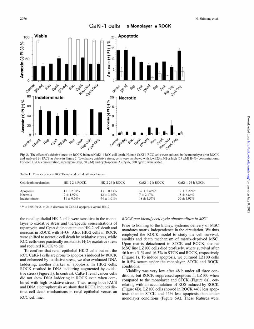
Because the mechanism of ROCK-induced cell death ap-pears to differ between renal epithelial and cancer cells(Figure 2 and Supplementary materials—Figure 1), we nextsought to evaluate the effect of oxidative stress in HK-2and CaKi-1 cells in the monolayer or ROCK using low[25 µM] or high [75 µM] H2O2 concentrations (Figures 3and 4). These concentrations were selected based on previ-ous studies in lymphoma cell lines [14,17]. We also studiedthe effect of co-incubation with cyclosporine A (CysA),an inhibitor of cyclophilin D, an essential component ofthe mitochondrial permeability transition pore, mediatingoxidative stress. The control for CysA was rapamycin, aninhibitor of the mammalian target of rapamycin (mTOR).While the CaKi-1 RCC line was highly resistant in themonolayer to oxidative stress or therapeutic concentrationsof rapamycin, its viability was reduced in ROCK (Figure 3).Furthermore, low and high oxidative stress or rapamycincould not augment ROCK-induced CaKi-1 cell death, indi-cating that ROCK results in a major breakdown of cellularhomeostasis. While CysA had no effect on CaKi-1 celldeath in the monolayer or ROCK, it attenuated necrotic celldeath induced by the combination of ROCK and oxidativestress (Figure 3). CysA decreased cell death in ROCK withlow-dose H2O2 by 31.1% and by 24.3% in ROCK withhigh-dose H2O2. Of note, because CysA exerted a similarimpact on the degree of both pure necrosis and PI-positiveannexin V-positive cells in both the two H2O2 concentra-tions (Figure 3), these dead cells of indeterminate causemay primarily reflect necrosis.
This finding was recapitulated in the combined effect ofROCK and oxidative stress on the indeterminate cell frac-tion, showing similarity to purely necrotic cells rather thanpurely apoptotic cells. Of special note, oxidative stress en-hanced necrosis (but not apoptosis) in CaKi-1 cells only inROCK but not in the monolayer (Figure 3). Most of theseeffects differed dramatically in HK-2 cells (Figure 4), i.e.
by guest on July 8, 2015http://ndt.oxfordjournals.org/
Dow
nloaded from
2076 N. Shimony et al.
Fig. 3. The effect of oxidative stress on ROCK-induced CaKi-1 RCC cell death. Human CaKi-1 RCC cells were cultured in the monolayer or in ROCKand analysed by FACS as above in Figure 2. To enhance oxidative stress, cells were incubated with low [25 µM] or high [75 µM] H2O2 concentrations.For each H2O2 concentration, rapamycin (Rap, 50 µM) and cyclosporine A (CysA, 300 ng/ml) were added.
Table 1. Time-dependent ROCK-induced cell death mechanism
Cell death mechanism HK-2 2-h ROCK HK-2 24-h ROCK CaKi-1 2-h ROCK CaKi-1 24-h ROCK
Apoptosis 11 ± 2.08% 13 ± 0.33% 37 ± 2.48%∗ 17 ± 3.29%∗Necrosis 2 ± 1.97% 12 ± 3.45% 7 ± 2.17% 15 ± 6.84%Indeterminate 11 ± 0.56% 44 ± 1.01% 18 ± 1.57% 36 ± 1.92%
∗P < 0.05 for 2- to 24-h decrease in CaKi-1 apoptosis versus HK-2.
the renal epithelial HK-2 cells were sensitive in the mono-layer to oxidative stress and therapeutic concentrations ofrapamycin, and CysA did not attenuate HK-2 cell death andnecrosis in ROCK with H2O2. Also, HK-2 cells in ROCKwere shifted to necrotic cell death by oxidative stress, whileRCC cells were practically resistant to H2O2 oxidative stressand required ROCK to die.
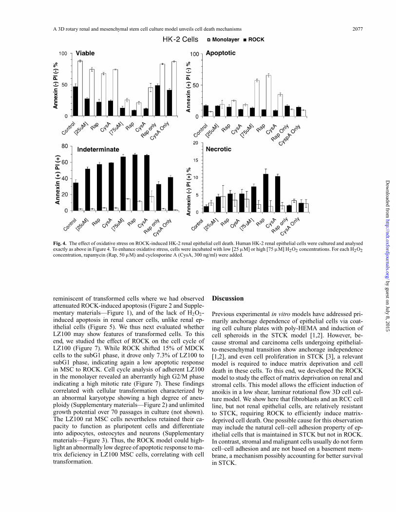
To confirm that renal epithelial HK-2 cells but not theRCC CaKi-1 cells are prone to apoptosis induced by ROCKand enhanced by oxidative stress, we also evaluated DNAladdering, another marker of apoptosis. In HK-2 cells,ROCK resulted in DNA laddering augmented by oxida-tive stress (Figure 5). In contrast, CaKi-1 renal cancer cellsdid not show DNA laddering in ROCK even when com-bined with high oxidative stress. Thus, using both FACSand DNA electrophoresis we show that ROCK induces dis-tinct cell death mechanisms in renal epithelial versus an
RCC cell line.
ROCK can identify cell cycle abnormalities in MSC
Prior to homing to the kidney, systemic delivery of MSCmandates matrix independence in the circulation. We thusemployed the ROCK model to study the cell survival,anoikis and death mechanism of matrix-deprived MSC.Upon matrix detachment in STCK and ROCK, the ratMSC line LZ100 cells died profusely, where survival after46 h was 31% and 16.3% in STCK and ROCK, respectively(Figure 1). To induce apoptosis, we cultured LZ100 cellsin 0.5% serum under the monolayer, STCK and ROCKconditions.
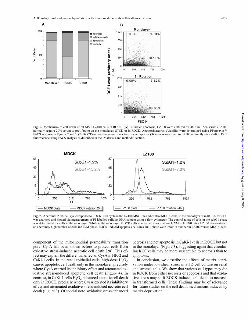
Viability was very low after 48 h under all three con-ditions, but ROCK suppressed apoptosis in LZ100 whencompared to the monolayer and STCK (Figure 6a), cor-relating with an accumulation of ROS induced by ROCK(Figure 6B). LZ100 cells showed in ROCK 44% less apop-tosis than in STCK and 45% less apoptosis than undermonolayer conditions (Figure 6A). These features were
by guest on July 8, 2015http://ndt.oxfordjournals.org/
Dow
nloaded from
A 3D rotary renal and mesenchymal stem cell culture model unveils cell death mechanisms 2077
Fig. 4. The effect of oxidative stress on ROCK-induced HK-2 renal epithelial cell death. Human HK-2 renal epithelial cells were cultured and analysedexactly as above in Figure 4. To enhance oxidative stress, cells were incubated with low [25 µM] or high [75 µM] H2O2 concentrations. For each H2O2concentration, rapamycin (Rap, 50 µM) and cyclosporine A (CysA, 300 ng/ml) were added.
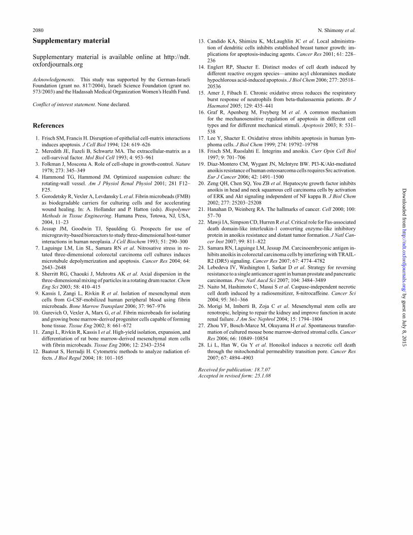
reminiscent of transformed cells where we had observedattenuated ROCK-induced apoptosis (Figure 2 and Supple-mentary materials—Figure 1), and of the lack of H2O2-induced apoptosis in renal cancer cells, unlike renal ep-ithelial cells (Figure 5). We thus next evaluated whetherLZ100 may show features of transformed cells. To thisend, we studied the effect of ROCK on the cell cycle ofLZ100 (Figure 7). While ROCK shifted 15% of MDCKcells to the subG1 phase, it drove only 7.3% of LZ100 tosubG1 phase, indicating again a low apoptotic responsein MSC to ROCK. Cell cycle analysis of adherent LZ100in the monolayer revealed an aberrantly high G2/M phaseindicating a high mitotic rate (Figure 7). These findingscorrelated with cellular transformation characterized byan abnormal karyotype showing a high degree of aneu-ploidy (Supplementary materials—Figure 2) and unlimitedgrowth potential over 70 passages in culture (not shown).The LZ100 rat MSC cells nevertheless retained their ca-pacity to function as pluripotent cells and differentiateinto adipocytes, osteocytes and neurons (Supplementarymaterials—Figure 3). Thus, the ROCK model could high-light an abnormally low degree of apoptotic response to ma-trix deficiency in LZ100 MSC cells, correlating with celltransformation.
Discussion
Previous experimental in vitro models have addressed pri-marily anchorage dependence of epithelial cells via coat-ing cell culture plates with poly-HEMA and induction ofcell spheroids in the STCK model [1,2]. However, be-cause stromal and carcinoma cells undergoing epithelial-to-mesenchymal transition show anchorage independence[1,2], and even cell proliferation in STCK [3], a relevantmodel is required to induce matrix deprivation and celldeath in these cells. To this end, we developed the ROCKmodel to study the effect of matrix deprivation on renal andstromal cells. This model allows the efficient induction ofanoikis in a low shear, laminar rotational flow 3D cell cul-ture model. We show here that fibroblasts and an RCC cellline, but not renal epithelial cells, are relatively resistantto STCK, requiring ROCK to efficiently induce matrix-deprived cell death. One possible cause for this observationmay include the natural cell–cell adhesion property of ep-ithelial cells that is maintained in STCK but not in ROCK.In contrast, stromal and malignant cells usually do not formcell–cell adhesion and are not based on a basement mem-brane, a mechanism possibly accounting for better survivalin STCK.
by guest on July 8, 2015http://ndt.oxfordjournals.org/
Dow
nloaded from
2078 N. Shimony et al.
Fig. 5. The effect of oxidative stress on ROCK-induced renal cell death.DNA gel electrophoresis showing apoptosis in HK-2 but not in CaKi-1 cells. Cells were cultured in the monolayer or in ROCK for 2 h with0.5% serum, with or without H2O2. After 2 h, cells were collected, DNAextracted and 2 µg was loaded in each lane. Lanes are: (1) HK-2 monolayer,(2) HK-2 in ROCK, (3) HK-2 in ROCK + 25 µM H2O2, (4) HK-2 inROCK + 75 µM H2O2, (5) CaKi-1 in ROCK and (6) CaKi-1 in ROCK+ 75 µM H2O2.
The ROCK model could provide valuable informationregarding apoptosis or necrosis as the primary cell deathmechanism in a variety of cell types, generally corre-lating with the cell transformation status. Activation ofnon-receptor tyrosine kinases in transformed cells was pre-viously shown to confer anoikis resistance via differentpathways, e.g. v-SRC [1], phosphorylated FAK [18], phos-phorylated SRC, the PI3-kinase/AKT pathway [19] or ERK[20]. Our data show that in addition to anoikis, necrosisis a major cell death mechanism induced by anoikis intransformed cells. This conclusion is based on the obser-vation that renal epithelial cells showed primarily ROCK-induced apoptosis, while malignant and transformed cellsmanifested substantial necrosis under ROCK conditions.
Two possible mechanisms may account for a higher rateof necrosis in transformed cells. First, upregulation of in-trinsic anti-apoptotic cellular pathways in transformed cells[21] may shift these cells to necrosis rather than apoptosisupon derangement of cell homeostasis by matrix depriva-tion. Furthermore, following the report that oxidative stressenhances necrosis in lymphoma cells [17], we observedthat oxidative stress induces apoptosis in the epithelial HK-2 cell line, in contrast to necrosis in CaKi-1 cells. Thecombination of matrix deprivation in ROCK and oxidativestress further accentuated the difference in cell death inthe epithelial versus malignant renal cell lines. Thus, while
oxidative stress drove HK-2 cells to apoptotic cell deathin the monolayer, it had practically no effect on adherentCaKi-1 cells. In contrast, while under ROCK conditionsCaKi-1 cells died profusely of both apoptosis and necrosis,oxidative stress, combined with ROCK enhanced necrosisbut decreased apoptosis. Recent data suggest that FLIP, anendogenous inhibitor of Fas death receptor signalling, andTRAIL, the ligand of DR5, may confer resistance to anoikisin prostate cancer and colorectal cancer cells, respectively[22,23]. It will be of interest to examine whether FLIP alsoplays a role in the resistance of circulating renal cancer cellsto matrix deprivation. Second, oxidative stress, resultingfrom matrix deprivation [7], may cause depletion of ATP.Because apoptosis is an energy-requiring process, ATP de-pletion may shift cells to necrosis [17]. Transformed cells,having a higher metabolic rate, may be thus more prone tonecrosis. In this regard, because oxidative and nitrosativestress may circumvent apoptosis resistance in cancer cellswhile sparing normal cells [24,25], induction of alterna-tive pathways of renal cancer cell death may circumventapoptosis resistance.
Our original observation of necrosis as a primary deathpathway in matrix-deprived cancer cells is supported by theresults of Jessup et al. [7]. While these authors referredonly to suspension-induced apoptosis, their flow cytom-etry results clearly show necrosis in colorectal cells sus-pended in the RWV model, as evident by 44% PI-positive,annexin V-negative cells versus only 2% PI-negative, an-nexin V-positive cells. Furthermore, these authors specifi-cally noted that ROCK-induced colorectal cancer cell deathwas not associated with DNA degradation, a typical findingin apoptosis [7]. This finding is in accord with our findingof enhancement of DNA degradation by oxidative stressin renal epithelial HK-2 cells in ROCK, but not in RCCCaKi-1 cells in ROCK (Figure 5). Thus, necrotic cell deathmay be an important general cancer cell death mechanismunder matrix-deficiency conditions, where enhancement ofcancer cell oxidative stress may be a potential therapeutictarget. This hypothesis is further supported by the previ-ous report that lymphoma cells exposed to severe oxidativestress are shifted from apoptosis to necrosis [17].
MSC have been suggested as renoprotective agents in thecontext of acute renal failure [26]. Because we observed inthe rat MSC LZ100 a lower degree of ROCK-induced apop-tosis than in STCK or the monolayer, it was of interest toassess their cell cycle response to ROCK. LZ100 displayeda baseline deranged cell cycle and an abnormal, low apop-totic response to ROCK. These abnormalities correlatedwith the karyotype aneuploidy of LZ100. Thus, despitetheir capacity to differentiate into various mesenchymallineages, showing even higher plasticity and differentiationcapacity than other primary rat MSC lines, LZ100 trans-formation raises concern regarding the use of high-passageMSC for clinical purposes, as has been recently suggestedfor mouse MSC that became spontaneously malignant [27].
CysA showed a dual effect on renal cells. In the HK-2 renal tubular epithelial cells, CysA enhanced apoptosis(Figure 4) while in RCC cells, it attenuated the additiveeffect of oxidative stress on ROCK-induced necrotic celldeath (Figure 3). These findings may be compatible withan inhibitory role of CysA on cyclophilin D, an essential
by guest on July 8, 2015http://ndt.oxfordjournals.org/
Dow
nloaded from
A 3D rotary renal and mesenchymal stem cell culture model unveils cell death mechanisms 2079
Fig. 6. Mechanism of cell death of rat MSC LZ100 cells in ROCK. (A) To induce apoptosis, LZ100 were cultured for 48 h in 0.5% serum (LZ100normally require 20% serum to proliferate) on the monolayer, STCK or in ROCK. Apoptosis/necrosis/viability were determined using PI/annexin VFACS as above in Figures 2 and 3. (B) ROCK-induced increase in reactive oxygen species (ROS) was measured in LZ100 indirectly via a shift in DCFfluorescence using FACS analysis as described in the ‘Materials and methods’ section.
Fig. 7. Aberrant LZ100 cell cycle response to ROCK. Cell cycle in the LZ100 MSC line and control MDCK cells, in the monolayer or in ROCK for 24 h,was analysed and plotted via measurement of PI-labelled cellular DNA content using a flow cytometer. The control range of cells in the subG1 phasewas determined for cells in the monolayer. While in the monolayer MDCK cells maintained a normal low G2/M to G1/G0 ratio, LZ100 demonstratedan aberrantly high number of cells in G2/M phase. ROCK-induced apoptosis cells in subG1 phase were lower in number in LZ100 versus MDCK cells.
component of the mitochondrial permeability transitionpore. CysA has been shown before to protect cells fromoxidative stress-induced necrotic cell death [28]. This ef-fect may explain the differential effect of CysA in HK-2 andCaKi-1 cells. In the renal epithelial cells, high-dose H2O2caused apoptotic cell death only in the monolayer, preciselywhere CysA exerted its inhibitory effect and attenuated ox-idative stress-induced apoptotic cell death (Figure 4). Incontrast, in CaKi-1 cells H2O2 enhanced necrotic cell deathonly in ROCK, precisely where CysA exerted its inhibitoryeffect and attenuated oxidative stress-induced necrotic celldeath (Figure 3). Of special note, oxidative stress enhanced
necrosis and not apoptosis in CaKi-1 cells in ROCK but notin the monolayer (Figure 3), suggesting again that circulat-ing RCC cells may be more susceptible to necrosis than toapoptosis.
In conclusion, we describe the effects of matrix depri-vation under low shear stress in a 3D cell culture on renaland stromal cells. We show that various cell types may diein ROCK from either necrosis or apoptosis and that oxida-tive stress may shift ROCK-induced cell death to necrosisin transformed cells. These findings may be of relevancefor future studies on the cell death mechanisms induced bymatrix deprivation.
by guest on July 8, 2015http://ndt.oxfordjournals.org/
Dow
nloaded from
2080 N. Shimony et al.
Supplementary material
Supplementary material is available online at http://ndt.oxfordjournals.org
Acknowledgements. This study was supported by the German-IsraeliFoundation (grant no. 817/2004), Israeli Science Foundation (grant no.573/2003) and the Hadassah Medical Organization Women’s Health Fund.
Conflict of interest statement. None declared.
References
1. Frisch SM, Francis H. Disruption of epithelial cell-matrix interactionsinduces apoptosis. J Cell Biol 1994; 124: 619–626
2. Meredith JE, Fazeli B, Schwartz MA. The extracellular-matrix as acell-survival factor. Mol Biol Cell 1993; 4: 953–961
3. Folkman J, Moscona A. Role of cell-shape in growth-control. Nature1978; 273: 345–349
4. Hammond TG, Hammond JM. Optimized suspension culture: therotating-wall vessel. Am J Physiol Renal Physiol 2001; 281 F12–F25.
5. Gorodetsky R, Vexler A, Levdansky L et al. Fibrin microbeads (FMB)as biodegradable carriers for culturing cells and for acceleratingwound healing. In: A. Hollander and P. Hatton (eds). BiopolymerMethods in Tissue Engineering. Humana Press, Totowa, NJ, USA,2004, 11–23
6. Jessup JM, Goodwin TJ, Spaulding G. Prospects for use ofmicrogravity-based bioreactors to study three-dimensional host-tumorinteractions in human neoplasia. J Cell Biochem 1993; 51: 290–300
7. Laguinge LM, Lin SL, Samara RN et al. Nitrosative stress in ro-tated three-dimensional colorectal carcinoma cell cultures inducesmicrotubule depolymerization and apoptosis. Cancer Res 2004; 64:2643–2648
8. Sherritt RG, Chaouki J, Mehrotra AK et al. Axial dispersion in thethree-dimensional mixing of particles in a rotating drum reactor. ChemEng Sci 2003; 58: 410–415
9. Kassis I, Zangi L, Rivkin R et al. Isolation of mesenchymal stemcells from G-CSF-mobilized human peripheral blood using fibrinmicrobeads. Bone Marrow Transplant 2006; 37: 967–976
10. Gurevich O, Vexler A, Marx G, et al. Fibrin microbeads for isolatingand growing bone marrow-derived progenitor cells capable of formingbone tissue. Tissue Eng 2002; 8: 661–672
11. Zangi L, Rivkin R, Kassis I et al. High-yield isolation, expansion, anddifferentiation of rat bone marrow-derived mesenchymal stem cellswith fibrin microbeads. Tissue Eng 2006; 12: 2343–2354
12. Baatout S, Herradji H. Cytometric methods to analyze radiation ef-fects. J Biol Regul 2004; 18: 101–105
13. Candido KA, Shimizu K, McLaughlin JC et al. Local administra-tion of dendritic cells inhibits established breast tumor growth: im-plications for apoptosis-inducing agents. Cancer Res 2001; 61: 228–236
14. Englert RP, Shacter E. Distinct modes of cell death induced bydifferent reactive oxygen species—amino acyl chloramines mediatehypochlorous acid-induced apoptosis. J Biol Chem 2006; 277: 20518–20536
15. Amer J, Fibach E. Chronic oxidative stress reduces the respiratoryburst response of neutrophils from beta-thalassaemia patients. Br JHaematol 2005; 129: 435–441
16. Graf R, Apenberg M, Freyberg M et al. A common mechanismfor the mechanosensitive regulation of apoptosis in different celltypes and for different mechanical stimuli. Apoptosis 2003; 8: 531–538
17. Lee Y, Shacter E. Oxidative stress inhibits apoptosis in human lym-phoma cells. J Biol Chem 1999; 274: 19792–19798
18. Frisch SM, Ruoslahti E. Integrins and anoikis. Curr Opin Cell Biol1997; 9: 701–706
19. Diaz-Montero CM, Wygant JN, McIntyre BW. PI3-K/Akt-mediatedanoikis resistance of human osteosarcoma cells requires Src activation.Eur J Cancer 2006; 42: 1491–1500
20. Zeng QH, Chen SQ, You ZB et al. Hepatocyte growth factor inhibitsanoikis in head and neck squamous cell carcinoma cells by activationof ERK and Akt signaling independent of NF kappa B. J Biol Chem2002; 277: 25203–25208
21. Hanahan D, Weinberg RA. The hallmarks of cancer. Cell 2000; 100:57–70
22. Mawji IA, Simpson CD, Hurren R et al. Critical role for Fas-associateddeath domain-like interleukin-1 converting enzyme-like inhibitoryprotein in anoikis resistance and distant tumor formation. J Natl Can-cer Inst 2007; 99: 811–822
23. Samara RN, Laguinge LM, Jessup JM. Carcinoembryonic antigen in-hibits anoikis in colorectal carcinoma cells by interfering with TRAIL-R2 (DR5) signaling. Cancer Res 2007; 67: 4774–4782
24. Lebedeva IV, Washington I, Sarkar D et al. Strategy for reversingresistance to a single anticancer agent in human prostate and pancreaticcarcinomas. Proc Natl Aacd Sci 2007; 104: 3484–3489
25. Naito M, Hashimoto C, Masui S et al. Caspase-independent necroticcell death induced by a radiosensitizer, 8-nitrocaffeine. Cancer Sci2004; 95: 361–366
26. Morigi M, Imberti B, Zoja C et al. Mesenchymal stem cells arerenotropic, helping to repair the kidney and improve function in acuterenal failure. J Am Soc Nephrol 2004; 15: 1794–1804
27. Zhou YF, Bosch-Marce M, Okuyama H et al. Spontaneous transfor-mation of cultured mouse bone marrow-derived stromal cells. CancerRes 2006; 66: 10849–10854
28. Li L, Han W, Gu Y et al. Honoikol induces a necrotic cell deaththrough the mitochondrial permeability transition pore. Cancer Res2007; 67: 4894–4903
Received for publication: 18.7.07Accepted in revised form: 25.1.08
by guest on July 8, 2015http://ndt.oxfordjournals.org/
Dow
nloaded from
Related Documents