A 1-Megadalton Translocation Complex Containing Tic20 and Tic21 Mediates Chloroplast Protein Import at the Inner Envelope Membrane W Shingo Kikuchi, a Maya Oishi, a Yoshino Hirabayashi, a Dong Wook Lee, b Inhwan Hwang, b and Masato Nakai a,1 a Institute for Protein Research, Osaka University, Suita, Osaka 565-0871, Japan b Division of Integrative Biosciences and Biotechnology and Division of Molecular and Life Sciences, POSTECH, Pohang 790-784, Korea Chloroplast protein import is mediated by two hetero-oligomeric protein complexes, the Tic and Toc translocons, which are located in the inner and outer envelope membranes. At the inner membrane, many Tic components have been identified and characterized, but it remains unclear how these Tic proteins are organized to form a protein-conducting channel or whether a stable Tic core complex that binds a translocating preproteins exists. Here, we report the identification of a 1-megadalton (MD) translocation complex as an intermediate during protein translocation across the inner membrane in Arabidopsis thaliana and pea (Pisum sativum). This complex can be detected by blue native PAGE using the mild detergent digitonin without any chemical cross-linkers. The preprotein arrested in the 1-MD complex can be chased into its fully translocated form after a subsequent incubation. While Tic20 and Tic21 appear to be involved in the 1-MD complex, Tic110, a well- characterized Tic component, exists as a distinct entity from the complex. Several lines of evidence suggest that the 1-MD complex functions in between the Toc and Tic110-containing complexes, most likely as a protein-conducting channel at the inner envelope. INTRODUCTION Nuclear-encoded chloroplast proteins are synthesized in the cytosol as preproteins with N-terminal targeting signals, called transit peptides. These proteins are then posttranslationally imported across the double envelope membranes of chloro- plasts. The chloroplastic outer and inner envelope membranes contain multisubunit machinery for the import of preproteins, termed the Toc (translocon at the outer envelope membrane of chloroplasts) and the Tic (translocon at the inner envelope membrane of chloroplasts) complexes, respectively (for reviews, see Soll and Schleiff, 2004; Be ´ dard and Jarvis, 2005; Jarvis, 2008; Kessler and Schnell, 2009). During import, the Toc and Tic complexes are thought to come together at contact sites where the outer and inner membranes are in proximity, allowing the preprotein to pass through both membranes simultaneously (Schnell and Blobel, 1993). During or shortly after import, the transit peptide is removed by a stromal processing peptidase, and the mature protein is then folded or targeted to one of the internal compartments. Protein import into chloroplasts requires ATP hydrolysis. In the presence of low concentrations of ATP (<100 mM), irreversible binding of preproteins to the translocon components occurs. Although it is not clear where the ATP-utilizing component may reside, previous studies have shown that a stable association of preproteins with the translocon components, the so-called early import intermediate, was generated under low ATP concentra- tions (Waegemann and Soll, 1991; Perry and Keegstra, 1994; Schnell et al., 1994; Ma et al., 1996; Akita et al., 1997; Kouranov and Schnell, 1997; Nielsen et al., 1997; Young et al., 1999; Inoue and Akita, 2008). In the presence of higher concentrations of ATP (>1 mM), complete translocation of preproteins across the dou- ble envelope of chloroplasts occurs. This high ATP level is probably required by stromal molecular chaperones believed to provide the driving force for unidirectional translocation of pre- proteins. At the outer membrane, Toc75, Toc159, and Toc34 form a stable complex and mediate the transfer of preproteins through the outer membrane (Schleiff et al., 2003; Kikuchi et al., 2006). At the inner membrane, eight proteins (Tic110, Tic40, Tic20, Tic21, Tic22, Tic55, Tic62, and Tic32) are reported to be involved in the import process (Soll and Schleiff, 2004; Be ´ dard and Jarvis, 2005; Teng et al., 2006; Jarvis, 2008; Kessler and Schnell, 2009). Tic20 has been proposed to function as the protein-conducting chan- nel of the inner membrane. Tic20 was identified by chemical cross-linking to translocating preproteins in pea (Pisum sativum) chloroplasts (Kouranov et al., 1998). Tic20 is an integral mem- brane protein and is predicted to have three or four transmem- brane helices. The reduction of Tic20 levels in Arabidopsis thaliana antisense plants produced a specific defect in protein translocation across the inner membrane (Chen et al., 2002). Tic110 is predicted to have two transmembrane helices at its N terminus and a large hydrophilic C-terminal domain, which was 1 Address correspondence to [email protected]. The author responsible for distribution of materials integral to the findings presented in this article in accordance with the policy described in the Instructions for Authors (www.plantcell.org) is: Masato Nakai ([email protected]). W Online version contains Web-only data. www.plantcell.org/cgi/doi/10.1105/tpc.108.063552 This article is a Plant Cell Advance Online Publication. The date of its first appearance online is the official date of publication. The article has been edited and the authors have corrected proofs, but minor changes could be made before the final version is published. Posting this version online reduces the time to publication by several weeks. The Plant Cell Preview, www.aspb.org ã 2009 American Society of Plant Biologists 1 of 17

Welcome message from author
This document is posted to help you gain knowledge. Please leave a comment to let me know what you think about it! Share it to your friends and learn new things together.
Transcript

A 1-Megadalton Translocation Complex Containing Tic20 andTic21 Mediates Chloroplast Protein Import at the InnerEnvelope Membrane W
Shingo Kikuchi,a Maya Oishi,a Yoshino Hirabayashi,a Dong Wook Lee,b Inhwan Hwang,b and Masato Nakaia,1
a Institute for Protein Research, Osaka University, Suita, Osaka 565-0871, Japanb Division of Integrative Biosciences and Biotechnology and Division of Molecular and Life Sciences, POSTECH, Pohang
790-784, Korea
Chloroplast protein import is mediated by two hetero-oligomeric protein complexes, the Tic and Toc translocons, which are
located in the inner and outer envelope membranes. At the inner membrane, many Tic components have been identified and
characterized, but it remains unclear how these Tic proteins are organized to form a protein-conducting channel or whether
a stable Tic core complex that binds a translocating preproteins exists. Here, we report the identification of a 1-megadalton
(MD) translocation complex as an intermediate during protein translocation across the inner membrane in Arabidopsis
thaliana and pea (Pisum sativum). This complex can be detected by blue native PAGE using the mild detergent digitonin
without any chemical cross-linkers. The preprotein arrested in the 1-MD complex can be chased into its fully translocated
form after a subsequent incubation. While Tic20 and Tic21 appear to be involved in the 1-MD complex, Tic110, a well-
characterized Tic component, exists as a distinct entity from the complex. Several lines of evidence suggest that the 1-MD
complex functions in between the Toc and Tic110-containing complexes, most likely as a protein-conducting channel at the
inner envelope.
INTRODUCTION
Nuclear-encoded chloroplast proteins are synthesized in the
cytosol as preproteins with N-terminal targeting signals, called
transit peptides. These proteins are then posttranslationally
imported across the double envelope membranes of chloro-
plasts. The chloroplastic outer and inner envelope membranes
contain multisubunit machinery for the import of preproteins,
termed the Toc (translocon at the outer envelope membrane of
chloroplasts) and the Tic (translocon at the inner envelope
membrane of chloroplasts) complexes, respectively (for reviews,
see Soll and Schleiff, 2004; Bedard and Jarvis, 2005; Jarvis,
2008; Kessler and Schnell, 2009). During import, the Toc and Tic
complexes are thought to come together at contact sites where
the outer and inner membranes are in proximity, allowing the
preprotein to pass through both membranes simultaneously
(Schnell and Blobel, 1993). During or shortly after import, the
transit peptide is removed by a stromal processing peptidase,
and the mature protein is then folded or targeted to one of the
internal compartments.
Protein import into chloroplasts requires ATP hydrolysis. In the
presence of low concentrations of ATP (<100 mM), irreversible
binding of preproteins to the translocon components occurs.
Although it is not clear where the ATP-utilizing component may
reside, previous studies have shown that a stable association of
preproteins with the translocon components, the so-called early
import intermediate, was generated under low ATP concentra-
tions (Waegemann and Soll, 1991; Perry and Keegstra, 1994;
Schnell et al., 1994; Ma et al., 1996; Akita et al., 1997; Kouranov
and Schnell, 1997; Nielsen et al., 1997; Young et al., 1999; Inoue
and Akita, 2008). In the presence of higher concentrations of ATP
(>1 mM), complete translocation of preproteins across the dou-
ble envelope of chloroplasts occurs. This high ATP level is
probably required by stromal molecular chaperones believed to
provide the driving force for unidirectional translocation of pre-
proteins.
At the outer membrane, Toc75, Toc159, and Toc34 form a
stable complex and mediate the transfer of preproteins through
the outer membrane (Schleiff et al., 2003; Kikuchi et al., 2006). At
the inner membrane, eight proteins (Tic110, Tic40, Tic20, Tic21,
Tic22, Tic55, Tic62, and Tic32) are reported to be involved in the
import process (Soll and Schleiff, 2004; Bedard and Jarvis, 2005;
Teng et al., 2006; Jarvis, 2008; Kessler and Schnell, 2009). Tic20
has been proposed to function as the protein-conducting chan-
nel of the inner membrane. Tic20 was identified by chemical
cross-linking to translocating preproteins in pea (Pisum sativum)
chloroplasts (Kouranov et al., 1998). Tic20 is an integral mem-
brane protein and is predicted to have three or four transmem-
brane helices. The reduction of Tic20 levels in Arabidopsis
thaliana antisense plants produced a specific defect in protein
translocation across the inner membrane (Chen et al., 2002).
Tic110 is predicted to have two transmembrane helices at its N
terminus and a large hydrophilic C-terminal domain, which was
1Address correspondence to [email protected] author responsible for distribution of materials integral to thefindings presented in this article in accordance with the policy describedin the Instructions for Authors (www.plantcell.org) is: Masato Nakai([email protected]).WOnline version contains Web-only data.www.plantcell.org/cgi/doi/10.1105/tpc.108.063552
This article is a Plant Cell Advance Online Publication. The date of its first appearance online is the official date of publication. The article has been
edited and the authors have corrected proofs, but minor changes could be made before the final version is published. Posting this version online
reduces the time to publication by several weeks.
The Plant Cell Preview, www.aspb.org ã 2009 American Society of Plant Biologists 1 of 17
shown to be exposed to the stromal compartment (Jackson
et al., 1998). The stromal domain of Tic110 has been proposed to
function as a molecular scaffold by binding the preprotein and
recruiting the stromal chaperone Hsp93 with the assistance of
the putative cochaperone Tic40 (Akita et al., 1997; Nielsen et al.,
1997; Chou et al., 2003; Chou et al., 2006). These three proteins
(Tic110, Tic40, and Hsp93) are thought to drive protein import
into the stroma through repeated cycles of binding and release.
Although an alternative model for the topology and function of
Tic110 has also been proposed, in which Tic110 is a polytopic
membrane protein that functions as a protein-conducting channel
(Heins et al., 2002; Balsera et al., 2009), a truncated version of
Tic110 lacking the N-terminal transmembrane helices was shown
to exist as a soluble protein when expressed in Escherichia coli or
in the stroma of transgenic Arabidopsis (Inaba et al., 2003).
However, the existence of a stable Tic complex containing a
protein-conducting channel remains unclear. Here, we report the
identification of a 1-MD translocation complex as an intermedi-
ate during protein translocation across the inner membrane. This
complex can be detected by blue native PAGE (BN-PAGE) using
the mild detergent digitonin without any chemical cross-linkers.
The preprotein arrested in the 1-MD translocation complex can
be chased into its fully translocated form after a subsequent
incubation. Antibody-shift BN-PAGE, immunodepletion, and im-
munoprecipitation assays suggest that Tic20 and Tic21 are
involved in the 1-MD translocation complex but that Tic110 is
not involved in this complex.
RESULTS
A Translocation Intermediate Complex Was Observed
by BN-PAGE
BN-PAGE allows the separation of membrane protein com-
plexes under nondenaturing conditions (Schagger and von
Jagow, 1991; Schagger et al., 1994). We examined whether
BN-PAGE is applicable for the analysis of preproteins in the
process of translocation across the double envelope mem-
branes of chloroplasts. The precursor of the small subunit of
ribulose-1,5-bisphosphate carboxylase/oxygenase (pSSU) was
used as a model protein. pSSU was synthesized in vitro in the
presence of [35S]Met. In vitro import reactions were performed in
the presence of different concentrations of ATP using pea
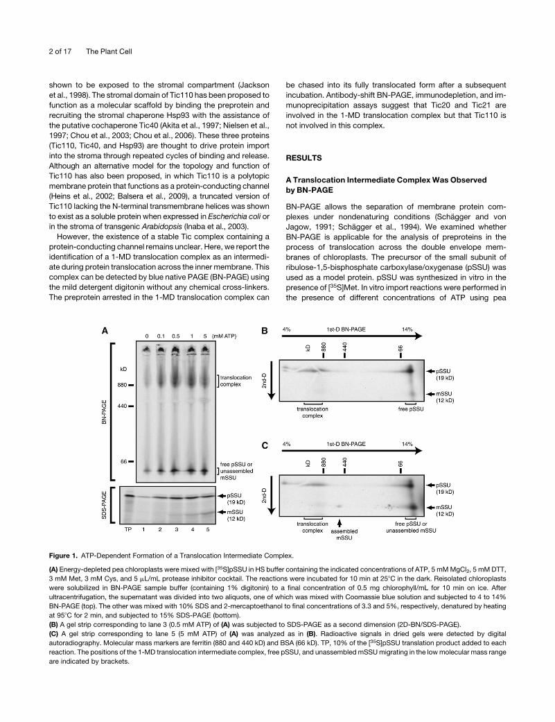
Figure 1. ATP-Dependent Formation of a Translocation Intermediate Complex.
(A) Energy-depleted pea chloroplasts were mixed with [35S]pSSU in HS buffer containing the indicated concentrations of ATP, 5 mMMgCl2, 5 mMDTT,
3 mM Met, 3 mM Cys, and 5 mL/mL protease inhibitor cocktail. The reactions were incubated for 10 min at 258C in the dark. Reisolated chloroplasts
were solubilized in BN-PAGE sample buffer (containing 1% digitonin) to a final concentration of 0.5 mg chlorophyll/mL for 10 min on ice. After
ultracentrifugation, the supernatant was divided into two aliquots, one of which was mixed with Coomassie blue solution and subjected to 4 to 14%
BN-PAGE (top). The other was mixed with 10% SDS and 2-mercaptoethanol to final concentrations of 3.3 and 5%, respectively, denatured by heating
at 958C for 2 min, and subjected to 15% SDS-PAGE (bottom).
(B) A gel strip corresponding to lane 3 (0.5 mM ATP) of (A) was subjected to SDS-PAGE as a second dimension (2D-BN/SDS-PAGE).
(C) A gel strip corresponding to lane 5 (5 mM ATP) of (A) was analyzed as in (B). Radioactive signals in dried gels were detected by digital
autoradiography. Molecular mass markers are ferritin (880 and 440 kD) and BSA (66 kD). TP, 10% of the [35S]pSSU translation product added to each
reaction. The positions of the 1-MD translocation intermediate complex, free pSSU, and unassembled mSSUmigrating in the lowmolecular mass range
are indicated by brackets.
2 of 17 The Plant Cell

chloroplasts. Chloroplasts were reisolated, solubilized with 1%
digitonin, and subjected to BN-PAGE and autoradiography (Fig-
ure 1A, top). Radioactive signals were found at, approximately,
the 1-MD area and at a low molecular mass (<66 kD). By SDS-
PAGE, the precursor form of SSU was observed at all tested
concentrations of ATP, and the mature form of SSU (mSSU) was
observed at relatively high concentrations of ATP (>1mM) (Figure
1A, bottom).
Figure 1B shows two-dimensional (2D)-BN/SDS-PAGE anal-
ysis of lane 3 (0.5 mM ATP) from Figure 1A. pSSU was found in
two peaks: one was in the 1-MD area, demonstrating that pSSU
was arrested in the 1-MD translocation complex, and the other
was in the low molecular mass range, probably representing
pSSU dissociated from the translocation machinery in the pres-
ence of detergent. Figure 1C shows 2D-BN/SDS-PAGE analysis
of lane 5 (5mMATP) from Figure 1A. pSSUwas also found in this
1-MD area. In addition,mSSUwas observed in the lowmolecular
mass range. Formation of the 1-MD translocation intermediate
complex wasmaximal at 0.5mMATP (Figure 1A, top). Therefore,
we decided to use this concentration to generate the transloca-
tion intermediate complex for subsequent experiments.
The 1-MD translocation complex was observed only when the
mild detergent digitonin was used. All tested detergents except
digitonin failed to preserve the 1-MD complex (i.e., 1% Triton
X-100, 1% Nonidet P-40, 1% dodecyl maltoside, 1% decyl
maltoside, or 1% octyl glucoside; data not shown). Moreover,
addition of either dodecyl maltoside or Triton X-100 to the
digitonin-solubilized BN-PAGE sample caused dissociation of
pSSU from the 1-MD complex (Figure 2A). These observations
suggest that pSSU is loosely associated with the 1-MD complex.
The possibility that the association of pSSU occurred artificially
after solubilization of the chloroplasts can be excluded because
pSSU was efficiently associated with the 1-MD complex in the
presence of ATP only when incubated with intact chloroplasts
but not with solubilized chloroplast extract (see Supplemental
Figure 1 online). A similar 1-MD translocation complex was also
observed when the precursor of ferredoxin:NADP+ oxidoreduc-
tase was used (see Supplemental Figure 2 online).
Formationof the1-MDTranslocationComplex IsBlockedby
Slowly Hydrolyzable Analogs of GTP and ATP
It is well known that Toc34 and Toc159 have GTP binding motifs,
and the binding of preproteins to the Toc complex is inhibited by
non- or slowly hydrolyzable analogs of GTP (Young et al., 1999;
Soll and Schleiff, 2004; Inoue and Akita, 2008; Jarvis, 2008;
Kessler and Schnell, 2009). To test whether the 1-MD translo-
cation intermediate complex is formed after translocation
Figure 2. Characteristic Features of the 1-MD Translocation Complex.
(A) After pea chloroplasts carrying [35S]pSSU were solubilized with 1% digitonin (Dig), either dodecyl maltoside (DDM, lane 2) or Triton X-100 (lane 3)
was added to give a final concentration of 1%. After incubation for 30 min on ice, samples were subjected to BN-PAGE.
(B) After pea chloroplasts were preincubated with the indicated concentrations of GTP-g-S or ATP-g-S for 10 min at 258C in the dark, [35S]pSSU and
ATP (final 0.5 mM) were added and incubated for another 10 min at 258C in the dark. Reisolated chloroplasts were solubilized and subjected to
BN-PAGE (top) and SDS-PAGE (bottom).
The 1-MDa Tic complex of chloroplasts 3 of 17

through the Toc complex via the general import pathway, we
examined the effect of a slowly hydrolyzable analog of GTP
(GTP-g-S). Chloroplasts were preincubatedwith GTP-g-S before
addition of pSSU and ATP. GTP-g-S drastically reduced the
formation of the 1-MD complex (Figure 2B), suggesting that
the 1-MD complex is formed after translocation through the
well-defined Toc complex. In addition, by preincubating chloro-
plasts with ATP-g-S, formation of the 1-MD complex was also
drastically reduced (Figure 2B). This result shows that hydrolysis
of ATP is also essential for the formation of the 1-MD complex.
ThePreproteinArrested in the1-MDTranslocationComplex
Can Be Chased into Its Fully Translocated Mature Form
Chase experiments indicated that, in the absence of ATP, almost
no import of pSSU, which had been arrested in the 1-MD
translocation complex, was observed (Figure 3, top). In the
presence of ATP (0.5 and 5 mM), the radiolabel in the 1-MD
complex gradually decreased during the chase period, while
radiolabeled ribulose-1,5-bis-phosphate carboxylase/oxygen-
ase (Rubisco) holoenzyme found at the 520-kDposition gradually
increased (Figure 3, top). SDS-PAGE analysis also showed that
pSSU gradually decreased, while fully imported mature SSU
gradually increased (Figure 3, bottom). This means that pSSU
arrested in the 1-MD complex was translocated, processed into
its mature form, and further assembled into the 520-kD Rubisco
holoenzyme in organello in the presence of higher concentra-
tions of ATP. Sincemature SSU contains only three of the sixMet
residues that are present in precursor SSU, the percentage of
bound pSSU that was chased into mSSU under 5 mM ATP
conditions for 30 min was estimated as 90%. The possibility that
pSSU is associated with a stromal chaperonin can be excluded
because addition of higher concentrations of ATP after solubi-
lization of chloroplasts did not affect the signal intensity of the
1-MD complex (see Supplemental Figure 1 online). Therefore, we
conclude that the radioactive 1-MD signal represents a chase-
able genuine translocation intermediate complex.
Protease Treatments Revealed That the Translocation
Intermediate Complex Is Thermolysin Resistant and
Partially Sensitive to Trypsin
To determine where the 1-MD translocation complex was accu-
mulated in chloroplasts, we performed a selective proteolysis
using exogenous thermolysin and trypsin. Thermolysin is an
outer envelope–impermeable protease that selectively digests
chloroplast surface-exposed proteins, whereas a certain con-
centration of trypsin is able to destroy the membrane integrity of
the outer envelope and partially digest proteins within the inter-
membrane space while leaving the stromal proteins undigested
(Kouranov et al., 1998; Hirohashi et al., 2001). Toc159, which has
a large cytosolically exposed domain, was easily degraded by
thermolysin and trypsin, as demonstrated by the resultant 52-kD
protease-resistant fragment (Figure 4B). Toc34, which also has a
cytosolically exposed domain, was resistant to up to 10mg/mL of
thermolysin (Chen et al., 2000) but was degraded by 100 mg/mL
of thermolysin and trypsin. Toc75, an integral membrane protein,
was largely resistant to thermolysin and trypsin. Tic22, which is
known to reside in the intermembrane space (Kouranov et al.,
1998), was resistant to thermolysin but sensitive to trypsin.
Stromal Hsp70 and Tic110, an inner envelopemembrane protein
that is oriented toward the stroma (Jackson et al., 1998; Inaba
et al., 2003), were completely resistant to both thermolysin and
trypsin (Figure 4B).
The 1-MD complex was largely resistant to exogenous ther-
molysin (Figure 4A, top, lanes 2 and 3). In SDS-PAGE, degrada-
tion products of SSU (SSU-DPs) were observed (Figure 4A,
bottom, lanes 2 and 3), indicating that theC-terminal tail of pSSU,
which was exposed to the surface of chloroplasts, was digested
by exogenous thermolysin, while the N-terminal part of pSSU,
which was probably deeply buried in a translocation channel,
was resistant to proteolysis. Furthermore, 2D-BN/SDS-PAGE of
lane 3 fromFigure 4A confirmed that the radioactive signals in the
1-MD area remaining after thermolysin treatment were derived
mostly from SSU-DP (Figure 4D). When chloroplasts carrying
pSSU were treated with 100 mg/mL trypsin, the radiolabeled
Figure 3. The Preprotein Arrested in the Translocation Intermediate
Complex Can Be Chased into Its Fully Imported Mature Form under High
ATP Concentrations.
The translocation intermediate was generated under 0.5-mM ATP as
described in Methods. Reisolated pea chloroplasts carrying [35S]pSSU
were resuspended in HS buffer containing the indicated concentrations
of ATP, 5 mM MgCl2, 5 mM DTT, 3 mM Met, 3 mM Cys, and 5 mL/mL
protease inhibitor cocktail on ice. Translocation (import) was resumed by
transferring the reaction tubes to a water bath (258C). The reactions were
terminated by chilling and centrifuging the chloroplasts at the indicated
time points. These samples were then solubilized and subjected to BN-
PAGE (top) and SDS-PAGE (bottom). As a control, chloroplasts carrying
preproteins were directly solubilized without the subsequent chase
reaction (lane 1). The positions of the 1-MD translocation intermediate
complex, the 520-kD Rubisco holoenzyme, free pSSU, and unassem-
bled mSSU are indicated by brackets.
4 of 17 The Plant Cell

Figure 4. Protease Treatments of the Chloroplast Surface Reveal That the 1-MD Translocation Complex Is Resistant to Thermolysin but Partially
Sensitive to Trypsin.
(A) to (E) The translocation intermediate was generated under 0.5-mM ATP as described in Methods. The pea chloroplast suspension was divided into
six aliquots (lanes 1 to 6) and washed twice with HS buffer in the absence of protease inhibitor cocktail. Chloroplasts carrying [35S]pSSU were treated
with the indicated concentrations of thermolysin (Thl) or trypsin (Trp) for 20 min on ice. After inactivation of the proteases, the chloroplast pellet was
solubilized and subjected to BN-PAGE ([A], top) and SDS-PAGE ([A], bottom). When using trypsin, trypsin inhibitor was added to BN-PAGE sample
buffer.
(B) Aliquots of the same samples in (A) were subjected to SDS-PAGE and immunoblotting with antibodies against indicated proteins. Arrows indicate
intact Toc159, 86- and 52-kD fragments of Toc159, and Tic22. An asterisk denotes a nonspecific cross-reacting band.
(C) A gel strip corresponding to lane 1 (without protease) of (A) was subjected to 2D-BN/SDS-PAGE.
(D) A gel strip corresponding to lane 3 (with thermolysin) of (A) was analyzed as in (C).
The 1-MDa Tic complex of chloroplasts 5 of 17

complex shifted slightly to a lower molecular mass on BN-PAGE
(Figure 4A, top, lane 6), indicating partial degradation of the 1-MD
complex. SSU-DPs were also found in the trypsin-treated sam-
ples by SDS-PAGE (Figure 4A, bottom, lanes 5 and 6). Figure 4E
shows 2D-BN/SDS-PAGE of lane 6 from Figure 4A, confirming
that the radioactive signals in the shifted complex were also
mostly derived from SSU-DP.
As we have shown previously, the intact Toc complex is 800 to
1000 kD on BN-PAGE (Kikuchi et al., 2006). As a result, we
initially suspected that the observed radiolabeled 1-MD complex
corresponded to the Toc complex itself due to their almost
identical sizes. Contrary to our expectations, the 1-MD complex
shown here was resistant to thermolysin. This observation was
inconsistent with the protease accessibilities of the Toc complex.
When chloroplasts were isolated in the absence of protease
inhibitor cocktail, the Toc complex became 350 to 500 kD in size
because the cytosolically exposed A-domain of Toc159 was
degraded by endogenous proteases during isolation of chloro-
plasts (Kikuchi et al., 2006; see Supplemental Figure 3 online,
lane 5). Irrespective of the presence or absence of protease
inhibitor cocktail during the isolation procedure, the radiolabeled
1-MD translocation complex was generated at nearly the same
levels (see Supplemental Figure 3 online, top, lanes 1 and 3).
Furthermore, as described above, while the 1-MD complex
remained resistant by thermolysin treatment, the Toc complex
was degraded to a 250- to 350-kD subcomplex in the same
samples (lanes 6 and 8). Therefore, we conclude that the 1-MD
complex does not correspond to the Toc complex itself. More
importantly, the observed partial degradation of the transloca-
tion complex after trypsin treatment suggests that the 1-MD
complex has an intermembrane space-facing domain.
TheTranslocation IntermediateAppears toBeAccumulated
in the Inner Envelope Membrane
To determine where [35S]pSSU was accumulated in chloro-
plasts, we separated envelope membrane vesicles into fractions
enriched in outer envelope membrane vesicles and inner enve-
lope membrane vesicles using sucrose density gradient centrif-
ugation (see Supplemental Figure 4 online). pSSU was found
mainly in the mixed outer-inner membrane fraction (fractions 4
and 5). A very minor portion of pSSU found in the outer mem-
brane fraction (fraction 8) may represent pSSU bound to Toc
complex that was not associated with contact sites. Thermoly-
sin-resistant SSU-DP was found exclusively in the mixed outer-
inner membrane fraction but not in the outer membrane fraction.
These observations suggest that the 1-MD complex in which the
N-terminal region of pSSU is stably associated resides in the
inner membrane rather than in the outer membrane.
Tic21 Is Involved in the 1-MD Translocation Complex
To identify which components are involved in the 1-MD complex,
we screened antisera against known Toc and Tic proteins by
antibody-shift BN-PAGE. In antibody-shift BN-PAGE, chloro-
plasts carrying pSSUwere first solubilized in digitonin-containing
buffer, followed by the addition of antibodies. The specific
binding of antibodies to a subunit(s) within the complex was
predicted to result in a shift of the complex to a higher molecular
mass on BN-PAGE. This assay was used in several reports (e.g.,
Johnston et al., 2002; Truscott et al., 2002). Arabidopsis chloro-
plasts were used in addition to pea chloroplasts for the assay
because some of the antibodies used were raised against
Arabidopsis antigens. We confirmed that incubation of Arabi-
dopsis chloroplasts with pSSU and ATP resulted in a virtually
identical translocation intermediate complex to that of the pea
complex (see Supplemental Figure 5 online). By thorough
screening of antisera, we found that an antibody against Arabi-
dopsis Tic21 caused a significant shift in this assay when
Arabidopsis chloroplasts were used (Figure 5A, lane 2). Tic21
was recently identified by Arabidopsis genetics (Teng et al.,
2006). When antibodies against pea Tic110 and SPA-820 mono-
clonal antibody (StressGen), which recognizes an as yet uniden-
tified intermembrane space Hsp70 (Schnell et al., 1994; Becker
et al., 2004), were added, the mobility of the 1-MD complex on
BN-PAGE was not affected (Figure 5A). All other antibodies,
which we tested, against known translocon components failed
to shift the 1-MD complex by antibody-shift BN-PAGE (i.e.,
antibodies against Toc159, Toc34, Tic40, Tic22, Cpn60a,
Cpn60b, and stromal Hsp70) (see Supplemental Figure 6 online).
These results indicate that at least Tic21 is involved in the 1-MD
complex.
The inability of anti-Tic110 antibody to shift the 1-MD trans-
location complex suggests that Tic110 is not involved in the
1-MD complex. To establish the absence of Tic110 in the 1-MD
complex, we removed Tic110 proteins from the solubilized
Arabidopsis chloroplast extract containing the translocation
intermediate by immunodepletion with anti-Tic110 antibody
bound to rProtein A-Sepharose. BN-PAGE followed by immu-
noblotting of the protein complexes remaining in the supernatant
showed almost complete depletion of a 200- to 300-kD Tic110
complex (Figure 5B, lane 4). By contrast, the amount of radio-
labeled 1-MD complex was not affected (Figure 5B, compare
lanes 1 and 2). The size of the Tic110 complex that migrated in
the 200- to 300-kD range is well consistent with previous reports
(Caliebe et al., 1997; Kuchler et al., 2002; Kikuchi et al., 2006).
Based on observations of antibody-shift BN-PAGE and immu-
nodepletion together with the substantial size difference be-
tween the 1-MD complex and the 200- to 300-kD Tic110
Figure 4. (continued).
(E) A gel strip corresponding to lane 6 (with trypsin) of (A)was analyzed as in (C). The three excised first-dimension BN-PAGE gel strips used in (C) to (E)
were derived from the same first-dimension BN-PAGE gel. The positions of the 1-MD translocation intermediate complex, a partially degraded
translocation intermediate complex, free pSSU, unassembled mSSU, and dissociated SSU-DP are indicated by brackets. mSSU assembled into the
520-kD Rubisco holoenzyme is indicated by arrows ([C] to [E]).
6 of 17 The Plant Cell

complex, we conclude that Tic110 is not involved in the 1-MD
translocation complex.
We also performed immunodepletion with anti-Tic40, -Hsp93,
and -Toc159 antibodies. Immunoblot analyses showed almost
complete depletion of these proteins from digitonin-solubilized
extract (see Supplemental Figure 7 online), demonstrating the
binding ability of these antibodies to respective solubilized
proteins. Nevertheless, these antibodies were unable to shift
the 1-MD complex. Therefore, we think that not only Tic110 but
also Tic40, Hsp93, and Toc159 are not involved in the 1-MD
translocation complex. We were unable to conclude, at this
stage, whether Tic20 is involved in the 1-MD complex either by
antibody-shift BN-PAGE or by immunodepletion, since all avail-
able Tic20 antisera appear not to recognize solubilized native
Tic20 proteins (see below).
Tic20 and a Minor Population of Tic21 Migrated at 1 MD
on BN-PAGE
We analyzed the size of native Tic21-containing complex in
Arabidopsis chloroplasts by BN-PAGE. Immunoblotting with
anti-Tic21 antibody identified several populations of the Tic21
complexes (Figure 6A, top). The majority of Tic21 was found in
the 100-kD area and thus is not present in the 1-MD complex.
However, a minor population of Tic21 was found in the 1 MD
area, which supports the hypothesis that Tic21 is involved in the
1-MD translocation complex.
We next analyzed the size of native Tic20-containing complex
since Tic20 is proposed to form the inner membrane transloca-
tion channel. Almost all Tic20 proteins were found in the 1-MD
area (Figure 6A, second). Although the Tic20 protein itself was
largely resistant to both thermolysin and trypsin, the Tic20
complex was resistant to thermolysin but partially sensitive to
trypsin (Figure 6A, bottom two panels), suggesting that some
other subunit(s) in the Tic20 complex has an intermembrane
space-facing domain. The BN-PAGE profile of the 1-MD Tic20
complex and its protease accessibility were very similar to those
of the 1-MD translocation complex shown in Figure 4. We also
analyzed the pea Tic20 complex by the same procedure and
obtained almost identical results (Figure 6F). In addition, by size
exclusion chromatography on a Superose 6 column, Tic20 was
found at, approximately, the 1-MD position, whereas Tic110 was
not cofractionated with Tic20 (Figure 6C), consistent with the
above observations using BN-PAGE.
Figure 5. Antibody-Shift BN-PAGE and Immunodepletion Experiments.
The translocation intermediate was generated using Arabidopsis chloroplasts by the same method described in Methods.
(A) The digitonin-solubilized chloroplastic extract containing [35S]translocation intermediate complex was mixed with purified anti-At Tic21 (8 mg), anti-
Ps Tic110 (5 mg), or monoclonal SPA-820 (5 mg) antibodies and incubated for 30 min on ice. Samples were subjected to BN-PAGE and digital
autoradiography. As a control, the solubilized chloroplastic extract without any antibody addition was applied (lane 1). The positions of a shifted
complex, the 1-MD translocation intermediate complex, free pSSU, and unassembled mSSU are indicated by brackets.
(B) The digitonin-solubilized chloroplastic extract containing [35S]translocation intermediate complex was immunodepleted with anti-Ps Tic110
antibody that had been bound to rProtein A-Sepharose. The immunodepleted fractions were subjected to BN-PAGE and autoradiography (lanes 1 and
2). The same samples were subjected to BN-PAGE and immunoblotting with anti-Ps Tic110 antibody (lanes 3 and 4). The native Tic110 complex is
indicated. An asterisk denotes a nonspecific cross-reacting band corresponding to the native Rubisco complex, which migrates in large quantities at
this position.
The 1-MDa Tic complex of chloroplasts 7 of 17

Immunodepletion Experiments Suggest That Tic20 Is
Associated with Tic21
Wenext investigated if there are direct interactions among Tic21,
Tic20, and Tic110. In Tic21-depleted extract, the majority of
Tic20 was depleted, while the amount of Tic110 was not affected
(Figure 6D). By contrast, neither Tic21 nor Tic20 was reduced in
Tic110-depleted extract. These results suggest that Tic21 and
Tic20 are associated and form the 1-MD complex under steady
state conditions but that Tic110 does not form any detectable
complex with Tic20 or Tic21 after solubilization with digitonin.
Tic20 Is a Core Component of the 1-MD Complex, whereas
Tic21 Appears to Be Loosely Associated with the Complex
Next, we looked for conditions in which Tic21 protein(s) are
dissociated from the Tic20 complex. After the Tic21-containing
Figure 6. Two-Dimensional BN/SDS-PAGE Analyses and Size Exclusion Chromatography of Tic Proteins.
(A) Intact Arabidopsis chloroplasts were treated with 100 mg/mL thermolysin (Thl), 100 mg/mL trypsin (Trp), or without proteases (Prot-) for 20 min on ice.
Reisolated chloroplasts were solubilized in BN-PAGE sample buffer (containing 1% digitonin) and subjected to 2D-BN/SDS-PAGE followed by
immunoblotting with anti-At Tic21 or anti-At Tic20 ES antibodies.
(B) Arabidopsis chloroplasts were solubilized in 1 M NaCl-containing BN-PAGE sample buffer (1% [w/v] water-soluble digitonin, 50 mM BisTris-HCl, pH
7.0, 500 mM 6-amino-n-caproic acid, 1 M NaCl, 10% [w/v] glycerol, and 10 mL/mL protease inhibitor cocktail) and subjected to 2D-BN/SDS-PAGE.
Immunoblotting was performed as in (A).
(C) Arabidopsis chloroplasts were solubilized with 1% digitonin and subjected to size exclusion chromatography on a Superose 6 column equilibrated
with 0.1% digitonin, 50 mMHEPES-KOH, pH 7.5, and 150mMNaCl. Immunoblotting was performed with anti-At Tic20 ES or anti-Ps Tic110 antibodies.
Molecular mass markers are ferritin (880 and 440 kD) and BSA (132 and 66 kD).
(D) The digitonin (1%)-solubilized Arabidopsis chloroplastic extract was immunodepleted with anti-At Tic21 or anti-Ps Tic110 antibodies that had been
bound to rProtein A-Sepharose. The immunodepleted fractions were subjected to SDS-PAGE and immunoblotting with anti-At Tic21, -At Tic20 ES, or
-Ps Tic110 antibodies.
(E) The dodecyl maltoside (1%)-solubilized Arabidopsis chloroplastic extract was immunoprecipitated with anti-At Tic21 antibody that had been cross-
linked to rProtein A-Sepharose by dimethyl pimelimidate. After the beads were washed with 0.5% Triton X-100 in TBS (25 mM Tris-HCl, pH 7.5, 150 mM
NaCl, and 10% [w/v] glycerol), the beads were further washed with various stringent conditions (2% Triton X-100 [TX], 0.2% SDS, 1%SDS, 1%CHAPS,
0.5 M NaCl, or 1 M NaCl; all reagents were in TBS). The proteins remained associated with the beads were eluted with 0.1 M glycine, pH 2.5, and 0.5%
Triton X-100 and subjected to SDS-PAGE followed by immunoblotting with anti-At Tic21 or -At Tic20 ES antibodies.
(F) Experiments were performed as in (A), except that pea chloroplasts were used.
8 of 17 The Plant Cell
Tic20 complex was immunoprecipitated with anti-At Tic21 anti-
body-immobilized (cross-linked) beads, the beads were incu-
bated under various severe conditions to disrupt the interaction
between Tic21 and the Tic20 complex. Incubation of this
immunoprecipitated complex under high salt conditions resulted
in a significant loss of Tic20 from the complex, while Tic21
remained associated with the beads (Figure 6E, lanes 6 and 7).
Similar results were obtained when the immunoprecipitated
complex was incubated with SDS (lanes 3 and 4). By contrast,
in the presence of stringent detergent conditions of 2% Triton
X-100 and 1% 3-[(3-cholamidopropyl)dimethylammonio]-1-
propanesulfonate (CHAPS), approximately half of the Tic20 sub-
complex remained associated with the beads via Tic21 (lanes 2
and 5).
Then, we asked how the 1-MD complex containing Tic20 and
Tic21 was influenced by treatment with high salt. To this end, we
performed 2D-BN/SDS-PAGE analysis of Arabidopsis chloro-
plasts after solubilization with digitonin in the presence of 1 M
NaCl. Almost all Tic21 was recovered in the 100-kD area, and a
minor population of Tic21 found in the 1-MD area in the absence
of NaCl disappeared (cf. Figures 6A with 6B, top panels). Cor-
respondingly, the size of the Tic20 complex appeared to be
slightly shifted to a lower molecular mass range (cf. Figures 6A,
second panel, with 6B, bottom panel). Notably, Tic20 proteins
that appeared as a smeared band around 1 MD and larger in the
absence of NaCl migrated as an intense spot in the presence of
1 M NaCl. This observation suggests that Tic21 was dissociated
from the Tic20 complex under high salt conditions, and the size
of the Tic20 complex was decreased slightly because of the
removal of Tic21 protein(s) and other potential unidentified
protein(s). More importantly, this observation suggests that
Tic20 exists as a core component of the 1-MD complex, whereas
Tic21 is loosely associated with the 1-MD complex.
Both the tic20 and tic21Mutants Were Defective in Plastid
Protein Import
There are two conflicting reports about the function of Tic21.
Teng et al. (2006) identified Tic21 as a preprotein translocon at
the inner envelope membrane, whereas Duy et al. (2007) inde-
pendently identified the same protein as an iron transporter
(PERMEASE IN CHLOROPLASTS1 [PIC1]). To clarify these con-
troversial findings, we employed two independent experimental
approaches. First, we performed transient expression and tar-
geting of preproteins using Arabidopsis knockout mutants. Be-
cause both homozygous tic21/pic1 and tic20 mutants show
severe albino phenotypes (Teng et al., 2006), standard import
experiments using isolated plastids are difficult. Therefore, we
used transient expression system in protoplasts (Jin et al., 2001;
Lee et al., 2008). In this system, plasmids encoding a fusion
protein consisting of the N-terminal transit peptide (79 amino
acids) of the small subunit of Rubisco and green fluorescent
protein (RbcS-nt:GFP) or a fusion protein consisting of the
N-terminal transit peptide (80 amino acids) of the pyruvate
dehydrogenase E1a subunit and GFP (E1a-nt:GFP) were intro-
duced into protoplasts by polyethylene glycol–mediated trans-
formation (Jin et al., 2001). RbcS-nt:GFP was used as a model
for photosynthetic proteins, whereas E1a-nt:GFP was used as a
model for nonphotosynthetic proteins (Smith et al., 2004). Protein
import into plastids was assessed by immunoblotting with anti-
GFP antibody using protein extracts prepared from transformed
protoplasts (Lee et al., 2008).
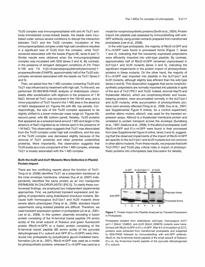
In the wild-type protoplasts, the majority of RbcS-nt:GFP and
E1a-nt:GFP were found in processed forms (Figure 7, lanes
1 and 2), indicating that the transiently expressed preproteins
were efficiently imported into wild-type plastids. By contrast,
approximately half of RbcS-nt:GFP remained unprocessed in
tic21/pic1 and tic20 mutants (lanes 3 and 5), indicating the
significant impairments in the protein import of photosynthetic
proteins in these mutants. On the other hand, the majority of
E1a-nt:GFP was imported into plastids in the tic21/pic1 and
tic20 mutants, although slightly less efficient than the wild type
(lanes 4 and 6). This observation suggests that some nonphoto-
synthetic preproteins are normally imported into plastids in spite
of the lack of Tic21/PIC1 and Tic20. Indeed, stromal Hsp70 and
thylakoidal Albino3, which are nonphotosynthetic and house-
keeping proteins, were accumulated normally in the tic21/pic1
and tic20 mutants, while accumulation of photosynthetic pro-
teins were severely affected (Teng et al., 2006; Duy et al., 2007;
see Supplemental Figure 8 online). As a control experiment,
another albino mutant, albino3, was used for the transient ex-
pression assay. Albino3 is a thylakoidal membrane protein and
unrelated to protein transport across the envelope (Sundberg
et al., 1997; Asakura et al., 2008). In the albino3 protoplasts, both
RbcS-nt:GFP and E1a-nt:GFP were found in their processed
form (see Supplemental Figure 9 online, lanes 3 and 4), suggest-
ing that the observed impairments of the import of RbcS-nt:GFP
are specific to the tic21/pic1 and tic20mutants but not common
in other albinomutants. From these results, we propose that both
Tic21/PIC1 and Tic20 play critical roles in import of photosyn-
thetic proteins into chloroplasts (see Discussion for details).
Figure 7. Protein Import into Plastids Analyzed by Transient Expression
in Protoplasts.
Protoplasts isolated from Arabidopsis wild-type, homozygous tic21/
pic1-1 (SALK_104852), and tic20 (SALK_039676) mutants were trans-
formed with RbcS-nt:GFP or E1a-nt:GFP. After 8 h of incubation at 228C,
proteins were extracted from transformed protoplasts and subjected
to SDS-PAGE followed by immunoblotting with anti-GFP antibody.
RbcS-nt, the N-terminal transit peptide of the small subunit of Rubisco;
E1a-nt, the N-terminal transit peptide of the pyruvate dehydrogenase
E1a subunit.
The 1-MDa Tic complex of chloroplasts 9 of 17

Upregulation of Iron Homeostasis Proteins Are Common in
Some Albino Mutants
In a second approach, we examined expression levels of iron
homeostasis proteins in the tic21/pic1 mutant and other albino
mutants. One reason that Duy et al. (2007) termed Tic21/PIC1 as
an iron transporter is based on the observation that genes related
to iron homeostasis are upregulated in the tic21/pic1 mutant.
Supplemental Figure 8 online shows immunoblot analyses of
several plastid-localized proteins in the tic21/pic1, tic20, and
albino3 mutants, all of which show albino phenotypes. As
reported by Duy et al. (2007), in the tic21/pic1 mutant, upregu-
lation of ferritin and copper superoxide dismutase 1 (CSD1) and
CSD2, which are related to iron homeostasis in plastids, was
reproduced. However, this upregulation was also observed in
other albino mutants, tic20 and albino3. This means that the
upregulation of these proteins is not specific to the tic21/pic1
mutant but is seen in other albino mutants.
Chemical Cross-Linking of the Preprotein to Tic20
To obtain direct evidence that Tic20 associates with the prepro-
tein in the translocation intermediate, chemical cross-linking of
the preprotein to translocon components was performed. In this
experiment, a fusion preprotein pSSU-DHFR, which consists of
full-length pSSU and dihydrofolate reductase (DHFR), was used
because it produced a much higher amount of cross-linked
products than pSSU. We then selectedm-maleimidobenzoyl-N-
hydroxysuccinimide ester (MBS) as a noncleavable cross-linker
after screening of more than a dozen reagents. Arabidopsis
chloroplasts were incubated with pSSU-DHFR under either low
or high ATP conditions. After reisolation, chloroplasts were
cross-linked with MBS followed by SDS-PAGE and autoradiog-
raphy. Several cross-linked products were observed, and the
relative amounts of these products varied between low and high
ATP conditions (e.g., a 48-kD and a 100-kDbandwere prominent
under low ATP conditions, whereas a 140-kD band was prom-
inent under high ATP conditions) (Figure 8A, compare lanes 2 and
3). This indicates dynamic changes of association partners in the
translocation event.
To examine whether Tic20 is cross-linked with the preprotein,
immunoprecipitation was performed after denaturation with
SDS. All four anti-At Tic20 antisera which we prepared and
raised in different rabbits can recognize denatured At Tic20
antigens, but are unable to immunoprecipitate or immunode-
plete native Tic20 proteins. The most probable explanation for
this is that Tic20 is deeply embedded within its large complex
and does not have surface-exposed epitopes. Therefore, cross-
linked products were denatured with 2% SDS to dissociate the
1-MD Tic20 complexes and allow Tic20 to be immunoprecipi-
tated. Anti-At Tic20 antibody immunoprecipitated the single
48-kD cross-linked band (Figure 8B, lane 3), demonstrating a
direct interaction between the preprotein and Tic20. Please note
that a slight shift of this 48-kD band to a lower molecular mass
was caused by an overlap of IgG that migrated in large quantities
at this position (cf. lanes 1 and 3).
We also performed immunoprecipitations using several Toc
and Tic antibodies. Immunoprecipitation with anti-Toc75 anti-
body gave a strong band at 100 kD, which most likely represents
a 1:1 cross-linked product with Toc75, and smeared bands of
>120 kD, which probably represent products including at least
one molecule of Toc75 (lane 4). Immunoprecipitation with anti-At
Tic21 antibody did not detect any specific band (data not shown),
probably because Tic21 is not positioned close to the prepro-
tein-translocating channel. Also, immunoprecipitation with anti-
Tic110 antibody after MBS cross-linking did not detect any
specific band (see Supplemental Figure 10 online). Almost all
major cross-linked bands except a 55-kD band observed under
low ATP conditions were immunoprecipitated with either anti-
Toc75 or -Tic20 antibodies, strongly suggesting that Toc75 and
Tic20 are the proteins in the closest contact with the trans-
locating preprotein.
Moreover, we used the cleavable cross-linker dithiobis(succi-
nimidyl propionate) (DSP), which has been successfully used to
cross-link preproteins to Toc and Tic proteins (Akita et al., 1997;
Chou et al., 2003). Consistent with the results of MBS cross-
linking, anti-Toc75 and -Tic20 antibodies immunoprecipitated
pSSU-DHFR more efficiently under low ATP conditions than
under high ATP conditions (Figure 8C, lanes 3, 4, 7, and 8). In
addition, immunoprecipitation with anti-Tic110 antibody was
able to capture a small amount of pSSU-DHFR under low ATP
conditions (lane 5) and both pSSU-DHFR and mSSU-DHFR
under high ATP conditions (lane 9), which are consistent with the
results of Chou et al. (2003).
DISCUSSION
We report the identification of a 1-MD translocation complex as
an intermediate during preprotein import into chloroplasts. Char-
acteristic features of the 1-MD translocation complex are sum-
marized as follows. (1) This complex is formed under limited ATP
conditions and can be detected by BN-PAGE with the mild
detergent digitonin. (2) Preproteins arrested in this complex can
be chased under high ATP conditions. (3) Protease accessibility
assays and sucrose density gradient centrifugation revealed that
this complex resides in the inner membrane. (4) Tic20 forms a
1-MD complex with a minor population of Tic21 at the inner
membrane under steady state conditions, and this complexmost
likely corresponds to the 1-MD translocation complex.
All tested detergents except digitonin failed to preserve the
1-MD translocation complex (Figure 2A; data not shown), sug-
gesting a loose association between the preprotein and the
translocation complex. This feature may have impeded previous
identification of the 1-MD translocation complex.
Although the 1-MD translocation complex described in this
study was derived from the inner membrane, protease treat-
ments shown in Figure 4 revealed that the C-terminal tail of the
preprotein was exposed to the surface of chloroplasts, suggest-
ing that the arrested preprotein spans both Toc and Tic com-
plexes. Indeed, in the presence of cross-linkers, the preprotein
was cross-linked with Toc75 and Tic20 (Figure 8). On BN-PAGE,
high concentrations of Coomassie blue (0.125%), which has an
anionic feature, was added to the detergent-solubilized protein
complexes, potentially causing dissociation of loosely associ-
ated proteins (Schagger and Pfeiffer, 2000; Gavin et al., 2003;
10 of 17 The Plant Cell
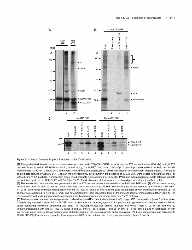
Figure 8. Chemical Cross-Linking of a Preprotein to Toc/Tic Proteins.
(A) Energy-depleted Arabidopsis chloroplasts were incubated with [35S]pSSU-DHFR under either low ATP concentrations (100 mM) or high ATP
concentrations (4 mM) in HS buffer containing 5 mM MgCl2, 5 mM DTT, 3 mM Met, 3 mM Cys, 5 mL/mL protease inhibitor cocktail, and 20 mM
methotrexate (MTX) for 10 min at 258C in the dark. The DHFR fusion protein, pSSU-DHFR, was used in this experiment instead of pSSU. Reisolated
chloroplasts carrying [35S]pSSU-DHFR, at 0.24 mg chlorophyll/mL in HS buffer in the presence of 20 mM MTX, were treated with (lanes 2 and 3) or
without (lane 1) 0.2 mM MBS. Noncleavable cross-linked products were subjected to 7.5% SDS-PAGE and autoradiography. Single asterisks indicate
cross-linked products of pSSU-DHFR with Toc75 or Tic20. The double asterisk indicates a cross-linked product with unidentified protein.
(B) The translocation intermediate was generated under low ATP concentrations and cross-linked with 0.2 mM MBS as in (A). Chloroplasts carrying
cross-linked products were solubilized under denaturing conditions containing 2% SDS. The resulting extract was diluted 18.5-fold with 0.5% Triton
X-100 in TBS followed by immunoprecipitation with anti-At Tic20 FL (lane 3) or anti-Ps Toc75 (lane 4) antibodies or with preimmune serum (lane 2). The
eluates were subjected to 7.5% SDS-PAGE and autoradiography. Input represents 30% of the material used for immunoprecipitation (lane 1). The
region marked with a diamond probably represents cross-linked products containing at least one Toc75 molecule.
(C) The translocation intermediate was generated under either low ATP concentrations (lanes 1 to 5) or high ATP concentrations (lanes 6 to 9) as in (A).
Cross-linking was performed with 0.2 mM DSP, which is cleavable with reducing agents. Chloroplasts carrying cross-linked products were solubilized
under denaturing conditions containing 2% SDS. The resulting extract was diluted 18.5-fold with 0.5% Triton X-100 in TBS followed by
immunoprecipitation with anti-At Tic20 FL (lanes 3 and 7), anti-Ps Toc75 (lanes 4 and 8), or anti-Ps Tic110 (lanes 5 and 9) antibodies or with
preimmune serum (lane 2). Bound proteins were eluted by boiling in 23 Laemmli sample buffer containing 10% 2-mercaptoethanol and subjected to
12.5% SDS-PAGE and autoradiography. Input represents 30% of the material used for immunoprecipitation (lanes 1 and 6).
The 1-MDa Tic complex of chloroplasts 11 of 17
Wittig and Schagger, 2005). It is highly probable that the Toc
complex was dissociated from the preprotein under the condi-
tions of BN-PAGE, whereas the Tic complex remained associ-
atedwith the preprotein during BN-PAGE andwas observable as
the 1-MD complex. Considering unidirectional protein transport
from the outer membrane to the inner membrane, it seems
reasonable that the translocating preprotein at this stage binds
more tightly to the Tic complex than to the Toc complex. When
chemical cross-linkingwasperformed to stabilize the associations
of Toc and Tic proteins prior to solubilization, the 1-MD translo-
cation complex was shifted to a higher molecular mass range
around 1.4 to 1.8MDonBN-PAGE,whichmost likely corresponds
to a Toc-Tic supercomplex (see Supplemental Figure 11 online).
The 1-MD translocation complex characterized in this study is
neither the Toc complex, to which preproteins initially bind, nor
the Tic110-containing complex, which should mediate a later
translocation step on the stromal side. We propose that the
1-MD translocation complex functions in between the Toc- and
Tic110-containing complexes. Tic110 andHsp93migrated in the
range of 200 to 300 kD on BN-PAGE (see Supplemental Figure 7
online; Caliebe et al., 1997; Kuchler et al., 2002), and Tic22
migrated at a low molecular mass (<66 kD) (data not shown),
supporting the idea that these Tic components are not involved in
the 1-MD translocation complex. Figure 8C shows that Toc75 is
preferentially associated with the precursor form of the prepro-
tein, whereas Tic110 is associated with both the precursor and
mature forms of the preprotein, consistent with the results of
Chou et al. (2003). Meanwhile, Tic20 is preferentially associated
with the precursor form of the preprotein, supporting the idea
that the Tic20-containing 1-MD complex functions in between
the Toc- and Tic110-containing complexes. The preprotein
arrested in the 1-MD complex would be transferred to the
Tic110/Hsp93 complex in a subsequent step, where the transit
peptide would be processed.
By analogy with mitochondrial import machinery, it is often
stated that Tic110 is an analogous component to Tim44 (Bedard
and Jarvis, 2005), which serves as a binding site for matrix Hsp70.
The situation that the 1-MD complex containing Tic20 and Tic21
holds preproteins in the absence of Tic110 is very similar to that of
the mitochondrial import since a Tim core complex containing
Tim23 and Tim17 can hold preproteins in the absence of Tim44 in
digitonin extracts (Dekker et al., 1997). It should be noted that,
despite the absence of any significant sequence similarities, Tic20
and Tic21, and mitochondrial Tim23 and Tim17, all have similar
molecular weights and contain three or four predicted transmem-
brane domains, suggesting functional similarity.
We have observed the protease-resistant fragment SSU-DPs
arrested in the 1-MD complex (Figure 4). Earlier studies have
depicted similar protease-resistant fragments (Friedman and
Keegstra, 1989; Waegemann and Soll, 1991, 1993; Chigri et al.,
2005; Inoue and Akita, 2008). Protease-resistant fragments are
called deg and classified into deg1, deg2, deg3, or deg4 based
on different molecular sizes, from the largest (deg1) to the
smallest (deg4). deg1 and deg2 were shown to cofractionate
with the outer membrane, whereas deg3 and deg4, which would
correspond to SSU-DPs in this study, were shown to cofrac-
tionate with the inner membrane (Waegemann and Soll, 1993;
Soll and Tien, 1998), supporting our observations.
Akita et al. (1997) have shown that a translocation intermediate
complex of ;600 kD could be isolated using chemical cross-
linkers. They generated the intermediate under incubation con-
ditions on ice for 20 min in the presence of 75 mM ATP, whereas
we incubated at 258C for 10 min in the presence of 0.5 mM ATP.
Under their conditions, an early intermediate is predicted to be
formed; therefore, the 600-kD complex most likely corresponds
to the Toc subcomplex (II) in our previous study (Kikuchi et al.,
2006). In addition, Chen and Li (2007) have recently shown that
two intermediate complexes of;880 and 1320 kD, which were
referred to as C1 and C2, respectively, were observed by BN-
PAGE. They concluded that both C1 and C2 contained the Toc
complex, while C2 additionally contained Tic110, Hsp93, and the
intermembrane space Hsp70. There are significant experimental
differences between their study and ours. After the import
reaction at 208C for 20 min, they first performed chemical
cross-linking to preserve the translocation intermediate com-
plexes. Then, isolated total membranes were solubilized with the
more stringent detergent decyl maltoside and fractionated by
sucrose density gradient centrifugation. C1 most likely corre-
sponds to the intact Toc complex characterized in our previous
study (Kikuchi et al., 2006). C2 likely corresponds to the Toc-Tic
supercomplex shown in Supplemental Figure 11 online. How-
ever, the involvement of Tic21 and/or Tic20 in formation of C2
was not analyzed. Moreover, since there are significant exper-
imental differences, some Tic proteins may be added to or
removed from the C2 complex.
Kouranov and Schnell (1997) have shown that the association
of the preprotein with Tic20 was increased in the presence of
high ATP (2 mM) compared with low ATP (0.1 mM). By contrast,
the cross-linked product between the preprotein and Tic20 was
more prominent in low ATP (0.1 mM) than in high ATP (4 mM) in
this study (Figure 8A). This difference can be explained by the
following reasons. In the study by Kouranov and Schnell (1997),
they used a urea-denatured preprotein that had been overex-
pressed in E. coli and purified from inclusion bodies. By
contrast, we used in vitro–translated soluble preproteins. In
addition, the preprotein used by Kouranov and Schnell con-
tained an IgG binding domain of Protein A at the C terminus.
Perhaps this are why the preprotein they used seems to require
more ATP to be unfolded and reach the Tic20-containing
complex than that required for the in vitro–synthesized prepro-
tein used in this study.
There are two conflicting reports about the function of Tic21,
which has been reported to be a component of the protein import
machinery at the inner envelope membrane (Teng et al., 2006) or
an iron transporter (Duy et al., 2007). To clarify the function of
Tic21, we performed transient expression and targeting of pre-
proteins in mutant protoplasts and compared expression levels
of metal homeostasis proteins in mutants. Figure 7 shows a
defect in protein import of photosynthetic proteins in the tic21/
pic1 mutant, which is comparable to that in the tic20 mutant.
Supplemental Figure 8 online shows that upregulation of ferritin,
CSD1, and CSD2, all of which are iron homeostasis-related
proteins, is not specific to the tic21/pic1 mutant but is seen in
other albino mutants. These observations support the proposal
by Teng et al. (2006) that Tic21/PIC1 functions in chloroplast
protein import.
12 of 17 The Plant Cell
The results shown in Figures 6A and 6B suggest that Tic21 is
not a central component of the 1-MD translocation complex but
loosely associated component of the complex. Nevertheless, the
lack of Tic21 causes severe defects in chloroplast protein import,
similar to those observed in the tic20 mutant. Preliminary anal-
ysis of the Tic20 complex in the tic21 mutant by 2D-BN/SDS-
PAGE showed that the Tic20 complex of the tic21mutant did not
migrate at the same position as that of the wild type but
accumulated at the top of the separation gel in BN-PAGE (S.
Kikuchi and M. Nakai, unpublished data), suggesting that an
improper assembly or an aggregation of the Tic20 complex
probably occurs in the tic21 mutant. Tic21 may function in the
proper assembly of the Tic20 complex. This hypothesis can
explain similar severe albino phenotypes of the tic20 and tic21
mutants (Teng et al., 2006).
Although the tic20 and tic21 mutants show severe albino
phenotypes and are seedling lethal, they are able to produce
albino leaves and occasionally inflorescence tissues on synthetic
media supplemented with sucrose. This suggests residual im-
port capacities in the tic20 and tic21mutants. As shown in Figure
7, import defects in the tic20 and tic21 mutants were clearly
observed using photosynthetic preprotein (RbcS-nt:GFP). How-
ever, interestingly, less severe import defects were observed
using nonphotosynthetic preprotein (E1a-nt:GFP). Indeed, stro-
mal Hsp70, ferritin, and thylakoidal Albino3, which are nonpho-
tosynthetic and housekeeping proteins, accumulated normally in
the plastids of the tic20 and tic21 mutants (see Supplemental
Figure 8 online). From these observations, we propose that Tic20
and Tic21 have substrate specificity for photosynthetic prepro-
teins. The phenotypes of the tic20 and tic21 mutants are very
similar to that of an Arabidopsis ppi2mutant in which the Toc159
gene is disrupted (Bauer et al., 2000; Teng et al., 2006). Many
photosynthetic proteins are deficient in theppi2mutant, whereas
nonphotosynthetic proteins seem to accumulate normally. In the
ppi2 mutant, Toc132 and Toc120, which are homologs of
Toc159, compensate for the absence of Toc159, at least to
some extent. The Tic20 family consists of four genes in Arabi-
dopsis: Tic20-I, Tic20-IV, Tic20-II, and Tic20-V (Jarvis, 2008).
Tic20-I characterized in this study is the most abundant isoform
among four proteins and is the closest homolog to the biochemi-
cally identified pea Tic20 (Kouranov et al., 1998). The other three
Tic20 isoforms in Arabidopsis may be responsible for the import
of nonphotosynthetic and housekeeping proteins. Preliminary
experiments indicated that certain double knockout mutations
introduced into the fourArabidopsis Tic20 genes resulted inmore
severe embryo lethal phenotype (S. Kikuchi, Y. Hirabayashi, and
M. Nakai, unpublished data), suggesting that the residual import
capacities observed in the tic20 and tic21 mutants may be due
to the presence of another Tic20 isoform-containing channel
that probably has different substrate specificity. These issues are
currently under investigation.
To date, the Tic110/Tic40/Hsp93 complex, which mediates
stromal side events, has received considerable attention with
regard to translocation across the inner membrane. However, a
Tic core complex that functions in between the Toc complex and
the Tic110/Tic40/Hsp93 complex has not yet been reported. We
believe that the 1-MD translocation complex characterized in
this study corresponds to this Tic core complex, which should
contain a protein-conducting channel. While Tic20 and Tic21
would play a crucial role in the 1-MD complex, we can easily
speculate that most constituents of the 1-MD complex have not
yet been identified. Therefore, further work will be required to
identify new components.
METHODS
Plant Material and Growth Conditions
Pea (Pisum sativum var Alaska) seedlings were grown on vermiculite in a
growth chamber under 14 h light at 258C/10 h dark at 238Ccycles for 12 to
13 d. The Arabidopsis thaliana mutants tic20 (SALK_039676) and tic21/
pic1-1 (SALK_104852) carrying T-DNA insertion(s) were kindly provided
by the Salk Institute Genomic Analysis Laboratory (Alonso et al., 2003).
Arabidopsis ecotype Columbia was used as the wild type. Arabidopsis
(wild type and mutants) were grown on MS plates (13 Murashige and
Skoog salts [Sigma-Aldrich], 13 Gamborg’s B5 vitamin [Sigma-Aldrich],
2% sucrose, pH 5.8, and 0.3% phytagel [Sigma-Aldrich]) in a growth
chamber under 16 h light at 238C /8 h dark at 218C cycles for 18 to 21 d
(Yabe et al., 2004; Asakura et al., 2008).
Isolation of Chloroplasts
Chloroplast isolation frompea leaveswasdescribed in our previous report
(Kikuchi et al., 2006). Arabidopsis chloroplasts were isolated by direct
homogenization method as described (Bruce et al., 1994; Aronsson and
Jarvis, 2002; Schulz et al., 2004) with the following modifications. Ap-
proximately 20 g of aerial parts were homogenized in 400 mL of blending
buffer (50 mM HEPES-KOH, pH 7.8, 330 mM sorbitol, 2 mM EDTA, 1 mM
MnCl2, 1 mM MgCl2, and 50 mM sodium ascorbate [freshly added in
powder]) with or without 5 mL/mL protease inhibitor cocktail (for plant
extracts, P-9599; Sigma-Aldrich), in five 2-s pulses in a kitchen blender
equipped with disposable razor blades. The homogenate was filtered
through four layers of Miracloth (Calbiochem) and then centrifuged at
4000g for 3 min. The crude chloroplast pellet was resuspended in HS
buffer (50 mM HEPES-KOH, pH 7.8, and 330 mM sorbitol) and overlaid
onto 30% (v/v) Percoll in HSbuffer. After centrifugation at 1350g for 15min
in a swinging bucket rotor, the pellet was washed twice with HS buffer.
Purified intactchloroplastswere kepton ice in thedarkandusedwithin3h.
In Vitro Transcription and Translation of Preproteins
Plasmid pGEM-4Z-pSSU for the in vitro expression of a precursor protein
of SSUwas constructedby inserting the coding sequencederived from its
cDNA prepared from pea into pGEM-4Z vector (Promega) at a XbaI/SalI
site. ThemRNAofpSSUwassynthesized from linearizedpGEM-4Z-pSSU
construct usingRiboMAX in vitro transcription system (Promega)withSP6
RNA polymerase. The resulting mRNA was translated in a wheat germ
extract (Promega) at 258C for 2 h in the presence of [35S]Met. A plasmid for
the in vitro expression of pSSU-DHFR, which consists of the full-length
precursor to SSU fused to the entire mouse dihydrofolate reductase, was
constructed by inserting the pSSU coding sequence lacking the stop
codon into pPC-DHFR/SP (Endo et al., 1994) at a PstI/BamHI site. The
fusion protein pSSU-DHFR was synthesized using TNT SP6-coupled
reticulocyte lysate system (Promega) at 308C for 90min in the presence of
[35S]Met. The translation mixtures were kept on ice and used within 3 h.
Formation of a Translocation Intermediate, Protease Treatments,
and Gel Electrophoresis
Isolated intact chloroplasts in HS buffer containing 5 mL/mL protease
inhibitor cocktail were preincubated for 10 min at 258C in the dark to
The 1-MDa Tic complex of chloroplasts 13 of 17

deplete endogenous ATP. Energy-depleted chloroplasts weremixedwith
in vitro–translated preproteins in HS buffer containing 0.5 mMMg-ATP, 5
mM MgCl2, 5 mM DTT, 3 mM Met, and 3 mM Cys. The reactions were
incubated for 10 min at 258C in the dark to form a translocation interme-
diate. Each reaction contained chloroplasts equivalent to 100 mg chlo-
rophyll in a reaction volume of 400 mL. The reactions were terminated by
reisolation of chloroplasts by centrifugation at 1500g for 1 min at 48C, and
chloroplasts were washed once with HS buffer containing 5 mL/mL
protease inhibitor cocktail. When chase experiments or protease treat-
ments were performed, the above reactions were performed as a batch
reaction corresponding to the number of various conditions. Before
protease accessibility assays, chloroplasts carrying preproteins were
washed twice with HS buffer in the absence of protease inhibitor cocktail.
For thermolysin treatment, chloroplasts in HS buffer were incubated with
10 to 100 mg/mL of thermolysin (Sigma-Aldrich) and 1 mM CaCl2 for 20
min on ice. Thermolysin was inactivated by the addition of 10 mM EDTA.
For trypsin treatment, chloroplasts in HS buffer were incubated with 10 to
100 mg/mL of trypsin (Sigma-Aldrich) for 20 min on ice. Trypsin was
inactivated by the addition of a fivefold excess of soybean trypsin inhibitor
(0.5 mg/mL; Sigma-Aldrich). Proteolyzed chloroplasts were pelleted and
washed twice with HS buffer containing either 5 mL/mL protease inhibitor
cocktail or 0.5 mg/mL trypsin inhibitor.
Samples prepared by the above methods were analyzed by two
different electrophoresis methods: BN-PAGE and SDS-PAGE. The
BN-PAGE method was described in detail in our previous report (Kikuchi
et al., 2006). The chloroplast pellet was solubilized in freshly prepared
BN-PAGE sample buffer (1% [w/v] water-soluble digitonin, 50 mM
BisTris-HCl, pH 7.0, 500 mM 6-amino-n-caproic acid, and 10% [w/v]
glycerol) containing 10 mL/mL protease inhibitor cocktail to a final
concentration of 0.5 mg chlorophyll/mL for 10 min on ice. Water-soluble
digitonin was prepared as described (Mori et al., 1999). When using
trypsin as a protease, trypsin inhibitor was added to a final concentration
of 0.5mg/mL toBN-PAGE sample buffer. Insolublematerial was removed
by ultracentrifugation at 100,000g for 10 min at 48C. The supernatant was
divided into two aliquots. One of which (40 mL) was mixed with
Coomassie Brilliant Blue G 250 solution (5% [w/v] Serva blue G, 50 mM
BisTris-HCl, pH 7.0, and 500 mM 6-amino-n-caproic acid) to give a
detergent:Coomassie ratio of 8:1 (w:w) and subjected to 4 to 14%
BN-PAGE. The other was mixed with 10% SDS and 2-mercaptoethanol
to final concentrations of 3.3 and 5%, respectively. The SDS-denatured
sample was heated at 958C for 2 min and subjected to SDS-PAGE. The
radioactive signals in dried gels were detected using a BAS-2000II image
analysis system (FujiFilm) and quantified using Image Gauge version 3.45
software (FujiFilm).
Antibody Preparation, Purification, and Immunoblotting
Two overlapping but different antigens of Arabidopsis Tic20 were
overexpressed as N-terminal hexahistidine-tagged fusion proteins in
Escherichia coli using recently developed cold shock–inducible system
(Qing et al., 2004). An antigen ofArabidopsis Tic21 was overexpressed as
a fusion protein with N-terminal 260 amino acids of T7 gene10 protein in
E. coli. An antigen of pea Tic22 was overexpressed as a C-terminal
hexahistidine-tagged fusion protein in E. coli. Regions of antigens used
were as follows: At Tic20 FL (full-length sequence not including transit
peptide), At Tic20 ES (C-terminal half part, amino acids 157 to 274), At
Tic21 (C-terminal part, amino acids 210 to 296), and Ps Tic22 (full-length
sequence including transit peptide). Recombinant At Tic20 FL, At Tic20
ES, and Ps Tic22 proteins were purified from inclusion bodies with nickel
chelate chromatography under denaturing conditions. Recombinant At
Tic21 protein was purified from inclusion bodies using SDS-PAGE and
subsequent electroelution. Purified recombinant At Tic20 FL, At Tic20 ES,
At Tic21, and Ps Tic22 proteins were injected into rabbits using Freund’s
complete adjuvant and (for subsequent boosts) Freund’s incomplete
adjuvant (Harlow and Lane, 1988). Preparation of antibodies against Ps
Toc75, Ps Toc159, Ps Toc34, Ps Tic110, So cpHsp70 (Spinacia oleracea
stromal Hsp70), So Cpn60a, and So Cpn60b was described in our
previous reports (Nishio et al., 1999; Asakura et al., 2004; Kikuchi et al.,
2006). Anti-At Tic21, -Ps Toc75, -Ps Toc159, and -Ps Toc34 antibodies
were affinity purified using the purified antigens as described previously
(Kikuchi et al., 2006). Anti-Ps Tic110 antibody was purified using rProtein
A-Sepharose (GE Healthcare). Anti-At Tic20 ES antibody was purified by
blot affinity purification (Tang, 1993).
Immunoblotting was performed as described previously (Kikuchi et al.,
2006). Toc75, Toc159, Toc34, Tic110, Tic22, and cpHsp70were detected
by an enhanced chemiluminescence system (GE Healthcare). Tic20 and
Tic21 were detected by the ECL plus system (GE Healthcare). We found
that At Tic21 protein band was lost in SDS-PAGE/immunoblotting after
standard denaturation by heating at 958C in Laemmli sample buffer;
therefore, when detecting Tic21 and distantly related Tic20, samples
were incubated at 378C for 30 min.
Antibody-Shift BN-PAGE
Antibody-shift BN-PAGE is used in several reports (e.g., Johnston et al.,
2002; Truscott et al., 2002). Chloroplasts carrying [35S]translocation
intermediate were solubilized in BN-PAGE sample buffer containing
10 mL/mL protease inhibitor as described above. After ultracentrifugation
at 100,000g for 10 min at 48C, 35 mL of the supernatant was mixed with
each purified antibody of known concentrations (0 to 10 mg) and incu-
bated for 30 min on ice with occasional mixing. After a clarifying spin, the
supernatant was mixed with Coomassie Brilliant Blue G 250 solution as
described above and subjected to BN-PAGE and autoradiography.
Immunodepletion
Chloroplasts carrying [35S]translocation intermediate were solubilized in
BN-PAGE sample buffer containing 10 mL/mL protease inhibitor as
described above. Insoluble material was removed by ultracentrifugation
at 100,000g for 10 min at 48C. Fifty to 100 mL of the supernatant was
incubated twice with 10 mL packed volume of rProtein A-Sepharose that
had been coupled with IgG for 1 h in cold room (68C) with rotational
mixing. The unbound fraction (supernatant) was recovered by centrifu-
gation and was filtered through a 0.2-mm membrane filter by centrifuga-
tion to remove residual beads.
Cross-Linking and Immunoprecipitation under
Denaturing Conditions
Chloroplasts carrying [35S]pSSU-DHFR, at 0.24 mg chlorophyll/mL in HS
buffer in the presence of 20 mM MTX and in the absence of protease
inhibitor cocktail, were treated with 0.2 mMMBS (Pierce) or 0.2 mM DSP
(Pierce) for 15 min on ice. The MBS cross-linking was quenched by
adding glycine and 2-mercaptoethanol to final concentrations of 20 mM
and 2%, respectively, and incubating on ice for another 15 min. The DSP
cross-linking was quenched by adding glycine to a final concentrations of
20 mM and incubating on ice for another 15 min. Chloroplasts were
recovered by centrifugation, washed with HS buffer containing 20 mM
MTX and 5 mL/mL protease inhibitor cocktail. Immunoprecipitation under
denaturing conditions was performed as described (Cline andMori, 2001)
with the following modifications. Chloroplasts carrying cross-linked pre-
proteins were solubilized under denaturing conditions (2% SDS, 0.5%
Triton X-100, 50 mM Tris-HCl, pH 7.5, 150 mM NaCl, and 10 mL/mL
protease inhibitor cocktail) at 258C for 5 min at 1 mg chlorophyll/mL.
Insoluble material was removed by centrifugation, and the supernatant
was diluted 18.5-fold with 0.5% Triton X-100 in TBS (50 mM Tris-HCl, pH
7.5, and 150 mM NaCl) to enable immunoprecipitations. The diluted
sample (370 mL) was mixed with 10 mL packed volume of rProtein
14 of 17 The Plant Cell

A-Sepharose that had been coupled with IgG and incubated for 2 h at
258Cwith rotational mixing. After the beadswerewashed twicewith 0.5%
Triton X-100 in TBS, bound proteins were eluted by boiling 2 min in 23
Laemmli sample buffer. When using DSP as the cross-linker, bound
proteins were eluted by boiling 5 min in 23 Laemmli sample buffer
containing 10% 2-mercaptoethanol to cleave the cross-linker. The elu-
ates were subjected to SDS-PAGE and autoradiography.
Transient Expression in Protoplasts
Arabidopsis (wild type and mutants) were grown on MS plates. Homozy-
gous tic20 and tic21mutantswere selected frompools of plants based on
the visible albino phenotypes. Protoplasts were prepared as described
previously (Jin et al., 2001). The GFP fusion constructs were introduced
into protoplasts by polyethylene glycol–mediated transformation (Jin
et al., 2001). Protein import into wild-type and mutant plastids was
analyzedby immunoblottingwithanti-GFPantibodyusingprotein extracts
from transformed protoplasts as described previously (Lee et al., 2008).
Miscellaneous
2D-BN/SDS-PAGE was performed as described previously (Kikuchi
et al., 2006) except when detecting At Tic21, At Tic20, and Ps Tic20,
where excised first-dimension native gels were incubated at 378C for 30
min. Size exclusion chromatography was also performed as described
previously (Kikuchi et al., 2006) except that 1% water-soluble digitonin
was used for solubilization and 0.1% water-soluble digitonin was used in
equilibration buffer instead of dodecyl maltoside. Water-soluble digitonin
was prepared as described (Mori et al., 1999). Monoclonal antibody
SPA-820 against Hsp70/Hsc70 was purchased from StressGen.
Accession Numbers
Sequence data from this article can be found in the Arabidopsis Genome
Initiative or GenBank/EMBL databases under the following accession
numbers: Tic20 (At1g04940), Tic21 (At2g15290), pea Tic22 (AF095284),
and pea pSSU (X00806).
Supplemental Data
The following materials are available in the online version of this article.
Supplemental Figure 1. The 1-MD Translocation Complex Is Not a
Stromal Chaperonin Complex.
Supplemental Figure 2. The 1-MD Translocation Complex Was
Generated Using Ferredoxin:NADP+ Oxidoreductase as a Preprotein.
Supplemental Figure 3. Partial Degradation of the Toc Complex,
Which Had Occurred during the Isolation of Chloroplasts, Did Not
Affect the Formation of the 1-MD Translocation Complex.
Supplemental Figure 4. Sucrose Density Gradient Separation of
Outer and Inner Envelope Membrane Vesicles.
Supplemental Figure 5. The Arabidopsis 1-MD Translocation Com-
plex Has Identical Protease Accessibility Properties to That of the Pea
Complex.
Supplemental Figure 6. Antibody-Shift BN-PAGE.
Supplemental Figure 7. Tests for the Ability of Anti-Tic and -Toc
Antibodies to Native Proteins by Immunodepletion.
Supplemental Figure 8. Immunoblot Analyses of Several Proteins in
Albino Mutants.
Supplemental Figure 9. Protein Import into Plastids in a Transient
Expression System Using Protoplasts from the albino3 Mutant.
Supplemental Figure 10. Immunoprecipitation after Chemical Cross-
Linking with MBS.
Supplemental Figure 11. Chemical Cross-Linking of the Transloca-
tion Intermediate.
ACKNOWLEDGMENTS
We thank Toshiharu Hase, Yoko Kimata-Ariga, Guy T. Hanke, Yukari
Asakura, Toshiki Yabe, Jocelyn Bedard, and coworkers of our labora-
tory for valuable discussions, Tazu Uchida for technical assistance, and
Rieko Nakanishi for secretarial assistance. We also thank the Salk
Institute Genomic Analysis Laboratory for providing the sequence-
indexed Arabidopsis T-DNA insertion mutants tic20 and tic21/pic1,
Hsou-min Li for the gift of anti-At Tic40 antibody, Brigitte Touraine for
the gift of anti-ferritin antibody, Marinus Pilon for the gift of anti-CSD2
antibody, Toshiya Hirohashi for the preparation of anti-Ps Tic22 anti-
body, Kazuaki Nishio for the preparation of anti-stromal So Hsp70, So
Cpn60a, and So Cpn60b antibodies, and Tetsuya Nohara for the gift of
pea first-strand cDNA. This work was supported by Grants-in-Aid for
Scientific Research on Priority Areas (17028034 and 17051020) from
MEXT of Japan to M.N. This work was also supported in part by Korea
Science and Engineering Foundation through the Creative Research
Initiatives Program (20090063529) to I.H. S.K. was supported by a
research fellowship from the Japan Society for the Promotion of
Science.
ReceivedOctober 3, 2008; revisedMay 22, 2009; accepted June 1, 2009;
published June 16, 2009.
REFERENCES
Akita, M., Nielsen, E., and Keegstra, K. (1997). Identification of protein
transport complexes in the chloroplastic envelope membranes via
chemical cross-linking. J. Cell Biol. 136: 983–994.
Alonso, J.M., Stepanova, A.N., Leisse, T.J., Kim, C.J., Chen, H.,
Shinn, P., Stevenson, D.K., Zimmerman, J., Barajas, P., Cheuk, R.,
Gadrinab, C., and Heller, C. (2003). Genome-wide insertional muta-
genesis of Arabidopsis thaliana. Science 301: 653–657.
Aronsson, H., and Jarvis, P. (2002). A simple method for isolating
import-competent Arabidopsis chloroplasts. FEBS Lett. 529:
215–220.
Asakura, Y., Hirohashi, T., Kikuchi, S., Belcher, S., Osborne, E.,
Yano, S., Terashima, I., Barkan, A., and Nakai, M. (2004). Maize
mutants lacking chloroplast FtsY exhibit pleiotropic defects in the
biogenesis of thylakoid membranes. Plant Cell 16: 201–214.
Asakura, Y., Kikuchi, S., and Nakai, M. (2008). Non-identical contri-
butions of two membrane-bound cpSRP components, cpFtsY and
Alb3, to thylakoid biogenesis. Plant J. 56: 1007–1017.
Balsera, M., Goetze, T.A., Kovacs-Bogdan, E., Schurmann, P.,
Wagner, R., Buchanan, B.B., Soll, J., and Bolter, B. (2009). Char-
acterization of Tic110, a channel-forming protein at the inner envelope
membrane of chloroplasts, unveils a response to Ca2+ and a stromal
regulatory disulfide bridge. J. Biol. Chem. 284: 2603–2616.
Bauer, J., Chen, K., Hiltbunner, A., Wehrli, E., Eugster, M., Schnell,
D.J., and Kessler, F. (2000). The major protein import receptor of
plastids is essential for chloroplast biogenesis. Nature 403: 203–207.
Becker, T., Hritz, J., Vogel, M., Caliebe, A., Bukau, B., Soll, J., and
Schleiff, E. (2004). Toc12, a novel subunit of the intermembrane
space preprotein translocon of chloroplasts. Mol. Biol. Cell 15: 5130–
5144.
The 1-MDa Tic complex of chloroplasts 15 of 17
Bedard, J., and Jarvis, P. (2005). Recognition and envelope translo-
cation of chloroplast preproteins. J. Exp. Bot. 56: 2287–2320.
Bruce, B.D., Perry, S., Froehlich, J., and Keegstra, K. (1994). In vitro
import of protein into chloroplasts. In Plant Molecular Biology Manual,
S.B. Gelvin and R.A. Schilperoot, eds (Belgium: Kluwer Academic
Publishers), pp. 1–15.
Caliebe, A., Grimm, R., Kaiser, G., Lubeck, J., Soll, J., and Heins, L.
(1997). The chloroplastic protein import machinery contains a Rieske-
type iron-sulfur cluster and a mononuclear iron-binding protein.
EMBO J. 16: 7342–7350.
Chen, K., Chen, X., and Schnell, D.J. (2000). Initial binding of prepro-
teins involving the Toc159 receptor can be bypassed during protein
import into chloroplasts. Plant Physiol. 122: 813–822.
Chen, K.-Y., and Li, H.-m. (2007). Precursor binding to an 880-kDa Toc
complex as an early step during active import of protein into chloro-
plasts. Plant J. 49: 149–158.
Chen, X., Smith, M.D., Fitzpatrick, L., and Schnell, D.J. (2002). In vivo
analysis of the role of atTic20 in protein import into chloroplasts. Plant
Cell 14: 641–654.
Chigri, F., Soll, J., and Vothknecht, U.C. (2005). Calcium regulation of
chloroplast protein import. Plant J. 42: 821–831.
Chou, M.-L., Fitzpatrick, L.M., Tu, S.-L., Budziszewski, G., Potter-
Lewis, S., Akita, M., Levin, J.Z., Keegstra, K., and Li, H.-m. (2003).
Tic40, a membrane-anchored co-chaperone homolog in the chloro-
plast protein translocon. EMBO J. 22: 2970–2980.
Chou, M.-L., Chu, C.-C., Chen, L.-J., Akira, M., and Li, H.-M. (2006).
Stimulation of transit-peptide release and ATP hydrolysis by a
cochaperone during protein import into chloroplasts. J. Cell Biol.
175: 893–900.
Cline, K., and Mori, H. (2001). Thylakoid DpH-dependent precursor
proteins bind to a cpTatC-Hcf106 complex before Tha4-dependent
transport. J. Cell Biol. 154: 719–729.
Dekker, P.J.T., Martin, F., Maarse, A.C., Bomer, U., Muller, H.,
Guiard, B., Meijer, M., Rassow, J., and Pfanner, N. (1997). The Tim
core complex defines the number of mitochondrial translocation
contact sites and can hold arrested preproteins in the absence of
matrix Hsp70-Tim44. EMBO J. 16: 5408–5419.
Duy, D., Wanner, G., Meda, A.R., von Wiren, N., Soll, J., and
Philippar, K. (2007). PIC1, an ancient permease in Arabidopsis
chloroplasts, mediates iron transport. Plant Cell 19: 986–1006.
Endo, T., Kawakami, M., Goto, A., America, T., Weisbeek, P., and
Nakai, M. (1994). Chloroplast protein import. Chloroplast envelopes
and thylakoids have different abilities to unfold proteins. Eur. J.
Biochem. 225: 403–409.
Friedman, A.L., and Keegstra, K. (1989). Chloroplast protein import
quantitative analysis of precursor binding. Plant Physiol. 89: 993–999.
Gavin, P.D., Devenish, R.J., and Prescott, M. (2003). FRET reveals
changes in the F1-stator stalk interaction during activity of F1F0-ATP
synthase. Biochim. Biophys. Acta 1607: 167–179.
Harlow, E., and Lane, D. (1988). Antibodies: A Laboratory Manual.
(Cold Spring Harbor, NY: Cold Spring Harbor Laboratory Press).
Heins, L., Mehrle, A., Hemmler, R., Wagner, R., Kuchler, M.,
Hormann, F., Sveshnikov, D., and Soll, J. (2002). The preprotein
conducting channel at the inner envelope membrane of plastids.
EMBO J. 21: 2616–2625.
Hirohashi, T., Hase, T., and Nakai, M. (2001). Maize non-photosyn-
thetic ferredoxin precursor is mis-sorted to the intermembrane space
of chloroplasts in the presence of light. Plant Physiol. 125: 2154–2163.
Inaba, T., Li, M., Alvarez-Huerta, M., Kessler, F., and Schnell, D.J.
(2003). atTic110 functions as a scaffold for coordinating the stromal
events of protein import into chloroplasts. J. Biol. Chem. 278: 38617–
38627.
Inoue, H., and Akita, M. (2008). Three sets of translocation intermedi-
ates are formed during the early stage of protein import into chloro-
plasts. J. Biol. Chem. 283: 7491–7502.
Jackson, D.T., Froehlich, J.E., and Keegstra, K. (1998). The hydro-
philic domain of Tic110, an inner envelope membrane component of
the chloroplastic protein translocation apparatus, faces the stromal
compartment. J. Biol. Chem. 273: 16583–16588.
Jarvis, P. (2008). Targeting of nucleus-encoded proteins to chloroplasts
in plants. New Phytol. 179: 257–285.
Jin, J.B., Kim, Y.A., Kim, S.J., Lee, S.H., Kim, D.H., Cheong, G.-W.,
and Hwang, I. (2001). A new dynamin-like protein, ADL6, is involved
in trafficking from the trans-Golgi network to the central vacuole in
Arabidopsis. Plant Cell 13: 1511–1526.
Johnston, A.J., Hoogenraad, J., Dougan, D.A., Truscott, K.N., Yano,
M., Mori, M., Hoogenraad, N.J., and Ryan, M.T. (2002). Insertion
and assembly of human Tom7 into the preprotein translocase com-
plex of the outer mitochondrial membrane. J. Biol. Chem. 277: 42197–
42204.
Kessler, F., and Schnell, D. (2009). Chloroplast biogenesis: Diversity
and regulation of the protein import apparatus. Curr. Opin. Cell Biol.
21: in press.
Kikuchi, S., Hirohashi, T., and Nakai, M. (2006). Characterization of
the preprotein translocon at the outer envelope membrane of chlo-
roplasts by blue native PAGE. Plant Cell Physiol. 47: 363–371.
Kouranov, A., Chen, X., Fuks, B., and Schnell, D.J. (1998). Tic20 and
Tic22 are new components of the protein import apparatus at the
chloroplast inner envelope membrane. J. Cell Biol. 143: 991–1002.
Kouranov, A., and Schnell, D.J. (1997). Analysis of the interactions of
preproteins with the import machinery over the course of protein
import into chloroplasts. J. Cell Biol. 139: 1677–1685.
Kuchler, M., Decker, S., Hormann, F., Soll, J., and Heins, L. (2002).
Protein import into chloroplasts involves redox-regulated proteins.
EMBO J. 21: 6136–6145.
Lee, D.W., Kim, J.K., Lee, S., Choi, S., Kim, S., and Hwang, I. (2008).
Arabidopsis nuclear-encoded plastid transit peptides contain multiple
sequence subgroups with distinctive chloroplast-targeting sequence
motifs. Plant Cell 20: 1603–1622.
Ma, Y., Kouranov, A., LaSala, S.E., and Schnell, D.J. (1996). Two
components of the chloroplast protein import apparatus, IAP86 and
IAP75, interact with the transit sequence during the recognition and
translocation of precursor proteins at the outer envelope. J. Cell Biol.
134: 315–327.
Mori, H., Summer, E.J., Ma, X., and Cline, K. (1999). Component
specificity for the thylakoidal sec and delta pH-dependent protein
transport pathways. J. Cell Biol. 146: 45–55.
Nielsen, E., Akita, M., Davila-Aponte, J., and Keegstra, K. (1997).
Stable association of chloroplastic precursors with protein transloca-
tion complexes that contain proteins from both envelope membranes
and a stromal Hsp100 molecular chaperone. EMBO J. 16: 935–946.
Nishio, K., Hirohashi, T., and Nakai, M. (1999). Chloroplast chaper-
onins: Evidence for heterogeneous assembly of alpha and beta Cpn60
polypeptides into a chaperonin oligomer. Biochem. Biophys. Res.
Commun. 266: 584–587.
Perry, S.E., and Keegstra, K. (1994). Envelope membrane proteins that
interact with chloroplastic precursor proteins. Plant Cell 6: 93–105.
Qing, G., et al. (2004). Cold-shock induced high-yield protein produc-
tion in Escherichia coli. Nat. Biotechnol. 22: 877–882.
Schagger, H., Cramer, W.A., and von Jagow, G. (1994). Analysis of
molecular masses and oligomeric states of protein complexes by blue
native electrophoresis and isolation of membrane protein complexes by
two-dimensional native electrophoresis. Anal. Biochem. 217: 220–230.
Schagger, H., and Pfeiffer, K. (2000). Supercomplexes in the respira-
tory chains of yeast and mammalian mitochondria. EMBO J. 19:
1777–1783.
16 of 17 The Plant Cell

Schagger, H., and von Jagow, G. (1991). Blue native electrophoresis
for isolation of membrane protein complexes in enzymatically active
form. Anal. Biochem. 199: 223–231.
Schleiff, E., Soll, J., Kuchler, M., Kuhlbrandt, W., and Harrer, R.
(2003). Characterization of the translocon of the outer envelope of
chloroplasts. J. Cell Biol. 160: 541–551.
Schnell, D.J., and Blobel, G. (1993). Identification of intermediates in
the pathway of protein import into chloroplasts and their localization
to envelope contact sites. J. Cell Biol. 120: 103–115.
Schnell, D.J., Kessler, F., and Blobel, G. (1994). Isolation of compo-
nents of the chloroplast protein import machinery. Science 266: 1007–
1012.
Schulz, A., Knoetzel, J., Scheller, H.V., and Mant, A. (2004). Uptake of
a fluorescent dye as a swift and simple indicator of organelle intact-
ness: Import-competent chloroplasts from soil-grown Arabidopsis. J.
Histochem. Cytochem. 52: 701–704.
Smith, M.D., Rounds, C.M., Wang, F., Chen, K., Afitlhile, M., and
Schnell, D.J. (2004). atToc159 is a selective transit peptide receptor
for the import of nucleus-encoded chloroplast proteins. J. Cell Biol.
165: 323–334.
Soll, J., and Schleiff, E. (2004). Protein import into chloroplasts. Nat.
Rev. Mol. Cell Biol. 5: 198–208.
Soll, J., and Tien, R. (1998). Protein translocation into and across the
chloroplastic envelope membranes. Plant Mol. Biol. 38: 191–207.
Sundberg, E., Slagter, J.G., Fridborg, I., Cleary, S.P., Robinson, C.,
and Coupland, G. (1997). ALBINO3, an Arabidopsis nuclear gene
essential for chloroplast differentiation, encodes a chloroplast protein
that shows homology to proteins present in bacterial membranes and
yeast mitochondria. Plant Cell 9: 717–730.
Tang, W.-J.Y. (1993). Blot-affinity purification of antibodies. Methods
Cell Biol. 37: 95–104.
Teng, Y.-S., Su, Y.-s., Chen, L.-J., Lee, Y.J., Hwang, I., and Li, H.-m.
(2006). Tic21 is an essential translocon component for protein trans-
location across the chloroplast inner envelope membrane. Plant Cell
18: 2247–2257.
Truscott, K.N., Wiedemann, N., Rehling, P., Muller, H., Meisinger, C.,
Pfanner, N., and Guiard, B. (2002). Mitochondrial import of the ADP/
ATP carrier: the essential TIM complex of the intermembrane space is
required for precursor release from the TOM complex. Mol. Cell. Biol.
22: 7780–7789.
Waegemann, K., and Soll, J. (1991). Characterization of the protein
import apparatus in isolated outer envelopes of chloroplasts. Plant J.
1: 149–158.
Waegemann, K., and Soll, J. (1993). Isolation and characterization of a
functionally active protein translocation apparatus from chloroplast
envelopes. In Molecular Mechanisms of Membrane Traffic, NATO ASI
Ser. H74, D.J. Morre, K.E. Howell, and J.J.M. Bergeron, eds (Berlin/
Heidelberg: Springer-Verlag), pp. 101–104.
Wittig, I., and Schagger, H. (2005). Advantages and limitations of clear-
native PAGE. Proteomics 5: 4338–4346.
Yabe, T., Morimoto, K., Kikuchi, S., Nishio, K., Terashima, I., and
Nakai, M. (2004). The Arabidopsis chloroplastic NifU-like protein CnfU,
which can act as an iron-sulfur cluster scaffold protein, is required for
biogenesis of ferredoxin and photosystem I. Plant Cell 16: 993–1007.
Young, M.E., Keegstra, K., and Froehlich, J.E. (1999). GTP promotes
the formation of early-import intermediates but is not required during
the translocation step of protein import into chloroplasts. Plant
Physiol. 121: 237–243.
The 1-MDa Tic complex of chloroplasts 17 of 17
DOI 10.1105/tpc.108.063552; originally published online June 16, 2009;Plant Cell
Shingo Kikuchi, Maya Oishi, Yoshino Hirabayashi, Dong Wook Lee, Inhwan Hwang and Masato NakaiProtein Import at the Inner Envelope Membrane
A 1-Megadalton Translocation Complex Containing Tic20 and Tic21 Mediates Chloroplast
This information is current as of June 20, 2018
Supplemental Data /content/suppl/2009/06/09/tpc.108.063552.DC1.html
Permissions https://www.copyright.com/ccc/openurl.do?sid=pd_hw1532298X&issn=1532298X&WT.mc_id=pd_hw1532298X
eTOCs http://www.plantcell.org/cgi/alerts/ctmain
Sign up for eTOCs at:
CiteTrack Alerts http://www.plantcell.org/cgi/alerts/ctmain
Sign up for CiteTrack Alerts at:
Subscription Information http://www.aspb.org/publications/subscriptions.cfm
is available at:Plant Physiology and The Plant CellSubscription Information for
ADVANCING THE SCIENCE OF PLANT BIOLOGY © American Society of Plant Biologists
Related Documents