MIT OpenCourseWare http://ocw.mit.edu 9.01 Introduction to Neuroscience Fall 2007 For information about citing these materials or our Terms of Use, visit: http://ocw.mit.edu/terms.

Welcome message from author
This document is posted to help you gain knowledge. Please leave a comment to let me know what you think about it! Share it to your friends and learn new things together.
Transcript

MIT OpenCourseWare http://ocw.mit.edu
9.01 Introduction to Neuroscience Fall 2007
For information about citing these materials or our Terms of Use, visit: http://ocw.mit.edu/terms.
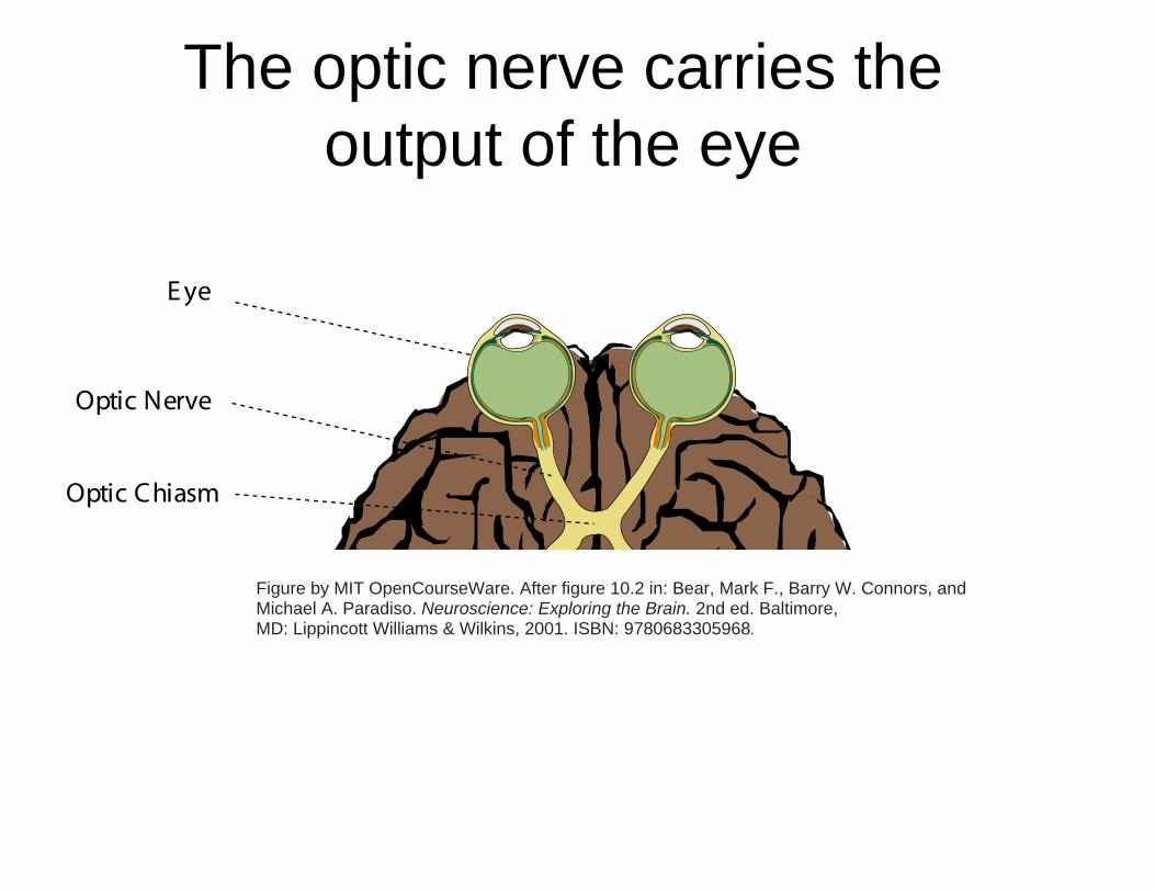
The optic nerve carries theoutput of the eye
Figure by MIT OpenCourseWare. After figure 10.2 in: Bear, Mark F., Barry W. Connors, and Michael A. Paradiso. Neuroscience: Exploring the Brain. 2nd ed. Baltimore, MD: Lippincott Williams & Wilkins, 2001. ISBN: 9780683305968.
E ye
Optic Nerve
Optic Chiasm
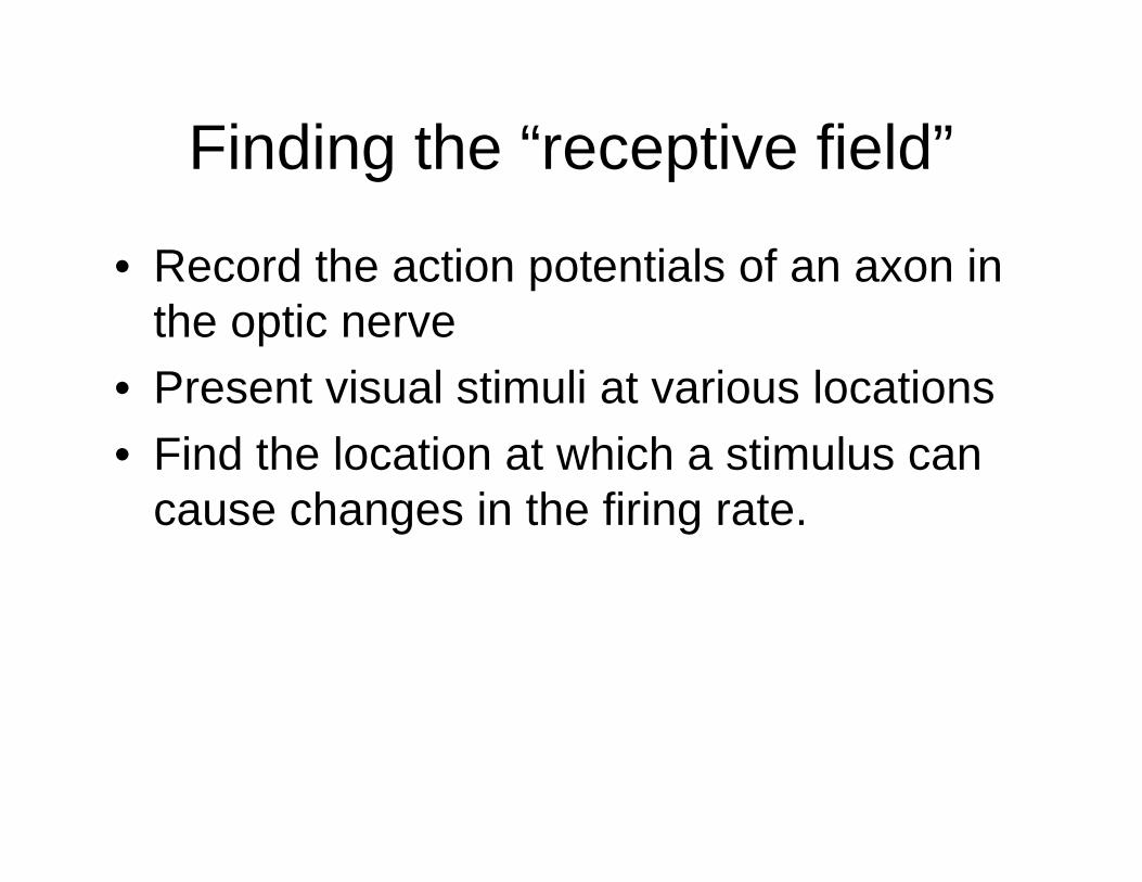
Finding the “receptive field”
• Record the action potentials of an axon in the optic nerve
• Present visual stimuli at various locations
• Find the location at which a stimulus can cause changes in the firing rate.
ON-center cell
• There is a background firing rate.
• The rate increases when the stimulus is in the receptive field (drawn circle).
Image removed due to copyright restrictions.
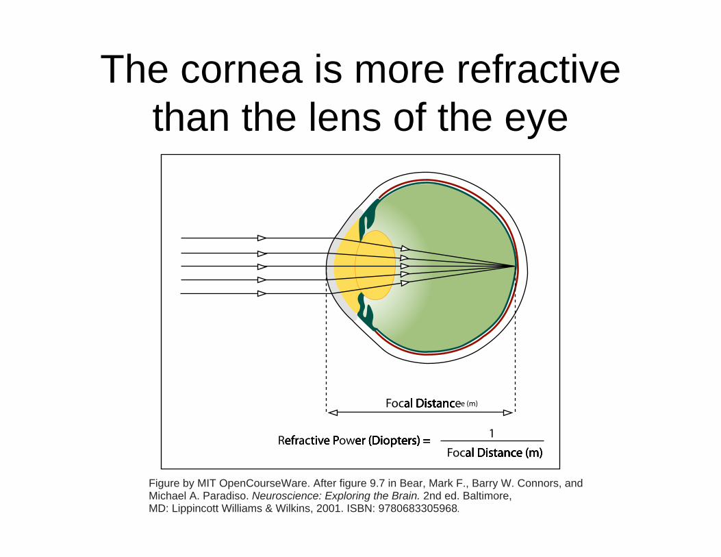
The cornea is more refractivethan the lens of the eye
Figure by MIT OpenCourseWare. After figure 9.7 in Bear, Mark F., Barry W. Connors, and Michael A. Paradiso. Neuroscience: Exploring the Brain. 2nd ed. Baltimore, MD: Lippincott Williams & Wilkins, 2001. ISBN: 9780683305968.
Focal Dal Dal Dal Distancistancistancistancistancistance
Refrefrefracactivtivtive Pe Pe Power (Der (Der (Der (Der (Dioptioptioptiopters) = ers) = ers) = ers) = ers) = ers) = ers) = 1
Focal Dal Dal Dal Distancistancistancistancistancistance (m)e (m)e (m)e (m)
e (m)
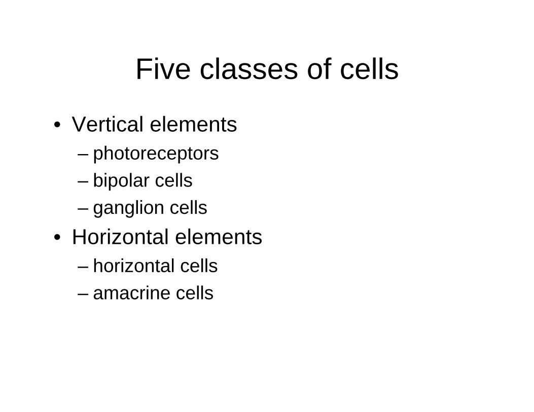
Five classes of cells
• Vertical elements – photoreceptors – bipolar cells – ganglion cells
• Horizontal elements – horizontal cells – amacrine cells
The retinahas layers
Image removed due to copyright restrictions.Cross section electron microscope image of the human retina.Figure 1 (Plate 32) in Boycott B. B. and J. E. Dowling. "Organizationof the Primate Retina: Light Microscopy." Phil Trans R Soc B 255, no. 799 (March 27, 1969): 109-184. doi: 10.1098/rstb.1969.0004.
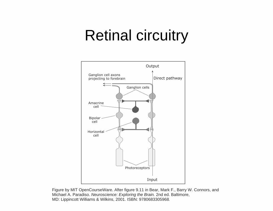
Retinal circuitry
Figure by MIT OpenCourseWare. After figure 9.11 in Bear, Mark F., Barry W. Connors, and Michael A. Paradiso. Neuroscience: Exploring the Brain. 2nd ed. Baltimore, MD: Lippincott Williams & Wilkins, 2001. ISBN: 9780683305968.
Ganglion cells
Direct pathway
Output
Input
Photoreceptors
Ganglion cell axonsprojecting to forebrain
Amacrinecell
Bipolarcell
Horizontalcell

Opthalmoscopic view
Image removed due to copyright restrictions.The retina, as viewed through an opthalmoscope.Figure 9.5 in Bear, Mark F., Barry W. Connors, and Michael A. Paradiso.Neuroscience: Exploring the Brain. 3rd ed. Baltimore, MD: Lippincott Williams& Wilkins, 2007. ISBN: 9780781760034.
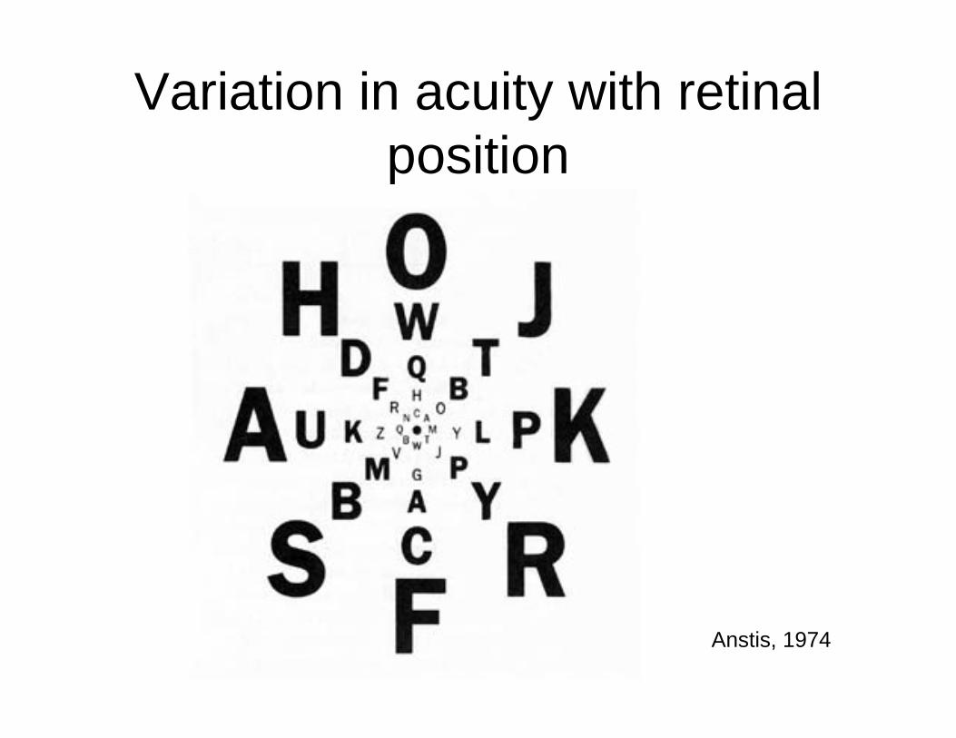
Variation in acuity with retinal
Anstis, 1974
position
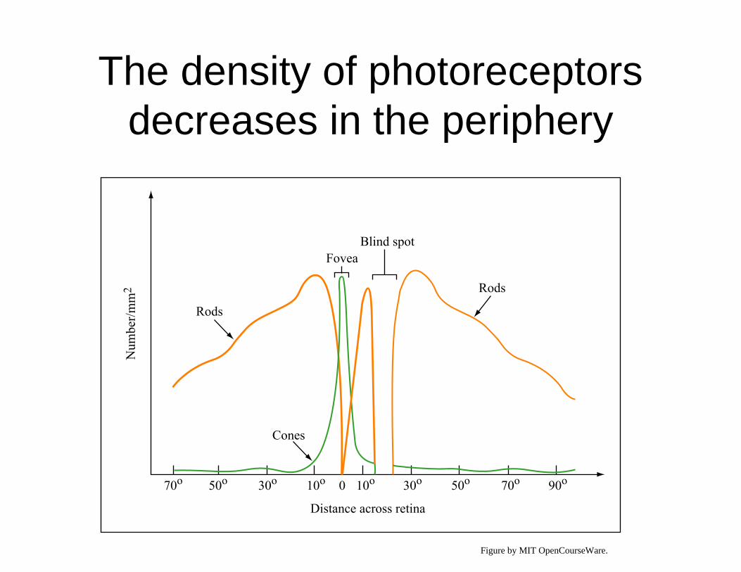
The density of photoreceptorsdecreases in the periphery
90o70o50o30o10o10o30o 050o70o
Distance across retina
Rods
Cones
FoveaBlind spot
Num
ber/m
m2 Rods
Figure by MIT OpenCourseWare.
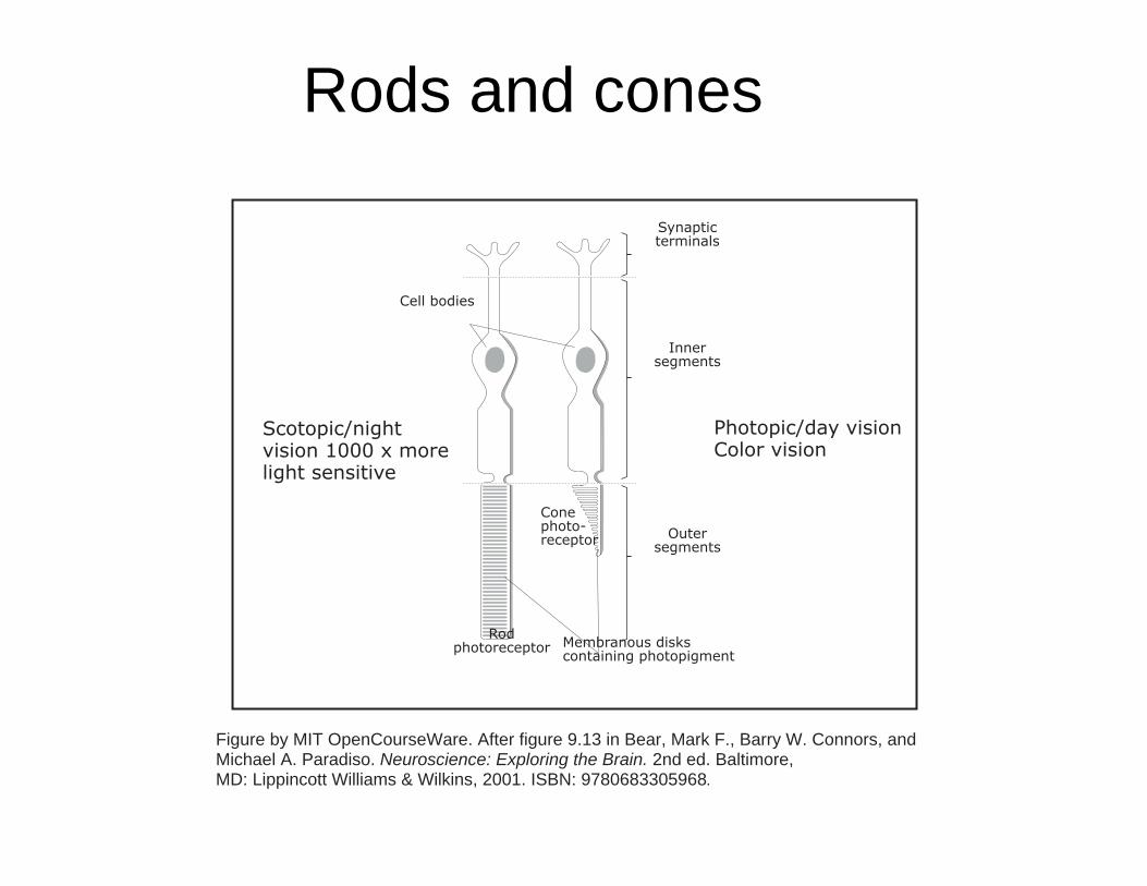
Rods and cones
Figure by MIT OpenCourseWare. After figure 9.13 in Bear, Mark F., Barry W. Connors, and Michael A. Paradiso. Neuroscience: Exploring the Brain. 2nd ed. Baltimore, MD: Lippincott Williams & Wilkins, 2001. ISBN: 9780683305968.
Conephoto-receptor
Scotopic/night vision 1000 x more light sensitive
Photopic/day visionColor vision
Outersegments
Innersegments
Synapticterminals
Cell bodies
Membranous diskscontaining photopigment
Rodphotoreceptor
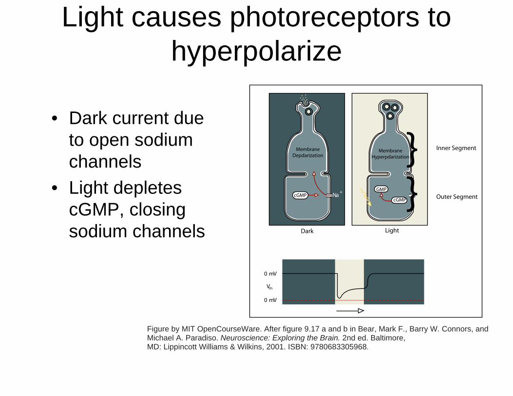
Light causes photoreceptors tohyperpolarize
• Dark current due to open sodium channels
• Light depletes cGMP, closing sodium channels
Figure by MIT OpenCourseWare. After figure 9.17 a and b in Bear, Mark F., Barry W. Connors, and Michael A. Paradiso. Neuroscience: Exploring the Brain. 2nd ed. Baltimore, MD: Lippincott Williams & Wilkins, 2001. ISBN: 9780683305968.
0 mV
0 mV
Vm
}Na
+
Inner Segment
Outer Segment
LightDark
MembraneDepdarization
MembraneHyperpdarization
GMP
cGMPcGMP }
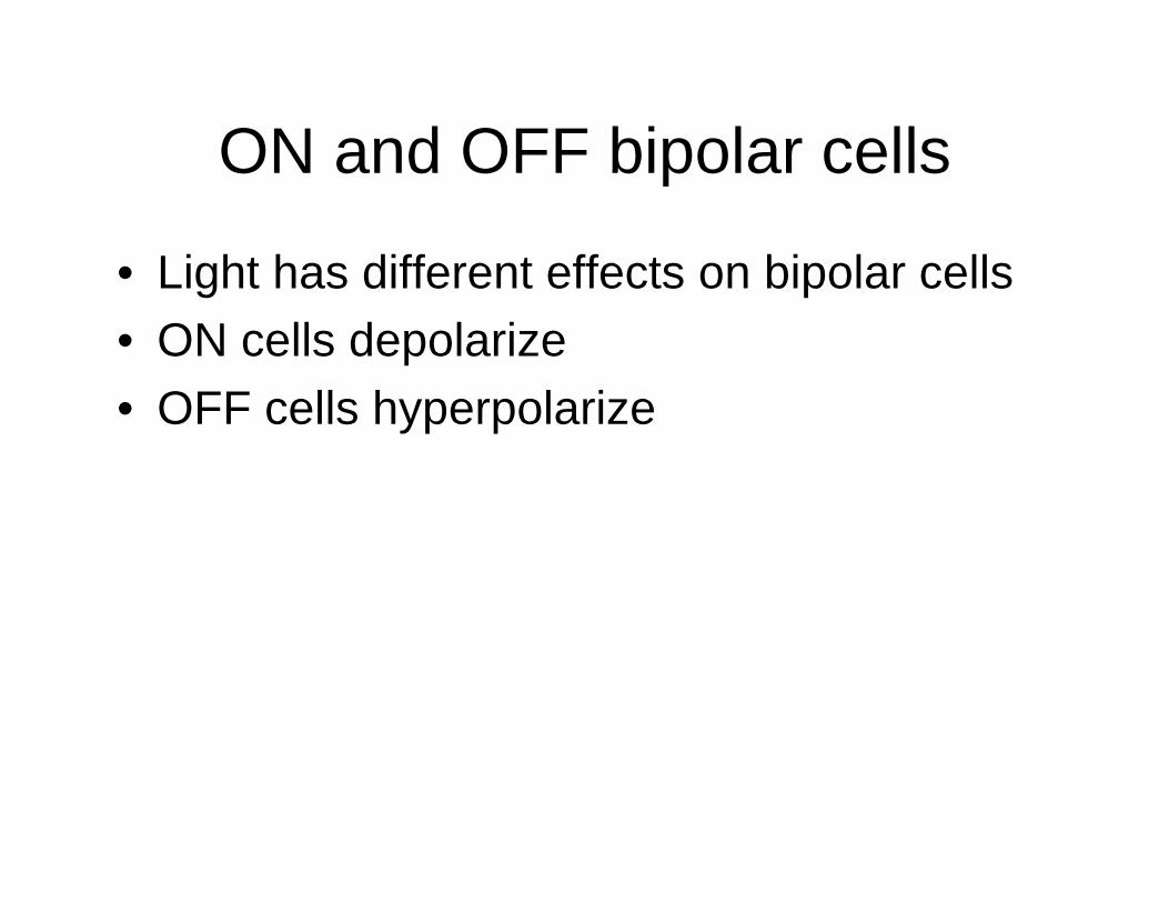
ON and OFF bipolar cells
• Light has different effects on bipolar cells
• ON cells depolarize • OFF cells hyperpolarize
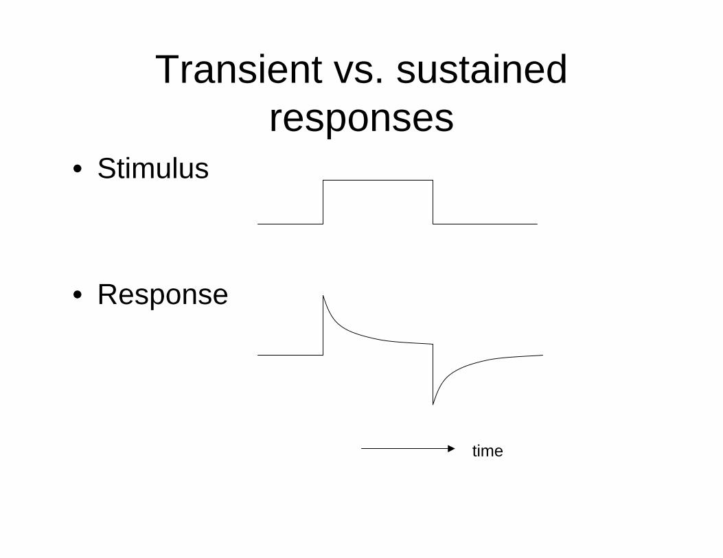
Transient vs. sustainedresponses
• Stimulus
• Response
time
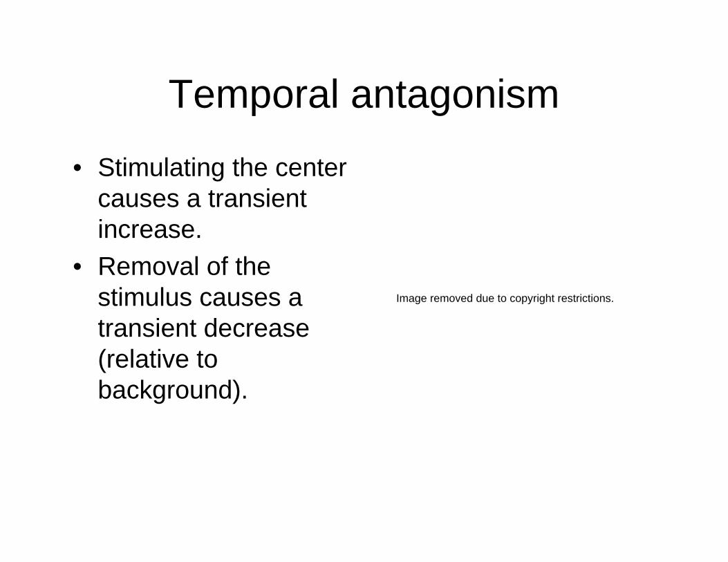
Temporal antagonism
• Stimulating the center causes a transient increase.
• Removal of the stimulus causes a transient decrease (relative to background).
Image removed due to copyright restrictions.
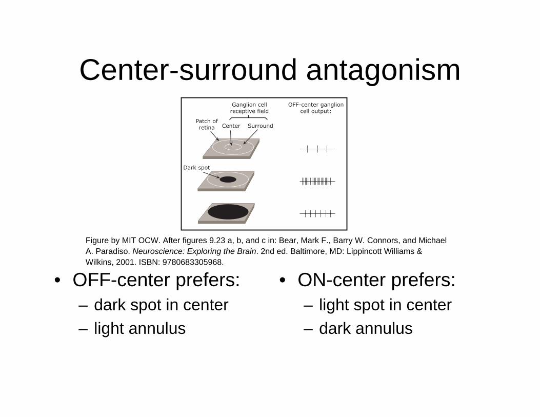
Center-surround antagonism
• OFF-center prefers: • ON-center prefers:– dark spot in center – light spot in center – light annulus – dark annulus
Ganglion cellreceptive field
OFF-center ganglioncell output:
Center
Dark spot
Patch ofretina Surround
Figure by MIT OCW. After figures 9.23 a, b, and c in: Bear, Mark F., Barry W. Connors, and MichaelA. Paradiso. Neuroscience: Exploring the Brain. 2nd ed. Baltimore, MD: Lippincott Williams &Wilkins, 2001. ISBN: 9780683305968.
Horizontal cells are coupled bygap junctions
Xin and Bloomfield
Photo removed due to copyright restrictions.
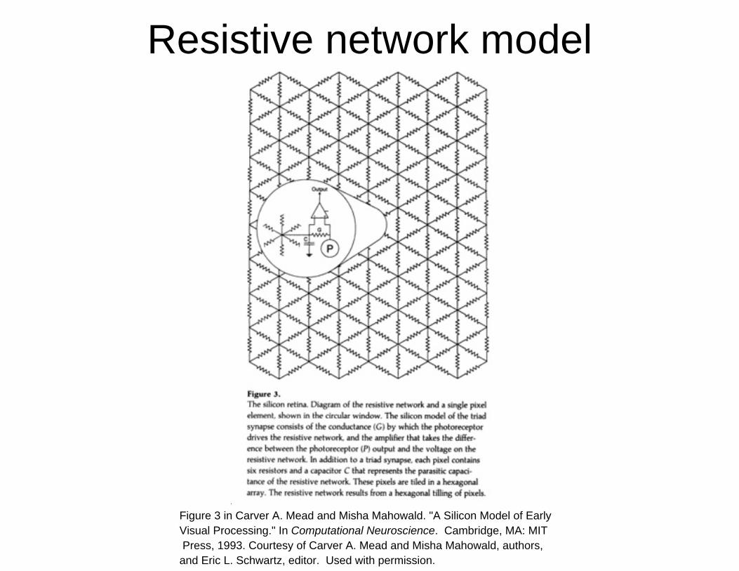
Resistive network model
Figure 3 in Carver A. Mead and Misha Mahowald. "A Silicon Model of EarlyVisual Processing." In Computational Neuroscience. Cambridge, MA: MIT Press, 1993. Courtesy of Carver A. Mead and Misha Mahowald, authors,and Eric L. Schwartz, editor. Used with permission.
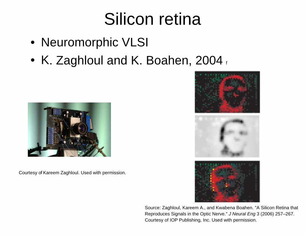
Silicon retinaNeuromorphic VLSI K. Zaghloul and K. Boahen, 2004f
Kareem Zaghloul. Used with permission.
Source: Zaghloul, Kareem A., and Kwabena Boahen. "A Silicon Retina that
• •
Courtesy o
Reproduces Signals in the Optic Nerve." J Neural Eng 3 (2006) 257–267.Courtesy of IOP Publishing, Inc. Used with permission.
f
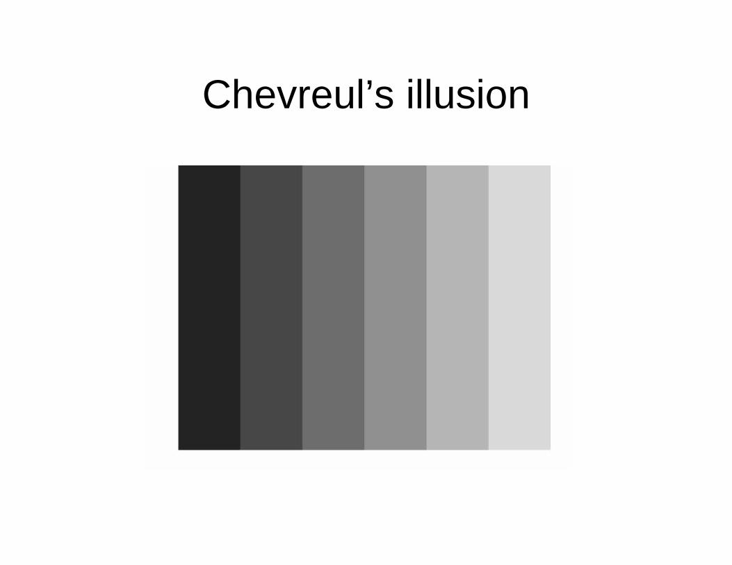
Chevreul’s illusion
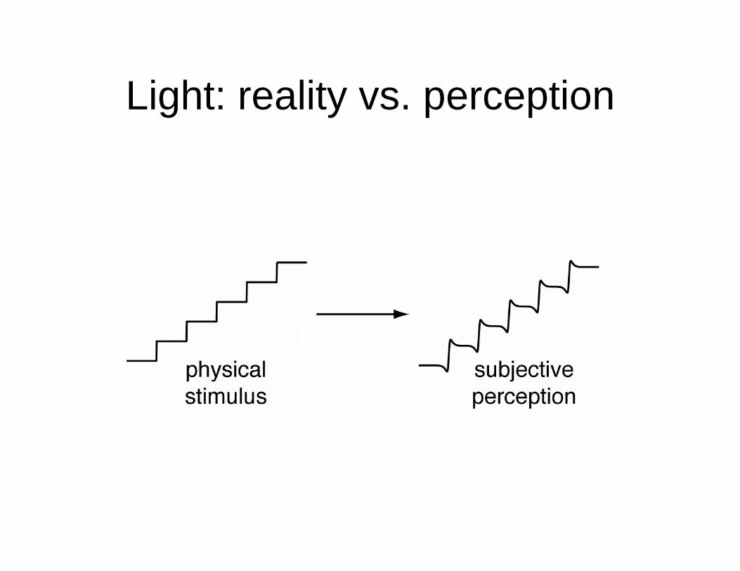
Light: reality vs. perception
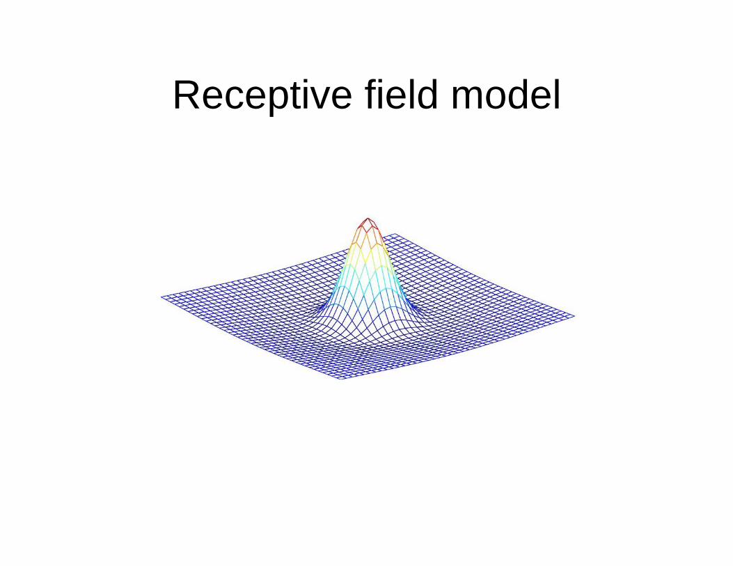
Receptive field model
Related Documents