E-ISSN: 1308-5263 TURKISH JOURNAL OF HEMATOLOGY • VOL.: 38 ISSUE: 1 MARCH 2021 Review A Retrospective Single-Center Study from Turkey Can Baykal, Sıla Kılıç Sayar, Kurtuluş Didem Yazganoğlu, Nesimi Büyükbabani; İstanbul, Turkey 1 Cover Picture: Moeinadin Safavi, Zohreh Nozarian, Farzad Kompani Post-Chemotherapy Foamy Histiocytes in Bone Marrow Aspiration of a Child with Acute Lymphoblastic Leukemia PROCEEDINGS ABSTRACTS Pieter Sonneveld, Rotterdam, Netherlands Alexander Popov, Moscow, Russian Federation Gunnur Deniz, İstanbul, Turkey Kala Kamdar, Texas, USA Monica Gramatges, Texas, USA Eva Kimby, Sweden Koen van Besien, New York, USA Hartmut Döhner, Ulm, Germany Andrew Wei, Melbourne, Australia Bastian von Tresckow, Essen, Germany Dennis Eichenauer, Köln, Germany Nicolas Boissel, Paris, France Oliver Ottmann, Cardiff, UK Andrés J. M. Ferreri, Milano, Italy Marion Subklewe, Munich, Germany ORAL PRESENTATIONS POSTER PRESENTATIONS 8 th International Congress on Leukemia Lymphoma Myeloma May 21-22, 2021 • VIRTUAL CONGRESS Volume 38 Issue 1 Suppl. 1 March 2021

Welcome message from author
This document is posted to help you gain knowledge. Please leave a comment to let me know what you think about it! Share it to your friends and learn new things together.
Transcript

Issue 1Volume 38 March 2021
E-IS
SN: 1
308-
5263
TURKISH
JOU
RNAL O
F HEM
ATOLO
GY • VO
L.: 38 ISSUE: 1 M
ARCH 2021
ReviewPrimary Immune Regulatory Disorders and Targeted TherapiesBurcu Kolukısa, Safa Barış; İstanbul, Turkey
Research ArticlesPrognostic Value of Antithrombin Levels in COVID-19 Patients and Impact of Fresh Frozen Plasma Treatment: A Retrospective Studyİlkay Anaklı, Perihan Ergin Özcan, Özlem Polat, Günseli Orhun, Gülçin Hilal Alay, Verda Tuna, Emre Çeliksoy, Mehmet Kılıç, Mutlu Mercan, Achmet Ali, Sevgi Beşışık, Figen Esen; İstanbul, Turkey
Current Practice in FFP Preparation and Use in Greece: A National SurveyAspasia Argyrou, Serena Valsami, Abraham Pouliakis, Maria Gavalaki, Antonis Aggelidis, Vasiliki Voulgaridou, Vasiliki Pliatsika, Theofanis Adraktas, Andreas Papachronis, Chrysoula Alepi, Vasiliki Giannopoulou, Panagiotis Siourounis, Sofia Tsagia, Georges Martinis, Eftihia Kontekaki, Eleftheria Zervou, Spiridon Koliofotis, Elias Kyriakou, Athina Mougiou, Lempousi Dimitra, Afrodite Chairopoulou, Aggeliki Tsakania, Maria Baka, Ioanna Apostolidou, Dimitra Moschandreou, Anastasia Livada, Marianna Politou, Fragoula Roussinou, Christina Pappa, Vasiliki Koika, Niki Vgontza, Anthippi Gafou, Ioanna Dendrinou, Fotini Sakellaridi, Lampothea Labrianou, Zafeiria Alexandropoulou, Vasiliki Sochali, Kostas Malekas, Areti Skordilaki, Georgia Kakava, Konstantinos Lebesopoulos, Konstantinos Stamoulis, Elisavet Grouzi; Athens, Thessaloniki, Piraeus, Alexandroupolis, Ioannina, Patras, Greece
Evaluation of Prognostic Significance of the International Staging System According to Glomerular Filtration Rate in Newly Diagnosed Multiple Myeloma Patients Eligible for Autologous Stem Cell Transplantation Rafiye Çiftçiler, Hakan Göker, Haluk Demiroğlu, İbrahim Celalettin Haznedaroğlu, Nilgün Sayınalp, Salih Aksu, Osman Özcebe, Yahya Büyükaşık; Ankara, Turkey
Original Versus Generic Lenalidomide in Patients with Relapsed/Refractory Multiple Myeloma: Comparison of Efficacy and Adverse EventsAli Zahit Bolaman, Atakan Turgutkaya, Birsen Sahip, Cem Selim, Hilal Eroğlu Küçükerdiler, Şehmus Ertop, Gökhan Sargın, İrfan Yavaşoğlu; Aydın, Zonguldak, Turkey
Evaluation of Associated Lymphomas and Their Risk Factors in Patients with Lymphomatoid Papulosis: A Retrospective Single-Center Study from TurkeyCan Baykal, Sıla Kılıç Sayar, Kurtuluş Didem Yazganoğlu, Nesimi Büyükbabani; İstanbul, Turkey
1Cover Picture: Moeinadin Safavi, Zohreh Nozarian, Farzad Kompani
Post-Chemotherapy Foamy Histiocytes in Bone Marrow Aspiration of a Child with Acute Lymphoblastic Leukemia
PROCEEDINGS
ABSTRACTS
Pieter Sonneveld, Rotterdam, Netherlands
Alexander Popov, Moscow, Russian Federation
Gunnur Deniz, İstanbul, Turkey
Kala Kamdar, Texas, USA
Monica Gramatges, Texas, USA
Eva Kimby, Sweden
Koen van Besien, New York, USA
Hartmut Döhner, Ulm, Germany
Andrew Wei, Melbourne, Australia
Bastian von Tresckow, Essen, Germany
Dennis Eichenauer, Köln, Germany
Nicolas Boissel, Paris, France
Oliver Ottmann, Cardiff, UK
Andrés J. M. Ferreri, Milano, Italy
Marion Subklewe, Munich, Germany
ORAL PRESENTATIONS
POSTER PRESENTATIONS
8th International Congress on Leukemia Lymphoma MyelomaMay 21-22, 2021 • VIRTUAL CONGRESS
Volume 38 Issue 1 Suppl. 1 March 2021

International Review Board
Nejat AkarGörgün Akpek Serhan Alkan Çigdem Altay Koen van Besien M. Sıraç Dilber Ahmet Dogan Peter Dreger Thierry FaconJawed Fareed Gösta Gahrton Dieter Hoelzer Marilyn Manco-Johnson Andreas JostingEmin Kansu Winfried Kern Nigel Key Korgün Koral Abdullah KutlarLuca Malcovati Robert Marcus Jean Pierre Marie Ghulam MuftiGerassimos A. Pangalis Antonio PigaAnanda Prasad Jacob M. RoweJens-Ulrich Rüffer Norbert Schmitz Orhan Sezer Anna Sureda Ayalew Tefferi Nükhet Tüzüner Catherine Verfaillie Srdan VerstovsekClaudio Viscoli
TOBB University of Economics and Technology Hospital, Ankara, Turkey Maryland School of Medicine, Baltimore, USA Cedars-Sinai Medical Center, Los Angeles, USAAnkara, TurkeyWeill Cornell Medicine, New York, USA Karolinska University, Stockholm, Sweden Memorial Sloan Kettering Cancer Center, New York, USA Heidelberg University, Heidelberg, GermanyLille University, Lille, France Loyola University, Maywood, USA Karolinska University Hospital, Stockholm, SwedenFrankfurt University, Frankfurt, GermanyUniversity of Colorado Anschutz Medical Campus, Aurora, USAUniversity Hospital Cologne, Cologne, Germany Hacettepe University, Ankara, Turkey Albert Ludwigs University, Freiburg im Breisgau, GermanyUniversity of North Carolina School of Medicine, NC, USASouthwestern Medical Center, Texas, USAMedical College of Georgia at Augusta University, Augusta, USA Pavia Medical School University, Pavia, Italy Kings College Hospital, London, UK Pierre et Marie Curie University, Paris, France King’s Hospital, London, UK Athens University, Athens, Greece Torino University, Torino, Italy Wayne State University School of Medicine, Detroit, USA Hebrew University of Jerusalem, Jerusalem, Israel University of Köln, Köln, GermanyAK St Georg, Hamburg, Germany Charité Comprehensive Cancer Center, Berlin, GermanySanta Creu i Sant Pau Hospital, Barcelona, Spain Mayo Clinic, Rochester, Minnesota, USA İstanbul Cerrahpasa University, İstanbul, Turkey Katholieke Universiteit Leuven, Leuven, BelgiumThe University of Texas MD Anderson Cancer Center, Houston, USA San Martino University, Genoa, Italy
Past EditorsErich Frank Orhan UlutinHamdi AkanAytemiz Gürgey
Senior Advisory BoardYücel Tangün Osman İlhan Muhit ÖzcanTeoman SoysalAhmet Muzaffer Demir
Language EditorLeslie Demir
Statistic EditorHülya Ellidokuz
Editorial Officeİpek Durusu Bengü Timoçin Efe
Editor-in-Chief Reyhan Küçükkaya
İstanbul, [email protected]
Associate Editors A.EmreEşkazan
İstanbul University-Cerrahpasa, İstanbul, [email protected]
AliİrfanEmreTekgündüzMemorial Bahçelievler Hospital, İstanbul, [email protected]
AyşegülÜnüvarİstanbul University, İstanbul, Turkey [email protected]
CengizBeyanAnkara, [email protected]
Hale ÖrenDokuz Eylül University, İzmir, Turkey [email protected]
İbrahimC.HaznedaroğluHacettepe University, Ankara, Turkey [email protected]
Selami Koçak ToprakAnkara University, Ankara, Turkey [email protected]
SemraPaydaşÇukurova University, Adana, Turkey [email protected]
ŞuleÜnalHacettepe University, Ankara, [email protected]
Assistant Editors ClaudioCerchione
University of Naples Federico II Napoli, Campania, Italy
EbruKocaBaskent University Ankara Hospital, Clinic of Hematology, Ankara, Turkey
ElifÜnalİnceAnkara University, Ankara, Turkey
İnciAlacacıoğluDokuz Eylül University, İzmir, Turkey
MarioTiribelliUniversity of Udine, Udine, Italy
MügeSayitoğluİstanbul University, İstanbul, Turkey
Nil GülerOndokuz Mayıs University, Samsun, Turkey
OlgaMeltemAkayKoç University, İstanbul, Turkey
VeyselSabriHançerİstinye University, İstanbul, Turkey
Zühre KayaGazi University, Ankara, Turkey
Publishing Services
GALENOS PUBLISHERMolla Gürani Mah. Kaçamak Sk. No: 21/1, Fındıkzade, İstanbul, Turkey Phone: +90 212 621 99 25 • Fax: +90 212 621 99 27 • www. galenos.com.tr
A-I
International Review Board
Nejat AkarGörgün Akpek Serhan Alkan Çigdem Altay Koen van Besien M. Sıraç Dilber Ahmet Dogan Peter Dreger Thierry FaconJawed Fareed Gösta Gahrton Dieter Hoelzer Marilyn Manco-Johnson Andreas JostingEmin Kansu Winfried Kern Nigel Key Korgün Koral Abdullah KutlarLuca Malcovati Robert Marcus Jean Pierre Marie Ghulam MuftiGerassimos A. Pangalis Antonio PigaAnanda Prasad Jacob M. RoweJens-Ulrich Rüffer Norbert Schmitz Orhan Sezer Anna Sureda Ayalew Tefferi Nükhet Tüzüner Catherine Verfaillie Srdan VerstovsekClaudio Viscoli
TOBB University of Economics and Technology Hospital, Ankara, Turkey Maryland School of Medicine, Baltimore, USA Cedars-Sinai Medical Center, Los Angeles, USAAnkara, TurkeyWeill Cornell Medicine, New York, USA Karolinska University, Stockholm, Sweden Memorial Sloan Kettering Cancer Center, New York, USA Heidelberg University, Heidelberg, GermanyLille University, Lille, France Loyola University, Maywood, USA Karolinska University Hospital, Stockholm, SwedenFrankfurt University, Frankfurt, GermanyUniversity of Colorado Anschutz Medical Campus, Aurora, USAUniversity Hospital Cologne, Cologne, Germany Hacettepe University, Ankara, Turkey Albert Ludwigs University, Freiburg im Breisgau, GermanyUniversity of North Carolina School of Medicine, NC, USASouthwestern Medical Center, Texas, USAMedical College of Georgia at Augusta University, Augusta, USA Pavia Medical School University, Pavia, Italy Kings College Hospital, London, UK Pierre et Marie Curie University, Paris, France King’s Hospital, London, UK Athens University, Athens, Greece Torino University, Torino, Italy Wayne State University School of Medicine, Detroit, USA Hebrew University of Jerusalem, Jerusalem, Israel University of Köln, Köln, GermanyAK St Georg, Hamburg, Germany Charité Comprehensive Cancer Center, Berlin, GermanySanta Creu i Sant Pau Hospital, Barcelona, Spain Mayo Clinic, Rochester, Minnesota, USA İstanbul Cerrahpasa University, İstanbul, Turkey Katholieke Universiteit Leuven, Leuven, BelgiumThe University of Texas MD Anderson Cancer Center, Houston, USA San Martino University, Genoa, Italy
Past EditorsErich Frank Orhan UlutinHamdi AkanAytemiz Gürgey
Senior Advisory BoardYücel Tangün Osman İlhan Muhit ÖzcanTeoman SoysalAhmet Muzaffer Demir
Language EditorLeslie Demir
Statistic EditorHülya Ellidokuz
Editorial Officeİpek Durusu Bengü Timoçin Efe
Editor-in-Chief Reyhan Küçükkaya
İstanbul, [email protected]
Associate Editors A.EmreEşkazan
İstanbul University-Cerrahpasa, İstanbul, [email protected]
AliİrfanEmreTekgündüzMemorial Bahçelievler Hospital, İstanbul, [email protected]
AyşegülÜnüvarİstanbul University, İstanbul, Turkey [email protected]
CengizBeyanAnkara, [email protected]
Hale ÖrenDokuz Eylül University, İzmir, Turkey [email protected]
İbrahimC.HaznedaroğluHacettepe University, Ankara, Turkey [email protected]
Selami Koçak ToprakAnkara University, Ankara, Turkey [email protected]
SemraPaydaşÇukurova University, Adana, Turkey [email protected]
ŞuleÜnalHacettepe University, Ankara, [email protected]
Assistant Editors ClaudioCerchione
University of Naples Federico II Napoli, Campania, Italy
EbruKocaBaskent University Ankara Hospital, Clinic of Hematology, Ankara, Turkey
ElifÜnalİnceAnkara University, Ankara, Turkey
İnciAlacacıoğluDokuz Eylül University, İzmir, Turkey
MarioTiribelliUniversity of Udine, Udine, Italy
MügeSayitoğluİstanbul University, İstanbul, Turkey
Nil GülerOndokuz Mayıs University, Samsun, Turkey
OlgaMeltemAkayKoç University, İstanbul, Turkey
VeyselSabriHançerİstinye University, İstanbul, Turkey
Zühre KayaGazi University, Ankara, Turkey
Publishing Services
GALENOS PUBLISHERMolla Gürani Mah. Kaçamak Sk. No: 21/1, Fındıkzade, İstanbul, Turkey Phone: +90 212 621 99 25 • Fax: +90 212 621 99 27 • www. galenos.com.tr
A-IDigital
Publishing Services
BAYTBilimsel Araştırmalar Basın Yayın ve Tanıtım Ltd. Şti.Ziya Gökalp Cad. 30/31, 06420 Kızılay, AnkaraTel: 0312 431 30 62 • Faks: 0312 431 36 02 • www.bayt.com.tr • [email protected]
Contact InformationEditorial Correspondence should be addressed to Dr. Reyhan KüçükkayaE-mail : [email protected]
All Inquiries Should be Addressed toTURKISH JOURNAL OF HEMATOLOGYAddress : Turan Güneş Bulv. İlkbahar Mah. Fahreddin Paşa Sokagı (eski 613. Sok.) No: 8 06550 Çankaya, Ankara / TurkeyPhone : +90 312 490 98 97Fax : +90 312 490 98 68 E-mail : [email protected]
E-ISSN: 1308-5263
Publishing ManagerMuhlis Cem Ar
Management AddressTürk Hematoloji Dernegi
Turan Güneş Bulv. İlkbahar Mah. Fahreddin Paşa Sokagı (eski 613. Sok.) No: 8 06550 Çankaya, Ankara / Turkey
Online Manuscript Submissionhttp://mc.manuscriptcentral.com/tjh
Web Pagewww.tjh.com.tr
Owner on Behalf of the Turkish Society of HematologyGüner Hayri Özsan
Publishing HouseMolla Gürani Mah. Kaçamak Sk. No: 21, 34093 Fındıkzade, İstanbul / Turkey Tel: +90 212 621 99 25
Fax: +90 212 621 99 27 E-mail: [email protected] Publisher Certificate Number: 14521
Publication Date24.02.2021
Cover PictureMoeinadin Safavi, Zohreh Nozarian, Farzad Kompani
Post-Chemotherapy Foamy Histiocytes in Bone Marrow Aspiration of a Child with Acute Lymphoblastic Leukemia
Smears showed multiple foamy histiocytes (A, B).
International scientific journal published quarterly.
The Turkish Journal of Hematology is published by the commercial enterprise of the Turkish Society of Hematology with Decision Number 6 issued by the Society on 7 October 2008.
A-IIII
8th International Congress on Leukemia Lymphoma MyelomaMay 21-22, 2021 • VIRTUAL CONGRESS
Digital Publishing
BAYT
Bilimsel Araştırmalar Basın Yayın ve Tanıtım Ltd. Şti.
Ziya Gökalp Cad. 30/31, 06420 Kızılay, Ankara
Tel: 0312 431 30 62 • Faks: 0312 431 36 02 • www.bayt.com.tr
Publication Date
20.05.2021
Cover Picture
Moeinadin Safavi, Zohreh Nozarian, Farzad Kompani
Post-Chemotherapy Foamy Histiocytes in Bone Marrow Aspiration
of a Child with Acute Lymphoblastic Leukemia
Smears showed multiple foamy histiocytes (A, B).

Contact InformationEditorial Correspondence should be addressed to Dr. Reyhan KüçükkayaE-mail : [email protected]
All Inquiries Should be Addressed toTURKISH JOURNAL OF HEMATOLOGYAddress : Turan Güneş Bulv. İlkbahar Mah. Fahreddin Paşa Sokagı (eski 613. Sok.) No: 8 06550 Çankaya, Ankara / TurkeyPhone : +90 312 490 98 97Fax : +90 312 490 98 68 E-mail : [email protected]
E-ISSN: 1308-5263
Publishing ManagerMuhlis Cem Ar
Management AddressTürk Hematoloji Dernegi
Turan Güneş Bulv. İlkbahar Mah. Fahreddin Paşa Sokagı (eski 613. Sok.) No: 8 06550 Çankaya, Ankara / Turkey
Online Manuscript Submissionhttp://mc.manuscriptcentral.com/tjh
Web Pagewww.tjh.com.tr
Owner on Behalf of the Turkish Society of HematologyGüner Hayri Özsan
Publishing HouseMolla Gürani Mah. Kaçamak Sk. No: 21, 34093 Fındıkzade, İstanbul / Turkey Tel: +90 212 621 99 25
Fax: +90 212 621 99 27 E-mail: [email protected] Publisher Certificate Number: 14521
Publication Date24.02.2021
Cover PictureMoeinadin Safavi, Zohreh Nozarian, Farzad Kompani
Post-Chemotherapy Foamy Histiocytes in Bone Marrow Aspiration of a Child with Acute Lymphoblastic Leukemia
Smears showed multiple foamy histiocytes (A, B).
International scientific journal published quarterly.
The Turkish Journal of Hematology is published by the commercial enterprise of the Turkish Society of Hematology with Decision Number 6 issued by the Society on 7 October 2008.
A-II
AIMS AND SCOPE The Turkish Journal of Hematology is published quarterly (March, June, September, and December) by the Turkish Society of Hematology. It is an independent, non-profit peer-reviewed international English-language periodical encompassing subjects relevant to hematology. The Editorial Board of The Turkish Journal of Hematology adheres to the principles of the World Association of Medical Editors (WAME), International Council of Medical Journal Editors (ICMJE), Committee on Publication Ethics (COPE), Consolidated Standards of Reporting Trials (CONSORT) and Strengthening the Reporting of Observational Studies in Epidemiology (STROBE). The aim of The Turkish Journal of Hematology is to publish original hematological research of the highest scientific quality and clinical relevance. Additionally, educational material, reviews on basic developments, editorial short notes, images in hematology, and letters from hematology specialists and clinicians covering their experience and comments on hematology and related medical fields as well as social subjects are published. As of December 2015, The Turkish Journal of Hematology does not accept case reports. Important new findings or data about interesting hematological cases may be submitted as a brief report.General practitioners interested in hematology and internal medicine specialists are among our target audience, and The Turkish Journal of Hematology aims to publish according to their needs. The Turkish Journal of Hematology is indexed, as follows: - PubMed Medline- PubMed Central- Science Citation Index Expanded- EMBASE- Scopus- CINAHL- Gale/Cengage Learning- EBSCO- DOAJ- ProQuest- Index Copernicus- Tübitak/Ulakbim Turkish Medical Database- Turk Medline- Hinari- GOALI- ARDI- OAREImpact Factor: 1.685Open Access PolicyTurkish Journal of Hematology is an Open Access journal. This journal provides immediate open access to its content on the principle that making research freely available to the public supports a greater global exchange of knowledge.Open Access Policy is based on the rules of the Budapest Open Access Initiative (BOAI) http://www.budapestopenaccessinitiative.org/.Subscription Information The Turkish Journal of Hematology is published electronically only as of 2019. Therefore, subscriptions are not necessary. All published volumes are available in full text free-of-charge online at www.tjh.com.tr.
Address: Turan Güneş Bulv. İlkbahar Mah. Fahreddin Paşa Sokagı (eski 613. Sok.) No: 8 06550 Çankaya, Ankara / TurkeyTelephone: +90 312 490 98 97 Fax: +90 312 490 98 68Online Manuscript Submission: http://mc.manuscriptcentral.com/tjh Web page: www.tjh.com.tr E-mail: [email protected] Requests for permission to reproduce published material should be sent to the editorial office.Editor: Professor Dr. Reyhan KüçükkayaAdress: Turan Güneş Bulv. İlkbahar Mah. Fahreddin Paşa Sokagı (eski 613. Sok.) No: 8 06550 Çankaya, Ankara / TurkeyTelephone: +90 312 490 98 97 Fax: +90 312 490 98 68 Online Manuscript Submission: http://mc.manuscriptcentral.com/tjh Web page: www.tjh.com.tr E-mail: [email protected] YayıneviMolla Gürani Mah. Kaçamak Sk. No:21 34093 Fındıkzade-İstanbul, TurkeyTelephone : +90 212 621 99 25Fax : +90 212 621 99 [email protected] for AuthorsInstructions for authors are published in the journal and at www.tjh.com.trMaterial DisclaimerAuthors are responsible for the manuscripts they publish in The Turkish Journal of Hematology. The editor, editorial board, and publisher do not accept any responsibility for published manuscripts.If you use a table or figure (or some data in a table or figure) from another source, cite the source directly in the figure or table legend.Editorial PolicyFollowing receipt of each manuscript, a checklist is completed by the Editorial Assistant. The Editorial Assistant checks that each manuscript contains all required components and adheres to the author guidelines, after which time it will be forwarded to the Editor in Chief. Following the Editor in Chief’s evaluation, each manuscript is forwarded to the Associate Editor, who in turn assigns reviewers. Generally, all manuscripts will be reviewed by at least three reviewers selected by the Associate Editor, based on their relevant expertise. Associate editor could be assigned as a reviewer along with the reviewers. After the reviewing process, all manuscripts are evaluated in the Editorial Board Meeting. Turkish Journal of Hematology’s editor and Editorial Board members are active researchers. It is possible that they would desire to submit their manuscript to the Turkish Journal of Hematology. This may be creating a conflict of interest. These manuscripts will not be evaluated by the submitting editor(s). The review process will be managed and decisions made by editor-in-chief who will act independently. In some situation, this process will be overseen by an outside independent expert in reviewing submissions from editors.
A-IIIIII
8th International Congress on Leukemia Lymphoma MyelomaMay 21-22, 2021 • VIRTUAL CONGRESS
Digital Publishing
BAYT
Bilimsel Araştırmalar Basın Yayın ve Tanıtım Ltd. Şti.
Ziya Gökalp Cad. 30/31, 06420 Kızılay, Ankara
Tel: 0312 431 30 62 • Faks: 0312 431 36 02 • www.bayt.com.tr
Publication Date
20.05.2021
Cover Picture
Moeinadin Safavi, Zohreh Nozarian, Farzad Kompani
Post-Chemotherapy Foamy Histiocytes in Bone Marrow Aspiration
of a Child with Acute Lymphoblastic Leukemia
Smears showed multiple foamy histiocytes (A, B).

TURKISH JOURNAL OF HEMATOLOGY INSTRUCTIONS FOR AUTHORSThe Turkish Journal of Hematology accepts invited review articles, research articles, brief reports, letters to the editor, and hematological images that are relevant to the scope of hematology, on the condition that they have not been previously published elsewhere. Basic science manuscripts, such as randomized, cohort, cross-sectional, and case-control studies, are given preference. All manuscripts are subject to editorial revision to ensure they conform to the style adopted by the journal. There is a double-blind reviewing system. Review articles are solicited by the Editor-in-Chief. Authors wishing to submit an unsolicited review article should contact the Editor-in-Chief prior to submission in order to screen the proposed topic for relevance and priority.
The Turkish Journal of Hematology does not charge any article submission or processing charges.
Manuscripts should be prepared according to ICMJE guidelines (http://www.icmje.org/). Original manuscripts require a structured abstract. Label each section of the structured abstract with the appropriate subheading (Objective, Materials and Methods, Results, and Conclusion). Letters to the editor do not require an abstract. Research or project support should be acknowledged as a footnote on the title page. Technical and other assistance should be provided on the title page.
Original Manuscripts
Title Page
Title: The title should provide important information regarding the manuscript’s content. The title must specify that the study is a cohort study, cross-sectional study, case-control study, or randomized study (i.e. Cao GY, Li KX, Jin PF, Yue XY, Yang C, Hu X. Comparative bioavailability of ferrous succinate tablet formulations without correction for baseline circadian changes in iron concentration in healthy Chinese male subjects: A single-dose, randomized, 2-period crossover study. Clin Ther 2011;33:2054-2059).
The title page should include the authors’ names, degrees, and institutional/professional affiliations and a short title, abbreviations, keywords, financial disclosure statement, and conflict of interest statement. If a manuscript includes authors from more than one institution, each author’s name should be followed by a superscript number that corresponds to their institution, which is listed separately. Please provide contact information for the corresponding author, including name, e-mail address, and telephone and fax numbers.
Important Notice: The title page should be submitted separately.
Running Head: The running head should not be more than 40 characters, including spaces, and should be located at the bottom of the title page.
Word Count: A word count for the manuscript, excluding abstract, acknowledgments, figure and table legends, and references, should be provided and should not exceed 2500 words. The word count for the abstract should not exceed 300 words.
Conflict of Interest Statement: To prevent potential conflicts of interest from being overlooked, this statement must be included in each manuscript. In case there are conflicts of interest, every author should complete the ICMJE general declaration form, which can be obtained at http://www.icmje.org/downloads/coi_disclosure.zip
Abstract and Keywords: The second page should include an abstract that does not exceed 300 words. For manuscripts sent by authors in Turkey, a title and abstract in Turkish are also required. As most readers read the abstract first, it is critically important. Moreover, as various electronic databases integrate only abstracts into their index, important findings should be presented in the abstract.
Objective: The abstract should state the objective (the purpose of the study and hypothesis) and summarize the rationale for the study.
Materials and Methods: Important methods should be written respectively.
Results: Important findings and results should be provided here.
Conclusion: The study’s new and important findings should be highlighted and interpreted.
Other types of manuscripts, such as reviews, brief reports, and editorials, will be published according to uniform requirements. Provide 3-10 keywords below the abstract to assist indexers. Use terms from the Index Medicus Medical Subject Headings List (for randomized studies a CONSORT abstract should be provided: http://www.consort-statement.org).
Introduction: The introduction should include an overview of the relevant literature presented in summary form (one page), and whatever remains interesting, unique, problematic, relevant, or unknown about the topic must be specified. The introduction should conclude with the rationale for the study, its design, and its objective(s).
Materials and Methods: Clearly describe the selection of observational or experimental participants, such as patients, laboratory animals, and controls, including inclusion and exclusion criteria and a description of the source population. Identify the methods and procedures in sufficient detail to allow other researchers to reproduce your results. Provide references to established methods (including statistical methods), provide references to brief modified methods, and provide the rationale for using them and an evaluation of their limitations. Identify all drugs and chemicals used, including generic names, doses, and routes of administration. The section should include only information that was available at the time the plan or protocol for the study was devised
A-IV
(https://www.strobe-statement.org/fileadmin/Strobe/uploads/checklists/STROBE_checklist_v4_combined.pdf).
Statistics: Describe the statistical methods used in enough detail to enable a knowledgeable reader with access to the original data to verify the reported results. Statistically important data should be given in the text, tables, and figures. Provide details about randomization, describe treatment complications, provide the number of observations, and specify all computer programs used.
Results: Present your results in logical sequence in the text, tables, and figures. Do not present all the data provided in the tables and/or figures in the text; emphasize and/or summarize only important findings, results, and observations in the text. For clinical studies provide the number of samples, cases, and controls included in the study. Discrepancies between the planned number and obtained number of participants should be explained. Comparisons and statistically important values (i.e. p-value and confidence interval) should be provided.
Discussion: This section should include a discussion of the data. New and important findings/results and the conclusions they lead to should be emphasized. Link the conclusions with the goals of the study, but avoid unqualified statements and conclusions not completely supported by the data. Do not repeat the findings/results in detail; important findings/results should be compared with those of similar studies in the literature, along with a summarization. In other words, similarities or differences in the obtained findings/results with those previously reported should be discussed.
Study Limitations: Limitations of the study should be detailed. In addition, an evaluation of the implications of the obtained findings/results for future research should be outlined.
Conclusion: The conclusion of the study should be highlighted.
ReferencesCite references in the text, tables, and figures with numbers in square brackets. Number references consecutively according to the order in which they first appear in the text. Journal titles should be abbreviated according to the style used in Index Medicus (consult List of Journals Indexed in Index Medicus). Include among the references any paper accepted, but not yet published, designating the journal followed by “in press”.
Examples of References: 1. List all authors
Deeg HJ, O’Donnel M, Tolar J. Optimization of conditioning for marrow transplantation from unrelated donors for patients with aplastic anemia after failure of immunosuppressive therapy. Blood 2006;108:1485-1491.
2. Organization as author
Royal Marsden Hospital Bone Marrow Transplantation Team. Failure of syngeneic bone marrow graft without preconditioning in post-hepatitis marrow aplasia. Lancet 1977;2:742-744.
3. BookWintrobe MM. Clinical Hematology, 5th ed. Philadelphia, Lea & Febiger, 1961.
4. Book Chapter
Perutz MF. Molecular anatomy and physiology of hemoglobin. In: Steinberg MH, Forget BG, Higs DR, Nagel RI, (eds). Disorders of Hemoglobin: Genetics, Pathophysiology, Clinical Management. New York, Cambridge University Press, 2000.
5. AbstractDrachman JG, Griffin JH, Kaushansky K. The c-Mpl ligand (thrombopoietin) stimulates tyrosine phosphorylation. Blood 1994;84:390a (abstract).
6. Letter to the EditorRao PN, Hayworth HR, Carroll AJ, Bowden DW, Pettenati MJ. Further definition of 20q deletion in myeloid leukemia using fluorescence in situ hybridization. Blood 1994;84:2821-2823.
7. SupplementAlter BP. Fanconi’s anemia, transplantation, and cancer. Pediatr Transplant 2005;9(Suppl 7):81-86.
Brief ReportsAbstractlength:Not to exceed 150 words.
Articlelength:Not to exceed 1200 words.
Introduction: State the purpose and summarize the rationale for the study.
MaterialsandMethods: Clearly describe the selection of the observational or experimental participants. Identify the methods and procedures in sufficient detail. Provide references to established methods (including statistical methods), provide references to brief modified methods, and provide the rationale for their use and an evaluation of their limitations. Identify all drugs and chemicals used, including generic names, doses, and routes of administration.
Statistics: Describe the statistical methods used in enough detail to enable a knowledgeable reader with access to the original data to verify the reported findings/results. Provide details about randomization, describe treatment complications, provide the number of observations, and specify all computer programs used.
Results: Present the findings/results in a logical sequence in the text, tables, and figures. Do not repeat all the findings/results in the tables and figures in the text; emphasize and/or summarize only those that are most important.
Discussion: Highlight the new and important findings/results of the study and the conclusions they lead to. Link the conclusions with the goals of the study, but avoid unqualified statements and conclusions not completely supported by your data.
Invited Review ArticlesAbstractlength:Not to exceed 300 words.
Articlelength:Not to exceed 4000 words.
Review articles should not include more than 100 references. Reviews should include a conclusion, in which a new hypothesis or study about the subject may be posited. Do not publish methods for literature search or level of evidence. Authors who will prepare review articles should already have published research articles on the relevant subject. The study’s new and
A-VIV
8th International Congress on Leukemia Lymphoma MyelomaMay 21-22, 2021 • VIRTUAL CONGRESS

(https://www.strobe-statement.org/fileadmin/Strobe/uploads/checklists/STROBE_checklist_v4_combined.pdf).
Statistics: Describe the statistical methods used in enough detail to enable a knowledgeable reader with access to the original data to verify the reported results. Statistically important data should be given in the text, tables, and figures. Provide details about randomization, describe treatment complications, provide the number of observations, and specify all computer programs used.
Results: Present your results in logical sequence in the text, tables, and figures. Do not present all the data provided in the tables and/or figures in the text; emphasize and/or summarize only important findings, results, and observations in the text. For clinical studies provide the number of samples, cases, and controls included in the study. Discrepancies between the planned number and obtained number of participants should be explained. Comparisons and statistically important values (i.e. p-value and confidence interval) should be provided.
Discussion: This section should include a discussion of the data. New and important findings/results and the conclusions they lead to should be emphasized. Link the conclusions with the goals of the study, but avoid unqualified statements and conclusions not completely supported by the data. Do not repeat the findings/results in detail; important findings/results should be compared with those of similar studies in the literature, along with a summarization. In other words, similarities or differences in the obtained findings/results with those previously reported should be discussed.
Study Limitations: Limitations of the study should be detailed. In addition, an evaluation of the implications of the obtained findings/results for future research should be outlined.
Conclusion: The conclusion of the study should be highlighted.
ReferencesCite references in the text, tables, and figures with numbers in square brackets. Number references consecutively according to the order in which they first appear in the text. Journal titles should be abbreviated according to the style used in Index Medicus (consult List of Journals Indexed in Index Medicus). Include among the references any paper accepted, but not yet published, designating the journal followed by “in press”.
Examples of References: 1. List all authors
Deeg HJ, O’Donnel M, Tolar J. Optimization of conditioning for marrow transplantation from unrelated donors for patients with aplastic anemia after failure of immunosuppressive therapy. Blood 2006;108:1485-1491.
2. Organization as author
Royal Marsden Hospital Bone Marrow Transplantation Team. Failure of syngeneic bone marrow graft without preconditioning in post-hepatitis marrow aplasia. Lancet 1977;2:742-744.
3. BookWintrobe MM. Clinical Hematology, 5th ed. Philadelphia, Lea & Febiger, 1961.
4. Book Chapter
Perutz MF. Molecular anatomy and physiology of hemoglobin. In: Steinberg MH, Forget BG, Higs DR, Nagel RI, (eds). Disorders of Hemoglobin: Genetics, Pathophysiology, Clinical Management. New York, Cambridge University Press, 2000.
5. AbstractDrachman JG, Griffin JH, Kaushansky K. The c-Mpl ligand (thrombopoietin) stimulates tyrosine phosphorylation. Blood 1994;84:390a (abstract).
6. Letter to the EditorRao PN, Hayworth HR, Carroll AJ, Bowden DW, Pettenati MJ. Further definition of 20q deletion in myeloid leukemia using fluorescence in situ hybridization. Blood 1994;84:2821-2823.
7. SupplementAlter BP. Fanconi’s anemia, transplantation, and cancer. Pediatr Transplant 2005;9(Suppl 7):81-86.
Brief ReportsAbstractlength:Not to exceed 150 words.
Articlelength:Not to exceed 1200 words.
Introduction: State the purpose and summarize the rationale for the study.
MaterialsandMethods: Clearly describe the selection of the observational or experimental participants. Identify the methods and procedures in sufficient detail. Provide references to established methods (including statistical methods), provide references to brief modified methods, and provide the rationale for their use and an evaluation of their limitations. Identify all drugs and chemicals used, including generic names, doses, and routes of administration.
Statistics: Describe the statistical methods used in enough detail to enable a knowledgeable reader with access to the original data to verify the reported findings/results. Provide details about randomization, describe treatment complications, provide the number of observations, and specify all computer programs used.
Results: Present the findings/results in a logical sequence in the text, tables, and figures. Do not repeat all the findings/results in the tables and figures in the text; emphasize and/or summarize only those that are most important.
Discussion: Highlight the new and important findings/results of the study and the conclusions they lead to. Link the conclusions with the goals of the study, but avoid unqualified statements and conclusions not completely supported by your data.
Invited Review ArticlesAbstractlength:Not to exceed 300 words.
Articlelength:Not to exceed 4000 words.
Review articles should not include more than 100 references. Reviews should include a conclusion, in which a new hypothesis or study about the subject may be posited. Do not publish methods for literature search or level of evidence. Authors who will prepare review articles should already have published research articles on the relevant subject. The study’s new and
A-VV
8th International Congress on Leukemia Lymphoma MyelomaMay 21-22, 2021 • VIRTUAL CONGRESS
important findings should be highlighted and interpreted in the Conclusion section. There should be a maximum of two authors for review articles.
Perspectives in Hematology
“Perspectives” are articles discussing significant topics relevant to hematology. They are more personal than a Review Article. Authors wishing to submit a Perspective in Hematology article should contact the Editor in Chief prior to submission in order to screen the proposed topic for relevance and priority. Articles submitted for “Perspectives in Hematology” must advance the hot subjects of experimental and/or clinical hematology beyond the articles previously published or in press in TJH. Perspective papers should meet the restrictive criteria of TJH regarding unique scientific and/or educational value, which will impact and enhance clinical hematology practice or the diagnostic understanding of blood diseases. Priority will be assigned to such manuscripts based upon the prominence, significance, and timeliness of the content. The submitting author must already be an expert with a recognized significant published scientific experience in the specific field related to the “Perspectives” article.
Abstract length: Not to exceed 150 words.
Article length: Not to exceed 1000 words.
References: Should not include more than 50 references
Images in Hematology
Articlelength: Not to exceed 200 words.
Authors can submit for consideration illustrations or photos that are interesting, instructive, and visually attractive, along with a few lines of explanatory text and references. Images in Hematology can include no more than 200 words of text, 5 references, and 3 figures or tables. No abstract, discussion, or conclusion is required, but please include a brief title.
Letters to the Editor
Article length: Not to exceed 500 words.
Letters can include no more than 500 words of text, 5-10 references, and 1 figure or table. No abstract is required, but please include a brief title. The total number is usually limited to a maximum of five authors for a letter to the editor.
Tables
Supply each table in a separate file. Number tables according to the order in which they appear in the text, and supply a brief caption for each. Give each column a short or abbreviated heading. Write explanatory statistical measures of variation, such as standard deviation or standard error of mean. Be sure that each table is cited in the text.
Figures
Figures should be professionally drawn and/or photographed. Authors should number figures according to the order in which they appear in the text. Figures include graphs, charts, photographs, and illustrations.
Each figure should be accompanied by a legend that does not exceed 50 words. Use abbreviations only if they have been introduced in the text. Authors are also required to provide the level of magnification for histological slides. Explain the internal scale and identify the staining method used. Figures should be submitted as separate files, not in the text file. High-resolution image files are not preferred for initial submission as the file sizes may be too large. The total file size of the PDF for peer review should not exceed 5 MB.
Authorship
Each author should have participated sufficiently in the work to assume public responsibility for the content. Any portion of a manuscript that is critical to its main conclusions must be the responsibility of at least one author.
Contributor’s Statement
All submissions should contain a contributor’s statement page. Each statement should contain substantial contributions to idea and design, acquisition of data, and analysis and interpretation of findings. All persons designated as an author should qualify for authorship, and all those that qualify should be listed. Each author should have participated sufficiently in the work to take responsibility for appropriate portions of the text.
Acknowledgments
Acknowledge support received from individuals, organizations, grants, corporations, and any other source. For work involving a biomedical product or potential product partially or wholly supported by corporate funding, a note stating, “This study was financially supported (in part) with funds provided by (company name) to (authors’ initials)”, must be included. Grant support, if received, needs to be stated and the specific granting institutions’ names and grant numbers provided when applicable.
Authors are expected to disclose on the title page any commercial or other associations that might pose a conflict of interest in connection with the submitted manuscript. All funding sources that supported the work and the institutional and/or corporate affiliations of the authors should be acknowledged on the title page.
Ethics
When reporting experiments conducted with humans indicate that the procedures were in accordance with ethical standards set forth by the committee that oversees human subject research. Approval of research protocols by the relevant ethics committee, in accordance with international agreements (Helsinki Declaration of 1975, revised 2013 available at https://www.wma.net/policies-post/wma-declaration-of-helsinki-ethical-principles-for-medical-research-involving-human-subjects/), is required for all experimental, clinical, and drug studies. Patient names, initials, and hospital identification numbers should not be used. Manuscripts reporting the results of experimental investigations conducted with humans must state that the study protocol received
A-VI
institutional review board approval and that the participants provided informed consent.
Non-compliance with scientific accuracy is not in accord with scientific ethics. Plagiarism: To re-publish, in whole or in part, the contents of another author’s publication as one’s own without providing a reference. Fabrication: To publish data and findings/results that do not exist. Duplication: Use of data from another publication, which includes re-publishing a manuscript in different languages. Salami slicing: To create more than one publication by dividing the results of a study unnecessarily.
We disapprove of such unethical practices as plagiarism, fabrication, duplication, and salami slicing, as well as efforts to influence the review process with such practices as gifting authorship, inappropriate acknowledgments, and references. Additionally, authors must respect participants‘ right to privacy.
On the other hand, short abstracts published in congress books that do not exceed 400 words and present data of preliminary research, and those that are presented in an electronic environment, are not considered as previously published work. Authors in such a situation must declare this status on the first page of the manuscript and in the cover letter.
(The COPE flowchart is available at http://publicationethics.org.)
We use iThenticate to screen all submissions for plagiarism before publication.
Conditions of Publication
All authors are required to affirm the following statements before their manuscript is considered: 1. The manuscript is being submitted only to The Turkish Journal of Hematology; 2. The manuscript will not be submitted elsewhere while under consideration by The Turkish Journal of Hematology; 3. The manuscript has not been published elsewhere, and should it be published in The Turkish Journal of Hematology it will not be published elsewhere without the permission of the editors (these restrictions do not apply to abstracts or to press reports for presentations at scientific meetings); 4. All authors are responsible for the manuscript’s content; 5. All authors participated in the study concept and design, analysis and interpretation of the data, and drafting or revising of the manuscript and have approved the manuscript as submitted. In addition, all authors are required to disclose any professional affiliation, financial agreement, or other involvement with any company whose product figures prominently in the submitted manuscript.
Authors of accepted manuscripts will receive electronic page proofs and are responsible for proofreading and checking the entire article within two days. Failure to return the proof in two days will delay publication. If the authors cannot be reached by email or telephone within two weeks, the manuscript will be rejected and will not be published in the journal.
Copyright At the time of submission all authors will receive instructions for submitting an online copyright form. No manuscript will be considered for review until
all authors have completed their copyright form. Please note, it is our practice not to accept copyright forms via fax, e-mail, or postal service unless there is a problem with the online author accounts that cannot be resolved. Every effort should be made to use the online copyright system. Corresponding authors can log in to the submission system at any time to check the status of any co-author’s copyright form. All accepted manuscripts become the permanent property of The Turkish Journal of Hematology and may not be published elsewhere, in whole or in part, without written permission.
Note: We cannot accept any copyright form that has been altered, revised, amended, or otherwise changed. Our original copyright form must be used as is.
Units of MeasurementMeasurements should be reported using the metric system, according to the International System of Units (SI). Consult the SI Unit Conversion Guide, New England Journal of Medicine Books, 1992.
An extensive list of conversion factors can be found at https://www.nist.gov/sites/default/files/documents/pml/wmd/metric/SP1038.pdf. For more details, see http://www.amamanualofstyle.com/oso/public/jama/si_conversion_table.html.
Abbreviations and SymbolsUse only standard abbreviations. Avoid abbreviations in the title and abstract. The full term for an abbreviation should precede its first use in the text, unless it is a standard abbreviation. All acronyms used in the text should be expanded at first mention, followed by the abbreviation in parentheses; thereafter the acronym only should appear in the text. Acronyms may be used in the abstract if they occur 3 or more times therein, but must be reintroduced in the body of the text. Generally, abbreviations should be limited to those defined in the AMA Manual of Style, current edition. A list of each abbreviation (and the corresponding full term) used in the manuscript must be provided on the title page.
Online Manuscript Submission ProcessThe Turkish Journal of Hematology uses submission software powered by ScholarOne Manuscripts. The website for submissions to The Turkish Journal of Hematology is http://mc.manuscriptcentral.com/tjh. This system is quick and convenient, both for authors and reviewers.
Setting Up an AccountNew users to the submission site will need to register and enter their account details before they can submit a manuscript. Log in, or click the “Create Account” button if you are a first-time user. To create a new account: After clicking the “Create Account” button, enter your name and e-mail address, and then click the “Next” button. Your e-mail address is very important. Enter your institution and address information, as appropriate, and then click the “Next” Button. Enter a user ID and password of your choice, select your area of expertise, and then click the “Finish” button.
If you have an account, but have forgotten your log-in details, go to “Password Help” on the journal’s online submission system and enter your e-mail address. The system will send you an automatic user ID and a new temporary password.
A-VIIVI
8th International Congress on Leukemia Lymphoma MyelomaMay 21-22, 2021 • VIRTUAL CONGRESS
institutional review board approval and that the participants provided informed consent.
Non-compliance with scientific accuracy is not in accord with scientific ethics. Plagiarism: To re-publish, in whole or in part, the contents of another author’s publication as one’s own without providing a reference. Fabrication: To publish data and findings/results that do not exist. Duplication: Use of data from another publication, which includes re-publishing a manuscript in different languages. Salami slicing: To create more than one publication by dividing the results of a study unnecessarily.
We disapprove of such unethical practices as plagiarism, fabrication, duplication, and salami slicing, as well as efforts to influence the review process with such practices as gifting authorship, inappropriate acknowledgments, and references. Additionally, authors must respect participants‘ right to privacy.
On the other hand, short abstracts published in congress books that do not exceed 400 words and present data of preliminary research, and those that are presented in an electronic environment, are not considered as previously published work. Authors in such a situation must declare this status on the first page of the manuscript and in the cover letter.
(The COPE flowchart is available at http://publicationethics.org.)
We use iThenticate to screen all submissions for plagiarism before publication.
Conditions of Publication
All authors are required to affirm the following statements before their manuscript is considered: 1. The manuscript is being submitted only to The Turkish Journal of Hematology; 2. The manuscript will not be submitted elsewhere while under consideration by The Turkish Journal of Hematology; 3. The manuscript has not been published elsewhere, and should it be published in The Turkish Journal of Hematology it will not be published elsewhere without the permission of the editors (these restrictions do not apply to abstracts or to press reports for presentations at scientific meetings); 4. All authors are responsible for the manuscript’s content; 5. All authors participated in the study concept and design, analysis and interpretation of the data, and drafting or revising of the manuscript and have approved the manuscript as submitted. In addition, all authors are required to disclose any professional affiliation, financial agreement, or other involvement with any company whose product figures prominently in the submitted manuscript.
Authors of accepted manuscripts will receive electronic page proofs and are responsible for proofreading and checking the entire article within two days. Failure to return the proof in two days will delay publication. If the authors cannot be reached by email or telephone within two weeks, the manuscript will be rejected and will not be published in the journal.
Copyright At the time of submission all authors will receive instructions for submitting an online copyright form. No manuscript will be considered for review until
all authors have completed their copyright form. Please note, it is our practice not to accept copyright forms via fax, e-mail, or postal service unless there is a problem with the online author accounts that cannot be resolved. Every effort should be made to use the online copyright system. Corresponding authors can log in to the submission system at any time to check the status of any co-author’s copyright form. All accepted manuscripts become the permanent property of The Turkish Journal of Hematology and may not be published elsewhere, in whole or in part, without written permission.
Note: We cannot accept any copyright form that has been altered, revised, amended, or otherwise changed. Our original copyright form must be used as is.
Units of MeasurementMeasurements should be reported using the metric system, according to the International System of Units (SI). Consult the SI Unit Conversion Guide, New England Journal of Medicine Books, 1992.
An extensive list of conversion factors can be found at https://www.nist.gov/sites/default/files/documents/pml/wmd/metric/SP1038.pdf. For more details, see http://www.amamanualofstyle.com/oso/public/jama/si_conversion_table.html.
Abbreviations and SymbolsUse only standard abbreviations. Avoid abbreviations in the title and abstract. The full term for an abbreviation should precede its first use in the text, unless it is a standard abbreviation. All acronyms used in the text should be expanded at first mention, followed by the abbreviation in parentheses; thereafter the acronym only should appear in the text. Acronyms may be used in the abstract if they occur 3 or more times therein, but must be reintroduced in the body of the text. Generally, abbreviations should be limited to those defined in the AMA Manual of Style, current edition. A list of each abbreviation (and the corresponding full term) used in the manuscript must be provided on the title page.
Online Manuscript Submission ProcessThe Turkish Journal of Hematology uses submission software powered by ScholarOne Manuscripts. The website for submissions to The Turkish Journal of Hematology is http://mc.manuscriptcentral.com/tjh. This system is quick and convenient, both for authors and reviewers.
Setting Up an AccountNew users to the submission site will need to register and enter their account details before they can submit a manuscript. Log in, or click the “Create Account” button if you are a first-time user. To create a new account: After clicking the “Create Account” button, enter your name and e-mail address, and then click the “Next” button. Your e-mail address is very important. Enter your institution and address information, as appropriate, and then click the “Next” Button. Enter a user ID and password of your choice, select your area of expertise, and then click the “Finish” button.
If you have an account, but have forgotten your log-in details, go to “Password Help” on the journal’s online submission system and enter your e-mail address. The system will send you an automatic user ID and a new temporary password.
A-VIIVII
8th International Congress on Leukemia Lymphoma MyelomaMay 21-22, 2021 • VIRTUAL CONGRESS
Full instructions and support are available on the site, and a user ID and password can be obtained during your first visit. Full support for authors is provided. Each page has a “Get Help Now” icon that connects directly to the online support system. Contact the journal administrator with any questions about submitting your manuscript to the journal ([email protected]). For ScholarOne Manuscripts customer support, click on the “Get Help Now” link on the top right-hand corner of every page on the site.
The Electronic Submission ProcessLog in to your author center. Once you have logged in, click the “Submit a Manuscript” link in the menu bar. Enter the appropriate data and answer the questions. You may copy and paste directly from your manuscript. Click the “Next” button on each screen to save your work and advance to the next screen.
Upload FilesClick on the “Browse” button and locate the file on your computer. Select the appropriate designation for each file in the drop-down menu next to the “Browse” button. When you have selected all the files you want to upload, click the “Upload Files” button. Review your submission before sending to the journal. Click the “Submit” button when you are finished reviewing. You can use ScholarOne Manuscripts at any time to check the status of your submission. The journal’s editorial office will inform you by e-mail once a decision has been made. After your manuscript has been submitted, a checklist will then be completed by the Editorial Assistant. The Editorial Assistant will check that the manuscript contains all required components and adheres to the author guidelines. Once the Editorial Assistant is satisfied with the manuscript it will be forwarded to the Senior Editor, who will assign an editor and reviewers.
The Review ProcesssEach manuscript submitted to The Turkish Journal of Hematology is subject to an initial review by the editorial office in order to determine if it is aligned with the journal’s aims and scope and complies with essential requirements. Manuscripts sent for peer review will be assigned to one of the journal’s associate editors that has expertise relevant to the manuscript’s content. All accepted manuscripts are sent to a statistical and English language editor before publishing. Once papers have been reviewed, the reviewers’ comments are sent to the Editor, who will then make a preliminary decision on the paper. At this stage, based on the feedback from reviewers, manuscripts can be accepted or rejected, or revisions can be recommended. Following initial peer-review, articles judged worthy of further consideration often require revision. Revised manuscripts generally must be received within 3 months of the date of the initial decision. Extensions must be requested from the Associate Editor at least 2 weeks before the 3-month revision deadline expires; The Turkish Journal of Hematology will reject manuscripts that are not received within the 3-month revision deadline. Manuscripts with extensive revision recommendations will be sent for further review (usually by the same reviewers) upon their re-submission. When a
manuscript is finally accepted for publication, the Technical Editor undertakes a final edit and a marked-up copy will be e-mailed to the corresponding author for review and any final adjustments.
Submission of Revised PapersWhen revising a manuscript based on the reviewers’ and Editor’s feedback, please insert all changed text in red. Please do not use track changes, as this feature can make reading difficult. To submit revised manuscripts, please log in to your author center at ScholarOne Manuscripts. Your manuscript will be stored under “Manuscripts with Decisions”. Please click on the “Create a Revision” link located to the right of the manuscript title. A revised manuscript number will be created for you; you will then need to click on the “Continue Submission” button. You will then be guided through a submission process very similar to that for new manuscripts. You will be able to amend any details you wish. At stage 6 (“File Upload”), please delete the file for your original manuscript and upload the revised version. Additionally, please upload an anonymous cover letter, preferably in table format, including a point-by-point response to the reviews’ revision recommendations. You will then need to review your paper as a PDF and click the “Submit” button. Your revised manuscript will have the same ID number as the original version, but with the addition of an R and a number at the end, for example, TJH-2011-0001 for an original and TJH-2011-0001.R1, indicating a first revision; subsequent revisions will end with R2, R3, and so on. Please do not submit a revised manuscript as a new paper, as revised manuscripts are processed differently. If you click on the “Create a Revision” button and receive a message stating that the revision option has expired, please contact the Editorial Assistant at [email protected] to reactivate the option.
English Language and Statistical EditingAll manuscripts are professionally edited by an English language editor prior to publication. After papers have been accepted for publication, manuscript files are forwarded to the statistical and English language editors before publishing. Editors will make changes to the manuscript to ensure it adheres to TJH requirements. Significant changes or concerns are referred to corresponding authors for editing.
Online EarlyThe Turkish Journal of Hematology publishes abstracts of accepted manuscripts online in advance of their publication. Once an accepted manuscript has been edited, the authors have submitted any final corrections, and all changes have been incorporated, the manuscript will be published online. At that time the manuscript will receive a Digital Object Identifier (DOI) number. Both forms can be found at www.tjh.com.tr. Authors of accepted manuscripts will receive electronic page proofs directly from the printer and are responsible for proofreading and checking the entire manuscript, including tables, figures, and references. Page proofs must be returned within 48 hours to avoid delays in publication.
A-VIII
Research Articles101 Impact of the HEAD-US Scoring System for Observing the Protective Effect of Prophylaxis in Hemophilia Patients: A Prospective, Multicenter, Observational Study Kaan Kavaklı, Süha Süreyya Özbek, Ali Bülent Antmen, Fahri Şahin, Şevkiye Selin Aytaç, Alphan Küpesiz, Bülent Zülfikar, Mehmet Sönmez, Ümran Çalıskan, Can Balkan, Tuğana Akbas, Taner Arpacı, İpek Tamsel, Turgut Seber, Berna Oğuz, Can Çevikol, Mesut Bulakçı, Polat Kosucu, Demet Aydoğdu, İlgen Şasmaz, Gülen Tüysüz, Basak Koç, Hüseyin Tokgöz, Zuhal Mehrekula, Burcu Özkan; İzmir, Adana, Ankara, İstanbul, Trabzon, Konya, Antalya, Turkey
111 Highlighting the Prognostic Importance of Measurable Residual Disease Among Acute Myeloid Leukemia Risk Factors Zehra Narlı Özdemir, Uğur Şahin, Klara Dalva, Mehmet Akif Baltacı, Atilla Uslu, Cemaleddin Öztürk, Güldane Cengiz Seval, Selami Koçak Toprak, Meltem Kurt Yüksel, Pervin Topçuoğlu, Önder Arslan, Muhit Özcan, Meral Beksaç, Osman İlhan, Günhan Gürman, Sinem Civriz Bozdağ; Ankara, Turkey
119 Impact of Concomitant Aberrant CD200 and BCL2 Overexpression on Outcome of Acute Myeloid Leukemia: A Cohort Study from a Single Center Mario Tiribelli, Angela Michelutti, Margherita Cavallin, Sara Di Giusto, Renato Fanin, Daniela Damiani; Udine, Italy
126 Allogeneic Hematopoietic Cell Transplantation in Extranodal Natural Killer/T-Cell Lymphoma Yin-yin Peng, Yi-ying Xiong, Li-xia Zhang, Jing Wang, Hong-bin Zhang, Qing Xiao, Shu-liang Guo; Chongqing, China
138 Comparison of Multiple Risk Assessment Models Risk Scoring Systems In HLA-Matched Related Allogeneic Hematopoietic Stem Cell Transplantation: A Retrospective Cohort Study Elifcan Aladağ, Haluk Demiroğlu, Yahya Büyükasık, Hakan Göker; Ankara, Turkey
Brief Report145 Autoimmune Lymphoproliferative Syndrome in Children with Nonmalignant Organomegaly, Chronic Immune Cytopenia, and Newly Diagnosed Lymphoma Zühre Kaya, Melek Isık, Nihan Oruklu, Serap Kirkiz, Emin Ümit Bağrıaçık, Luis M. Allende, María J. Díaz-Madroñero, Raquel Ruiz-García, Faruk Güçlü Pınarlı, Pınar Göçün Uyar, Ülker Koçak; Ankara, Turkey, Madrid, Spain
Images in Hematology151 Unusual Spherical Bodies in Bone Marrow of a Patient with Monoclonal Gammopathy of Undetermined Significance Habib Moshref Razavi; Vancouver, British Colombia
153 Flower-Like Plasma Cell Nuclei in Multiple Myeloma Abibatou Sall, Moussa Seck, Diama Samb, Blaise Faye, Macoura Gadji, Saliou Diop, Awa Oumar Touré; Dakar, Senegal
Letters to the Editor155 Immune Thrombotic Thrombocytopenic Purpura in a Patient with Suspected COVID-19: Hydroxychloroquine Culprit or Just Happenstance? Tajamul H. Mir; Srinagar, India
CONTENTS
A-IXVIII
8th International Congress on Leukemia Lymphoma MyelomaMay 21-22, 2021 • VIRTUAL CONGRESS

Research Articles101 Impact of the HEAD-US Scoring System for Observing the Protective Effect of Prophylaxis in Hemophilia Patients: A Prospective, Multicenter, Observational Study Kaan Kavaklı, Süha Süreyya Özbek, Ali Bülent Antmen, Fahri Şahin, Şevkiye Selin Aytaç, Alphan Küpesiz, Bülent Zülfikar, Mehmet Sönmez, Ümran Çalıskan, Can Balkan, Tuğana Akbas, Taner Arpacı, İpek Tamsel, Turgut Seber, Berna Oğuz, Can Çevikol, Mesut Bulakçı, Polat Kosucu, Demet Aydoğdu, İlgen Şasmaz, Gülen Tüysüz, Basak Koç, Hüseyin Tokgöz, Zuhal Mehrekula, Burcu Özkan; İzmir, Adana, Ankara, İstanbul, Trabzon, Konya, Antalya, Turkey
111 Highlighting the Prognostic Importance of Measurable Residual Disease Among Acute Myeloid Leukemia Risk Factors Zehra Narlı Özdemir, Uğur Şahin, Klara Dalva, Mehmet Akif Baltacı, Atilla Uslu, Cemaleddin Öztürk, Güldane Cengiz Seval, Selami Koçak Toprak, Meltem Kurt Yüksel, Pervin Topçuoğlu, Önder Arslan, Muhit Özcan, Meral Beksaç, Osman İlhan, Günhan Gürman, Sinem Civriz Bozdağ; Ankara, Turkey
119 Impact of Concomitant Aberrant CD200 and BCL2 Overexpression on Outcome of Acute Myeloid Leukemia: A Cohort Study from a Single Center Mario Tiribelli, Angela Michelutti, Margherita Cavallin, Sara Di Giusto, Renato Fanin, Daniela Damiani; Udine, Italy
126 Allogeneic Hematopoietic Cell Transplantation in Extranodal Natural Killer/T-Cell Lymphoma Yin-yin Peng, Yi-ying Xiong, Li-xia Zhang, Jing Wang, Hong-bin Zhang, Qing Xiao, Shu-liang Guo; Chongqing, China
138 Comparison of Multiple Risk Assessment Models Risk Scoring Systems In HLA-Matched Related Allogeneic Hematopoietic Stem Cell Transplantation: A Retrospective Cohort Study Elifcan Aladağ, Haluk Demiroğlu, Yahya Büyükasık, Hakan Göker; Ankara, Turkey
Brief Report145 Autoimmune Lymphoproliferative Syndrome in Children with Nonmalignant Organomegaly, Chronic Immune Cytopenia, and Newly Diagnosed Lymphoma Zühre Kaya, Melek Isık, Nihan Oruklu, Serap Kirkiz, Emin Ümit Bağrıaçık, Luis M. Allende, María J. Díaz-Madroñero, Raquel Ruiz-García, Faruk Güçlü Pınarlı, Pınar Göçün Uyar, Ülker Koçak; Ankara, Turkey, Madrid, Spain
Images in Hematology151 Unusual Spherical Bodies in Bone Marrow of a Patient with Monoclonal Gammopathy of Undetermined Significance Habib Moshref Razavi; Vancouver, British Colombia
153 Flower-Like Plasma Cell Nuclei in Multiple Myeloma Abibatou Sall, Moussa Seck, Diama Samb, Blaise Faye, Macoura Gadji, Saliou Diop, Awa Oumar Touré; Dakar, Senegal
Letters to the Editor155 Immune Thrombotic Thrombocytopenic Purpura in a Patient with Suspected COVID-19: Hydroxychloroquine Culprit or Just Happenstance? Tajamul H. Mir; Srinagar, India
CONTENTS
A-IX
X ORGANIZING COMMITTEE
XI SCIENTIFIC PROGRAM
XV ORAL PRESENTATIONS LIST
PROCEEDINGS3 Antibodies Upfront or at Relapse in Multiple Myeloma? Pieter Sonneveld
5 FLOW-MRD in the Era of BITE & CART Therapies Alexander Popov
7 Towards Shaping a High-Quality Network of FLOW-MRD Labs in Turkey Günnur Deniz
9 Obesity and Metabolic Syndrome in Childhood Acute Lymphoblastic Leukemia Survivors Kala Kamdar
11 Early Aging, Chronic Conditions and Biological Indicators of Aging in Childhood Acute Lymphoblastic Leukemia Survivors Monica Gramatges
12 Follicular Lymphoma with Focus on Therapy Eva Kimby
15 Cellular Therapy for Follicular Lymphoma Koen van Besien
19 Combining New Agents with „3+7“ Chemotherapy in Fit Patients Hartmut Döhner
22 New Agents for the Treatment of Older Patients with AML Andrew Wei
24 Relapsed and Refractory HL: Innovative Therapies Bastian von Tresckow
25 Update on Nodular lymphocyte-Predominant Hodgkin lymphoma Dennis Eichenauer
27 Current Status and Future Prospects in T-ALL Nicolas Boissel
29 Progress in Ph+/Ph-like Acute Lymphoblastic Leukemia Oliver Ottmann
32 Primary CNS Lymphoma: Updates and Breaking News Andrés J. M. Ferreri
35 CAR T in DLBCL – “Came to Stay” Marion Subklewe
ABSTRACTS40 ORAL PRESENTATIONS
63 POSTER PRESENTATIONS
80 AUTHORS INDEX
IX8th International Congress on Leukemia Lymphoma Myeloma
May 21-22, 2021 • VIRTUAL CONGRESS

XA-X
8th International Congress on Leukemia Lymphoma MyelomaMay 21-22, 2021 • VIRTUAL CONGRESS
Organizing Commitee
Congress President Güner Hayri Özsan Dokuz Eylul University, Turkey
Congress Secretary Muhlis Cem Ar İstanbul University-Cerrahpaşa, Turkey Meltem Kurt Yüksel Ankara University, Turkey Şule Ünal Cangül Hacettepe University, Turkey
3rd İstanbul Immunohematology Summit Secretary Meral Beksaç Ankara University, Turkey
Scientific Chairs – Program Planners
Acute Lymphoblastic Leukemia Dieter Hoelzer Goethe University of Frankfurt, Germany
Acute Myeloid Leukemia Hartmut Doehner University of Ulm, Germany
Aggressive Lymphomas Martin Dreyling Munich University, Germany
Chronic Lymphocytic Leukemia Michael Hallek University Hospital of Cologne, Germany
Chronic Myeloid Leukemia Susanne Saussele University Hospital Mannheim, Germany
Hodgkin Lymphoma Bastian von Tresckow University Hospital Esse, Germany
Indolent Lymphomas Eva Kimby Karolinska University, Sweden
Multiple Myeloma Pieter Sonneveld Erasmus MC, Netherlands
Pediatric Leukemias-I Michael Dworzak St. Anna Children’s Hospital, Austria
Pediatric Leukemias-II Mehmet Fatih Okcu Texas Children’s Hospital, USA
ORGANIZING COMMITTEE

XIA-XI 8th International Congress on Leukemia Lymphoma Myeloma
May 21-22, 2021 • VIRTUAL CONGRESS
SCIENTIFIC PROGRAMMAY 21, 2021
TIME HALL A HALL B
13:40 – 13:45 Opening CeremonySpeakers: Güner Hayri Özsan (Dokuz Eylül University, Turkey), Muhlis Cem Ar (İstanbul University -Cerrahpaşa, Turkey), Şule Ünal Cangül (Hacettepe University, Turkey), Meltem Kurt Yüksel (Ankara University, Turkey), Reyhan Küçükkaya (Turkey), Neslihan Andıç (Eskişehir Osmangazi University, Turkey), Leylagül Kaynar (Erciyes University, Turkey)
13:45 – 14:00 Break
14:00-15:30 SESSION-1MULTIPLE MYELOMA
SESSION-2PEDIATRIC LEUKEMIAS-I
Scientific Chair: Ömür Gökmen Sevindik (Medipol University, Turkey), Pieter Sonneveld (Erasmus MC, Netherlands)
Antibodies Upfront or at Relapse?: Pieter Sonneveld (Erasmus MC, Netherlands)
What is the Best Treatment Sequence for RRMM?: Thierry Facon (Lille University Hospital, France)
Is MRD the New Outcome in Clinical Practice?: Francesca Gay (City of Health and Science University Hospital of Turin, Italy)
Scientific Chair: Hale Ören (Dokuz Eylül University, Turkey), Michael Dworzak (St. Anna Children’s Hospital, Austria)
Advancements of the I-BFM FLOW Network: Innovative Solutions for Diagnosis and MRD Assessment in Acute Leukemias: Michael Dworzak (St. Anna Children’s Hospital, Austria)
FLOW-MRD in the Era of BITE & CART Therapies: Alexander Popov (Federal Research and Clinical Centre, Russia)
Towards Shaping a High-Quality Network of FLOW-MRD Labs in Turkey: Günnur Deniz (Istanbul University, Turkey)
15:30 – 16:00 Break
16:00 – 16:45 SATELLITE SYMPOSIUM
New Dimension in Efficacy: Darzalex in RRMMScientific Chair: Meral Beksaç (Ankara University, Turkey)Speakers: Erdal Kurtoğlu (Antalya Training and Research Hospital, Turkey), Ömür Gökmen Sevindik (Medipol University, Turkey)
16:45 – 17:15 Break
17:15 – 18:45 SESSION-3CHRONIC MYELOID LEUKEMIA
SESSION-4PEDIATRIC LEUKEMIAS-II
Scientific Chair: Ahmet Emre Eşkazan (İstanbul University -Cerrahpaşa, Turkey), Susanne Saussele (University Hospital Mannheim, Germany)
Modern CML Treatment According to the New ELN Recommendations: Mario Tiribelli (University of Udine, Italy)
Treatment Free Remission. A Goal for All CML Patients?: Susanne Saussele (University Hospital Mannheim, Germany)
New Options for Patients After 1st Line: Ahmet Emre Eşkazan (İstanbul University -Cerrahpaşa, Turkey)
Scientific Chair: Volkan Hazar (Medstar Hospital, Turkey), Fatih Okcu (Texas Children’s Hospital, USA)
Epidemiology of Late Effects in Children with Acute Lymphoblastic Leukemia: Fatih Okcu (Texas Children’s Hospital, USA)
Obesity and Metabolic Syndrome in Childhood Acute Lymphoblastic Leukemia Survivors: Kala Kamdar (Texas Children’s Hospital, USA)
Early Aging, Chronic Conditions and Biological Indicators of Aging in Childhood Acute Lymphoblastic Leukemia Survivors: Monica Gramatges (Texas Children’s Hospital, USA)
18:45 – 19:15 Break

XIIA-XII
8th International Congress on Leukemia Lymphoma MyelomaMay 21-22, 2021 • VIRTUAL CONGRESS
SCIENTIFIC PROGRAMMAY 21, 2021
TIME HALL A HALL B
19:15 – 20:45 SESSION-5INDOLENT LYMPHOMAS
Scientific Chair: Olga Meltem Akay ( Koç University , Turkey), Eva Kimby (Karolinska Institute, Sweden)
Follicular Lymphoma with Focus on Therapy: Eva Kimby (Karolinska Institute, Sweden)
Cellular Therapies for Follicular Lymphoma: Koen Van Besien (Presbyterian Hospital, USA)
Management of Marginal Zone Lymphoma: Catherine Thieblemont (Hôpital Saint-Louis, France)
20:45 – 21:00 Break
21:00-21:45 SATELLITE SYMPOSIUM
Carfilzomib Treatment in Relapsed/Refractory Multiple Myeloma
Scientific Chair: Tülin Tuğlular (Marmara University, Turkey)
Speaker: Joseph Mikhael (Translational Genomic Research Institute, USA)
21:45 – 22:00 Break
22:00 – 23:00 ORAL PRESENTATIONS

XIIIA-XIII 8th International Congress on Leukemia Lymphoma Myeloma
May 21-22, 2021 • VIRTUAL CONGRESS
SCIENTIFIC PROGRAMMAY 22, 2021
TIME HALL A
09:30 – 11:00 ACUTE MYELOID LEUKEMIA
Scientific Chair: İnci Alacacıoğlu (Dokuz Eylül University, Turkey), Hartmut Döhner (University of Ulm Germany)
Molecular Heterogeneity and Clonal Evolution of AML: Lars Bullinger (Charité Universitätsmedizin Berlin, Germany)
Combining New Agents with “3+7“ Chemotherapy in Fit Patients: Hartmut Döhner (University of Ulm, Germany)
New Agents For The Treatment of Older Patients: Andrew Wei (Alfred Hospital, Melbourne, Australia)
11:00 – 11:30 BREAK
11:30 – 13:00 CHRONIC LYMPHOCYTIC LEUKEMIA
Scientific Chair: Fatih Demirkan (Dokuz Eylül University, Turkey), Michael Hallek (University Hospital of Cologne, Germany)
State-of-the Art First Line Therapy of CLL: Michael Hallek (University Hospital of Cologne, Germany)
Management of Relapsed CLL and Richter Transformation: Davide Rossi (Institute of Oncology Research, Switzerland)
Modelling of Response Pattern and Cloned Evolution of CLL: Othman Al-Sawaf (University Hospital of Cologne, Germany)
13:00 – 13:30 BREAK
13:30 -14:15 SATELLITE SYMPOSIUM
The Evolving Role of Venetoclax in the Era of Novel R/R CLL Therapies
Scientific Chair: Burhan Ferhanoğlu (Koç University, Turkey)
Speaker: Michael Hallek (University Hospital of Cologne, Germany)
14:15 – 14:45 BREAK
14:45- 16:15 HODGKIN LYMPHOMA
Scientific Chair: Muhit Özcan (Ankara University, Turkey), Bastian von Tresckow (University Hospital Essen, Germany)
Firstline Treatment of HL: Paul Bröckelmann (University Hospital of Cologne, Germany)
Update on NLPHL: Dennis Eichenauer (University Hospital of Cologne, Germany)
Relapsed and Refractory HL: Innovative Therapies: Bastian von Tresckow (University Hospital Essen, Germany)
16:15 – 16:45 BREAK
16:45 – 17:30 SATELLITE SYMPOSIUMOptimizing Outcomes for Patients with CLL in 2021: Challenging the GeneticsScientific Chair: Ahmet Muzaffer Demir (Trakya University, Turkey)
How Biology is Informing Treatment Decisions in Firstline CLL ?: Fatih Demirkan (Dokuz Eylül University, Turkey)
Breakthrough CLL Disease Control in Relapsed & Refractory Settings: Long Term Ibrutinib Outcomes: Önder Arslan (Ankara University, Turkey)
17:30 – 18:00 BREAK
MAY 21, 2021
TIME HALL A HALL B
19:15 – 20:45 SESSION-5INDOLENT LYMPHOMAS
Scientific Chair: Olga Meltem Akay ( Koç University , Turkey), Eva Kimby (Karolinska Institute, Sweden)
Follicular Lymphoma with Focus on Therapy: Eva Kimby (Karolinska Institute, Sweden)
Cellular Therapies for Follicular Lymphoma: Koen Van Besien (Presbyterian Hospital, USA)
Management of Marginal Zone Lymphoma: Catherine Thieblemont (Hôpital Saint-Louis, France)
20:45 – 21:00 Break
21:00-21:45 SATELLITE SYMPOSIUM
Carfilzomib Treatment in Relapsed/Refractory Multiple Myeloma
Scientific Chair: Tülin Tuğlular (Marmara University, Turkey)
Speaker: Joseph Mikhael (Translational Genomic Research Institute, USA)
21:45 – 22:00 Break
22:00 – 23:00 ORAL PRESENTATIONS

XIVA-XIV
8th International Congress on Leukemia Lymphoma MyelomaMay 21-22, 2021 • VIRTUAL CONGRESS
SCIENTIFIC PROGRAMMAY 22, 2021
TIME HALL A
18:00 – 19:30 ACUTE LYMPHOBLASTIC LEUKEMIA
Scientific Chair: Önder Arslan (Ankara University, Turkey), Dieter Hoelzer (Goethe University of Frankfurt, Germany) Immunotherapies in B-Lineage ALL: Dieter Hoelzer (Goethe University of Frankfurt, Germany)
Current Status and Future Prospects in T-ALL: Nicolas Boissel (Hôpital Saint-Louis, France)
Progress in Ph+/Ph-like ALL: Oliver Ottmann (Cardiff University, Wales)
19:30 – 20:00 BREAK
20:00 – 21:30 AGGRESSIVE LYMPHOMAS
Scientific Chair: Burhan Ferhanoğlu (Koç University, Turkey), Martin Dreyling (Munich University, Germany) Primary CNS Lymphoma: Andres Ferreri (Vita-Salute San Raffaele University, Italy)
Mantle Cell Lymphoma: Martin Dreyling (Munich University, Germany)
CAR T-Cells in DLBCL: Marion Subklewe (Gene Center, Germany)
21:30 – 21:45 BREAK
21:45 – 22:00 Closing Remarks

XVA-XV 8th International Congress on Leukemia Lymphoma Myeloma
May 21-22, 2021 • VIRTUAL CONGRESS
ORAL PRESENTATIONS LISTMULTIPLE MYELOMA
Oral Presentation Room-1
Scientific Chair: Fahir Özkalemkaş (Uludağ Univesity, Bursa), Mutlu Arat (Florence Nightingale Hospital, Turkey)
TITLE PERESENTER NAME
FREE LIGHT CHAIN ESCAPE IN MULTIPLE MYELOMA: IS IT AN EARLY CLUE OF AGGRESSIVE PROGRESSION? BORAN YAVUZ
COMPARISON OF CANCER AND AGING RESEARCH GROUP SCORE (CARG) AND COMORBIDITY INDEX SCORES IN MULTIPLE MYELOMA PATIENTS MEHMET BAYSAL
POSTINDUCTION FDG-PET IMAGING IMPROVES THE IMPACT OF BIOCHEMICAL RESPONSE ASSESSMENT ON TRANSPLANT OUTCOME GÜLDANE CENGİZ SEVAL
PATIENT RELATED FACTORS OVERRIDE LENALIDOMIDE MAINTENANCE AS A FACTOR OF SEVERITY FOR COVID-19 INFECTION EKİN KIRCALI
PACE-LIKE REGIMENS IN THE TREATMENT OF RELAPSED/ REFRACTORY MULTIPLE MYELOMA AYLİN FATMA KARATAŞ
THE IMPACT OF PRETRANSPLANT IMMUNOMODULATORY DRUGS ON CMV REACTIVATION ATİLLA USLU
PRECLINIC/GENETIC Oral Presentation Room-2
Scientific Chair: Muhlis Cem Ar (İstanbul University -Cerrahpaşa, Turkey), Alphan Küpesiz (Akdeniz University, Turkey)
MYD88 EXPRESSION IN PRIMARY AND SECONDARY CNS LYMPHOMAS BERRİN BALIK AYDIN
PHENOTYPES OF BONE MARROW MONOCYTES IN STEM CELL TRANSPLANTATION FOR ACUTE LEUKEMIA: A DESCRIPTIVE PILOT STUDY EKİN KIRCALI
THE IGLV3-21 LIGHT CHAIN ANALYSIS IN IR-RELATED CHRONIC LYMPHOCYTIC LEUKEMIA PATIENTS NADİİA BİLOUS
KILLER IMMUNOGLOBULIN LIKE HAPLOTYPE BB IS OBSERVED MORE FREQUENTLY AMONG MYELOMA CASES COMPARED TO HEALTHY CONTROLS YALIM AKIN
MUTATION PROFILE OF THE PATIENTS TESTED WITH NEXT GENERATION SEQUENCING AND CLINICAL IMPLICATIONS YAŞA GÜL MUTLU
INFECTION/QUALITY OF LIFE Oral Presentation Room-3
Scientific Chair: Adalet Meral Güneş (Uludağ University, Turkey), Mustafa Nuri Yenerel (İstanbul University, Turkey)
DETERMINATION OF INFECTION FREQUENCY IN PATIENTS USING RUXOLITINIB DUE TO GRAFT VERSUS HOST DISEASE HÜLYA YILMAZ
EFFICACY OF ANTI-IL-6 ANTIBODY IN THREE PATIENTS WITH COVID-19 INFECTION AND MULTIPLE MYELOMA ANICA DIVAC
ASSESSMENT OF POSSIBLE RISK FACTORS FOR THE DEVELOPMENT OF CORONAVIRUS INFECTION IN PATIENTS WITH HEMATOLOGICAL CANCERS INNA KAMAEVA
OUR CENTER EXPERIENCE OF MULTIPLE MYELOMA PATIENTS WITH COVID-19 MEHMET SEZGİN PEPELER
EVALUATION OF POSSIBLE EFFECTS OF THE COVID-19 PANDEMIC ON FEBRILE NEUTROPENIA EPISODES IN CHILDREN WITH ACUTE LEUKEMIA ŞEBNEM YILMAZ
QUALITY OF LIFE MEASURES OF THE PATIENTS WHO ARE DIAGNOSED WITH HEMATOLOGICAL MALIGNANCIES AND TIME EFFECT ON PARAMETERS BERRİN BALIK AYDIN

XVIA-XVI
8th International Congress on Leukemia Lymphoma MyelomaMay 21-22, 2021 • VIRTUAL CONGRESS
ORAL PRESENTATIONS LISTLEUKEMIA/LYMPHOMA Oral Presentation Room-4
Scientific Chair: Düzgün Özatlı (Ondokuz Mayıs University, Turkey), Şebnem Yılmaz (Dokuz Eylül University, Turkey)
TITLE PERESENTER NAME
EVALUATION OF CLINICAL AND LABORATORY FINDINGS AT DIAGNOSIS AND RELAPSE IN CHILDREN WITH ACUTE LEUKEMIA ŞEBNEM YILMAZ
PERSISTENT POLYCLONAL B-CELL LYMPHOCYTOSIS WITH BINUCLEATED LYMPHOCYTES (PPBL) BERRİN BALIK AYDIN
ANALYSIS OF FACTORS PREDICTING EFFICACY OF IMATINIB IN PATIENTS WITH CHRONIC MYELOID LEUKEMIA: A RETROSPECTIVE ANALYSIS MESUT TIĞLIOĞLU
DIAGNOSTIC CHALLENGES AND CONSEQUENT THERAPEUTIC DILEMMAS ENCOUNTERED IN CLASSIFYING ACUTE MYELOID LEUKEMIAS MÜRÜVVET SEDA AYDIN
CLINICAL OUTCOMES AND TREATMENT PATTERNS OF PRIMARY CENTRAL NERVOUS SYSTEM LYMPHOMA:MULTICENTER RETROSPECTIVE ANALYSIS SERKAN GÜVEN
GIANT MASS IN THE EYELID: T CELL LYMPHOMA FERDA CAN
LYMPHOMA Oral Presentation Room-5
Scientific Chair: Şule Mine Bakanay Öztürk (Ankara Yıldırım Beyazıt University, Turkey), Ebru Koca (Başkent University, Turkey)
IS SURGICAL EXCISIONAL BIOPSY STILL THE GOLD STANDARD DIAGNOSTIC APPROACH IN LYMPHOMAS? YAŞA GÜL MUTLU
BRENTUXIMAB VEDOTIN CONSOLIDATION AFTER AUTO-SCT IN HIGH-RISK HODGKIN’S LYMPHOMA: MULTI-CENTER RETROSPECTIVE STUDY OLGA MELTEM AKAY
EVALUATION OF PD-1/ PD-L1 EXPRESSION, TUMOR MICROENVIRONMENT AND PROGNOSTIC FACTORS IN DIFFUSE LARGE B CELL LYMPHOMA GÜLDİDAR BASMACI
BROWN ADIPOSE TISSUE FORMATION DUE TO NIVOLUMAB TREATMENT ELÇİN ERDOĞAN YÜCEL
PRIMARY GASTRIC NK/T CELL LYMPHOMA WITH T CELL PHENOTYPE: A RARE EBV RELATED LOCALLY INVOLVED AGRESSIVE LYMPHOMA CASE. DERYA KOYUN
THE EFFECT OF THE CELL OF ORIGIN USING HANS ALGORITHM ON PROGNOSIS IN DIFFUSE LARGE B CELL LYMPHOMAS TAHA ULUTAN KARS

PROCEEDINGS

3
PROCEEDINGS
8th International Congress on Leukemia Lymphoma MyelomaMay 21-22 2021 • VIRTUAL CONGRESS
The treatment of newly diagnosed Multiple Myeloma (MM) in transplant-eligible patients (TE-NDMM) has been defined by a backbone of High-dose therapy (HDT) and autologous transplant (ASCT) plus induction therapy and followed by maintenance. According to the recently updated ESMO/EHA guidelines, induction treatment may consist of 4 cycles of VCD or VTD, while VRD is not yet approved(1). Recently Daratumumab added to VTD was approved by EMA and this combination is now reimbursed in several EU countries. In transplant-ineligible patients Daratumumab in combination with Lenalidomide and Dexamethasone (DRd) has been approved and this combination is now recommended for use as first-line treatment, while Dara-VMP is a valuable alternative based on the proteasome inhibitor combination.
TE- NDMM
Dara-VTD has been compared with VTD for induction and consolidation treatment before and after HDT/ASCT, followed by Dara maintenance versus no maintenance based on the outcome of the Cassiopeia trial, conducted by the French IFM group and the Dutch HOVON group(2). The results of the first randomization showed a higher response rate (sCR 29% vs 20%), higher MRD-negativity rate (64% vs 44%) and superior PFS (93% vs 85% at 18 months, p<0.001) with Daratumumab. The results of the 2nd randomization for daratumumab versus no maintenance will be available soon. Another trial, the Griffin study compared Dara-VRd with VRd alone for induction and consolidation(3). Again, response rates and MRD-negativity were superior with Dara, while PFS and OS require longer follow-up. Currently, the European Myeloma Network EMN is conducting three trials in these patients, one being the Perseus trial comparing Dara-VRD with VRD followed by HDT/ASCT and consolidation followed by Dara/Len maintenance, including a stopping option for patients in sustained MRD for 1 year. The other study is the ISKIA trial, comparing Carfizomib, Lenalidomide, Dexamethasone with the same combination plus Isatuxmab. Finally, EMN18 investigates
the addition of Daratumumab to VCD or VTD. These trials are still enrolling patients. Other trials investigate the effects of Elotuzumab in high-risk patients.
TNE-NDMM
The first antibody combination which was approved in elderly or non-transplant eligible patients was Dara-VMP based on the Alcyone trial(4). At 30 months PFS was 60% vs 28% and superiority of Dara-VMP for response, MRD-negativity and overall survival was confirmed at a recent longer follow-up. Alternatively, the MAIA study compared Lenalidomide/Dexamethasone with the same regimen to which Daratumumab was added, all given until progressive disease(5). The overwhelming superiority of DaraRd is illustrated by the median PFS of approximately 55 months, which is currently unprecedented in the transplant-ineligible patients.
RRMM
Numerous trials have investigated monoclonal antibodies in the relapse and/or refractory setting. Early trials such as Castor (Dara-VD vs VD), Pollux (Dara-Rd vs Rd) demonstrated the superiority of the antibody combinations in patients not previously exposed to anti-CD38 treatment. Even at long follow-up sustained MRD was observed in some patients. Also the Apollo trial (Dara-Pom/Dex vs Pom/Dex), the Icaria trial (Isatuximab-Pom/Dex vs Pom/Dex) and early reports of the Ikema trial (Isatuximab-Carfilzomib/Dex vs Carfilzomib/Dex) showed the superiority for PFS of the triplet combinations. Taken together, these trials have defined the right indication for antibody triplets in naive patients.
Nowadays, the use of anti-CD38 antibodies in newly diagnosed patients has become the standard, and their use was approved by EMA in the Dara-VTD, Dara-VMP and Dara-Rd schedules. Consequently, few patients will be anti-CD38 antibody naïve
Antibodies Upfront or at Relapse in Multiple Myeloma?
Pieter Sonneveld
Department of Hematology, Erasmus MC, Rotterdam, The Netherlands

4 8th International Congress on Leukemia Lymphoma MyelomaMay 21-22 2021 • VIRTUAL CONGRESS
when they progress after initial therapy. Therefore, in the near future the use of these antibodies will be restricted to patients who relapse after a long treatment-free interval or had received suboptimal treatment. The choice when to apply CD38 antibodies clearly is in favor of front-line therapy.
The recently updated ESMO-EHA guidelines for Multiple Myeloma reflect the current role of Daratumumab and Isatuximab in first-line and later lines of therapy.
References1. Dimopoulos MA, Moreau P, Terpos E, Mateos MV, Zweegman S, Cook G, et
al. Multiple Myeloma: EHA-ESMO Clinical Practice Guidelines for Diagnosis, Treatment and Follow-up. Hemasphere. 2021;5(2):e528.
2. Moreau P, Attal M, Hulin C, Arnulf B, Belhadj K, Benboubker L, et al. Bortezomib, thalidomide, and dexamethasone with or without daratumumab before and after autologous stem-cell transplantation for newly diagnosed multiple myeloma (CASSIOPEIA): a randomised, open-label, phase 3 study. Lancet. 2019;394(10192):29-38.
3. Voorhees PM, Kaufman JL, Laubach J, Sborov DW, Reeves B, Rodriguez C, et al. Daratumumab, lenalidomide, bortezomib, and dexamethasone for transplant-eligible newly diagnosed multiple myeloma: the GRIFFIN trial. Blood. 2020;136(8):936-45.
4. Mateos MV, Cavo M, Blade J, Dimopoulos MA, Suzuki K, Jakubowiak A, et al. Overall survival with daratumumab, bortezomib, melphalan, and prednisone in newly diagnosed multiple myeloma (ALCYONE): a randomised, open-label, phase 3 trial. Lancet. 2020;395(10218):132-41.
5. Facon T, Kumar S, Plesner T, Orlowski RZ, Moreau P, Bahlis N, et al. Daratumumab plus Lenalidomide and Dexamethasone for Untreated Myeloma. N Engl J Med. 2019;380(22):2104-15.

5
PROCEEDINGS
8th International Congress on Leukemia Lymphoma MyelomaMay 21-22 2021 • VIRTUAL CONGRESS
Immunotherapy is the most rapidly evolving field in clinical malignant hematology. Targeting of the B-lineage surface antigen CD19 in B-cell precursor acute lymphoblastic leukemia (BCP-ALL) is one of the most successful examples of T-cell-based immunotherapies. Two CD19-targeted approaches were recently approved for clinical application: the CD3/CD19 bi-specific T-cell engager, blinatumomab; and CD19-directed chimeric antigen receptor T-cells (CD19 CAR-T cells). Both approaches gave an excellent response in adult and pediatric patients with relapsed and refractory B-cell leukemia. In addition, blinatumomab was approved for the treatment of primary MRD-positive ALL.
However, a significant proportion of patients does not respond to therapy or experiences relapse. Acting as a strong selective factor, CD19-directed immunotherapy can drive the specific immune escape mechanism by the loss of CD19 expression on leukemic blasts, thereby leading to CD19-negative relapses. Moreover, CD19-targeted treatment of some BCP-ALL, especially with KMT2A gene (former MLL) rearrangements, has been reported to increase the number of cases of lineage switch to acute myeloid leukemia (AML), which is, in fact, a fundamentally different kind of CD19-negative relapse. Although the loss of CD19 usually does not interfere with flow cytometric detection of relapse, it significantly challenges the monitoring of the minimal residual disease (MRD), which is critically important for evaluating treatment effectiveness.
In the era of targeted treatment, MRD persistence has become one of the main indications for immunotherapeutic drug administration. In addition, complete MRD elimination is a crucial point in assessment of treatment effectiveness. PCR-based MRD monitoring with patient-specific systems provides reliable results regardless of the presence of CD19. At the same time, genetic methods are unable to detect the expression of this surface antigen on leukemic cells. This evaluation is essential
for monitoring the effectiveness of CD19-directed therapy and for guiding further treatment. MRD detection based on multicolor flow cytometry (MFC) is faster, less expensive and easier to perform than molecular techniques. Moreover, MFC helps to assess the expression of CD19 and other markers suitable for further targeting (CD22, BCL-2, etc.). Additionally, the use of MFC in MRD monitoring allows investigation of other hematopoietic cell lineages.
Flow cytometric MRD monitoring was previously shown to be a valuable prognostic factor for gauging the risk of relapse in both primary and relapsed BCP-ALL in patients who underwent either conventional chemotherapy or HSCT. Since cytometric residual leukemia detection is based on B-cell compartment studies, CD19 is a vital antigen for conventional flow cytometric MRD monitoring in BCP-ALL. The algorithm used for MFC data analysis in patients treated with anti-CD19 agents differs from that used in patients undergoing standard chemotherapy. In B-lineage ALL patients treated with chemotherapy, MFC-based detection of residual leukemic cells is performed among CD19(+) cells. In regard to CD19-directed therapy, the standard algorithm is limited by possible partial or complete elimination of the surface CD19. Therefore, alternative pan-B-cell markers must be used for B-lineage compartment restriction, e.g., CD22, CD24, or intracellular (i) CD79a, which are generally detectable on blasts of B-lineage ALL.
It is known that CD19 is lost in nearly 20-30% of patients after blinatumomab application and in up to nearly 65% of relapses after CAR-T therapy with the huge disproportion in their frequency between 4-1BB- and CD28-containing platforms (85% vs 22% of relapsed cases respectively). If modulation in the expression of other antigens also occurs, cytometric MRD studies could become very tricky. For this reason, new gating strategies with an extended antibody panel should be developed
FLOW-MRD in the Era of BITE & CART Therapies
Alexander Popov
Dmitry Rogachev National Medical Research Center of Pediatric Hematology, Oncology and Immunology, Moscow, Russian Federation

6 8th International Congress on Leukemia Lymphoma MyelomaMay 21-22 2021 • VIRTUAL CONGRESS
for evaluating patients with BCP-ALL after T-cell-engaging therapies. As suggested by S. Cherian et al, CD22 and CD24 could be added to aid in monitoring of BCP ALL if CD19-negativity develops. However, both of these markers could be negative on leukemic cells particularly when KMT2A gene rearrangement occurs. Other antigens such as CD10 or iCD79a also could be used for primary gating, although their application might not be acceptable in every case.
Moreover, the expression of other markers commonly used for MRD evaluation (CD10, CD20, CD34, CD45, CD58, CD38), frequently changes both in the percentage of positive cells and distribution of the positivity level, and the frequency of these changes is different for blinatumomab and CAR-T groups.
CD19-directed therapy may influence the normal BM background as well. Previously, an immature B-cell precursor (BCP) population of CD19(-)iCD79a(+)CD22(+) cells was identified in normal BM. These CD19(-) BCPs can be detected in the BM of patients with the B-lineage ALL during MFC-based MRD monitoring. It was shown that CD19(-) BCPs are most often found in patients after CD19-directed therapy. The overall immunophenotype of CD19(-) BCPs differs from that of CD19(+) BCPs. Cells displaying such immunophenotypic features could be erroneously interpreted as leukemic and even most-used machine-based tools for leukemia immunophenotyping, also fail to identify CD19(-) BCPs as a population of normal hematogones and interpreted their immunophenotype as
leukemic. The significance of CD19(-) BCPs increases when CD19 needs to be replaced by another pan-B-cell marker, i.e., for MFC-MRD monitoring in patients after CD19-directed therapy. When CD22/CD24/iCD79a/CD10-based gating is applied for analysis, the location of “empty spaces” on dot plots becomes different from that after CD19-based gating. This fact complicates the use of this method for MRD detection and stresses the importance of considering individual leukemic immunophenotypes.
Our data show that expression of CD19 and all other markers that are useful for MRD monitoring in BCP-ALL could be changed in various directions between ALL diagnosis, MRD and relapse. This suggests that flow cytometric MRD monitoring after CD19 targeting should be based on a sophisticated approach with combinations of multiple marker and flexible gating strategies in order to minimize the possibility of false negative results. On the other hand, relative expansion of CD19-negative normal very early BCPs after CD19-targetting could lead to false-positive MFC-MRD results. Taking into account both changes of leukemic and normal bone-marrow cells under selective pressure of T-cell engagers and CAR-T, we could develop cytometric approach with specificity comparable with PCR-based or NGS-based techniques with only insignificant differences in sensitivity.
Even in the era of targeted treatment, modern multicolor approaches allows MFC to remain the most applicable technique for MRD-monitoring in ALL patients.
7
PROCEEDINGS
8th International Congress on Leukemia Lymphoma MyelomaMay 21-22 2021 • VIRTUAL CONGRESS
Assessment of minimal residual disease (MRD) during first months of therapy gives information on timely response to treatment and is shown to be a powerful and independent indicator of treatment outcome in patients with acute lymphoblastic leukemia (ALL). Detecting submicroscopic levels of leukemia cells in bone marrow on the 15th day of treatment is associated with prognosis. Assessment of MRD with flow cytometry is faster and cheaper when compared to molecular methods and it is the main reason for utilization in many centers. According to Associazione Italiana Ematologia Oncologia Pediatrica - AIEOP and Berlin-Frankfurt-Münster - BFM protocol, evaluation of MRD with flow cytometry is related to comparison of expression levels of major antigens at different time points of remission induction therapy of B- and T-cell precursor ALL.
Aziz Sancar Institute of Experimental Medicine, Department of Immunology had an experience on flow cytometry since 1989. Flow cytometry technology bears the methodological advantage of being relatively simple and quick. It is a commonly used technique in the department, being utilized by both graduate students and academic staff alike. It is also an essential part of our diagnostic laboratory.
With the recommendation of Dr. Lebriz Yuksel Soycan, the flow team decided to start MRD detection in the department of Immunology, and in 2009, Dr. Suzan Adin Cinar and Dr. Gunnur Deniz spent some time in Dr. Michael Dworzak lab, Vienna for MRD training. In 2010, MRD course organized in the Aziz Sancar Institute of Experimental Medicine, Dr. Michael Dworzak and Angela Schumich shared their valuable knowledge with the flow team.
After training in Vienna, first B-ALL samples were analyzed according to standard operating procedure and followed the iBFM flow twinning program maturation in the department of Immunology. Maturation was granted upon completion of a series of 25 different patient sample pairs per lineage jointly assessed
and provided that no gross failures to identify and quantify day 15 MRD were recorded in the most recent half of the series. After B-ALL maturation, the same procedure was followed for T-ALL.
MRD lab has been running ring test trials based on exchange of non-selected (spotted by time-point) LMD files, or of patient samples and spiked specimens (mixtures of leukemic cells from samples at diagnosis with normal peripheral blood or bone marrow) are of great value to determine the quality of performance when multiple laboratories co-operate. LMD file exchange is particularly useful to assess the ability of staff in terms of post-acquisition skills, which is most crucial in MRD assessment because depending on the human factor in dot plot interpretation. It also shows the quality of acquired samples per center. UK-NEQAS issues stabilized whole blood with laboratories required to determine the level of MRD by flow cytometry, 2 samples are issued per trial and this program issues samples of 4 times per annum in our MRD lab.
For B-ALL MRD detection, CD10, CD11a, CD19, CD20, CD34, CD38, CD45 and CD58 expression levels were determined by 4-color flow cytometry. Nucleated cell counts were determined by Syto16 staining and blast counts among nucleated CD19+ B cells were determined. The first center in Turkey who received certificate of proficiency from AIEOP-BFM partner for B-ALL in August 2011, for T-ALL in August 2013 can evaluate its own cases. For T-ALL MRD detection, blasts were detected in bone marrow samples from T-ALL patients on the 15th day of treatment and the MRD ratio and relapse risk were evaluated. CD45, CD3, CD4, CD5, CD7, CD8, CD99, cytoplasmic CD3 and cytoplasmic TdT antigen expressions in bone marrow samples were detected by 8-color flow cytometry. Leukemic cell ratio detected in nucleated CD7+ T cell population stained by Syto41 was determined. Risk scores were determined according to blast ratios as follows: Lower than 0.1% was determined as low risk (FLR), between 0.1% - 10% was termed as medium risk (FMR) and over 10% was determined as high risk (FHR).
Towards Shaping a High-Quality Network of FLOW-MRD Labs in Turkey
Günnur Deniz
Istanbul University, Aziz Sancar Institute of Experimental Medicine, Department of Immunology, Istanbul, Turkey

8 8th International Congress on Leukemia Lymphoma MyelomaMay 21-22 2021 • VIRTUAL CONGRESS
Analyses of day 33, 78 and after high-risk negative and relapse cycles of B-ALL samples have been set up in our center and the certificate received in 2018.
Gunnur Deniz became a coordinator of Turkish MRD group cooperation with BFM group. Now, her team is running the MRD in B & T ALL patients. The network has the goal of bringing the samples at the center together with the collaboration with the other centers around Turkey.
After establishing and getting the experience in B- and T-ALL, the institute has started to share their experience with other flow centers around Turkey. So far Ankara-I, Kocaeli, Bursa, Izmir, Gaziantep and Van have already got the maturation on 15-day B-ALL. Ankara-II, Trabzon and Antalya are in training period and hoping to complete their certification period soon. There is no center that has been completed maturation for T-ALL yet.
MRD is now used in several clinical trials for risk assignment and to guide clinical management overall. The time points at which MRD testing is performed and the threshold levels that trigger treatment intensification vary according to the methodology available, and protocol design. Although there are many problems to solve, centers who are able to run the standardized protocols around Turkey would be helping to MRD assessment.
References1. Ratei R., Basso G., Dworzak M., Gaipa G., Veltroni M., Rhein P., Biondi A.,
Schrappe M., Ludwig W.D., Karawajew L. H. Monitoring treatment response of childhood precursor B-cell acute lymphoblastic leukemia in the AIEOP-BFM-ALL 2000 protocol with multiparameter flow cytometry: predictive impact of early blast reduction on the remission status after induction.H Leukemia. 2009:23(3):528-534.
2. Dworzak M.N., Schumich A., Printz D., Potschger U., Husak Z., Attarbaschi A., Basso G., Gaipa G., Ratei R., Mann G., Gadner H. for the AIEOP-BFM-ALL-FCM-MRD- Study Group. CD20 up-regulation in pediatric B-cell precursor acute lymphoblastic leukemia during induction treatment: setting the stage for anti-CD20 directed immuno- therapy. Blood. 2008:112:3982-3988
3. Gaipa G, Basso G, Maglia O, et al. Drug-induced immunophenotypic modulation in childhood ALL: implications for minimal residual disease detection. Leukemia. 2005;19:49-56.
4. Gaipa G, Basso G, Aliprandi S, et al. Prednisone induces immunophenotypic modulation of CD10 and CD34 in non-apoptotic B-cell precursor acute lymphoblastic leukemia cells. HCytometry B (Clin CytometryH). 2008;74:150-155.
5. Dworzak MN, Gaipa G, Veltroni M, et al. Standardization of flow cytometric minimal residual disease evaluation in acute lymphoblastic leukemia: Multi-centric assessment is feasible. Results of the AIEOP-BFM-ALL-FCM-MRD-Study Group. Cytometry B (Clin Cytometry). 2008:74:331-340.
6. Dworzak MN, Froschl G, Printz D, et al. Prognostic significance and modalities of flow cytometric minimal residual disease detection in childhood acute lymphoblastic leukemia. Blood. 2002;99:1952-1958.
7. Dworzak MN, Buldini B, Gaipa G, Ratei R, Hrusak O, Luria D, Rosenthal E, Bourquin JP, Sartor M, Schumich A, Karawajew L, Mejstrikova E, Maglia O, Mann G, Ludwig WD, Biondi A, Schrappe M, Basso G; International-BFM-FLOW-network. AIEOP-BFM consensus guidelines 2016 for flow cytometric immunophenotyping of Pediatric acute lymphoblastic leukemia. Cytometry B Clin Cytom. 2018;94(1):82-93.
8. Schumich A, Maurer-Granofszky M, Attarbaschi A, Potschger U, Buldini B, Gaipa G, Karawajew L, Printz D, Ratei R, Conter V, Schrappe M, Mann G, Basso G, Dworzak MN; AIEOP-BFM-ALL-FLOW-Study Group. Flow-cytometric minimal residual disease monitoring in blood predicts relapse risk in pediatric B-cell precursor acute lymphoblastic leukemia in trial AIEOP-BFM-ALL 2000. Pediatr Blood Cancer. 2019;66(5):e27590.
9. Dworzak MN, Froschl G, Printz D, De Zen L, Gaipa G, Ratei R, Basso G, Biondi A, Ludwig W-D, Gadner H. C99 expression in T-lineage ALL: implications for flow cytometric detection of minimal residual disease. Leukema. 2004;18(4):703-8.
10. Dworzak MN, Panzer-Grümayer ER. Flow cytometric detection of minimal residual disease in acute lymphoblastic leukemia. Leuk Lymphoma. 2003;44(9):1445-55.

9
PROCEEDINGS
8th International Congress on Leukemia Lymphoma MyelomaMay 21-22 2021 • VIRTUAL CONGRESS
Despite excellent survival rates with contemporary therapy, survivors of childhood acute lymphoblastic leukemia (ALL) are at risk for long-term metabolic and cardiovascular disease, including obesity, diabetes mellitus, and hyperlipidemia. Cranial irradiation is an important risk factor for obesity and diabetes, but young age at leukemia diagnosis, early weight gain, and female gender are also associated with long-term obesity. Several potential mechanisms have been postulated for these findings, including alterations in the leptin and adiponectin pathways, suboptimal dietary habits, inadequate physical activity, and gut microbiota changes during chemotherapy. Additionally, recent studies have identified potential genetic risk factors for obesity in both the general population and in childhood ALL survivors. Intervention studies in pediatric and adult cancer survivors have shown limited impacts on primary metabolic outcomes, and continued research is needed to identify effective interventions that reduce long-term cardiovascular risks.
References1. Armstrong GT, Oeffinger KC, Chen Y, et al. Modifiable risk factors and major
cardiac events among adult survivors of childhood cancer. J Clin Oncol. 2013;31(29):3673-3680.
2. Barnea D, Raghunathan N, Friedman DN, Tonorezos ES. Obesity and Metabolic Disease After Childhood Cancer. Oncology (Williston Park). 2015;29(11):849-855.
3. Braam KI, van der Torre P, Takken T, Veening MA, van Dulmen-den Broeder E, Kaspers GJ. Physical exercise training interventions for children and young adults during and after treatment for childhood cancer. Cochrane Database Syst Rev. 2016;3:CD008796.
4. Brown AL, Lupo PJ, Danysh HE, Okcu MF, Scheurer ME, Kamdar KY. Prevalence and Predictors of Overweight and Obesity Among a Multiethnic Population of Pediatric Acute Lymphoblastic Leukemia Survivors: A Cross-Sectional Assessment. J Pediatr Hematol Oncol. 2016;38(6):429-436.
5. Burcelin R. Gut microbiota and immune crosstalk in metabolic disease. Mol Metab. 2016;5(9):771-781.
6. Chow EJ, Pihoker C, Hunt K, Wilkinson K, Friedman DL. Obesity and hypertension among children after treatment for acute lymphoblastic leukemia. Cancer. 2007;110(10):2313-2320.
7. Friedman DN, Tonorezos ES, Cohen P. Diabetes and Metabolic Syndrome in Survivors of Childhood Cancer. Horm Res Paediatr. 2019;91(2):118-127.
8. Fuemmeler BF, Pendzich MK, Clark K, et al. Diet, physical activity, and body composition changes during the first year of treatment for childhood acute leukemia and lymphoma. J Pediatr Hematol Oncol. 2013;35(6):437-443.
9. Garmey EG, Liu Q, Sklar CA, et al. Longitudinal changes in obesity and body mass index among adult survivors of childhood acute lymphoblastic leukemia: a report from the Childhood Cancer Survivor Study. J Clin Oncol. 2008;26(28):4639-4645.
10. Garmey EG, Liu Q, Sklar CA, et al. Longitudinal changes in obesity and body mass index among adult survivors of childhood acute lymphoblastic leukemia: a report from the Childhood Cancer Survivor Study. J Clin Oncol. 2008;26(28):4639-4645.
11. Green DM, Cox CL, Zhu L, et al. Risk factors for obesity in adult survivors of childhood cancer: a report from the Childhood Cancer Survivor Study. J Clin Oncol. 2012;30(3):246-255.
12. Green DM, Cox CL, Zhu L, et al. Risk factors for obesity in adult survivors of childhood cancer: a report from the Childhood Cancer Survivor Study. J Clin Oncol. 2012;30(3):246-255.
13. Janiszewski PM, Oeffinger KC, Church TS, et al. Abdominal obesity, liver fat, and muscle composition in survivors of childhood acute lymphoblastic leukemia. J Clin Endocrinol Metab. 2007;92(10):3816-3821.
14. Janiszewski PM, Oeffinger KC, Church TS, et al. Abdominal obesity, liver fat, and muscle composition in survivors of childhood acute lymphoblastic leukemia. J Clin Endocrinol Metab. 2007;92(10):3816-3821.
15. Jarvela LS, Kemppainen J, Niinikoski H, et al. Effects of a home-based exercise program on metabolic risk factors and fitness in long-term survivors of childhood acute lymphoblastic leukemia. Pediatr Blood Cancer. 2012;59(1):155-160.
16. Lupo PJ, Brown AL, Arroyo VM, et al. DNA methylation and obesity in survivors of pediatric acute lymphoblastic leukemia: A report from the Childhood Cancer Survivor Study. Genes Chromosomes Cancer. 2019;58(1):52-59.
17. Malhotra J, Tonorezos ES, Rozenberg M, et al. Atherogenic low density lipoprotein phenotype in long-term survivors of childhood acute lymphoblastic leukemia. J Lipid Res. 2012;53(12):2747-2754.
18. Meacham LR, Chow EJ, Ness KK, et al. Cardiovascular risk factors in adult survivors of pediatric cancer--a report from the childhood cancer survivor study. Cancer Epidemiol Biomarkers Prev. 2010;19(1):170-181.
19. Meacham LR, Gurney JG, Mertens AC, et al. Body mass index in long-term adult survivors of childhood cancer: a report of the Childhood Cancer Survivor Study. Cancer. 2005;103(8):1730-1739.
20. Meacham LR, Sklar CA, Li S, et al. Diabetes mellitus in long-term survivors of childhood cancer. Increased risk associated with radiation therapy: a report for the childhood cancer survivor study. Arch Intern Med. 2009;169(15):1381-1388.
Obesity and Metabolic Syndrome in Childhood Acute Lymphoblastic Leukemia Survivors
Kala Kamdar
Texas Children’s Hospital, USA
10 8th International Congress on Leukemia Lymphoma MyelomaMay 21-22 2021 • VIRTUAL CONGRESS
21. Oeffinger KC, Mertens AC, Sklar CA, et al. Obesity in adult survivors of childhood acute lymphoblastic leukemia: a report from the Childhood Cancer Survivor Study. J Clin Oncol. 2003;21(7):1359-1365.
22. Oudin C, Berbis J, Bertrand Y, et al. Prevalence and characteristics of metabolic syndrome in adults from the French childhood leukemia survivors’ cohort: a comparison with controls from the French population. Haematologica. 2018;103(4):645-654.
23. Oudin C, Simeoni MC, Sirvent N, et al. Prevalence and risk factors of the metabolic syndrome in adult survivors of childhood leukemia. Blood. 2011;117(17):4442-4448.
24. Richard MA, Brown AL, Belmont JW, et al. Genetic variation in the body mass index of adult survivors of childhood acute lymphoblastic leukemia: A report from the Childhood Cancer Survivor Study and the St. Jude Lifetime Cohort. Cancer. 2021;127(2):310-318.
25. Robien K, Ness KK, Klesges LM, Baker KS, Gurney JG. Poor adherence to dietary guidelines among adult survivors of childhood acute lymphoblastic leukemia. J Pediatr Hematol Oncol. 2008;30(11):815-822.
26. Ross JA, Oeffinger KC, Davies SM, et al. Genetic variation in the leptin receptor gene and obesity in survivors of childhood acute lymphoblastic leukemia: a report from the Childhood Cancer Survivor Study. J Clin Oncol. 2004;22(17):3558-3562.
27. Saultier P, Auquier P, Bertrand Y, et al. Metabolic syndrome in long-term survivors of childhood acute leukemia treated without hematopoietic stem cell transplantation: an L.E.A. study. Haematologica. 2016;101(12):1603-1610.
28. Slater ME, Steinberger J, Ross JA, et al. Physical Activity, Fitness, and Cardiometabolic Risk Factors in Adult Survivors of Childhood Cancer with a History of Hematopoietic Cell Transplantation. Biol Blood Marrow Transplant. 2015;21(7):1278-1283.
29. Tonorezos ES, Robien K, Eshelman-Kent D, et al. Contribution of diet and physical activity to metabolic parameters among survivors of childhood leukemia. Cancer Causes Control. 2013;24(2):313-321.
30. Tonorezos ES, Robien K, Eshelman-Kent D, et al. Contribution of diet and physical activity to metabolic parameters among survivors of childhood leukemia. Cancer Causes Control. 2013;24(2):313-321.
31. Tonorezos ES, Vega GL, Sklar CA, et al. Adipokines, body fatness, and insulin resistance among survivors of childhood leukemia. Pediatr Blood Cancer. 2012;58(1):31-36.
32. Zhang FF, Liu S, Chung M, Kelly MJ. Growth patterns during and after treatment in patients with pediatric ALL: A meta-analysis. Pediatr Blood Cancer. 2015;62(8):1452-1460.
33. Zhang FF, Parsons SK. Obesity in Childhood Cancer Survivors: Call for Early Weight Management. Adv Nutr. 2015;6(5):611-619.
34. Zhang FF, Roberts SB, Parsons SK, et al. Low Levels of Energy Expenditure in Childhood Cancer Survivors: Implications for Obesity Prevention. J Pediatr Hematol Oncol. 2015;37(3):232-236.
35. Zwielehner J, Lassl C, Hippe B, et al. Changes in human fecal microbiota due to chemotherapy analyzed by TaqMan-PCR, 454 sequencing and PCR-DGGE fingerprinting. PLoS One. 2011;6(12):e28654.

11
PROCEEDINGS
8th International Congress on Leukemia Lymphoma MyelomaMay 21-22 2021 • VIRTUAL CONGRESS
Emerging evidence suggests that survivors of childhood cancer experience premature aging, a phenomenon that underlies the numerous and often complex adverse health conditions prevalent in survivor populations. Up to one third of childhood cancer survivors develop a severe, disabling, or life-threatening chronic health condition within 20 years of completing therapy, with an 80% cumulative prevalence in survivors over the age of 45 years. Survivors of acute lymphoblastic leukemia (ALL) who were treated in more recent eras, without cranial radiation and with risk-stratified therapy, have fewer chronic health conditions (age-adjusted) than those treated in the 1970s or earlier. Childhood cancer survivors are also more likely to report poor health, functional impairment, and activity limitations than their age-based peers. Frailty is a term often used in geriatrics to indicate the progressive decline in physiologic reserve that occurs with aging. Frail individuals are more vulnerable to adverse health outcomes, such as falls, fractures, disability, frequent hospitalization, and early mortality. In two recent studies conducted in large cohorts of childhood cancer survivors, the prevalence of frailty among survivors in their 30’s was similar to that of non-survivor populations at least three decades older. Twenty percent of ALL survivors are either frail or pre-frail, an outcome that is significantly associated with smoking. Frail survivors are more likely to have frequent, severe chronic health conditions, and are at higher risk for mortality. We will review the incidence and risk factors associated with chronic health conditions and frailty in survivors, and discuss current approaches to early detection and intervention.
References1. Gibson TM, Mostoufi-Moab S, Stratton KL, et al., Temporal patterns in the
risk of chronic health conditions in survivors of childhood cancer diagnosed 1970-99: a report from the Childhood Cancer Survivor Study cohort. Lancet Oncol. 2018 Dec;19(12):1590-1601. doi: 10.1016/S1470-2045(18)30537-0. PMID: 30416076; PMCID: PMC6309183.
2. Dixon SB, Chen Y, Yasui Y, et al., Reduced Morbidity and Mortality in Survivors of Childhood Acute Lymphoblastic Leukemia: A Report From the Childhood Cancer Survivor Study. J Clin Oncol. 2020 Oct 10;38(29):3418-3429. doi: 10.1200/JCO.20.00493. PMID: 32706634; PMCID: PMC7527155.
3. Ness KK, Kirkland JL, Gramatges MM, et al., Premature Physiologic Aging as a Paradigm for Understanding Increased Risk of Adverse Health Across the Lifespan of Survivors of Childhood Cancer. J Clin Oncol. 2018 Jul 20;36(21):2206-2215. doi: 10.1200/JCO.2017.76.7467. PMID: 29874132; PMCID: PMC6553838.
4. Henderson TO, Ness KK, Cohen HJ. Accelerated aging among cancer survivors: from pediatrics to geriatrics. Am Soc Clin Oncol Educ Book. 2014:e423-30. doi: 10.14694/EdBook_AM.2014.34.e423. PMID: 24857133.
5. Wilson CL, Chemaitilly W, Jones KE, et al., Modifiable Factors Associated With Aging Phenotypes Among Adult Survivors of Childhood Acute Lymphoblastic Leukemia. J Clin Oncol. 2016 Jul 20;34(21):2509-15. doi: 10.1200/JCO.2015.64.9525. PMID: 27001572; PMCID: PMC4962734.
6. Spiers GF, Kunonga TP, Hall A, et al., Measuring frailty in younger populations: a rapid review of evidence. BMJ Open. 2021 Mar 22;11(3):e047051. doi: 10.1136/bmjopen-2020-047051. PMID: 33753447; PMCID: PMC7986767.
7. Guida JL, Agurs-Collins T, Ahles TA, et al., Strategies to Prevent or Remediate Cancer and Treatment-Related Aging. J Natl Cancer Inst. 2021 Feb 1;113(2):112-122. doi: 10.1093/jnci/djaa060. PMID: 32348501; PMCID: PMC7850536.
Early Aging, Chronic Conditions and Biological Indicators of Aging in Childhood Acute Lymphoblastic Leukemia Survivors
Monica Gramatges
Texas Children’s Hospital, USA
12
PROCEEDINGS
8th International Congress on Leukemia Lymphoma MyelomaMay 21-22 2021 • VIRTUAL CONGRESS
Background
Follicular lymphoma (FL) represents a heterogeneous disease both clinically and biologically. The diagnosis FL grade 1, 2 and 3 is based on a surgical specimen/excisional lymph node (LN) biopsy and on a core biopsy only if not accessible LNs. A pathological review by an expert hematopathologist is advised especially for distinguishing grade 3A and 3B and to rule out transformation to a more aggressive lymphoma. The hallmark of FL the t(14;18)(q32;q21) places the B-cell leukemia/lymphoma 2 (BCL2) oncogene under control of the Ig heavy-chain enhancer, and is found in 80–90% of FL tumors, but is no longer seen as the primary genetic driver. Several other recurrent genetic alterations are found in FL also in in epigenetic regulators. Furthermore, sequencing studies have discovered additional genetic aberrations. As an example, gain of p110δ increases the recurrence risk of FL and could be a predictor of aggressiveness. Co-operation between the genetics and epigenetics and with the lymphoma microenvironment is also important for the FL prognosis and for finding new therapeutics 1,2,3, 4.
Clinical prognostic factors
The FL International Prognostic Index (FLIPI) was built on a cohort of patients treated before the rituximab-era, and is based on five bio-clinical parameters and is well-established for predicting overall survival (OS)5. The FLIPI2, likewise based on five, but different, bio-clinical parameters, uses progression-free survival (PFS) as the main endpoint6. Recently a simplified prognostic score, the PRIMA-PI, was published, based on only two parameters; lymphoma bone marrow involvement and serum ß2-microglobulin (ß2m)7. The FLIPI and the FLIPI2 were developed in patients treated with chemotherapy, only some with the addition of R, while the PRIMA-PI was built on the PRIMA-trial cohort, in which all patients received R-chemotherapy combinations followed by randomization to
rituximab maintenance or observation7. The PRIMA-prognostic index has been shown to be useful also in patients with first-line chemo-free rituximab-based therapy8
Baseline total metabolic tumor volume (TMTV), computed on positron-emission tomography (PET), can be used for stratification of FL patients9, but require sophisticated methods as does the M7-FLIPI using mutations in key genes to distinguish low-risk from high-risk patients 10,11.
First-line therapy
No curative therapy is established for FL as yet and the natural course of the disease is characterised by spontaneous regressions in 10%-20% of cases. Therefore, therapy should be initiated only if symptomatic disease, including B symptoms, impairment of heamatopoesis and clear progression. Compression of vital organs, ascites, pleural effusion or rapid lymphoma progression is always an indication for therapy. According to the 2020 ESMO guidelines12 an anti-CD20 mAb, either obinutuzumab (O) or rituximab (R), is to be combined with chemotherapy12,13,14. In the GALLIUM study, a superiority of O over R was seen when combined wih chemotherapy (anthracycline-based regimen CHOP, bendamustine or CVP) for induction followed by 2 years of antibody-only maintenance15. Three-year PFS rates were highest in the bendamustine group and lowest in the CVP group. This finding is consistent with results of the randomized FOLL-05 study of R plus chemotherapy in patients with FL16 In all trials R-bendamustine shows lower toxicity than R-CHOP and is often favoured by patients. However, a bendamustine containing induction often leads to long-term T lymphocytopaenia, foremost of CD4-positive cells and with anti-CD20 maintenance an extended anti-infectious prophylaxis is needed. However, after most other induction regimens R-maintenance every 2 months for 2 years is indicated due to improved PFS, but still without impact on OS.
Follicular Lymphoma with Focus on Therapy
Eva Kimby
Karolinska Institute, Sweden
138th International Congress on Leukemia Lymphoma MyelomaMay 21-22 2021 • VIRTUAL CONGRESS
Patients with FL histological grade 3B or signs of transformation (preferable histologically proven) should be treated with R-CHOP or a DLBCL regimen.
For patients with a low tumor burden disease, R-monotherapy and for all symptomatic patiens R in combination with immunomodulary drugs is an alternative to R-chemo17,18. Lenalidomide triggers T-cell effector functions in patients with FL leading to more effective ADCC in combination with antibodies. The R2 regimen (rituximab+ revlimid = lenalidomide) has been used in several controlled trial with favorable results19 and in an international phase III trial, R2 appeared to have a similar efficacy as immunochemotherapy20, but is as yet an approved therapy only at relapse21 (see below).
Relapse Therapy
In relapses a switch of chemotherapy and antibody is mostly recommended. In the GADOLIN study an OS benefit was seen in patients with R-refractory indolent NHL who were randomized to obinutuzumab plus bendamustine induction with obinutuzumab maintenance compared to bendamustine monotherapy22.
In patients with relapse lenalidomide in combination with an anti- CD20 mAb may be considered. The R2 regimen (rituximab+ revlimid = lenalidomide) is approved for all FL patients with relapse as several trials has shown a high efficay23. The GALEN, using obinutuzumab combined with lenalidomide for relapsed/refractory FL was also a postive trial.
For double-refractory disease phosphatidylinositol-3-kinase (PI3K) inhibitors as idelalisib, an oral drug, is approved. With idelalisib treatment an anti-infectious prophylaxis (co-trimoxazole/acyclovir) and CMV monitoring is needed. Another problem with idelalisib is late-onset colitis and pulmonary toxicity (atypical pneumonias/pneumonitis). Some newer PI3K inhibitors seem to have a more favourable toxicity profile, as copanalisib (PI3K-α and -δ inhibitor), umbralisib (a dual PI3Kδ/CK1ε Inhibitor, with high selectivity for PI3Kδ) and duvelisib (inhibitor of PI3Kδ and PI3Kγ) all approved in US in the third-line setting for FL and umbralisib for R/R FL with ≥ 3 prior lines of systemic therapy 24.25.
Tazemetostat is an EZH2-targeted drug, and a clinical trial has shown that monotherapy gives durable responses and is well tolerated in heavily pretreated patients with R/R especially in patients with an EZH mutation26.
Early relapse of FL, POD24, especially after R-chemo defines patients at high risk for death and new therapeutics option are needed 27,28,29. Based on the results of phase II and observational studies, high-dose chemotherapy with ASCT may prolong PFS and OS and should be considered, especially in young patients
who experience a very short remission after anti-CD20 antibody-containing chemotherapy regimens. These studies and data on bispecific antibodies, CAR-T-cells and allo transplantation will be discussed further by Professor Koen van Besien.
References1. Stevens WBC, Mendeville M, Redd R, et al. Prognostic relevance of CD163
and CD8 combined with EZH2 and gain of chromosome 18 In follicular lymphoma: A Study By The Lunenburg Lymphoma Biomarker Consortium. Haematologica 2017; 102: 1413-23.
2. Wahlin BE, Sundström C, Holte H, … KimbyE. T cells in tumors and blood predict outcome in follicular lymphoma treated with rituximab. Clin Cancer Res. 2011.15;17(12):4136-44.
3. Cédric Ménard et al, Lenalidomide triggers T-cell effector functions in vivo in patients with follicular lymphoma Blood Adv 2021 Apr 27;5(8):2063-2074.
4. Longley J, Johnson PWM. Epigenetics of Indolent Lymphoma and how It drives novel therapeutic approaches-focus on EZH2-targeted drugs. Tazematostat. Curr Oncol Rep. 2021.3;23(7):76.
5. Solal-Céligny P, Roy P, Colombat P, et al. Follicular Lymphoma International Prognostic Index. Blood. 2004 Sep1;104: 1258-65.
6. Federico M, Caballero Barrigón MD, Marcheselli L, et al. Follicular lymphoma international prognostic index 2: a new prognostic index for follicular lymphoma developed by the international follicular lymphoma prognostic factor project. J Clin Oncol. 2009 ;27(27):4555-62.
7. Bachy E, Maurer MJ, Habermann TM, et al. A simplified scoring system in de novo follicular lymphoma treated initially with immunochemotherapy. Blood. 2018 Jul 5;132(1):49-58.
8. Eva Kimby, Sandra Lockmer, Harald Holte, et al. The simplified follicular lymphoma PRIMA-prognostic index is useful in patients with first-line chemo-free rituximab-based therapy Br J Haematology 2020.191(5):738-747.
9. Meignan M, Cottereau AS, Versari A, et al. Baseline metabolic tumor volume predicts outcome in high-tumor-burden follicular lymphoma: a pooled analysis of three multicenter studies. J Clin Oncol. 2016 Oct 20;34(30):3618-3626.
10. Pastore A, Jurinovic V, Kridel, R, et al. Integration of gene mutations in risk prognostication for patients receiving first-line immunochemotherapy for follicular lymphoma: a retrospective analysis of a prospective clinical trial and validation in a population-based registry. The Lancet Oncology, 2015. 16, 1111–1122.
11. Lockmer S, Ren W, Brodtkorb M, …Kimby E. M7-FLIPI is not prognostic in Follicular Lymphoma patients with First-Line Rituximab Chemo-Free Therapy. Br J Haematol. 2020. 188(2):259-267.
12. M. Dreyling, M. Ghielmini, S. Rule, G. Salles, M. Ladetto, et al. Newly diagnosed and relapsed follicular lymphoma: ESMO Clinical Practice Guidelines for diagnosis, treatment and follow-up. Ann Oncol 2021. 32, 3, 298-308.
13. Rummel M et al. Bendamustine plus rituximab versus CHOP plus rituximab as first-line treatment for patients with indolent and mantle-cell lymphomas: an open-label, multicentre, randomised, phase 3 non-inferiority trial. The Lancet 2013. 381, 9873, 1203-1210.
14. Bachy E., Seymour J.F., Feugier P. et al. Sustained progression-free survival benefit of rituximab maintenance in patients with follicular lymphoma: long-term results of the PRIMA study. J Clin Oncol. 2019; 37: 2815-2824.
15. Hiddemann W., Barbui A.M., Canales M.A. et al. Immunochemotherapy with obinutuzumab or rituximab for previously untreated follicular lymphoma in the GALLIUM study: influence of chemotherapy on efficacy and safety. J Clin Oncol. 2018; 36: 2395-2404.
14 8th International Congress on Leukemia Lymphoma MyelomaMay 21-22 2021 • VIRTUAL CONGRESS
16. Luminari S., Ferrari A., Manni M.et al. Long-term results of the FOLL05 trial comparing R-CVP versus R-CHOP versus R-FM for the initial treatment of patients with advanced-stage symptomatic follicular lymphoma. J Clin Oncol. 2018; 36: 689-696.
17. Taverna C.J., Martinell G.,Hitz F. et al. Rituximab maintenance for a maximum of 5 years after single-agent rituximab induction in follicular lymphoma: results of the randomized controlled phase III trial SAKK 35/03.J Clin Oncol. 2016; 34: 495-500.
18. Lockmer S, Østenstad B, Hagberg H, … Kimby E. Chemotherapy-Free Initial Treatment of Advanced Indolent Lymphoma has Durable Effect With Low Toxicity: Results From Two Nordic Lymphoma Group Trials With More Than 10 Years of Follow-Up. J Clin Oncol. 2018. 20;26(33):3315-3323.
19. Zucca E, Rondeau S, Vanazzi A, .. Kimby E. Swiss Group for Clinical Cancer Research and the Nordic Lymphoma Group. Short regimen of rituximab plus lenalidomide in follicular lymphoma patients in need of first-line therapy. Blood. 2019 Jul 25;134(4):353-362.
20. Morschhauser F, Fowler NH, Feugier, P, et al. Rituximab plus Lenalidomide in advanced untreated follicular lymphoma. N Engl J Med 2018; 379:934-947.
21. Flowers CR, Leonard JP, Fowler NH. Review. Lenalidomide in follicular lymphom Blood 2020 11;135(24):2133-2136.
22. Cheson B.D., Chua N., Mayer J.,et al. Overall survival benefit in patients with rituximab-refractory indolent non-hodgkin lymphoma who received obinutuzumab plus bendamustine induction and obinutuzumab maintenance in the GADOLIN study.J Clin Oncol. 2018; 36: 2259-2266.
23. Leonard J.P., Trneny M., Izutsu K.et al. AUGMENT: a phase III study of lenalidomide plus rituximab versus placebo plus rituximab in relapsed or refractory indolent lymphoma. J Clin Oncol. 2019; 37: 1188-1199.
24. Ayesha F C. and Cheson BD. Copanlisib in the Treatment of Relapsed Follicular Lymphoma: Utility and Experience from the ClinicCancer Manag Res. 2021; 13: 677–692.
25. Fowler NH, Samaniego F, Jurczak W, ….and Zinzani PL. Umbralisib, a dual PI3Kdelta/CK1epsilon inhibitor in patients with relapsed or refractory indolent lymphoma. J Clin Oncol. 2021 Mar 8.
26. Franck Morschhauser, Hervé Tilly, Aristeidis Chaidos, et al. Tazemetostat for patients with relapsed or refractory follicular lymphoma: an open-label, single-arm, multicentre, phase 2 trial Lancet Oncol. 2020. 21(11):1433-1442.
27. Casulo C, Byrtek M, Dawson KL, et al. Early relapse of follicular lymphoma after rituximab plus cyclophosphamide, doxorubicin, vincristine, and prednisone defines patients at high risk for death: Analysis from the National LymphoCare Study. J Clin Oncol. 2015; 33:2516-2522.
28. Seymour JF, Marcus R, Davies A, et al Association of early disease progression and very poor survival in the GALLIUM study in follicular lymphoma: benefit of obinutuzumab in reducing the rate of early progression. Haematologica 2019; 104(6):1202-1208.
29. Moccia AA, Schär S, Hayoz S, et al. Prognostic value of POD24 validation in follicular lymphoma patients initially treated with chemotherapy-free regimens in a pooled analysis of three randomized trials of the Swiss Group for Clinical Cancer Research (SAKK). Br J Haematol. 2021.192(6):1031-1034.
15
PROCEEDINGS
8th International Congress on Leukemia Lymphoma MyelomaMay 21-22 2021 • VIRTUAL CONGRESS
Despite major advances in the treatment of follicular lymphoma, the disease generally remains incurable. Novel targeted agents often require prolonged or continuous administration with issues of cost, compliance and cumulative toxicity. Autologous stem cell transplantation results in very prolonged remissions and cure in up to 50% of patients with chemotherapy-sensitive recurrence. Recent data indicate that salvage autologous transplantation leads to improved survival for patients with early treatment failure, i.e. recurrence within 2 years after appropriate initial treatment. It may be the preferred treatment for such patients. Autologous transplantation has also been extensively investigated in the consolidation treatment of younger patients with high-risk features, but has largely been abandoned in that setting because of concerns over late therapy-related MDS/AML. Purging techniques to reduce graft contamination have been associated with decreased rates of disease recurrence after autologous transplantation, as has post-transplant rituximab maintenance. Allogeneic transplantation has low rates of disease recurrence but a higher rate of complications, despite widespread use of reduced-intensity conditioning. Haplo-transplant, umbilical cord blood transplant or haplo-cord transplants are excellent graft sources for those lacking HLA-identical donors. CART cells have revolutionized management of follicular lymphoma and were recently approved for this indication.
Introduction
Follicular lymphoma is an exquisitely chemosensitive disorder, with a high response rate, but upon treatment with conventional chemotherapy also a very high recurrence rate. A dogma, emerging in the early days of combination chemotherapy, and hard to dispel is that follicular lymphoma is an incurable disorder. Dose intensification with autologous stem cell rescue was one of the earliest methods available to overcome
inherent resistance of residual lymphoma cells and has proven remarkably effective. Allogeneic transplantation avoids some of the problems associated with autologous transplantation such as the issue of bone marrow involvement and also exploits GVL effects. Both procedures therefore convincingly prove that follicular lymphoma is curable.
Very recently CAR T (chimeric antigen receptor T-cells) therapy, has been approved for treatment of follicular lymphoma. We briefly discuss outcomes with each of these procedures.
Autologous Transplantation Initial Experience
Groups at Dana Farber in Boston (1) and at St Bart’s in London (2) were the first to systematically investigate autologous transplantation for follicular lymphoma. They used a TBI containing conditioning regimen and reported that patients transplanted in second or third remission obtained durable remissions in approximately 50% of cases. These data have been repeatedly updated and most remissions have been durable. With a length of follow up of a minimum of 12 years, 48% of patients were free from disease progression and 54% were alive at 10 years.(3) But these initial studies also found cumulative rate of t-MDS of approximately 10% (4) In subsequent studies, TBI has been mostly avoided for this very reason and instead BEAM chemotherapy (BCNU –Etoposide-Cytarabine-Melphalan) has been used. Other studies suggest that chemotherapy exposure prior to transplant contributes to a substantial degree to the occurrence of t-MDS after transplant.(5, 6) Numerous other studies have confirmed the curative potential of autologous transplant for follicular lymphoma. Collectively, these analyses establish a number of findings.
• All studies showed a plateau for PFS curves evident at approximately 6 years and beyond post autologous transplant. Therefore, follicular lymphoma can be
Cellular Therapy for Follicular Lymphoma
Koen van Besien
Hematology/Oncology Weill Cornell Medical College/New York Presbyterian, New York, USA

16 8th International Congress on Leukemia Lymphoma MyelomaMay 21-22 2021 • VIRTUAL CONGRESS
cured by autologous transplantation.
• Several studies showed that prior treatment with rituximab improves long-term survival and progression-free survival after autologous transplantation (7, 8)
• TBI based conditioning was associated with higher rates of long term NRM due to secondary malignancies, specifically MDS/AML (4, 9-11) and therefore has largely been replaced with BEAM. But t-MDS is also associated with older age (10,11) and with more extensive pre-treatment (4,10) which may induce pre-malignant lesions prior to transplant and cell collection.
• The optimal time for autologous transplantation may be in second remission as it is in this situation that the benefits optimally outweigh the risks as compared to 1st or later remissions (3, 8, 9) Some have recently even argued -again -for its use as consolidation of first remission (7) This will be discussed in the next section.
• The only phase 3 randomized study which prospectively investigated the use of autologous transplant in relapsed follicular lymphoma was the EBMT (European Bone Marrow Transplant) sponsored C.U.P. (Conventional chemotherapy, Unpurged graft, Purged graft) trial (12) and was conducted before rituximab became available. It demonstrated a notable improvement in 2 year progression-free and overall survival (55% and 71% respectively) of transplanted patients, when compared to the CHOP like chemotherapy alone (26% and 46% respectively) cohort.
Autologous stem cell transplantation has been extensively studied in randomized studies as consolidation of first remission, with most studies conducted in the pre-rituximab era and all comparing high dose chemotherapy to CHOP-like regimens. Most studies found an improvement in progression-free survival but due to transplant associated toxicities, no definitive overall survival advantage was ever established (13-17). A meta-analysis including 3 randomized clinical trials which included 701 patients confirmed the lack of overall survival benefit of autologous transplant as upfront consolidative therapy (18) The lack of convincing survival data has led to a consensus statement by the EBMT-Lymphoma working party supporting autologous transplant after relapse, but not as consolidation of first remission (19). But the Spanish group found – with a median follow up of 12 years (interquartile range 8-15 years) - a projected 12-year PFS of 74% for patients transplanted in first remission. They argue that previous studies lacked sufficient follow-up, that autologous transplant remains a superior treatment and that it should be considered for patients in first remission (7).
As indirect evidence accumulated of autograft contamination by residual lymphoma cells in the early 1990’s (1, 20) the objective of eliminating these cells from the graft with various “purging” techniques became an important area of investigation. This concept found particular pertinence in follicular and mantle cell lymphomas in which overt or occult bone marrow infiltration is a common feature (21, 22) These purging techniques have involved the use of monoclonal antibodies or chemotherapeutic drugs applied with both ex-vivo and in-vivo approaches. The CIBMTR (Center of the International Bone Marrow Transplant Registry) compared the outcomes of syngeneic, purged and unpurged autologous transplant with allo-SCT in NHL patients (23) Recipients of purged autologous transplant had a lower risk of relapse (p=.0009), with increased progression-free survival (p=.003) and overall survival (p=.04) compared to their unpurged counterparts. In a similar study restricted to follicular lymphoma patients, these observations were confirmed. (24). Ex-vivo purging methods are technically arduous, labor- and cost- intensive (24, 25). Therefore, in-vivo purging with rituximab became the preferred field of inquiry. Based on preliminary data indicating concurrent administration of rituximab with high-dose AraC as a safe and efficient method for in-vivo purging in FL and MCL (26), an Italian group conducted a multicenter prospective trial investigating purging with Rituxan and chemotherapy prior to autologous transplant in 64 patients with refractory or relapsed FL (27). Using bcl-2 PCR as a marker of residual lymphoma, all 33 patients in whom this data was available obtained PCR negative harvests and experienced favorable results compared to historical chemotherapy only programs. Furthermore, bcl-2 negativity in the blood, bone marrow and leukapheresis product was associated with persistence of clinical remission after autologous transplant.
A prospective randomized trial by the EBMT Lymphoma working party evaluated rituximab for in-vivo purging in 280 patients with a median follow up period of 8.3 years (28). The authors reported a 10 year PFS of 48% for the purged vs 42% for the unpurged groups (P=0.18). The same study also evaluated the use of rituximab maintenance – a strategy widely used after front line treatment for follicular lymphoma. Patients were randomized to four post autologous transplant doses of rituximab each given two months apart vs no maintenance. Ten year PFS at 54% was significantly superior for those receiving maintenance vs 37% for those receiving no maintenance. (P=0.01) Neither purging nor maintenance affected overall survival. This study was recently updated and continues, with 12 years median follow-up, to show an important PFS advantage for those receiving maintenance rituximab.(29)
The introduction of rituximab was a watershed event in the treatment of lymphoma in the treatment of lymphoma leading to dramatically improved survival.(30) Since then several new drugs and classes have been added to the armamentarium
178th International Congress on Leukemia Lymphoma MyelomaMay 21-22 2021 • VIRTUAL CONGRESS
including newer monoclonals, imids, BTK inhibitors, PI3 kinase inhibitors, bendamustine and most recently bcl2 inhibitors, toxin labeled antibodies, bispecific antibodies and CAR T cells. (31) There are a plethora of treatment options for patients with recurrent disease, but autologous transplant continues to have a role, particularly in the treatment of patients with early progression - i.e. failure within two years after initial therapy (32). Such patients constitute approximately 20% of all patients with follicular lymphoma and are increasingly recognized as those with the worst prognosis (33, 34) Three independent studies have shown a survival benefit fro autologous transplantation in POD24 patients. (32, 35, 36) Retrospective studies demonstrate that definitive therapy with autologous transplant results in better quality of life compared with non-curative approaches (37)
Allogeneic Transplantation
Allogeneic transplantation was initially investigated as a treatment of last resort in patients with very advanced low grade lymphoma (38). The rates of disease recurrence after allogeneic transplantation have been remarkably low, establishing it as a highly curative therapy that can often be effective in patients with considerable amounts of residual disease. The advantage of an assuredly lymphoma free graft – exemplified by the low recurrence rates after syngeneic transplant (23)- acting synergistically with graft versus lymphoma (GVL) effect –demonstrated through observations of disease regression after donor lymphocyte infusion (39, 40) all contribute to these low rates of disease recurrence. The data on the largest studies are summarized in Table 2 Data on conditioning intensity, GVHD prophylaxis and relative outcomes compared to autologous transplantation will be briefly discussed.
CAR-T Cells
CAR- T cell technology (Chimeric antigen receptor) has revolutionized the management of B-cell malignancies. CAR-T cells are patient-derived- lymphocytes that are transduced in-vitro with a chimeric receptor, part antibody, part co-signaling domain, part T-cell signaling domain. Anti CD19 CAR T have resulted in impressive and durable responses in patients with refractory ALL and large cell lymphoma (41) The CAR T cell field is developing rapidly with studies of modified CARs, and new targets being reported daily. Most of the studies have been conducted in aggressive and transformed lymphoma, where approximately 50% of treated patients obtain durable remissions- a rate of response that is unheard of with other therapies. A recent study of follicular lymphoma led to approval of Axicabtagene Ciloleucel (Yescarta ®).(42) The toxicity of CAR-T cell therapy is considerable and includes severe cytokine release syndrome and neurological toxicity (43)
References1. J. G. Gribben et al., Immunologic purging of marrow assessed by PCR before
autologous bone marrow transplantation for B-cell lymphoma. N. Engl. J. Med 325, 1525-1533 (1991).
2. A. Z. S. Rohatiner, C. G. A. Price, S. Amott, e. al., Myeloablative therapy with autologous bone marrow transplantation as consolidation of remission in patients with follicular lymphoma. Ann. Oncol 2(suppl2), 147 (1991).
3. A. Z. Rohatiner et al., Myeloablative therapy with autologous bone marrow transplantation for follicular lymphoma at the time of second or subsequent remission: long-term follow-up. J Clin Oncol 25, 2554-2559 (2007).
4. J. O. Armitage, Myelodysplasia and acute leukemia after autologous bone marrow transplantation. J Clin Oncol 18, 945-946 (2000).
5. E. Abruzzese et al., Detection of abnormal pretransplant clones in progenitor cells of patients who developed myelodysplasia after autologous transplantation. Blood 94, 1814-1819 (1999).
6. N. J. Chao et al., Importance of bone marrow cytogenetic evaluation before autologous bone marrow transplantation for Hodgkin’s disease. J. Clin. Oncol 9, 1575-1579 (1991).
7. A. Jimenez-Ubieto et al., Autologous Stem Cell Transplantation for Follicular Lymphoma: Favorable Long-Term Survival Irrespective of Pretransplantation Rituximab Exposure. Biol Blood Marrow Transplant 23, 1631-1640 (2017).
8. J. Kothari et al., Autologous stem cell transplantation for follicular lymphoma is of most benefit early in the disease course and can result in durable remissions, irrespective of prior rituximab exposure. Br J Haematol 165, 334-340 (2014).
9. S. Montoto et al., Long-term follow-up of high-dose treatment with autologous haematopoietic progenitor cell support in 693 patients with follicular lymphoma: an EBMT registry study. Leukemia 21, 2324-2331 (2007).
10. I. N. Micallef et al., Therapy-related myelodysplasia and secondary acute myelogenous leukemia after high-dose therapy with autologous hematopoietic progenitor-cell support for lymphoid malignancies. J Clin Oncol 18, 947-955 (2000).
11. C. Tarella et al., Risk factors for the development of secondary malignancy after high-dose chemotherapy and autograft, with or without rituximab: a 20-year retrospective follow-up study in patients with lymphoma. J. Clin. Oncol 29, 814-824 (2011).
12. H. C. Schouten et al., High-dose therapy improves progression-free survival and survival in relapsed follicular non-Hodgkin’s lymphoma: results from the randomized European CUP trial. J Clin.Oncol. 21, 3918-3927 (2003).
13. G. Lenz, Myeloablative radiochemotherapy followed by autologous stem cell transplantation in first remission prolongs progression-free survival in follicular lymphoma: results of a prospective, randomized trial of the German Low-Grade Lymphoma Study Group. Blood 104, 2667-2674 (2004).
14. C. Sebban et al., Standard chemotherapy with interferon compared to CHOP followed by high-dose therapy with autologous stem cell transplantation in untreated patients with advanced follicular lymphoma: the GELF-94 randomized study from the GELA. Blood, (2006).
15. E. Deconinck et al., High-dose therapy followed by autologous purged stem-cell transplantation and doxorubicin-based chemotherapy in patients with advanced follicular lymphoma: a randomized multicenter study by GOELAMS. Blood 105, 3817-3823 (2005).
16. E. Gyan et al., High-dose therapy followed by autologous purged stem cell transplantation and doxorubicin-based chemotherapy in patients with advanced follicular lymphoma: a randomized multicenter study by the GOELAMS with final results after a median follow-up of 9 years. Blood 113, 995-1001 (2009).
17. M. Ladetto et al., Prospective, multicenter randomized GITMO/IIL trial comparing intensive (R-HDS) versus conventional (CHOP-R) chemoimmunotherapy in high-risk follicular lymphoma at diagnosis: the superior disease control of R-HDS does not translate into an overall survival advantage. Blood 111, 4004-4013 (2008).

18 8th International Congress on Leukemia Lymphoma MyelomaMay 21-22 2021 • VIRTUAL CONGRESS
18. M. Al Khabori, J. R. de Almeida, G. H. Guyatt, J. Kuruvilla, M. Crump, Autologous stem cell transplantation in follicular lymphoma: a systematic review and meta-analysis. J Natl Cancer Inst 104, 18-28 (2012).
19. S. Montoto et al., Indications for hematopoietic stem cell transplantation in patients with follicular lymphoma: a consensus project of the EBMT-Lymphoma Working Party. Haematologica 98, 1014-1021 (2013).
20. J. G. Gribben, L. M. Nadler, Detection of minimal residual disease in patients with lymphomas using the polymerase chain reaction. [Review]. Important. Advances. in Oncology, 117-129 (1994).
21. L. E. Damon et al., Immunochemotherapy and autologous stem-cell transplantation for untreated patients with mantle-cell lymphoma: CALGB 59909. Journal of Clinical Oncology 27, 6101-6108 (2009).
22. H. Liu et al., Detection of minimal residual disease following induction immunochemotherapy predicts progression free survival in mantle cell lymphoma: final results of CALGB 59909. Haematologica 97, 579-585 (2012).
23. P. Bierman et al., Syngeneic hematopoietic stem-cell transplantation for Non-Hodgkin’s lymphoma: A comparison with allogeneic and autologous transplantation - The lymphoma working committee of the International Bone Marrow Transplant Registry and the European Group for Blood and Marrow Transplantation. Journal of Clinical Oncology 21, 3744-3753 (2003).
24. K. van Besien et al., Comparison of autologous and allogeneic hematopoietic stem cell transplantation for follicular lymphoma. Blood 102, 3521-3529 (2003).
25. K. Belhadj, Y. Hicheri, C. Pautas, Benefit of the adjunction of rituximab in salvage regimen before autologous stem cell transplantation in follicular lymphoma at relapse. A single institution study. Blood 106, 2075a (2005).
26. L. Arcaini et al., A model of in vivo purging with Rituximab and high-dose AraC in follicular and mantle cell lymphoma. Bone Marrow Transplant 34, 175-179 (2004).
27. L. Arcaini et al., Immunochemotherapy with in vivo purging and autotransplant induces long clinical and molecular remission in advanced relapsed and refractory follicular lymphoma. Ann Oncol 19, 1331-1335 (2008).
28. R. Pettengell et al., Rituximab purging and/or maintenance in patients undergoing autologous transplantation for relapsed follicular lymphoma: a prospective randomized trial from the lymphoma working party of the European group for blood and marrow transplantation. J Clin Oncol 31, 1624-1630 (2013).
29. R. Pettengell et al., Durable benefit of rituximab maintenance post-autograft in patients with relapsed follicular lymphoma: 12-year follow-up of the EBMT lymphoma working party Lym1 trial. Bone Marrow Transplant., (2021).
30. R. I. Fisher, Overview of southwest oncology group clinical trials in non-hodgkin lymphoma. Clin. Adv. Hematol. Oncol 3, 544 (2005).
31. C. Y. Cheah, N. H. Fowler, Novel agents for relapsed and refractory follicular lymphoma. Best Pract Res Clin Haematol 31, 41-48 (2018).
32. C. Casulo et al., Autologous Transplantation in Follicular Lymphoma with Early Therapy Failure: A National LymphoCare Study and Center for International Blood and Marrow Transplant Research Analysis. Biol Blood Marrow Transplant, (2017).
33. C. Casulo et al., Early Relapse of Follicular Lymphoma After Rituximab Plus Cyclophosphamide, Doxorubicin, Vincristine, and Prednisone Defines Patients at High Risk for Death: An Analysis From the National LymphoCare Study. J Clin Oncol 33, 2516-2522 (2015).
34. V. Jurinovic et al., Clinicogenetic risk models predict early progression of follicular lymphoma after first-line immunochemotherapy. Blood 128, 1112-1120 (2016).
35. V. Jurinovic et al., Autologous Stem Cell Transplantation for Patients with Early Progression of Follicular Lymphoma: a Follow-Up Study of Two Randomized Trials From the German Low Grade Lymphoma Study Group. Biol Blood Marrow Transplant, (2018).
36. M. Manna et al., Autologous transplantation improves survival rates for follicular lymphoma patients who relapse within 2-years of chemoimmunotherapy: A multicentre retrospective analysis of consecutively treated patients in the real world. Leuk Lymphoma, (2018).
37. S. Andresen et al., The impact of high-dose chemotherapy, autologous stem cell transplant and conventional chemotherapy on quality of life of long-term survivors with follicular lymphoma. Leuk Lymphoma 53, 386-393 (2012).
38. K. vanBesien et al., Allogeneic bone marrow transplantation for low grade lymphoma. British Journal of Haematology 93, 548-548 (1996).
39. C. Mandigers, J. P. Meijerink, J. M. Raemaekers, A. Schattenberg, E. J. Mensink, Graft vs lymphoma effect of donor leucocyte infusions shown by real-time quantitative PCR analysis of t(14;18). Lancet 352, 1522-1523 (1998).
40. K. W. Van Besien et al., Management of lymphoma recurrence after allogeneic transplantation: the relevance of graft-versus-lymphoma effects. Bone Marrow Transplant 19, 977-982 (1997).
41. J. N. Brudno, J. N. Kochenderfer, Chimeric antigen receptor T-cell therapies for lymphoma. Nat Rev Clin Oncol 15, 31-46 (2018).
42. C. Jacobson et al., Primary Analysis of Zuma-5: A Phase 2 Study of Axicabtagene Ciloleucel (Axi-Cel) in Patients with Relapsed/Refractory (R/R) Indolent Non-Hodgkin Lymphoma (iNHL). Blood 136, 40-41 (2020).
43. S. S. Neelapu et al., Toxicity management after chimeric antigen receptor T cell therapy: one size does not fit ‘ALL’. Nat Rev Clin Oncol 15, 218 (2018).
44. F. L. Locke, C. Anasetti, G. Moffitt Immunotherapy Working, P. the Immune Cell Therapy, Transplanters drive CARs to the clinic by brewing ICE-T: the Moffitt roadmap. J Immunother Cancer 5, 59 (2017).
19
PROCEEDINGS
8th International Congress on Leukemia Lymphoma MyelomaMay 21-22 2021 • VIRTUAL CONGRESS
With the advent of novel molecular techniques, in particular the next-generation sequencing techniques, important insights have been gained in the understanding of the genetic basis of acute myeloid leukemia (AML).1,2 These insights have led to the development of a number of novel agents that in part have been approved by US Food and Drug Administration (FDA) and European Medicines Ageny (EMA). This progress is currently best illustrated by the successful development of FLT3-, IDH1/IDH2-, and BCL-2 inhibitors.
Activating FLT3 mutations, including FLT3 internal tandem duplications (ITD) and tyrosine kinase domain (TKD) mutations, are among the most frequent gene mutations in AML.1 FLT3-ITDs have consistently been associated with worse outcome, in particular in cases with a high mutant-to-wildtype allelic ratio.3 Tyrosine kinase inhibitors with activity against FLT3 have been in development for many years. Midostaurin is a first-generation, type I multi-targeted kinase inhibitor with inhibitory activity against FLT3-ITD and -TKD mutations. In the randomized, placebo-controlled phase III CALGB 10603/RATIFY study which evaluated midostaurin in patients aged 18 to 59 years with newly diagnosed FLT3-mutated AML in combination with intensive induction and consolidation therapy followed by a one-year oral maintenance therapy, midostaurin significantly improved overall survival (OS) and event-free survival (EFS).4 Gilteritinib, quizartinib and crenolanib are next-generation FLT3-inhibitors with a much higher selectivity for FLT3. In the randomized phase III trial comparing single-agent gilteritinib with a physician’s choice of low- and high-intensity regimens in adults with relapsed and/or refractory FLT3-mutated AML, gilteritinib significantly improved median OS and response rates.5 Both agents are approved by FDA and EMA, midostaurin for first-line therapy in combination with intensive chemotherapy for patients with newly diagnosed FLT3-mutated AML, gilteritinib as single agent for patients with
FLT3-mutated relapsed or refractory AML. Ongoing clinical trials include trials using intensive chemotherapy plus midostaurin versus gilteritinib (NCT04027309), using FLT3-inhibitors for maintenance after allogeneic hematopoietic cell transplantation (HCT) (NCT02997202), or using the combination of gilteritinib with other novel agents (e.g. venetoclax; NCT03625505).
IDH1 and IDH2 mutations are found in approximately 15-25% of patients with newly diagnosed AML; the incidence of the mutations increases with older age.1 Ivosidenib and enasidenib are selective, orally available inhibitors of mutant IDH1 and IDH2, respectively. Both agents were first evaluated as single agents in patients with relapsed/refractory IDH-mutated AML.6,7 When using the inhibitors as single agents, complete remissions (CR) were achieved in approximately 20% of patients. The results from these early single-arm phase II studies led to approval of ivosidenib and enasidenib by FDA for the treatment of relapsed/refractory IDH1- and IDH2-mutated AML. Ongoing clinical trials are now combining ivosidenib and enasidenib as first-line therapy for patients with IDH1/IDH2-mutant AML with the hypomethylating agent (HMA) azacitidine, or with intensive chemotherapy. Both combinations have yielded initial encouraging results.8-10
Targeting the apoptotic pathway has become another very successful approach for the treatment of AML as demonstrated by studies combining the BCL-2 inhibitor venetoclax with HMAs or low-dose cytarabine.11,12 The pivotal VIALE-A trial was a randomized, phase III trial which compared azacitidine plus venetoclax versus azacitidine plus placebo in patients with newly diagnosed AML and considered ineligible for intensive chemotherapy.13 The median OS was 14.7 months in the azacitidine plus venetoclax group versus 9.6 months in the azacitidine plus placebo group (HR, 0.66; 95-CI, 0.52 to 0.85; P<0.001). The composite CR rate (CR or CR with incomplete
Combining New Agents with „3+7“ Chemotherapy in Fit Patients
Hartmut Döhner
Department of Internal Medicine III, University Hospital of Ulm, Ulm, Germany
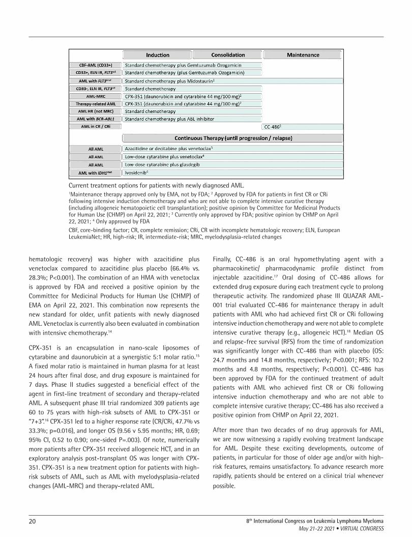
20 8th International Congress on Leukemia Lymphoma MyelomaMay 21-22 2021 • VIRTUAL CONGRESS
hematologic recovery) was higher with azacitidine plus venetoclax compared to azacitidine plus placebo (66.4% vs. 28.3%; P<0.001). The combination of an HMA with venetoclax is approved by FDA and received a positive opinion by the Committee for Medicinal Products for Human Use (CHMP) of EMA on April 22, 2021. This combination now represents the new standard for older, unfit patients with newly diagnosed AML. Venetoclax is currently also been evaluated in combination with intensive chemotherapy.14
CPX-351 is an encapsulation in nano-scale liposomes of cytarabine and daunorubicin at a synergistic 5:1 molar ratio.15 A fixed molar ratio is maintained in human plasma for at least 24 hours after final dose, and drug exposure is maintained for 7 days. Phase II studies suggested a beneficial effect of the agent in first-line treatment of secondary and therapy-related AML. A subsequent phase III trial randomized 309 patients age 60 to 75 years with high-risk subsets of AML to CPX-351 or “7+3”.16 CPX-351 led to a higher response rate (CR/CRi, 47.7% vs 33.3%; p=0.016), and longer OS (9.56 v 5.95 months; HR, 0.69; 95% CI, 0.52 to 0.90; one-sided P=.003). Of note, numerically more patients after CPX-351 received allogeneic HCT, and in an exploratory analysis post-transplant OS was longer with CPX-351. CPX-351 is a new treatment option for patients with high-risk subsets of AML, such as AML with myelodysplasia-related changes (AML-MRC) and therapy-related AML.
Finally, CC-486 is an oral hypomethylating agent with a pharmacokinetic/ pharmacodynamic profile distinct from injectable azacitidine.17 Oral dosing of CC-486 allows for extended drug exposure during each treatment cycle to prolong therapeutic activity. The randomized phase III QUAZAR AML-001 trial evaluated CC-486 for maintenance therapy in adult patients with AML who had achieved first CR or CRi following intensive induction chemotherapy and were not able to complete intensive curative therapy (e.g., allogeneic HCT).18 Median OS and relapse-free survival (RFS) from the time of randomization was significantly longer with CC-486 than with placebo (OS: 24.7 months and 14.8 months, respectively; P<0.001; RFS: 10.2 months and 4.8 months, respectively; P<0.001). CC-486 has been approved by FDA for the continued treatment of adult patients with AML who achieved first CR or CRi following intensive induction chemotherapy and who are not able to complete intensive curative therapy; CC-486 has also received a positive opinion from CHMP on April 22, 2021.
After more than two decades of no drug approvals for AML, we are now witnessing a rapidly evolving treatment landscape for AML. Despite these exciting developments, outcome of patients, in particular for those of older age and/or with high-risk features, remains unsatisfactory. To advance research more rapidly, patients should be entered on a clinical trial whenever
possible.
Current treatment options for patients with newly diagnosed AML.1Maintenance therapy approved only by EMA, not by FDA; 2 Approved by FDA for patients in first CR or CRi following intensive induction chemotherapy and who are not able to complete intensive curative therapy (including allogeneic hematopoietic cell transplantation); positive opinion by Committee for Medicinal Products for Human Use (CHMP) on April 22, 2021; 3 Currently only approved by FDA; positive opinion by CHMP on April 22, 2021; 4 Only approved by FDACBF, core-binding factor; CR, complete remission; CRi, CR with incomplete hematologic recovery; ELN, European LeukemiaNet; HR, high-risk; IR, intermediate-risk; MRC, myelodysplasia-related changes

218th International Congress on Leukemia Lymphoma MyelomaMay 21-22 2021 • VIRTUAL CONGRESS
References1. Bullinger L, Döhner K, Döhner H. Genomics of acute myeloid leukemia
diagnosis and pathways. J Clin Oncol. 2017;35(9):934-46.
2. Papaemmanuil E, Gerstung M, Bullinger L, et al. Genomic classification and prognosis in acute myeloid leukemia. N Engl J Med. 2016;374(23):2209-21.
3. Döhner H, Estey E, Grimwade D, et al. Diagnosis and management of acute myeloid leukemia in adults: 2017 ELN recommendations from an international expert panel. Blood. 2017;129(4):424-447.
4. Stone RM, Mandrekar SJ, Sanford BL, et al. Midostaurin plus chemotherapy for acute myeloid leukemia with a FLT3 mutation. N Engl J Med. 2017;377(5):454-464.
5. Perl AE, Martinelli G, Cortes JE, et al. Gilteritinib significantly prolongs overall survival in patients with FLT3-mutated (FLT3mut+) relapsed/refractory (R/R) acute myeloid leukemia (AML): Results from the Phase III ADMIRAL trial. N Eng J Med. 2019;381(18):1728-1740.
6. DiNardo CD, Stein EM, de Botton S, et al. Durable remissions with ivosidenib in IDH1-mutated relapsed or refractory AML. N Engl J Med. 2018;378:2386-2398.
7. Stein EM, DiNardo CD, Pollyea DA, et al. Enasidenib in mutant IDH2 relapsed or refractory acute myeloid leukemia. Blood. 2017;130:722-731.
8. DiNardo CD, Schuh AC, Stein EM, et al. Enasidenib plus azacitidine significantly improves complete remission and overall response compared with azacitidine alone in patients with newly diagnosed acute myeloid leukemia (AML) with isocitrate dehydrogenase 2 (IDH2) mutations: interim phase II results from an ongoing, randomized study. Blood. 2019;133(Suppl 1):643(abstr).
9. DiNardo CD, Stein AS, Stein EM, et al. Mutant IDH1 inhibitor ivosidenib in combination with azacitidine for newly diagnosed acute myeloid leukemia. J Clin Oncol. 2021;39:57-65.
10. Stein EM, DiNardo CD, Fathi AT, et al. Ivosidenib or enasidenib combined with intensive chemotherapy in patients with newly diagnosed AML: A phase 1 study. Blood. 2021;137(13):1792-1803.
11. DiNardo CD, Pratz KW, Letai A, et al. Safety and preliminary efficacy of venetoclax with decitabine or azacitidine in elderly patients with previously untreated acute myeloid leukaemia: a non-randomised, open-label, phase 1b study. Lancet Oncol. 2018;19:216-228.
12. Wei AH, Strickland SA Jr, Hou JZ, et al. Venetoclax combined with low-dose cytarabine for previously untreated patients with acute myeloid leukemia: results from a phase Ib/II study. J Clin Oncol. 2019;37:1277-1284.
13. DiNardo CD, Jonas BA, Pullarkat V, Thirman MJ, Garcia JS, Wei AH, Konopleva M, Döhner H, Letai AG, Fenaux P, Koller E, Havelange V, Leber B, Esteve J, Wang J, Pejsa V, Hajek R, Porkka K, Egyed M, Lavie D, Lemoli RM, Yamamoto K, Yoon SS, Jang JH, Yeh SP, Turgut M, Hong WJ, Zhou Y, Potluri J, Pratz KW. Venetoclax and azacitidine combination for treatment-naïve patients with AML. N Engl J Med. 2020;383(7):617-62.
14. Lachowiez C, Konopleva M, Kadia TM, et al. Interim analysis of the phase 1b/2 study of the BCL-2 inhibitor venetoclax in combination with standard intensive AML induction/consolidation therapy with FLAG-IDA in patients with newly diagnosed or relapsed/refractory AML. Presented at: American Society of Hematology (ASH) Annual Meeting; December 5-8, 2020; virtual. Abstract 3329
15. Lim WS, Tardi PG, Dos Santos N, Xie X, Fan M, Liboiron BD, Huang X, Harasym TO, Bermudes D, Mayer LD. Leukemia-selective uptake and cytotoxicity of CPX-351, a synergistic fixed-ratio cytarabine:daunorubicin formulation, in bone marrow xenografts. Leuk Res. 2010;34(9):1214-23.
16. Lancet JE, Uy GL, Cortes JE, Newell LF, Lin TL, Ritchie EK, Stuart RK, Strickland SA, Hogge D, Solomon SR, Stone RM, Bixby DL, Kolitz JE, Schiller GJ, Wieduwilt MJ, Ryan DH, Hoering A, Banerjee K, Chiarella M, Louie AC, Medeiros BC. CPX-351 (cytarabine and daunorubicin) liposome for injection versus conventional cytarabine plus daunorubicin in older patients with newly diagnosed secondary acute myeloid leukemia. J Clin Oncol. 2018;36(26):2684-92.
17. Garcia-Manero G, Gore SD, Cogle C, et al. Phase I study of oral azacitidine in myelodysplastic syndromes, chronic myelomonocytic leukemia, and acute myeloid leukemia. J Clin Oncol. 2011;29(18):2521-7.
18. Wei AH, Döhner H, Pocock C, Montesinos P, Afanasyev B, Dombret H, Ravandi F, Sayar H, Jang JH, Porkka K, Selleslag D, Sandhu I, Turgut M, Giai V, Ofran Y, Cakar MK, de Sousa AB, Rybka J, Frairia C, Borin L, Beltrami G, Cermak J, Ossenkoppele GJ, La Torre I, Skikne B, Kumar K, Dong Q, Beach CL, Roboz GJ; QUAZAR AML-001 Trial Investigators. Maintenance therapy with CC-486 for patients with acute myeloid leukemia in first remission after chemotherapy. N Engl J Med. 2020;383(26):2526-2537.
22
PROCEEDINGS
8th International Congress on Leukemia Lymphoma MyelomaMay 21-22 2021 • VIRTUAL CONGRESS
Barriers and recent progress
Historically, therapeutic progress among patients with AML older than 60 years has been limited. Recently, a number of trials have demonstrated improved outcomes for fit older patients, including CPX-351 vs 7+3 for patients with secondary and therapy related AML aged between 60 and 75 years and oral azacitidine vs placebo for patients after attaining remission after intensive chemotherapy aged ≥55 years. Among older unfit patients, improved survival has been demonstrated for glasdegib + LDAC vs LDAC and venetoclax vs placebo combined with either azacitidine or low-dose cytarabine. Of therapies investigated for older patients unfit for intensive chemotherapy, VEN+ AZA appears to be the most active option.
Lessons learned
Based on promising phase 1-2 studies which showed high rates of response with low early mortality, two randomised phase 3 studies were conducted which showed significant improvements in response and overall survival for the addition of VEN with AZA (VIALE-A) or VEN with LDAC (VIALE-C). Key lessons learned from using these regimens include the high rate of response achieved after 1–2 cycles of therapy, the low rate of tumor lysis syndrome if preventative measures were followed and the potential risks of prolonged marrow suppression if therapy was not interrupted or duration of venetoclax dosing not truncated according to protocols followed in the trials.
Among patients aged 75 years or older or with comorbidities rendering patients unfit for intensive chemotherapy, VEN plus AZA was associated with response rates of 66% and median overall survival of 14.7 months. Sub-group analyses showed that this regimen was particularly beneficial for patients with IDH mutations. On the contrary, patients with TP53 or FLT3-ITD mutations did not have a survival benefit, despite improved early
responses. This has led to new trials examining the addition of FLT3 inhibitors to VEN+AZA and alternative therapies with TP53 independent activity in an attempt to overcome this poor-risk mutation subgroup.
A common experience among physicians treating patients with VEN + AZA is the occurrence of marrow toxicity. Principles of management include:
• Consider a BMA on day 21 so that VEN can be interrupted if BM blasts are <5%
• Deferring commencement of the next cycle until robust count recovery has occurred (either CRh or CR)
• If blood count recovery is delayed > 14 days, reduce VEN duration next cycle
• If blood counts remain low and the marrow is very hypocellular, consider AZA dose reduction
• If the marrow is very hypocellular and the patient has adverse risk disease, consider a transplant strategy in remission
Future questions and challenges
Despite progress being made in the treatment of older patients with AML, many unanswered questions remain:
1. Is it appropriate to use VEN + AZA for older patients fit for intensive chemotherapy (e.g. between the ages of 60-75). Currently there is no randomised data to support this practice, therefore intensive chemotherapy should remain the standard of care unless patients are enrolled to clinical trials.
2. Should patients with a prior history of myeloproliferative disease, including MF, receive VEN+AZA? Although excluded
New Agents for the Treatment of Older Patients with AML
Andrew Wei
The Alfred Hospital, Melbourne, Australia

238th International Congress on Leukemia Lymphoma MyelomaMay 21-22 2021 • VIRTUAL CONGRESS
from phase 3 trials, single centre data suggests limited benefit in this poor risk sub-group (Masarova et al, Blood Advances 2021).
3. Should all patients unfit for intensive therapy receive VEN + AZA? Enrolment to clinical trials invariably represents a selected sub-group and it is likely that patients in the community will be frailer than those enrolled to clinical trials. It is unknown how well this regimen will be tolerated among very frail patients. If this regimen is considered for use in patients with poor performance, close inpatient monitoring and support is strongly recommended.
4. Should patients with a FLT3 mutation receive a FLT3 inhibitor in first line? Currently the role of a FLT3 inhibitor as first line therapy in older unfit patients remains uncertain. A phase 3 trial comparing gilteritinib + AZA vs AZA alone has been reported to be negative but clinical results have yet to be published. Clinical trials examining triple VEN+AZA+gilteritinib are in progress.
5. What is the role of AZA-VEN in patients with HMA failure? The VIALE-A trial excluded patients with prior HMA exposure. The VIALE-C trial with LDAC + VEN included 20% patients with prior HMA exposure. The response rate was only 25% and median survival remained < 6 months, suggesting alternative treatment options need to be explored.
6. Can venetoclax be combined with more intensive chemotherapy in older patients? A trial (CAVEAT) has examined 5+2 in combination with venetoclax and found this combination to be well tolerated and associated with a high response rate in de novo AML. Delayed blood count recovery was noted after consolidation that included ara-C and an anthracycline. The optimal consolidation approach remains to be defined. Another approach includes the addition of cladribine to venteoclax + LDAC alternating with AZA + VEN (Kadia et al, ASH 2020). Both approaches are likely best reserved for fitter older patients with AML
References1. DiNardo, C.D., Jonas, B.A., Pullarkat, V., Thirman, M.J., Garcia, J.S., Wei, A.H.,
Konopleva, M., Dohner, H., Letai, A., Fenaux, P., Koller, E., Havelange, V., Leber, B., Esteve, J., Wang, J., Pejsa, V., Hajek, R., Porkka, K., Illes, A., Lavie, D., Lemoli, R.M., Yamamoto, K., Yoon, S., Jang, J., Yeh, S., Turgut, M., Hong, W., Zhou, Y., Potluri, J., & Pratz, K.W. (2020). Azacitidine and Venetoclax in Previously Untreated Acute Myeloid Leukemia. The New England Journal of Medicine, 383(7), 617-629. doi: 10.1056/NEJMoa2012971
2. DiNardo, C.D., Tiong, I.S., Quaglieri, A., MacRaild, S., Loghavi, S., Brown, F., Thijssen, R., Pomilio, G., Ivey, A., Salmon, J., Glytsou, C., Fleming, S., Zhang, Q., Ma, H., Patel, K., Kornblau, S., Xu, Z., Chua, C., Chen, X., Blombery, P., Flensburg, C., Aifantis, I., Kantarjian, H., Huang, D., Roberts, A., Majewski, I.J., Konopleva, M., & Wei, A. (2020). Molecular patterns of response and treatment failure after frontline venetoclax combinations in older patients with AML. Blood. doi: 10.1182/blood.2019003988
3. Chua, C.C., Roberts, A.W., Reynolds, J., Fong, C.Y., Ting, S.B., Salmon, J.M., MacRaild, S., Ivey, A., Tiong, I.S., Fleming, S., Brown, F.C., Loo, S., Majewski, I.J., Bohlander, S.K., & Wei, A.H. (2020). Chemotherapy and Venetoclax in Elderly Acute Myeloid Leukemia Trial (CAVEAT): A Phase Ib Dose-Escalation Study of Venetoclax Combined With Modified Intensive Chemotherapy. Journal of Clinical Oncology, 38(30), 3506-3517. doi: 10.1200/JCO.20.00572
4. Wei, A.H., Döhner, H., Pocock, C., Montesinos, P., Afanasyev, B., Dombret, H., Ravandi, F., Sayar, H., Jang, J., Porkka, K., Selleslag, D., Sandhu, I., Turgut, M., Giai, V., Ofran, Y., Çakar, M., Botelho de Sousa, A., Rybka, J., Frairia, C., Borin, L., Beltrami, G., Čermák, J., Ossenkoppele, G., La Torre, I., Skikne, B., Kumar, K., Dong, Q., Beach, C.L., & Roboz, G.J. (2020). Oral Azacitidine Maintenance for Acute Myeloid Leukemia in First Remission. New England Journal of Medicine, 383(26), 2526-2537. doi: 10.1056/NEJMoa2004444
5. Wei, A., Montesinos, P., Ivanov, V., DiNardo, C.D., Novak, J., Laribi, K., Kim, I., Stevens, D.A., Fiedler, W., Pagoni, M., Samoilova, O., Hu, Y., Anagnostopoulos, A., Bergeron, J., Hou, J., Murthy, V., Yamauchi, T., McDonald, A., Chyla, B., Gopalakrishnan, S., Jiang, Q., Mendes, W., Hayslip, J., & Panayiotidis, P. (2020). Venetoclax plus LDAC for patients with untreated AML ineligible for intensive chemotherapy: phase 3 randomized placebo-controlled trial. Blood. doi: 10.1182/blood.2020004856
6. DiNardo, C.D., & Wei, A.H. (2019). How I treat acute myeloid leukemia in the era of new drugs. Blood, 135(2), 85-96. doi: 10.1182/blood.2019001239
7. Lancet, J. E., et al. (2018). “CPX-351 (cytarabine and daunorubicin) Liposome for Injection Versus Conventional Cytarabine Plus Daunorubicin in Older Patients With Newly Diagnosed Secondary Acute Myeloid Leukemia.” Journal of Clinical Oncology 36(26): 2684-2692.
8. Cortes, J. E., et al. (2019). “Randomized comparison of low dose cytarabine with or without glasdegib in patients with newly diagnosed acute myeloid leukemia or high-risk myelodysplastic syndrome.” Leukemia 33(2): 379.

24
PROCEEDINGS
8th International Congress on Leukemia Lymphoma MyelomaMay 21-22 2021 • VIRTUAL CONGRESS
First-line cure rates in young patients with Hodgkin Lymphoma (HL) are excellent; however still 10%-20% of patients suffer from relapsed or refractory disease. Reinduction chemotherapy followed by high-dose chemotherapy (HDCT) and autologous stem cell transplant (ASCT) is standard of care for suitable patients with relapsed or refractory HL and allows for cure in approximately 50% (Eichenauer, et al 2018, Hoppe, et al 2020). Due to the poor prognosis of high-risk patients even with HDCT and ASCT, consolidation strategies have been evaluated to improve the cure rates (Moskowitz, et al 2018). Current consolidation strategies will be discussed. For patients with recurrence after HDCT and ASCT, treatment is palliative in most cases. The anti-CD30 antibody-drug conjugate brentuximab vedotin (BV) has been shown to induce high response rates in these patients but durable responses were reported in a small percentage of patients only (Chen, et al 2016). Anti-programmed death-1 (PD1) antibodies show even more impressive results in terms of response rates and progression-free survival; however, as extended follow-up data become available, most patients seem to relapse sooner or later (Armand, et al 2018, Chen, et al 2019). More recently, brentuximab vedotin was challenged by pembrolizumab as new standard of care in patients with relapsed or refractory HL if HDCT is not an option (Kuruvilla, et al 2021). New combination studies with anti-PD1 antibodies, e.g. with chemotherapy or double checkpoint blockade, aiming at more durable responses are currently ongoing, with first highly promising results (Moskowitz, et al 2020). Additionally, clinical trials with PD1 antibodies in first relapsed HL assess the role of these new class of drugs in reinduction therapy and more recently even in the replacement of HDCT. For carefully selected patients with multiple relapses, dose-reduced allogeneic transplant (RICallo) is a potentially curative option. The role of RICallo in the era of anti-PD1 antibodies is currently being re-evaluated (Merryman, et al 2019).
References1. Armand, P., Engert, A., Younes, A., Fanale, M., Santoro, A., Zinzani, P.L.,
Timmerman, J.M., Collins, G.P., Ramchandren, R., et al. (2018) Nivolumab for Relapsed/Refractory Classic Hodgkin Lymphoma After Failure of Autologous Hematopoietic Cell Transplantation: Extended Follow-Up of the Multicohort Single-Arm Phase II CheckMate 205 Trial. J Clin Oncol, 36, 1428-1439.
2. Chen, R., Gopal, A.K., Smith, S.E., Ansell, S.M., Rosenblatt, J.D., Savage, K.J., Connors, J.M., Engert, A., Larsen, E.K., et al. (2016) Five-year survival and durability results of brentuximab vedotin in patients with relapsed or refractory Hodgkin lymphoma. Blood, 128, 1562-1566.
3. Chen, R., Zinzani, P.L., Lee, H.J., Armand, P., Johnson, N.A., Brice, P., Radford, J., Ribrag, V., Molin, D., et al. (2019) Pembrolizumab in relapsed or refractory Hodgkin lymphoma: 2-year follow-up of KEYNOTE-087. Blood, 134, 1144-1153.
4. Eichenauer, D.A., Aleman, B.M.P., Andre, M., Federico, M., Hutchings, M., Illidge, T., Engert, A., Ladetto, M. & [email protected], E.G.C.E.a. (2018) Hodgkin lymphoma: ESMO Clinical Practice Guidelines for diagnosis, treatment and follow-up. Ann Oncol, 29 Suppl 4, iv19-iv29.
5. Hoppe, R.T., Advani, R.H., Ai, W.Z., Ambinder, R.F., Armand, P., Bello, C.M., Benitez, C.M., Bierman, P.J., Boughan, K.M., et al. (2020) Hodgkin Lymphoma, Version 2.2020, NCCN Clinical Practice Guidelines in Oncology. J Natl Compr Canc Netw, 18, 755-781.
6. Kuruvilla, J., Ramchandren, R., Santoro, A., Paszkiewicz-Kozik, E., Gasiorowski, R., Johnson, N.A., Fogliatto, L.M., Goncalves, I., de Oliveira, J.S.R., et al. (2021) Pembrolizumab versus brentuximab vedotin in relapsed or refractory classical Hodgkin lymphoma (KEYNOTE-204): an interim analysis of a multicentre, randomised, open-label, phase 3 study. Lancet Oncol, 22, 512-524.
7. Merryman, R.W., Castagna, L., Corradini, P., Ho, V.T., Bond, D.A., Jaglowski, S., Spinner, M.A., Arai, S., Lowsky, R., et al. (2019) Safety and Efficacy of Allogeneic Hematopoietic Stem Cell Transplant after Programmed Cell Death 1 (PD-1) / Programmed Cell Death Ligand 1 (PD-L1) Blockade for Classical Hodgkin Lymphoma: Analysis of a Large International Cohort. Blood, 134, 775-775.
8. Moskowitz, A.J., Shah, G., Schöder, H., Ganesan, N., Hancock, H., Davey, T., Sohail, S., Santarosa, A., Pomerantz, L., et al. (2020) Phase II Study of Pembrolizumab Plus GVD As Second-Line Therapy for Relapsed or Refractory Classical Hodgkin Lymphoma. Blood, 136, 17-18.
9. Moskowitz, C.H., Walewski, J., Nademanee, A., Masszi, T., Agura, E., Holowiecki, J., Abidi, M.H., Chen, A.I., Stiff, P., et al. (2018) Five-year PFS from the AETHERA trial of brentuximab vedotin for Hodgkin lymphoma at high risk of progression or relapse. Blood, 132, 2639-2642.
Relapsed and Refractory HL: Innovative Therapies
Bastian von Tresckow
University Hospital Essen, Germany
25
PROCEEDINGS
8th International Congress on Leukemia Lymphoma MyelomaMay 21-22 2021 • VIRTUAL CONGRESS
Nodular lymphocyte-predominant Hodgkin lymphoma (NLPHL) is a rare B cell-derived malignancy accounting for roughly 5% of all Hodgkin lymphoma (HL) cases. Pathological and clinical characteristics of NLPHL differ from classical HL (cHL) [1].
The malignant cells in NLPHL are termed lymphocyte predominant (LP) cells. They stain consistently positive for CD20 but lack CD30. Six different histopathological growth patterns have been described in NLPHL. Patients with a typical histopathological growth pattern (pattern A and B) are distinguished from individuals with a variant histology (pattern C, D, E and F). The clinical course of NLPHL is usually rather indolent. However, there is a tendency towards late and multiple relapses. In addition, histological transformation into aggressive B-cell non-Hodgkin lymphoma (B-NHL) occurs in a significant minority of patients [2]there are also patients with advanced NLPHL who frequently present with spleen and liver involvement, B-symptoms and a more aggressive clinical course. Different clinical presentations correlate with distinct histopathological characteristics. NLPHL can be divided into typical and variant histopathological growth patterns. The clinical course of most patients with a typical growth pattern is indolent whereas patients with a variant histology more often present with advanced stage disease and relapse occurs more frequently and earlier. Despite these differences, the prognosis after stage-adapted treatment is favourable for both patient groups. Some cases presenting with a variant histology show a histopathological and clinical overlap with T-cell/histiocyte rich large B-cell lymphoma (THRLBCL. In comparison with cHL, NLPHL is diagnosed at a somewhat higher median age (43 years according to an analysis using the SEER database) and patients are more often male (approximately 75% of cases). At initial diagnosis, patients mostly present with early-stage disease [3].
First-line treatment of NLPHL is stage-adapted. Patients presenting with stage IA disease without risk factors are usually treated with radiotherapy (RT) alone [4]we performed an analysis using the database of the German Hodgkin Study Group. PATIENTS
AND METHODS: The long-term outcome of 256 patients with stage IA NLPHL was evaluated. Patients had received combined-modality treatment (CMT; n = 72. As compared with RT alone, single-agent rituximab is associated with an increased relapse rate [5]. Patients with early-stage NLPHL other than stage IA without risk factors are commonly treated very similarly to cHL. Thus, 2 cycles of ABVD chemotherapy followed by RT are given in most cases. Treatment of patients with intermediate-stage disease consists of 4 cycles of chemotherapy followed by RT. Although data are scarce, the addition of rituximab can be discussed in individuals with early and intermediate stages [6]. The optimal treatment for advanced NLPHL is undefined. Activity has been demonstrated for different protocols such as escalated BEACOPP, R-CHOP and BR [6–8].
At 10 years, progression-free survival for patients with newly diagnosed NLPHL is ranging between 70% (advanced stages) and 90% (stage IA without risk factors). The 10-year overall survival is in excess of 90% [4, 6]we performed an analysis using the database of the German Hodgkin Study Group. PATIENTS AND METHODS: The long-term outcome of 256 patients with stage IA NLPHL was evaluated. Patients had received combined-modality treatment (CMT; n = 72.
Unlike patients with relapsed cHL, individuals with NLPHL recurrence usually do not require aggressive salvage therapy with high-dose chemotherapy and autologous stem cell transplantation (ASCT). In the majority of cases, long-term remission is achieved with second-line treatment consisting of rituximab or conventional chemotherapy optionally followed by RT [9]we performed an analysis using the database of the German Hodgkin Study Group (GHSG.
Patients with histological transformation into aggressive B-NHL are mostly treated with R-CHOP or high-dose chemotherapy and ASCT. Treatment in the individual patient is chosen based on factors such as prior therapies and age [10, 11]most commonly diffuse large B-cell lymphoma (DLBCL.
Update on Nodular lymphocyte-Predominant Hodgkin lymphoma
Dennis Eichenauer
University Hospital of Cologne, Germany

26 8th International Congress on Leukemia Lymphoma MyelomaMay 21-22 2021 • VIRTUAL CONGRESS
References1. Eichenauer DA, Engert A (2017) Nodular lymphocyte-predominant Hodgkin
lymphoma: a unique disease deserving unique management. Hematol Am Soc Hematol Educ Progr 2017:324–328. https://doi.org/10.1182/asheducation-2017.1.324
2. Hartmann S, Eichenauer DA (2020) Nodular lymphocyte predominant Hodgkin lymphoma: pathology, clinical course and relation to T-cell/histiocyte rich large B-cell lymphoma. Pathology 52:142–153. https://doi.org/10.1016/j.pathol.2019.10.003
3. Shivarov V, Ivanova M (2018) Nodular lymphocyte predominant Hodgkin lymphoma in USA between 2000 and 2014: an updated analysis based on the SEER data. Br J Haematol 182:727–730. https://doi.org/10.1111/bjh.14861
4. Eichenauer DA, Plutschow A, Fuchs M, et al (2015) Long-Term Course of Patients With Stage IA Nodular Lymphocyte-Predominant Hodgkin Lymphoma: A Report From the German Hodgkin Study Group. J Clin Oncol 33:2857–2862. https://doi.org/10.1200/JCO.2014.60.4363
5. Eichenauer DA, Plutschow A, Fuchs M, et al (2020) Rituximab in newly diagnosed stage IA nodular lymphocyte-predominant Hodgkin lymphoma: long-term follow-up of a phase 2 study from the German Hodgkin Study Group. Leukemia 34:953–956. https://doi.org/10.1038/s41375-019-0609-3
6. Eichenauer DA, Plutschow A, Fuchs M, et al (2020) Long-Term Follow-Up of Patients With Nodular Lymphocyte-Predominant Hodgkin Lymphoma Treated in the HD7 to HD15 Trials: A Report From the German Hodgkin Study Group. J Clin Oncol 38:698–705. https://doi.org/10.1200/JCO.19.00986
7. Fanale MA, Cheah CY, Rich A, et al (2017) Encouraging activity for R-CHOP in Advanced Stage Nodular Lymphocyte Predominant Hodgkin Lymphoma. Blood. https://doi.org/10.1182/blood-2017-02-766121
8. Prusila REI, Haapasaari KM, Marin K, et al (2018) R-Bendamustine in the treatment of nodular lymphocyte-predominant Hodgkin lymphoma. Acta Oncol (Madr) 1–3. https://doi.org/10.1080/0284186X.2018.1450522
9. Eichenauer DA, Plutschow A, Schroder L, et al (2018) Relapsed and refractory nodular lymphocyte-predominant Hodgkin lymphoma: an analysis from the German Hodgkin Study Group. Blood 132:1519–1525. https://doi.org/10.1182/blood-2018-02-836437
10. Al-Mansour M, Connors JM, Gascoyne RD, et al (2010) Transformation to aggressive lymphoma in nodular lymphocyte-predominant Hodgkin’s lymphoma. J Clin Oncol 28:793–799. https://doi.org/10.1200/JCO.2009.24.9516
11. Kenderian SS, Habermann TM, Macon WR, et al (2016) Large B-cell transformation in nodular lymphocyte-predominant Hodgkin lymphoma: 40-year experience from a single institution. Blood 127:1960–1966. https://doi.org/10.1182/blood-2015-08-665505

27
PROCEEDINGS
8th International Congress on Leukemia Lymphoma MyelomaMay 21-22 2021 • VIRTUAL CONGRESS
T-cell acute lymphoblastic leukemia (T-ALL) are clonal proliferation derived from thymic T-cell progenitors. T-ALL are less than B-cell ALL and account for approximatively 15% of ALL in children and up to 25% in adults, with a relative peak of frequency reached in adolescent and young adult (AYA) populations.1 Therefore, patients with T-ALL are usually younger than patients with B-ALL in adult protocols. T-cell lymphoblastic lymphoma (T-LBL) is the lymphomatous counterpart of T-ALL, with by definition less than 20% of bone marrow infiltrating blasts. Overall, both diseases share a common biology and overall prognosis when exposed to similar therapeutic strategies.
T-ALL emerge from the disruption of normal T-cell differentiation by different genetic alterations leading to the arrest of normal differentiation, excess of proliferation and survival advantage.2 Leukemia driving events result in the aberrant expression of transcription factors that confer both a specific gene expression profile and a define stage of maturation arrest of the disease. Immature T-ALL are characterized by the overexpression of HOXA or LMO genes, early cortical T-ALL are mostly driven by the TLX1 (HOX11) and TLX3 (HOX11L2) homeobox genes, whereas late cortical are associated with TAL1 rearrangements. In contrary to B-ALL, none of these primary events has been clearly associated with a specific prognosis or broadly integrated in risk stratification strategies.
In the last two decades, a myriad of secondary events mostly gene mutations or deletions have been described, the most frequent involving CDKN2A/B cell cycle inhibitors and NOTCH1 pathway in over 70% of cases.2 These gene alterations lead to the dysregulation of different pathways including NOTCH1 (NOTCH1, FBXW7), JAK-STAT (IL7R, JAK1, JAK3, DNM2…), PI3K-AKT-mTOR (PTEN, AKT…), transcription factors (PHF6, WT1, RUNX1…), methylation and chromatin modifiers (PRC2, DNMT3A, TET2, IDH1/2…). NOTCH1 pathway alterations are mostly due to mutations involving either NOTCH1 receptor or FBXW7, which controls the degradation of intracellular NOTCH1 domain. NOTCH1 and FBXW7 mutations, which may coexist in
some cases, were associated with a favorable prognosis in both T-ALL and T-LBL.3 On contrary, PTEN or RAS mutations were suggested to be associated with a higher risk of relapse.4
Among immature T-ALL, early T-cell progenitor (ETP)-ALL is a specific subgroup of resistant ALL initially defined by a specific gene expression signature and later by a specific immunophenotype (CD1a-, CD8-, CD5-/dim, aberrant expression of myeloid or stem cell markers).5 This subgroup of T-ALL display an increased frequency of gene mutations associated with cytokine receptor and RAS signaling, transcription factors, epigenetic regulators, and a decreased frequency of NOTCH1 pathway and CDKN2A/B mutations.6 ETP-ALL are usually associated with high levels of post-induction MRD and a higher risk of relapse.5,7 However, pediatric and adult studies have suggested that this poor prognosis may be abrogated by minimal residual disease (MRD)-based risk stratification.6,8
In young adults aged up to 60 years old, the prognosis of T-ALL has clearly benefited from dose-intensive approaches inspired from pediatric protocols.9 In recently published adult or pediatric/AYA protocols, complete remission rates exceed 90% and 3/5-year survival range from 60 to 74%. This improvement is supposed to be linked to a wider use of asparaginase and methotrexate, but there is a lack of supportive data in adults. The major prognostic impact of post-induction MRD has been validated in adult and pediatric cohorts. It remains unclear whether MRD should be assessed at the same timelines than for B-ALL, a late MRD assessment being considered as more informative by some pediatric cooperative groups.10 Allogeneic stem cell transplant (allo-SCT) was historically proposed to a large proportion of patients and its benefit mostly observed in AYAs. Many groups have now incorporated poor MRD response as one of the most important predictors of allo-SCT benefit in adult ALL.11 Other factors frequently used to indicate allo-SCT are a high white blood cell count at diagnosis (> 100 G/L) or an early or mature phenotype. A benefit of allo-SCT was indeed observed in ETP-ALL, which usually display a slow response to induction therapy.6
Current Status and Future Prospects in T-ALL
Nicolas Boissel
Saint-Louis Hospital, Institut de Recherche Saint-Louis, Université de Paris, France
28 8th International Congress on Leukemia Lymphoma MyelomaMay 21-22 2021 • VIRTUAL CONGRESS
Nelarabine is a soluble prodrug of Ara-G approved in the treatment of relapsed/refractory (R/R) T-ALL in 3rd line. This approval was supported by phase 2 trials conducted in pediatric/AYA or adult patients.12–14 The expected CR/CRi rate range from 26 to 36%. Nelarabine was combined to an HyperCVAD backbone in a phase 2 trial by the MD Anderson Cancer Center with no evidence of benefit compared to an historical cohort treated with HyperCVAD alone.15 Nelarabine is associated with a risk of neurotoxicity and should not be given with intrathecal chemotherapy. Of note, an inferior rate of response was noted in R/R T-LBL compared to T-ALL in the phase 2 studies by the COG and the GMALL.13,14 In the COG phase 3 ALL0434 study, a benefit of nelarabine was demonstrated in children and AYA patients with T-ALL but not T-LBL patients.16 Interestingly, a reduction of the CNS relapse risk was observed without excess of CNS toxicity despite a wide use of CNS irradiation.
The better understanding of T-ALL biology is currently leading to the development of new drug strategies in T-ALL/LBL. The first generation of drugs targeting NOTCH1 pathway including gamma-secretase inhibitors has been confronted to narrow therapeutic indexes. New strategies are being developed. The dependency of ETP-ALL to BCL-2 and of more mature T-ALL to BCL-XL has supported the investigation of BH3 mimetics.17 As an example, an ongoing trial combining venetoclax and navitoclax yielded a 53% overall response rate in R/R T-ALL patients. Incorporation to frontline chemotherapy at different phases of the treatment is ongoing.18 In terms of immunotherapy development, T-ALL lags far behind B-ALL. However preclinical and first anecdotal cases suggest that targeting CD38, which is highly expressed in T-ALL including ETP subgroup, may be of interest to eradicate MRD in this disease. The development of CAR-T was impaired by both the risk of fratricide and of sustained T-cell aplasia in patients. Nonetheless, CAR-T programs targeting different T-cell antigens including CD5, CD7, or TCRB chain have been initiated with first promising results.
To conclude, many progresses have been accomplished during the last two decades to both understand the biology of T-ALL and improve patient outcome. T-ALL adult patients are mostly AYA who have benefited from pediatric-inspired regimen and whose outcome may be predicted by post-induction MRD assessment and baseline characteristics. Promising therapeutics include NOTCH1 pathway inhibitors, BH3 mimetics, and immunotherapies such as anti-CD38 mAb and new generation CAR T cells.
References1. Chiaretti S, Vitale A, Cazzaniga G, et al. Clinico-biological features of 5202
patients with acute lymphoblastic leukemia enrolled in the Italian AIEOP and GIMEMA protocols and stratified in age cohorts. Haematologica. 2013;98(11):1702–1710.
2. Gianni F, Belver L, Ferrando A. The Genetics and Mechanisms of T-Cell Acute Lymphoblastic Leukemia. Cold Spring Harb. Perspect. Med. 2020;10(3):a035246.
3. Asnafi V, Buzyn A, Le Noir S, et al. NOTCH1/FBXW7 mutation identifies a large subgroup with favorable outcome in adult T-cell acute lymphoblastic leukemia (T-ALL): a Group for Research on Adult Acute Lymphoblastic Leukemia (GRAALL) study. Blood. 2009;113(17):3918–3924.
4. Trinquand A, Tanguy-Schmidt A, Ben Abdelali R, et al. Toward a NOTCH1/FBXW7/RAS/PTEN-based oncogenetic risk classification of adult T-cell acute lymphoblastic leukemia: a Group for Research in Adult Acute Lymphoblastic Leukemia study. J. Clin. Oncol. Off. J. Am. Soc. Clin. Oncol. 2013;31(34):4333–4342.
5. Coustan-Smith E, Mullighan CG, Onciu M, et al. Early T-cell precursor leukaemia: a subtype of very high-risk acute lymphoblastic leukaemia. Lancet Oncol. 2009;10(2):147–156.
6. Bond J, Graux C, Lhermitte L, et al. Early response-based therapy stratification improves survival in adult ETP-ALL: a GRAALL study. J Clin Oncol. 2017;in press:
7. Gutierrez A, Dahlberg SE, Neuberg DS, et al. Absence of biallelic TCRgamma deletion predicts early treatment failure in pediatric T-cell acute lymphoblastic leukemia. J. Clin. Oncol. Off. J. Am. Soc. Clin. Oncol. 2010;28(24):3816–3823.
8. Wood BL, Winter SS, Dunsmore KP, et al. T-Lymphoblastic Leukemia (T-ALL) Shows Excellent Outcome, Lack of Significance of the Early Thymic Precursor (ETP) Immunophenotype, and Validation of the Prognostic Value of End-Induction Minimal Residual Disease (MRD) in Children’s Oncology Group (COG) Study AALL0434. Blood. 2014;124(21):1–1.
9. Patel AA, Thomas J, Rojek AE, Stock W. Biology and Treatment Paradigms in T Cell Acute Lymphoblastic Leukemia in Older Adolescents and Adults. Curr. Treat. Options Oncol. 2020;21(7):57.
10. Schrappe M, Valsecchi MG, Bartram CR, et al. Late MRD response determines relapse risk overall and in subsets of childhood T-cell ALL: results of the AIEOP-BFM-ALL 2000 study. Blood. 2011;118(8):2077–2084.
11. Giebel S, Marks DI, Boissel N, et al. Hematopoietic stem cell transplantation for adults with Philadelphia chromosome-negative acute lymphoblastic leukemia in first remission: a position statement of the European Working Group for Adult Acute Lymphoblastic Leukemia (EWALL) and the Acute Leukemia Working Party of the European Society for Blood and Marrow Transplantation (EBMT). Bone Marrow Transplant. 2019;54(6):798–809.
12. DeAngelo DJ, Yu D, Johnson JL, et al. Nelarabine induces complete remissions in adults with relapsed or refractory T-lineage acute lymphoblastic leukemia or lymphoblastic lymphoma: Cancer and Leukemia Group B study 19801. Blood. 2007;109(12):5136–5142.
13. Berg SL, Blaney SM, Devidas M, et al. Phase II study of nelarabine (compound 506U78) in children and young adults with refractory T-cell malignancies: a report from the Children’s Oncology Group. J. Clin. Oncol. Off. J. Am. Soc. Clin. Oncol. 2005;23(15):3376–3382.
14. Gokbuget N, Basara N, Baurmann H, et al. High single-drug activity of nelarabine in relapsed T-lymphoblastic leukemia/lymphoma offers curative option with subsequent stem cell transplantation. Blood. 2011;118(13):3504–3511.
15. Abaza Y, M Kantarjian H, Faderl S, et al. Hyper-CVAD plus nelarabine in newly diagnosed adult T-cell acute lymphoblastic leukemia and T-lymphoblastic lymphoma. Am. J. Hematol. 2018;93(1):91–99.
16. Dunsmore KP, Winter SS, Devidas M, et al. Children’s Oncology Group AALL0434: A Phase III Randomized Clinical Trial Testing Nelarabine in Newly Diagnosed T-Cell Acute Lymphoblastic Leukemia. J. Clin. Oncol. 2020;38(28):3282–3293.
17. Chonghaile TN, Roderick JE, Glenfield C, et al. Maturation stage of T-cell acute lymphoblastic leukemia determines BCL-2 versus BCL-XL dependence and sensitivity to ABT-199. Cancer Discov. 2014;4(9):1074–1087.
18. Lacayo NJ, Pullarkat VA, Stock W, et al. Safety and efficacy of venetoclax in combination with navitoclax in adult and pediatric relapsed/refractory acute lymphoblastic leukemia and lymphoblastic lymphoma. American Society of Hematology Washington, DC; 2019.

29
PROCEEDINGS
8th International Congress on Leukemia Lymphoma MyelomaMay 21-22 2021 • VIRTUAL CONGRESS
BCR-ABL1–positive ALL
Treatment and prognosis of patients with BCR-ABL1–positive ALL has changed dramatically during the past decade, owing primarily to addition of ABL-directed tyrosine kinase inhibitors (TKI) to first-line therapy. TKIs either with corticosteroids (CS) or in combination with chemotherapy and result in CR rates in excess of 90% irrespective of which TKI is used. single-arm studies suggest that 2nd and 3rd generation TKI may be superior to imatinib, at least in patients who do not undergo HSCT, but the optimal choice of TKI in specific contexts remains to be resolved. With imatinib-based therapy frontline therapy followed by allo-HSCT, long-term DFS rates of 60% to 75% have been reported. For imatinib there is evidence of a dose dependency during the induction period with superiority of a higher imatinib dose (800 mg/day), while second generation TKI have the theoretical advantages of greater potency and clinical activity against a broader panel of kinase domain mutations conferring resistance. To date, no prospective comparative trials to determine whether any TKI is superior have yet been performed in adults, and comparisons with historical imatinib-based studies are inconclusive, partly due to the confounding effect of HSCT. For non-transplanted adult patients, the current consensus position is that TKI should be continued indefinitely, if possible.
The third-generation TKI ponatinib has attracted particular interest because of its inhibitory activity towards the BCR-ABL kinase and the T315I TKD mutation, which confers resistance to all other clinically approved ABL-TKI. In combination with the Hyper-CVAD regimen ponatinib induced deep molecular responses in the majority of patients with newly diagnosed Ph+ ALL. Notably, it was associated with excellent outcome even in patients not undergoing allogeneic SCT in the only study published to date.(1) These data are particularly relevant for elderly patients with BCR-ABL1-positive ALL in whom allo-
HSCT may be perceived as posing too great a risk. Cardiovascular ischemic as well as embolic or thrombotic peripheral vascular events appear to be mitigated by dose reduction to 30 mg during initial therapy. Randomized comparative trials to compare regimens incorporating ponatinib or other TKIs are ongoing.
Evidence has been accumulating that in conjunction with TKI the intensity of induction chemotherapy can be reduced substantially without compromising efficacy while significantly decreasing toxicity. In fact, more intensive induction was associated with higher morbidity and mortality as demonstrated by a randomized trial of the GRAALL Study Group and had no survival benefit.(2) A low-intensity induction combining TKI with CS or CS plus vincristine has been adopted by the GIMEMA and in EWALL protocols for elderly Ph+ ALL respectively, thereby nearly abrogating induction mortality.(3, 4)
Recent innovative studies have evaluated the combination of TKI with immunotherapy as front-line therapy to further minimize the treatment toxicity while maximizing effectiveness. In what may be a practice-changing trial by the Italian GIMEMA study group an 85-day induction period with dasatinib and corticosteroids was followed by blinatumomab for up to 5 cycles, given concurrently with dasatinib.(5) Ninety-eight percent of patients achieved a CR and 60% were MRD negative after two cycles of blinatumomab. At a median follow-up of 18 months, disease-free survival was 88%. In this trial blinatumomab demonstrated clinical activity against TKD mutations including T315I. Chemotherapy-free (except for intrathecal prophylaxis) first-line therapy for Ph+ ALL, including combination of blinatumomab and TKI is also being tested in several randomized and non-randomized trials in Europe.
Maintaining remission and selecting the best post-remission therapy in patients with Ph+ ALL remains a challenge, even more so as the role of allogeneic hematopoietic stem cell
Progress in Ph+/Ph-like Acute Lymphoblastic Leukemia
Oliver Ottmann
Division of Cancer and Genetics School of Medicine, Cardiff University, Cardiff, UK

30 8th International Congress on Leukemia Lymphoma MyelomaMay 21-22 2021 • VIRTUAL CONGRESS
transplantation (HSCT) has become controversial. While allogeneic HSCT remains the gold standard against which other forms of treatment for Ph+ ALL are compared, the risk of non-relapse mortality (NRM) associated with transplant remains considerable. Thus age, comorbidities, and performance status remain critical parameters for deciding whether to proceed to HSCT.(6, 7) This issue has gained importance following realization that a subset of Ph+ ALL patients, specifically those with a very good molecular response, may remain in remission for prolonged periods even without HSCT. These results have given rise to the notion that patients with low level or negative MRD may not need to undergo HSCT to be cured.(2, 8) This is relevant particularly for patients at higher risk of TRM due to age or comorbidities in whom the superior anti-leukemic efficacy of HSCT may be outweighed by early mortality. In addition, the level of MRD at the time of HSCT has been shown to be an important predictor of outcome by some but not all studies. Methodological considerations including specific MRD thresholds and timepoints as well as clinical context are important to develop clinically applicable and validated algorithms for decisions on HSCT.(9) Donor availability is no longer a limitation given the bigger registries and feasibility of haploidentical HSCT with outcome at least comparable to conventional transplant procedures.
More recently, autologous SCT has been reconsidered as an option for a select subset of patients with Ph+ ALL who have achieved a very good molecular response to induction therapy.(10) In the prospective GRAAPH-2005 trial, survival after ASCT and alloHSCT was identical in a subset of patients with low MRD levels (BCR-ABL1/ABL1 ratio ≤0.01%).(2) A randomized comparison of these treatment modalities should be awaited before routinely adopting ASCT as therapy for Ph+ ALL.
TKIs are an integral component of therapy leading up to HSCT, and best continued indefinitely with chemotherapy-based regimens or autologous stem cell transplantation (ASCT). Its use after HSCT is supported by large retrospective analyses and several prospective trials, with similar outcome when used prophylactically or triggered by MRD.(11-13) Second generation TKI may be superior to imatinib for high-risk patients, but TKI alone have limited efficacy in patients transplanted beyond CR1. MRD should be monitored frequently, preference should be given to BM as a source of material, and close attention should be paid to the assay sensitivity. Superiority of clinical intervention based on detectable MRD rather than at morphologic relapse has been demonstrated for blinatumomab, leading to FDA and EMA approval of blinatumomab initially for MRD positive Ph-negative and more recently Ph+ ALL. The concept of clinical intervention for molecular failure or relapse in Ph+ ALL is supported by clinical trials and is applicable to not only blinatumomab.
The frequent association of clinical resistance to TKI with point mutations in the tyrosine kinase domain (KD) of BCR-ABL1 mandates rising levels of BCR-ABL1 transcripts should prompt KD mutation analysis. Clinical relapse or a significant rise in MRD should trigger testing for BCR-ABL1 KD mutations, as the results will inform subsequent therapy in relation to which TKI to switch to. NGS has become the method of choice for mutation testing, providing a sensitivity of 1-5%.
Although Ph+ ALL is considered a very high-risk subtype in adults several additional parameters are indicative of a particularly poor prognosis. As for Ph-negative ALL, age, WBC >30/nl and CNS involvement are inversely correlated with outcome, as are additional chromosomal abnormalities and supernumerary Ph chromosomes at diagnosis. More recently recurring genomic abnormalities in genes involved in B cell development, e.g. IKZF1 and CDKN2A/B deletions have been linked with less favorable outcome, including patients receiving highly effective first-line treatment with TKI plus blinatumomab or those undergoing HSCT in CR1. Current data also indicate that the number of affected genes is prognostically relevant.
Ph-like/BCR-ABL1-like ALL
A unique subgroup referred to as either Philadelphia-like or BCR-ABL1-like ALL was first identified in pediatric patients based on gene expression signatures resembling those observed in Ph+ ALL, but in the absence of the BCR-ABL1 translocation. It is found at varying frequencies in all age groups, ranging from 10-15% in children up to nearly 30% in young adults, and is considered to contribute to the inferior outcome of AYA patients compared with children.(14) Diagnosis is challenging because the group is genetically very heterogeneous, encompassing multiple rearrangements that affect more than 15 kinase or cytokine receptor genes, most fusions involving ABL-class genes (ABL1, ABL2, CSF1R, LYN, PDGFRA, PDGFRB); (ABL1, ABL2, PDGFRA, PDGFRB, CSF1R and LYN), alterations driving JAK-STAT signaling (e.g. rearrangements and mutations/ deletions of CRLF2, JAK2, EPOR, TYK2, IL7R, SH2B3, JAK1, JAK3, TYK2, IL2RB), mutations activating Ras signaling (NRAS, KRAS, PTPN11 and less frequently others (FLT3, FGFR1, NTRK3). Cytokine receptor–like factor 2 (CRLF2) rearrangements/overexpression are present in nearly half of BCR-ABL1-like ALL in AYAs and adults.(15) Unfortunately, there are no universally agreed diagnostic criteria for this subgroup, even though the provisional entity of “B-ALL with translocations involving tyrosine kinases or cytokine receptors (BCR/ABL1–like ALL)” has been added to the 2016 World Health Organization classification of myeloid neoplasms and acute leukemias. Different gene panels used for the early hierarchical clustering and prediction analysis of microarray (PAM) classifier showed little overlap and the two methods showed incomplete concordance in assigning patients to the BCR-ABL1-like subgroup. Several groups have devised
318th International Congress on Leukemia Lymphoma MyelomaMay 21-22 2021 • VIRTUAL CONGRESS
their own simplified algorithms to identify BCR-ABL1-like ALL based on 9 to 15 gene panels or by combining quantification of gene expression with other techniques including flow cytometry for CRLF2 expression, FISH analysis for JAK2 and other gene mutations and WES, WGS and RNA-seq. Availability of standardized criteria and assays are needed as the diagnosis of BCR-ABL1-like ALL has prognostic and therapeutic implications.
Patients with BCR-ABL1-like ALL experience a lower CR rate, higher MRD levels after induction, more frequent relapse and inferior survival. The Ph-like signature was associated with inferior survival rates in adolescents and young adults treated in the pediatric-inspired CALGB10403 trial. The question of whether intensive treatment is capable of ameliorating the negative impact of this subtype in adult patients was examined in the pediatric-based, MRD-driven LAL1913 GIMEMA front-line protocol. Patients with Ph-like ALL had a significantly lower CR rate, EFS and DFS, as well as a greater MRD persistence than the other Ph-negative patients, indicating the need for other alternative therapeutic interventions. Theoretically, most alterations in Ph-like ALL can be targeted by FDA-approved TKIs: JAK-STAT signaling (JAK inhibition); ABL-class fusions (ABL inhibitor); FLT3 and NTRK3 fusions (FLT3 and NTRK3 inhibitor). Some of these (ruxolitinib and ABL-directed TKI) are being tested in frontline studies. Combination of kinase inhibitors against multiple signaling pathways may provide an opportunity for tolerable, highly effective therapy given in addition to established treatment regimens. Considering the heterogeneity of lesions observed and consequently small number of patients, design of trials to demonstrate efficacy are challenging, the implications of which will be discussed.
References1. Jabbour E, Short NJ, Ravandi F, Huang X, Daver N, DiNardo CD, et al.
Combination of hyper-CVAD with ponatinib as first-line therapy for patients with Philadelphia chromosome-positive acute lymphoblastic leukaemia: long-term follow-up of a single-centre, phase 2 study. Lancet Haematol. 2018;5(12):e618-e27.
2. Chalandon Y, Thomas X, Hayette S, Cayuela JM, Abbal C, Huguet F, et al. Randomized study of reduced-intensity chemotherapy combined with imatinib in adults with Ph-positive acute lymphoblastic leukemia. Blood. 2015;125(24):3711-9.
3. Chiaretti S, Ansuinelli M, Vitale A, Elia L, Matarazzo M, Piciocchi A, et al. A multicenter total therapy strategy for de novo adult Philadelphia chromosome positive acute lymphoblastic leukemia patients. Final results of the GIMEMA LAL1509 protocol. Haematologica. 2021.
4. Rousselot P, Coude MM, Gokbuget N, Gambacorti Passerini C, Hayette S, Cayuela JM, et al. Dasatinib and low-intensity chemotherapy in elderly patients with Philadelphia chromosome-positive ALL. Blood. 2016;128(6):774-82.
5. Foa R, Vitale A, Vignetti M, Meloni G, Guarini A, De Propris MS, et al. Dasatinib as first-line treatment for adult patients with Philadelphia chromosome-positive acute lymphoblastic leukemia. Blood. 2011;118(25):6521-8.
6. Litzow MR, Fielding AK, Luger SM, Paietta E, Ofran Y, Rowe JM, et al. The evolving role of chemotherapy and hematopoietic cell transplants in Ph-positive acute lymphoblastic leukemia in adults. Bone Marrow Transplant. 2017;52(12):1592-8.
7. Ravandi F. Current management of Philadelphia chromosome positive ALL and the role of stem cell transplantation. Hematology Am Soc Hematol Educ Program. 2017;2017(1):22-7.
8. Ravandi F, Jorgensen JL, Thomas DA, O’Brien S, Garris R, Faderl S, et al. Detection of MRD may predict the outcome of patients with Philadelphia chromosome-positive ALL treated with tyrosine kinase inhibitors plus chemotherapy. Blood. 2013;122(7):1214-21.
9. Bruggemann M, Kotrova M. Minimal residual disease in adult ALL: technical aspects and implications for correct clinical interpretation. Blood Adv. 2017;1(25):2456-66.
10. Lyu M, Jiang E, He Y, Yang D, Ma Q, Pang A, et al. Comparison of autologous and allogeneic stem cell transplantation for patients with Philadelphia chromosome-positive acute lymphoblastic leukemia. Hematology. 2021;26(1):65-74.
11. Giebel S, Czyz A, Ottmann O, Baron F, Brissot E, Ciceri F, et al. Use of tyrosine kinase inhibitors to prevent relapse after allogeneic hematopoietic stem cell transplantation for patients with Philadelphia chromosome-positive acute lymphoblastic leukemia: A position statement of the Acute Leukemia Working Party of the European Society for Blood and Marrow Transplantation. Cancer. 2016;122(19):2941-51.
12. Pfeifer H, Wassmann B, Bethge W, Dengler J, Bornhauser M, Stadler M, et al. Randomized comparison of prophylactic and minimal residual disease-triggered imatinib after allogeneic stem cell transplantation for BCR-ABL1-positive acute lymphoblastic leukemia. Leukemia. 2013;27(6):1254-62.
13. Warraich Z, Tenneti P, Thai T, Hubben A, Amin H, McBride A, et al. Relapse Prevention with Tyrosine Kinase Inhibitors after Allogeneic Transplantation for Philadelphia Chromosome-Positive Acute Lymphoblast Leukemia: A Systematic Review. Biol Blood Marrow Transplant. 2020;26(3):e55-e64.
14. Chiaretti S, Messina M, Foa R. BCR/ABL1-like acute lymphoblastic leukemia: How to diagnose and treat? Cancer. 2019;125(2):194-204.
15. Inaba H, Mullighan CG. Pediatric acute lymphoblastic leukemia. Haematologica. 2020;105(11):2524-39.

32
PROCEEDINGS
8th International Congress on Leukemia Lymphoma MyelomaMay 21-22 2021 • VIRTUAL CONGRESS
Modern treatment of primary central nervous system lymphoma (PCNSL) includes induction and consolidation phases. Usually, induction consists of polychemotherapy containing high-dose methotrexate (HDMTX; ≥1 g/m2) as main drug. Different combinations have been tested in single-arm phase II studies, mostly with addition of alkylating agents and/or high-dose cytarabine (HDARAC), with or without rituximab. However, these combinations are currently used in limited geographical areas, and their routine use is not supported by a randomized trial. As exception, the IELSG20 randomized trial has demonstrated that a HDMTX-HDARAC combination is associated with significantly better outcome than HDMTX alone. The recent trial called IELSG32with a factorial double randomization comparing three different induction combinations and two major consolidation strategies demonstrated that the addition of rituximab and thiotepa to conventional HDMTX-HDARAC combination (called MATRix regimen) is associated with significantly improved outcome and acceptable toxicity. Importantly, results of the second randomization of this trial demonstrate that both whole-brain irradiation (WBRT) and autologous stem cell transplantation (ASCT) are effective consolidative options; however, patients treated with WBRT showed a higher decline in some cognitive functions. Hopefully, ASCT and other intensified options like reduced-dose WBRT and consolidative non-myeloablative chemotherapy will significantly improve survival among young and fit patients. Conversely, results remain poor in elderly patients who should be assessed in ad hoc trials. A recent randomized trial suggests that a combination of HDMTX, procarbazine and vincristine is equally active to MTX-temozolomide combination, whereas encouraging results with temozolomide maintenance were recently reported. De-escalated approaches like consolidation by non-cross resistant conventional chemotherapy or maintenance chemotherapy are being also investigated both in young and elderly patients. The most important ongoing randomized trials are aimed to establish the most effective and better tolerated consolidative
therapy after induction chemoimmunotherapy. Additionally, new molecules are being assessed in patients with relapsed/refractory PCNSL enrolled. Encouraging preliminary results with lenalidomide and ibrutinib were reported. International cooperation and multidisciplinary approach will be mandatory to achieve further progress in this field.
References 1. Ferreri AJ, Abrey LE, Blay JY, et al: Summary statement on primary central
nervous system lymphomas from the eighth international conference on malignant lymphoma, lugano, switzerland, june 12 to 15, 2002. J Clin Oncol 21:2407-2414, 2003
2. Deckert M, Engert A, Bruck W, et al: Modern concepts in the biology, diagnosis, differential diagnosis and treatment of primary central nervous system lymphoma. Leukemia 25:1797-1807, 2011
3. Kluin PM, Deckert M, Ferry JA: Primary diffuse large B-cell lymphoma of the CNS. in : WHO Classification of Tumours of Haematopoietic and Lymphoid Tissues. (ed Revised 4th edition). Lyon, IARC, 2017, pp 300-302
4. Shiels MS, Pfeiffer RM, Besson C, et al: Trends in primary central nervous system lymphoma incidence and survival in the U.S. Br J Haematol 174:417-424, 2016
5. Zeremski V, Koehler M, Fischer T, et al: Characteristics and outcome of patients with primary CNS lymphoma in a “real-life” setting compared to a clinical trial. Ann Hematol 95:793-799, 2016
6. Ferreri AJM: Therapy of primary CNS lymphoma: Role of intensity, radiation, and novel agents. Hematology Am Soc Hematol Educ Program 2017:565-577, 2017
7. Ferreri AJ, Cwynarski K, Pulczynski E, et al: Chemoimmunotherapy with methotrexate, cytarabine, thiotepa, and rituximab (MATRix regimen) in patients with primary CNS lymphoma: Results of the first randomisation of the international extranodal lymphoma study group-32 (IELSG32) phase 2 trial. Lancet Haematol 3:e217-27, 2016
8. Weller M, Martus P, Roth P, et al: Surgery for primary CNS lymphoma? challenging a paradigm. Neuro Oncol 14:1481-1484, 2012
9. Hoang-Xuan K, Bessell E, Bromberg J, et al: Diagnosis and treatment of primary CNS lymphoma in immunocompetent patients: Guidelines from the european association for neuro-oncology. Lancet Oncol 16:e322-32, 2015
10. Citterio G, Reni M, Gatta G, et al: Primary central nervous system lymphoma. Crit Rev Oncol Hematol 113:97-110, 2017
Primary CNS Lymphoma: Updates and Breaking News
Andrés J. M. Ferreri
Lymphoma Unit, Department of Onco-Hematology, IRCCS San Raffaele Scientific Institute, Milano, Italy

338th International Congress on Leukemia Lymphoma MyelomaMay 21-22 2021 • VIRTUAL CONGRESS
11. Ferreri AJ, Donadoni G, Cabras MG, et al: High doses of antimetabolites followed by high-dose sequential chemoimmunotherapy and autologous stem-cell transplantation in patients with systemic B-cell lymphoma and secondary CNS involvement: Final results of a multicenter phase II trial. J Clin Oncol 33:3903-3910, 2015
12. Ferreri AJ, Reni M, Pasini F, et al: A multicenter study of treatment of primary CNS lymphoma. Neurology 58:1513-1520, 2002
13. Hegde U, Filie A, Little RF, et al: High incidence of occult leptomeningeal disease detected by flow cytometry in newly diagnosed aggressive B-cell lymphomas at risk for central nervous system involvement: The role of flow cytometry versus cytology. Blood 105:496-502, 2005
14. Fend F, Ferreri AJ, Coupland SE: How we diagnose and treat vitreoretinal lymphoma. Br J Haematol 173:680-692, 2016
15. Ferreri AJ, Blay JY, Reni M, et al: Prognostic scoring system for primary CNS lymphomas: The international extranodal lymphoma study group experience. J Clin Oncol 21:266-272, 2003
16. Abrey LE, Ben-Porat L, Panageas KS, et al: Primary central nervous system lymphoma: The memorial sloan-kettering cancer center prognostic model. J Clin Oncol 24:5711-5715, 2006
17. Houillier C, Taillandier L, Lamy T, et al: Whole brain radiotherapy (WBRT) versus intensive chemotherapy with haematopoietic stem cell rescue (IC + HCR) for primary central nervous system lymphoma (PCNSL) in young patients: An intergroup anocef-goelams randomized phase II trial (PRECIS). Blood 128 (22):Abstract #782, 2016 (abstr)
18. Ferreri AJM, Cwynarski K, Pulczynski E, et al: Whole-brain radiotherapy or autologous stem-cell transplantation as consolidation strategies after high-dose methotrexate-based chemoimmunotherapy in patients with primary CNS lymphoma: Results of the second randomisation of the international extranodal lymphoma study group-32 phase 2 trial. Lancet Haematol 4:e510-e523, 2017
19. Schorb E, Fox CP, Fritsch K, et al: High-dose thiotepa-based chemotherapy with autologous stem cell support in elderly patients with primary central nervous system lymphoma: A european retrospective study. Bone Marrow Transplant, 2017
20. Ferreri AJ, Reni M, Foppoli M, et al: High-dose cytarabine plus high-dose methotrexate versus high-dose methotrexate alone in patients with primary CNS lymphoma: A randomised phase 2 trial. Lancet 374:1512-1520, 2009
21. Bromberg JEC, Issa S, Bakunina K, et al: Rituximab in patients with primary CNS lymphoma (HOVON 105/ALLG NHL 24): A randomised, open-label, phase 3 intergroup study. Lancet Oncol, 2019
22. Batchelor TT, Kolak G, Ciordia R, et al: High-dose methotrexate for intraocular lymphoma. Clin Cancer Res 9:711-715, 2003
23. Rubenstein JL, Fridlyand J, Abrey L, et al: Phase I study of intraventricular administration of rituximab in patients with recurrent CNS and intraocular lymphoma. J Clin Oncol 25:1350-1356, 2007
24. Villano JL, Koshy M, Shaikh H, et al: Age, gender, and racial differences in incidence and survival in primary CNS lymphoma. Br J Cancer 105:1414-1418, 2011
25. Kasenda B, Ferreri AJ, Marturano E, et al: First-line treatment and outcome of elderly patients with primary central nervous system lymphoma (PCNSL)--a systematic review and individual patient data meta-analysis. Ann Oncol 26:1305-1313, 2015
26. Batchelor T, Carson K, O’Neill A, et al: Treatment of primary CNS lymphoma with methotrexate and deferred radiotherapy: A report of NABTT 96-07. J Clin Oncol 21:1044-1049, 2003
27. Omuro A, Chinot O, Taillandier L, et al: Methotrexate and temozolomide versus methotrexate, procarbazine, vincristine, and cytarabine for primary CNS lymphoma in an elderly population: An intergroup ANOCEF-GOELAMS randomised phase 2 trial. Lancet Haematol 2:e251-9, 2015
28. Illerhaus G, Marks R, Muller F, et al: High-dose methotrexate combined with procarbazine and CCNU for primary CNS lymphoma in the elderly: Results of a prospective pilot and phase II study. Ann Oncol, 2008
29. Ramsey LB, Balis FM, O’Brien MM, et al: Consensus guideline for use of glucarpidase in patients with high-dose methotrexate induced acute kidney injury and delayed methotrexate clearance. Oncologist 23:52-61, 2018
30. Batchelor TT, Grossman SA, Mikkelsen T, et al: Rituximab monotherapy for patients with recurrent primary CNS lymphoma. Neurology 76:929-930, 2011
31. Birnbaum T, Stadler EA, von Baumgarten L, et al: Rituximab significantly improves complete response rate in patients with primary CNS lymphoma. J Neurooncol 109:285-291, 2012
32. Holdhoff M, Ambady P, Abdelaziz A, et al: High-dose methotrexate with or without rituximab in newly diagnosed primary CNS lymphoma. Neurology 83:235-239, 2014
33. Kurzwelly D, Glas M, Roth P, et al: Primary CNS lymphoma in the elderly: Temozolomide therapy and MGMT status. J Neurooncol 97:389-392, 2010
34. Nayak L, Abrey LE, Drappatz J, et al: Multicenter phase II study of rituximab and temozolomide in recurrent primary central nervous system lymphoma. Leuk Lymphoma 54:58-61, 2013
35. Grommes C, Nayak L, Tun HW, et al: Introduction of novel agents in the treatment of primary CNS lymphoma. Neuro Oncol, 2018
36. Ferreri AJ, Illerhaus G: The role of autologous stem cell transplantation in primary central nervous system lymphoma. Blood 127:1642-1649, 2016
37. Nelson DF, Martz KL, Bonner H, et al: Non-hodgkin’s lymphoma of the brain: Can high dose, large volume radiation therapy improve survival? report on a prospective trial by the radiation therapy oncology group (RTOG): RTOG 8315. Int J Radiat Oncol Biol Phys 23:9-17, 1992
38. DeAngelis LM, Yahalom J, Thaler HT, et al: Combined modality therapy for primary CNS lymphoma. J Clin Oncol 10:635-643, 1992
39. Thiel E, Korfel A, Martus P, et al: High-dose methotrexate with or without whole brain radiotherapy for primary CNS lymphoma (G-PCNSL-SG-1): A phase 3, randomised, non-inferiority trial. Lancet Oncol 11:1036-1047, 2010
40. Rubenstein JL, Gupta NK, Mannis GN, et al: How I treat CNS lymphomas. Blood 122:2318-2330, 2013
41. Correa DD, Rocco-Donovan M, DeAngelis LM, et al: Prospective cognitive follow-up in primary CNS lymphoma patients treated with chemotherapy and reduced-dose radiotherapy. J Neurooncol 91:315-321, 2009
42. Sun A, Bae K, Gore EM, et al: Phase III trial of prophylactic cranial irradiation compared with observation in patients with locally advanced non-small-cell lung cancer: Neurocognitive and quality-of-life analysis. J Clin Oncol 29:279-286, 2011
43. Abrey LE, Moskowitz CH, Mason WP, et al: Intensive methotrexate and cytarabine followed by high-dose chemotherapy with autologous stem-cell rescue in patients with newly diagnosed primary CNS lymphoma: An intent-to-treat analysis. J Clin Oncol 21:4151-4156, 2003
44. Colombat P, Lemevel A, Bertrand P, et al: High-dose chemotherapy with autologous stem cell transplantation as first-line therapy for primary CNS lymphoma in patients younger than 60 years: A multicenter phase II study of the GOELAMS group. Bone Marrow Transplant 38:417-420, 2006
45. Omuro A, Correa DD, DeAngelis LM, et al: R-MPV followed by high-dose chemotherapy with TBC and autologous stem-cell transplant for newly diagnosed primary CNS lymphoma. Blood 125:1403-1410, 2015
46. Illerhaus G, Kasenda B, Ihorst G, et al: High-dose chemotherapy with autologous haemopoietic stem cell transplantation for newly diagnosed primary CNS lymphoma: A prospective, single-arm, phase 2 trial. Lancet Haematol 3:e388-97, 2016
47. Soussain C, Suzan F, Hoang-Xuan K, et al: Results of intensive chemotherapy followed by hematopoietic stem-cell rescue in 22 patients with refractory or recurrent primary CNS lymphoma or intraocular lymphoma. J Clin Oncol 19:742-749, 2001

34 8th International Congress on Leukemia Lymphoma MyelomaMay 21-22 2021 • VIRTUAL CONGRESS
48. Boehme V, Schmitz N, Zeynalova S, et al: CNS events in elderly patients with aggressive lymphoma treated with modern chemotherapy (CHOP-14) with or without rituximab: An analysis of patients treated in the RICOVER-60 trial of the german high-grade non-hodgkin lymphoma study group (DSHNHL). Blood 113:3896-3902, 2009
49. Wieduwilt MJ, Valles F, Issa S, et al: Immunochemotherapy with intensive consolidation for primary CNS lymphoma: A pilot study and prognostic assessment by diffusion-weighted MRI. Clin Cancer Res 18:1146-1155, 2012
50. Rubenstein JL, Hsi ED, Johnson JL, et al: Intensive chemotherapy and immunotherapy in patients with newly diagnosed primary CNS lymphoma: CALGB 50202 (alliance 50202). J Clin Oncol 31:3061-3068, 2013
51. Birsen R, Willems L, Pallud J, et al: Efficacy and safety of high-dose etoposide cytarabine as consolidation following rituximab methotrexate temozolomide induction in newly diagnosed primary central nervous system lymphoma in immunocompetent patients. Haematologica 103:e296-e299, 2018
52. Langner-Lemercier S, Houillier C, Soussain C, et al: Primary CNS lymphoma at first relapse/progression: Characteristics, management, and outcome of 256 patients from the french LOC network. Neuro Oncol 18:1297-1303, 2016
53. Grommes C, DeAngelis LM: Primary CNS lymphoma. J Clin Oncol 35:2410-2418, 2017
54. Hans CP, Weisenburger DD, Greiner TC, et al: Confirmation of the molecular classification of diffuse large B-cell lymphoma by immunohistochemistry using a tissue microarray. Blood 103:275-282, 2004
55. Fukumura K, Kawazu M, Kojima S, et al: Genomic characterization of primary central nervous system lymphoma. Acta Neuropathol 131:865-875, 2016
56. Braggio E, Van Wier S, Ojha J, et al: Genome-wide analysis uncovers novel recurrent alterations in primary central nervous system lymphomas. Clin Cancer Res 21:3986-3994, 2015
57. Vater I, Montesinos-Rongen M, Schlesner M, et al: The mutational pattern of primary lymphoma of the central nervous system determined by whole-exome sequencing. Leukemia 29:677-685, 2015
58. Grommes G, Gavrilovic IT, Kaley TJ, et al: Updated results of single-agent ibrutinib in recurrent/refractory primary (PCNSL) and secondary CNS lymphoma (SCNSL). J Clin Oncol 35 (suppl):abstr 7515, 2017 (abstr)
59. Chapuy B, Roemer MG, Stewart C, et al: Targetable genetic features of primary testicular and primary central nervous system lymphomas. Blood 127:869-881, 2016
60. Kraan W, Horlings HM, van Keimpema M, et al: High prevalence of oncogenic MYD88 and CD79B mutations in diffuse large B-cell lymphomas presenting at immune-privileged sites. Blood Cancer J 3:e139, 2013
61. Cobbers JM, Wolter M, Reifenberger J, et al: Frequent inactivation of CDKN2A and rare mutation of TP53 in PCNSL. Brain Pathol 8:263-276, 1998
62. Korfel A, Schlegel U, Herrlinger U, et al: Phase II trial of temsirolimus for Relapsed/Refractory primary CNS lymphoma. J Clin Oncol 34:1757-1763, 2016
63. Kuhn JG, Chang SM, Wen PY, et al: Pharmacokinetic and tumor distribution characteristics of temsirolimus in patients with recurrent malignant glioma. Clin Cancer Res 13:7401-7406, 2007
64. Wen PY, Touat M, Alexander BM, et al: Buparlisib in patients with recurrent glioblastoma harboring phosphatidylinositol 3-kinase pathway activation: An open-label, multicenter, multi-arm, phase II trial. J Clin Oncol:JCO1801207, 2019
65. Choquet S, Houillier C, Bijou F, et al: Ibrutinib monotherapy in relapsed or refractory poimary CNS lymphoma (PCNSL) and primary vitero-retinal lymphoma (PVRL). results of the interim analysis of the iLOC phase II study from the LYSA and the french LOC network. Blood 128 (22):abst 614, 2016 (abstr)
66. Lionakis MS, Dunleavy K, Roschewski M, et al: Inhibition of B cell receptor signaling by ibrutinib in primary CNS lymphoma. Cancer Cell 31:833-843.e5, 2017
67. Grommes C, Pastore A, Palaskas N, et al: Ibrutinib unmasks critical role of bruton tyrosine kinase in primary CNS lymphoma. Cancer Discov 7:1018-1029, 2017
68. Grommes C, Tang SS, Wolfe J, et al: Phase 1b trial of an ibrutinib-based combination therapy in recurrent/refractory CNS lymphoma. Blood 133:436-445, 2019
69. Rubenstein JL, Geng H, Fraser EJ, et al: Phase 1 investigation of lenalidomide/rituximab plus outcomes of lenalidomide maintenance in relapsed CNS lymphoma. Blood Adv 2:1595-1607, 2018
70. Ghesquieres H, Houillier C, Chinot O, et al: Rituximab-lenalidomide (REVRI) in relapsed or refractory primary CNS lymphoma (PCNSL) or primary vitreo-retinal lymphoma (PVRL). results of a “proof of concept” phase II study of the french LOC network. Blood 128 (22):abst 6695, 2016 (abstr)
71. Tun HW, Johnston PB, Grommes C, et al: Phase I clinical trial on pomalidomide and dexamethasone in treating patients with relapsed/refractory primary central nervous system lymphoma (PCNSL) or primary vitreoretinal lymphoma (PVRL). J Clin Oncol 35 (suppl):abstr 7516, 2017
72. Nayak L, Iwamoto FM, LaCasce A, et al: PD-1 blockade with nivolumab in relapsed/refractory primary central nervous system and testicular lymphoma. Blood, 2017
73. Ambady P, Szidonya L, Firkins J, et al: Combination immunotherapy as a non-chemotherapy alternative for refractory or recurrent CNS lymphoma. Leuk Lymphoma:1-4, 2018
74. Abramson JS, McGree B, Noyes S, et al: Anti-CD19 CAR T cells in CNS diffuse large-B-cell lymphoma. N Engl J Med 377:783-784, 2017
75. Morris PG, Correa DD, Yahalom J, et al: Rituximab, methotrexate, procarbazine, and vincristine followed by consolidation reduced-dose whole-brain radiotherapy and cytarabine in newly diagnosed primary CNS lymphoma: Final results and long-term outcome. J Clin Oncol 31:3971-3979, 2013
76. Glass J, Won M, Schultz CJ, et al: Phase I and II study of induction chemotherapy with methotrexate, rituximab, and temozolomide, followed by whole-brain radiotherapy and postirradiation temozolomide for primary CNS lymphoma: NRG oncology RTOG 0227. J Clin Oncol 34:1620-1625, 2016
77. Pulczynski EJ, Kuittinen O, Erlanson M, et al. Successful change of treatment strategy in elderly patients with primary central nervous system lymphoma by de-escalating induction and introducing temozolomide maintenance: results from a phase II study by the Nordic Lymphoma Group. Haematologica 100(4):534-540, 2014
78. Fritsch K, Kasenda B, Schorb E, et al. High-dose methotrexate-based immuno-chemotherapy for elderly primary CNS lymphoma patients (PRIMAIN study). Leukemia 31(4):846-852, 2016
79. Vu K, Mannis G, Hwang J et al. Low-dose lenalidomide maintenance after induction therapy in older patients with primary central nervous system lymphoma. Br J Haematol 2019, in press
80. Olivier G, Clavert A, Lacotte-Thierry L et al. A phase 1 dose escalation study of idarubicin combined with methotrexate, vindesine, and prednisolone for untreated elderly patients with primary central nervous system lymphoma. The GOELAMS LCP 99 trial. Am J Hematol 89:1024–1029, 2014
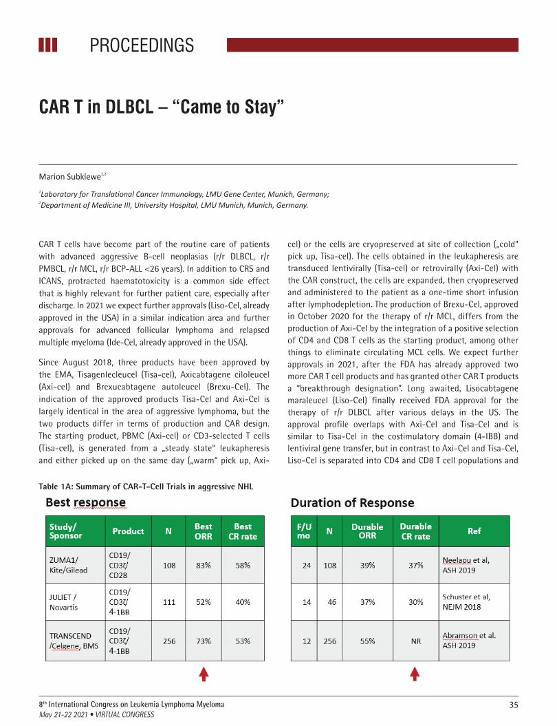
35
PROCEEDINGS
8th International Congress on Leukemia Lymphoma MyelomaMay 21-22 2021 • VIRTUAL CONGRESS
CAR T cells have become part of the routine care of patients with advanced aggressive B-cell neoplasias (r/r DLBCL, r/r PMBCL, r/r MCL, r/r BCP-ALL <26 years). In addition to CRS and ICANS, protracted haematotoxicity is a common side effect that is highly relevant for further patient care, especially after discharge. In 2021 we expect further approvals (Liso-Cel, already approved in the USA) in a similar indication area and further approvals for advanced follicular lymphoma and relapsed multiple myeloma (Ide-Cel, already approved in the USA).
Since August 2018, three products have been approved by the EMA, Tisagenlecleucel (Tisa-cel), Axicabtagene ciloleucel (Axi-cel) and Brexucabtagene autoleucel (Brexu-Cel). The indication of the approved products Tisa-Cel and Axi-Cel is largely identical in the area of aggressive lymphoma, but the two products differ in terms of production and CAR design. The starting product, PBMC (Axi-cel) or CD3-selected T cells (Tisa-cel), is generated from a „steady state“ leukapheresis and either picked up on the same day („warm“ pick up, Axi-
cel) or the cells are cryopreserved at site of collection („cold“ pick up, Tisa-cel). The cells obtained in the leukapheresis are transduced lentivirally (Tisa-cel) or retrovirally (Axi-Cel) with the CAR construct, the cells are expanded, then cryopreserved and administered to the patient as a one-time short infusion after lymphodepletion. The production of Brexu-Cel, approved in October 2020 for the therapy of r/r MCL, differs from the production of Axi-Cel by the integration of a positive selection of CD4 and CD8 T cells as the starting product, among other things to eliminate circulating MCL cells. We expect further approvals in 2021, after the FDA has already approved two more CAR T cell products and has granted other CAR T products a “breakthrough designation”. Long awaited, Lisocabtagene maraleucel (Liso-Cel) finally received FDA approval for the therapy of r/r DLBCL after various delays in the US. The approval profile overlaps with Axi-Cel and Tisa-Cel and is similar to Tisa-Cel in the costimulatory domain (4-IBB) and lentiviral gene transfer, but in contrast to Axi-Cel and Tisa-Cel, Liso-Cel is separated into CD4 and CD8 T cell populations and
CAR T in DLBCL – “Came to Stay”
Marion Subklewe1,2
1Laboratory for Translational Cancer Immunology, LMU Gene Center, Munich, Germany; 2Department of Medicine III, University Hospital, LMU Munich, Munich, Germany.
Table 1A: Summary of CAR-T-Cell Trials in aggressive NHL
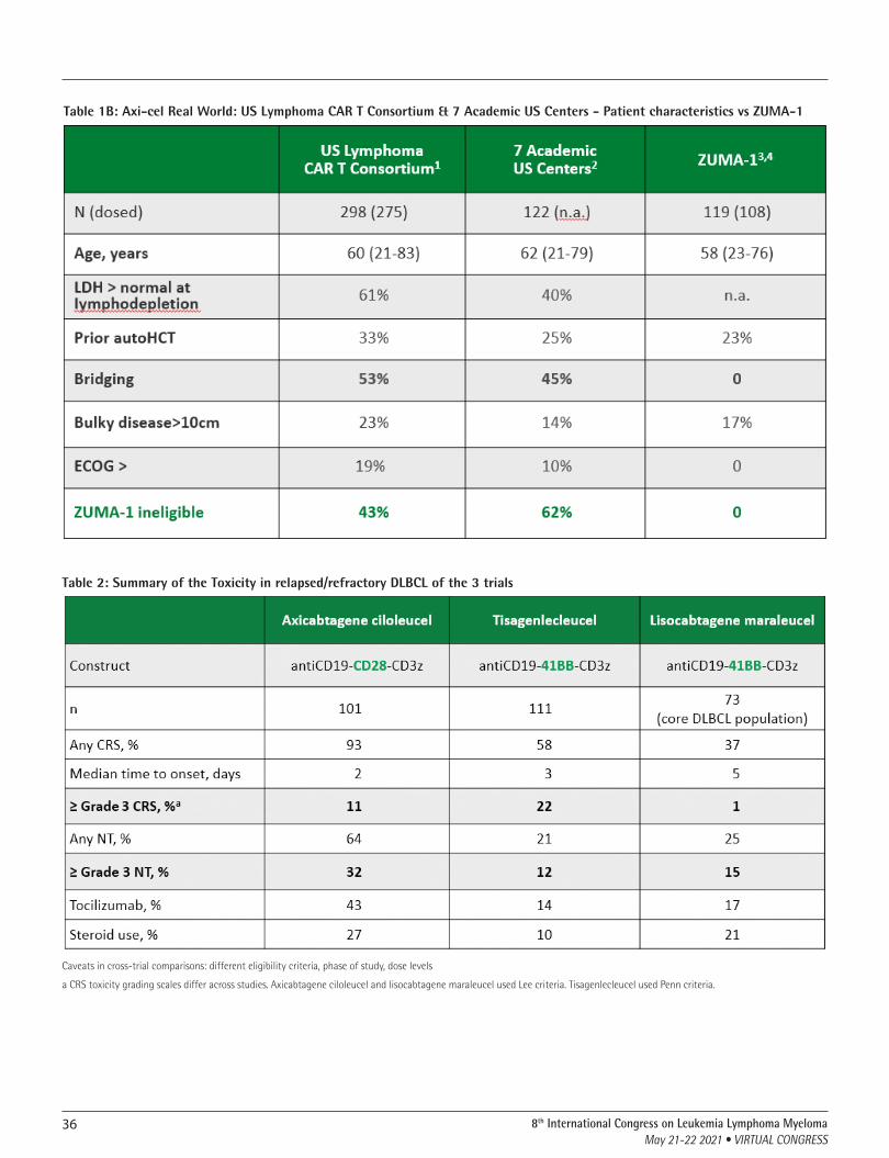
36 8th International Congress on Leukemia Lymphoma MyelomaMay 21-22 2021 • VIRTUAL CONGRESS
Table 1B: Axi-cel Real World: US Lymphoma CAR T Consortium & 7 Academic US Centers - Patient characteristics vs ZUMA-1
Table 2: Summary of the Toxicity in relapsed/refractory DLBCL of the 3 trials
Caveats in cross-trial comparisons: different eligibility criteria, phase of study, dose levels
a CRS toxicity grading scales differ across studies. Axicabtagene ciloleucel and lisocabtagene maraleucel used Lee criteria. Tisagenlecleucel used Penn criteria.

378th International Congress on Leukemia Lymphoma MyelomaMay 21-22 2021 • VIRTUAL CONGRESS
applied sequentially in a CD4: CD8 ratio of 1: 1. In relation to toxicity and outcome there are differences between the 3 products (Axi-Cel, Tisa-Cel and Liso-Cel). The results of toxicity and outcome are depicted in table 1 and table 2. Importantly, the comparison between trials is compromised by i) different patient population included within the clinical trials with differences in inclusion and exclusion criterae, ii) differences in trial design, iii) different grading of toxicity and different guidelines of intervention, iv) differences in outcome reports. So far, no biomarkers have been identified to advice which CAR T cell product is most suitable for an individual patient. However, long term follow up data confirmed a low relapse rate in patients that have achieved a metabolic remission at 3 months after CAR T cell transfusion.
While CRS and ICANS have a high priority as classic immune-mediated side effects in acute care after CAR T-cell transfusion, hematotoxicity in the “real-world” setting represents one of the most common acute and long-term side effects associated with CAR T-cells and is for the non -relapse mortality mainly responsible. It predisposes to sometimes severe infectious complications and is an important part of the multifactorial immunosuppression after CAR T-cell therapy. The CAR-T-cell registration studies had restrictive hematological inclusion criteria. Patients with an ANC <1000 G / l, severe thrombocytopenia (ZUMA-1: <75,000 / µl, JULIET: <50,000 / µl) or anemia (JULIET: Hb <8 g / dl) were excluded. In this respect, the data on haematological reconstitution after therapy reported in these studies may not be sufficiently representative for a significant proportion of the patients treated. However, several studies underline the high incidence of cytopenias after
CAR T cell therapy. A special characteristic of neutropenia is the often bimodal course with transient regeneration of leukocytes in the peripheral blood after G-CSF stimulation. Nevertheless, leukopenia and neutropenia, some of them pronounced, often recur as a result, so that close monitoring of the patient is necessary in the first few weeks after CAR-T-cell therapy. Thrombocytopenia, on the other hand, is often delayed and often only reaches its nadir in the 2nd month after therapy.
In summary, it should be noted that CAR T cells have become part of standard care in the US and in Europe for treatment of r/r DLBCL and we expect an increase in approved CAR T products, indications and, as a result, the number of CAR T cell treated patients. The complexity will increase in perspective due to the different CAR T cell products with different side effect profiles for different tumor entities.
References1. Locke FL, Ghobadi A, Jacobson CA, et al. Long-term safety and activity of
axicabtagene ciloleucel in refractory large B-cell lymphoma (ZUMA-1): a single-arm, multicentre, phase 1–2 trial. Lancet Oncol 2018. doi:10.1016/s1470-2045(18)30864-7
2. Schuster, S.J., et al., Tisagenlecleucel in Adult Relapsed or Refractory Diffuse Large B-Cell Lymphoma. N Engl J Med, 2019. 380(1): p. 45-56.
3. Abramson JS, Palomba ML, Gordon LI, et al. Lisocabtagene maraleucel for patients with relapsed or refractory large B-cell lymphomas (TRANSCEND NHL 001): a multicentre seamless design study. Lancet 2020; 396: 839–852. doi:10.1016/s0140-6736(20)31366-0
4. Logue, J.M., et al., Immune reconstitution and associated infections following axicabtagene ciloleucel in relapsed or refractory large B-cell lymphoma. Haematologica, 2020.
5. Fried, S., et al., Early and late hematologic toxicity following CD19 CAR-T cells. Bone Marrow Transplant,2019. 54(10): p. 1643-1650.

ABSTRACTS


ORAL PRESENTATIONS
40 8th International Congress on Leukemia Lymphoma MyelomaMay 21-22 2021 • VIRTUAL CONGRESS
ORAL PRESENTATIONS
� Stem Cell Transplantation
OP-01 Abstract Referance: 20
BRENTUXIMAB VEDOTIN CONSOLIDATION AFTER AUTO-SCT IN HIGH-RISK HODGKIN’S LYMPHOMA: MULTI-CENTER RETROSPECTIVE STUDYOlga Meltem Akay1, Murat Ozbalak2, Mustafa Pehlivan3, Birol Yıldız4, Ant Uzay5, Tuğçe Nur Yiğenoğlu6, Tuğrul Elverdi7, Leylagül Kaynar8, Orhan Ayyıldız9, İpek Yonal Hindilerden2, Hasan Sami Göksoy10, Şebnem İzmir Güner11, Ahmet Kürşad Güneş12, Mehmet Sönmez13, Meltem Kurt Yüksel14, Sinem Civriz Bozdağ14, Zübeyde Nur Özkurt15, Tayfur Toptaş16, Mehmet Hilmi Doğu17, Ozan Salim18, Güray Saydam19, İrfan Yavaşoğlu20, Meltem Aylı4, Gülsüm Özet21, Murat Albayrak22, Elif Birtaş Ateşoğlu23, Selami K. Toprak14, Rahşan Yıldırım24, Özgür Mehtap25, Sevgi Kalayoğlu Beşışık2, Meliha Nalçacı2, Fevzi Altuntaş6, Burhan Ferhanoğlu1--26
1Koç University Medical Faculty, Department of Internal Medicine, Division of Hematology, Istanbul, Turkey 2Istanbul University, Istanbul Medical Faculty, Department of Internal Medicine, Division of Hematology, Istanbul, Turkey 3Gaziantep University Medical Faculty, Department of Internal Medicine, Division of Hematology, Gaziantep, Turkey 4Gulhane Research and Training Hospital, Department of Internal Medicine, Division of Medical Oncology, Ankara, Turkey 5Acıbadem University Medical Faculty, Depatment of Hematology, Istanbul, Turkey 6Dr Abdurrahman Yurtaslan Ankara Oncology Research and Training Hospital, Division of Hematology, Ankara, Turkey 7Istanbul University Cerrahpaşa, Medical Faculty, Department of Internal Medicine, Division of Hematology, Istanbul, Turkey 8Erciyes University Medical Faculty, Department of Internal Medicine, Division of Hematology, Kayseri, Turkey 9Dicle University Medical Faculty, Department of Internal Medicine, Division of Hematology, Diyarbakır, Turkey 10Yeniyuzyıl University Gaziosmanpaşa Hospital, Depatment of Hematology, Istanbul, Turkey 11Memorial Şişli Hospital, Depatment of Hematology, Istanbul, Turkey 12Mehmet Akif İnan Hospital, Depatment of Hematology, Şanlıurfa, Turkey 13Karadeniz Technical University, Department of Internal Medicine, Division of Hematology, Trabzon, Turkey 14Ankara University Medical Faculty, Department of Internal Medicine, Division of Hematology, Ankara, Turkey 15Gazi University Medical Faculty, Department of Internal Medicine, Division of Hematology, Ankara, Turkey 16Marmara University Medical Faculty, Department of Internal Medicine, Division of Hematology, Istanbul, Turkey 17Istanbul Research and Training Hospital, Division of Hematology, Istanbul, Turkey 18Akdeniz University Medical Faculty, Department of Internal Medicine, Division of Hematology, Antalya, Turkey 19Ege University Medical Faculty, Department of Internal Medicine, Division of Hematology, Izmir, Turkey 20Adnan Menderes University Medical Faculty, Department of Internal Medicine, Division of Hematology, Aydın, Turkey 21Ankara City Hospital, Division of Hematology, Ankara, Turkey 22Dışkapı Research and Training Hospital, Division of Hematology, Ankara, Turkey 23Anadolu Medical Center, Division of Hematology, Izmıt, Turkey 24Ataturk University Medical Faculty, Department of Internal Medicine, Division of Hematology, Erzurum, Turkey 25Kocaeli University Medical Faculty, Department of Internal Medicine, Division of Hematology, İzmit, Turkey 26v.k.v. American Hospital Division of Hematology, İstanbul, T
Background: The standard of care for patients with relapsed/refractory (R/R) Hodgkin Lymphoma (HL) is high dose chemotherapy with autologous stem cell support (ASCT). The AETHERA trial reported an increased progres-sion-free survival (PFS) when Brentuximab Vedotin (BV) was used as mainte-nance therapy in high-risk HL after ASCT.
Aim: We aimed to determine the impact and safety of BV as maintenance after ASCT in real-world patients.
Patients and Methods: Patients with relapsed/refractory HL started on BV consolidation therapy after ASCT due to high risk of relapse, between January 2016 and July 2019, from 25 institutions, were retrospectively
analyzed. Eligible patients had at least one of the following risk factors for progression: 1) primary refractory HL (defined as progression during or failure to achieve a complete remission after frontline therapy) or relapse < 12 months, 2) extranodal disease at relapse, 3) B symptoms at relapse, 4) ≥ two prior salvage therapies, 5) partial response (PR) or stable disease (SD) to most recent salvage therapy. There was no restriction for BV-use in the pre-ASCT setting.
The median follow-up time was 26 months. The most common high-risk features were primary refractory or relapsed disease < 12 months (n = 61), lack of CR to the last salvage regimen (n = 51), and having had at least two salvage regimens (n = 29).
BV consolidation was initiated within 6 months of ASCT and administered at a dose of 1.8 mg/kg intravenous infusion over 30 min every 3 weeks for up to 16 cycles in an outpatient setting.
The primary endpoint of the study was PFS; secondary endpoints were safe-ty and OS. The study was approved by the local ethical committee. All data analyses were performed using Stata 11.1 SE software.
Results: Seventy-five patients were included in the final analysis. Patients’ baseline characteristics, initial treatment strategies, and treatment respons-es are summarized in Table 1. A high proportion of our cohort had relapsed disease within 12 months from the completion of frontline therapy (46.7%) or had primary refractory HL (34.6%).
At the time of analysis, 42 patients completed consolidation courses, and BV was discontinued in 33 patients. The reason for discontinuation of BV were PD (n = 15), adverse events (n = 12), patient’s (n = 3) and physician’s (n = 2) decision, and death due to sepsis (n = 1).
Fifty patients had an ongoing response (CR in 41, PR in six, and SD in three patients), 25 had progressed. Ten died in the follow-up, eight with progres-sive disease and two due to infection while in CR. The 2-years PFS and OS rates were 67.75% (95% CI:0.55–0.77) and 87.61% (95% CI:0.76–0.94), respec-tively (Figure 1). Patients with 1–3 risk factors had similar PFS and OS com-pared to patients with 4–5 risk factors. Seventeen patients (23%) received BV in the pre-ASCT treatment lines, and there was no survival difference between the BV naïve and BV exposed groups.
The most common adverse events were neutropenia (27%) and peripheral neuropathy (21%). Sixteen patients (21.3%) experienced grade 3 or 4 toxicity (Table 2). BV was discontinued due to AE in 12 (16%) patients [neuropathy (n = 9; grade 2 in one, grade 3 in seven, and grade 4 in one case), pulmonary toxicity (n = 1), neutropenia and infection (n = 2)].
In our cohort, 42 patients completed BV consolidation therapy. BV was discontinued in 17 patients due to adverse events (n = 12) and patients’ (n = 3) and physician’s (n = 2) decision. The 2-year PFS rates were 87.62% (95% CI: 0.69–0.95) and 81.45% (95% CI 0.52–0.93) in the completed and discontinued groups, respectively (p = 0.74). The OS rates for the completed and discontinued groups were 100% and 94.12% (95% CI: 0.65–0.99), respec-tively (p = 0.46).
Conclusions: BV consolidation after ASCT in patients with high-risk HL in a real-life setting seems to be a reasonable therapeutic option, especially given the increased possibility of cure and decreased subsequent applica-tion of highly toxic approaches.Keywords: Hodgkin Lymphoma; Brentuximab Vedotin; Autologous Stem Cell Transplantation
References
1. Moskowitz CH, Nademanee A, Masszi T, et al. Brentuximab vedotin as consolidation therapy after autologous stem-cell transplantation in patients with Hodgkin’s lym-phoma at risk of relapse or progression (AETHERA): a randomised, double-blind, placebo-controlled, phase 3 trial. Lancet. 2015;385(9980):1853-1862.
2. Moskowitz CH, Walewski J, Nademanee A, et al. Five-year PFS from the AETHERA trial of brentuximab vedotin for Hodgkin lymphoma at high risk of progression or relapse. Blood. 2018;132(25):2639-2642.
ORAL PRESENTATIONS
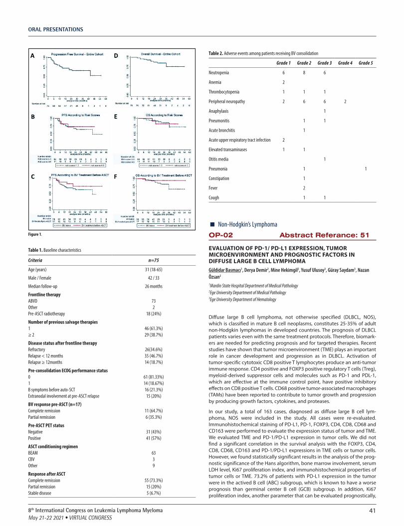
ORAL PRESENTATIONS
418th International Congress on Leukemia Lymphoma MyelomaMay 21-22 2021 • VIRTUAL CONGRESS
Figure 1.
Table 1. Baseline characteristics
Criteria n=75
Age (years) 31 (18-65)
Male / Female 42 / 33
Median follow-up 26 months
Frontline therapyABVDOtherPre-ASCT radiotherapy
732
18 (24%)
Number of previous salvage therapies1≥ 2
46 (61.3%)29 (38.7%)
Disease status after frontline therapyRefractoryRelapse < 12 monthsRelapse ≥ 12months
26(34.6%)35 (46.7%)14 (18.7%)
Pre-consolidation ECOG performance status01B symptoms before auto-SCTExtranodal involvement at pre-ASCT relapse
61 (81.33%)14 (18.67%)16 (21.3%)15 (20%)
BV response pre-ASCT (n=17)Complete remissionPartial remission
11 (64.7%)6 (35.3%)
Pre-ASCT PET statusNegativePositive
31 (43%)41 (57%)
ASCT conditioning regimenBEAMCBVOther
6339
Response after ASCTComplete remissionPartial remissionStable disease
55 (73.3%)15 (20%)5 (6.7%)
Table 2. Adverse events among patients receiving BV consolidation
Grade 1 Grade 2 Grade 3 Grade 4 Grade 5
Neutropenia 6 8 6
Anemia 2
Thrombocytopenia 1 1 1
Peripheral neuropathy 2 6 6 2
Anaphylaxis 1
Pneumonitis 1 1
Acute bronchitis 1
Acute upper respiratory tract infection 2
Elevated transaminases 1 1
Otitis media 1
Pneumonia 1 1
Constipation 1
Fever 2
Cough 1 1
� Non-Hodgkin’s LymphomaOP-02 Abstract Referance: 51
EVALUATION OF PD-1/ PD-L1 EXPRESSION, TUMOR MICROENVIRONMENT AND PROGNOSTIC FACTORS IN DIFFUSE LARGE B CELL LYMPHOMAGüldidar Basmacı1, Derya Demir2, Mine Hekimgil2, Yusuf Ulusoy3, Güray Saydam3, Nazan Özsan2
1Mardin State Hospital Department of Medical Pathology 2Ege University Department of Medical Pathology 3Ege University Department of Hematology
Diffuse large B cell lymphoma, not otherwise specified (DLBCL, NOS), which is classified in mature B cell neoplasms, constitutes 25-35% of adult non-Hodgkin lymphomas in developed countries. The prognosis of DLBCL patients varies even with the same treatment protocols. Therefore, biomark-ers are needed for predicting prognosis and for targeted therapies. Recent studies have shown that tumor microenvironment (TME) plays an important role in cancer development and progression as in DLBCL. Activation of tumor-specific cytotoxic CD8 positive T lymphocytes produce an anti-tumor immune response. CD4 positive and FOXP3 positive regulatory T cells (Treg), myeloid-derived suppressor cells and molecules such as PD-1 and PDL-1, which are effective at the immune control point, have positive inhibitory effects on CD8 positive T cells. CD68 positive tumor-associated macrophages (TAMs) have been reported to contribute to tumor growth and progression by producing growth factors, cytokines, and proteases.
In our study, a total of 163 cases, diagnosed as diffuse large B cell lym-phoma, NOS were included in the study. All cases were re-evaluated. Immunohistochemical staining of PD-L1, PD-1, FOXP3, CD4, CD8, CD68 and CD163 were performed to evaluate the expression status of tumor and TME. We evaluated TME and PD-1/PD-L1 expression in tumor cells. We did not find a significant correlation in the survival analysis with the FOXP3, CD4, CD8, CD68, CD163 and PD-1/PD-L1 expressions in TME cells or tumor cells. However, we found statistically significant results in the analysis of the prog-nostic significance of the Hans algorithm, bone marrow involvement, serum LDH level, Ki67 proliferation index, and immunohistochemical properties of tumor cells or TME. 73.2% of patients with PD-L1 expression in the tumor were in the actived B cell (ABC) subgroup, which is known to have a worse prognosis than germinal center B cell (GCB) subgroup. In addition, Ki67 proliferation index, another parameter that can be evaluated prognostically,
ORAL PRESENTATIONS
42 8th International Congress on Leukemia Lymphoma MyelomaMay 21-22 2021 • VIRTUAL CONGRESS
was significantly higher in patients with PD-L1 expression in the tumor cells. Although PD-1, which is the target of immune control point inhibitors, was found to be positive in a small number of tumors, it was found that the risk of progression was increased by 0.359 times with increasing expression rate (p <0.001). PD-L1 positivity in the TME was significantly higher in the GCB subgroup.
Although PD-L1 expression in neoplastic cells was not correlated with survival analysis in DLBCL cases, PD-L1 expression in neoplastic cells may be associated with poor prognosis due to Ki67 proliferation index and ABC type relationship. A detailed understanding of tumor microenvironment and tumor cell interactions will shed light on identifying new therapeutic options and developing new therapeutic agents.Keywords: PD-1, PD-L1, DBBHL, tumor microenvironment, prognosis
� Multiple Myeloma
OP-03 Abstract Referance: 76
KILLER IMMUNOGLOBULIN LIKE HAPLOTYPE BB IS OBSERVED MORE FREQUENTLY AMONG MYELOMA CASES COMPARED TO HEALTHY CONTROLSYalim Akin1, Pinar Ataca Atilla2, Pinar Yurdakul Mesutoglu3, Guldane Cengiz Seval4, Taner Otlu1, Ridvan Alniacik4, Klara Dalva4, Gunhan Gurman2--4, Meral Beksac1--4
1Ankara University Cord Blood Bank 2Ankara University Stem Cell Institute 3Istinye University School of Medicine Department of Microbiology 4Ankara University School of Medicine Department of Hematology
Background: Natural killer (NK) cells play an important role in immuno-therapeutic approaches due to their anti-tumoral cytotoxic effects. Effector NK-cell functions are controlled by interactions of inhibitory and activating killer-cell immnuglobulin-like receptors (iKIRs and aKIRs) on NK cells with human leukocyte antigen (HLA) class I ligands (HLA A,B,C,E) on target cells. KIR and KIR ligands are highly polymorphic genetic systems segregat-ing independently, creating a great diversity among individuals. The KIR haplotype definitions are continuously evolving and presenting valuable information for interpretation and inclusive evaluation of the simultaneous effects of both inhibitory and activating KIR genotypes. Previously, we have demonstrated that KIR2DL3 and KIR2DS2 were significantly less frequent among MM patients in comparison to healthy controls (Beksac, 2017). The aim of this study was to investigate the frequency of any KIR genotypes and their cognate ligands in a larger study population.
Material and Methods: 178 MM patients previously diagnosed at our center or elsewhere between 2007-2018 enrolled to the study. The median age of patients was 63 (range, 33-95. The known patient characteristics were ISS:I/II/III:49/36/37; IgG/IgA/Light chain: 58/22/40. Median lines of treatment was 3 (range, 1-6) with 38% of the patients had received at least one ASCT. As a control group, we included 242 healthy subjects screened for HLA and KIR genotyping aged between 18-65 as related/unrelated hematopoietic stem cell donors.
KIR and KIR ligand genotyping: The Olerup SSP KIR Genotyping Kit (Olerup, Stockholm, Sweden) enabled us to detect 14 KIRs: KIR2DL1, 2DL2, 2DL3, 2DL4, 2DL5A/B, 2DS1, 2DS2, 2DS3, 2DS4, 2DS5, 3DL1, 3DL2, 3DL3, 3DS1. The Olerup KIR HLA Ligand Detection Kit was used to type HLA-ABw4+, HLA-BBw4+Thr80, HLA-BBw4+Ile80, HLA-BBw4+Asp77,Thr80, HLA-CAsn80, and HLA-CLys80.
Statistical Analysis: The Statistical Package for the Social Sciences (SPPS version 26, for windows, IBM, USA) was used for data analysis. Pearson’s chi-square test and Fisher’s exact test were applied for categorical variables. For continuous variables, t test was applied. P<0.05 was considered as statistical significance.
Results: KIR genotypes were studied in 178 MM patients and 242 healthy controls given in Table 1. Among aKIR genotypes 2DS2, 2DS3 and 3DS1 were found to be less frequent in MM patients compared to healthy subjects
(P=0.003; P=0.012; P=0.029). iKIR genotypes 2DL2, 2DL3 were significiant-ly less expressed in MM patients (P=0.014; P<0.0001). The frequency of patients with aKIR ≥5 was significantly lower in patient groups (P<0.0001). Haplotype BB was more frequent in patient group (P<0.0001) whereas haplotype AB was less frequent (P=0.001). When KIR receptor genes were evaluated along with their ligands the frequency of expression of KIR2DL2 and C1, KIR2DL3 and C1, KIR2DS1 and C2 as well as KIR3DS1 and Bw4 were found to be decreased in patients compared to healthy subjects (P=0.002; P<0.0001; P=0.001; P=0.024) (Table 2).
Conclusion: This study confirms our previous results and revealed addition-al significance on less frequent expression of KIR2DL2, KIR2DS3 and KIR3DS1 in MM compared to healthy controls in a larger study population. The signif-icant differences of individual KIR genes between the healthy subjects and MM patients are mostly located on the centromeric region and belongs to B group haplotypes. Haplotype BB was observed more frequently in MM com-pared to healthy controls. It should be noted that NK mediated responses are complex and require additional analysis involving cell surface expression on effector and myeloma cells.Keywords: Multiple Myeloma, KIR, KIRligand, Haplotype
Table 1. KIR genetype frequendes among healthy control group and MM patients.

ORAL PRESENTATIONS
438th International Congress on Leukemia Lymphoma MyelomaMay 21-22 2021 • VIRTUAL CONGRESS
Table 2. KIR receptor genes along with their ligands
� Chronic Lymphocytic Leukemia
OP-04 Abstract Referance: 73
THE IGLV3-21 LIGHT CHAIN ANALYSIS IN IR-RELATED CHRONIC LYMPHOCYTIC LEUKEMIA PATIENTSNadiia Bilous1, Iryna Abramenko1, Iryna Dyagil1, Zoya Martina1, Anatoliy Chumak1
1Research Centre For Radiation Medicine
Background: While the radiogenicity of chronic lymphocytic leukemia (CLL) remains debatable, several epidemiological studies showed an elevated radiation-associated risk for it. Our previous studies revealed some immu-nogenetic features of IR-exposed CLL, in particular, a low representation of IGHV3-21 gene among mutated (M) immunoglobulin heavy chain variable region (IgHV) sequences. In CLL, the subset #2 stereotyped Ig receptor (IgHV3-21/IgLV3-21) expression as well as unmutated (UM) IgHV status is associated with poor prognosis. The IgLV3-21 light chain usage was recently shown to confer unfavourable prognosis irrespective of IgHV status or sub-set #2 stereotyped receptor.
Aim: To analyse the frequency of IgLV3-21 light chain expression in IR-related CLL and correlate it with clinical outcome.
Material and methods: Samples of 107 CLL patients who were exposed to ionizing radiation (IR) due to Chornobyl NPP accident (83 clean-up work-ers, 16 inhabitants of radionuclide contaminated areas, and 8 evacuees) were studied. The Ig light chain rearrangements were analysed by Sanger sequencing using BIOMED-2 protocol in 26 patients, and in 81 patients the IgLV3-21 chain presence was tested with real-time PCR method and primer strategy developed by Stamatopoulos et al. [2018]. Time-to-first-treatment (TTFT) and overall survival (OS) were analyzed by Kaplan-Meier statistic. Patient characteristics were as follows: 88% males; median age: 58; Binet stages B or C: 55%; IgHV status: 65% IgHV UM (more or equal 98% identity to germline); 35% IgHV M (less than 98% germline identity), recurrent muta-tions: TP53: 11.5% (12 of 104 cases), NOTCH1: 7.7% (8 of 104 cases); SF3B1: 10.2% (9 of 88 cases); major stereotyped subsets assigned using the ARResT/AssignSubsets tool: 11.2%, the most frequent: subset #1 (2 of 107 cases, 1.9%). The subset #2 – one case (0.9%).
Results: The IgLV3-21 light chain expression was found in 7 (6.5%) cases in studied group, that is comparable with published data. Among them 3 cases (42.8%) were IgHV M, including one subset #2 case, and one non-subset #2 IgHV3-21 case. Except in stereotyped subset #2, no other major stereo-typed subset was represented. The frequency of subset #2 among IgLV3-21 expressing cases in studied CLL group (14.3%) was almost two times lower as compared to the reported CLL cohorts (27%). [Stamatopoulos et al., 2018]. The IgLV3-21 tended to be associated with SF3B1 mutations (P = 0.113), while no associations with TP53 or NOTCH1 mutations were found. In line with previous reports patients with the IgLV3-21 chain showed a significant-ly shorter median OS and TTFT compared to IgHV M (61 vs. 174 months, P =
0.017, and 6 vs. 88 months, P = 0.021, respectively), but comparable to IgHV UM cases (80 months, P = 0.716, and 12 months, P = 0.619, respectively).
Conclusions: Our results indicate some possible features of B-cell receptor repertoire in IR-related CLL. The IgLV3-21 light chain expression is confirmed as an unfavourable prognosis marker.Keywords: Chronic lymphocytic leukemia, IgHV, Ig light chain
� Non-Hodgkin’s Lymphoma
OP-05 Abstract Referance: 77
CLINICAL OUTCOMES AND TREATMENT PATTERNS OF PRIMARY CENTRAL NERVOUS SYSTEM LYMPHOMA:MULTICENTER RETROSPECTIVE ANALYSISSerkan Guven1, Boran Yavuz1, Aylın Fatma Karakus1, Elcın Erdogan Yucel1, Fatih Demirkan1, Hayri Guner Ozsan1, Mehmet Ali Ozcan1, Bulent Undar1, Tugba Cetintepe2, Hatice Demet Kiper2, Mesut Gocer3, Erdal Kurtoglu3, Omur Gokmen Sevindik4, Sureyya Yigit Kaya5, İnci Alacacioglu1
19 Eylul Universty Hospital 2Ataturk Education and Research Hospital 3Antalya Education and Research Hospital 4Medipol University Hospital 5Manisa Sehir Hospital
Aim: Primary central nervous system lymphoma (PCNSL) is a rare disease which has poor prognosis. Because of low incidence, it is very difficult to make treatment recommendations. There is still no concensus on treatment. In this study, we want to evaluate treatment options and outcomes for PCNSL during the period 2007-2020.
Material-Methods: Thirty-four PCNSL patients diagnosed between 2007-2021 from 5 centers were included. Data from all centers were collected ret-rospectively. Patients under age of 18 years and having systemic lymphoma were excluded.
Results: The median age was 59 years (range: 22-78 years) with male to female ratio as 1.3/1. Nineteen patients (55.9%) had ECOG ≥ 2 performance score. Fifteen patients (44.1%) had normal LDH levels and only 14.7% had B symptoms at diagnosis. A single mass lesion was detected on MRI of 25 patients. MTX-based regimen was applied to twenty nine patients (85.3%) as an induction therapy. With the first line treatment, 52.9% of the patients had complete remission (CR), 17.6% partial remission (PR), 23.5% stable disease (SD). Treatment was completed to 4-6 cycles in patients who had CR and PR with MTX-based therapy. Four patients with PR were consolidated by autologous transplantation. Seven patients with CR (38.9%) were relapsed, 11 patients with SD and PR progressed during follow-up period. 32.4% of patients received RT during follow-up period as a part of induction therapy. Only whole brain radio therapy (WBRT) was given to 2 elderly patients due to their poor performance. Both are still alive. Sixteen of all patients died after a median follow-up of 29.1542.3 months. The median progression free survival (PFS) and overall survival (OS) of all patients were 88,4 18.7, 67.09 months respectively. It was found that survival was longer in those who had CR in the first line treatment than the others (CR: 87.8618.3 X PR:12.252.7 X SD:9.62.5 months ; p:0.02). The median OS of the patients receiving MTX-based regimen was 60.08 14.4months. Ibrutinib was used in five patients (due to relapse in 4 cases, as maintenance after autologous transplantation in one case).
Conclusion: Although there is still no consensus regarding induction thera-py used in PCNSL, survival is better with well-tolerated MTX-based regimens. Since CR obtained with induction therapy has an effect on long-term surviv-al, achieving the best response with regimens combined with new agents may prolong survival.Keywords: Primary central nervous system lymphoma; Treatment;

ORAL PRESENTATIONS
44 8th International Congress on Leukemia Lymphoma MyelomaMay 21-22 2021 • VIRTUAL CONGRESS
� Multiple Myeloma
OP-06 Abstract Referance: 27
COMPARISON OF CANCER AND AGING RESEARCH GROUP SCORE (CARG) AND COMORBIDITY INDEX SCORES IN MULTIPLE MYELOMA PATIENTSSerap Baysal1, Mehmet Baysal2, Nihan Alkıs2, Zafer Serenli Yeğen2
1Gürsu District Health Directorate, Bursa, Turkey 2Bursa City Hospital, Bursa, Turkey
Introduction: Multiple Myeloma is accounted for approximately 10 -15% of hematological malignancies and 1% of all cancers. The median age at the time of diagnosis is 66. Although overall survival of the patients has increased in recent years with the developments in the treatment, major-ity of patients and deaths are encountered in patients over 65 years of age. Similarly, the incidence of solid cancers increases with advanced age. Besides aging also brings trouble in treatment due to comorbidities and toxicities. Recently Cancer and Aging Research Group (CARG) has reported and demonstrated CARG score to predict chemotherapy toxicity for older adults with solid malignancies. In this study, we tried to show the value and utility of the CARG score in newly diagnosed multiple myeloma patients with a focus on R-MCI (Revised Myeloma comorbidity Index) and Freiburg Comorbidity Index (FCI).
Material and Methods: Sixty newly diagnosed Multiple Myeloma patients which were identified between 2019 and 2020 at a single center were included in our study. Patients were evaluated retrospectively. Patients were divided into three risk groups according to CARG score low risk (0-5 points), intermediate-risk (6-9 points), and high risk (10-19 points). Two validated comorbidity indexes (R-MCI and FCI) for Multiple Myeloma were also eval-uated and CARG score compared with both of them. Categorical variables were evaluated with Chi-square test was used. Correlation analysis was performed using Spearmen tests. SPSS 22.0 (SPSS Inc, Chicago, IL, USA) was used for statistical analysis, and a two-sided p-value below or equal to 0.05 was considered as statistically significant.
Results: Mean age of the patients were 63, 25 ranging from 35 to 82. Thirty-two of the patients were male and 28 were female. According to the CARG score 14 patients were found to be low risk, 28 patients were found to be intermediate risk and 18 patients were evaluated as high risk. For R-MCI 26 patients were evaluated as low risk, 28 patients were evaluated as interme-diate risk and 6 patients were evaluated as high risk. For FCI 35 patients had a score of 0, 20 patients had a score of 1 and there were 5 patients with an FCI score of 2-3. CARG score was significantly associated with R-MCI and FCI. We have analyzed the relationship between CARG score and comorbid-ity indexes with Spearmen correlation analysis and found a strong positive correlation.
Discussion: CARG score was generated to predict chemotherapy toxicity for elderly patients with solid malignancies. Data of the CARG score in its use in hematological cancers is lacking. In this study, we have shown that CARG score can be associated with comorbidity indexes in Multiple Myeloma. Since the follow-up periods of the patients were short we could not report overall survival data. Regardless, we think that the CARG score can be used and interpreted in hematologic cancers and especially in Multiple Myeloma which the incidence increases with aging.Keywords: Multiple Myeloma, Chemoterapy Toxicity, Comorbidity,
Table 1: Characteristic and Clinical Data of the Patients
Table 2: Comparison of CARG score and R-MCI
CARG Score R-MCI P-value
Low Risk Intermediate Risk High Risk
Low Risk 13 1 0 0,001
Intermediate Risk 13 15 0
High Risk 0 12 6
Table 3: Comparison of CARG score and Freiburg Comorbidity Index
CARG Score FCI p-value
Low Risk Intermediate Risk High Risk
Low Risk 13 1 0 0,001
Intermediate Risk 21 7 0
High Risk 1 12 5
Table 4: Spearmen Correlation Analysis Results between CARG score, R-MCI, and FCI
CARG Score R-MCI FCI p-value
CARG Score 0,725 0,694 0,001
R-MCI 0,725 0,695 0,001
FCI 0,694 0,695 0,001
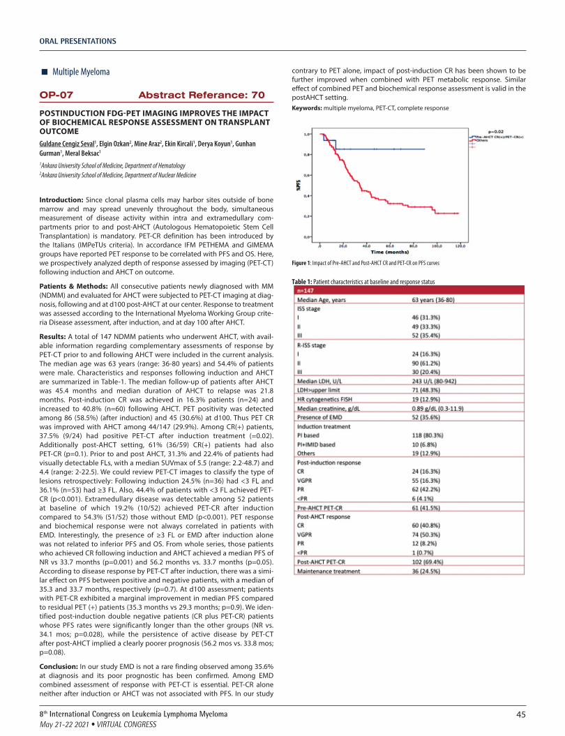
ORAL PRESENTATIONS
458th International Congress on Leukemia Lymphoma MyelomaMay 21-22 2021 • VIRTUAL CONGRESS
� Multiple Myeloma
OP-07 Abstract Referance: 70
POSTINDUCTION FDG-PET IMAGING IMPROVES THE IMPACT OF BIOCHEMICAL RESPONSE ASSESSMENT ON TRANSPLANT OUTCOMEGuldane Cengiz Seval1, Elgin Ozkan2, Mine Araz2, Ekin Kircali1, Derya Koyun1, Gunhan Gurman1, Meral Beksac1
1Ankara University School of Medicine, Department of Hematology 2Ankara University School of Medicine, Department of Nuclear Medicine
Introduction: Since clonal plasma cells may harbor sites outside of bone marrow and may spread unevenly throughout the body, simultaneous measurement of disease activity within intra and extramedullary com-partments prior to and post-AHCT (Autologous Hematopoietic Stem Cell Transplantation) is mandatory. PET-CR definition has been introduced by the Italians (IMPeTUs criteria). In accordance IFM PETHEMA and GIMEMA groups have reported PET response to be correlated with PFS and OS. Here, we prospectively analyzed depth of response assessed by imaging (PET-CT) following induction and AHCT on outcome.
Patients & Methods: All consecutive patients newly diagnosed with MM (NDMM) and evaluated for AHCT were subjected to PET-CT imaging at diag-nosis, following and at d100 post-AHCT at our center. Response to treatment was assessed according to the International Myeloma Working Group crite-ria Disease assessment, after induction, and at day 100 after AHCT.
Results: A total of 147 NDMM patients who underwent AHCT, with avail-able information regarding complementary assessments of response by PET-CT prior to and following AHCT were included in the current analysis. The median age was 63 years (range: 36-80 years) and 54.4% of patients were male. Characteristics and responses following induction and AHCT are summarized in Table-1. The median follow-up of patients after AHCT was 45.4 months and median duration of AHCT to relapse was 21.8 months. Post-induction CR was achieved in 16.3% patients (n=24) and increased to 40.8% (n=60) following AHCT. PET positivity was detected among 86 (58.5%) (after induction) and 45 (30.6%) at d100. Thus PET CR was improved with AHCT among 44/147 (29.9%). Among CR(+) patients, 37.5% (9/24) had positive PET-CT after induction treatment (=0.02). Additionally post-AHCT setting, 61% (36/59) CR(+) patients had also PET-CR (p=0.1). Prior to and post AHCT, 31.3% and 22.4% of patients had visually detectable FLs, with a median SUVmax of 5.5 (range: 2.2-48.7) and 4.4 (range: 2-22.5). We could review PET-CT images to classify the type of lesions retrospectively: Following induction 24.5% (n=36) had <3 FL and 36.1% (n=53) had ≥3 FL. Also, 44.4% of patients with <3 FL achieved PET-CR (p<0.001). Extramedullary disease was detectable among 52 patients at baseline of which 19.2% (10/52) achieved PET-CR after induction compared to 54.3% (51/52) those without EMD (p<0.001). PET response and biochemical response were not always correlated in patients with EMD. Interestingly, the presence of ≥3 FL or EMD after induction alone was not related to inferior PFS and OS. From whole series, those patients who achieved CR following induction and AHCT achieved a median PFS of NR vs 33.7 months (p=0.001) and 56.2 months vs. 33.7 months (p=0.05). According to disease response by PET-CT after induction, there was a simi-lar effect on PFS between positive and negative patients, with a median of 35.3 and 33.7 months, respectively (p=0.7). At d100 assessment; patients with PET-CR exhibited a marginal improvement in median PFS compared to residual PET (+) patients (35.3 months vs 29.3 months; p=0.9). We iden-tified post-induction double negative patients (CR plus PET-CR) patients whose PFS rates were significantly longer than the other groups (NR vs. 34.1 mos; p=0.028), while the persistence of active disease by PET-CT after post-AHCT implied a clearly poorer prognosis (56.2 mos vs. 33.8 mos; p=0.08).
Conclusion: In our study EMD is not a rare finding observed among 35.6% at diagnosis and its poor prognostic has been confirmed. Among EMD combined assessment of response with PET-CT is essential. PET-CR alone neither after induction or AHCT was not associated with PFS. In our study
contrary to PET alone, impact of post-induction CR has been shown to be further improved when combined with PET metabolic response. Similar effect of combined PET and biochemical response assessment is valid in the postAHCT setting.Keywords: multiple myeloma, PET-CT, complete response
Figure 1: Impact of Pre-AHCT and Post-AHCT CR and PET-CR on PFS curves
Table 1: Patient characteristics at baseline and response status
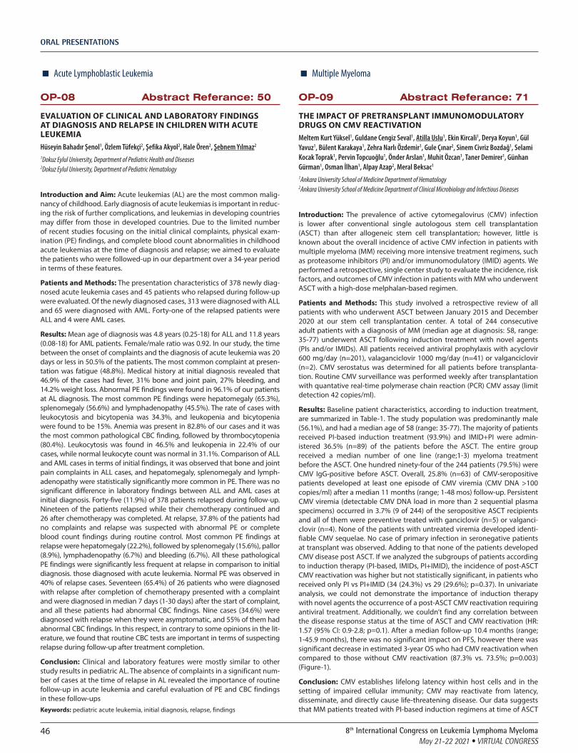
ORAL PRESENTATIONS
46 8th International Congress on Leukemia Lymphoma MyelomaMay 21-22 2021 • VIRTUAL CONGRESS
� Acute Lymphoblastic Leukemia
OP-08 Abstract Referance: 50
EVALUATION OF CLINICAL AND LABORATORY FINDINGS AT DIAGNOSIS AND RELAPSE IN CHILDREN WITH ACUTE LEUKEMIAHüseyin Bahadır Şenol1, Özlem Tüfekçi2, Şefika Akyol2, Hale Ören2, Şebnem Yılmaz2
1Dokuz Eylul University, Department of Pediatric Health and Diseases 2Dokuz Eylul University, Department of Pediatric Hematology
Introduction and Aim: Acute leukemias (AL) are the most common malig-nancy of childhood. Early diagnosis of acute leukemias is important in reduc-ing the risk of further complications, and leukemias in developing countries may differ from those in developed countries. Due to the limited number of recent studies focusing on the initial clinical complaints, physical exam-ination (PE) findings, and complete blood count abnormalities in childhood acute leukemias at the time of diagnosis and relapse; we aimed to evaluate the patients who were followed-up in our department over a 34-year period in terms of these features.
Patients and Methods: The presentation characteristics of 378 newly diag-nosed acute leukemia cases and 45 patients who relapsed during follow-up were evaluated. Of the newly diagnosed cases, 313 were diagnosed with ALL and 65 were diagnosed with AML. Forty-one of the relapsed patients were ALL and 4 were AML cases.
Results: Mean age of diagnosis was 4.8 years (0.25-18) for ALL and 11.8 years (0.08-18) for AML patients. Female/male ratio was 0.92. In our study, the time between the onset of complaints and the diagnosis of acute leukemia was 20 days or less in 50.5% of the patients. The most common complaint at presen-tation was fatigue (48.8%). Medical history at initial diagnosis revealed that 46.9% of the cases had fever, 31% bone and joint pain, 27% bleeding, and 14.2% weight loss. Abnormal PE findings were found in 96.1% of our patients at AL diagnosis. The most common PE findings were hepatomegaly (65.3%), splenomegaly (56.6%) and lymphadenopathy (45.5%). The rate of cases with leukocytosis and bicytopenia was 34.3%, and leukopenia and bicytopenia were found to be 15%. Anemia was present in 82.8% of our cases and it was the most common pathological CBC finding, followed by thrombocytopenia (80.4%). Leukocytosis was found in 46.5% and leukopenia in 22.4% of our cases, while normal leukocyte count was normal in 31.1%. Comparison of ALL and AML cases in terms of initial findings, it was observed that bone and joint pain complaints in ALL cases, and hepatomegaly, splenomegaly and lymph-adenopathy were statistically significantly more common in PE. There was no significant difference in laboratory findings between ALL and AML cases at initial diagnosis. Forty-five (11.9%) of 378 patients relapsed during follow-up. Nineteen of the patients relapsed while their chemotherapy continued and 26 after chemotherapy was completed. At relapse, 37.8% of the patients had no complaints and relapse was suspected with abnormal PE or complete blood count findings during routine control. Most common PE findings at relapse were hepatomegaly (22.2%), followed by splenomegaly (15.6%), pallor (8.9%), lymphadenopathy (6.7%) and bleeding (6.7%). All these pathological PE findings were significantly less frequent at relapse in comparison to initial diagnosis. those diagnosed with acute leukemia. Normal PE was observed in 40% of relapse cases. Seventeen (65.4%) of 26 patients who were diagnosed with relapse after completion of chemotherapy presented with a complaint and were diagnosed in median 7 days (1-30 days) after the start of complaint, and all these patients had abnormal CBC findings. Nine cases (34.6%) were diagnosed with relapse when they were asymptomatic, and 55% of them had abnormal CBC findings. In this respect, in contrary to some opinions in the lit-erature, we found that routine CBC tests are important in terms of suspecting relapse during follow-up after treatment completion.
Conclusion: Clinical and laboratory features were mostly similar to other study results in pediatric AL. The absence of complaints in a significant num-ber of cases at the time of relapse in AL revealed the importance of routine follow-up in acute leukemia and careful evaluation of PE and CBC findings in these follow-upsKeywords: pediatric acute leukemia, initial diagnosis, relapse, findings
� Multiple Myeloma
OP-09 Abstract Referance: 71
THE IMPACT OF PRETRANSPLANT IMMUNOMODULATORY DRUGS ON CMV REACTIVATIONMeltem Kurt Yüksel1, Guldane Cengiz Seval1, Atilla Uslu1, Ekin Kircali1, Derya Koyun1, Gül Yavuz1, Bülent Karakaya1, Zehra Narlı Özdemir1, Gule Çınar2, Sinem Civriz Bozdağ1, Selami Kocak Toprak1, Pervin Topcuoğlu1, Önder Arslan1, Muhit Özcan1, Taner Demirer1, Günhan Gürman1, Osman İlhan1, Alpay Azap2, Meral Beksac1
1Ankara University School of Medicine Department of Hematology 2Ankara University School of Medicine Department of Clinical Microbiology and Infectious Diseases
Introduction: The prevalence of active cytomegalovirus (CMV) infection is lower after conventional single autologous stem cell transplantation (ASCT) than after allogeneic stem cell transplantation; however, little is known about the overall incidence of active CMV infection in patients with multiple myeloma (MM) receiving more intensive treatment regimens, such as proteasome inhibitors (PI) and/or immunomodulatory (IMID) agents. We performed a retrospective, single center study to evaluate the incidence, risk factors, and outcomes of CMV infection in patients with MM who underwent ASCT with a high-dose melphalan-based regimen.
Patients and Methods: This study involved a retrospective review of all patients with who underwent ASCT between January 2015 and December 2020 at our stem cell transplantation center. A total of 244 consecutive adult patients with a diagnosis of MM (median age at diagnosis: 58, range: 35-77) underwent ASCT following induction treatment with novel agents (PIs and/or IMIDs). All patients received antiviral prophylaxis with acyclovir 600 mg/day (n=201), valaganciclovir 1000 mg/day (n=41) or valganciclovir (n=2). CMV serostatus was determined for all patients before transplanta-tion. Routine CMV surveillance was performed weekly after transplantation with quantative real-time polymerase chain reaction (PCR) CMV assay (limit detection 42 copies/ml).
Results: Baseline patient characteristics, according to induction treatment, are summarized in Table-1. The study population was predominantly male (56.1%), and had a median age of 58 (range: 35-77). The majority of patients received PI-based induction treatment (93.9%) and IMID+PI were admin-istered 36.5% (n=89) of the patients before the ASCT. The entire group received a median number of one line (range;1-3) myeloma treatment before the ASCT. One hundred ninety-four of the 244 patients (79.5%) were CMV IgG-positive before ASCT. Overall, 25.8% (n=63) of CMV-seropositive patients developed at least one episode of CMV viremia (CMV DNA >100 copies/ml) after a median 11 months (range; 1-48 mos) follow-up. Persistent CMV viremia (detectable CMV DNA load in more than 2 sequential plasma specimens) occurred in 3.7% (9 of 244) of the seropositive ASCT recipients and all of them were preventive treated with ganciclovir (n=5) or valganci-clovir (n=4). None of the patients with untreated viremia developed identi-fiable CMV sequelae. No case of primary infection in seronegative patients at transplant was observed. Adding to that none of the patients developed CMV disease post ASCT. If we analyzed the subgroups of patients according to induction therapy (PI-based, IMIDs, PI+IMID), the incidence of post-ASCT CMV reactivation was higher but not statistically significant, in patients who received only PI vs PI+IMID (34 (24.3%) vs 29 (29.6%); p=0.37). In univariate analysis, we could not demonstrate the importance of induction therapy with novel agents the occurrence of a post-ASCT CMV reactivation requiring antiviral treatment. Additionally, we couldn’t find any correlation between the disease response status at the time of ASCT and CMV reactivation (HR: 1.57 (95% CI: 0.9-2.8; p=0.1). After a median follow-up 10.4 months (range; 1-45.9 months), there was no significant impact on PFS, however there was significant decrease in estimated 3-year OS who had CMV reactivation when compared to those without CMV reactivation (87.3% vs. 73.5%; p=0.003) (Figure-1).
Conclusion: CMV establishes lifelong latency within host cells and in the setting of impaired cellular immunity; CMV may reactivate from latency, disseminate, and directly cause life-threatening disease. Our data suggests that MM patients treated with PI-based induction regimens at time of ASCT

ORAL PRESENTATIONS
478th International Congress on Leukemia Lymphoma MyelomaMay 21-22 2021 • VIRTUAL CONGRESS
seem to have higher risk of developing symptomatic CMV reactivation and adding IMID to PI does not influence this. However, further studies on a large number of patients are warranted to clarify these findingsKeywords: multiple myeloma, autologous stem cell transplantation, cytomegalovirus
Figure 1. Shows the Kaplan-Meier curves for overall survival (OS) in patients based on the presence of CMV reactivation
Table 1: Baseline patient characteristics by induction treatment
� Myeloprofilerative DisordersOP-10 Abstract Referance: 68
MUTATION PROFILE OF THE PATIENTS TESTED WITH NEXT GENERATION SEQUENCING AND CLINICAL IMPLICATIONSYaşa Gül Mutlu1, Berrin Balık Aydın1, Ömür Gökmen Sevindik1
1Medipol Istanbul University, Department of Hematology
Background and Aim: Next Generation Sequencing (NGS) was a ground-breaking advent in the field of genetics. It allowed to better and pre-cisely identify both driver and secondary mutations in both hematological and solid organ malignancies. Besides, it allowed to even precisely follow-up the response via Variant Allele Frequencies (VAF) of those mutations. In this regard, we have established a Myeloid NGS panel including 30 commonly mutated genes. We wanted to present the first report among the rates and VAF’s of the mutations both obtained in the time of diagnosis and also during the follow up of first 50 patients.
Material and Methods: A 30 gene Myeloid NGS panel was used to deter-mine the presence of any driver or secondary mutations of the patients who were diagnosed with any kind of myeloid hematological malignancy. The
rate of positivity and also the mean and standard devaitions of the mutated genes were recorded with the demographics.
Results: The median age of the 50 patients were 55 (18-86), and 56% of the patients were male. The distribution of the diagnosis were as follows; AML (56%), MDS (14%), CMML (10%), Myelofibrosis (10%), other MPNs (PV or ET 10%). Most frequently observed mutation was FLT3 with a frequency of 23.5% in all 63 samples, others were given in Figure 1. The VAF’s of the mutations which were found to be positive in 63 samples of 50 patients were given in Figure 2. When the samples of the patients who were diagnosed with AML or MDS, the most common first 5 mutations were as follows; FLT3 (34.3%), DNMT3A (20.5%), NPM1 (20.5%), ASXL1 (14.6%) and IDH2 (11.9%). Regarding the patients who were diagnosed with AML or MDS, the positiv-ity rates and VAF’s were given in Figure 3 and 4. Co-mutation rates of the most common 5 mutations were also analyzed in this particular group and presented in Table 1. The follow-up of the positive clones in patients with myeloid neoplasms have allowed us to offer a reliable follow up and timely institution of some targeted therapies and to offer appropriate initial thera-py even in patients who had no crude cytogenetic abnormalities.
Conclusion: NGS had allowed us to better prognosticate and to offer more precise therapies in patients who were diagnosed with myeloid neoplasms.Keywords: Next Generation Sequencies, Myeloid Neoplasms, Prognosis, Genetics
Figure 1.
Figure 2.
Figure 3.
Figure 4

ORAL PRESENTATIONS
48 8th International Congress on Leukemia Lymphoma MyelomaMay 21-22 2021 • VIRTUAL CONGRESS
Table 1: Co-Mutations
� Stem Cell TransplantationOP-11 Abstract Referance: 63
PHENOTYPES OF BONE MARROW MONOCYTES IN STEM CELL TRANSPLANTATION FOR ACUTE LEUKEMIA: A DESCRIPTIVE PILOT STUDYMeltem Kurt Yüksel1, Ekin Kırcalı1, Cemaleddin Öztürk1, Hülya Yilmaz1, Derya Koyun1, Şenay İpek2, Güldane Cengiz Seval1, Sinem Civriz Bozdağ1, Selami Koçak Toprak1, Pervin Topçuoğlu1, Klara Dalva1
1Ankara University Medical School, Hematology Department 2Ankara University Medical School, Flow Cytometry Laboratory
Introduction and Aim: Monocytes probably take a major role in immune regulation and hematological reconstitution after hematopoietic cell trans-plant. Phenotypically, monocytes are classified in three different subsets according to the clusters of differentiation they express: Classical MO1 (CD14 bright/ CD16 -), intermediate MO2 (CD14 bright/ CD16+) and non-classical MO3 (CD14 dim/ CD16 +) by flow cytometry. In this series of acute leukemia patients, we aimed to investigate the relation of monocyte subsets and clin-ical outcomes of allotransplant recipients.
Methods: We retrospectively investigated acute leukemia patients who underwent allogeneic transplantation at Ankara University Hematology Department and acquired both pretransplant and the +28th day monocyte subsets by flow cytometry. Nine (n= 9) patients were included in the trial.
Findings: The demographics and details about transplants are shown on Table 1. Flow cytometric analysis of pretransplant and post- transplant bone marrow aspirations are shown on Table 2. As we identified few patients, a statistical analysis was not made at this point. Two of the cases suffered from acute graft vs host disease, none of whom had distinctive subsets of monocytes (Table 2).
Conclusion: Bone marrow monocyte subtypes (eg. non-classical) might play a critical role in foreseeing acute leukemia relapse, engraftmant or graft vs host disease after allogeneic hematopoietic cell transplantation. In this retrospective pilot trial, we were not able to prove this hypothesis right. Prospective studies are needed to verify the findings in the literature.Keywords: monocyte subtypes, non-classical monocytes, hematopoietic cell transplant
References
1. A Pilot Study of Circulating Monocyte Subsets in Patients Treated with Stem Cell Transplantation for High-Risk Hematological Malignancies. Ida Marie Rundgren 1,2, Elisabeth Ersvær 1, Aymen Bushra Ahmed 3, Anita Ryningen 1 and Øystein Bruserud 2,3,* 1: Department of Biomedical Laboratory Scientist Education and Chemical Engineering, Faculty of Engineering and Natural Sciences, Western Norway University of Applied Sciences, 5020 Bergen, Norway; [email protected] (I.M.R.); [email protected] (E.E.); [email protected] (A.R.) 2: Department of Clinical Science, University of Bergen, 5020 Bergen, Norway 3: Section for Hematology, Department of Medicine, Haukeland University Hospital, 5021 Bergen, Norway
Table 1: Patient Demographics
Table 2: Flow Cytometric Parameters
� Acute Lymphoblastic Leukemia
OP-12 Abstract Referance: 55
EVALUATION OF POSSIBLE EFFECTS OF THE COVID-19 PANDEMIC ON FEBRILE NEUTROPENIA EPISODES IN CHILDREN WITH ACUTE LEUKEMIAİrem Ceren Erbaş1, Özlem Tüfekçi2, Şefika Akyol2, Hatice Karaoğlu Kısrak1, Ayşe Çakıl Güzin1, Şilem Özdem Alataş1, Şebnem Yılmaz2, Hale Ören2, Nurşen Belet1
1Dokuz Eylul University, Department of Pediatric Infectious Diseases 2Dokuz Eylul University, Department of Pediatric Hematology
Introduction: Febrile neutropenia (FN) is one of the most common and mor-tal complications in cancer patients receiving chemotherapy. The effects of COVID-19 pandemic conditions on febrile neutropenia episodes of children with acute leukemia have not been investigated in detail yet. In this study, we aimed to investigate the effects of the COVID-19 pandemic on frequency, etiology, and prognosis of FN episodes in children with acute leukemia.
Material and Methods: Relevant data about FN episodes that observed between March 2018 - March 2021 were collected retrospectively from medical records of patients diagnosed with acute leukemia. Episodes were classified into two groups as before and after the pandemic. The data were presented as (%) for categorical variables and as median (25-75p) for numer-ic variables.
Results: Fourty-six patients (47.8% girls) with a median age of 4.7 (2.6-12.6) years were included in the study. The most common underlying disease was acute lymphoblastic leukemia (69.9%). A total of 113 FN episodes of the patients during the study period were evaluated (75.2% before the

ORAL PRESENTATIONS
498th International Congress on Leukemia Lymphoma MyelomaMay 21-22 2021 • VIRTUAL CONGRESS
pandemic). The remission status during the episodes and the risk groups of the patients were similar among two groups (p = 0.689 and 0.054, respec-tively). The number of episodes per patient was two (1-3) in both groups (p = 0.476). Episodes with a fever of known origin and the duration of febrile days were similar between groups (p > 0.05). Although there was an increase in the frequency of bacterial agents in the etiology during the post-pandemic period, there was no statistically significant difference between the two groups (28.2% vs 42.9%; p = 0.150). The presence of resistant organisms and the frequency of polymicrobial agents were similar in both groups (p > 0.05). While the frequency of viral respiratory tract agents was 25.9% before and 10.7% after the pandemic, no statistically significant difference was found among groups (p = 0.094). Among the viral respiratory tract agents, Rhinovirus (n=12), Respiratory syncytial virus (n=3), and Parainfluenza virus (n=3) were the most frequent ones before the pandemic; whereas COVID-19 (n=2), Rhinovirus (n=1), and Adenovirus (n=1) were detected commonly after the pandemic. Both of two patients with COVID-19 did not need oxygen support and recovered without any complications. There was no difference between two groups in terms of the frequency of suspected, probable, or proven fungal infections (8.5% vs 14.5%; p = 0.381). Treatment durations were similar in FN episodes before and after the pandemic (12 vs 10.5 days; p = 0.734). Only one patient died during the FN episode in the post-pandemic period.
Conclusion: Despite the strict isolation rules that were followed during the pandemic, there was no change in the frequency of FN episodes of the patients. We found that the distribution of etiological causes, duration of febrile days and treatment periods were not affected by the COVID-19 pandemic.Keywords: acute leukemia, febrile neutropenia, COVID-19, children
� Non-Hodgkin’s Lymphoma
OP-13 Abstract Referance: 82
IS SURGICAL EXCISIONAL BIOPSY STILL THE GOLD STANDARD DIAGNOSTIC APPROACH IN LYMPHOMAS?Yaşa Gül Mutlu1, Berrin Balık Aydın1, Ömür Gökmen Sevindik1
1Medipol Istanbul University, Department of Hematology
Background and Aims: Surgical Excisional Biopsy (SEB) is accepted as the standard of care approach in the diagnosis of lymphomas according to both national and international guidelines. However, both financial issues related with the increased cost and the invasive nature of the procedure forced phy-sicians to use some alternative diagnostic methods. One of these methods is the Core Needle Biopsy (CNB), which gained a reputation for the diagnosis of lymphomas with the advent of improved pathological and immunohisto-chemical analysis, which made it possible to have an accurate diagnosis with limited tissue samples. In this retrospective study, we aimed to compare the diagnostic yield of SEB and CNB.
Material and Methods: 131 patients who were diagnosed with either Hodgkin (HL) or Non-Hodgkin Lymphoma (NHL) with a nodal biopsy which was acquired via SEB or CNB were included in the study between 2014-2020 in our center. 68 patients were undergone SEB and the remaining 63 were undergone CNB. Samples were re-classified according to the diagnostic ability relying on a pre-defined criterion as fully diagnostic, partially diag-nostic and inadequate. Samples which allowed to identify the exact tumor type and/or subtype were accepted as fully diagnostic. Sufficient tissue that pathologist could have any suspicious findings considering malignant lym-phoma classified as partial diagnostic group. Inadequate samples were the ones which were not enough to report any final diagnosis.
Results: The patients who underwent to a CNB were significantly younger than the patients who underwent to SEB (47.6 vs 56.8, p = 0.003). Despite the fully diagnostic ability of SEB outperformed CNB (95.2% vs 83.8%, p = 0.035), in 92.6% of the patients whose tissue samples were obtained via CNB were accepted to have a sufficient diagnosis to initiate the treatment
and not required a second CNB or SEB, which was comparable with the ones achieved by SEB (92.6% vs 95.2%, p = 0.720) (Tables 1-3)
Conclusion: According to the results obtained in our study, we may con-clude that CNB is a viable and comparable alternative to SEB, offering a less invasive and less expansive approach to diagnose a lymphoma.Keywords: Surgical Excional Biopsy, Core Needle Biopsy, Lymphoma, Diagnosis
Table 1:Patient Demographics, Biopsy Locations and Diagnosis Among CNB and SEB Groups
Table 2: Diagnostic Sub-Categories of CNB versus SEB
Table 3: Need for a Second Biopsy

ORAL PRESENTATIONS
50 8th International Congress on Leukemia Lymphoma MyelomaMay 21-22 2021 • VIRTUAL CONGRESS
� Multiple Myeloma
OP-14 Abstract Referance: 64
PATIENT RELATED FACTORS OVERRIDE LENALIDOMIDE MAINTENANCE AS A FACTOR OF SEVERITY FOR COVID-19 INFECTIONEkin Kırcalı, Güldane Cengiz Seval, Selami Koçak Toprak, Pervin Topçuoğlu, Önder Arslan, Muhit Özcan, Osman İlhan, Meral Beksaç
Ankara University Medical School, Hematology Department
Introduction: Maintenance with lenalidomide after high dose melphalan consolidation is known to extend progression free survival (PFS) in multiple myeloma (MM) patients [1]. Lenalidomide may induce neutropenia and lymphopenia, exposing some patients to infections. On the other hand, there have been small case series and brief reports suggesting IMIDs might actually protect from severe Covid-19 [2] [3]. Here, we resumed our set of MM patients who were diagnosed with Covid-19 infection and compared outcomes based on continuous lenalidomide maintenance. A novel Covid-19 risk prediction model has been integrated to compare patient groups on or off maintenance.
Patients and Methods: 60 MM patients were diagnosed with Covid-19 in our department between March 2020- April 2021. Here, we will be reporting the data of those who were on lenalidomide when infected (n= 29), not on lenalidomide maintenance (n= 31), and as a control group, the patients who were not infected with Covid-19 while on lenalidomide (n= 20). Results were compared on Lenalidomide but no Covid-19 (Table 1).
Ji et al published a study of 208 patients in order to predict high risk Covid- 19 patients [4], using a set of data (CALL score- comorbidities, age, lymph-openia, high lactate dehydrogenase [LDH]). CALL score varies between 4 and 13, 4-6 being low, 7- 9 intermediate, 10- 13 high risk. We repurposed CALL score on our set of patients, too (Table 1). All statistical analyses were performed via SPSS statistics version 21.0 software.
Results: Table 1 summarizes patient demographics and essential laboratory data. Infection fatality rate for lenalidomide maintenance and no lenalido-mide groups were 17.2% and 19.3%, respectively. Table 2 compares severity of Covid-19 infection and mortality across subgroups.
Conclusion: Based on our analysis a risk score at the onset of Covid-19 infection identifies similar distribution among patients who were on or off Lenalidomide maintenance. Clinical severity is determined by risk score and not Lenalidomide maintenance, supporting continuation of therapy to pre-vent recurrence of myelomaKeywords: Covid-19, lenalidomide, risk factors for Covid severity
References
1. Palumbo, A., et al., Autologous transplantation and maintenance therapy in multiple myeloma. New England Journal of Medicine, 2014. 371(10): p. 895-905.
2. Tailor, I.K., et al., Outcome of myeloma patients with COVID-19 on active lenalido-mide-based therapy: Does lenalidomide protect from severe COVID-19? Hematology/oncology and stem cell therapy, 2020.
3. Chowdhury, K.H., et al., Drug Repurposing Approach against Novel Coronavirus Disease (COVID-19) through Virtual Screening Targeting SARS-CoV-2 Main Protease. Biology, 2021. 10(1): p. 2.
4. Ji, D., et al., Prediction for progression risk in patients with COVID-19 pneumonia: the CALL score. Clinical Infectious Diseases, 2020. 71(6): p. 1393-1399.
Table 1: Patient Demographics (median when applicable)
Table 2: Comparison of Groups in Tems of Risk and Mortality
� Multiple Myeloma
OP-15 Abstract Referance: 74
PACE-LIKE REGIMENS IN THE TREATMENT OF RELAPSED/ REFRACTORY MULTIPLE MYELOMAAylin Fatma Karatas, Boran Yavuz, Mehmet Ali Ozcan, Elçin Erdogan Yucel, Serkan Guven, Fatih Demirkan, Inci Alacacıoglu, Guner Hayri Ozsan
Dokuz Eylul University Hospital
Introduction: Multiple myeloma (MM) is a plasma cell malignancy with propensity to cause bone lesions, hypercalcemia, renal failure and anemia. Treatmen options in multiple myelome changing. With the availability of novel chemotherapeutic agents including proteasome inhibitors (PIs), and immunomodulatory drugs (IMiDs), and increased use of high dose therapy with autologous stem cell transplantation (ASCT), the overall survival (OS) in patients with MM has improved.(1.2). Patients with multiple relapses and/or refractory MM (RRMM) are difficult to manage as the therapeutic options become limited and the response to new therapy resulted in lower response rate and shorter duration. (3) In eligible patients, PACE (cisplatin, doxorubi-cin, cyclophosphamide, and etoposide) regimens is often utilized.
Patients and Methods: 12 patients with multiple myeloma who were followed up in Dokuz Eylül University Hematology Unit were included in the study.We analyzed the outcomes of 13 patients with RRMM who received PACE regimens at our center between 2019 and 2021 in an intent-to-treat analysis.
Results: Median age was 51 years, and 66.7% male. Patients have diagnosed between 2012 and 2019 and received PACE regimens at a median of 50 months from diagnosis. Patients were heavily pretreated with a median of 5 prior regimens (range, 2-8), prior autologous stem cell transplant [ASCT] (except one). PACE regimens of patient were listed in Table I.
Patients who were not completed therapy due to toxicity and died in a short time were excluded. 1 patient achieved Complete Response (CR) (%8.3) , 5 of patients achieved Partial Response (PR) (%41,7), 1 patient achieved Minimal Response (MR) (%8,3). Alternative treatments were started after 1 course due to renal failure and gram negative infection in 1 patient, grade 4 neutropenia and opportunistic infection in 1 patient. 3 patients died in 1 month. 2 due to toxic hepatitis and sepsis, 1 due to sepsis. 3 patient)s with PR and CR underwent allogenic stem cell transplantation. One of them died 1 month after AKIT due to Graft versus host disease (GVHD) and Venooclusive disease (VOD).

ORAL PRESENTATIONS
518th International Congress on Leukemia Lymphoma MyelomaMay 21-22 2021 • VIRTUAL CONGRESS
7 patients (%58) had extramedullary involvement. Median overall survival of patients after pace regimens were 7 months (Figure I). 5 patients are still alive. Median progression free survival of patients after pace regimens were 4 months (Figure II).
Conclusion: In patients for suitable PACE like regimens have a high response rate even if they were heavily pretreated (4). PACE like regimens can be mod-ified by adding novel agents to the regimen. Their use can be considered in heavily pretreated patients especially if they have extramedullary disease manifestations. And may be a bridge therapy for allogenic transplatation.Keywords: Autologus stem cell transplant,PACE like regimens, Overall Survival
References1. Kumar SK, Rajkumar SV, Dispenzieri A, et al. Improved survival in multiple myeloma
and the impact of novel therapies. Blood. 2008; 111(5):2516–2520.
2. Kumar SK, Dispenzieri A, Lacy MQ, et al. Continued improvement in survival in multi-ple myeloma: changes in early mortality and outcomes in older patients. Leukemia. 2014;28(5):1122–1128
3. Lee CK, Barlogie B, Munshi N, et al. DTPACE: an effective, novel combination chemo-therapy with thalidomide for previously treated patients with myeloma. J Clin Oncol. 2003;21:2732–2739
4. Gerrie, A.S., Mikhael, J.R., Cheng, L., Jiang, H., Kukreti, V., Panzarella, T., Reece, D., Stewart, K.A., Trieu, Y., Trudel, S. and Chen, C.I. (2013), D(T)PACE as salvage therapy for aggressive or refractory multiple myeloma. Br J Haematol, 161: 802-810. https://doi.org/10.1111/bjh.12325
Figure 1. Overall Survival
Figure 2. Progression Free Survival
Table 1: Pace Regimens
n %
CAR D PACE (Carfilzomib Dexamethasone) 3 25
POM D PACE (Pomalidomide Dexamethasone) 4 33.3
VD PACE (Bortezomib Dexamethasone) 2 16.7
D PACE (Dexamethasone) 1 8.3
VDT PACE (Bortezomib Dexamethasone Talidomide) 2 16.7
Total 12 100
� Palliative Care–Supportive Therapy
OP-16 Abstract Referance: 69
QUALITY OF LIFE MEASURES OF THE PATIENTS WHO ARE DIAGNOSED WITH HEMATOLOGICAL MALIGNANCIES AND TIME EFFECT ON PARAMETERSBerrin Balık Aydın, Yaşa Gül Mutlu, Ömür Gökmen Sevindik
Medipol Istanbul University, Department of Hematology
Background and Aim: The World Health Organization defines the quality of life as “perceiving one’s own life in a culture and value system according to one’s own goals, expectations, standards and interests”.The name of the cancer disease, the fear given by the name, anxiety for the future, the stress caused by what may be experienced during the disease process and the undesirable effects related to the treatment significantly affect the quality of life of the patients and their relatives. In this study, it was aimed to determine the quality of life, social support levels and factors affecting patients who were diagnosed with hematological malignancy and received chemothera-py, with regard to the certain time points passed after diagnosis.
Material and Methods: The EORTC QLQ-C30 Quality of Life Version 3.0 Turkish Scale, which was previously validated in Turkish, was used to assess QoL measures. A total of 89 hematological cancer patients included who were admitted to our hospital between December 2020 and February 2021. The data obtained from the scale were evaluated by demographic character-istics, diagnosis and the time from diagnosis of the patients.
Results: A total of 89 patients, 37 (42%) female and 52 (59%) male, were included in the study. Median age was 55 (18 - 85). According to their diagno-sis, patients were categorized into 5 different groups as leukemias, lympho-mas, plasma cell diseases, mds-mpn and others. The time after the diagnosis was analyzed by dividing them into groups as the first 6 months and after, the first 12 months and after. The disease diagnosis did not show a statistically significant difference on quality of life parameters. The scales resulted to be similar across gender groups. When the age groups were compared, it was found statistically significant that the scores in the physical function and role function scales were higher and the scores in the fatigue, loss of appetite, constipation and diarrhea scales were found to be statistically significant in the group <55 years old. Quality of life resulted in higher in this age group. When the patients were compared with the time from diagnosis as <6 months and ≥6 months, it was observed that the complaint of constipation was higher in the first 6 months, and no significant dissimilarity was observed between groups regarding the remaining QoL measures, interestingly this was also true for a comparison between 1 year period and more. Validaion of the parame-ters were confirmed by high concordance with Cronbach alfa scores.
Conclusion: In conclusion, it was seen that the Turkish version of the QLQ-30 Scale is a measurement tool with valid and reliable indicators in measuring the quality of life of patients with hematological cancer. The time from diagnosis had no apperant impact on QoL measures in our patient group. It makes it essential to support our patients throughout the therapy and even after the end of treatment, both medically and socially with experts in the field. We will try to establish an interdisciplinary support group in this manner.Keywords: Quality of Life, Hematology, Cancer, Support

ORAL PRESENTATIONS
52 8th International Congress on Leukemia Lymphoma MyelomaMay 21-22 2021 • VIRTUAL CONGRESS
� Non-Hodgkin’s Lymphoma
OP-18 Abstract Referance: 6
THE EFFECT OF THE CELL OF ORIGIN USING HANS ALGORITHM ON PROGNOSIS IN DIFFUSE LARGE B CELL LYMPHOMASTaha Ulutan Kars, Atakan Tekinalp
Necmettin Erbakan University, Meram Faculty of Medicine, Department of Internal Medicine, Division of Hematology
Introduction: Diffuse large B-cell lymphoma (DLBCL) is the most common type of non-Hodgkin lymphoma (NHL)1. The prognosis of DLBCL depends on the clinical features such as International Prognostic Index (IPI) and gene expression profiling2. Various immunohistochemical (IHC) algorithms have been developed by using molecular studies. Hans algorithm (HA) by was widely accepted as a mechanism to divide DLBCL into germinal centre (GC) and non-GC subtypes according to the cell of origin (COO)3. While there are many studies showing that this algorithm is successful in predicting progno-sis, there are also studies showing that it is not successful 4,5
Materials and Methods: There were 84 cases of DLBCL reported from the beginning of 2012 to early 2020.
Results: The mean age was 59,7±14,8 years. There was a might male domi-nancy. 27 of patients (32,1%) were in germinal center (GC) group and 57 of (67,9%) in non-GC group. Table-1 shows the relationship between COO and sex, nodal/extranodal involvement, IPI score, response to the first line treat-ment. The difference of hemogram, inflammatory parameters and maximum fluorodeoxyglucose (FDG) uptake on positron emission tomography (PET/CT) imaging in these groups were not statistically significant, as document-ed in Table-2.
3-year overall survivial (OS) in GC group and non-GC groups were 72% and 70%, respectively. There was no significant difference in median survival between two groups (log-rank p: 0,867). Kaplan-Meier survival curve is seen in Figure-1.
Discussion: DLBCL is the most common type of NHL and considered an aggressive lymphoma. Predicting prognosis in an individual patient is very difficult as DLBCL comprises a group of morphologically, immunohisto-chemically, and clinically heterogeneous tumours rather than one single entity. IHC is presently the cheaper option to determine COO of DLBCL. One of the methods most commonly used is the HA, which uses the expression of CD10, BCL6 and MUM1 by the DLBCL cells to classify patients as GC or non-GC3. The advantage of using HA is that it uses only three antibodies, which made it widely acceptable as compared to other algorithms that were developed later to subtype DLBCL according to COO. However, the clinico-pathological differences between GC and non-GC as well as the prognostic and predictive value of the HA have not been fully evaluated.
The predictive role of the HA has been previously evaluated in small studies with conflicting Results: Ilic et. al. showed that patients with the GCB subtype of DLBCL had an outcome similar to that of patients with the non-GCB sub-type6. Zinzani et al. reported that projected 4-year OS was 100% for GCB and 82% for non-GCB patients (p=0,12)7. A recent meta-analysis by Fang et al. has shown a statistical trend towards an association between IHC profile and overall response rate8. In the present study, we have attempted to critically evaluate the HA in relationship to clinical and pathological characteristics as well as response to therapy and survival in patients with DLBCL. Similar to the literature, the non-GC group in higher than the GC group in present study. Our study showed that there is no statistical significance of 3-year OS between the two subtypes. Our study also, showed that there is no signif-icant difference the relationship between COO and presence of extranodal involvement, IPI score, response to first line treatment. However, compared to other published studies, our study has a small number of patients which may affect the statistical analysis.
This study suggests that the HA does not have prognostic value in DLBCL patients. This result may be related to the development of the HA before
the period in which rituximab was widely used. Additional efforts should be directed at elucidating more reliable easy-to-use IHC-based algorithms to identify DLBCL subtypes with future aims of not only risk-stratifying patients but also directing therapy to improve outcomes.Keywords: Lymphoma, Large B-Cell, Diffuse, Prognosis, Immunohistochemistry
References
1 Siegel, R. L., Miller, K. D. & Jemal, A. Cancer statistics, 2019. CA. Cancer J. Clin. 69, 7–34 (2019).
2. Sehn, L. H. & Gascoyne, R. D. Diffuse large B-cell lymphoma: optimizing outcome in the context of clinical and biologic heterogeneity. Blood 125, 22–32 (2015).
3. Hans, C. P. Confirmation of the molecular classification of diffuse large B-cell lympho-ma by immunohistochemistry using a tissue microarray. Blood 103, 275–282 (2004).
4. Lin, Y. H. & Yang-Yen, H. F. The Osteopontin-CD44 Survival Signal Involves Activation of the Phosphatidylinositol 3-Kinase/Akt Signaling Pathway. J. Biol. Chem. 276, 46024–46030 (2001).
5. Castillo, J. J. et al. The Hans algorithm is not prognostic in patients with diffuse large B-cell lymphoma treated with R-CHOP. Leuk. Res. 36, 413–417 (2012).
6. Ilić, I. et al. Lack of prognostic significance of the germinal-center phenotype in dif-fuse large B-cell lymphoma patients treated with CHOP-like chemotherapy with and without rituximab. Int. J. Hematol. 90, 74–80 (2009).
7. Zinzani, P. L. et al. Immunophenotype and intermediate-high international prognos-tic index score are prognostic factors for therapy in diffuse large B-cell lymphoma patients. Cancer 116, 5667–5675 (2010).
8. Fang, C., Xu, W. & Li, J.-Y. A systematic review and meta-analysis of rituximab-based immunochemotherapy for subtypes of diffuse large B cell lymphoma. Ann. Hematol. 89, 1107–1113 (2010).
Figure 1. Cumulative Survival by Cell of Origin

ORAL PRESENTATIONS
538th International Congress on Leukemia Lymphoma MyelomaMay 21-22 2021 • VIRTUAL CONGRESS
Table 1. The Relationship Between COO and Sex, Nodal/Extranodal Involvement, IPI-risk, Response to the First Line Treatment
GC Non-GC p
Sex, n (%)
Female 13 (48,1) 24 (42,1) 0,602
Male 14 (51,9) 33 (57,9)
Nodal/Extranodal Involvement, n (%)
Nodal 9 (33,3) 21 (36,8) 0,754
Extranodal 18 (66,6) 36 (63,2)
IPI score, n (%)
Low 5 (18,5) 8 (14,0) 0,568
Low-Intermediate 8 (29,6) 18 (31,6)
High-Intermediate 3 (11,1) 13 (22,8)
High 11 (40,7) 18 (31,6)
Response to First Line Treatment, n (%)
Complete 13 (48,1) 36 (63,2) 0,110
Partial 11 (40,7) 11 (19,3)
Progression 3 (11,1) 10 (17,5)
Tablo 2. The Relationship Between COO and Sex, Laboratory Findings
GC (n:27) Non-GC (n:57) p
Age (year) 57,11 ± 14 60,9 ± 15,2 0,276
Hb (g/dL) 13,6 ± 1,4 12,2 ± 2,2 0,277
WBC (µL) 8.000 (5.300-17.500) 7.900 (1.500-28.900) 0,534
Neutrophil (µL) 5.500 (3.400-12,500) 5.100 (700-12.400) 0,490
Lymphocyte (µL) 1.300 (5000-10.200) 1.500 (400-22.300) 0,950
Monocyte (µL) 796 ± 260 756 ± 300 0,554
Platelet (x1000/µL) 297,8 ± 104,9 277,1 ± 133,3 0,480
MPV (Fl) 10,4 (6,4-12,3) 9,8 (5,9-12,6) 0,399
LDH (IU) 355 (163-1228) 331 (134-1840) 0,527
Ferritin (ml/ng) 246,4 (15-2818) 242,6 (9,6-1418) 0,554
Β2 MG (mg/dL) 3,3 (1,8-5,8) 3,4 (1,7-16) 0,808
Sedimentasyon (mm/h) 27 (3-105) 31 (2-98) 0,871
CRP (mg/L) 36 (0,1-231) 27,2 (0,1-257) 0,308
Maksimum SUVmax 20,1 ± 7,6 21,6 ± 11,1 0,463
� Stem Cell Transplantation
OP-19 Abstract Referance: 61
DETERMINATION OF INFECTION FREQUENCY IN PATIENTS USING RUXOLITINIB DUE TO GRAFT VERSUS HOST DISEASEHülya Yilmaz, Ekin Kırcalı, Cemaleddin Öztürk, Güldane Cengiz Seval, Sinem Civriz Bozdağ, Selami Koçak Toprak, Pervin Topçuoğlu, Günhan Gürman, Meltem Kurt Yüksel
Ankara Univercity Medical Faculty, Department of Hematology
Introduction: Graft versus Host Disease (GVHD) is an important life-threat-ening complication in patients undergoing allogeneic stem cell transplanta-tion (ASCT). Illumination of the pathogenesis of GVHD, albeit partially, has
increased the variety of drugs used in its treatment in recent years. Although there is no license indication, with the identification of mechanisms that can play a role in pathogenesis such as JAK-STAT and BTK pathway, many new drugs are being used in our country in the management of GVHD with off-label approval. Ruxolitinib is one of these drugs. The side effects of ruxolitinib, which is a non-selective TKI, whose efficacy in GVHD has also been demonstrated by clinical studies, is limited. Prevention and treatment of infections, especially latent infections are as important as effectiveness in the success of GVHD treatment.
Purpose: To determine the effect of Ruxolitinib use on viral (CMV, HBV, BKV, EBV), bacterial, fungal infection risk in GVHD patients.
Method: Patients who were not able to use steroids due to steroid refractory GVHD or side effects, after allogeneic stem cell transplantation between 2016-2020 were included. Patient files and patient registry system were reviewed retrospectively. CMV reactivation was defined as patients with a CMV copy number> 500.
Results: A total of 34 patients were included in the study. The median age of the patients was 46 (22-64), the female / male ratio was 15/19. 35% (n = 12) of the cases were diagnosed with ALL, 26% (n = 9) AML, 11% (n=4) MDS and 11 (n=4)% lymphoma. Organ involvement in patients with GVHD is shown in figure 1. While the majority of the patients took ruxolitinib due to steroid unresponsiveness, 26% (n = 9) of the patients received ruxolitinib because of steroid side effects or dependence. Bacterial infection requiring hospitaliza-tion was detected in 3 cases during follow-up. Under ruxolitinib treatment, a total of 13 times CMV reactivation was observed in 11 patients and BKV viremia was observed in 2 patients. The details are shown in table 1. In a patient who received cidofovir treatment due to hemorrhagic cystitis (grade 3) and regressed to grade 0 after treatment, BKV copy number increased after ruxolitinib, but hemorrhagic cystitis was not observed in the patient. After 2 weeks, she came with grade 4 hemorrhagic cystitis.
Discussion: When steroid-refractory acute GVHD develops, patients con-tinue to receive third and fourth-line treatments, respectively, when there is no response to second-line GVHD treatment, and without discontinuing other drugs. Although the dose of steroid treatment is reduced, discon-tinuation of drug therapy, especially steroid, is an important problem. It is also very difficult to determine in these patients which of the drugs they are receiving causes to develop infections since those patients are receiving multiple immunosuppressive therapies all of which causes a susceptability to infections.
Conclusion: In this study, infection (viral + bacterial) was found in 47% of the patients who took ruxolitinib. During the use of ruxolitinib, the Jak1-2 inhib-itor, which we frequently use in steroid refractory patients, it is important to monitor patients especially for CMV and BKV infections.Keywords: GVHD, Ruxolitinib, CMV, BKV
References
1. Penack, O., Marchetti, M., Ruutu, T., Aljurf, M., Bacigalupo, A., Bonifazi, F., ... & Basak, G. W. (2020). Prophylaxis and management of graft versus host disease after stem-cell transplantation for haematological malignancies: updated consensus recommen-dations of the European Society for Blood and Marrow Transplantation. The Lancet Haematology, 7(2), e157-e167.
2. Liu, Y., Fan, Y., Zhang, W., Chen, J., Cheng, Q., Ma, X., ... & Xu, Y. (2021). Efficiency and toxicity of ruxolitinib as the salvage treatment in steroid-refractory acute graft-ver-sus-host disease after haplo-identical stem cell transplantation. Transplantation and Cellular Therapy.
3. Alvarnas, J. C. (2021). Ruxolitinib for the Treatment of Steroid-Refractory Chronic Graft-vs-Host Disease—Another Hopeful Step Forward. JAMA Network Open, 4(1), e2035719-e2035719.
4. Wu, H., Shi, J., Luo, Y., Tan, Y., Zhang, M., Lai, X., ... & Zhao, Y. (2021). Evaluation of Ruxolitinib for Steroid-Refractory Chronic Graft-vs-Host Disease After Allogeneic Hematopoietic Stem Cell Transplantation. JAMA network open, 4(1), e2034750-e2034750.

ORAL PRESENTATIONS
54 8th International Congress on Leukemia Lymphoma MyelomaMay 21-22 2021 • VIRTUAL CONGRESS
Table 1.Type of infections are detailed
Figure 1. Organ involvement in 34 patients with GVHD
� Non-Hodgkin’s LymphomaOP-20 Abstract Referance: 81
MYD88 EXPRESSION IN PRIMARY AND SECONDARY CNS LYMPHOMASBerrin Balık Aydın1, Yaşa Gül Mutlu1, Aslı Çakır2, Ömür Gökmen Sevindik1
1Medipol Istanbul University, Department of Hematology 2Medipol Istanbul University, Department of Pathology
Background and Aim: Myeloid differentiation primary response 88 (MYD88) is a common adaptor protein that is responsible for signaling from several receptors; is encoded by the MYD88 gene. We aimed to assess the level of MYD88 expression, and their associations with clinicopathological parame-ters in CNS Lymphomas.
Material and Methods: A total of 11 patients were included in the study. MYD88 protein expression was evaluated by immunohistochemistry (IHC) using two different scoring systems. All samples were diagnosed and selected by a hematopathologist. Tissue samples were collected from all patients before treatment. MYD88 cytoplasmic expression was classified as four categories according to the staining intensity on a scale from 0 to 3 as
follows: 0, no reaction; 1, weak reaction; 2, moderate reaction; and 3, strong reaction. This classification was called as the first classification model. In the second classification model, the extent of staining scored as 0 (0% of tumor area stained), 1 (< 10%), 2 (10–50%), or 3 (> 50%). Staining intensity and the percentage of tumor cell positivity were evaluated and recorded by a hematopathologist .
Results: A majority of the patients in our CNSL cohort had an ABC-like immunophenotype, as has been previously reported. MYD88 protein expression was seen in 8/9 cases (88,8%) and varied widely by intensity and density of expression. Five patients (55,5%) showed high-level of MYD88 expression.1 patient didn’t show any MYD-88 expression. 3 of the patients (patient 2,3and 9) were dead after the diagnose who showed a low expres-sion of MYD 88 (Table 1).
Conclusion: MYD-88 protein expression was found to be positive in both primary and secondary CNS lymphomas regardless of the lymphoma subtype. A large multi-institutional cohort should help to assess the role of MYD-88 expression on the prognosis of CNS lymphomas. We will try to collobarate with some further institutions to achieve this goal.Keywords: CNS lymphoma, MYD88, Expression
Table 1.
PatientMYD 88 first cl.
MYD 88 second cl. pathology localization
Lymphoma type
1 3+ 90% (3) dlbcl-abc temporal lobe primary cns
2 1+ 20% (2) extranodal marginal zone lymphoma
spinal mass na
3 1+ 30% (2) dlbcl-abc cerebellum primary cns
4 - - dlbcl-abc foramen luschka primary cns
5 2+ 100% (3) follicular lymphoma-3a epidural mass secondary cns
6 3+ 95% (3) dlbcl-abc parietal lobe primary cns
7 1+ 100% (3) dlbcl-abc temporal lobe secondary cns
8 3+ 100% (3) dlbcl-abc cerebellum secondary cns
9 1+ 40% (2) dlbcl-abc occipital lobe primary cns
� Multiple Myeloma
OP-21 Abstract Referance: 21
OUR CENTER EXPERIENCE OF MULTIPLE MYELOMA PATIENTS WITH COVID-19Mehmet Sezgin Pepeler, Esra Cengiz, Merve Ecem Erdoğan Yön, Funda Ceran, Simten Dağdaş, Gülsüm Özet
Ankara Bilkent City Hospital
Cancer patients are at higher risk to develop a severe form of COVID-19.MM is a cancer of the mature B-cell lineage and associated with both humoral and cellular immune dysfunction(1,2). We evaluated MM patients, diagnosed with COVID-19 followed up, in our center
Methods-Results: We collected the patient’s clinical characteristics, labora-tory parameters,chest CT imaging, treatment approach and clinical outcome and MM treatment history. 25th of May 2020-28th of February 2021, we evaluated 11 MM patients who tested swab-positive or thorax-CT findings for COVID-19 (Median age: 60 years (range 55-75); Male/female: 6(54.5%)/5 (45.5%). 8 patients had clinical symptoms in the course of COVID-19. The most common symptoms were fever, cough,dyspnea. ASCT was performed 10 patients. Before COVID, 80% patients ECOG were 0 and 20% were 1. Durie Salmon stages were 45.5% III-A, 27.5% III-B, 18% I-A, 9% I-B.45.5% patient’s MM subtype were IgG kappa,%27.5 were IgG lambda, 9% were lambda, 9% were IgA kappa, 9% were non-secratory. At the time of COVID-19 diagnosis,
ORAL PRESENTATIONS
558th International Congress on Leukemia Lymphoma MyelomaMay 21-22 2021 • VIRTUAL CONGRESS
the patient’s disease status were 36.3% VGPR, 45.5% PR, 18.2% relapse. They received median 2 lines treatment. 7 patients were receiving active chemo-therapy at the time of covid-19 diagnosis (3 Len-dex, 1 Ixa-Len-Dex, 1 Bor-Len-Dex, 1 Bor-Dex,1Bor-Cyc-Dex)1 patient was on the 6th post-transplant day. 5 patients were followed at home, 4 in the CoVID-19 service, 2 in the intensive care unit. Among those receiving active chemotherapy; 4 of them were followed up COVID service(Len-Dex, Bor-Cyc-Dex, Ixa-Len-Dex, Bor-Dex). The others were followed up at home whose chemotherapy were Len-Dex, Bor-Len-Dex. One of the 2 patients who were followed up in the inten-sive care unit was on the 6th post-transplant day and the other was receiving active chemotherapy (lenalidomid-deksametasone). One patient needed a mechanical ventilator. Average follow-up time was 14.1 days at hospital. Average time from MM diagnosis to COVID-19 diagnosis 67.3 months. The median absolute lymphocyte count at presentation with COVID-19 was 1060 cells/ml (range 20-2550), the median absolute neutrophile count was 2570 cells/ml (range 10-6490). The median ferritin level was 170 ug/L (14.8-486). The median C-reactive protein 11.6 mg/L (1.6-192). IgG hypogamaglobulin-emia were detected 2 patients. These patients folowed up at intensive care units. İmmunoparesis were detected 8 patients. Among 8 patients, 3 patients followed up at intensive care units. Favipravir and anticoagulan prophylaxis was given to all patients as corona treatment. Steroid therapy was given to 3 patients. Only one patient was died who had undergone autologous stem cell transplantation. At the time of COVID-19 diagnosis, this patient was on the 6th post -transplant day. This pateint was died post-transplant 28th day.
Discussion: Patients with MM seem to be at increased risk for more severe COVID-19 infection and associated complications due to their immuno-compromised state, the older age and comorbidities. In our study 72.7% (8/11) patients had immunoparesis. In a retrospective study by Bo Wang et al, immunoparesis was present in 89% (51/57) of patients. Immunoparesis alone was not significantly associated with hospitalization or mortality (3). Our study immunoparesis was not significantly associated mortality. MM specific disease characteristics and the type of MM treatment were not associated with increased mortality. Although several demographic factors and comorbidities increased the risk of hospitalization and mortality, MM therapy and immunoparesis did not influence outcomes(4). In our study 8/11 patients had immunoparesis. Only one patients , on the 28th day of autologous stem cell transplant, was died. In this patient, the post-transplant immunosuppressive state was more responsible for this statusThis study has the limitations of single institution, retrospective reporting of a smaller cohort of patients.Keywords: Multiple myeloma, COVID-19, immunoparesis
References
1. Cowan AJ, Allen C, Barac A, Basaleem H, et al. Global Burden of Multiple Myeloma: A Systematic Analysis for the Global Burden of Disease Study 2016. JAMA Oncol. 2018;4(9):1221-7.
2. Teh BW, Worth LJ, Harrison SJ, Thursky KA, et al. Risks and burden of viral respiratory tract infections in patients with multiple myeloma in the era of immunomodulatory drugs and bortezomib: experience at an Australian Cancer Hospital. Support Care Cancer. 2015;23(7):1901-6
3. Bo Wang, Oliver Van Oekelen, Torek H Mouhieddine, et al. A tertiary center experience of multiple myeloma patients with COVID-19: lessons learned and the path forward.J Hematol Oncol.2020 Jul 14;13(1):94
4. Hultcrantz M, Richter J, Rosenbaum C, et al. COVID-19 infections and outcomes in patients with multiple myeloma in New York City: a cohort study from five academic centers. medRxiv, 2020; 2020.06.09.20126516.
� Chronic Myeloid Leukemia
OP-22 Abstract Referance: 30
ANALYSIS OF FACTORS PREDICTING EFFICACY OF IMATINIB IN PATIENTS WITH CHRONIC MYELOID LEUKEMIA: A RETROSPECTIVE ANALYSISMesut Tığlıoğlu1, Murat Albayrak1, Abdulkerim Yıldız2, Pınar Akyol1, Buğra Sağlam1, Fatma Yılmaz1, Merih Reis Aras1, Ümit Yavuz Malkan1, Hacer Berna Afacan Öztürk1
1Diskapi Yildirim Beyazit Training and Research Hospital, Department of Hematology, Ankara, Turkey 2Hitit University ,department of Hematology, Corum, Turkey
Background: Imatinib is commonly used first generation tyrosine kinase inhibitor for patients with chronic myeloid leukemia (CML). The efficacy have been reported as very high even in recent studies.
Patients and methods: A retrospective analysis was made with newly diagnosed CML patients who treated with imatinib as a first line agent from January 2010 to January 2020. We classified the patients as those achieved adequate response and those discontinuated due to inadequate efficacy. Patients in whom imatinib was cessated due to adverse events or other caus-es were excluded. Two groups were compared to analyze factors predicting efficacy of the agent.
Results: Totally 47 CML patients with median age of 55 years were included. There were 20 female (%42,6) and 27 male (%57,4) subjects. Among them, imatinib was discontinued in 19 patients because of inadequate response where as 28 patients were still going on at the end of median 33,3 months follow-up duration. At the end of follow-up, there were 44 survivors (%93,6), and 3 nonsurvivors (%6,4). Median BCR ABL1 at the time of diagnosis was 67,6 [0,0-291,4] in patients with response whereas it was 41,9 [0,0-208,5] in cessated group (p=0,022). All other disease and demographic characteristics were similar between groups (p>0,05). Logistic regression analysis revealed no factor had impact on efficacy (p>0,05).
Conclusion: Almost 10 years of follow-up demonstrated that there is no fac-tor predicting response to imatinib in CML patients including demographic and disease characteristics. Larger population based studies are needed to determine significant factors.Keywords: Key words: imatinib, efficacy, response, failure
References
1. Sawyers, C.L., Chronic myeloid leukemia. New England Journal of Medicine, 1999. 340(17): p.1330-1340.
2. Society, A.C., Cancer facts & figures 2014. 2014: American Cancer Society.
3. Huang, X., J. Cortes, and H. Kantarjian, Estimations of the increasing prevalence and plateau prevalence of chronic myeloid leukemia in the era of tyrosine kinase inhibitor therapy. Cancer, 2012. 118(12): p. 3123-3127.
4. Deininger, M., et al., International randomized study of interferon vs STI571 (IRIS) 8-year follow up: sustained survival and low risk for progression or events in patients with newly diagnosed chronic myeloid leukemia in chronic phase (CML-CP) treated with imatinib. 2009, American Society of Hematology.
5. O’Brien, S.G., et al., Imatinib compared with interferon and low-dose cytarabine for newly diagnosed chronic-phase chronic myeloid leukemia. New England Journal of Medicine, 2003. 348(11): p. 994-1004.
6. Hochhaus, A., et al., Long-term outcomes of imatinib treatment for chronic myeloid leukemia. New England Journal of Medicine, 2017. 376(10): p. 917-927.
7. Kantarjian, H., et al., on Behalf of IRIS Study Group: Declining rates of adverse events (AEs), rare occurrence of serious AEs, and no unexpected long-term side effects at 5 years in patients with newly diagnosed chronic myeloid leukemia in chronic phase initially treated with imatinib in the international randomized study of interferon vs STI571 (IRIS). Blood, 2006. 108.
ORAL PRESENTATIONS
56 8th International Congress on Leukemia Lymphoma MyelomaMay 21-22 2021 • VIRTUAL CONGRESS
� Acute Myeloid Leukemia
OP-23 Abstract Referance: 15
DIAGNOSTIC CHALLENGES AND CONSEQUENT THERAPEUTIC DILEMMAS ENCOUNTERED IN CLASSIFYING ACUTE MYELOID LEUKEMIASMuruvvet Seda Aydin, Funda Ceran, Simten Dagdas, Gulsum Ozet
Department of Hematology, Ankara City Hospital
Background: The suspicion of acute promyelocytic leukemia (APL) occurs with the morphology and flow cytometric findings. There are studies report-ing acute myeloid leukemia (AML) patients with a rare APL-like phenotype (1). Although PCR is the gold standard for PML-RARA, we may encounter false positive (contamination artifacts) results and this can also cause con-fusion (2).
Cases
Case 1: A 46-year-old female patient presented with hyperleukocytosis (117000*109/L). Peripheral blood and bone marrow aspiration revealed granular blasts. Flow cytometry revealed MPO+, CD13+, CD33+, CD38+, CD99+, CD117+ but CD34-, HLA-DR-, CD11b-, CD7-, CD15- blasts and the mvAPL was considered. Induction chemotherapy was started with all-trans-retinoic acid (ATRA). PCR, FISH and conventional karyotyping for t(15;17) were negative. ATRA treatment was discontinued subsequent to grade 3-4 dermatological toxicity. Only NPM-1A mutation was detected in the patient.
Case 2: A 35-year-old male patient presented with pancytopenia developed during refractory sarcoma treatment. Undifferentiated blasts were observed in the bone marrow aspiration. Flow cytometry revealed MPO+, CD33 +, CD38+, CD64+, CD65+,CD99+, CD117+, but CD34-, HLA-DR-, CD11b-, CD15- blasts and mvAPL was considered. ATRA + Arsenic trioxide regimen was started. The PCR of t (15;17) in bcr-2 break point was found to be positive as NCN 0.05 and contamination was considered. Translocation (15; 17) was negative in FISH and conventional karyotyping. ATRA+ATO treatment was discontinued subsequent to differentiation syndrome.
Case 3: A 68-year-old female patient presented with pancytopenia and blasts with granules were observed in bone marrow aspiration. Flow cytom-etry revealed MPO+, CD33+, CD38+, CD44+, CD117+, but CD34-, HLA-DR-, CD11b-, CD7-, CD15- blasts and mvAPL was considered. Chemotherapy was started in combination with ATRA. The patient developed respiratory dis-tress, which was thought to be associated with ATRA. ATRA has been discon-tinued. PCR of t(15; 17) was negative in FISH and conventional karyotyping. WT-1 mutation and 11q23 rearrangement were detected in the patient.
Case 4: A 42-year-old male patient presented with pancytopenia. Blasts containing suspicious Auer rods were observed in bone marrow aspiration. Flow cytometry revealed MPO+, CD33+, CD34+, CD38+, CD99+ , CD117+, HLA-DR+, CD7 and CD56 aberrant positive blasts and AML M1-M2 was con-sidered. 3 + 7 induction regimen was started. The PCR of t (15; 17) in bcr-3 break point was found to be positive as NCN 0.02 and contamination was considered. Translocation (15; 17) was negative in FISH and conventional karyotyping. Case 5: A 37-year-old female patient presented with hyper-leukocytosis (61000*109/L). In the peripheral blood flow cytometry, MPO+, CD4+, CD13+, CD14+, CD33+, CD34+, CD38+, CD99+, CD117+, HLA-DR+ blasts (15%) were observed, but 22% blasts were observed in the bone mar-row. 3+7 induction chemotherapy was started. The PCR of t (15; 17) in bcr-2 break point was found to be positive as NCN 0.76 and contamination was considered. Translocation (15; 17) was negative in FISH and conventional karyotyping. Case 6: A 41-year-old male patient presented to us with leuko-cytosis (14000*109/L). In the bone marrow flow cytometry MPO+, CD13+, CD33+, CD36+, CD38+, CD64+, CD99+, CD117+, CD11b+, HLA-DR+ blasts were observed, compatible with AML-M4. 3+7 induction chemotherapy was started. The PCR of t (15; 17) in bcr-2 break point was found to be positive as NCN 0.08 and contamination was considered. Translocation (15; 17) was negative in FISH and conventional karyotyping.
Discussion: In the cases with mv-APL-like phenotype, NPM mutation was shown in one, WT-1 and 11q23 mutation was shown in one, and the third case was treatment-related AML. It should be kept in mind that, very low titers of PML/RARA PCR positivity may also be contamination artifacts in cases with AML and other clinical and laboratory parameters can aid in diagnosis.Keywords: Acute Promyelocytic Leukemia, acute myeloid leukemia, flow cytometry
References
1. Arana Rosainz MJ, Nguyen N, Wahed A, Lelenwa LC, Aakash N, Schaefer K, et al. Acute myeloid leukemia with mutated NPM1 mimics acute promyelocytic leukemia presen-tation. International journal of laboratory hematology. 2021;43(2):218-26.
2. Wu Q, Zhang R, Peng R, Fu Y, Zhang J, Chen K, et al. Clinician-friendly reports of molecular measurable residual disease monitoring in acute promyelocytic leukemia. Annals of hematology. 2019;98(10):2347-55.
� Multiple Myeloma
OP-24 Abstract Referance: 46
EFFICACY OF ANTI-IL-6 ANTIBODY IN THREE PATIENTS WITH COVID-19 INFECTION AND MULTIPLE MYELOMAAnica Divac1, Marija Zdravkovic2--3, Olivera Markovic1--3
1Clinical Hospital Centre Bezanijska Kosa-department of Hematology 2Clinical Hospital Centre Bezanijska Kosa-department Od Cardiology 3Faculty of Medicine İn Belgrade, Serbia
Literature data suggest that patients with hematological malignancies have high risk for developing severe form COVID-19 infection. Especially suscep-tible to severe complications from COVID-19 infection are multiple myeloma patients, due to impaired immunity caused by the disease itself and previously applied chemotherapy. SARS-CoV-2 infection induces a dose-dependent production of IL-6 from bronchial epithelial cells. There are insufficient data to recommend either for or against the use of tocilizumab for the treatment of COVID-19 patients in general as well as for treatment patietns with multiple myeloma. We present three patients with multiple myeloma and severe form covid19 infection. In our institution, 7 multiple myeloma patients were treated for the concomitant COVID-19 infection. Seven patients had severe form of SARS-CoV-2 19 infection defined as need of hospitalization and oxygen. The most severe forms of the disease were experienced by patients receiving che-motherapy at the time of diagnosis of covid 19 infection. Tocilizumab has been applied in three patients. Two patients had severe form of COVID 19 infection, and one patient had critical form of infection. In patient with critical forme of disease diagnosis of multiple myeloma and covid 19 infection was established simultaneously. This is 47- year-old women with massive bilateral pneumonia (score 25/25) and with elevation of CRP (19.5mg/L), ferritin (534μg/L), D-dimer (1817ng/ml) IL-6 (53.44pg/mL) in laboratory. She also had advamdec of dis-ease of multiple myeloma: very high level of calcemia (4.4mmol/L), hyperpro-teinemia (83g/L), IgG paraprotein, anemia (103g/L), creatinine (248µmol/L), diffuse osteolytic lessions and massive bone marrow infiltration with plasma cells (90%). FISH analysis showed del13q14. As respiratore status rapidly agra-vated she needed ventilatory support and patient was transferred to intensive care unit where she put on noninvasive mechanical ventilation. Since the con-dition worsened with life-threatening condition despite the complete therapy we decided to apply tocilizumab. Significant clinical improvement in respirato-ry status was reported after tocilizumab. Second and third patients are 60 and 66 years old with a history of symptomatic MM which was diagnosed in 2015 and 2018. They have severe form of covid 19 infection during chemotherapy due to recidive of multiple myeloma. Both of them had prolonged disease duration and worsening after 2 weeks after initial improvement. Their clinical conditions gradually recovered after tocilizumab treatment. Patients with multiple myeloma have a predisposition to severe forms of the disease. Our results suggest the usefulness of tocilizumab in a selected group of patients with severe COVID19 infection and multiple myeloma who do not respond well to previous therapy. Our patients had severe forms COVID19 infection. In contrast to general population, where clinical deterioration commonly develops between 7th and 10th day, our patients had worsend after 14th day.Keywords: multiple myeloma, COVID19, tocilizumab

ORAL PRESENTATIONS
578th International Congress on Leukemia Lymphoma MyelomaMay 21-22 2021 • VIRTUAL CONGRESS
References
1. Zhang X, Song K, Tong F, Fei M, Guo H, Lu Z, Wang J, Zheng C. First case of COVID-19 in a patient with multiple myeloma successfully treated with tocilizumab. Blood Adv. 2020 Apr 14;4(7):1307-1310.
2. Chaidos A, Katsarou A, Mustafa C, Milojkovic D, Karadimitris A. Interleukin 6-blockade treatment for severe COVID-19 in two patients with multiple myeloma. Br J Haematol. 2020 Jul;190(1):e9-e11.
3. Elsabah H, Aldapt MB, Taha R, Soliman DS, Elomri H, Ibrahim F. The Outcome of Six Patients with COVID-19 Infection and Multiple Myeloma. Mediterr J Hematol Infect Dis. 2020 Nov 1;12(1):e2020082.
� Multiple Myeloma
OP-25 Abstract Referance: 40
FREE LIGHT CHAIN ESCAPE IN MULTIPLE MYELOMA: IS IT AN EARLY CLUE OF AGGRESSIVE PROGRESSION?Boran Yavuz, Aylin Fatma Karataş, Elçin Erdoğan Yücel, Serkan Güven, İnci Alacacıoğlu, Fatih Demirkan, Güner Hayri Özsan
Dokuz Eylül University School of Medicine Department of Hematology
Free light chain escape is a rare phenomenon seen in 3% of multiple myelo-ma patients [1]. It is characterized by rise of involved free light chain levels without involvement the heavy chain component. It often accompanies extramedullary disease progression[2] or rapid renal impairment[3]. Here we present 3 multiple myeloma cases with free light chain escape and a review of the literature. Case A: 52-year-old female patient, who has no known comorbid conditions, undergoes an operation after a tarsal bone fracture in September 2019 and applies to our clinic after the pathology result is compatible with plasmacytoma. IgGλ paraproteinemia and multiple bone lesions are detected and is considered to be R-ISS stage III and treatment is started immediately with weekly bortezomib + cyclophosphamide + dexamethasone (VCD). When she came for the 15th day of the first course it was observed that her general condition deteriorated; anemia, hypercal-cemia, and a high creatinine level compatible with acute kidney damage is detected. At the same time, free λ light chain escape is detected. The patient is hospitalized and 1 volume plasma exchange is performed 4 times every other day. In our patient, a 40% free λ reduction was achieved with 4 sessions of plasma exchange. This data appears to be consistent with the Mayo Clinic data, where free λ reduction of 36.6–93.3% was observed with a median of 8 sessions (4 to 23) of plasma exchange [4]. Case B: A 46-year-old male patient was evaluated in June 2010 with complaints of low back pain and fatigue and IgAλ paraproteinemia was detected. He had anemia and diffuse bone involvement and diagnosed with multiple myeloma R-ISS: I. The patient has a history of treatment with VCD, autologous stem cell transplantation (in 2011 and 2018), lenalidomide use has been followed up with pomalidomide treatment since July 2019, he applied to the orthopedic clinic due to back pain in July 2020. Magnetic resonance imaging with T2-weighted images revealed an extramedullary mass lesion with significant compression of the spinal cord at C7-T1-T2 and free chain escape is simultaneously detected. Patient underwent allogeneic stem cell transplant(ASCT) after radiotherapy and remission induction. Case C: 56-year-old female patient is referred with mild anemia and hypercalcemia and diagnosed with IgGκ multiple myeloma R-ISS: II. She received high dose chemotherapy with autologous stem cell support after achieving partial response with four courses of VCD. Relapsed after 8 months and received lenalidomide+dexamethasone (LenDex) with no response. Carfilzomib is added to the treatment and she progressed with many cervical lymph nodes and left pleural effusion. Light chain escape has been detected. She underwent ASCT after remission induction. Our cases show that in during the treatment of myeloma, a clone that may be selected which can lead to light chain escape and disease progression. Also light chain escape can accompany extramedullary/extraosseous disease progres-sion or rapid renal impairment as suggested by literature. It has also been put forward that serum free light chain follow-up during treatment could be useful in identifying this phenomenon [3]. We suggest surveying free light chain levels in addition to intact immunoglobulin levels and serum protein electrophoresis during the course of treatment of myeloma patients.Keywords: Multiple Myeloma, Recurrence
References
1. Caillon, H., et al., Free Light Chain Escape in Multiple Myeloma : an Exceptional Phenomenon. Blood, 2016. 128(22): p. 4428-4428.
2. Patel, U.H., et al., Nonsecretory and Light Chain Escape in Patients With Multiple Myeloma. Clin Lymphoma Myeloma Leuk, 2018. 18(12): p. e515-e519.
3. Kraj, M., et al., Light Chain Escape in 3 Cases: Evidence of Intraclonal Heterogeneity in Multiple Myeloma from a Single Institution in Poland. Case Rep Hematol, 2015. 2015: p. 809840.
4. Burnette, B.L., N. Leung, and S.V. Rajkumar, Renal improvement in myeloma with bortezomib plus plasma exchange. N Engl J Med, 2011. 364(24): p. 2365-6.
Figure 1. Case A IgG and free Lamda levels
Figure 2. Case B IgA and free Lambda Levels
Figure 3.Case B Magnetic Resonance Image (T2-Weighed)

ORAL PRESENTATIONS
58 8th International Congress on Leukemia Lymphoma MyelomaMay 21-22 2021 • VIRTUAL CONGRESS
Figure 4. Case C IgG and free Kappa Levels
Figure 5. Case C Computerized Tomography Scan with IV Contrast (Coronal Plane)
� Other
OP-26 Abstract Referance: 12
ASSESSMENT OF POSSIBLE RISK FACTORS FOR THE DEVELOPMENT OF CORONAVIRUS INFECTION IN PATIENTS WITH HEMATOLOGICAL CANCERSInna Kamaeva, Irina Lysenko, Aleksandr Sagakyants, Elena Bondarenko, Oksana Shulgina, Nadezhda Nikolaeva, Elena Kapuza, Tatiana Pushkareva, Yakha Gaysultanova, Oleg Kit, Aleksey Velichko
National Medical Research Centre For Oncology
Background: Literary data do confirm that patients with hematological can-cers are more likely to be infected with COVID-19 than healthy people (both groups are “naive” to the virus); however, the infection is much more severe in hematological patients (with mortality rate of about 20%). Lymphomas and leukemias are among the ten most common comorbid diseases in
people who have died from coronavirus infection. The purpose of the study was to identify possible risk factors for the development of coronavirus infection in patients with hematological cancers and to distribute the groups of patients (depending on the tumor, age, and concomitant pathology) most susceptible to the coronavirus infection.
Material and methods: The study included 21 patients receiving chemo-therapy for lymphoproliferative diseases with PCR-confirmed COVID-19 and clinical and radiological manifestations of pneumonia recruited from March to July 2020. Data processing and charting was performed in Microsoft Excel 2016.
Results: The majority of patients had confirmed Hodgkin’s lymphoma (N=7) - 35%; 6 patients (30%) were diagnosed with multiple myeloma. The age groups were: 20-29 years old (I) - 2 patients, 40-49 years old (II) - 4 patients, 50-59 years old (III) - 5 patients, 60-69 years old (IV) - 8 patients, 70-79 years old (V) - 1 patient, and 1 patient over 80 years old (VI). Both patients who were over 70 years old (2 of 21) died from infectious com-plications. The majority of patients received first-line therapy - 12 patients (57%), the remaining 9 people received second and subsequent treatment lines; in 4 patients the condition was regarded as a continuously progressive tumor course. The duration of cancer history at the time of infection with coronavirus ranged from 3 weeks to 5 years (Ʃ 11 months). All patients had concomitant diseases: metabolic syndrome in 5 patients (23%), cardiovascu-lar pathology in 13 patients (59%), diabetes mellitus in 3 (4%), and chronic viral hepatitis B in 1 patient. Changes in the hematopoietic system were observed in 15 patients (71.4%), with grade I anemia in 3 patients, grade II anemia in 8 patients, grade I leukopenia in 2 patients, grade III leukopenia in 1 patient, grade I thrombocytopenia in 4 patients. 9 patients (42.8%) were assigned to receive GCSF. 17 patients (80%) received chemotherapy regi-mens employing glucocorticoids. Disorders in the blood coagulation system were observed in 4 patients (19%).
Conclusions: Coronavirus infection was the most common in patients with non-Hodgkin’s lymphomas, as well as Hodgkin’s lymphoma in the age group from 60-69 years. The presence of concomitant pathology and the age of patients increased the risk of coronavirus infection.Keywords: COVID-19, lymphoma, hematological cancer, risk factors
� Other
OP-27 Abstract Referance: 59
BROWN ADIPOSE TISSUE FORMATION DUE TO NIVOLUMAB TREATMENTElcin Erdogan Yucel1, Aylin Fatma Karatas1, Erkan Derebek2, Inci Alacacioglu1, Mustafa Secil3, Guner Hayri Ozsan1
1Dokuz Eylul University, Faculty of Medicine, Department of Hematology 2Dokuz Eylul University, Faculty of Medicine, Department of Nuclear Medicine 3Dokuz Eylul University, Faculty of Medicine, Department of Radiology
Introduction: Human adipose tissue is classified into two groups as white adipose tissue and brown adipose tissue (BAT) basicly. Brown adipose tissue is responsible of thermogenesis in mammals and especially in newborns. Here, we present a short case of BAT formation in a patient after nivolumab treatment.
Case Report: A 29-year-old female was diagnosed with stage 3B Hodgkin lymphoma. 6 cycles of ABVD, 2 cycles of DHAPand brentuximab were administered. The patient underwent autologous stem cell transplantation. Nivolumab immunotherapy was initiated in the following process. PET/CT was performed at baseline (figure 1), month 2 (figure 2) and month 4 (fig-ure3). After the third cycle of nivolumab, brown adipose tissue (BAT) forma-tion was detected by PET CT. Other causes of BAT formation were excluded as tyroid disorders and drug use except nivolumab.
Discussion: Mukherjee et al. described drugs into 4 major classes due to site of BAT activation. Class 1 drugs are the B3AR agonists, class 2 drugs acts on norepinephrine formation, class 3 drugs are activators of peroxisome

ORAL PRESENTATIONS
598th International Congress on Leukemia Lymphoma MyelomaMay 21-22 2021 • VIRTUAL CONGRESS
proliferator-activated receptor-g(PPAR-g) and class 4 are the other drugs [1]. We observed BAT formation after administration of nivolumab in a patient with Hodgkin disease. Nivolumab acts on programmed death receptor-1 (PD1) and PDL1 receptors. Besides, Ingram et al. showed that PDL1 reseptor expression is higher in BAT than many tissues as white adipose tissue, spleen etc [2]. Although there is a need for experimental studies on this subject; the relationship between nivolumab and BAT formation may be through the PD1 pathway. Moreover,nivolumab may be added as a PD-1 antibody drug that causing BAT formation to the literature.
Conclusion: In this case, we aimed to draw attention to BAT formation as a reactive process to nivolumab treatment. However, more cases are needed in this regard, this is the first report in the literature about the BAT formation due to nivolumab treatment.Keywords: Brown Adipose Tissue, Nivolumab
References
1. Mukherjee J, Baranwal A, Schade KN. Classification of Therapeutic and Experimental Drugs for Brown Adipose Tissue Activation: Potential Treatment Strategies for Diabetes and Obesity. Curr Diabetes Rev. 2016;12(4):414-428
2. Ingram JR, Dougan M, Rashidian M, Knoll M, Keliher EJ, Garrett S, Garforth S, Blomberg OS, Espinosa C, Bhan, Almo SC,Weissleder R, Lodish H, Dougan SK, Ploegh HL: PD-L1 is an activation-independent marker of brown adipocytes. Nat Commun 2017;8(1):647.
Figure 1. PET/CT imaging before nivolumab treatment.
Figure 2. PET/CT imaging at the end of the second month of nivolumab treatment
Figure 3. PET/CT imaging at the end of the fourth month of nivolumab treatment. FDG uptake of BAT is observed.

ORAL PRESENTATIONS
60 8th International Congress on Leukemia Lymphoma MyelomaMay 21-22 2021 • VIRTUAL CONGRESS
� Other
OP-28 Abstract Referance: 8
PERSISTENT POLYCLONAL B-CELL LYMPHOCYTOSIS WITH BINUCLEATED LYMPHOCYTES (PPBL)Berrin Balık Aydın, Yaşa Gül Mutlu, Ömür Gökmen Sevindik
Istanbul Medipol University, Department of Hematology
Introduction: Persistent polyclonal B-cell lymphocytosis (PPBL) is an infre-quent benign disease was first described by Gordon et al. in 1982, character-ized by a polyclonal B-cell lymphocytosis with binucleated lymphocytes (1). Many of the patients are middle-aged asymptomatic female smokers with absolute lymphocytosis in the complete blood count (CBC). Cytogenetic profile demonstrates an additional isochromosome for the long arm of chromosome 3—1i(3q)—and most patients are HLA-DR7- positive (2). The immunophenotype of B lymphocytes in PBBL shows an expansion of B-cells that usually express CD19, CD20, CD22, CD27, and CD79b, and are common-ly negative for CD5, CD10, CD23, and CD38, with a normal Kappa/Lambda light chain ratio (3,4).
Case: A female patient who was 46 year-old has admitted to our hematol-ogy outpatient clinic suffering from a long-standing leukocytosis. She was evaluated at another hospital regarding this lymphocytic leukocytosis (plus a mild monocytosis) and a peripheral blood - flow cytometry and bone marrow aspiration and biopsy was applied. Patient was diagnosed with chronic neutrophilic leukemia and offered a treatment plan according to this diagnosis made in that clinic. She wanted to have a second opinion. Past medical history revealed no comorbidities except being a heavy smoker for at least 50-pack years. Regarding family history, she had two relatives with solid organ malignancies, an aunt with hepatocellular carcinoma and an uncle with lung cancer.
We wanted to re-assess the underlying disease and ordered a new complete blood count and a peripheral blood smear (Figure 1). We have noticed the abundance of some binucleated lymphocytes in the peripheral smear, which potentially related to a polyclonal b lymphocytosis. A new flow cytometry was ordered and revealed a pathognomonic phenotype of polyclonal b lymphocytosis (expression patterns and Figure 2).
Patient was diagnosed with “Polyclonal B Lymphocytosis” according to these further work-up and informed among the benign nature of the disease.No treatment was recommended.
Discussion: PPBL often shows an indolent, stable course over many years or slight progress with continued cigarette consumption. To avoid giving a misdiagnosis of malignant LPD, PPBL must be recognized, so to diagnose of PPBL is crucial in order to avoid unnecessary procedures and therapeutic measures.Keywords: lymphocytosis , binucleated, Polyclonal
References
1. Gordon DS, Jones BM, Browning SW, Spira TJ, Lawrence DN. Persistent polyclonal lymphocytosis of B lymphocytes. N Engl J Med 1982;307:232–236.
2. Troussard X, Cornet E, Lesesve JF, Kourel C, Mossafa H. Polyclonal B-cell lymphocytosis with binucleated lymphocytes (PPBL). Onco Targets Ther 2008;1:59–66.
3. Cornet E, Lesesve JF, Mossafa H, Sebahoun G, Levy V, Davi F, Troussard X. Long-term follow-up of 111 patients with persistent polyclonal B-cell lymphocytosis with binu-cleated lymphocytes. Leukemia 2009;23:419–422.
4. Del Giudice I, Pileri SA, Rossi M, Sabattini E, Campidelli C, Starza ID, De Propris MS, Mancini F, Perrone MP, Gesuiti P, et al. Histopathological and molecular features of persistent polyclonal B-cell lymphocytosis (PPBL) with progressive splenomegaly. Br J Haematol 2009;144:726–731.
Figure 1. Peripheral blood smear
Figure 2. Flow cytometry scattergrams
� OtherOP-29 Abstract Referance: 60
PRIMARY GASTRIC NK/T CELL LYMPHOMA WITH T CELL PHENOTYPE: A RARE EBV RELATED LOCALLY INVOLVED AGRESSIVE LYMPHOMA CASE.Derya Koyun1, Seher Yüksel2, Güldane Cengiz Seval1, Işinsu Kuzu2, Muhit Özcan1, Sinem Civriz Boz1Ankara University School of Medicine Hematology Department 2Ankara University School of Medicine Pathology Department
Introduction: Extranodal NK/T cell lymphoma is a rare subtype of Non Hodgkin lymphoma which shows association with EBV and has a poor porognosis.1
Case: A 56-years old woman had been admitted to our clinic with history of abdominal pain, weight loss and night sweat for 7 months. On her endo-scopic examination which has been performed in another center, multiple erosions, mucosal thickening on gastric antrum and duodenum were shown. CT scan was consistent with gastric and proximal intestinal wall and multiple small mesentery lymph nodes. The endoscopic biopsies had been diagnosed as active ulcer and mesentery lymph node biopsies had been reported as paracortical hyperplasia. These biopsies were consultated in our hospital and the biopsy pathology showed active gastritis, ulceration with increased atypical cytotoxic T cells with CD4 and CD30 expression but negative with CD56. EBV in biopsy was demonstrated by EBER in situ hybridisation. As a consequence of plasma EBV load and pathology con-sistent with EBV associated lymphoproliferative disease; patient has been diagnosed as chronic active EBV disease which is a rare lmyphoproliferative and poor prognostic entity. Treatment schedule which was described previ-ously by Yonese et al. was started.2 Symptoms and plasma EBV load of the
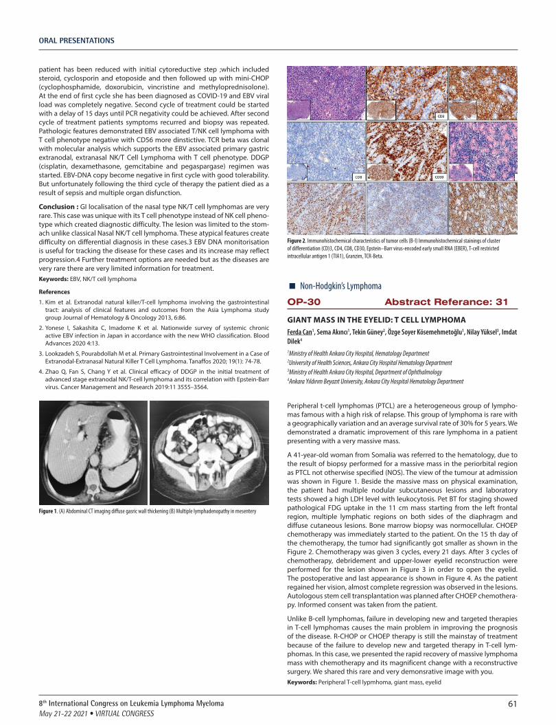
ORAL PRESENTATIONS
618th International Congress on Leukemia Lymphoma MyelomaMay 21-22 2021 • VIRTUAL CONGRESS
patient has been reduced with initial cytoreductive step ;which included steroid, cyclosporin and etoposide and then followed up with mini-CHOP (cyclophosphamide, doxorubicin, vincristine and methyloprednisolone). At the end of first cycle she has been diagnosed as COVID-19 and EBV viral load was completely negative. Second cycle of treatment could be started with a delay of 15 days until PCR negativity could be achieved. After second cycle of treatment patients symptoms recurred and biopsy was repeated. Pathologic features demonstrated EBV associated T/NK cell lymphoma with T cell phenotype negative with CD56 more dinstictive. TCR beta was clonal with molecular analysis which supports the EBV associated primary gastric extranodal, extranasal NK/T Cell Lymphoma with T cell phenotype. DDGP (cisplatin, dexamethasone, gemcitabine and pegaspargase) regimen was started. EBV-DNA copy become negative in first cycle with good tolerability. But unfortunately following the third cycle of therapy the patient died as a result of sepsis and multiple organ disfunction.
Conclusion : GI localisation of the nasal type NK/T cell lymphomas are very rare. This case was unique with its T cell phenotype instead of NK cell pheno-type which created diagnostic difficulty. The lesion was limited to the stom-ach unlike classical Nasal NK/T cell lymphoma. These atypical features create difficulty on differential diagnosis in these cases.3 EBV DNA monitorisation is useful for tracking the disease for these cases and its increase may reflect progression.4 Further treatment options are needed but as the diseases are very rare there are very limited information for treatment.Keywords: EBV, NK/T cell lymphoma
References
1. Kim et al. Extranodal natural killer/T-cell lymphoma involving the gastrointestinal tract: analysis of clinical features and outcomes from the Asia Lymphoma study group Journal of Hematology & Oncology 2013, 6:86.
2. Yonese I, Sakashita C, Imadome K et al. Nationwide survey of systemic chronic active EBV infection in Japan in accordance with the new WHO classification. Blood Advances 2020 4:13.
3. Lookzadeh S, Pourabdollah M et al. Primary Gastrointestinal Involvement in a Case of Extranodal-Extranasal Natural Killer T Cell Lymphoma. Tanaffos 2020; 19(1): 74-78.
4. Zhao Q, Fan S, Chang Y et al. Clinical efficacy of DDGP in the initial treatment of advanced stage extranodal NK/T-cell lymphoma and its correlation with Epstein-Barr virus. Cancer Management and Research 2019:11 3555–3564.
Figure 1. (A) Abdominal CT imaging diffuse gasric wall thickening (B) Multiple lymphadenopathy in mesentery
Figure 2. Immunohistochemical characteristics of tumor cells (B-I) Immunohistochemical stainings of cluster of differentiation (CD)3, CD4, CD8, CD30, Epstein–Barr virus-encoded early small RNA (EBER), T-cell restricted intracellular antigen 1 (TIA1), Granzim, TCR-Beta.
� Non-Hodgkin’s Lymphoma
OP-30 Abstract Referance: 31
GIANT MASS IN THE EYELID: T CELL LYMPHOMAFerda Can1, Sema Akıncı1, Tekin Güney2, Özge Soyer Kösemehmetoğlu1, Nilay Yüksel3, Imdat Dilek4
1Ministry of Health Ankara City Hospital, Hematology Department 2University of Health Sciences, Ankara City Hospital Hematology Department 3Ministry of Health Ankara City Hospital, Department of Ophthalmology 4Ankara Yıldırım Beyazıt University, Ankara City Hospital Hematology Department
Peripheral t-cell lymphomas (PTCL) are a heterogeneous group of lympho-mas famous with a high risk of relapse. This group of lymphoma is rare with a geographically variation and an average survival rate of 30% for 5 years. We demonstrated a dramatic improvement of this rare lymphoma in a patient presenting with a very massive mass.
A 41-year-old woman from Somalia was referred to the hematology, due to the result of biopsy performed for a massive mass in the periorbital region as PTCL not otherwise specified (NOS). The view of the tumour at admission was shown in Figure 1. Beside the massive mass on physical examination, the patient had multiple nodular subcutaneous lesions and laboratory tests showed a high LDH level with leukocytosis. Pet BT for staging showed pathological FDG uptake in the 11 cm mass starting from the left frontal region, multiple lymphatic regions on both sides of the diaphragm and diffuse cutaneous lesions. Bone marrow biopsy was normocellular. CHOEP chemotherapy was immediately started to the patient. On the 15 th day of the chemotherapy, the tumor had significantly got smaller as shown in the Figure 2. Chemotherapy was given 3 cycles, every 21 days. After 3 cycles of chemotherapy, debridement and upper-lower eyelid reconstruction were performed for the lesion shown in Figure 3 in order to open the eyelid. The postoperative and last appearance is shown in Figure 4. As the patient regained her vision, almost complete regression was observed in the lesions. Autologous stem cell transplantation was planned after CHOEP chemothera-py. Informed consent was taken from the patient.
Unlike B-cell lymphomas, failure in developing new and targeted therapies in T-cell lymphomas causes the main problem in improving the prognosis of the disease. R-CHOP or CHOEP therapy is still the mainstay of treatment because of the failure to develop new and targeted therapy in T-cell lym-phomas. In this case, we presented the rapid recovery of massive lymphoma mass with chemotherapy and its magnificent change with a reconstructive surgery. We shared this rare and very demonsrative image with you.Keywords: Peripheral T-cell lypmhoma, giant mass, eyelid

ORAL PRESENTATIONS
62 8th International Congress on Leukemia Lymphoma MyelomaMay 21-22 2021 • VIRTUAL CONGRESS
Figure 1. Patient’s view at admission
Figure 2. View of the tumour on 15th day of the first chemotherapy
Figure 3. View of the lession before operation
Figure 4. Final image of the patient

POSTER PRESENTATIONS
638th International Congress on Leukemia Lymphoma MyelomaMay 21-22 2021 • VIRTUAL CONGRESS
� Chronic Myeloid Leukemia
PP-01 Abstract Referance: 29
ANALYSIS OF DEMOGRAPHIC AND DISEASE CHARACTERISTICS OF PATIENTS WITH CHRONIC MYELOID LEUKEMIA: A SINGLE CENTRE ANALYSISMesut Tığlıoğlu1, Murat Albayrak1, Abdulkerim Yıldız2, Pınar Akyol1, Buğra Sağlam1, Fatma Yılmaz1, Merih Reis Aras1, Ümit Yavuz Malkan1, Senem Maral1
1Diskapi Yildirim Beyazit Training and Research Hospital, Department of Hematology, Ankara, Turkey 2Hitit University, Department of Hematology, Corum, Turkey
Background: Chronic myeloid leukemia (CML), is a clonal myeloproliferative disorder characterized by overproduction of cells of the myeloid series by the presence of the Philadelphia chromosome (Ph). With the development of tyrosine kinase inhibitors (TKIs), treatment options for CML have changed significantly.
Patients and methods: This retrospective study was conducted on patients diagnosed with BCR-ABL positive CML in the Hematology department of our tertiary care hospital between 2010 to 2020. Clinical and demographic characteristics of CML patients, as well as treatment efficacy, side effects, resistance to treatment, possible complications, and survival were analyzed.
Results: A total of 59 patients with the mean age of 55.59 ± 14.48 (years) were included. 30 patients (50.8%) were female, and median total follow-up period was 33.9 [0.2-172.0] months At the last follow-up, 54 (91.5%) patients were still alive, and 5 (8.5%) patients were nonsurvivors. The median sokal score was 1.0 [0.6-20.0]. All patients were given imatinib as the first line treatment. During the follow-up, imatinib was discontinued in 19 patients due to insufficient response, and in 10 patients for other reasons, and second generation TKIs were started. 2 patients were included in a trial of imatinib cessation at another center. As second generation TKIs, dasatinib was pre-ferred in 14 (46.7%) patients and nilotinib was preferred in 15 patients (50%). Pleural effusion was observed in 4 patients who received dasatinib and 3 of them had to discontinue treatment due to side effects, and no significant side effects were observed in patients using nilotinib. T315I mutation was detected in a patient who was unresponsive to imatinib, dasatinib and nilo-tinib and as a result of the mutation analysis bosutinib treatment was given. Blastic transformation was detected in 2 patients during follow-up.
Conclusion: The result of the current study demonstrated that, treatment options, response rates and side effects were all comparable with results of other real world studies. Larger patient based studies are needed to cover the course of the disease and to better mamage the patients.Keywords: CML, management, treatment
References
1. Hochhaus, A., et al., Dasatinib induces notable hematologic and cytogenetic respons-es in chronic-phase chronic myeloid leukemia after failure of imatinib therapy. Blood, 2007. 109(6): p. 2303-2309.
2. Faderl, S., et al., The biology of chronic myeloid leukemia. New England Journal of Medicine, 1999. 341(3): p. 164-172.
3. Jabbour, E. and H. Kantarjian, Chronic myeloid leukemia: 2018 update on diagnosis, therapy and monitoring. American journal of hematology, 2018. 93(3): p. 442-459.
4. Hehlmann, R., Innovation in hematology. Perspectives: CML 2016. haematologica, 2016. 101(6): p. 657.
5. Goldman, J.M. and J.V. Melo, Chronic myeloid leukemia—advances in biology and new approaches to treatment. New England Journal of Medicine, 2003. 349(15): p. 1451-1464.
6. Silver, R.T., et al., An Evidence-Based Analysis of the Effect of Busulfan, Hydroxyurea, Interferon, and Allogeneic Bone Marrow Transplantation in Treating the Chronic Phase of Chronic Myeloid Leukemia: Developed for the American Society of Hematology: Presented in part at the Education Session of the American Society of Hematology, December 5, 1998, Miami Beach, FL. Blood, The Journal of the American Society of Hematology, 1999. 94(5): p. 1517-1536.
7. Martins, J.R.B., et al., Comparison of microRNA Expression Profile in Chronic Myeloid Leukemia Patients Newly Diagnosed and Treated by Allogeneic Hematopoietic Stem Cell Transplantation. Frontiers in oncology, 2020. 10: p. 1544.
� Chronic Lymphocytic Leukemia
PP-02 Abstract Referance: 7
DIAGNOSIS AND TREATMENT OPTIONS IN LOW-RISK AND PROGRESSIVE CHRONIC LYMPHOCYTIC LEUKEMIALarisa Musteata1--2, Vasile Musteata1--2, Victor Munteanu2
1State University of Medicine and Pharmacy “n. Testemitanu” 2Institute of Oncology
Background: The increased rates of morbidity, prevalence and disability, common relapses and complications, negative socio-economic impact1,2 point out chronic lymphocytic leukemia (CLL) as an actual problem of public health and oncology.
Materials and Methods: We performed a clinico-analytical, descriptive and cohort study. The study enrolled 82 patients with CLL, who were treated and supervised at the Institute of Oncology between the years 2011 – 2020. The age range was 45-87 years (average – 66.3 years old). Females were 34 (41.4%), males – 48 (58.6%). The diagnosis was asserted by the immuno-histochemical, cytological and immunophenotyping examinations of the biopsied lymph nodes, bone marrow and blood3,4. Staging was realized according to Binet Classification. The diagnosis of CLL was established in cases of the increased lymphocyte count more than 5 x 1000 [MICRO]/L in the peripheral blood, and more than 30% in the bone marrow aspirates. Stage A patients didn’t require chemotherapy until progression. Single-agent chemotherapy with chlorambucil and rituximab were indicated in cases with stage B. Combination chemotherapy (COP, CHOP, R-COP) and radiotherapy were administered in patients, who progressed into stage C.
Results: CLL developed in 67 (81.7%) patients during the first 6 months from the onset. The patients of 60-79 years old formed the predominant age category. 17p deletion remained undetected in stage A and B. Stage A was revealed in 53 (64.6%) cases, stage B – in 29 (35.4%). CLL progressed into stage B in 22 (41.5%) patients, and into stage C in 10 (34.5%). Of 53 patients with stage A, 21 (39.6%) didn’t manifest any clinical signs at diagnosis. In stage B peripheral lymph nodes were enlarged in 27 (93.1%) cases, spleno-megaly occurred in 22 (78.6%), hepatomegaly – in 13 (46.4%). In stage B, 10 (34.5%) patients developed autoimmune hemolytic anemia, 13 (44.8%) – metaplastic anemia, and 5 (17.2%) – autoimmune thrombocytopenia. Infections were registered in 11 (20.8%) cases with stage A, and in 10 (34.5%) with stage B. Leukocyte count varied between 12.8-525x1000 [MICRO]/L, (average – 93.7 x 1000 [MICRO]/L), and lymphocytosis – between 52-97% (average – 76.2%). The bone marrow aspiration revealed the increased lymphocyte count between 33-91%. Chemotherapy with chlorambucil was administered in 22 (41.5%) patients with stage A and comorbidities, who progressed into stage B. Only partial responses were obtained. Rituximab was adjoined to chemotherapy in CD20 positive cases with stage B, and contributed to 57.1% of complete responses. Radiotherapy did not eliminate completely the tumor sites, which re-grew within 1.5 months after the last irradiation procedure. The 3- and 5-year overall survival in the totality of patients reached 91.2% and 77.4%, being higher after the rituximab-con-taining regimens and lower in stage B (84.8% and 55.4%, respectively). Complete clinical and hematologic responses were obtained in 2 of 4 refrac-tory stage B and C patients. 66 (80.5%) patients have been followed up nowadays, with the ECOG-WHO score of 0-2.
Conclusions: CLL commonly affected males, and was diagnosed in stage A. The overall survival correlated with CLL stage, and turned out to be lower in stage B and higher in the cases treated with combined regimens.Keywords: chronic lymphocytic leukemia, immunophenotyping, chemotherapy, survival
References
1. Halek M, Shanafelt TD, Eichhorst B. Chronic Lymphocytic Leukemia. The Lancet. 2018; 391 (10129): 1524-1537.
2. Tresckow J, Eichhorst B, Bahlo J. The Treatment of Chronic Lymphocytic Leukemia. Deutsches Arzteblatt International. 2019; 116(4): 41-46.
POSTER PRESENTATIONS

POSTER PRESENTATIONS
64 8th International Congress on Leukemia Lymphoma MyelomaMay 21-22 2021 • VIRTUAL CONGRESS
3. Kersting S, Levin MD, Mous R. Clinical Practice Guidelines for Diagnosis and Treatment of Chronic Lymphocytic Leukemia in the Netherlands. Clin. Lymph. Myel. Leuk. 2018; 18(1): 52-57.
4. Jaksic B, Pejsa V, Ostojic-Kolonic S, Kardum-Skelin I et. al. Guidelines for Diagnosis and Treatment of Chronic Lymphocytic Leukemia. KROHEM B-CLL 2017. Acta Clin. Croat. 2018; 57: 190-215.
� Acute Lymphoblastic Leukemia
PP-03 Abstract Referance: 49
ACUTE LYMPHOBLASTIC LEUKEMIA IN A CHILD WITH DOWN SYNDROME AND STURGE WEBER SYNDROME: FIRST IN LITERATUREZeynep Sena Akgiray1, Enes Candir1, Nihan Bayram1, Yontem Yaman1, Murat Elli1, Sema Anak1
1Istanbul Medipol University
Background: Sturge Weber syndrome (SWS) is a rare vascular disorder characterized by capillary hemangiomas (port-wine stains) on the face. Leptomeningeal malformations, seizures, motor mental retardation and glaucoma may accompany. There is no genetic inheritance.
Down syndrome (trisomy 21) is the most common chromosomal abnormali-ty in newborns. Patients with Down syndrome are at increased risk for some hematologic malignancies.
We present the first case of ALL in a 6.5-year-old boy with Down syndrome and Sturge Weber syndrome in the literature.
Case: A 6.5-year-old boy with Struge Weber and Down syndrome presented with complaints of fever for 5 days, swelling and pain in the left knee. Cervical lymphadenopathy and splenomegaly were present. Laboratory investiga-tions revealed leukopenia, trombocythopenia and anemia. Peripheral blood smear was suggestive of acute leukemia with blasts of L1 morphology. The bone marrow was diffusely infiltrated with blasts that accounted for 100% of bone marrow cellularity. MRI of the left knee showed leukemic infiltra-tion in all bone structures of the knee joint. There was no CSF involvement. Chemotherapy was immidiately started according to ALL IC BFM 2009 pro-tocol. The bone marrow still remains in remission.
Discussion and conclusion: The risk of hematopoietic malignancies such as acute myeloproliferative diseases, acute myeloid leukemia and acute lymphoblastic leukemia (ALL) is increased in children with Down syndrome. The most common genetic abnormality in DS -associated ALL cases is over-expression of the CRFL2 gene (62%) and most of these are associated with JAK -2 mutations (50%). No pathological/possible pathological mutation was detected in our patient. Bone marrow was negative for inv (16), t (15,17), t (8.21), t (9.22). The risk of chemotherapy-related toxicity (especially with regimens that include methotrexate therapy) is significantly higher in indi-viduals with Down syndrome. We administered low doses of methotrexate and did not encounter any serious or life-threatening toxicities.In literature, there is no clearly defined association between Sturge Weber syndrome and ALL or Down syndrome. Our case is the first case in the literature where the diagnoses of Down syndrome, Sturge Weber Syndrome and ALL are seen together.Keywords: Sturge-Weber Syndrome, Down Syndrome, Acute Lymphoblastic Leukemia
� Non-Hodgkin’s Lymphoma
PP-04 Abstract Referance: 34
HIGH GRADE NON-HODGKIN LYMPHOMA PATIENTS WHO WERE PREVIOUSLY TREATED FOR HODGKIN LYMPHOMAFatma Keklik Karadağ1, Nur Soyer1, Fahri Şahin1, Filiz Vural1, Mahmut Töbü1, Güray Saydam1
1Ege University Hospital, Department of Hematology
Introduction: Lymphoma is a malignancy that is the most seen including our immune system. It is divided into two cathegories: Hodgkin (HL) and Non-Hodgkin Lymphoma (NHL). High Grade B-cell Lymphomas (HGBL) have been defined as a new separate entity in 2016 revised WHO classification of lymphoid neoplasms. The previously well-known Double- and Triple-Hit Lymphomas (DHL/THL) are included in this umbrella category under the name of HGBL with MYC and BCL2 and/or BCL6 rearrangements. However the incidence for NHL after primary HL is not exactly known, it was reported as 15 and 6% in some studies and HGBL after therapy of HL is an extremely rare condition. In this report, we aimed to present two rare cases with HGBL after treatment of HL.
Case- 1: A 38 year old man presented weakness, weight loss and fever in 2006. In his family history, his all four siblings were diagnosed HL, two of them were died due to HL. He diagnosed mixt-celluler HL with cervical lymph node biopsy. His ann-arbor stage was 3B and he was treated with doxorubicin, bleomycin, vinblastine and dacarbazine (ABVD chemothera-py regimen) for 6 cycles. He was in complete remission during four years. Because of mediastineal progression was detected on his control PET-CT scan, he was treated autolog stem cell transplantation in 2010 following 2 cycles of ICE regimen. In post-transplant period, he was also given radi-ation therapy on mediastinal side. He was relapsed from inguinal lymph node in 2014. Brentuximab vedotin (BV) and Bendamustine (B) combined chemotheraphy was given to the patient for second relaps for 6 cycles and continued BV alone. On the twenty night cycles of BV, hypermetabolic some regions and hyperdens nodulary formations on jejunal loops was detected on his PET/CT. He was diagnosed with diffuse large B cell lymphoma from jejunal biopsy. He was treated with 2 cycles of R-ICE regimen. The last PET-CT scan and bone marrow biopsy were evaluated in favor of stable disease. The patient was offered for allogeneic stem cell transplantation but he refused to further treatment.
Case- 2: A 51 year-old male patient presented with abdominal pain in 2020. On his history, he was diagnosed with Mixed cellularity classical Hodgkin lymphoma (2004) his ann arbor stage was 3. he was treated with doxorubi-cin, bleomycin, vinblastine and dacarbazine (ABVD chemotherapy regimen). After 6 cycles of ABVD, he was followed in complete remission during 16 years. He had no fewer, diarrhea or constipation. Organomegaly was not detected. A 2x2 cm mass palpated right to the umblicus. He had multiple lymphadenopathie in right paratracheal region and mass lesion in the form of aneurysmatic dilatation in the lower right quadrant with asymmetric wall thickening on the ileal ans in CT scan. Patient underwent excisional biopsy from that mass and detected diffuse infiltration of CD10, CD20 and CD38 positive neoplastic medium lymphoid cells with an high mitotic index. Both of c-myc and Bcl-6 were positive. The Ki67 proliferation index was %98. He was treated with autologous stem cell transplantation after following 2 cycles of Hyper-Cvad regimen.
Discussion: Lymphomas contain so many different subgroups in itselfs and analyze which of them is the main diagnosis is supported by pathologic, immunochemical and genetic methods. Transforming of these diseases seems to be releated with EBV reactivation in immuncompromised patient on a period of T-cell suppression by disrupting interaction between the T and B lymphocytes or otoactivation of B lymphocytes. Both of chemothera-py agents and radiotherapy may trigger the variations on this interactions in a way that we dont know yet.Keywords: Hodgkin lymphoma, non-Hodgkin lymphoma, incidence, treatment, cancer

POSTER PRESENTATIONS
658th International Congress on Leukemia Lymphoma MyelomaMay 21-22 2021 • VIRTUAL CONGRESS
References
1. Rueffer U, et al. Non-Hodgkin’s lymphoma after primary Hodgkin’s disease in the German Hodgkin’s Lymphoma Study Group: incidence, treatment, and prognosis. J Clin Oncol. 2001 Apr 1;19(7):2026-32. doi: 10.1200/JCO.2001.19.7.2026. PMID: 11310450.
2. Eichenauer DA, et al. Non-Hodgkin lymphoma after treatment for classical Hodgkin lymphoma: a report from the German Hodgkin Study Group. Br J Haematol. 2021 Jan 24. doi: 10.1111/bjh.17327. Epub ahead of print. PMID: 33486762.
3. Schaapveld M, et al. Second Cancer Risk Up to 40 Years after Treatment for Hodgkin’s Lymphoma. N Engl J Med. 2015; 373: 2499– 511.
� Acute Lymphoblastic Leukemia
PP-05 Abstract Referance: 41
SEVERE HYPERTRIGLICERIDEMİA DUE TO L-ASPARAGINASE IN AN ADULT ACUTE LYMPHOBLASTIC LEUKEMIA PATIENTFerda Can1, Sema Akıncı1, Tekin Güney2, Ozge Soyer Kösemehmetoğlu1, Imdat Dilek3
1Ministry of Health Ankara City Hospital Hematoloji Department 2University of Science Ankara City Hospital Hematoloji Department 3Ankara Yıldırım Beyazıt University Ankara City Hospital Hematoloji Department
Asparaginase is one of the main drugs in the treatment of acute lympho-blastic leukemia (ALL). Side effects such as hypersensitivity, coagulation disorder, pancreatitis, hyperlipidemia and hypertriglyceridemia can be seen with asparaginase. In our case, a patient with the diagnosis of ALL who required plasmapheresis due to severe hypertriglyceridemia caused by L-asparaginase is presented.
A 39-year-old male patient was diagnosed with ALL in December 2020 and Linker remission induction chemotherapy was started. Four doses of L-asparaginase were administered as part of remission induction therapy. No complications were observed. CALGB 10403 chemotherapy was initiat-ed to the patient as remission could not be achieved in the bone marrow aspiration performed after induction. After 3 doses of L-asparaginase, the triglyceride level was 3799 mg / dl. The image of the patient’s blood sample after 30 minutes is presented in Figure 1. An appropriate diet and fenofibrate were initiated to the patient. Plasmapheresis was started when the triglycer-ide level increased to 5133 mg/dl during the follow-up. The procedure was continued for six days. Triglyceride level decreased to 483 mg / dl. Serum triglyceride levels before, during and after plasmapheresis has shown in the Figure 2. The patient had no clinical symptoms in terms of pancreatitis, amy-lase lipase levels did not exceed 1.5 times the upper limit, and the pancreas was normal on tomography. In the clinical follow-up, the patient died of sepsis in the cytopenic period.
Patients who had to go under plasmapheresis procedure due to hyper-triglyceridemia, which is one of the known side effects of L-asparaginase, presented in the literature as cases, and we wanted to present our patient. We think that it is necessary to be careful in terms of hypertriglyceridemia with clinical-laboratory findings before and during L-asparaginase treatment when necessary.Keywords: Hypertriglyceridemia; plasmapheresis, asparaginase
References
1. Zawitkowska, Joanna, et al. Severe drug-induced hypertriglyceridemia treated with plasmapheresis in children with acute lymphoblastic leukemia. Transfusion and Apheresis Science 58.5 (2019): 634-637.
2. Hinson, Ashley, Dorothee Newbern, and Corinne M. Linardic. Asparaginase-induced hypertriglyceridemia presenting as pseudohyponatremia during leukemia treat-ment. Case reports in pediatrics 2014 (2014).
3. Aljabri K et al. Hypertriglyceridemia and Hypercholesterolemia Induced by L-Asparaginase. Annals of Saudi medicine. 23 (2003) 173-4.
Figure 1. Patient’s blood sample image
Figure 2. Serum triglyceride levels before, during and after plasmapheresis
� Stem Cell Transplantation
PP-06 Abstract Referance: 9
MARKED AND ROBUST IMPROVEMENT OF REFRACTORY CHRONIC GRAFT VERSUS HOST DISEASE OF THE LUNG WITH RUXOLITINIB: CASE REPORTFehmi Hindilerden1, Yüksel Aslı Öztürkmen1, Emine Gültürk1, Mutlu Arat2
1Sağlık Bilimleri Üniversitesi Bakırköy Dr. Sadi Konuk Eğitim Ve Araştırma Hastanesi Hematoloji Kliniği 2İstanbul Florence Nightingale Hastanesi Hematopoietik Kök Hücre Nakil Ünitesi
Introduction: Graft-versus-host disease (GVHD) is the main cause of mor-bidity and mortality after allogeneic hematopoietic stem cell transplantation (aHSCT). First-line systemic treatment consists of high doses of cortico-steroids. Unfortunately, more than 50% of the patients will not respond adequately, thus requiring second-line treatment. Due to the key role of JAK-STAT pathways on T cells activation, JAK inhibitors may reduce GVHD by inhibiting donor T-cell expansion and inflammatory cytokine production, regulatory T-cell function and viability.

POSTER PRESENTATIONS
66 8th International Congress on Leukemia Lymphoma MyelomaMay 21-22 2021 • VIRTUAL CONGRESS
Case: 60 years old male with prior history of diabetes mellitus was diagnosed with acute myeloid leukemia with complex karyotype and subsequently underwent aHSCT from his matched female donor after achieving 1st hema-tological remission. Methotrexate and cyclosporine A were used as GVHD prophylaxis. No acute GVHD developed at follow up and cyclosporine A taper was initiated at day 75 after aHSCT. By day 150, he developed moderate chron-ic GVHD (skin score 2, lung score 1, mouth score 1) for which methylpred-nisolone 0.5 mg/kg/day along with inhaler corticosteroids and B2mimetics were initiated. By day 210, methylprednisolone was stopped but the patient required insulin for uncontrolled hyperglycemia. By day 300, he presented with dry cough and bilateral lower extremity edema. Thorax CT showed bibasiler reticuler abnormalities consistent with lung cGVHD. Nephrotic range proteinuria (6.6 gr/day) was detected. Renal biopsy showed membranous nephropathy. Screening for potential causes of membranous nephropathy was excluded and membranous nephropathy was attributed to cGVHD. Diagnosed with severe cGVHD, 1 mg/kg/day MP, rituximab 375 mg/m2/week for 4 doses and cyclosporine were initiated. MP was needed to be tapered due to uncontrolled hyperglycemia. By the 6th week of treatment, there was moderate decrease in proteinuria (3,5 gr/day) but the patient complained of progressive dyspnea requiring oxygen treatment. Thorax CT showed gener-alised reticulonodular and ground glass opacities and honeycombing,which were findings compatible with severe lung cGVHD (Figure 1). Bronchoalveolar lavage analysis identified no infectious agent. Diagnosed with refractory severe cGVHD of the lung, ruxolitinib 2x10 mg/day was initiated. By the 4th week of ruxolitinib, the patient reported marked improvement in his dyspnea and had no need for oxygen support. By the 3rd month of ruxolitinib, radio-logical findings on thorax CT showed marked regression (Figure 2). Moreover, proteinuria remained less than 1 gr/day under ruxolitinib.
Conclusion: Ruxolitinib in the real life setting has been shown as an effective and safe treatment option for GVHD patients, with overall response rate of 57.1% for refractory cGVHD among heavily pretreated patients. Ruxolitinib also gives an oppurtunity to spare doses of steroids as well. In lung cGVHD, organ response for lung involvement was reported to be 10%. The dramatic and robust response achieved in our multiple drug refractory patient needs to be further confirmed by other studies. Keywords: Ruxolitinib, lung chronic graft versus host disease
Figure 1.
Figure 2.
� Chronic Myeloid Leukemia
PP-08 Abstract Referance: 4
TARGETED THERAPIES IN CHRONIC LEUKEMIASVasile Musteata1--2
1State University of Medicine and Pharmacy “n. Testemitanu” 2Institute of Oncology From Moldova
Background: Chronic leukemias are the most frequent hematological malig-nancies within the structure of morbidity by blood tumors with primary bone marrow (BM) involvement, being characterized in the advanced stages by a recurrent evolution, unfavorable prognosis and negative socio-eco-nomic impact1,2,3. Chronic leukemias may be perceived as an actual issues of public health and hematologic oncology due to the increased incidence, disability rates and disease burden1,3,4.
Methods: This clinico-analytical, descriptive study enrolled 42 patients with chronic myeloid leukemia (CML) and 85 patients with chronic lymphocytic leukemia (CLL), who were managed at the Institute of Oncology during the period of 2009–2020. CML cases were diagnosed in chronic and accelerated phases by the molecular tests of the BM and peripheral blood (PB). The quan-titative RT-PCR was accomplished with the aim to determine the expression of the BCR-ABL p210 and p190 transcripts2,5. Five transcription products (b2a2, b3a2, b2a3, b3a3 si e1a2) were analyzed by the usage of the quanti-tative PCR test. CLL was diagnosed by cytological, immunophenotyping and immunohistochemical tests of the BM, PB and biopsied lymph nodes3,6. The CLL stage was established according to Binet Classification. All CLL cases were CD20 positive.
Results: CML was diagnosed mostly in a workable population – 27 (64.3%) patients aged below 60 years. CLL occurred in 57 (67.1%) patients with the age over 60 years. CML patients received the first- or second-line kinase-targeting therapy with imatinib or nilotinib. The complete hemato-logic response was achieved under the single-agent chemotherapy with TKIs in all cases. The complete cytogenetic response was obtained within 12–18 months after the TKIs therapy in 37.4% of patients. The complete molecular response emerged in 26.8% of cases. Under the TKIs treatment, the 1- and 5-year overall survival (OS) was 99.2 and 81.7%, being superior to the 5-year OS (39.2%) in patients managed by the conventional chemotherapy (CChT) with antimetabolites and alkylating antineoplastic agents. Single-agent che-motherapy with chlorambucil was administered in 23 (42.6%) CLL patients with comorbidities, who evolved from stage A into stage B, and in 28 patients with stage B at diagnosis. Twelve stage C cases were managed with ibrutinib, CChT alone (COP, CHOP) or in combination with rituximab (R-COP, R-CHOP, R-CVClP). CChT resulted only into clinico-hematological improve-ment. The OS of CLL patients at one year was 98.2%, at 5 years – 77.3%, and proved to be superior in stage A (100% and 95.7%, respectively) and after the rituximab-containing combined treatment. Three of 5 refractory stage B and C patients responded to the treatment with ibrutinib, without serious side effects.
Conclusions: Targeted antineoplastic therapy proved to be efficient in the newly diagnosed and relapsed cases regardless of the type of chronic leu-kemias and patient’s age. CLL relapse after rituximab-containing regimens may be considered as the indication for the kinase-targeting therapy with ibrutinib.Keywords: chronic leukemias, targeted therapy, overall survival
References
1. Hughes TP, Ross DM, Melo JV. Handbook of chronic myeloid leukemia. Springer International Publishing Switzerland, 2016: 1-66.
2. Jabbour E, Kantarjian H. Chronic myeloid leukemia: 2018 update on diagnosis, thera-py and monitoring. Am. J. Hematol. 2018; 93: 442-459.
3. Halek M, Shanafelt TD, Eichhorst B. Chronic Lymphocytic Leukemia. The Lancet. 2018; 391: 1524-1537.
4. Fitzmaurice C, Akinyemiju TF, Al Lami FH et.al. Global, Regional, and National Cancer Incidence, Mortality, Years of Life Lost, Years Lived With Disability, and Disability-Adjusted Life-Years for 29 Cancer Groups, 1990 to 2016. A Systematic Analysis for the Global Burden of Disease Study. JAMA Oncol. 2018; 4(11):1553-1568.

POSTER PRESENTATIONS
678th International Congress on Leukemia Lymphoma MyelomaMay 21-22 2021 • VIRTUAL CONGRESS
5. Dorfman LE, Floriani MA, Oliveira TM, Cunegatto B, Rosa RF, Zen PR. The role of cytogenetics and molecular biology in the diagnosis, treatment and monitoring of patients with chronic myeloid leukemia J. Bras. Patol. Med. Lab. 2018; 54 (2): 83-91.
6. Hallek M, Cheson BD, Catovsky D, Caligaris-Cappio F et. al. Guidelines for Diagnosis, Indications for Treatment, Response Assessment, and Supportive Management of Chronic Lymphocytic Leukemia. Blood. 2018; 131: 2745-2760.
� Multiple Myeloma
PP-09 Abstract Referance: 35
SUCCESSFULL AUTOLOGOUS STEM CELL TRANSPLANTAION FOR POEMS SYNDROME: A CASE REPORTFatma Keklik Karadağ1, Nur Soyer1, Fahri Şahin1, Güray Saydam1
1Ege University Hospital, Department of Hematology
Introductıon: POEMS syndrome (Polyneuropathy, Organomegaly, Endocrinopathy, Monoclonal protein, Skin changes) is a rare condition char-acterized by the presence of a monoclonal plasma cell disorder and periph-eral neuropathy, along with other systemic symptoms. The International Myeloma Working Group (IMGW) criteria for diagnosis for POEMS requires the presence of at least three major criteria (ie, polyneuropathy plus monoclonal plasma cell disorder plus any one of the following three: osteo-sclerotic bone lesion, Castleman disease, or elevated serum or plasma vascular endothelial growth factor (VEGF) levels), along with the presence of at least one of the six minor criteria (Organomegaly, Extravascular vol-ume overload,Endocrinopathy ,Skin changes ,Papiledema ,Trombocytosis/polycythemia). The absence of either osteosclerotic myeloma or Castleman disease should make the diagnosis of POEMS syndrome suspect. There is no standard treatment for POEMS syndrome. Radiation therapy is appropriate option for those with limited disease (one to three isolated bone lesions), and chemotherapy similar to multiple myeloma for those with widespread bone lesions. Autologous stem cell transplantation (ASCT) with Melphalan is an option for patients with rapidly progressive neuropathy and for younger patients with widespread osteosclerotic lesions.
Case report: 62 years old man who has a history of Celiac disease eval-uated for 3 months of mononeuropathy multiplex ,1 month of diabetes insipidus and 1 month of vasculitic rash .on physical examination, there was clearly weakness of lower limbs. He has mononeuropathy multiplex in Electromyelography (EMG). Multiple myeloma was reported as IgG kappa monoclonal, CD 138 positive plasma cells reaching up to 40% in his bone marrow biopsy in Agust 2018. Also Diabetes insipitus was detected. The patient with Polyneuropathy, monoclonal plasma cell disorder, endocrinop-athy, skin changes was diagnosed POEMS. Bortezomib, Cyclophosphamid, Dexamethason therapy (VCD regimen) was started in September 2018. After 5 cycles of VCD, the patient was mobilized with G CSF. ASCT with melphalan as a conditoining regimen was performed and he was engrafted succesfully on the 20th day of the transplant. He was in complete remission for 2 years after ASCT. Neuropathy findings began to improve after ASCT and he has no complain about neuropathy any more.
Discussion: POEMS syndrome is a rare, chronic, multisystemic, paraneoplas-tic syndrome. Although the pathophysiology of POEMS syndrome is not fully known, the source of its symptoms is thought to be excessive VEGF produc-tion by neoplastic cells. VEGF may also use the increase for diagnostic pur-poses. Also similar to chronic inflammatory demyelinating polyneuropathy (CIDP) with its clinical and laboratory features. Patients diagnosed with CIDP should be examined in terms of possible gammopathy and M protein and bone lesions should be investigated. As in our patient, widespread osteo-sclerotic lesions and bone marrow involvement on bone marrow aspirate and biopsy has been treated ASCT after Bortezomib-based therapy .Keywords: POEMS, autologous stem cell transplantation, bortezomib, plasma cell
� Other
PP-10 Abstract Referance: 24
A VERY RARE CAUSE OF BICYTOPENIA; THE USE OF VITEX ACNU CASTUSMerih Reis Aras1, Murat Albayrak1, Fatma Yılmaz1, Senem Maral1, Pınar Akyol1, Hacer Berna Afacan Öztürk1
1University of Health Sciences Ankara Diskapi Yildirim Beyazit Training and Research Hospital, Hematology Department, Ankara, Türkiye
Introduction: Vitex agnus castus L. (VAC) is a bush type plant found in the Mediterranean parts of Europe and Central Asia (1). Casticin is a polymeth-ylflavonide derived mainly from the Vitex species of the Verbenaceae family. This substance isolated from VAC leaves has potent anti-inflammatory and lipoxygenase inhibitory activity. The molecular mechanism of its anti-in-flammatory action is the blockade of the NF-kβ, Akt and mitogen-activated protein kinase signaling pathway (2).
Case presentation: A 27-year-old female with no known chronic disease presented hospital with the complaints of weakness.
The laboratory test reulsts revealed bicytopenia. The patient was hospital-ized for further examination. There were no B symptoms. Drug and herbal substance use was questioned and the patient reported having used BNO 1095 (generic name Agnucaston), which is a preparation of the dry extract of Fructus acnu casti for accessory breast, for 3 months and most recently 1 month ago
Anemia parameters were within normal limits. A peripheral blood smear was taken and evaluated. Anisocytosis was determined in the red blood cell morphology, leukocyte count compatible with complete blood count, and no atypical cells were seen. Viral hepatitis markers, TORCH panel, brucella, ANA, Anti-Ds DNA and SARS-COV2 PCR test were performed, and all were negative.
In the investigation of the etiology, ultrasound imaging showed a large number of cervical, axillary and inguinal lymph nodes. Fine needle aspiration biopsy was performed on the right cervical lymph node, and it was reported as benign cytology.
Bone marrow aspiration and biopsy were performed. Flow cytometry was studied from the aspiration material. In the bone marrow aspiration eval-uation; “ Erythroid serial rate was increased (40%) and approximately 30% small, mature, narrow cytoplasmed atypical lymphoid cell infiltration was observed”. The flow cytometry result was reported as: “Significant lymphocy-tosis and granulocytopenia, CD4 / CD8 ratio impaired in increased number of T lymphocytes [ratio 0.45, (reference range 1.3-3.6)]”.
The bone marrow biopsy pathology was reported as “normocellular bone marrow showing increased CD3 (+) mature T cells, an increase in interstitial pattern containing interstitial multiple lymphoid aggregates” Considering possible lymphoproliferative or immunological processes, treatment iniated of 60 mg / day methylprednisolone .
The result of the clonality studied from the bone marrow biopsy were report-ed as; “Molecular findings supporting clonal T cell increase among poly-clonal T cells are considered features supporting autoimmune neutropenia. Immunophenotypic and molecular findings observed in the bone marrow primarily suggest that the increase in clonal cytotoxic T cells on a polyclonal basis may be associated with non-neoplastic autoimmune processes”.
As a result of bone marrow clonality, the methylprednisolone dose was reduced and then discontinued. The patient remains under follow-up and the hemogram parameters are completely normal.
Discussion: Over the last 50 years VAC has been used especially for the treatment of premenstrual syndrome and prevention of premenstrual mas-talgia (3).
The assessment of causality between fructus agnu casti extract and bicy-topenia using the Naranjo nomogram questionnaire yielded a score of 5,

POSTER PRESENTATIONS
68 8th International Congress on Leukemia Lymphoma MyelomaMay 21-22 2021 • VIRTUAL CONGRESS
indicating that the side effect probably caused which means side-effect is probably caused by the fructus agnu casti extract (Table 1) (4).
The aim of presenting this case was to emphasize that patients who are investigated for cytopenia should be questioned about the use of herbal substances in addition to medications and the possible side-effects of herbal products should be considered. This case also provides an example of the effectiveness of steroid therapy in the treatment of cytopenias triggered by autoimmunity.Keywords: Vitex Agnu Castus, Bicytopenia, Casticin
References:
1. Claudia D, Coon T, Max P. Vitex agnus castus: A Systematic Review of Adverse Events. Drug Safety. 2005;28:319-32..
2. Chan EWC, Wong SK, Chan HT. Casticin from Vitex species: a short review on its anticancer and anti-inflammatory properties. Journal of integrative medicine. 2018;16(3):147-52.
3. Seidlova-Wuttke D, Wuttke W. The premenstrual syndrome, premenstrual mastodyn-ia, fibrocystic mastopathy and infertility have often common roots: effects of extracts of chasteberry (Vitex agnus castus) as a solution. Clinical Phytoscience. 2017;3(1):6.
4. Naranjo CA, Busto U, Sellers EM, Sandor P, Ruiz I, Roberts E, et al. A method for estimating the probability of adverse drug reactions. Clinical Pharmacology & Therapeutics. 1981;30(2):239-45.
Table 1. Naranjo Advers drug reaction probability scale—items and score
To assess the adverse drug reaction, please answer the following questionnaire and give the pertinent score Yes No
Don’t know Score
Are there previous conclusion reports on this reaction? +1 0 0 0
Did the adverse event appear after the suspected drug was administered?
+2 -1 0 +2
Did the adverse reaction improve when the drug was discontinued, or a specific antagonist was administered?
+1 0 0 +1
Did the adverse reaction reappear when the drug was readministered?
+2 -1 0 0
Are there alternative causes (other than the drug) that could on their own have caused the reaction?
-1 +2 0 +2
Did the reaction reappear when a placebo was given? -1 +1 0 0
Was the drug detected in the blood (or other fluids) in concentrations known to be toxic?
+1 0 0 0
Was the reaction more severe when the dose was increased, or less severe when the dose was decreased?
+1 0 0 0
Did the patient have a similar reaction to the same or similar drug in any previous exposure?
+1 0 0 0
Was the adverse event confirmed by any objective evidence? +1 0 0 0
� Multiple Myeloma
PP-12 Abstract Referance: 45
SCLEROMYXEDEMA SKIN LESIONS AND ERECTILE DYSFUNCTION SUCCESSFULLY TREATED WITH BORTEZOMIBE DEXAMETHASONE REGIMEN:Murat Ozbalak1, Simge Erdem1, Ipek Yonal Hindilerden1, Sevgi Kalayoglu Besisik1
1Istanbul University Istanbul Medical Faculty, Department of Hematology
Scleromyxedema is a primary cutaneous mucinuous presenting with gen-eralized sclerodermoid cutaneous lesions, associated with monoclonal gammopathy. Patients develop many waxy firm papules and plaques. Histological examination shows mucin deposition, increased fibroblasts and fibrosis. Cardioascular, gastrointestinal, pulmonary, musculoskeletal, and renal or nervous systems may be involved [1].
A 44-year-old male presented to the dermatology clinic with generalized skin stiffness, erythema, decreased range of motion of the joints, dysphagia and weight loss. He also had popular lesions on the face and the hands. The skin biopsy revealed orthokeratosis in the epidermis, milg pigmentation of the basal layer, increased collagen buckets in mid- and upper dermis, increased number of fibroblasts accompanied with asidic mucin formation. The serum immunofixation electrophoresis reported Ig G Lambda monoclo-nal gammopathy. With the diagnosis of scleromyxedema, PUVA treatment was initiated but the lesions stayed stable. He then was referred to our hematology department.
The bone marrow biopsy revealed mildly increased plasma cells, with a ratio of 10%. The gastroscopic and manometric evaluation demonstrated decreased esophageal motility. The cardiac functions were within normal limits in both echocardiography and cardiac MRI. He had mild axonal type polyneuropathy affecting sensory nerves of the lower extremities. Oral thalidomide 100mg/day was initiated and was increased to 250 mg/day. By the first year of thalidomide treatment, the skin lesions resolved com-pletely. However, as the numbness of the legs progressed, the thalidomide dosage was adjusted to 150 mg/day. Although the skin lesions responded completely to thalidomide treatment, erectile dysfunction developed and thalidomide was stopped at the 20th month of treatment. The erectile dys-function was not improved with cessation of thalidomide. He was out of our follow-up for a while, then he presented with generalized recurrence of skin lesions, fatigue and weight loss at the 4th year of diagnosis. He had sick sinus syndrome and pace-maker was placed. Cardiac biopsy proved increased fibroblasts, however it was not accompanied with mucin accumulation. The work-up did not reveal any malignancy. Bone marrow biopsy was similar as the time of diagnosis, Kongo red always negative. He had still erectile dysfunction and numbness in the legs. Thalidomide 100 mg/day was re-ini-tiated. The skin lesions partially responded to therapy and then recurred extensively at the 90th month of diagnosis (Figure 1a).
We then stopped thalidomide and started bortezomibe 1.3 mg/m2 (D1,4,8,11) and dexamethasone 40 mg (D1,2,4,5,8,9,11,12) regimen was initiated. By the end of first cycle, the lesions began to regress slowly and by the end of third cycle, he had a satisfactory response (Figure 1b). The erectile dysfunction resolved completely. Following the 4th cycle, we evaluated our patient for autologous stem cell transplantation; however the SCT co-morbidity index was 5 due to cardiac and respiratory pathologies. Following the eight cycle of the regimen, we continue administering bortezomibe q2w and dexameth-asone 20 mg/week schedule. He is still in our follow-up at the 10th year of diagnosis with almost completely resolved skin lesions (Figure 1c).
Bortezomibe dexamethasone treatment may be effective both in skin lesions and improvement of neuropathy due to scleromyxedema. In cases ineligible for autologous stem cell transplantation, long term treatment may be an option.Keywords: Scleromyxedema, plasma cell dyscrasia, bortezomibe
References
1. Rongioletti F, Merlo G, Cinotti E, Fausti V, Cozzani E, Cribier B, Metze D, Calonje E, Kanitakis J, Kempf W, Stefanato CM, Marinho E, Parodi A. Scleromyxedema: a mul-ticenter study of characteristics, comorbidities, course, and therapy in 30 patients. J Am Acad Dermatol 2013;69:66-72.
Figure 1. Facial skin lesion of the patient a) At the time of bortezomibe dexamethasone initiation. b) Following 4 cycles of treatment. c) At the 2nd year of treatment.
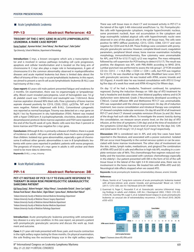
POSTER PRESENTATIONS
698th International Congress on Leukemia Lymphoma MyelomaMay 21-22 2021 • VIRTUAL CONGRESS
� Acute Lymphoblastic Leukemia
PP-13 Abstract Referance: 43
TRISOMY OF THE C-MYC GENE IN ACUTE LYMPHOBLASTIC LEUKEMIA: A RARE CASE REPORTGuray Saydam1, Aysenur Arslan1, İrem Toksoy1, Nur Akad Soyer1, Fahri Şahin1
1Ege University, School of Medicine, Department of Hematology
Introduction: C-myc, a known oncogene which acts a transcription fac-tor and is involved in various pathways including cell cycle progression, apoptosis and cellular transformation and is located on the long arm of chromosome 8. C-myc also plays a major role in hematopoiesis. A gain of chromosome 8 is the common abnormality observed in myeloproliferative diseases and acute myeloid leukemia but there is limited data about the effect of trisomy of the c-myc in acute lymphoblastic leukemia. In this report, we aimed to present a rare B-cell acute lymphoblastic leukemia (B-ALL) case had trisomy of the c-myc.
Case report: 63 years-old male patient presented fatigue and weakness for 2 months. On examination, there was no organomegaly or lympadenop-athy. Blood count revealed pancytopenia. Level of hemoglobin was 7.6 g/dl, platelet count was 112000/mm3 and neutrophil was 1120/mm3. Bone marrow aspiration showed 90% blast cells. Flow cytometry of bone marrow aspirate showed positivity for CD19, CD20, CD22, cyCD79a. TdT and C10 were negative. Patient diagnosed with B-ALL. Conventional cytogenetic was reported as normal karyotype. Trisomy was detected on c-myc gene on 8q24.1 loci by fluorescent in situ hybridization (FISH). Patient was treated with a hyper CVAD/arm A (cyclophosphamide, vincristine, doxorubicin and dexametasone) protocol. Bone marrow aspiration and FISH were repeated at the end of the fourth week of cycle. Blastic cells were less than 5 percent in bone marrow and FISH was negative for c-myc gene.
Conclusion: Although B-ALL is primarily a disease of children, there is a peak of incidence in adults >60 years old and adults have much worse prognosis than children. Isolated myc gene abnormalities, especially a gain of chromo-some 8 without other genetic abnormalities is rare in B lymphoblastic leu-kemia with some cases reported in pediatric patients with worse prognosis. The prognosis of trisomy of c-myc gene in adults is still unclear and there need to be more data to determine.Keywords: B-ALL, c-myc, trisomy
� Acute Myeloid Leukemia
PP-14 Abstract Referance: 38
PET-CT INSTEAD OF PCR 15:17 TO EVALUATE RESPONSE TO THERAPY IN HIGH RISK PROMYELOCYTIC GRANULOCYTIC SARCOMA PATIENTGül Yavuz Ermiş1, Bülent Yenigün2, Hülya Yilmaz1, Cemaleddin Öztürk1, Emre Can Sağlık3, Osman Fırat Duran3, Klara Dalva1, Elgin Özkan4, Işınsu Kuzu5, Meltem Kurt Yüksel1
1Ankara University Faculty of Medicine Internal Medicine Hematology Department 2Ankara University Faculty of Medicine Thoracic Surgery Department 3Ankara University Faculty of Medicine Internal Medicine Department 4Ankara University Faculty of Medicine Nuclear Medicine Department 5Ankara University Faculty of Medicine Pathology Department
Introduction: Acute promyelocytic leukemia presenting with extramedul-lary disease is a very rare condition. In this case report, we present a patient with promyelocytic granulocytic sarcoma and discuss the work up, treat-ment and outcome.
Case: A 21-year-old male presented with fever, pain, and muscle contraction in the right axillary region lasting for three months. On physical examination, the sole finding was the excisional biopsy scar in the right axillary region.
There was soft tissue mass in chest CT and increased activity in PET-CT in the lateral of the right 3-4th intercostal area(Picture 1a, 1b). Promyelocytic-like cells with hypergranular cytoplasm, irregular hyperchromatic nucleus, some prominent nucleoli, Auer rod accumulation in the cytoplasm and large eosinophilic isolated atypical cells with hyperchromatic nuclei were observed in one of the atypical cells in the soft tissue mass. The cells were positive for MPO (diffuse positivity), CD13, CD33, CD68, CD117 and were negative for CD34 and HLA-DR. These findings were consistent with promy-elocytic granulocytic sarcoma. However, complete blood count, coagulation parameters, peripheral blood smear, bone marrow examination including morphology, immunohistochemistry, flow cytometry were unremarkable for APL; and PCR for t(15;17) (q22; q12) was negative. The mass rebiopsied, followed by cell suspension for PCR testing to detect t(15:17). The result was positive, the diagnosis was APL with PML-RARA according to WHO 2016. Lumbar puncture was performed to assess the risk group1. LP FCM revealed CD 33+ CD13 - / + HLA DRp + CD34 +/- CD9 + cells, but PCR was negative for t(15;17). He was classifed as high-risk APML (Modified Sanz score 2017) with granulocytic sarcoma. He was treated with ATRA, arsenic trioxide and GO (Figure). A month later he was treated consolidation with ATO 0.15 mg/kg five days of a week four week and ATRA 45 mg/m2 for fifteen days.
On day 12 of he had a headache, Treatment continued, his symptoms regressed. During the induction therapy on 18th day of ATO treatment he had a headache and double vision, then bilateral papillary edema and 6.CN dysfunction were detected. Grade 2 Neurotoxicity developed according to CTAEvs5. Cranial diffusion MRI and Methionine PET-CT was unremarkable. ATO was suspended until the clinical improvement. On day 28 of induction treatment, two course consolidation and intratecal therapy was completed. LP showed complete molecular remission. On day 60 and 90 of all treatment PET-CT and chest CT showed complete remission (Picture 2a and 2b). None of the drugs had such side effects. To investigate the arsenic toxicity during the consolidation, we measure serum arsenic level, on the 3rd day of ATO infusion, at the time of symptoms (12th day) and at the time of resolution of the symptoms (22nd day).The serum level of arsenic for the days 3rd, 12th and 22nd were 53.45 mcg/l; 131,5 mcg/l; 52,47 mcg/l respectively.
Discussion: EM is considered rare in APL and only few cases have been reported in the literature, and associated with a poorer outcome2. Isolated EM can occur most frequently in the central nervous system or can be asso-ciated with bone marrow involvement. The other sites of involvement are the skin, testes, lymph nodes, mediastinum, and gingiva3.The combination of ATRA-ATO and GO is safe and effective in high-risk APL resulting in a com-plete remission rate of 96%. This chemotherapy-free regimen reduced mor-bidity and the toxicities associated with cytotoxic chemotherapy especially in the elderly1. Our patient presented with EM in the form of GS of the soft tissue tissue in the lateral of the right 3-4 th intercostal area, there was no involvement in the bone marrow. We treated him with ATRA+ATO and GO. Neurological side effects stopped when we ceased ATO.Keywords: Acute promyelocytic leukemia, extramedullary disease, arsenic trioxide
References
1. Abaza, Yasmin et al. “Long-term outcome of acute promyelocytic leukemia treated with all-trans-retinoic acid, arsenic trioxide, and gemtuzumab.” Blood vol. 129,10 (2017): 1275-1283. doi:10.1182/blood-2016-09-736686
2. Guermazi A, Feger C, Rousselot P, et al. Granulocytic sarcoma (chloroma): imag-ing findings in adults and children. AJR Am J Roentgenol 2002; 178(2): 319–325. 3-Ochs RC, Gormley RH, Luger S, et al. Isolated bowel relapse in acute promyelocytic leukemia: an unusual site of extramedullary recurrence. J Clin Oncol 2010; 28(29): e550–e553
Figure 1. Treatment scheme ATO, ATRA and GO
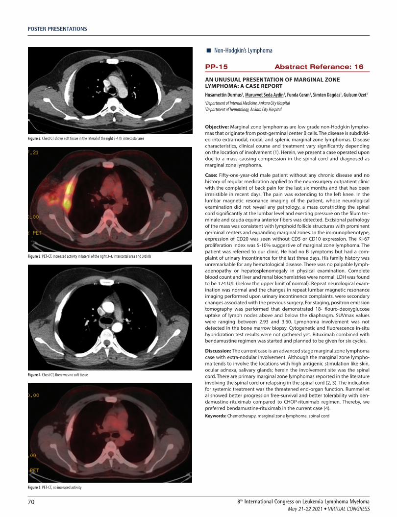
POSTER PRESENTATIONS
70 8th International Congress on Leukemia Lymphoma MyelomaMay 21-22 2021 • VIRTUAL CONGRESS
Figure 2. Chest CT shows soft tissue in the lateral of the right 3-4 th intercostal area
Figure 3. PET-CT, increased activity in lateral of the right 3-4. intercostal area and 3rd rib
Figure 4. Chest CT, there was no soft tissue
Figure 5. PET-CT, no increased activity
� Non-Hodgkin’s Lymphoma
PP-15 Abstract Referance: 16
AN UNUSUAL PRESENTATION OF MARGINAL ZONE LYMPHOMA: A CASE REPORTHusamettin Durmus1, Muruvvet Seda Aydin2, Funda Ceran1, Simten Dagdas1, Gulsum Ozet1
1Department of Internal Medicine, Ankara City Hospital 2Department of Hematology, Ankara City Hospital
Objective: Marginal zone lymphomas are low grade non-Hodgkin lympho-mas that originate from post-germinal center B cells. The disease is subdivid-ed into extra-nodal, nodal, and splenic marginal zone lymphomas. Disease characteristics, clinical course and treatment vary significantly depending on the location of involvement (1). Herein, we present a case operated upon due to a mass causing compression in the spinal cord and diagnosed as marginal zone lymphoma.
Case: Fifty-one-year-old male patient without any chronic disease and no history of regular medication applied to the neurosurgery outpatient clinic with the complaint of back pain for the last six months and that has been irresistible in recent days. The pain was extending to the left knee. In the lumbar magnetic resonance imaging of the patient, whose neurological examination did not reveal any pathology, a mass constricting the spinal cord significantly at the lumbar level and exerting pressure on the filum ter-minale and cauda equina anterior fibers was detected. Excisional pathology of the mass was consistent with lymphoid follicle structures with prominent germinal centers and expanding marginal zones. In the immunophenotype, expression of CD20 was seen without CD5 or CD10 expression. The Ki-67 proliferation index was 5-10% suggestive of marginal zone lymphoma. The patient was referred to our clinic. He had no B symptoms but had a com-plaint of urinary incontinence for the last three days. His family history was unremarkable for any hematological disease. There was no palpable lymph-adenopathy or hepatosplenomegaly in physical examination. Complete blood count and liver and renal biochemistries were normal. LDH was found to be 124 U/L (below the upper limit of normal). Repeat neurological exam-ination was normal and the changes in repeat lumbar magnetic resonance imaging performed upon urinary incontinence complaints, were secondary changes associated with the previous surgery. For staging, positron emission tomography was performed that demonstrated 18- flouro-deoxyglucose uptake of lymph nodes above and below the diaphragm. SUVmax values were ranging between 2.93 and 3.60. Lymphoma involvement was not detected in the bone marrow biopsy. Cytogenetic and fluorescence in-situ hybridization test results were not gathered yet. Rituximab combined with bendamustine regimen was started and planned to be given for six cycles.
Discussion: The current case is an advanced stage marginal zone lymphoma case with extra-nodular involvement. Although the marginal zone lympho-ma tends to involve the locations with high antigenic stimulation like skin, ocular adnexa, salivary glands; herein the involvement site was the spinal cord. There are primary marginal zone lymphomas reported in the literature involving the spinal cord or relapsing in the spinal cord (2, 3). The indication for systemic treatment was the threatened end-organ function. Rummel et al showed better progression free-survival and better tolerability with ben-damustine-rituximab compared to CHOP-rituximab regimen. Thereby, we preferred bendamustine-rituximab in the current case (4). Keywords: Chemotherapy, marginal zone lymphoma, spinal cord

POSTER PRESENTATIONS
718th International Congress on Leukemia Lymphoma MyelomaMay 21-22 2021 • VIRTUAL CONGRESS
References
1. Sindel A, Al-Juhaishi T, Yazbeck V. Marginal Zone Lymphoma: State-of-the-Art Treatment. Current treatment options in oncology. 2019;20(12):90.
2. Hojo Y, Ito M, Abumi K, Sudo H, Takahata M. Primary spinal marginal zone lymphoma relapse at a different spinal level after remission of the primary lesion. Global spine journal. 2013;3(4):261-4.
3. Ahmadi SA, Frank S, Hänggi D, Eicker SO. Primary spinal marginal zone lymphoma: case report and review of the literature. Neurosurgery. 2012;71(2):E495-508; discus-sion E.
4. Rummel MJ, Niederle N, Maschmeyer G, Banat GA, von Grünhagen U, Losem C, et al. Bendamustine plus rituximab versus CHOP plus rituximab as first-line treatment for patients with indolent and mantle-cell lymphomas: an open-label, multicentre, randomised, phase 3 non-inferiority trial. Lancet (London, England). 2013;381(9873):1203-10.
Figure 1. Spinal mass at L3-L4
� Non-Hodgkin’s Lymphoma
PP-16 Abstract Referance: 84
IBRUTINIB MONOTHERAPY FOR RELAPSED OR REFRACTORY DLBCL PATİENTS: A SİNGLE CENTER EXPERİENCEMustafa Merter1, Ayşe Uysal1, Ömer Ekinci2
1Fırat University School of Medicine Hematology Department 2Diyarbakir Gazi Yasargil Education and Research Hospital
Introduction: Ibrutinib is an oral covalent inhibitor of Bruton tyrosine kinase, which disrupts signaling from the B-cell receptor to NF-κB. Ibrutinib has shown activity in non–germinal center B-cell diffuse large B-cell lympho-ma (DLBCL). In this study we aimed to evaluate the effectiveness of ibrutinib as a single agent in our relapsed or refractory DLBCL patients.
Method: We evaluate 6 patients who receive ibrutinib as a monotherapy for R/R DLBCL between May 2018 and June 2019. Patients’ previous treatments, Ann-Arbor stages, comorbidities and responses to ibrutinib were identified.
Results: Patients’ characteristics were shown in table-1. Ibrutinib was admin-istered to all patients as a single dose of 560 mg peroral daily. Three patients had no response to ibrutinib. Two patients had central nervous system(CNS) involvement at the time of relapse and both did not respond to ibrutinib. One patient had Richter transformation and she was the only patient that complete response was achieved. One patient had stable disease and one has partial response.
Conclusion: In a phase 1/2 clinical trial that involved 80 subjects with relapsed or refractory DLBCL, ibrutinib produced complete or partial responses in 37%. Our results were consistent with this study. Interestingly ibrutinib had no activitiy in our CNS involved patients despite its well known CNS penetration. Another interesting finding of our study was that the only patient who achieved comlete response had Richter transformation. All our patients had poor performance status and were not eligible for high dose therapies. Ibrutinib were well tolerated in all our patients and no adverse event was observed. Our study suggests that ibrutinib is a feasible choice for patients who are multi refractory and have poor performance status.Keywords: ibrutinib, relapsed and refractory lymphoma DLBCL
Table 1.
Case-1 Case-2 Case Case-4 Case-5 Case-6
Age 71 69 44 80 64 84
Gender Female Female Female Female Female Female
Ann-Arbor stage
IVB IV IV IVS III IV
Previous treatments
R-CHOP, R-Bendamustin, R-lenalidomid
R-high dose metorexate
R-HD MTX+ARA-C,
autologous stem cell
transplatation
R-CHOP, R-Lenalidomid
CHOP, FC, R-FC, R-CHOP,
R-DHAP, R-Lenalidomid
R-CHOP, R-GDP,
R-Lenalidomid
Response Progressive disease
Progressive disease
Progressive disease
Partial response
Comlete response
Stable disease
� Acute Myeloid Leukemia
PP-17 Abstract Referance: 19
PLEURAL MYELOID SARCOMA: A RARE INVOLVEMENT SITE CAUSING PLEURAL EFFUSION AT ACUTE MYELOID LEUKEMIAFatma Yılmaz1, Murat Albayrak1, Buğra Sağlam1, Pınar Akyol1, Mesut Tığlıoğlu1, Merih Reis Aras1, Senem Maral1, Hacer Berna Afacan Öztürk1, Abdülkerim Yıldız2
1Diskapi Yildirim Beyazit Training and Research Hospital,department of Hematology 2Hitit University Erol Olçok Training and Research Hospital,department of Hematology Çorum,turkey
Introduction: Myeloid Sarcoma (MS) with extramedullary involvement occurs in less than 1% of all acute myeloid leukemia (AML) and is common with monocytic differentiation. According to the 2016 revision the World Health Organization classification of myeloid neoplasms and acute leuke-mia; classified as acute myeloid leukemia and related neoplasm (1).
MS which has more aggressive course than AML is frequently located in bone, periosteum, soft tissue and lymph node while it also has rare such as epidural, orbita, pleura etc (2).
Herein we reported a rare case of 63 year old man with AML-M5 and diag-nosed MS with pleural effusion cytology.
Case Report: A 63 –year-old man who had no history of chronic disease was admitted to emergeny department with complaints of fever and shortness of breath. In the blood test examination; white blood cells (WBC): 33.2x10⁹/L; neutrophil: 21.5x10⁹/L; monocyte:1.8x10⁹ /L, hemoglobin 8.8 g/dL, and plate-letes:26x109 /L and pleural effusion were detected and referred to our hema-tology department. Blastic cell infiltration was detected in peripheral smear and the patient underwent bone marrow biopsy. More than 70-80% mono-blasts were observed in bone marrow aspiration. Flowcytometric examination was also performed and were interpreted of AML M5. Azacitidine+Venetoklax treatment was initiated consider of the patient’s age and performance status. Flowcytometric examination of effusion with exudate quality could not be studied because the effusion was hemorrhagic. On the sixth day of treatment, effusion cytology was reported that cell block with atypical immature myeloid cells and AML involvemnet. Remission induction kemotherapy was planned for the patient diagnosed with MS.
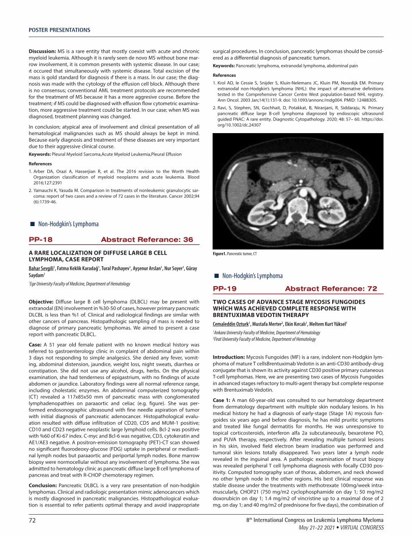
POSTER PRESENTATIONS
72 8th International Congress on Leukemia Lymphoma MyelomaMay 21-22 2021 • VIRTUAL CONGRESS
Discussion: MS is a rare entity that mostly coexist with acute and chronic myeloid leukemia. Although it is rarely seen de novo MS without bone mar-row involvement, it is common presents with systemic disease. In our case; ıt occured that simultaneously with systemic disease. Total excision of the mass is gold standard for diagnosis ıf there is a mass. İn our case; the diag-nosis was made with the cytology of the effusion cell block. Although there is no consensus; conventional AML treatment protocols are recommended for the treatment of MS because it has a more aggresive course. Before the treatment; ıf MS could be diagnosed with effusion flow cytometric examina-tion, more aggressive treatment could be started. In our case; when MS was diagnosed, treatment planning was changed.
In conclusion; atypical area of involvement and clinical presentation of all hematological malignancies such as MS should always be kept in mind. Because early diagnosis and treatment of these diseases are very important due to their aggressive clinical course.Keywords: Pleural Myeloid Sarcoma,Acute Myeloid Leukemia,Pleural Effusion
References
1. Arber DA, Orazi A, Hasserjian R, et al. The 2016 revision to the Worth Health Organization classification of myeloid neoplasms and acute leukemia. Blood 2016;127:2391
2. Yamauchi K, Yasuda M. Comparison in treatments of nonleukemic granulocytic sar-coma: report of two cases and a review of 72 cases in the literature. Cancer 2002;94 (6):1739-46.
� Non-Hodgkin’s Lymphoma
PP-18 Abstract Referance: 36
A RARE LOCALIZATION OF DIFFUSE LARGE B CELL LYMPHOMA, CASE REPORTBahar Sevgili1, Fatma Keklik Karadağ1, Tural Pashayev1, Ayşenur Arslan1, Nur Soyer1, Güray Saydam1
1Ege University Faculty of Medicine, Department of Hematology
Objective: Diffuse large B cell lymphoma (DLBCL) may be present with extranodal (EN) involvement in %30-50 of cases, however primary pancreatic DLCBL is less than %1 of. Clinical and radiological findings are similar with other cancers of pancreas. Histopathologic sampling of mass is needed to diagnose of primary pancreatic lymphomas. We aimed to present a case report with pancreatic DLBCL.
Case: A 51 year old female patient with no known medical history was referred to gastroenterology clinic in complaint of abdominal pain within 3 days not responding to simple analgesics. She denied any fever, vomit-ing, abdominal distension, jaundice, weight loss, night sweats, diarrhea or constipation. She did not use any alcohol, drugs, herbs. On the physical examination, she had tenderness of epigastrium, with no findings of acute abdomen or jaundice. Laboratory findings were all normal reference range, including cholestatic enzymes. An abdominal computerized tomography (CT) revealed a 117x85x50 mm of pancreatic mass with conglomerated lymphadenopathies on paraaortic and celiac (e.g. figure). She was per-formed endosonographic ultrasound with fine needle aspiration of tumor with initial diagnosis of pancreatic adenocancer. Histopathological evalu-ation resulted with diffuse infiltration of CD20, CD5 and MUM-1 positive; CD10 and CD23 negative neoplastic large lymphoid cells. Bcl-2 was positive with %60 of Ki-67 index. C-myc and Bcl-6 was negative, CD3, cytokeratin and AE1/AE3 negative. A positron-emission tomography (PET)-CT scan showed no significant fluorodeoxy-glucose (FDG) uptake in peripheral or mediasti-nal lymph nodes but paraaortic and periportal lymph nodes. Bone marrow biopsy were normocellular without any involvement of lymphoma. She was admitted to hematology clinic as pancreatic diffuse large B cell lymphoma of pancreas and treat with R-CHOP chemoterapy regimen.
Conclusion: Pancreatic DLBCL is a very rare presentation of non-hodgkin lymphomas. Clinical and radiologic presentation mimic adenocancers which is mostly diagnosed in pancreatic malignancies. Histopathological evalua-tion is essential to refer patients optimal therapy and avoid inappropriate
surgical procedures. In conclusion, pancreatic lymphomas should be consid-ered as a differential diagnosis of pancreatic tumors.Keywords: Pancreatic lymphoma, extranodal lymphoma, abdominal pain
References
1. Krol AD, le Cessie S, Snijder S, Kluin-Nelemans JC, Kluin PM, Noordijk EM. Primary extranodal non-Hodgkin’s lymphoma (NHL): the impact of alternative definitions tested in the Comprehensive Cancer Centre West population-based NHL registry. Ann Oncol. 2003 Jan;14(1):131-9. doi: 10.1093/annonc/mdg004. PMID: 12488305.
2. Ravi, S, Stephen, SN, Gochhait, D, Potakkat, B, Niranjani, R, Siddaraju, N. Primary pancreatic diffuse large B‐cell lymphoma diagnosed by endoscopic ultrasound guided FNAC: A rare entity. Diagnostic Cytopathology. 2020; 48: 57– 60. https://doi.org/10.1002/dc.24307
Figure1. Pancreatic tumor, CT
� Non-Hodgkin’s Lymphoma
PP-19 Abstract Referance: 72
TWO CASES OF ADVANCE STAGE MYCOSIS FUNGOIDES WHİCH WAS ACHİEVED COMPLETE RESPONSE WITH BRENTUXIMAB VEDOTIN THERAPYCemaleddin Ozturk1, Mustafa Merter2, Ekin Kırcalı1, Meltem Kurt Yüksel1
1Ankara University Faculty of Medicine, Department of Hematology 2Firat University Faculty of Medicine, Department of Hematology
Introduction: Mycosis Fungoides (MF) is a rare, indolent non-Hodgkin lym-phoma of mature T cellsBrentuximab Vedotin is an anti-CD30 antibody-drug conjugate that is shown its activity against CD30 positive primary cutaneous T-cell lymphomas. Here, we are presenting two cases of Mycosis Fungoides in advanced stages refractory to multi-agent therapy but complete response with Brentuximab Vedotin.
Case 1: A man 60-year-old was consulted to our hematology department from dermatology department with multiple skin nodulary lesions. In his medical history he had a diagnosis of early-stage (Stage 1A) mycosis fun-goides six years ago and before diagnosis, he has mild pruritic symptoms and treated like fungal dermatitis for months. He was unresponsive to topical corticosteroids, interferon alfa 2a subcutaneously, bexarotene PO, and PUVA therapy, respectively. After revealing multiple tumoral lesions in his skin, involved field electron beam irradiation was performed and tumoral skin lesions totally disappeared. Two years later a lymph node revealed in the inguinal area. A pathologic examination of trucut biopsy was revealed peripheral T cell lymphoma diagnosis with focally CD30 pos-itivity. Computed tomography scan of thorax, abdomen, and neck showed no other lymph node in the other regions. His best clinical response was stable disease under the treatments with methotrexate 100mg/week intra-muscularly, CHOP21 (750 mg/m2 cyclophosphamide on day 1; 50 mg/m2 doxorubicin on day 1; 1.4 mg/m2 of vincristine up to a maximal dose of 2 mg, on day 1; and 40 mg/m2 of prednisone for five days), the combination of

POSTER PRESENTATIONS
738th International Congress on Leukemia Lymphoma MyelomaMay 21-22 2021 • VIRTUAL CONGRESS
gemcitabine vinorelbine, respectively. After four cycles of the combination of gemcitabine and vinorelbine chemotherapy, the tumoral lesions were continued to progress. Brentuximab Vedotin (BV) was started intravenously every three weeks at a dose of 1,8 mg/kg. Before BV therapy clinical stage is IIB. After two cycles of therapy course with BV, all skin lesions and lymph nodes disappeared completely, and we continued to BV five cycles more and stopped. The only adverse event is grade2 polyneuropathy. After three years later from BV therapy, this patient is still in remission and no any other problem with treatment and disease.
Case 2: 58 year-old man was admitted to our hematology department with mycosis fungoides tumoral stage. In his medical history, he had a diagnosis of stage I foliculotrophic mycosis fungoides according to EORCT criteria before twenty years ago. Besides he had a history of various skin directed therapies such as topical steroids, PUVA, UVB-NB, he was unresponsive to also interferon alpha 2a and bexarotene orally. He was also unresponsive to systemic low-dose methotrexate and ECP (extracorporeal photopheresis) due to multiple tumoral skin lesions developed two years ago. Partially response was achieved with GDP regimen (gemcitabine 1000 mg/m2 on days 1 and 8; dexamethasone 40 mg IV on days 1 to 4, and cisplatin 75mg/m2) after six cycles. Due to unsatisfactory response with GDP regimen, we administered pralatrexate IV 15 mg/m2 once weekly for every three weeks. After eleven courses of pralatrexate, tumoral lesions continued to progress. We applied lymph node and skin biopsy again and the pathological exam-ination result is compatible with focal CD30 positivity. Brentuximab Vedotin (BV) was started intravenously every three weeks at a dose of 1,8 mg/kg. At the end of two-course of BV complete response was achieved dramatically. Unfortunately, the patient died due to pneumonia while continuing the 11th course of his treatment with sustained complete response.
Discussion: Patients with Foliculotrophic MF in advanced stages have a poor prognosis. In this group of patients, there is no consensus about the treatment modalities and the result of treatment is not satisfactory. Brentuximab Vedotin is a new and promising option for advanced stages MF patients without regard to CD30 expression rate.Keywords: Brentuximab vedotin, mycosis fungoides, non-Hodgkin lymhoma
References
1. Prince, H. Miles, et al. “Brentuximab vedotin or physician’s choice in CD30-positive cutaneous T-cell lymphoma (ALCANZA): an international, open-label, randomised, phase 3, multicentre trial.” The Lancet 390.10094 (2017): 555-566.
� Non-Hodgkin’s LymphomaPP-20 Abstract Referance: 3
PATIENT DIAGNOSED WITH PRIMARY OVARIAN DIFFUSE LARGE B CELL LYMPHOMA AND UNDERWENT CENTRAL NERVOUS SYSTEM PROPHYLAXISTaha Ulutan Kars1, Atakan Tekinalp1, Ali Öz2, Hatice Zeynep Dikici1, Kübra Uygun Yel1, Sinan Demircioğlu1, Özcan Çeneli1
1Necmettin Erbakan University, Meram Faculty of Medicine, Division of Hematology 2Necmettin Erbakan University, Meram Faculty of Medicine, Department of Internal Medicine
Introduction: Non Hodgkin Lymphoma (NHL) rarely involves the female genital system. This involvement can be caused by advanced disease or primary female genital system [1]. Ovarian involvement occurs in 7–25% of lymphomas with disseminated extranodal involvement [2]. Primary ovarian NHL is less common; accounts for 0.5% of all NHLs and 1.5% of all ovarian tumors [3]. Most common type of primary NHL seen in ovary is diffuse large B cell lymphoma (DLBCL), accounting for 20% of primary ovarian DLBCL (PODLBCL) cases [1].
Case Report: A 34-year-old female presented to the department of gyne-cology and obstetrics with the complaint of abdominal pain, pelvic lump and hypermenorrhea. Pelvic ultrasonography (USG) showed a hypoechoic right ovarian mass measuring 12cm×10cm. Total abdominal hysterectomy + bilateral salpingo-oophorectomy (TAH + BSO) was performed on the patient. Histopathological examination of the ovary reported as DLBCL
(CD20+, CD79a+, CD23+, CD5 rare+, CD3 rare+, bcl-6 rare+, bcl-2 rare+, CD10-), and the patient was referred to us. The patient did not have any com-plaints or B symptoms on admission. There was no lymphadenomegaly or organomegaly on physical examination. In complete blood count, all values were within normal limits, except hemoglobin (11.5 g/dL). Peripheral blood smear was normal. There was no abnormal value in biochemical parameters. Lymphoma infiltration was not detected in the bone marrow biopsy. PET/CT revealed lymph nodes in the left paraaortic and left parailiac regions with a size of 2.5cm and increased metabolic activity. The patient was accepted as stage IIE. R-IPI score was 1. R-CHOP was initiated for the patient. After 3 cycles, a complete response was found in the interim evaluation with PET/CT, and 6 cycles were completed. It was decided to perform CNS prophylaxis with intrathecal methotrexate (4 times, 15 mg every 28 days) for the patient whose complete response continued after six cycles, due to ovarian involve-ment. The patient, whose intrathecal treatment is still continuing, and is followed up without any complications.
Discussion: Patients diagnosed with PODLBCL have been shown rarely in the literature. It has been shown that primary ovarian DLBCL is less aggres-sive in terms of clinical course and has a better 5-year survival compared to DLBCL with secondary ovarian involvement [4]. Unlike radical surgery, chemotherapy is an optimal treatment in these patients; therefore, correct diagnosis is essential to avoid unnecessary procedures [5].
Fox et al. have suggested criteria for the diagnosis of primary ovarian lym-phoma. First, the tumor should be confined to the ovary, regional lymph nodes, or adjacent organs at the time of the diagnosis. Also bone marrow and peripheral blood should not have any abnormal cells, and if extraovarian disease appears later, there must be a gap of few months between the time of appearance of ovarian and extraovarian lesions [6]. In our case, involve-ment was limited only to the ovarian and ovarian lymph node. Also, having a complete response supports that it meets these criteria.
Because of its rarity, CNS involvement has been reported very rarely in PODLBCL. It has been shown in the literature that methotrexate can be safely administered simultaneously with R-CHOP and is associated with a lower risk of CNS recurrence in high-risk DBBHL; however, PODLBCL was not examined in these studies [7,8]. Hu et al. discussed with 3 cases that they considered to be PODLBCL. They reported that one of these cases had CNS involvement and this patient died of relapse after treatment [9]. However, it is doubtful whether the diagnosis is PODLBCL because of bone marrow involvement at the time of diagnosis in this case.
Although there are few data in the literature, we planned 4 cycles of intrathe-cal methotrexate for CNS prophylaxis for the patient in whom we achieved complete response after R-CHOP treatment. The patient is still receiving intrathecal prophylaxis.Keywords: Ovary, Lymphoma, Large B-Cell, Diffuse
References
1. Vang R, et al. Ovarian non Hodgkin’s lymphoma: a clinicopathologic study of eight primary cases. ModPathol. 2001;14:1093.
2. Chorlton I, et al. (1974) Malignant reticuloendothelial disease involving the ovary as a primary manifestation: a series of 19 lymphomas and 1 granulocytic sarcoma. Cancer 34:397–407
3. Vang R, et al. (2001) Non-Hodgkin’s lymphoma involving the gynecologic tract: a review of 88 cases. Adv Anat Pathol 8:200–217
4. Monterroso V, et al. Malignant lymphomas involving the ovary. A clinicopathologic analysis of 39 cases. Am J Surg Pathol. 1993;17:154–70.
5. Weingertner AS, et al. Non-Hodgkin malignant lymphoma revealed by an ovarian tumor case report and review of the literature. Gynecol Oncol. 2004;95(3):750–54.
6. Fox H, et al. Malignant lymphoma presenting as an ovarian tumour: a clinicopatho-logical analysis of 34 cases. BJOG Int J Obstet Gynaecol. 1988;95: 386–90.
7. O’Rourke K, et al. Intravenous methotrexate as central nervous system (CNS) prophy-laxis is associated with a low risk of CNS recurrence in high-risk patients with diffuse large B-cell lymphoma. Cancer. 2010
8. Abramson JS, et al. Intravenous methotrexate as central nervous system (CNS) prophylaxis is associated with a low risk of CNS recurrence in high-risk patients with diffuse large B-cell lymphoma. Cancer. 2010
9. Hu, R., et al. (2012). Ovary involvement of diffuse large B-cell lymphoma. The American journal of case reports, 13, 96–98.

POSTER PRESENTATIONS
74 8th International Congress on Leukemia Lymphoma MyelomaMay 21-22 2021 • VIRTUAL CONGRESS
� Other
PP-21 Abstract Referance: 57
CMV INFECTION WITH FATAL HEMOPHAGOCYTIC SYNDROMEErman Ozturk1, Işıl Erdoğan Özünal1, Tayfun Elibol1, Emrah Kılıçaslan1
1Istanbul Medeniyet University Prof Dr Süleyman Yalçın City Hospital Hematology
Hemophagocytic lymphohistiocytosis (HLH) is a life-threatening syndrome in which an exaggerated immune response leads to severe immune activa-tion. Mostly diagnosis in infants but disease also diagnosed in adults. HLH resulting from inherited or secondary hyperinflammation. If it is not treated excessive immune activation can cause to multiorgan damage and death. Fever, Splenomegaly, cytopenia, hypertriglyceridemia, hyperferritinemia are some findings. Diagnosis of HLH is based on the HLH-2004 criteria. Associated illnesses are infections, malignancy, rheumatologic disorders, and immunodeficiency. Treatment of HLH based on immune suppression and primary disorder that cause HLH. It is hard to recognize if it is primary HLH or infection exacerbated HLH, which immunosuppression may be complicated. We represent a patient with fatal HLH secondary to infection disease.
Our case is a 27-year-old male patient, previously healthy. Referred to our cen-ter for worsening fatigue, high grade fever, night sweet and blurred vision. History goes back to 6 months ago. On physical examination he had gener-alized lymphadenopathy, pleural effusion, ascites, edema, hepatomegaly, and splenomegaly with 2 cm below costal edge. His vision was reduced and choroid detachment, retinal infiltration was found. Ophthalmology depart-ment advise pulse steroid therapy urgently and 1 gr methylprednisolon was applied for 3 days. Blood tests are summarized at table 1. Disseminated intravascular coagulopathy was seen and supportive and fibrinogen replace-ment therapy was applied daily. Because of pancytopenia bone marrow biopsy was performed and hemophagocytosis was revealed (Figure 1). He diagnosed with hemophagocytic lymphohistiocytosis. Thoracentesis and bronchoscopy evaluation was also performed and exudative pleural effu-sion and endobronchial lesions which suggestive of tuberculosis was seen. At broncho alveolar lavage evaluation no acid resistant bacteria was found but CMV PCR was positive. Antituberculosis treatment with four agents and ganciclovir were given. Patient’s body temperature did not go down below 39ºC on follow up. At PET-CT scan he had generalized lymphadenopathy with high grade FDG avidity (SUV max 5-36), reticulonodular image at lung parenchyma. There was no sign of any malignancy at lymphnode excisional biopsy. Diagnosis of tuberculosis could not be proofed with microbiologic methods and we decide that HLH was develop secondary to CMW infection. HLH clinic was highly aggressive and DIC was not controlled with the current treatment. Immunosuppressive therapy with Etoposide was judged and due to active uncontrolled infection treatment was delayed. After 15th day of admission, he was death secondary to multiorgan failure.
Treatment of HLH is based on immunosuppressive treatment with etopo-side, glucocorticoids and methotrexate at central nervous system involve-ment settings. Secondary infections are not rare and immunosuppressive treatment is hard to decide. At this patient he had CMV infection and HLH did not respond to pulse steroid therapy. Etoposide treatment also could not be applied because of active infection. Anakinra or emapalumab may be treatment alternatives like this patient. Patients whose unresponsive the initial treatment alemtuzumab may be an option but at patients with CMV infection this agent can not be applied. Allogeneic stem cell transplantation is another treatment option to refractory HLH patients. It may be hard to decide the treatment options in infection settings.Keywords: Hemophagocytic lymphohistiocytosis, Hemophagocytic syndroma, CMV
Figure 1.
Tables 1.
WBC 1000/mL
Hb 8,7 g/dl
Plt 46000 /mL
Cr 0,6 mg/dl
AST 450 U/L
ALT 170 U/L
LDH 902 U/L
Bilirubine 8,23 mg/dl
D bilirubine 7,89 mg/dl
ALP 879 U/L
GGT 160 U/L
D-dimer 8,93 mg/dl
Fibrinogen 85 mg/dl
Ferritin 9760 mg/dl
� Non-Hodgkin’s Lymphoma
PP-22 Abstract Referance: 54
ENDEMIC BURKITT’S LYMPHOMA, CASE PRESENTATIONFerda Can1, Sema Akıncı1, Tekin Güney2, Özge Soyer Kösemehmetoğlu1, İmdat Dilek3
1Ministry of Health Ankara City Hospital, Hematology Department 2University of Health Science, Ankara City Hospital, Hematology Department 3Ankara Yıldırım Beyazıt University, Ankara City Hospital, Hematology Department
Burkitt’s lymphoma is an aggressive B-cell non-Hodgkin lymphoma. There are 3 subtypes: Sporadic, immunodeficiency related and endemic. Endemic Burkitt is a subtype that is seen 50 times more often in Africa than elsewhere and is often associated with facial involvement and Ebstein Barr Virus (EBV). In this case, a patient from African descent with dramatic clinical presenta-tion and rapid response to treatment is presented.
A 26-year-old male patient admitted to the clinic for evaluation of a mass on his face. Image of the patient on admission has shown in Figure. Patient’s chief complaint was dyspnea and this mass for one month. The patient came from Afghanistan and was working as a worker in our country. He visited a

POSTER PRESENTATIONS
758th International Congress on Leukemia Lymphoma MyelomaMay 21-22 2021 • VIRTUAL CONGRESS
local dentist and oral prophylaxis was performed. No clinical changes were evident post prophylaxis. His medical status was insignificant. All blood parameters were within normal limits except lactate dehydrogenase (LDH). LDH was 932 U/L. ELISA was negative for HIV. Peripheral blood smear was normal. The pathological evaluation of the biopsy taken from the mass was compatible with Burkitt’s lymphoma. Pozitron emission tomography for staging showed extensive lymphadenopathies below and above the diaphragm, cervical and thoracic vertebrae, and involvement in the rib and sacroiliac joint. Bone marrow aspiration and biopsy was normocellular. Rituksimab +HyperCVAD chemotherapy was immediately started to the patient. The dramatic improvement in the mass on the 8th day of che-motherapy is shown in Figure 2. Complete response was observed after 2 cycles of the patient’s chemotherapy. A total of 4 cycles of R-HyperCVAD and chemotherapy-adapted intrathecal treatments were given. The patient, who had a febrile neutropenia attack during chemotherapy responding to antibiotic treatments, was discharged at the end of the treatments without any problem.
We present an endemic subtype of Burkitt’s lymphoma which is a chemo-sensitive aggressive lymphoma that shows geographic distribution. We think that we will see these rare type more frequently due to the changing population distribution in our country due to migration.Keywords: Endemic, Burkitt’s lymphoma
References
1. Kalisz et al. An update on Burkitt lymphoma: a review of pathogenesis and mul-timodality imaging assessment of disease presentation, treatment response, and recurrence. Insights into Imaging (2019) 10:56.
2. Martinez L.O. The 2016 updated WHO classification of lymphoid neoplasias. Hematological Oncology. 2017;35(S1):37–45.
Figure 1. Image of the patient on admission
Figure 2. Image of the patient on the 8th day of the chemotherapy
� Chronic Myeloid Leukemia
PP-23 Abstract Referance: 17
VITREOUS HEMORRAGE; A RARE OPHTHALMIC ADVERS EFFECT DUE TO IMATINIB TREATMENTFatma Yılmaz1, Murat Albayrak1, Pınar Akyol1, Mesut Tığlıoğlu1, Buğra Sağlam1, Merih Reis Aras1, Senem Maral1, Hacer Berna Afacan Öztürk1
1Diskapi Yildirim Beyazit Training and Research Hospital,department of Hematology
İntroduction: Chronic myeloid leukemia (CML) characterized by unregu-lated growth of myeloid cells in the bone marrow and the accumulation of these cells in the peripheral blood. CML is classified as myeloproliferative neoplasm and has Philadelphia (Ph) chromosome that cause reciprocal translocation t(9;22).
Imatinib is commonly tolerated well by patients. The most common opthal-mic side effects are eyelid edema, periorbital edema. Other side effects which occured less than 1% blepharitis, blurred vision, conjunctival hemor-rhage, conjunctivitis, retinal hemorrhage etc.
Here we report a rare case involving 51-year- old man with Chronic myeloid leukemia (CML) who developed vitreous hemorrhage due to imatinib after 9 months of treatment.
Case report: A 51 - year- old man who had no history of smoking, alcohol or any chronic disease. The patient, was detected leukocytosis in the blood test examination and referred to hematology.
Bone marrow biopsy showing compatible with CML. Cytogenetic analysis by polymerase chain reaction were observed t(9:22)(q34:q11.2) and the quanti-fication of transcript level of BCR-ABL/ABL was 52.74%.
Imatinib treatment (400mg/day) was started. In the ninth month of imatinib treatment, the patient complained of sudden decrease in vision. Vitreous hemorrhage was detected in the left eye and the patient was operated. Vitreous hemorrhage recurred one month after the operation.
At the fourth day after the discontinuation of imatinib treatment, the patient’s opthalmic complaints improved significantly. Naranjo algorithm was applied and score was 9. Thus, bosutinib was planned instead of ima-tinib treatment.
POSTER PRESENTATIONS
76 8th International Congress on Leukemia Lymphoma MyelomaMay 21-22 2021 • VIRTUAL CONGRESS
Discussion: İmatinib is oral signal inhibitör that targets tyrosine kinase for BCR/ABL, platelet-derived growth factor, stem cell factor and c-KIT.
In conjunctiva and sclera have large amount of c-KIT positive mast cells which inhibited by imatinib. The inbition of c-KIT positive mast cells by ima-tinib, may be responsible for further exposure of the conjunctival mucosa to injuries.
In our case; imatinib treatment was discontinued due to recurrent vitreous hemorrage and because no other cause can be found. At the fourth day after the discontinuation of imatinib treatmet, the patient’s opthalmic com-plaints improved significantly. This patient’s Naranjo advers drug reaction probability scale was calculated to be 9. This result suggested that vitreous hemorrage was due to imatinib treatment.
We recommend that regular ophthalmologic control before and during ima-tinib treatment to prevent possible ocular complications, especially patients with comorbidities (diabetes mellitus, hypertension, myopia). It should be kept in mind rare side effects such as vitreous hemorrage may be seen in the patients who use imatinib.Keywords: İmatinib, vitreous hemorrhage, Chronic Myeloid Leukemia
� Non-Hodgkin’s LymphomaPP-24 Abstract Referance: 44
PRIMARY EXTRANODAL PANCREATIC NON-HODGKIN LYMPHOMA: A RARE CASE REPORTAyşenur Arslan1, Sercan Kamalak2, Nur Akad Soyer1, Guray Saydam1
1Ege University, School of Medicine, Department of Hematology 2Ege University, School of Medicine, Department of Internal Medicine
Introduction: Primary extranodal pancreatic non-hodgkin lymphoma (PPL) is extremely rare condition and is fewer than 1% in all non-Hodgkin’s lymphomas (NHL). PPL may have a similar clinical manifestation and sim-ilar radiographic appearance to pancreatic adenocancer and often not diagnosed until surgery. By the reason of diffirent prognosis and diffirent treatment strategies of these tumors, pathological diagnosis becomes very important. In this report, we aimed to present a rare case with PPL.
Case Report: A 57 year-old female patient presented to with jaundice, dark urine and light-coloured stool. She had no abdominal pain, fewer, diarrhea or constipation. Lymphadenomegaly or organomegaly was not detected. Her past medical history was unremerkable. Endoscopic retrograde cholan-giopancreatography (ERCP) was performed and revealed pancreatic mass in the head of pancreas which was compressed the bile and stent placed. Patient underwent Whipple surgery with an initial diagnosis of pancreatic adenocarcinoma. Immunohistochemical studies of resected tissue showed diffuse infiltration of CD20 positive neoplastic large lymphoid cells with an high mitotic index. CD3, cytokeratin AE1/AE3 and bcl-2 was negative. A positron-emission tomography (PET)-CT scan showed no significant fluoro-deoxy-glucose (FDG) uptake in lymph nodes. Only FDG uptake was detected in masses nearby surgical margin and caudal part of superior mesenteric artery. The diagnosis was PPL. She treated with R-CHOP (rituximab, cyclo-phosphamide, doxorubicine, vincristine and prednisone) regimen.
Discussion and Conclusion: PPL is extremely rare, comprising 1% of extra-nodal lymphomas and 0.5% of malignant pancreatic tumors. Although pre-surgical diagnosis provide to avoid surgery esprecially on young patients, diagnose is difficult due to similar clinical symptoms imaging tecnics as well as biochemical markers. PPL should always be suspected in the differential diagnosis of pancreatic tumor as long-term disease remission can be achieved with chemotherapy in patients. Keywords: Non-Hodgkin Lymphoma, extranodal pancreatic kymphoma, rare diseases
� Acute Lymphoblastic Leukemia
PP-25 Abstract Referance: 78
CHEMOTHERAPY EXTRAVASATION MANAGEMENT: SINGLE CENTER EXPERIENCEDefne Ay Tuncel1
1Sbu Adana City Training and Research Hospital
Chemotherapy extravasation may result in serious damage to patients, with irreversible local injures and disability. Evidence-based standardization on extravasation management is lacking and many institutions do not prac-tice adequate procedures to prevent the severer damages. Our aim was to explore the prevention and treatment of extravasation injuries, proposing a standard therapeutic protocol together with a review of the literature.
We examined extravasation injuries due to intravenous intervention medi-cation in our pediatric hematology oncology patients who were followed up in our clinic for a year. Although extravasation injuries are not common, such wounds cause functional, appearance and long-term healing problems. Patients may encounter surgical interventions in which skin graft operations are also applied after tissue loss. Our goal is to heal deep wounds without surgical intervention.
Method: Bioactive treatment protocol was used as a treatment protocol. Enzymatic debridement of the wound is provided by the collagenase of the product containing bacterial collagenase and hyaluronic acid. With hyal-uronic acid, new tissue is created from the bottom.
Wound care should be started by cleaning with an antiseptic solution. After the bioactive drug is applied to the wound area, wet dressing is applied with saline solution and the wound area is closed. After the granule texture is formed, the product containing hyaluronic acid, sodium salt and silver sulfo-diazine is applied to the wound area. The treatment is continued by closing the wound with wet dressing. When the wound floor is epithelialized, it is continued with the product containing only hyaluronic acid. Treatment is continued until the epithelization of the wound area is completed.
Discussion: Extravasation injuries can cause very mild to severe tissue loss. Severe wounds are very difficult to treat. After the necrotic tissue is debrided, the wound is reduced after serial dressings and a skin graft is placed in its follow-up.Since chemotherapy and radiotherapy treatments that oncolog-ical patients receive cause immune suppression and neutrophil functions are not sufficient, patients are susceptible to infections.The wound areas of our leukemia patients, who are under follow-up treatment within a year, are formed especially in the areas of the back of the hand, arm, back of the foot and wrist where intravenous applications are made. Severe extravasation was observed in six patients. 3 of the patients with acute lymphoblastic leu-kemia developed during induction treatment, one during consolidation and the other during protocol M treatments. Possible side effects of chemothera-py drugs were explained in detail to our patients and the application of port catheter system was recommended. However, because the families of our patients did not accept port catheter system. Extravasation was encountered in our patients during follow-up and treatments, and rapid intervention and bioactive wound care treatments were applied.With bioactive wound treat-ments, it was seen that a faster healing and granulation tissue formation was better in terms of classical wound. Due to the formation of necrotic tissue in the foot, surgical debridement was performed twice. The other patients were treated with enzymatic debridement, without the need for surgical debridement, in place of the tissue.
Result: Extravasation injuries can be seen especially in hematology oncol-ogy clinics where intravenous administration is performed frequently. It is a preventable injury. The main purpose is to ensure preventability. With the use of the port catheter system, the possibility of extravasation is minimized. The healthcare personnel working in these clinics should be knowledgeable and prepared about such injuries. In this way, thanks to early interventions, it is possible to treat the wounds without going into advanced stages.Keywords: Leukemia, extravazation ,childhood

POSTER PRESENTATIONS
778th International Congress on Leukemia Lymphoma MyelomaMay 21-22 2021 • VIRTUAL CONGRESS
References
1. Clark E, Giambra BK, Hingl J et al. Reducing Risk Of Harm From Extravasation: A3- tiered evidence basedlist of pediatric peripheral intravenous infusate.J Infus Nurs.2013 Jan -Feb;36(1):37-45.
2. Avdal EU, AydınogluN. Extravasation of vesicant drugs and evidence based manage-ment.International journal of caring sciences.2012;5:191-202.
Figure 1.
Figure 2.
Figure 3
Figure 4.
� Other
PP-26 Abstract Referance: 58
A VERY RARE DIAGNOSIS: NEUROCUTANEOUS MELANOMATOSIS IN A PEDIATRIC PATIENTZeynep Sena Akgiray1, Enes Çandır1, Aslı Çakır1, Elif Kuzucular1, Leyla Telhan1, İrem İşlek1, Mehmet Sait Dogan1, Alpay Çakmak1, Nihan Bayram1, Yontem Yaman1, Murat Elli1, Sema Anak1
1Istanbul Medipol University
Background: Primary melanocytic lesions of the central nervous system (CNS) are very rare; they account for 0.05% of primary brain tumors. The aggressive form of melanocytosis is called leptomeningeal melanomatosis, which is diffuse invasiveness of leptomeninges with or without nodular for-mation. Neurocutaneous melanomatosis (NCM) is a rare congenital disorder characterized by multiple congenital melanocytic cutaneous nevi associated with diffuse intracranial leptomeningeal melanomatosis. The disease may be asymptomatic or may cause neurological signs/symptoms depending on the size, location and progression.
CASE: A 6-year-old girl presented with headache, vomiting and seizure for 2 months. There are 20-25 hairy dark nevi up to 5 cm in diameter on the upper and lower extremities, trunk and posterior toracholumbar location. Craniospinal magnetic resonance imaging showed extensive

POSTER PRESENTATIONS
78 8th International Congress on Leukemia Lymphoma MyelomaMay 21-22 2021 • VIRTUAL CONGRESS
leptomeningeal enhancement accompanied by CSF entrapments and hydrocephalus. Histopathological examination of the CSF revealed melanin loaded cells, some with histiocytic morphology. A meningeal biopsy was performed to make a definitive diagnosis. The excisional biopsy of the brain, pia-arachnoid and brain tissue was reported as meningeal melanomatosis. During follow-up, the patient’s neurological status deteriorated. Status epilepticus was observed on EEG. The patient had intermittent hyperepnea and respiratory pattern disturbances. We started chemotherapy regimen with temozolomide, vincristine, and cyclophosphamide. She was treated with radiotherapy. Pembrolizumab was administered. Her clinical condition improved dramatically after treatment. She requires no respiratory sup-port.She is able to communicate but has difficulty articulating words and sentences.
Discussion and Conlusion: We wanted to present this case because of its rarity and lack of specificity of initial presentation. Neurocutaneous melano-matosis can mimic meningitis, encephalitis, tuberculosis, lymphoma, meta-static tumors, and neurosarcoidosis. In our case, meningeal melanomatosis affected the entire leptomeninx and spinal cord, which can cause various symptoms. The disease is extremely difficult to detect despite CT, MRI, CSF cytology and histopathology. Chemotherapy, radiotherapy and theu-ropeutic antibodies can be given but unfortunately the prognosis is poor. However, our patient’s clinic improved very significantly after treatment with chemotherapy, radiotherapy and pembrolizumab. Further studies are need-ed to make an early diagnosis, better understand the course of the disease and find the ideal way to treat neurocutaneous melanomatosis.Keywords: Brain Tumor, Neurocutaneous Melanomatosis, Leptomeningeal Melanomatosis,
� Multiple Myeloma
PP-27 Abstract Referance: 83
ALLOGENEIC STEM CELL TRANSPLANTATION IN MULTIPLE MYELOMA WITH CNS INVOLVEMENT: A CASE REPORTMustafa Merter1, Ayşe Uysal1, Ömer Ekinci2
1Fırat University School of Medicine Hematology Department 2Diyarbakir Gazi Yasargil Education Research Hospital Hematology Department
Case: A 42-year-old female patient was diagnosed with ISS stage 3 Ig G kappa MM five years ago. She received three cycles of VAD chemotherapy but she showed no improvement. Afterwards, she received two cycles of vel-dex to which she showed PR and underwent autologous stem cell transplan-tation(ASCT). She received no consolidation and maintenance therapyand relapsed 2 years later. She received 2 cycles of VCD therapy and achieved PR. Afterwards, she received two cycles of VRD and achieved PR and then under-went second ASCT. Afterwards, she did not receive consolidation and main-tenance therapy and was followed up for two years with CR. Two years later, while she was in biochemical complete response, headache started. Bilateral papillary edema was observed in the fundus examination. Therefore, cranial magnetic resonance imaging was performed. Only osteolytic lesions were observed. Magnetic resonance venography was normal. A diagnostic lumbar puncture was performed. The opening pressure was 60 mm/H2O. Cytology and flow cytometry analysis revealed 80% plasma cell infiltration in cerebro-spinal fluid (CSF). Intrathecal chemotherapy, CRd and CNS irradiation were simultaneously initiated. As a result, the patient reported fewer headaches. CSF analysis after five cycles of CRD and six cycles of intrathecal chemother-apy revealed no CNS-MM. However, she developed severe headaches, ptosis in the right eye, severe low back, and extremity pain during the planning process of the allogeneic stem cell transplantation. MRI revealed a contrast enhanced extra axial mass of 18 mm in diameter in the left temporal lobe.
CSF analysis showed plasma cell infiltration. She received pomalidomide and dexamethasone therapy. Her clinical status deteriorated due to pain and slow improvement, and therefore, she was also administered temo-zolomide. Her pain disappeared at the end of the first cycle. MRI performed at the end of the third cycle of the pomalidomide and temozolomide
therapy was assessed in line with the stable disease. She received two more cycles, and then, allogeneic stem cells were transplanted to her from a fully-matched unrelated male donor with a thiotepa (day 5, 275 mg/m2/day) and melphalan (day 2, 140 mg / m2/day) regimen. MRI scans in the first and second months after transplantation showed no parenchymal involvement of the CNS. CMV colitis developed on day +41 after trans-plantation. Intravenous ganciclovir improved the patient’ clinical status. However, refractory grade 3 liver and gastrointestinal graft versus host disease (GvHD) was observed on day +97 after transplantation and patient died due to GVHD on dat +114.
Discussion: The involvement of the CNS in patients with MM is about 1%. The median survival in those cases is in the range of 3 to 7 months. There is no standard treatment for CNS-MM because it is quite rare. The literature consists mostly of case reports and small case series. Proteosome inhibitors have little CNS penetration, however, they may be effective in patients with CNS involvement because such patients have impaired blood brain barrier. Lenalidomide and pomalidomide are drugs with high CNS penetration. Our patient received pomalidomide in combination with temozolomide, which has high CNS penetration. This treatment resulted in stable disease and served as a bridge for transplantation. To our knowledge, there is no research on the use of pomalidomide and temozolomide combination for the treatment of CNS-MM. Therefore, this study is the first case report, and the pomalidomide and temozolomide combination was effective.There is little information on allogeneic stem cell transplantation in patients with CNS-MM. Thiotepa-containing conditioning regimens followed by alloge-neic stem cell transplantation and careful GvHD monitoring may be ideal for long-term disease control in patients with CNS-MM. However, patients should be carefully selected to avoid treatment-related mortality.Keywords: multiple myeloma, temozolomide, CNS involvement, allogeneic
� Multiple MyelomaPP-28 Abstract Referance: 80
FREQUENCY AND PROGNOSTIC SIGNIFICANCE OF MEFV GENE MUTATIONS IN MULTIPLE MYELOMA AND OTHER PLASMA CELL NEOPLASMSIşık Kaygusuz Atagündüz1, Munir Azizy2, Tayfur Toptaş1, Fatih Eren3, Demet Yılmaz3, Tuğba Tolu2, Fatma Arıkan1, Fergün Yılmaz1, Tülin Fıratlı Tuğlular1
1Marmara University Pendik Training and Research Hospital, Department of Hematology 2Marmara University Pendik Training and Research Hospital, Department of Internal Medicine 3Marmara University Pendik Training and Research Hospital, Department of Medical Biology
Background: Mediterranean FeVer (MEFV) gene, a member of a highly conserved gene family that regulates embryonic development, hemato-poiesis, oncogenesis, and inflammation, encodes pyrin protein, an import-ant regulator of apoptosis, inflammation and communication between cytokines. The relationship of MEFV gene mutations with malignancies is an intriguing issue. It is suggested that MEFV gene mutations may cause a decrease in apoptosis, increase in inflammation and nuclear factor-kappa B (NF-kB) activity by disrupting the structure of normal pyrin protein and its interaction with other proteins. The role of increased NF-kB activity in the pathogenesis of multiple myeloma (MM) is well defined. However, there is no information that MEFV mutations may play a role in the pathogenesis of MM through a decrease in apoptosis and an increase in NF-kB activity.
Aims: In our study, we aimed to investigate the frequency of MEFV gene mutations in MM and the effect of these mutations on the disease prognosis.
Methods: Sixty-five patients diagnosed with plasma cell neoplasia followed in our haematology department were included in the study. MEFV gene mutation analyzes were performed from the blood samples of all patients. The gene mutation frequency was compared with the results of 186 healthy individuals. The clinical characteristics and survival rates of patients with and without mutations were compared by evaluating the data obtained from the patient files.

POSTER PRESENTATIONS
798th International Congress on Leukemia Lymphoma MyelomaMay 21-22 2021 • VIRTUAL CONGRESS
Results: Fifty-five patients diagnosed with multiple myeloma, and ten patients diagnosed with monoclonal gammopathy of undetermined signif-icance, solitary plasmacytoma, smoldering myeloma and amyloidosis were included in our study. MEFV gene mutation was observed in 11 (16.92%) patients in all patient group and it was found that the frequency did not increase compared to the control group. Nine MEFV gene mutations were found in eight patients (14.54%) in the MM group and it was observed that the frequency did not increase compared to the control group. However, it was found that the frequency of E148Q mutations decreased significantly compared to the control group (1.88% versus 10.71%), but the difference remained within the statistical significance limit (p=0.0524). When the clin-ical data of patients with and without mutations in the MM patient group were compared, it was observed that there was no significant difference between the two groups in terms of International Staging System (ISS) stag-es, anaemia and the frequency of renal involvement. In the survival analysis, there was no statistically significant difference between the 2 groups in terms of 3-year progression-free survival and overall survival rates.
Conclusion/Summary: In our study, we found that there is no increase in the frequency of MEFV gene mutations in MM patients compared to the con-trol group. However, the frequency of the E148Q mutation was significantly reduced in the patient group compared to the healthy control. We also observe that carrying the MEFV gene mutation does not have a significant effect on the clinical course and survival of the disease. However, due to the low number of patients with mutations, it seems necessary to conduct studies involving more patients for a more accurate evaluation. The fact that the frequency of E148Q mutations is significantly lower in MM patients compared to the healthy control group seems to be an area that needs to be investigated and confirmed by studies involving more patients, especially in order to understand the role of this gene mutation in the malignant trans-formation process.Keywords: Multiple myeloma, MEFV Gene Mutations, prognosis
AUTHORS INDEX
80 8th International Congress on Leukemia Lymphoma MyelomaMay 21-22 2021 • VIRTUAL CONGRESS
AUTHORS INDEX
AUTHORS INDEX
AAbramenko, Iryna 43Akay, Olga Meltem 40Akgiray, Zeynep Sena 64, 77Akıncı, Sema 61, 65, 74Akin, Yalim 42Akyol, Pınar 55, 63, 67, 71, 75Akyol, Şefika 46, 48Alacacıoglu, Inci 43, 50, 57, 58Alataş, Şilem Özdem 48Albayrak, Murat 40, 55, 63, 67,
71, 75Alkıs, Nihan 44Alniacik, Ridvan 42Altuntaş, Fevzi 40Anak, Sema 64, 77Aras, Merih Reis 55, 63, 67, 71, 75Arat, Mutlu 65Araz, Mine 45Arıkan, Fatma 78Arslan, Ayşenur 69, 72, 76Arslan, Önder 46, 50Atagündüz, Işık Kaygusuz 78Ateşoğlu, Elif Birtaş 40Atilla, Pinar Ataca 42Aydın, Berrin Balık 47, 49, 51, 54, 60Aydin, Muruvvet Seda 56, 70Aylı, Meltem 40Ayyıldız, Orhan 40Azap, Alpay 46Azizy, Munir 78
BBasmacı, Güldidar 41Bayram, Nihan 64, 77Baysal, Mehmet 44Baysal, Serap 44Beksac, Meral 42, 45, 46, 50Belet, Nurşen 48Besien, Koen van 15Besisik, Sevgi Kalayoglu 40, 68Bilous, Nadiia 43Boissel, Nicolas 27Bondarenko, Elena 58Bozdağ, Sinem Civriz 40, 46, 48, 53Boz, Sinem Civriz 60
C-ÇCandir, Enes 64Can, Ferda 61, 65, 74Cengiz, Esra 54Ceran, Funda 54, 56, 70Cetintepe, Tugba 43Chumak, Anatoliy 43Çakır, Aslı 54, 77Çakmak, Alpay 77Çandır, Enes 77Çeneli, Özcan 73Çınar, Gule 46
DDagdas, Simten 54, 56, 70Dalva, Klara 42, 48, 69Demircioğlu, Sinan 73Demir, Derya 41Demirer, Taner 46Demirkan, Fatih 43, 50, 57Deniz, Günnur 7Derebek, Erkan 58Dikici, Hatice Zeynep 73Dilek, Imdat 61, 65, 74Divac, Anica 56Dogan, Mehmet Sait 77Doğu, Mehmet Hilmi 40Döhner, Hartmut 19Duran, Osman Fırat 69Durmus, Husamettin 70Dyagil, Iryna 43
EEichenauer, Dennis 25Ekinci, Ömer 71, 78Elibol, Tayfun 74Elli, Murat 64, 77Elverdi, Tuğrul 40Erbaş, İrem Ceren 48Erdem, Simge 68Eren, Fatih 78Ermiş, Gül Yavuz 69
FFerhanoğlu, Burhan 40Ferreri, Andrés J. M. 32
GGaysultanova, Yakha 58Gocer, Mesut 43Göksoy, Hasan Sami 40Gramatges, Monica 11Gurman, Gunhan 42, 45Guven, Serkan 43, 50Gültürk, Emine 65Güner, Şebnem İzmir 40Güneş, Ahmet Kürşad 40Güney, Tekin 61, 65, 74Gürman, Günhan 46, 53Güven, Serkan 57Güzin, Ayşe Çakıl 48
HHekimgil, Mine 41Hindilerden, Fehmi 65Hindilerden, Ipek Yonal 40, 68
Iİlhan, Osman 46, 50İpek, Şenay 48İşlek, İrem 77
KKamaeva, Inna 58Kamalak, Sercan 76Kamdar, Kala 9Kapuza, Elena 58Karadağ, Fatma Keklik 64, 67, 72Karakaya, Bülent 46Karatas, Aylin Fatma 43, 50, 57, 58Kars, Taha Ulutan 52, 73Kaya, Sureyya Yigit 43Kaynar, Leylagül 40Kılıçaslan, Emrah 74Kırcalı, Ekin 48, 50, 53, 72Kısrak, Hatice Karaoğlu 48Kimby, Eva 12Kiper, Hatice Demet 43Kircali, Ekin 45, 46Kit, Oleg 58Koyun, Derya 45, 46, 48, 60Kösemehmetoğlu, Ozge Soyer 61,
65, 74Kurtoglu, Erdal 43Kuzucular, Elif 77Kuzu, Işınsu 60, 69
LLysenko, Irina 58
MMalkan, Ümit Yavuz 55, 63Maral, Senem 63, 67, 71, 75Markovic, Olivera 56Martina, Zoya 43Mehtap, Özgür 40Merter, Mustafa 71, 72, 78Mesutoglu, Pinar Yurdakul 42Munteanu, Victor 63Musteata, Larisa 63Musteata, Vasile 63, 66Mutlu, Yaşa Gül 47, 49, 51, 54, 60
NNalçacı, Meliha 40Nikolaeva, Nadezhda 58
O-ÖOtlu, Taner 42Ottmann, Oliver 29Özbalak, Murat 40, 68Özcan, Mehmet Ali 43, 50Özet, Gulsum 56, 70Özkan, Elgin 45Özsan, Guner Hayri 43, 50, 58Özturk, Cemaleddin 72Özturk, Erman 74Ören, Hale 46, 48Öz, Ali 73Özcan, Muhit 46, 50, 60Özdemir, Zehra Narlı 46Özet, Gülsüm 40, 54Özkan, Elgin 69Özkurt, Zübeyde Nur 40Özsan, Güner Hayri 57Özsan, Nazan 41Öztürk, Cemaleddin 48, 53, 69Öztürk, Hacer Berna Afacan 55, 67,
71, 75Öztürkmen, Yüksel Aslı 65Özünal, Işıl Erdoğan 74
PPashayev, Tural 72Pehlivan, Mustafa 40Pepeler, Mehmet Sezgin 54Popov, Alexander 5Pushkareva, Tatiana 58
S-ŞSagakyants, Aleksandr 58Sağlam, Buğra 55, 63, 71, 75Sağlık, Emre Can 69Salim, Ozan 40Saydam, Güray 40, 41, 64, 67, 69,
72, 76Secil, Mustafa 58Seval, Güldane Cengiz 42, 45, 46,
48, 50, 53, 60Sevgili, Bahar 72Sevindik, Ömür Gökmen 43, 47, 49,
51, 54, 60Shulgina, Oksana 58Sonneveld, Pieter 3Soyer, Nur 64, 67, 72Soyer, Nur Akad 69, 76Sönmez, Mehmet 40Subklewe, Marion 35Şahin, Fahri 64, 67, 69Şenol, Hüseyin Bahadır 46

AUTHORS INDEX
81
8th International Congress on Leukemia Lymphoma MyelomaMay 21-22 2021 • VIRTUAL CONGRESS
TTekinalp, Atakan 52, 73Telhan, Leyla 77Tığlıoğlu, Mesut 55, 63, 71, 75Toksoy, İrem 69Tolu, Tuğba 78Topçuoğlu, Pervin 46, 48, 50, 53Toprak, Selami Koçak 40, 46, 48,
50, 53Toptaş, Tayfur 40, 78Töbü, Mahmut 64Tresckow, Bastian von 24Tuğlular, Tülin Fıratlı 78Tuncel, Defne Ay 76Tüfekçi, Özlem 46, 48
U-ÜUlusoy, Yusuf 41Uslu, Atilla 46Uysal, Ayşe 71, 78Uzay, Ant 40 Ündar, Bulent 43
VVelichko, Aleksey 58Vural, Filiz 64
WWei, Andrew 22
YYaman, Yontem 64, 77Yavaşoğlu, İrfan 40Yavuz, Boran 43, 50, 57Yavuz, Gül 46Yeğen, Zafer Serenli 44Yel, Kübra Uygun 73Yenigün, Bülent 69Yıldırım, Rahşan 40Yıldız, Abdulkerim 55, 63, 71Yıldız, Birol 40Yılmaz, Demet 78Yılmaz, Fatma 55, 63, 67, 71, 75Yılmaz, Fergün 78Yılmaz, Şebnem 46, 48Yiğenoğlu, Tuğçe Nur 40Yilmaz, Hülya 48, 53, 69Yön, Merve Ecem Erdoğan 54Yücel, Elçin Erdoğan 43, 50, 57, 58Yüksel, Meltem Kurt 40, 46, 48, 53,
69, 72Yüksel, Nilay 61Yüksel, Seher 60
ZZdravkovic, Marija 56
Related Documents


![Azacytidine Enhances Efficacy of Tyrosine Kinase …...methylation in leukemia, lymphoma and multiple myeloma [73,74]. Moreover, over expression of bone marrow proteoglycan 2 (PRG2)](https://static.cupdf.com/doc/110x72/5f3350b79b285c36e55d3a8a/azacytidine-enhances-efficacy-of-tyrosine-kinase-methylation-in-leukemia-lymphoma.jpg)