3,350+ OPEN ACCESS BOOKS 108,000+ INTERNATIONAL AUTHORS AND EDITORS 115+ MILLION DOWNLOADS BOOKS DELIVERED TO 151 COUNTRIES AUTHORS AMONG TOP 1% MOST CITED SCIENTIST 12.2% AUTHORS AND EDITORS FROM TOP 500 UNIVERSITIES Selection of our books indexed in the Book Citation Index in Web of Science™ Core Collection (BKCI) Chapter from the book Microbes, Viruses and Parasites in AIDS Process Downloaded from: http://www.intechopen.com/books/microbes-viruses-and- parasites-in-aids-process PUBLISHED BY World's largest Science, Technology & Medicine Open Access book publisher Interested in publishing with IntechOpen? Contact us at [email protected]

Welcome message from author
This document is posted to help you gain knowledge. Please leave a comment to let me know what you think about it! Share it to your friends and learn new things together.
Transcript

3,350+OPEN ACCESS BOOKS
108,000+INTERNATIONAL
AUTHORS AND EDITORS115+ MILLION
DOWNLOADS
BOOKSDELIVERED TO
151 COUNTRIES
AUTHORS AMONG
TOP 1%MOST CITED SCIENTIST
12.2%AUTHORS AND EDITORS
FROM TOP 500 UNIVERSITIES
Selection of our books indexed in theBook Citation Index in Web of Science™
Core Collection (BKCI)
Chapter from the book Microbes, Viruses and Paras ites in AIDS ProcessDownloaded from: http://www.intechopen.com/books/microbes-viruses-and-paras ites-in-aids-process
PUBLISHED BY
World's largest Science,Technology & Medicine
Open Access book publisher
Interested in publishing with IntechOpen?Contact us at [email protected]

14
Cryptosporidiosis - From Epidemiology to Treatment
Anane Sonia Faculty of Medicine, Tunis,
Tunisia
1. Introduction
Cryptosporidiosis is a disease caused by Cryptosporidium spp, an obligate intracellular protozoan parasite. It is a frequent cause of diarrheal disease in humans, and several groups of humans are particularly susceptible to cryptosporidiosis. In developing countries, Cryptosporidium infections occur mostly in children younger than 5 years (Newman et al, 1999; Bern et al, 2000; Bhattacharya et al, 1997; Gatei et al, 2006). In immunocompromised persons such as human immunodeficiency virus-positive (HIV) patients, the incidence and severity of cryptosporidiosis increases as the CD4 lymphocyte cell count falls (Navin et al, 1999; Pozio et al, 1997; Sorvillo et al, 1998). Although cryptosporidiosis was initially described in mice in 1907 (Tyzzer, 1907), human disease was first identified almost three quarters of a century later. Two reports were published in 1976: one case in healthy child and one in an immunosuppressed adult (Meisel et al, 1976; Nime et al, 1976). The next significant milestone was the emergence of chronic and life-threatening cryptosporidiosis with HIV/AIDS in the early 1980s. The association with AIDS and the appearance of early clinical and epidemiological reports implicating cryptosporidiosis as a frequent cause of acute diarrhea in the general population firmly established that the infection was serious and widespread in humans. The first case of cryptosporidiosis in a homosexual man with AIDS was reported in 1982 (Ma & Soave, 1983) and by mid-1983, some 50 cases had been reported (Ma, 1984). The link with AIDS was so strong that cryptosporidiosis became one of the defining features of the syndrome before the discovery of the causative virus. Then, other immunocompromised patients and healthy people including veterinary workers and children were also diagnosed with cryptosporidiosis. Cryptosporidiosis is an opportunistic parasitosis. It is characterised by self-limiting gastroenteritis in otherwise healthy people, while it is more severe in immunocompromised subjects in HIV-infected patients and constitutes a serious threat leading to chronic or fulminant disease, wasting and death (Hunter & Nichols, 2002; Chen et al, 2002). While improved antiretroviral regimens have significantly reduced the prevalence of AIDS and AIDS-related opportunistic infections, cryptosporidiosis remains among the most common causes of diarrhoea in patients with AIDS (Hunter & Nichols, 2002; Morpeth & Thielman, 2006).
www.intechopen.com

Microbes, Viruses and Parasites in AIDS Process
290
During the three last decades our knowledge about cryptosporidiosis has expanded remarkably. The focus of this revue is to summarize current knowledge on taxonomy, epidemiology, diagnosis, treatment and prevention of cryptosporidiosis in AIDS patients.
2. Parasite
2.1 Taxonomy
Cryptosporidium species are apicomplexan parasites. Over 100 years have passed since Ernest Edward Tyzzer first made his observations on the genus Cryptosporidium. Cryptosporidium was so named because of the absence of sporocysts within the oocysts, a characteristic of other coccidia. The first species described was C. muris, from the gastric glands of laboratory mice (Tyzzer, 1907). Tyzzer later published a more complete description of the life cycle and subsequently described a second species, also from laboratory mice. C. parvum differed from C. muris not only by infecting the small intestine rather than the stomach but also because the oocysts were smaller (Tyzzer, 1910, 1912). Following the initial discovery of Cryptosporidium, over 50 years elapsed during which the parasite was commonly confused with other apicomplexan genera, especially members of the coccidian genus Sarcocystis. After the recognition of true differences between Cryptosporidium and Sarcocystis, the erroneous concept of strict host specificity was applied to Cryptosporidium spp. This led to the creation of multiple new species including C. agni in sheep, C. anserinum in geese, C. bovis in calves, C. cuniculus in rabbits, C. garnhami in humans, and C. rhesi in monkeys (Levine, 1980; Barker & Carbonell, 1974). Subsequent cross-transmission studies demonstrated that Cryptosporidium isolates from
different animals can frequently be transmitted from one host species to another, which
ended the practice of naming species based on host origin and the synonymization of many
of these new Cryptosporidium species as C. parvum.
In recent years, molecular characterizations of Cryptosporidium have helped to clarify the confusion in Cryptosporidium taxonomy and validate the existence of multiple species in each vertebrate class. As a result, several new species of Cryptosporidium have also been named, C. andersoni from cattle, C. canis from dogs, C. molnari from fish and C. hominis from humans (Xiao et al, 2004; Alvarez-Pellitero & Sitja-Bobadilla, 2002; Lindsay et al, 2000; Morgan-Ryan et al, 2002). Human disease has been traditionally attributed to C. parvum but it was apparent from both the early epidemiological questions raised regarding the zoonotic status and transmission of the organism that variants occurred. C. parvum, C. hominis were two confused species. The first observations of genetic heterogeneity among C. parvum (currently C. parvum and C. hominis) isolated from humans and livestock date back to 1992. Southern blotting of restriction enzyme-digested genomic DNA (Ortega et al, 1991), Western blotting (Nina et al, 1991) and isoenzyme profiles obtained from oocyst lysates (Ogunkolade et al, 1993) provided the first insights into the extent of heterogeneity in this species. Significantly, these studies showed for the first time that humans were infected with two subgroups within C. parvum, which were variously named “human” and “cattle”, H and C or Type 1 and Type 2, respectively. These observations, subsequently confirmed in many laboratories, were significant in showing that humans are part of two distinct transmission cycles, one comprising ruminants and humans
www.intechopen.com
Cryptosporidiosis - From Epidemiology to Treatment
291
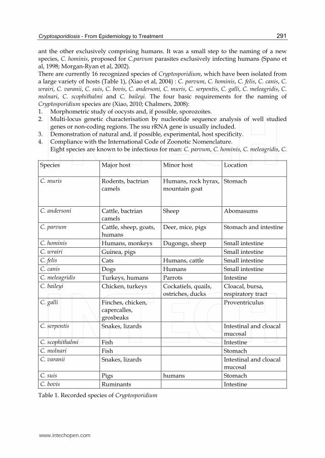
ant the other exclusively comprising humans. It was a small step to the naming of a new species, C. hominis, proposed for C.parvum parasites exclusively infecting humans (Spano et al, 1998; Morgan-Ryan et al, 2002). There are currently 16 recognized species of Cryptosporidium, which have been isolated from a large variety of hosts (Table 1), (Xiao et al, 2004) : C. parvum, C. hominis, C. felis, C. canis, C. wrairi, C. varanii, C. suis, C. bovis, C. andersoni, C. muris, C. serpentis, C. galli, C. meleagridis, C. molnari, C. scophithalmi and C. baileyi. The four basic requirements for the naming of Cryptosporidium species are (Xiao, 2010; Chalmers, 2008): 1. Morphometric study of oocysts and, if possible, sporozoites. 2. Multi-locus genetic characterisation by nucleotide sequence analysis of well studied
genes or non-coding regions. The ssu rRNA gene is usually included. 3. Demonstration of natural and, if possible, experimental, host specificity. 4. Compliance with the International Code of Zoonotic Nomenclature.
Eight species are known to be infectious for man: C. parvum, C. hominis, C. meleagridis, C.
Species
Major host Minor host Location
C. muris
Rodents, bactrian camels
Humans, rock hyrax, mountain goat
Stomach
C. andersoni Cattle, bactrian camels
Sheep Abomasums
C. parvum
Cattle, sheep, goats, humans
Deer, mice, pigs Stomach and intestine
C. hominis Humans, monkeys Dugongs, sheep Small intestine
C. wrairi Guinea, pigs Small intestine
C. felis Cats Humans, cattle Small intestine
C. canis Dogs Humans Small intestine
C. meleagridis Turkeys, humans Parrots Intestine
C. baileyi Chicken, turkeys Cockatiels, quails, ostriches, ducks
Cloacal, bursa, respiratory tract
C. galli Finches, chicken, capercalles, grosbeaks
Proventriculus
C. serpentis Snakes, lizards Intestinal and cloacal mucosal
C. scophithalmi Fish Intestine
C. molnari Fish Stomach
C. varanii Snakes, lizards Intestinal and cloacal mucosal
C. suis Pigs humans Stomach
C. bovis Ruminants Intestine
Table 1. Recorded species of Cryptosporidium
www.intechopen.com

Microbes, Viruses and Parasites in AIDS Process
292
felis, C. canis, C. suis, C. muris and C. andersoni, although some are reported extremely rarely, particularly the latter three species, and the pathogenicity of some species for man has not been proven (Chalmers, 2008). C. hominis and C. parvum are recognized globally as the most important Cryptosporidium species infecting humans (Dunand et al, 1997; Xiao & Ryan, 2004). Muthusamy et al characterized cryptosporidial infections in 48 human immunodeficiency virus-infected individuals in india by multilocus genotyping. C. hominis, C. parvum, C. felis, C. muris and C. meleagridis were identified (Muthusamy et al, 2006).
2.2 Life-cycle
Sporulated oocysts, containing 4 sporozoites, are excreted by the infected host through feces
and possibly other routes such as respiratory secretions. Following ingestion (and possibly
inhalation) by a suitable host, excystation occurs. The sporozoites are released and parasitize
epithelial cells of the gastrointestinal tract or other tissues such as the respiratory tract.
Cell invasion by sporozoites is followed by intracellular development to trophozoite.
In these cells, trophozoites undergo asexual multiplication (schizogony or merogony) to
form schizonts or meronts.
Asexual replication occurs by re-infection of merozoites, released by type I meronts.
Development of type II from type I meronts is the initial step of the asexual reproductive
cycle. Merozoites are released from type II meronts and re-infect neighbouring cells where
they develop into producing microgamonts (male) and macrogamonts (female) by sexual
multiplication (gametogony). Upon fertilization of the macrogamont by the microgamont
zygote develop, which undergoes further development into an oocyst.
Two different types of oocysts are produced, the thick-walled, which is commonly excreted
from the host, and the thin-walled oocyst, which is primarily involved in autoinfection.
Oocysts are infective upon excretion, thus permitting direct and immediate fecal-oral
transmission.
3. Epidemiology
3.1 Risk factors
The risk of infection increases in more profoundly immunosuppressed persons, as measured
by the CD4 T-lymphocyte counts (Houpt et al, 2005).
The incidence and severity of cryptosporidiosis increases as the CD4 T-lymphocyte cell
count falls, especially when it falls to below 200 cells/l (Navin et al, 1999; Pozio et al, 1997;
Sorvillo et al, 1999; Sara et al, 2008).
Various social and behavioral factors also increase the risk of infection. For example, in a
large multicenter European study, the risk of cryptosporidiosis was significantly lower for
intravenous drug users than for homosexual men and for women than for men, suggesting
that sexual behaviour may be an important risk factor (Pedersen et al, 1996).
3.2 Modes of transmission
Transmission of the parasite is facilitated by a relatively low infectious dose and by the
resistance of the parasite oocyst stage to commonly used disinfection techniques e.g
chlorination of dinking water.
www.intechopen.com

Cryptosporidiosis - From Epidemiology to Treatment
293
The infection is spread in a number of ways: from person to person, from animals, via food, and by water. Cryptosporidiosis is now the most common cause of waterborne disease in the world (Xiao & al, 2004). Because Cryptosporidium spp. infects humans and a wide variety of animals and because of
the ubiquitous presence of Cryptosporidium oocysts in the environment, humans can acquire
Cryptosporidium infections through several transmission routes (Clark, 1999; Griffiths, 1998).
In pediatric and elderly populations, especially in day care centers and nursing homes,
person-to-person transmission probably plays a major role in the spread of Cryptosporidium
infections (Xiao L et al, 2004; Neill et al, 1996; Tangermann, 1991). In rural areas, zoonotic
infections via direct contact with farm animals have been reported many times, but the
relative importance of direct zoonotic transmission of cryptosporidiosis is not entirely clear
(Miron et al, 1991).
Numerous outbreaks of cryptosporidiosis due to contaminated food or water (drinking or
recreational) have been reported in several industrialized nations, and studies have
sometimes identified water as a major route of Cryptosporidium transmission in areas where
the disease is endemic (Xiao et al, 2004; MacKenzie et al, 1991; Weinstein et al, 1993 ).
Many outbreaks in the United States have occurred in waterparks, community swimming
pools, and day care centers.
Large outbreaks due to the contamination of water supplies have been documented in
recent years (Richardson et al, 1991; Tzipori & Giovanni, 2008) and in one particular
outbreak, contamination of a water–treatment plant in Milwaukee (USA) was estimated to
result in infections in 403 000 people ( Mac Kenzie et al, 1994). This outbreak was associated
with municipal drinking water despite state-of-the art water treatment.
The magnitude of these outbreaks highlighted the significance of drinking contaminated
water as a major risk factor for contracting cryptosporidiosis in the USA (Tzipori &
Giovanni, 2008).
The sources and human infective potentials of Cryptosporidium oocysts in water, however,
are largely unclear.
One major problem in understanding the transmission of Cryptosporidium infection is the
lack of morphologic features that clearly differentiate one Cryptosporidium spp from many
others (Mac Kenzie et al, 1994). Hence, one cannot be sure which Cryptosporidium spp is
involved when one examines oocysts in clinical specimens under a microscope. Another
major problem is the inability to grow the organisms in large numbers from contaminated
sources. Adding to the diagnosis problem and technical difficulties is the confusion in the
taxonomy of Cryptosporidium spp., which is partially caused by the lack of consistency in the
classification of protozoan parasites in general.
C. hominis is believed to be transmitted exclusively between humans, whereas C. parvum is
transmitted between humans as well as through a zoonotic cycle usually involving
ruminants. Although calves are often implicated as the reservoir of C. parvum, the
importance of animals in transmission of C. parvum has been brought into question by
studies that found that humans are infected with subtypes that perpetuate almost
exclusively among humans (Siobhan & Tzipori, 2008).
The study of Cryptosporidium transmission dynamics is made more complex when atypical
species are considered. Human infections with C. meleagridis, C. muris, C. felis, C. canis, C.
andersoni and C. suis, though uncommon, have been reported (Mor MS & Tzipori, 2008; Xiao
& Ryan, 2004).
www.intechopen.com

Microbes, Viruses and Parasites in AIDS Process
294
The natural reservoirs of these species are believed to be poultry, rodents, cats, dogs, cattle, and pigs, respectively; however the broad host ranges are not well characterized. Host factors may also increase the likelihood of infection following exposure to these species, because they are more frequently identified in HIV-positive persons. Two recent studies in East Africa have attempted to correlate species diversity with area of residence. In Malawi, C. meleagridis and C. andersoni were detected only in children from a rural district, although 70% of C. parvum cases were identified in urban areas and C. hominis predominated in both settings (Mor & Tzipori, 2008; Mersha & Tiruneh, 1992). In Kenyan children, 4% of cases were due to C. canis, C. felis, C. meleagridis and C. muris, and there was no discernible difference by region (Gatei et al, 2006).
4. Clinical features
4.1 Gastrointestinal diseases
Cryptosporidium species infect the microvillus border of the gastrointestinal epithelium of a wide range of vertebrate hosts, including humans. Infected individuals show a wide spectrum of clinical presentations. The pathogenicity of Cryptosporidium varies with the species of parasites involved and the type, age and immune status of the hosts. In immunocompetent subjects, cryptosporidiosis is characterized by watery or mucoid diarrhoea and abdominal pain with spontaneous recovery following several days or weeks of symptoms (Mersha & Tiruneh, 1992). Immunocompetent hosts control and eliminate the infection, which typically causes acute, self-limited watery diarrhea lasting 5 to 10 days. However, in patients with defects in cellular immune responses (e.g. AIDS), Cryptosporidium spp frequently causes persistent or chronic diarrhea which can lead to death and can also involve the hepatobiliary and the respiratory tract. It is an opportunistic infection. The risk of infection increases in more profoundly immunosuppressed persons, as measured by the CD4-T-lymphocyte counts (Houpt et al, 2005; Morpeth & Thielman, 2006). Various presentations of cryptosporidiosis in HIV-positive patients were described (Blanshard et al, 1992; Mc Gowan et al, 1993; Manabe et al, 1998). U.S. workers described four clinical syndromes: chronic diarrhea (affecting 36% of patients), cholera-like disease (33%), transient diarrhea (15%), and relapsing illness (15%) (Blanshard et al, 1992, Manabe et al, 1998). Infected patients had a significantly shorter duration of survival from the time of diagnosis than did Cryptosporidium-negative AIDS patients (240 and 666 days, respectively; P = 0.0004). One aspect of chronic cryptosporidiosis in patients with AIDS is the large weight loss that many experience (Blanshard et al, 1992). One study from France reported that the severity of weight loss in such patients is independently associated with levels of nutrient intake (P < 0.005) and high stool frequency (P < 0.01) but not with nutrient malabsorption (Beaugerie et al, 1998). As well as developing a more severe form of typical gastrointestinal disease, people with HIV infection can develop atypical disease presentations, affecting body systems not usually affected in immunocompetent individuals. Some of these unusual presentations are discussed below.
4.2 Atypical gastrointestinal disease
Many cases of gastritis were described. One particularly problematic complication of gastric involvement is antral narrowing and gastric outlet obstruction (Iribarren et al, 1997; Moon et
www.intechopen.com

Cryptosporidiosis - From Epidemiology to Treatment
295
al, 1999). Such gastric outlet obstruction can lead to nausea and vomiting and eventually may cause a severe reduction in nutrient intake. A further unusual complication of cryptosporidiosis in AIDS patients is pneumatosis
cystoides intestinalis (Samson & Brown, 1996; Sidhu et al, 1994). This disease is
characterized by the presence of thin-walled, gas-containing cysts in the intestinal wall.
Sometimes these cysts can rupture, resulting in a pneumoretroperitoneum and
pneumomediastinum.
There is a case report of cryptosporidiosis affecting the oesophagus in a 2-year-old child and
resulting in vomiting and dysphagia (Kazlow et al, 1986). Finally, there is also a case report
of Cryptosporidium infection causing appendicitis (Oberhuber et al, 1991). The diagnosis was
confirmed histologically after an appendectomy was performed.
4.3 Biliary tract disease
Cholangitis, and particularly sclerosing cholangitis, is an important complication of AIDS.
Although not appearing to adversely affect survival, the disease can be a cause of significant
pain (Forbes et al, 1993). In a Spanish study of 43 AIDS patients with chronic diarrhea due to
Cryptosporidium infection, 8 patients (18.6%) were reported to have Cryptosporidium infection
of the common bile duct (Lopez-Velez et al, 1995).
4.4 Pancreatitis
A series of 15 autopsies on patients with AIDS and cryptosporidiosis showed that five had
evidence of infection of the pancreas (Godwin et al, 1991). Histological changes were
generally mild and were limited to hyperplastic squamous metaplasia.
Three people with AIDS presented with acute or chronic pancreatitis related to
cryptosporidiosis (Calzetti et al, 1997). All three patients had abdominal pain resistant to
analgesics, increased serum amylase levels, and abnormalities at both sonography and
computed tomography. Endoscopic retrograde cholangiopancreotography revealed
papillary stenosis in all three patients. It is difficult to assess the impact of cryptosporidiosis-
related pancreatic disease. Certainly, the first study does not suggest significant morbidity
due to Cryptosporidium in the pancreas (Hunter & Nichols, 2002).
4.5 Respiratory tract disease
In a study from Spain, 7 of 43 patients (16.3%) with chronic diarrhea due to Cryptosporidium
had Cryptosporidium oocysts detectable in the sputum (Lopez-Velez et al, 1995). Of these
seven patients, five had respiratory symptoms and an abnormal chest radiograph;
Mycobacterium tuberculosis was isolated in two of the five, and M. avium was isolated in
another two. The remaining two patients had no respiratory symptoms and normal chest
radiographs.
Other case series of patients with respiratory cryptosporidiosis were reported (Clavel et al,
1996; Hunter & Nichols, 2002). The majority of whom had another pathogen detected. So,
the exact role of Cryptosporidium in the respiratory symptoms is not clear.
Dunand et al reported on 5 of their own cases and reviewed 14 other cases of parasitic
sinusitis in HIV-positive patients from the literature (Dunand et al, 1997). Symptoms often
included fever and chills in addition to local tenderness and discharge. Although the
prognosis was frequently poor, this was due to other complications of HIV infection.
www.intechopen.com

Microbes, Viruses and Parasites in AIDS Process
296
5. Diagnosis
For intestinal cryptosporidiosis, a parasitological examination of the stools is not only readily accessible and repeatable, but also non invasive for the patient. Oocysts are eliminated at intermittent intervals so stool tests should be repeated three times at three-day intervals (Weber et al, 1991; John & Petri, 2006). Because of the small size of the Cryptosporidium oocysts, they are difficult to identify in fresh samples without specific coloration. Oocysts may be examined under phase-contrast microscopy after concentration by different techniques. Oocysts may be concentrated by the modified zinc sulfate centrifugal flotation technique or by Sheather’s sugar flotation. Another concentration technique involves formalin-ethyl acetate sedimentation followed by layering and flotation over hypertonic sodium chloride solution to separate oocysts from stool debris (Weber et al, 1991). Oocysts appear as highly refractile spherical bodies (4 to 6 µm). Several staining techniques can be used, applied to the swabs realized after concentration. Modified Ziehl Neelsen stain is the best staining technique. The oocysts appear as rose spherical elements and contain four sporozoites (fig 1). The background is stained blue or green depending on the counter-stain used (Fast Green, malachite green or aniline blue) (Weber et al, 1991; Sunnotel et al, 2006).
Fig. 1. Oocysts of Cryptosporidium sp stained by Modified Ziehl Neelsen stain
Cryptosporidium can also be detected by staining air-dried, methanol-fixed fecal smears with Giemsa’s stain. More recently, detection of oocysts is made by the use of copro-antigen detection kits, such as immunofluorescent antibody stains for microscopy, enzyme linked immunosorbent assays (ELISA) or immunochromatographic tests (Johnston et al, 2003). Cryptosporidium oocysts also have been detected in fecal specimens by fluorescent stain like auramine-rhodamine. The analytical sensitivity of these methods is generally in excess of 104 oocysts per gram faeces, depending on faecal consistency (Anusz et al, 1990; Weber et al, 1991), although immunofluorescence microscopy offers improved sensitivity (Arrowood, 1997).
www.intechopen.com

Cryptosporidiosis - From Epidemiology to Treatment
297
Cryptosporidiosis can be diagnosed by identifying organisms (meronts containing merozoites and gamonts containing micro- and macrogametes) in intestinal biopsy material. Organisms stain lightly with hematoxylin and eosin and appear as small round bodies on the mucosal surface of biopsy specimens. With Masson’s stain, a small red nucleus and blue cytoplasm can be distinguished in many of the organisms. Species identification is an important element of outbreak investigations particularly where the source is not clear, and it is important that this is underpinned by routine species identification to establish the background epidemiology (Chalmers, 2008). Even though oocysts of many Cryptosporidium spp are morphologically similar, morphometric measurement of oocysts can play a vital role in the differentiation of some Cryptosporidium spp. For example, the established species (interval) in birds and reptiles can easily be differentiated on the basis of the size and shape of oocysts. (Xiao et al, 2004). However, laboratory techniques are unable to discriminate between the two main species involved in human disease, C. hominis and C. parvum. Species identification can be achieved with molecular methods which provide a precious tool for detecting Cryptosporidium and differentiating species in biological samples of infected patients (Chalmers, 2008). However, Cryptosporidium presents two main problems for the isolation of DNA; the robust nature of the oocysts, requiring a disruption step prior to DNA extraction and the richness of PCR inhibitors in the sample matrix, requiring separation during DNA extraction. Currently, there is no international consensus of typing methods and many rely on DNA sequence analysis which can be time-consuming and costly (Chalmers, 2008). In 1991, Mark Laxer was the first to apply PCR to the detection of Cryptosporidium oocysts (Laxer et al, 1991). Among molecular methods, PCR combined with restriction fragment length polymorphism (PCR-RFLP), which was first applied to Cryptosporidium typing by Awad-El-Kariem (Awad-El-Kariem et al, 1994) is the most used. PCR-RFLP assay detecting an RsaI polymorphism in the Cryptosporidium oocyst wall protein (COWP) gene (Spano et al, 1997), and a species-specific assay targeting the small-subunit rRNA gene (Xiao et al, 1999) are the most popular. Papers describing such assays or their application to Cryptosporidium typing are too numerous to cite here. Other methods such as random amplification methods (Morgan et al, 1995), sequencing (Lopez-Velez et al, 1995), length polymorphisms of repetitive sequences (Feng et al, 2000) and conformational polymorphism detection methods (Gasser et al, 2001) should also be mentioned in this context. However, the relatively high cost of molecular methods at present has limited their application in developed and developing countries. The methodologies used in the detection of Cryptosporidium-specific antibodies vary widely, which complicates comparison of results. The use of the recombinant CP 41 antigen in a standardized serodiagnostic assay could provide a reliable and cost-effective method for assessing human exposure to Cryptosporidium in developing countries (Kjos et al, 2005).
6. Treatment
Despite the importance of cryptosporidiosis in mondial health, there has been any effective therapeutic specifically against Cryptosporidium infection, probably because of the unique intracellular extracytoplasmic location of Cryptosporidium and the poorly understood host-parasite interface. In addition, difficulties in laboratory propagation, including the absence of an ideal cell culture method, have limited high-throughput drug screening. Hundreds of drugs have been tested in the laboratory, and putative reports suggest that several,
www.intechopen.com

Microbes, Viruses and Parasites in AIDS Process
298
including paromomycin, macrolides (e.g., azithromycin and spiramycin) and albendazole, are partially effective (Mor & Tzipori, 2008; Tzipori, 1998). Clinical evaluations of these drugs have been disappointing, largely because they failed to clear the parasite from patients with HIV/AIDS. To date, the broad-spectrum, anti-infective nitazoxanide (NTZ) has shown the most promise against Cryptosporidium (Mor & Tzipori, 2008). It is used in many areas of the world because it appears to be well tolerated, it has a relatively low incidence of adverse effects, and it displays no significant known drug-to-drug interactions (Bobak, 2006). However, it is not effective against cryptosporidiosis in immunocompromised persons. A meta-analysis of randomized, placebo-controlled trial of NTZ (of which there are only 2) among immunocompromised patients concluded that NTZ was no more effective than placebo in resolving diarrhea and achieving parasitological clearance in HIV-positive persons (Abubakar, 2007). It has been speculated that HIV-positive persons may benefit from longer-duration regimens or higher doses of NTZ. However, a sustained clinical response was observed in only 59% of patients with HIV/AIDS who received off-label NTZ in a compassionate-use program (Rossignol, 2006). This study indicate that the drug should be administered 500 mg b.d. until clinical symptoms resolve and oocysts are eliminated from the stool. Doses may be escalated to 1000 mg b.d. to accelerate or improve parasitological response. Fourteen days of treatment are generally sufficient in patients with CD4 counts above 50 cells/mm3 while at least 8 weeks of treatment are likely required in patients with CD4 counts below 50 cells/mm3. Although refinement of the dosing regimen may improve clinical efficacy of NTZ, a prolonged therapeutic course will be impracticable in developing countries because of the expense and likely patient noncompliance. ColoPlus is a product which may be an important alternative or additional treatment in HIV-associated diarrhoea. It is a product based on bovine colostrums witch is the first milk the suckling calf receives from the cow. It is rich in immunoglobulins, growth factors, antibacterial peptides and nutrients. It supplies the calf with a passive immunity before its own active immunity is established. As well as having a high nutritional value, it is designed for slow passage through the gastrointestinal tract. Floren et al conducted a study on thirty patients with HIV-associated diarrhoea. The patients were treated with ColoPlus for 4 weeks in an open-labelled non-randomised study, after an observational period of one week. After a post-treatment period of another two weeks, treatment with anti-HIV drugs was started, if deemed appropriate. The effects on the frequency of stool evacuations per day, on body-weight, fatigue, haemoglobin, levels and CD4 + counts before and after treatment with ColoPlus were measured. There was a dramatic decrease in stool evacuations per day, a substantial decrease in body-weight and an increase in CD4 count by 125 % (Floren et al, 2006). In part because of the failure of other therapeutic approaches, there have been several attempts at passive antibody-based immunotherapy for cryptosporidial infections (Crabb, 1998). These have also had limited success. One therapeutic intervention that has a dramatic effect on cryptosporidiosis in AIDS patients is antiretroviral therapy leading to recovery of the CD4 count. (Foudraine, 1998; Maggi et al, 2000; Miao et al, 2000; Morpeth & Thielman, 2006). The authors noted that resolution of the diarrhea seemed to be related to an increased CD4 - cell count rather than
www.intechopen.com

Cryptosporidiosis - From Epidemiology to Treatment
299
to the viral load. These findings give further support to the observation that it is cellular immunity that is of paramount importance in clearing Cryptosporidium infection.
7. Prophylaxis
Because of the risk of acquiring a life-threatening disease, people with AIDS should take the
following specific measures to help reduce the risk of waternborne cryptosporidiosis: boil
drinking water for 1 minute, or filter drinking water with devices that remove particles 1 µm
and larger, or use bottled drinking water, especially water obtained from underground
sources (eg. springs or wells), which are less likely to be contamined by Cryptosporidium
oocysts. However, the boiling of water is the most certain method of killing Cryptosporidium
oocysts. They should take additional precautions, including avoiding contact with young
pets, and avoiding swallowing water while swimming (John & Petri, 2006).
The realization that Cryptosporidium oocysts are resistant to many chemical disinfectants
(Tzipori & Giovanni, 2008; Rochelle et al, 2005) led to a search for methods that can
inactivate oocysts without generating harmful by products. Much attention has focused on
UV irradiation and ozone as alternative methods capable of inactivating waterborne oocysts
(Tzipori & Giovanni, 2008; Lloyd & Drury, 2002). However, control of surface-water
contamination is being emphasized as a first measure to reduce the occurrence of
waterborne oocysts. Regulations aimed at reducing the risk of exposure to waterborne
oocysts have been put in place; for example, the Long Term 2 Enhanced Surface Water
Treatment Rule in the USA and regulations in the UK requiring continuous monitoring for
Cryptosporidium oocysts in drinking water. A treatment-based standard of one oocyst in 10 l
has been adopted (Sidhu et al, 1994).
8. Conclusion
We would conclude that research over the last three decades has dramatically increased our
knowledge on cryptosporidiosis, but key questions still remain unclear.
With the new interest in Cryptosporidium generated by the emergence of the latter as the
cause of human disease, much research is ongoing and will provide continuing information
concerning cryptosporidiosis in the future.
Future developments need to include harmonisation of rapid and more cost effective
methods.
Effective therapies are likely to become available in the near future. Access to endogenous
forms and immortalization of strains in culture or by cryopreservation remain major
challenges, which will require new ideas and new approaches.
Identification of Cryptosporidium isolates to species level and of subtyping is indispensable
for appropriate control measures during outbreaks.
9. References
Abubakar, I., Aliyu, SH, Arumugam, C, Usman, NK & Hunter, PR. (2007). Treatment of
cryptosporidiosis in immunocompromised individuals: systematic review and
meta-analysis. Br J Clin Pharmacol, Vol.63, No.4, (April 2007), pp. 387-393.
www.intechopen.com

Microbes, Viruses and Parasites in AIDS Process
300
Alvarez-Pellitero, P. & Sitja-Bobadilla, A. (2002). Cryptosporidium molnari n. sp.
(Apicomplexa: Cryptosporidiidae) infecting two marine fish species, Sparus aurata
L. and Dicentrarchus labrax. Int J Parasitol, Vol.32, No.8, (July 2002), pp. 1007-21.
Awad-el-Kariem, FM., Warhurst, DC & McDonald, V. (1994).Detection and species
identification of Cryptosporidium oocysts using a system based on PCR and
endonuclease restriction. Parasitology, Vol.109, No.1, (July 1994), pp. 19-22.
Barker, IK. & Carbonell, PL. (1974). Cryptosporidium agni sp.n. from lambs, and
Cryptosporidium bovis sp.n. from a calf, with observations on the oocyst. Z
Parasitenkd, Vol.44, No.4, (1974), pp. 289-98
Beaugerie, L., Carbonnel, F, Carrat, F, Rached, AA, Maslo, C, Gendre, JP, Rozenbaum, W &
Cosnes, J.(1998). Factors of weight loss in patients with HIV and chronic diarrhea. J
Acquired Immune Defic Syndr, Vol.19, No.1, (September 1998); pp. 34-9.
Bern, C., Hernandez, B, Lopez, MB, Arrowood, MJ, De Merida, AM. & Klein, R. E. (2000).
The contrasting epidemiology of Cyclospora and Cryptosporidium among outpatients
in Guatemala. Am J trop Med Hyg, Vol.63, No.(5-6), (Nov-Dec 2000), pp.231-5.
Bhattacharya, MK., Teka, T, Faruque, AS. & Fuchs, GJ.(1997). Cryptosporidium infection in
children in urban Bangladesh. J Trop Pediatr, Vol.43, No.5, (October 1997), pp.
282-6.
Blanshard, C., Jackson, AM, Shanson, DC, Francis, N. & B. G. Gazzard.(1992).
Cryptosporidiosis in HIV-seropositive patients. Q J Med, Vol.85, No. (307-308),
(Nov-Dec 1992), pp. 813-23.
Bobak, DA.(2006). Use of nitazoxanide for gastrointestinal tract infections: treatment of
protozoan parasitic infection and beyond. Curr Infect Dis Rep, Vol.8, No.2, (March
2006), pp. 91-5.
Calzetti, C., Magnani, G, Confalonieri, D, Capelli, A, Moneta, S, Scognamiglio, P. &
Fiaccadori, F.(1997). Pancreatite da Cryptosporidium parvum in pazienti con grave
deficit immunitario correlato ad infezione da HIV. Ann Ital Med Int, Vol.12, No.2,
(Apr-Jun 1997), pp. 63-6.
Chalmers RM. (2008). Cryptosporidium: from laboratory diagnosis to surveillance and
outbreaks. Parasite, Vol.15, No. 3, (September 2008), pp. 372-8.
Chen, X-M., Keithly, JS, Paya, CV. & LaRusso, NF. (2002). Cryptosporidiosis. N Engl J Med,
Vol.346, No.22, (May, 2002), pp. 1723-31.
Clark, DP. (1999). New insights into human cryptosporidiosis. Clin Microbiol Rev, Vol.12,
No.4, (October 1999), pp. 554-63.
Clavel, A., Arnal, AC, Sanchez, EC, Cuesta, J, Letona, S, Amiguet, JA, Castillo, FJ, Varea, M.
& Gomez-Lus, R.(1996). Respiratory cryptosporidiosis: case series and review of the
literature. Infection, Vol.24, No.5, (Sep-Oct 1996), pp. 341-6.
Crabb, JH. (1998). Antibody-based immunotherapy of cryptosporidiosis. Adv. Parasitol,
Vol.40, (1998), pp. 121-49.
Dann, SM., Wang, H-C, Robinson, P, Castellanos-Gonzalez, A, Lewis, DE & Clinton White,
A. (2008). Intestinal Immune Response to Human Cryptosporidium sp. infection
Infection and immunity, Vol.76, No.1, (January 2008), pp. 23-9.
Dunand, VA., Hammer, SM, Rossi, R, Poulin, M, Albrecht, MA, Doweiko, JP, DeGirolami,
PC, Coakley, E, Piessens, E. & Wanke, CA.(1997). Parasitic sinusitis and otitis in
www.intechopen.com

Cryptosporidiosis - From Epidemiology to Treatment
301
patients infected with human immunodeficiency virus: report of five cases and
review. Clin Infect Dis, Vol.25, No.2, (August 1997), pp. 267-72.
Fayer, R., Trout, JM, Xiao, L, Morgan, UM, Lai, AA. & Dubey, JP. (2001). Cryptosporidium
canis n. sp. from domestic dogs. J Parasitol, Vol.87, No.6, (December 2001), pp. 1415-
22.
Feng, X., Rich , SM, Akiyosh, D, Tumwine, JK, Kekitiinwa, A, Nabukeera, N, Tzipori, S &
Widmer, G.(2000). Extensive polymorphism in Cryptosporidium parvum identified
by multilocus microsatellite analysis. Appl Environ Microbiol, Vol.66, No.8, (August
2000), pp. 3344-9.
Floren, CH., Chinenye, S, Elfstrand, L, Hagman, C & Ihse, I. (2006); ColoPlus, a new product
based on bovine colostrum, alleviates HIV-associated diarrhoea. Scand J
Gatsroenterol, Vol.41, No.6, (June 2006), pp. 682-6.
Forbes, A., Blanshard, C & Gazzard, B. (1993). Natural history of AIDS related sclerosing
cholangitis: a study of 20 cases. Gut, Vol.34, No.1, (January 1993), pp. 116-21.
Foudraine, NA., Weverling, GJ, van Gool, T, Roos, MT, de Wolf, F, Koopmans, PP, van den
Broek, PJ, Meenhorst, PL, Leeuwen, Rvan, Lange JM & Reiss, P.(1998).
Improvement of chronic diarrhoea in patients with advanced HIV-1 infection
during potent antiretroviral therapy. AIDS, Vol.12, No.1, (January 1998), pp. 35-41
Gasser, RB., Zhu, X, Caccio, S, Chalmers, R, Widmer, G, Morgan, UM, Thompson, RC,
Pozio, E & Browning, GF.(2001).Genotyping Cryptosporidium parvum by single-
strand conformation polymorphism analysis of ribosomal and heat shock gene
regions. Electrophoresis, Vol.22, No.3, ( February 2001), pp. 433-7.
Gatei, W., Wamae, CN, Mbae, C, Waruru, A, Mulinge, E, Waithera, T, Gatika, SM, Kamwati,
SK, Revathi, G & Hart, CA. (2006). Cryptosporidiosis: prevalence, genotype
analysis, and symptoms associated with infections in children in Kenya. Am J Trop
Med Hyg, Vol.75, No.1, (July 2006), pp. 78-82.
Godwin, TA. (1991). Cryptosporidiosis in the acquired immunodeficiency syndrome: a
study of 15 autopsy cases. Hum Pathol, Vol.22, No.12, (December 1991), pp. 1215-24.
Griffiths, JK.(1998). Human cryptosporidiosis: epidemiology, transmission, clinical disease,
treatment, and diagnosis. Adv Parasitol, Vol.40, (1998), pp. 37–85.
Houpt, ER., Bushen, OY, Sam, NE, Kohli, A, Asgharpour, A, Ng, CT, Calfee, DP, Guerrant,
RL, Maro, V, Ole-Nguyaine, S & Shao, JF.(2005).Short report: asymptomatic
Cryptosporidium hominis infection among human immunodeficiency virus–infected
patients in Tanzania. Am J Trop Med Hyg, Vol.73, No.3, (September 2005), pp. 520-2.
Hunter, PR., Nichols G.(2002). Epidemiology and clinical features of Cryptosporidium
infection in immunocompromised patients. Clin Microbiol Rev, Vol.15, No.1,
(January 2002), pp. 145-54.
Iribarren, JA., Castiella, A, Lobo, C, Lopez, P, Wichmann, M A von, Arrizabalaga, JF,
Rodriguez-Arrondo, J. & Alzate, LF.(1997). AIDS associated cryptosporidiosis with
antral narrowing. A new case. J Clin Gastroenterol, Vol.25, No.4, (December 1997),
pp. 693-4.
John, DT Petri WA. (2006). Lumen-Dwelling Protozoa. In Medical Parasitology, Saunders
Elsevier, pp.(68-71).
www.intechopen.com

Microbes, Viruses and Parasites in AIDS Process
302
Johnston, sp., Ballar, MM, Beach, MJ, Causer, L. & Wilkins, PP. (2003). Evaluation of three
commercial assays for fecal speciments. J Clin Microbiol, Vol.41, No.2, (February
2003), pp. 623-6.
Kazlow, PG., Shah, K, Benkov, KJ, Dische, R. & LeLeiko, NS. (1986). Esophageal
cryptosporidiosis in a child with acquired immune deficiency syndrome.
Gastroenterology, Vol.91, No.5, (November 1986), pp. 1301-3.
Kjos, SA., Jenkins, M, Okhuysen, PC. & Chappell, CL. (2005). Evaluation of recombinant
oocyst protein CP 41 for detection of Cryptosporidium-specific antibodies. Clin Diag
Lab Immunol, Vol.12, No.2, (February 2005), pp. 268-72.
Laxer, MA., Timblin, BK & Patel, RJ. (1991). DNA sequences for the specific detection of
Cryptosporidium parvum by the polymerase chain reaction. Am J Trop Med Hyg,
Vol.45, No.6, (December 1991), pp. 688-94.
Levine, ND.(1984). Taxonomy and review of the coccidian genus Cryptosporidium (protozoa,
apicomplexa). J Protozool, Vol. 31, No.1, (February 1984), pp.94-98.
Lindsay, DS., Upton, SJ, Owens, DS, Morgan, UM, Mead, JR. & Blagburn, BL.(2000).
Cryptosporidium andersoni n. sp. (Apicomplexa: Cryptosporiidae) from cattle, Bos
taurus. J Eukaryot Microbiol, Vol. 47, No.1, (Jan-Feb 2000), pp. 91-5.
Lloyd, A & Drury, D.(2002). Continuous monitoring for Cryptosporidium–a novel approach
to public health protection. Water Sci Technol, Vol.46, No. (11-12), (2002), pp. 297-
301.
Lopez-Velez, R., Tarazona, R, Garcia Camacho, A, Gomez-Mampaso, E, Guerrero, A,
Moreira, V & Villanueva, R.(1995). Intestinal and extraintestinal cryptosporidiosis
in AIDS patients. Eur J Clin Microbiol Infect Dis, Vol.14, No.8, (August 1995), pp.
677-81.
Ma, P. & Soave, R. (1983). Three-step stool examination for cryptosporidiosis in 10
homosexual men with protracted watery diarrhea. J Infect Dis, Vol.147, No.5, (1983)
pp.824-8.
Ma P. (1984). Cryptosporidium and the enteropathy of immune deficiency. J Pediatr
Gastroenterol Nutr, Vol.3, No.4, (September 1984), pp. 488-90.
Mac Kenzie., WR, Hoxie, NJ, Proctor, ME, Gradus, MS, Blair, KA, Peterson, DE,
Kazmierczak, JJ, Addiss, DG, Fox, KR, Rose, JB. & al.(1994). A massive outbreak in
Milwaukee of Cryptosporidium infection transmitted through the public water
supply. N Engl J Med; Vol.331, No.3, (July 1994), pp. 161-7.
MacKenzie, WR., Hoxie, NJ, Proctor, ME, Gradus, MS, Blair, KA, Peterson, DE,
Kazmierczak, J J, Addiss, DG, Fox, K R, Rose, JB & al.(1994). A massive outbreak in
Milwaukee of Cryptosporidium infection transmitted through the public water
supply. N Engl J Med, Vol.331, No.3, (July 1994), pp. 161-7.
Maggi, P., Larocca, AM, Quarto, M, Serio, G, Brandonisio, O, Angarano, G & Pastore, G.
(2000). Effect of antiretroviral therapy on cryptosporidiosis and microsporidiosis in
patients infected with human immunodeficiency virus type 1. Eur J Clin Microbiol
Infect Dis, Vol.19, No.3, (March 2000), pp. 213-7.
Manabe, YC., Clark, DP, Moore, R D, Lumadue, JA, Dahlman, H R. Belitsos, PC, Chaisson
RE. & Sears. C. L. (1998). Cryptosporidiosis in patients with AIDS: correlates of
disease and survival. Clin Infect Dis, Vol.27, No.3, (September 1998), pp. 536-42.
www.intechopen.com

Cryptosporidiosis - From Epidemiology to Treatment
303
Mc Gowan, I., Hawkins, AS & Weller, IV.(1993).The natural history of cryptosporidial
diarrhoea in HIV-infected patients. AIDS, Vol.7, No.3, (March 1993), pp. 349-54.
Meisel, JL., Perera, DR, Meligro, C & Rubin, CE.(1976). Overwhelming watery diarrhoea
associated with a Cryptosporidium in an immunosuppressed patient.
Gastroenterology, Vol.70, No.6, (June 1976), pp. 1156-60.
Mersha, D & Tiruneh, M.(1992). Frequency of isolation of Cryptosporidium oocysts in
Ethiopian children with acute diarrhoeal disease. East Afr Med J, Vol.69, No.6, (June
1992), pp. 314-5.
Miao, YM., Awad-El-Kariem, FM, Franzen, C, Ellis, DS, Muller, A, Counihan, HM, Hayes,
PJ. & Gazzard, BG.(2000). Eradication of Cryptosporidia and Microsporidia following
successful antiretroviral therapy. J Acquired Immune Defic Syndr, Vol.25, No.2,
(October 2000), pp. 124-9.
Miron, D., Kenes, J & Dagan, R.(1991). Calves as a source of an outbreak of
cryptosporidiosis among young children in an agricultural closed community.
Pediatr Infect Dis J, Vol.10, No.6, (June 1991), pp. 438-41.
Moon, A., Spivak, W. & Brandt,L. J.(1999).Cryptosporidium-induced gastric obstruction in a
child with congenital HIV infection: case report and review of the literature. J
Pediatr Gastroenterol Nutr, Vol.28, No.1, (January 1999), pp. 108-11.
Mor, MS., & Tzipori, S. (2008). Cryptosporidiosis in Children in Sub-Saharan Africa: A
Lingering. Clin Infect Dis, Vol.47, No.7, (October 2008), pp. 15-21.
Morgan, UM, Constantine, CC, O'Donoghue, P, Meloni, BP, O'Brien, PA & Thompson
,RC.(1995). Molecular characterization of Cryptosporidium isolates from humans and
other animals using random amplified polymorphic DNA analysis. Am J Trop Med
Hyg, Vol.52, No.6, (June 1995), pp. 559-64.
Morgan-Ryan, UM., Fall, A, Ward, LA, Hijjawi, N, Sulaiman, I, Fayer, R, Thompson, RC,
Olson, M, Lal,A. & Xiao, L.(2002). Cryptosporidium hominis n. sp. (Apicomplexa:
Cryptosporidiidae) from Homo sapiens. J Eukaryot Microbiol, Vol.49, No.6,
(November 2002), pp. 433-40.
Morpeth, SC, Thielman, NM. (2006).Diarrhea in patients with AIDS. Curr Treat Options
Gastroenterol, Vol.9, No.1, (February 2006), pp. 23-37.
Muthusamy,D, Rao, SS, Ramani, S, Monica, B, Banerjee, I, Abraham, OC, Mathai, DC,
Primrose, B, Muliyil, J, Wanke, CA, Ward, HD & Kang, G.(2006). Multilocus
genotyping of Cryptosporidium sp. Isolates from human immunodeficiency virus-
infected individuals in south india. J Clin Microbiol, Vol.44, No.2, (February 2006),
pp. 632-4.
Navin, TR., Weber, R, Vugia, DJ, Rimland, DJ, Roberts, M, Addiss, DG, Visvesvara, GS,
Wahlquist, SP, Hogan, SE, Gallagher, L E, Juranek, DD, Schwartz, DA, Wilcox, CM,
Wilcox, JM, Stewart, SE, Thompson, r & Bryan, RT.(1999). Declining CD4 T-
lymphocyte count is associated with increased risk of enteric parasitosis and
chronic diarrhea: results of a 3-year longitudinal study. J Acquir Immune Defic Synd,
Vol.20, No.2, (February 1999), pp. 154-9.
Neill, MA., Rice, SK, Ahmad, NV & Flanigan, T. P.(1996). Cryptosporidiosis: an
unrecognized cause of diarrhea in elderly hospitalized patients. Clin Infect Dis,
Vol.22, No.1, (January 1996), pp. 168-70.
www.intechopen.com
Microbes, Viruses and Parasites in AIDS Process
304
Newman, RD., Sears, CL, Moore, SR, Nataro, JP, Wuhib, TD, Agnew, A, Guerrant R L. &
Lima, AAM. (1999). Longitudinal study of Cryptosporidium infection in children in
northeastern Brazil. J Infect Dis, Vol.180, No.1, (July 1999), pp. 167-75
Nime, FA., Burek, JD, Page, DL, Holscher, MA. & Yardley, JH.(1976). Acute enterocolitis in a
human being infected with the protozoan Cryptosporidium. Gastroenterology, Vol.70,
No.4, (April 1976), pp. 592-8.
Nina, JM, McDonald, V, Deer, RM, Wright, SE, Dyson, DA, Chiodini, PL & McAdam, KP.
1992. Comparative study of the antigenic composition of oocyst isolates of
Cryptosporidium parvum from different hosts. Parasite Immunol, Vol.14, No.2, (March
1992), pp. 227-32.
Oberhuber, G., Lauer, E, Stolte, M & Borchard, F.(1991). Cryptosporidiosis of the appendix
vermiformis: a case report. Z Gastroenterol, Vol.29, No.11, (November 1991), pp.
606-8.
Ogunkolade, BW, Robinson, HA, McDonald, V, Webster, K & Evans, DA.(1993). Isoenzyme
variation within the genus Cryptosporidium. Parasitol Res, Vol.79, No.5, (1993), pp.
385-8.
Ortega, YR., Sheehy, RR, Cama, VA, Oishi KK & Sterling CR. (1991). Restriction fragment
length polymorphism analysis of Cryptosporidium parvum isolates of bovine and
human origin. J Protozool, Vol.38, No.6, (Nov-Dec 1991), pp.(40S-41S).
Pedersen, C., Danner, S, Lazzarin, A, Glauser, M. P, Weber, R, Katlama, C, Barton, S. E &
Lundgren, J. D.( 1996). Epidemiology of cryptosporidiosis among European AIDS
patients. Genitourin Med, Vol. 72, No.2, (April 1996) pp.128–131.
Pozio, E., Rezza, G, Boschini, A, Pezzotti, P, Tamburrini, A, Rossi, P, Di Fine, M, Smacchia,
C, Schiesari, A, Gattei, E, Zucconi, R. & Ballarini, P. (1997). Clinical
cryptosporidiosis and human immunodeficiency virus (HIV)-induced
immunosuppression: findings from a longitudinal study of HIV-positive and HIV-
negative former injection drug users. J Infec Dis, Vol.176, No.4, (October 1997),
pp.969-75.
Richardson, AJ., Frankenberg, RA, Buck, AC, Selkon, JB, Colbourne, JS, Parsons, JW &
Mayon-White, RT.(1991).An outbreak of waterborne cryptosporidiosis in Swindon
and Oxfordshire. Epidemiol Infect, Vol.107, No.3, (December 1991), pp. (485-95).
Rochelle, PA., Upton, SJ, Montelone, BA & Woods, K.(2005). The response of
Cryptosporidium parvum to UV light. Trends Parasitol, Vol.21, No.2, (February 2005),
pp.81-7.
Rossignol, JF.(2006). Nitazoxanide in the treatment of acquired immune deficiency
syndrome-related cryptosporidiosis: results of the United States compassionate use
program in 365 patients. Aliment Pharmacol Ther, Vol.24, No.5, (September 2006),
pp.887-94.
Samson, VE. & Brown, W. R. (1996). Pneumatosis cystoides intestinalis in AIDS-associated
cryptosporidiosis. More than an incidental finding? J Clin Gastroenterol, Vol.22,
No.4, (June 1996), pp.311-2.
Tzipori, S & Widmer, G.(2008).A hundred-year retrospective on cryptosporidiosis.Trends
Parasitol, Vol.24, No.4, (April 2008), pp.184-9.
www.intechopen.com

Cryptosporidiosis - From Epidemiology to Treatment
305
Sidhu, S., Flamm, S & Chopra, S. (1994). Pneumatosis cystoides intestinalis: an incidental
finding in a patient with AIDS and cryptosporidial diarrhea. Am. J. Gastroenterol,
Vol.89, No.9, (September 1994), pp. 578-9.
Sorvillo, F., Beall, G, Turner, PA, Beer, VL, Kovacs, AA, Kraus, P, Masters, D. & Kerndt,
PR.(1998). Seasonality and factors associated with cryptosporidiosis among
individuals with HIV infection. Epidemiol Infect, Vol.21, No.1, (August 1998),
pp.197-204.
Spano, F., Putignani, L, Crisanti, A, Sallicandro, P, Morgan, UM, Le Blancq, SM, Tchack, L,
Tzipori, S & Widmer, G.(1998). Multilocus genotypic analysis of Cryptosporidium
parvum isolates from different hosts and geographical origins. J Clin Microbiol,
Vol.36, No.11, (November 1998), pp.3255-9.
Spano, F., Putignani, L, McLauchlin, J, Casemore, DP & Crisanti, A.(1997). PCR-RFLP
analysis of the Cryptosporidium oocyst wall protein (COWP) gene discriminates
between C. wrairi and C. parvum, and between C. parvum isolates of human and
animal origin. FEMS Microbiol Lett, Vol.150, No.2, (May 1997), pp.209-17.
Strong, WB., Gut, J & Nelson, RG.(2000). Cloning and sequence analysis of a highly
polymorphic Cryptosporidium parvum gene encoding a 60-kilodalton glycoprotein
and characterization of its 15- and 45-kilodalton zoite surface antigen products.
Infect Immun, Vol.68, No.7, (July 2000), pp. 4117-34.
Sunnotel, O., Lowery, CJ, Moore, JE, Dooley, JS, Xiao, L, Millar, BC, Rooney, PJ & Snelling,
WJ. (2006). Cryptosporidium. Lett Appll Microbiol, Vol.43, No.1, (July 2006), pp. 7-16.
Tangermann, RH., Gordon, S, Wiesner, P & Kreckman L.(1991). An outbreak of
cryptosporidiosis in a day-care center in Georgia. Am J Epidemiol, Vol.133, No.5,
(March 1991), pp. 471-6.
Tyzzer, E.E.(1907). A sporozoon found in the peptic glands of the common mouse. Proc Soc
Exp Biol Med, Vol.5, (1907), pp.12–13.
Tyzzer, E.(1910). An extracelluar coccidium, Cryptosporidium muris (gen. & sp. nov.), of the
gastric glands of the common mouse. J Med Res, Vol.18, (1910), pp.487–509.
Tyzzer, E. (1912). Cryptosporidium parvum (sp. nov.), a coccidium found in the small
intestine of the common mouse. Arch Protisenkd, Vol.26, (1912), pp.394–412.
Tzipori, S. (1998). Cryptosporidiosis: laboratory investigations and chemotherapy. Adv.
Parasitol, Vol.40, (1998), pp.187–221.
Weber, R., Bryuan, RT, Bishop, HS, Walquist, SP, Sullivan, JJ, & Juranek, D,D. (1991).
Threshold of detection of Cryptosporidium oocysts in human stool specimens:
evidence for low sensitivity of current methods. Journal of Clinical Microbiology,
Vol.29, No.7, (July 1991), pp.1323-7.
Weinstein, P., Macaitis, M, Walker, C & Cameron, S. (1993). Cryptosporidial diarrhoea in
South Australia. An exploratory case-control study of risk factors for transmission.
Med J Aust, Vol.158, No.2, (January 1993), pp.117-9.
Xiao, L., Escalante, L, Yang, C, Sulaiman, I, Escalante, AA, Montali, RJ, Fayer, R & Lal,
AA.(1999). Phylogenetic analysis of Cryptosporidium parasites based on the small-
subunit Rrna gene locus. Appl Environ Microbiol, Vol.65, No.4, (April 1999), pp.1578-
83.
www.intechopen.com

Microbes, Viruses and Parasites in AIDS Process
306
Xiao, L., Fayer, R, Ryan, U & Steve, J. (2004). Cryptosporidium Taxonomy: Recent Advances
and Implications for Public Health. Clinical Microbial Reviews, Vol.17, No.1,
(January 2004), pp.72-97.
Xiao, L. & Ryan, UM.(2004). Cryptosporidiosis: an update in molecular epidemiology. Curr
Opin Infect Dis, Vol.17, No.5, (October 2004), pp.483-90.
Xiao, L. (2010). Molecular epidemiology of cryptosporidiosis: An update. Experimental
Parasitology, Vol.124, No.1, (January 2010), pp.80-9.
www.intechopen.com

Microbes, Viruses and Parasites in AIDS ProcessEdited by Prof. VladimÃr Zajac
ISBN 978-953-307-601-0Hard cover, 390 pagesPublisher InTechPublished online 19, October, 2011Published in print edition October, 2011
InTech EuropeUniversity Campus STeP Ri Slavka Krautzeka 83/A 51000 Rijeka, Croatia Phone: +385 (51) 770 447 Fax: +385 (51) 686 166www.intechopen.com
InTech ChinaUnit 405, Office Block, Hotel Equatorial Shanghai No.65, Yan An Road (West), Shanghai, 200040, China Phone: +86-21-62489820 Fax: +86-21-62489821
The main goal in compiling this book was to highlight the situation in Africa in terms of AIDS and opportunisticdiseases. Several chapters reveal great poverty, an apocalyptic situation in many parts of Africa. Globalmigration of people resulted in their exposure to pathogens from all over the world. This fact has to beacknowledged and accepted as African reality. New, unconventional hypotheses, not determined byestablished dogmas, have been incorporated into the book, although they have not yet been sufficientlyvalidated experimentally. It still applies that any dogma in any area of science, and medicine in particular,has and always will hinder progress. According to some biologists, in the future, AIDS is very likely to occur in anumber of variations, as a direct result of the ongoing processes in the global human society. Thus, weurgently need a comprehensive solution for AIDS, in order to be ready to fight other, much more dangerousintruders.
How to referenceIn order to correctly reference this scholarly work, feel free to copy and paste the following:
Anane Sonia (2011). Cryptosporidiosis - From Epidemiology to Treatment, Microbes, Viruses and Parasites inAIDS Process, Prof. VladimÃr Zajac (Ed.), ISBN: 978-953-307-601-0, InTech, Available from:http://www.intechopen.com/books/microbes-viruses-and-parasites-in-aids-process/cryptosporidiosis-from-epidemiology-to-treatment
Related Documents


