3,350+ OPEN ACCESS BOOKS 108,000+ INTERNATIONAL AUTHORS AND EDITORS 114+ MILLION DOWNLOADS BOOKS DELIVERED TO 151 COUNTRIES AUTHORS AMONG TOP 1% MOST CITED SCIENTIST 12.2% AUTHORS AND EDITORS FROM TOP 500 UNIVERSITIES Selection of our books indexed in the Book Citation Index in Web of Science™ Core Collection (BKCI) Chapter from the book Advances in the Preclinical Study of Ischemic Stroke Downloaded from: http://www.intechopen.com/books/advances-in-the-preclinical- study-of-ischemic-stroke PUBLISHED BY World's largest Science, Technology & Medicine Open Access book publisher Interested in publishing with IntechOpen? Contact us at [email protected]

Welcome message from author
This document is posted to help you gain knowledge. Please leave a comment to let me know what you think about it! Share it to your friends and learn new things together.
Transcript

3,350+OPEN ACCESS BOOKS
108,000+INTERNATIONAL
AUTHORS AND EDITORS114+ MILLION
DOWNLOADS
BOOKSDELIVERED TO
151 COUNTRIES
AUTHORS AMONG
TOP 1%MOST CITED SCIENTIST
12.2%AUTHORS AND EDITORS
FROM TOP 500 UNIVERSITIES
Selection of our books indexed in theBook Citation Index in Web of Science™
Core Collection (BKCI)
Chapter from the book Advances in the Preclinical Study of Ischemic StrokeDownloaded from: http://www.intechopen.com/books/advances-in-the-preclinical-study-of-ischemic-stroke
PUBLISHED BY
World's largest Science,Technology & Medicine
Open Access book publisher
Interested in publishing with IntechOpen?Contact us at [email protected]
5
Cerebral Ischemia Induced Proteomic Alterations: Consequences for
the Synapse and Organelles
Willard J. Costain1, Arsalan S. Haqqani2, Ingrid Rasquinha1, Marie-Soleil Giguere2 and Jacqueline Slinn3
1Glycosyltransferases and Neuroglycomics, Institute for Biological Sciences, National Research Council, Ottawa, ON,
2Proteomics, Institute for Biological Sciences, National Research Council, Ottawa, ON, 3Cerebrovascular Research, Institute for Biological Sciences,
National Research Council, Ottawa, ON, Canada
1. Introduction
The synapse is the focal point for neuronal communication and neuron-glia interactions.
Synaptic structure and function are intimately related and many of the proteins that provide
structure to the synapse also regulate synaptic function (Abe et al. 2004, Couchman 2003,
Ehlers 2002, Passafaro et al. 2003). The synaptic structure - function relationship is highly
apparent during pathological conditions. This is exemplified in neurodegenerative
disorders, such as Alzheimer’s disease, where synaptic function is positively correlated with
neuronal function (Gasic & Nicotera 2003) and viability (Deisseroth et al. 2003), with
synaptic pathology preceding cell death (Gasic & Nicotera 2003).
Maintenance of synaptic structure and functionality is a process that is highly energy
dependent. Studies of synaptosomal morphology and metabolism have indicated that the
synapse is highly susceptible to ischemic damage (Pastuszko et al. 1982, Rafalowska et al.
1980, Sulkowski et al. 2002, Enright et al. 2007, Zhang & Murphy 2007). This exceptional
requirement for energy necessitates the localization of numerous mitochondria proximal to
the synaptic bouton and mitochondria are a commonly observed feature in synaptosomal
preparations (Costain et al. 2008). Synaptosomal metabolic activity (Rafalowska et al. 1980)
and the rate of neurotransmitter re-uptake (Pastuszko et al. 1982) are decreased following
acute hypoxia. Furthermore, Sulkowski et al (2002) observed that ischemia decreases
synaptosomal oxygen consumption and metabolic capacity for a period of 24 hours.
Cerebral ischemia induces a marked depletion in synaptic vesicle content and increases the number of damaged mitochondria (Sulkowski et al. 2002, Costain et al. 2008). Importantly, ischemia-induced alterations in synaptosomal morphology are indistinguishable from that of brain slices (Sulkowski et al. 2002). Ischemia-like conditions (hypoxic stress or excitotoxicity) induce dramatic and rapidly reversible structural/morphological changes in dendritic spines (Hasbani et al. 2001, Mattson et al. 1998, Park et al. 1996, Enright et al. 2007,
www.intechopen.com
Advances in the Preclinical Study of Ischemic Stroke
86
Zhang & Murphy 2007). The structural (Park et al. 1996) and biochemical alterations (Martone et al. 1999) at the synapse rapidly return to normal following cessation of ischemic stressors. This initial period of apparent recovery is followed by a period of morphological and biochemical alterations that persist for upwards of 24 hours (Martone et al. 1999). Similarly, dysregulation of synaptic adhesion is observed prior to the onset of neuronal cell death and continues thereafter (Costain et al. 2008). These studies indicate that the synapse is highly responsive to ischemia and is an important modulator of post-ischemic neuronal fate. Signals triggered at the synapse propagate toward the cell body and instigate delayed post-ischemic neuronal death in a process termed as synaptic apoptosis (Mattson et al. 1998). Synaptically localized signals, either anti- or pro-apoptotic, can be propagated to the cell body in both an anterograde and retrograde manner (Mattson & Duan 1999). Apoptotic stimuli have been shown to induce caspase-3 activation, mitochondrial membrane depolarization and phospholipid asymmetry in isolated synaptosomes (Mattson et al. 1998). Similarly, trophic factor withdrawal increases axonal caspase-3 activity, but not within the neuronal soma (Mattson & Duan 1999). In hippocampal neurons, apoptotic signals initiated at the dendrites have been shown to subsequently spread toward the cell body (Mattson & Duan 1999). Synaptic apoptosis may be a mechanism that is necessary for synaptic remodeling under non-pathological conditions as well as contributing to or initiating neuronal apoptosis during pathological conditions. Pro-apoptotic proteins have been found to play a role in non-pathological processes such as neurogenesis, neurite outgrowth and synaptic plasticity (Mattson & Gleichmann 2005). This suggests that signals triggered at the synapse may propagate toward the cell body and instigate post-ischemic neuronal death. Cellular protein levels are determined by the balance between the rates of synthesis and
degradation, and cerebral ischemia has a pronounced effect on these processes. The
transcriptional response to cerebral ischemia has been studied using high throughput
methods under a variety of experimental conditions (Gilbert et al. 2003, MacManus et al.
2004). Similarly, cerebral ischemia-induced protein degradation has been examined for a
variety of individual proteins. Activation of a variety of proteases, such as members of the
caspase, calpain and cathepsin families, is a well-described consequence of cerebral ischemia
(Vanderklish & Bahr 2000, Kagedal et al. 2001). Complicating this is the observation that
cerebral ischemia causes proteosomal (DeGracia et al. 2002), lysosomal (Costain et al. 2010),
mitochondrial (Costain et al. 2010) and endoplasmic reticulum dysfunction (Ge et al. 2007).
Thus, it is almost impossible to predict post-ischemic cellular protein levels from gene
expression data alone. When focusing on a subcellular structure, such as the synapse, an
additional mechanism will impact protein levels. Intracellular transport mechanisms can
target a protein to a specific region or be involved in sequestering proteins away from their
original location (Zhao et al. 2005, Vanderklish & Bahr 2000). As a result of these factors, the
best approach for determining post-ischemic synaptic protein levels is to perform a direct
assessment using proteomic methodologies.
An understanding of the cell death processes that are precipitated by exposure to cerebral ischemia is necessary for designing rational therapeutic intervention. Considering that cell death can be mediated by multiple inter-related mechanisms, it is perhaps unsurprising that the majority of single target small molecules have failed in clinical trials for stroke (Ginsberg 2008). The role of apoptotic cell death in cerebral ischemia has long been studied (Hou &
www.intechopen.com
Cerebral Ischemia Induced Proteomic Alterations: Consequences for the Synapse and Organelles
87
MacManus 2002), and more recently ischemia induced autophagy has become an active area of interest (Liu et al. 2010). While necrotic cell death is well known to occur in cerebral infarcts, the recent identification of programed necrosis, or ‘necroptosis’, has reinvigorated research in ischemia-induced necrotic cell death.
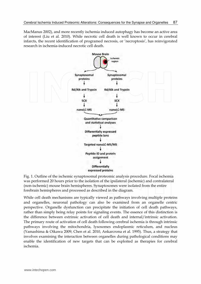
Fig. 1. Outline of the ischemic synaptosomal proteomic analysis procedure. Focal ischemia was performed 20 hours prior to the isolation of the ipsilateral (ischemic) and contralateral (non-ischemic) mouse brain hemispheres. Synaptosomes were isolated from the entire forebrain hemispheres and processed as described in the diagram.
While cell death mechanisms are typically viewed as pathways involving multiple proteins
and organelles, neuronal pathology can also be examined from an organelle centric
perspective. Organelle dysfunction can precipitate the initiation of cell death pathways,
rather than simply being relay points for signaling events. The essence of this distinction is
the difference between extrinsic activation of cell death and internal/intrinsic activation.
The primary route of activation of cell death following cerebral ischemia is through intrinsic
pathways involving the mitochondria, lysosomes endoplasmic reticulum, and nucleus
(Yamashima & Oikawa 2009, Chen et al. 2010, Ankarcrona et al. 1995). Thus, a strategy that
involves examining the interaction between organelles during pathological conditions may
enable the identification of new targets that can be exploited as therapies for cerebral
ischemia.
www.intechopen.com
Advances in the Preclinical Study of Ischemic Stroke
88
The effects of cerebral ischemia are often, by necessity, described from a highly reductionist point of view, with most studies focusing on specific signaling pathways or individual molecules. Conversely, genomic and proteomic datasets offer the opportunity to expand the scope of understanding and enable the interpretation of systematic responses. While there is a wealth of data available describing neuronal proteins localized in synaptosomes as well as pre-synaptic and post-synaptic preparations, to date studies on the effects of cerebral ischemia on the synaptic proteome are limited (Costain et al. 2008). The aim of this chapter is to integrate new and existing genomic and proteomic datasets to provide a comprehensive understanding of the effect of cerebral ischemia on the function of neuronal organelles, as well as their role in mediating cell death and / or neuroprotection.
2. Materials and methods
2.1 Animal care A local committee for the Canadian Council on Animal Care approved all procedures using mice. The C57B mice were purchased from Charles River Canada (St-Constant, PQ). Under temporary isoflurane anesthesia, the mice (20-23 g) were subjected to occlusion of the left middle cerebral artery (MCAO) using an intraluminal filament as previously described (Costain et al. 2008). After 1 hr of ischemia, the animals were briefly reanesthetized, the filament withdrawn and wounds sutured. After 20 hrs of reperfusion, mice were briefly anesthetized with isoflurane and the brain rapidly excised and dissected on ice.
2.2 Synaptosome preparation Contralateral (CT) and ischemic (IS) hemispheres from one mouse were manually homogenized in 2 ml of HM buffer (0.32 M sucrose, 1 mM EDTA, 0.25 mM DTT, 1 U/ml RNasin (Promega)) using a dounce homogenizer, 800 rpm 13 strokes at 4 °C. The homogenates were centrifuged (1000 g for 10 min at 4 C) and the supernatant retained. The pellet was homogenized and centrifuged (as before) a second time. The first and second supernatants were transferred to 2 ml polycarbonate tubes and centrifuged at 20,000 g for 20
min at 4 C. The resultant pellet was resuspended in 2 ml HM buffer using a dounce homogenizer. Discontinuous sucrose:percoll gradients were prepared by layering 2 ml each, in order, of 25% (percoll in HM buffer), 15%, 10% and 3% into 10 ml polycarbonate centrifuge tubes. One ml of the sample was then layered on top of a gradient and centrifuged at 32,000 g for 5 min at 4 C. Five fractions were collected following centrifugation: F1 - 3% percoll (cytoplasm), F2 - interface between 3% and 10% percoll (myelin), F3 - interface between 10% and 15% percoll (small synaptosomes, myelin & mitochondria), F4 - interphase between 15% and 25% percoll (intact synaptosomes), and F5 - pellet (mitochondria). Fractions F1 - F4 were made up to 3 ml with HM buffer and
centrifuged at 12,000 g for 20 min at 4 C. The supernatant was removed and the pellet washed with 1 x PBS, twice (each time spinning at 12000 x g for 10min). The final pellet was resuspended in either HM buffer or 50 mM Tris pH 8.5, 0.1% SDS. The physical and biochemical characteristics of the synaptosome preparation used here are described in further detail in Costain et al. (2008).
2.3 Protein preparation, digestion and ion exchange chromatography Proteins from each synaptosome sample were precipitated by adding 10 volumes of cold
acetone and incubating at 1 h at -20 C followed by centrifugation at 5000g for 5 min. Pellets
www.intechopen.com
Cerebral Ischemia Induced Proteomic Alterations: Consequences for the Synapse and Organelles
89
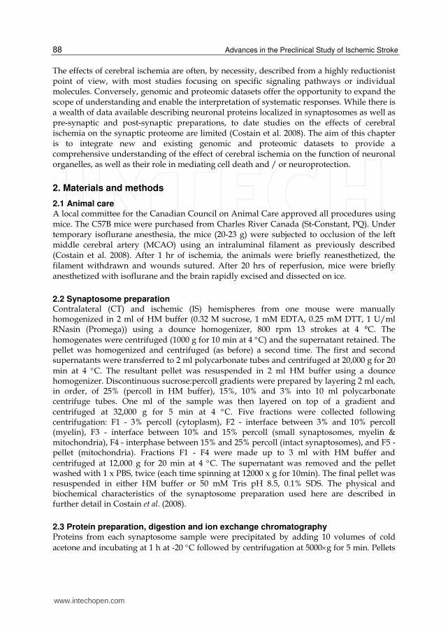
Fig. 2. NanoLC-MS analysis of contralateral (control) and ipsilateral (stroke) samples. Shown are images representing the nanoLC-MS data from each sample, where each spot represents a peptide ion. MatchRx software was used to extract peptide data, align the control and stroke datasets and correct retention time variations. Shown are the merged images before and after MatchRx-dependent correction. More than 5000 ions were detected per sample.
were dissolved in an appropriate volume of denaturing buffer (50 mM Tris-HCl, pH 8.5,
0.1% SDS) to a final protein concentration of 2 mg/mL. One hundred µg of each protein was
transferred to a fresh tube. The proteins were reduced using dithiothrietol (4 mM, 10 min at
95 C), alkylated using iodoacetamide (10 mM, 20 min at room temperature in dark), and
digested using 5 µg of MS-grade trypsin gold (Promega, 12-18 h at 37 C). The digested
peptides were diluted 10-fold in 10 mM KH2PO4, pH 3.0, 25% acetonitrile and loaded onto a
cation exchange column (POROS® 50 HS, 50-µm particle size, 4.0 mm x 15 mm, Applied
Biosystems) for separation. Five fractions were eluted using step-gradient of 0-350 mM KCl.
Each fraction was evaporated to dryness and dissolved in 5% acetronitrile, 1% acetic acid for
analysis by mass spectrometry (MS).
www.intechopen.com
Advances in the Preclinical Study of Ischemic Stroke
90
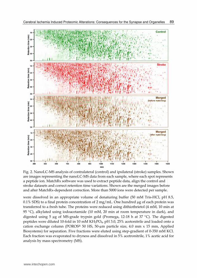
Fig. 3. Scatter plot showing relative expression of peptides in stroke and control synaptosomes. Peptide intensities were extracted from nanoLC-MS runs using MatchRx and ratio-intensity plots of stroke-vs-control samples after global median normalization were plotted. Peptides showing up (red) or down (green) regulation by 1.5-fold in stroke relative to control samples and showing a p <0.05 were considered differentially expressed. These corresponded to ~27% of the peptides and were used for targeted identification using nanoLC-MS/MS.
2.4 MS analysis and protein identification A hybrid quadrupole time-of-flight MS (Q-TOF Ultima, Waters, Millford, MA) with an electrospray ionization source (ESI) and with an online reverse-phase nanoflow liquid chromatography column (nanoLC, 0.075 mm × 150 mm PepMap C18 capillary column, Dionex/LC-Packings, San Francisco, CA) was used for all analyses. Samples were separated on the nanoLC column using a gradient of 5-75% acetonitrile and 0.2% formic acid in 90 min, at 350 nL/min supplied by a CapLC HPLC pump (Waters). Five percent of each sample was first analyzed by nanoLC-MS in a survey (MS-only) mode for quantitation using MatchRx software as described recently (Haqqani et al. 2008). Briefly, each scan was background-subtracted, Savitzky-Golay-based smoothed, and centroided using Masslynx software v4.0 (Waters) and exported as an mzXML file (Pedrioli et al. 2004). Using MatchRx software, isotopic distribution pattern, charge state, and quantitative abundance of peptides in each nanoLC-MS run were determined. The peptides were then aligned across multiple nanoLC-MS runs through a neighboring-peak finding algorithm (Haqqani et al. 2008) followed by quantitatively comparing the levels of each peptide in the ipsilateral and contralateral fractions to identify differentially expressed peptides. Peptides showing consistent log2 fold-change of >1.5 or <-1.5 among biological replicates and showing significant difference from mean expression levels (p < 0.05, Wilcoxon matched pairs test) were considered differentially expressed. Images of each run were also generated to visually validate the differentially expressed peptides using MatchRx (Fig. 4). To sequence the differentially expressed peptides, they were included in a 'include list'. Another 5% of each sample was then re-injected into the mass spectrometer, and only the peptides included in the 'include list' were sequenced in MS/MS mode (targeted nanoLC-MS/MS). All MS/MS spectra were obtained on 2+, 3+, and 4+ ions. Peak lists were submitted to a probability-based search engine, Mascot version 2.2.0 (Matrix Science Ltd., London, U.K.) (Hirosawa et
www.intechopen.com
Cerebral Ischemia Induced Proteomic Alterations: Consequences for the Synapse and Organelles
91
al. 1993). The initial database utilized was a composite of forward and reverse Uniprot-Swiss-Prot Mus musculus protein database (Aug 2011 containing 16,390 sequences). Unmatched peptides were subsequently searched against the remaining Uniprot-Swiss-Prot database (Aug 2011 containing 531,473 sequences). Searches were performed with a specified trypsin enzymatic cleavage with one possible missed cleavage. The false-positive rates (FPR) in database searching by Mascot were calculated as described earlier (Peng et al. 2003): FPR = (2 × Nrev)/(Nrev + Nfwd), where Nrev is the number of peptides identified (after filtering) from the reverse-database, and Nfwd is the number of peptides identified (after filtering) from the forward database. To maximize the number of peptides and keep the FPR < 0.5%, ion scores > 20, parent ion tolerance of < 0.1 Da, fragment ion tolerance of < 0.2 Da, and minimal number of missed cleavages were chosen. As an independent statistical measure of peptide identification, Peptide Prophet probabilities were also measured. All identified peptides had p ≥ 0.90. The MS/MS spectrum of each differentially expressed peptide pair was manually examined and confirmed.
Fig. 4. Examples of differentially expressed proteins on nanoLC-MS images. Shown are merged images representing nanoLC-MS data of control (green image) and stroke (red image) samples. The two down-regulated peptides (green) are from 3-Oxoacid CoA Transferase 1 (Q9D0K2, Oxct1) and the four up-regulated peptides (red) are from glial high affinity glutamate transporter (P43006, Slc1a2). Peptides were identified by targeted nanoLC-MS/MS. In each image, inset shows a close-up image of the indicated peptide and the fold-change in stroke sample relative to control. For detailed list see Tables 1 and 2.
3. Results
3.1 Label-free proteomic analysis of ischemic synaptosomes An outline of the experimental procedure is provided in Fig. 1. Following cerebral ischemia, the ipsilateral and contralateral hemispheres were separated and synaptosomal proteins isolated. The proteins from each sample were digested into sequenceable peptides, separated into 4 cation exchange chromatography fractions and analyzed by nanoLC-MS to quantify the level of each peptide. Each cation exchange fraction contained more than 12,000 peptide peaks as identified by nanoLC-MS analysis (Fig. 2). MatchRx software was thus used to identify quantitative differences in the nanoLC-MS runs between all the ipsilateral and contralateral fractions. The software extracts peptide-peak intensities and enables correction of retention time variations amongst multiple nanoLC-MS runs (Fig. 2), thus allowing accurate peptide alignment and quantitative comparison of the samples. Statistical
www.intechopen.com

Advances in the Preclinical Study of Ischemic Stroke
92
analyses were carried out to identify differentially expressed peaks, resulting in the determination that 27% of the peaks showed differential expression (≥ 1.5 fold difference) between the two hemispheres (Fig. 3). As the variability between two biological synaptosomal preps was found to be about 10% (unpublished data), the observed differences were primarily attributable to the effects of cerebral ischemia and much less due to biological variability.
Fig. 5. Linear regression analysis of bias in label-free proteomics data. Expression data were plotted against protein mass and mascot score in panels A and B, respectively. Error values were plotted against mascot score in panel C. Linear regression analyses were performed on the up (red)- and down (green)-regulated proteins independently. Regression lines are plotted in panels B and C. Analyses of the regression slopes did not detect significant
www.intechopen.com
Cerebral Ischemia Induced Proteomic Alterations: Consequences for the Synapse and Organelles
93
departures from zero, indicating that there was no correlation between expression and protein size / mascot score or error and mascot score. A runs test indicated that there was no departure from linearity in the regression analysis of error versus mascot score. Furthermore, no significant differences were detected between the up and down regulated proteins in any of the analyses.
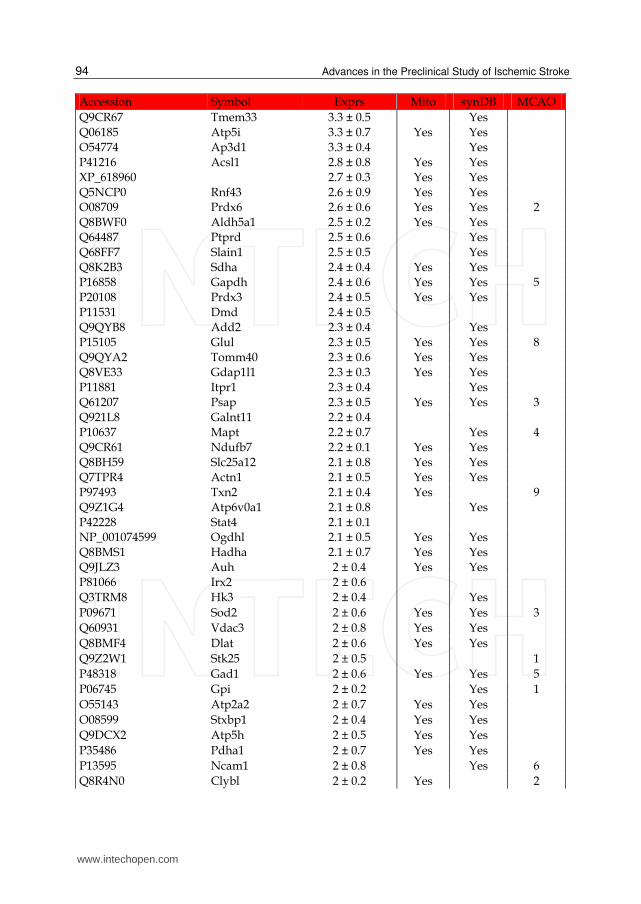
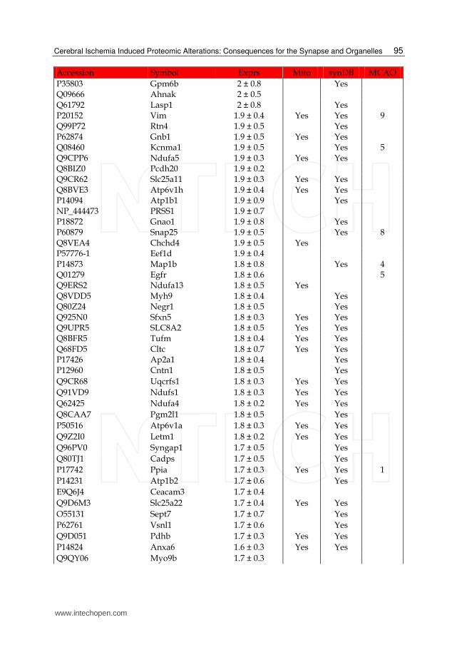
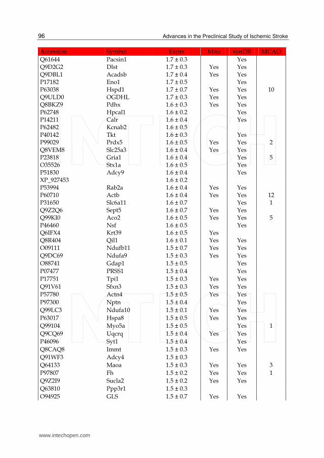
The differentially expressed peaks were then sequenced by re-injecting each fraction and analyzing with targeted nanoLC-MS/MS using an include list containing the masses and retention times of each targeted peak. The sequenced peptides were used to generate a list of ischemia-responsive proteins. A total of 371 proteins were identified as responsive to cerebral ischemia (hereafter referred to as either the IS dataset or IS proteins) (Tables 1 and 2). Two-thirds (68%) of these proteins were up-regulated and the remaining were down-regulated. Examples of up- and down-regulated proteins are shown in Fig. 4. A series of linear regression analyses were performed to ensure that the expression data was free of systematic bias. The results of the linear regression analyses indicate that the expression values were not correlated to protein mass (Fig. 5A) or Mascot score (Fig. 5B). Similarly, the measurement error was not correlated to Mascot score (Fig. 5C). These results indicate that the observed expression and error values are due to biological effects.
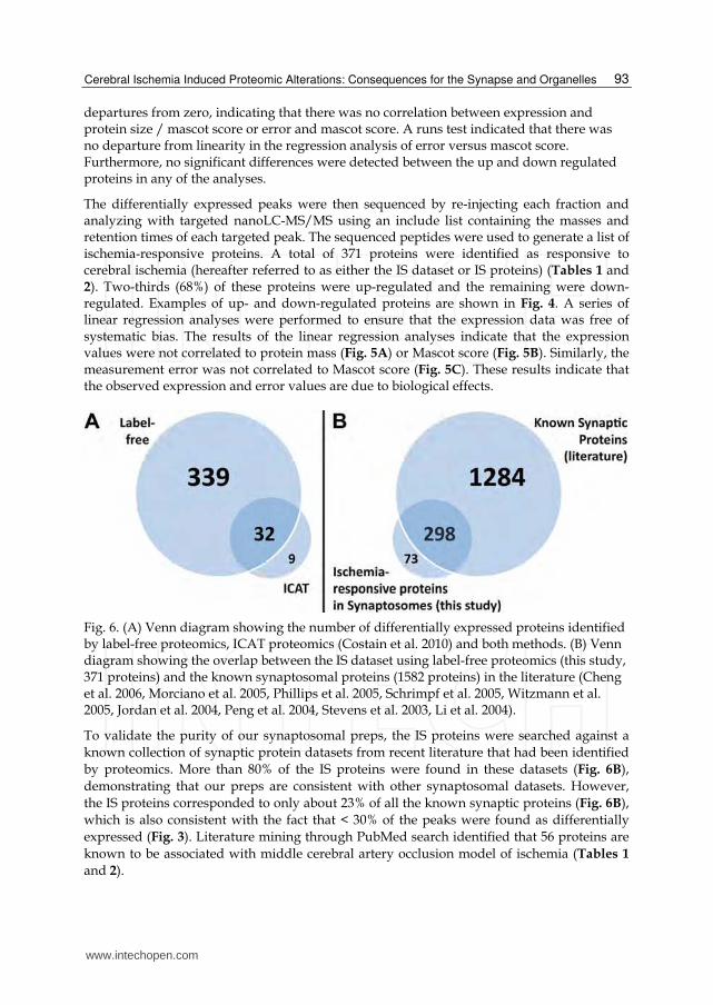
Fig. 6. (A) Venn diagram showing the number of differentially expressed proteins identified by label-free proteomics, ICAT proteomics (Costain et al. 2010) and both methods. (B) Venn diagram showing the overlap between the IS dataset using label-free proteomics (this study, 371 proteins) and the known synaptosomal proteins (1582 proteins) in the literature (Cheng et al. 2006, Morciano et al. 2005, Phillips et al. 2005, Schrimpf et al. 2005, Witzmann et al. 2005, Jordan et al. 2004, Peng et al. 2004, Stevens et al. 2003, Li et al. 2004).
To validate the purity of our synaptosomal preps, the IS proteins were searched against a
known collection of synaptic protein datasets from recent literature that had been identified
by proteomics. More than 80% of the IS proteins were found in these datasets (Fig. 6B),
demonstrating that our preps are consistent with other synaptosomal datasets. However,
the IS proteins corresponded to only about 23% of all the known synaptic proteins (Fig. 6B),
which is also consistent with the fact that < 30% of the peaks were found as differentially
expressed (Fig. 3). Literature mining through PubMed search identified that 56 proteins are
known to be associated with middle cerebral artery occlusion model of ischemia (Tables 1
and 2).
www.intechopen.com
Advances in the Preclinical Study of Ischemic Stroke
94
Accession Symbol Exprs Mito synDB MCAO
Q9CR67 Tmem33 3.3 ± 0.5 Yes Q06185 Atp5i 3.3 ± 0.7 Yes Yes O54774 Ap3d1 3.3 ± 0.4 Yes P41216 Acsl1 2.8 ± 0.8 Yes Yes XP_618960 2.7 ± 0.3 Yes Yes Q5NCP0 Rnf43 2.6 ± 0.9 Yes Yes O08709 Prdx6 2.6 ± 0.6 Yes Yes 2 Q8BWF0 Aldh5a1 2.5 ± 0.2 Yes Yes Q64487 Ptprd 2.5 ± 0.6 Yes Q68FF7 Slain1 2.5 ± 0.5 Yes Q8K2B3 Sdha 2.4 ± 0.4 Yes Yes P16858 Gapdh 2.4 ± 0.6 Yes Yes 5 P20108 Prdx3 2.4 ± 0.5 Yes Yes P11531 Dmd 2.4 ± 0.5 Q9QYB8 Add2 2.3 ± 0.4 Yes P15105 Glul 2.3 ± 0.5 Yes Yes 8 Q9QYA2 Tomm40 2.3 ± 0.6 Yes Yes Q8VE33 Gdap1l1 2.3 ± 0.3 Yes Yes P11881 Itpr1 2.3 ± 0.4 Yes Q61207 Psap 2.3 ± 0.5 Yes Yes 3 Q921L8 Galnt11 2.2 ± 0.4 P10637 Mapt 2.2 ± 0.7 Yes 4 Q9CR61 Ndufb7 2.2 ± 0.1 Yes Yes Q8BH59 Slc25a12 2.1 ± 0.8 Yes Yes Q7TPR4 Actn1 2.1 ± 0.5 Yes Yes P97493 Txn2 2.1 ± 0.4 Yes 9 Q9Z1G4 Atp6v0a1 2.1 ± 0.8 Yes P42228 Stat4 2.1 ± 0.1 NP_001074599 Ogdhl 2.1 ± 0.5 Yes Yes Q8BMS1 Hadha 2.1 ± 0.7 Yes Yes Q9JLZ3 Auh 2 ± 0.4 Yes Yes P81066 Irx2 2 ± 0.6 Q3TRM8 Hk3 2 ± 0.4 Yes
P09671 Sod2 2 ± 0.6 Yes Yes 3
Q60931 Vdac3 2 ± 0.8 Yes Yes
Q8BMF4 Dlat 2 ± 0.6 Yes Yes
Q9Z2W1 Stk25 2 ± 0.5 1
P48318 Gad1 2 ± 0.6 Yes Yes 5
P06745 Gpi 2 ± 0.2 Yes 1
O55143 Atp2a2 2 ± 0.7 Yes Yes
O08599 Stxbp1 2 ± 0.4 Yes Yes
Q9DCX2 Atp5h 2 ± 0.5 Yes Yes
P35486 Pdha1 2 ± 0.7 Yes Yes
P13595 Ncam1 2 ± 0.8 Yes 6
Q8R4N0 Clybl 2 ± 0.2 Yes 2
www.intechopen.com
Cerebral Ischemia Induced Proteomic Alterations: Consequences for the Synapse and Organelles
95
Accession Symbol Exprs Mito synDB MCAO
P35803 Gpm6b 2 ± 0.8 Yes Q09666 Ahnak 2 ± 0.5 Q61792 Lasp1 2 ± 0.8 Yes P20152 Vim 1.9 ± 0.4 Yes Yes 9 Q99P72 Rtn4 1.9 ± 0.5 Yes P62874 Gnb1 1.9 ± 0.5 Yes Yes Q08460 Kcnma1 1.9 ± 0.5 Yes 5 Q9CPP6 Ndufa5 1.9 ± 0.3 Yes Yes Q8BIZ0 Pcdh20 1.9 ± 0.2 Q9CR62 Slc25a11 1.9 ± 0.3 Yes Yes Q8BVE3 Atp6v1h 1.9 ± 0.4 Yes Yes P14094 Atp1b1 1.9 ± 0.9 Yes NP_444473 PRSS1 1.9 ± 0.7 P18872 Gnao1 1.9 ± 0.8 Yes P60879 Snap25 1.9 ± 0.5 Yes 8 Q8VEA4 Chchd4 1.9 ± 0.5 Yes P57776-1 Eef1d 1.9 ± 0.4 P14873 Map1b 1.8 ± 0.8 Yes 4 Q01279 Egfr 1.8 ± 0.6 5 Q9ERS2 Ndufa13 1.8 ± 0.5 Yes Q8VDD5 Myh9 1.8 ± 0.4 Yes Q80Z24 Negr1 1.8 ± 0.5 Yes Q925N0 Sfxn5 1.8 ± 0.3 Yes Yes Q9UPR5 SLC8A2 1.8 ± 0.5 Yes Yes Q8BFR5 Tufm 1.8 ± 0.4 Yes Yes Q68FD5 Cltc 1.8 ± 0.7 Yes Yes P17426 Ap2a1 1.8 ± 0.4 Yes P12960 Cntn1 1.8 ± 0.5 Yes
Q9CR68 Uqcrfs1 1.8 ± 0.3 Yes Yes
Q91VD9 Ndufs1 1.8 ± 0.3 Yes Yes
Q62425 Ndufa4 1.8 ± 0.2 Yes Yes
Q8CAA7 Pgm2l1 1.8 ± 0.5 Yes
P50516 Atp6v1a 1.8 ± 0.3 Yes Yes
Q9Z2I0 Letm1 1.8 ± 0.2 Yes Yes
Q96PV0 Syngap1 1.7 ± 0.5 Yes
Q80TJ1 Cadps 1.7 ± 0.5 Yes
P17742 Ppia 1.7 ± 0.3 Yes Yes 1
P14231 Atp1b2 1.7 ± 0.6 Yes
E9Q6J4 Ceacam3 1.7 ± 0.4
Q9D6M3 Slc25a22 1.7 ± 0.4 Yes Yes
O55131 Sept7 1.7 ± 0.7 Yes
P62761 Vsnl1 1.7 ± 0.6 Yes
Q9D051 Pdhb 1.7 ± 0.3 Yes Yes
P14824 Anxa6 1.6 ± 0.3 Yes Yes
Q9QY06 Myo9b 1.7 ± 0.3
www.intechopen.com
Advances in the Preclinical Study of Ischemic Stroke
96
Accession Symbol Exprs Mito synDB MCAO
Q61644 Pacsin1 1.7 ± 0.3 Yes Q9D2G2 Dlst 1.7 ± 0.3 Yes Yes Q9DBL1 Acadsb 1.7 ± 0.4 Yes Yes P17182 Eno1 1.7 ± 0.5 Yes P63038 Hspd1 1.7 ± 0.7 Yes Yes 10 Q9ULD0 OGDHL 1.7 ± 0.3 Yes Yes Q8BKZ9 Pdhx 1.6 ± 0.3 Yes Yes P62748 Hpcal1 1.6 ± 0.2 Yes P14211 Calr 1.6 ± 0.4 Yes P62482 Kcnab2 1.6 ± 0.5 P40142 Tkt 1.6 ± 0.3 Yes P99029 Prdx5 1.6 ± 0.5 Yes Yes 2 Q8VEM8 Slc25a3 1.6 ± 0.4 Yes Yes P23818 Gria1 1.6 ± 0.4 Yes 5 O35526 Stx1a 1.6 ± 0.5 Yes P51830 Adcy9 1.6 ± 0.4 Yes XP_927453 1.6 ± 0.2 P53994 Rab2a 1.6 ± 0.4 Yes Yes P60710 Actb 1.6 ± 0.4 Yes Yes 12 P31650 Slc6a11 1.6 ± 0.7 Yes 1 Q9Z2Q6 Sept5 1.6 ± 0.7 Yes Yes Q99KI0 Aco2 1.6 ± 0.5 Yes Yes 5 P46460 Nsf 1.6 ± 0.5 Yes Q6IFX4 Krt39 1.6 ± 0.5 Yes Q8R404 Qil1 1.6 ± 0.1 Yes Yes O09111 Ndufb11 1.5 ± 0.7 Yes Yes
Q9DC69 Ndufa9 1.5 ± 0.3 Yes Yes
O88741 Gdap1 1.5 ± 0.5 Yes
P07477 PRSS1 1.5 ± 0.4 Yes
P17751 Tpi1 1.5 ± 0.3 Yes Yes
Q91V61 Sfxn3 1.5 ± 0.3 Yes Yes
P57780 Actn4 1.5 ± 0.5 Yes Yes
P97300 Nptn 1.5 ± 0.4 Yes
Q99LC3 Ndufa10 1.5 ± 0.1 Yes Yes
P63017 Hspa8 1.5 ± 0.5 Yes Yes
Q99104 Myo5a 1.5 ± 0.5 Yes 1
Q9CQ69 Uqcrq 1.5 ± 0.4 Yes Yes
P46096 Syt1 1.5 ± 0.4 Yes
Q8CAQ8 Immt 1.5 ± 0.3 Yes Yes
Q91WF3 Adcy4 1.5 ± 0.3
Q64133 Maoa 1.5 ± 0.3 Yes Yes 3
P97807 Fh 1.5 ± 0.2 Yes Yes 1
Q9Z2I9 Sucla2 1.5 ± 0.2 Yes Yes
Q63810 Ppp3r1 1.5 ± 0.3
O94925 GLS 1.5 ± 0.7 Yes Yes
www.intechopen.com
Cerebral Ischemia Induced Proteomic Alterations: Consequences for the Synapse and Organelles
97
Accession Symbol Exprs Mito synDB MCAO
Q9R1T4 Sept6 1.5 ± 0.3 Yes P61922 Abat 1.5 ± 0.3 Yes Yes Q61206 Pafah1b2 1.5 ± 0.4 P06151 Ldha 1.5 ± 0.2 Yes Yes P54227 Stmn1 1.5 ± 0.3 Yes 3 P31648 Slc6a1 1.4 ± 0.5 Yes 1 P28652 Camk2b 1.4 ± 0.3 Yes Q61548 Snap91 1.4 ± 0.2 Yes Yes P48962 Slc25a4 1.4 ± 0.2 Yes Yes P68254 Ywhaq 1.4 ± 0.2 Yes Yes P50518 Atp6v1e1 1.4 ± 0.1 Yes Yes Q8VHW2 Cacng8 1.4 ± 0.4 Yes O55125 Nipsnap1 1.4 ± 0.3 Yes Yes O70566 Diaph2 1.4 ± 0.2 Yes Yes P20936 RASA1 1.4 ± 0.2 Q9ES97 Rtn3 1.4 ± 0.3 Yes P50114 S100b 1.4 ± 0.2 Yes 7 O08553 Dpysl2 1.4 ± 0.4 Yes Yes 2 Q60930 Vdac2 1.4 ± 0.5 Yes Yes Q02053 Uba1 1.4 ± 0.2 Yes Q9R0P9 Uchl1 1.4 ± 0.2 Yes 2 P10126 Eef1a1 1.4 ± 0.6 Yes Yes Q60932 Vdac1 1.4 ± 0.3 Yes Yes P06837 Gap43 1.4 ± 0.3 Yes 14 P62835 Rap1a 1.4 ± 0.2 Yes Yes
Q9D0F9 Pgm1 1.3 ± 0.2 Yes
P62204 Calm1 1.3 ± 0.2 Yes 25
P47753 Capza1 1.3 ± 0.4 Yes
P52480 Pkm2 1.3 ± 0.4 Yes Yes
Q9QZD8 Slc25a10 1.3 ± 0.4 Yes Yes
Q9EQF6 Dpysl5 1.3 ± 0.5 Yes 1
Q5SUA5 Myo1g 1.3 ± 0.2 Yes Yes
Q9D0S9 Hint2 1.3 ± 0.3 Yes Yes
Q62261 Sptbn1 1.3 ± 0.2 Yes
Q8BVI4 Qdpr 1.3 ± 0.2 Yes Yes
O70443 Gnaz 1.3 ± 0.2 Yes Yes
P18760 Cfl1 1.3 ± 0.3 Yes Yes
Q62315 Jarid2 1.3 ± 0.2
Q03963 Eif2ak2 1.3 ± 0.2
P35802 Gpm6a 1.3 ± 0.4 Yes
Q9D6R2 Idh3a 1.3 ± 0.2 Yes Yes 1
O08749 Dld 1.3 ± 0.4 Yes Yes
P38647 Hspa9 1.3 ± 0.4 Yes Yes 1
O88737 Bsn 1.3 ± 0.3 Yes
P70268 Pkn1 1.3 ± 0.3
www.intechopen.com
Advances in the Preclinical Study of Ischemic Stroke
98
Accession Symbol Exprs Mito synDB MCAO
Q61879 Myh10 1.3 ± 0.3 Yes P62814 Atp6v1b2 1.3 ± 0.2 Yes P07901 Hsp90aa1 1.3 ± 0.3 Yes Yes P63328 Ppp3ca 1.3 ± 0.3 Yes Yes P56382 Atp5e 1.2 ± 0.2 Yes Yes P99028 Uqcrh 1.2 ± 0 Yes Yes P05201 Got1 1.2 ± 0.2 Yes Yes P16277 Blk 1.2 ± 0.3 Yes Q9CQA3 Sdhb 1.2 ± 0.3 Yes Yes P07146 Prss2 1.2 ± 0.2 Q9WV55 Vapa 1.2 ± 0.3 Yes P28663 Napb 1.2 ± 0.2 Yes O35658 C1qbp 1.1 ± 0.2 Yes Yes Q9CZW5 Tomm70a 1.1 ± 0.2 Yes Yes Q9Z0J4 Nos1 1.1 ± 0.3 Yes Yes 66 Q80XN0 Bdh1 1.1 ± 0.2 Yes Yes P34884 Mif 1.1 ± 0.2 Yes 3 Q8K314 Atp2b1 1.1 ± 0.3 Yes Q640R3 Hepacam 1.1 ± 0.1 Yes Q5SWU9 Acaca 1.1 ± 0.1 Yes Yes Q00690 Sele 1.1 ± 0 26 A2AJ76 Hmcn2 1 ± 0.3 Q9JI46 Nudt3 1 ± 0.2 Yes Q6PCP5 Mff 1 ± 0.1 Yes Yes O70283 Wnt2b 1 ± 0.2 P70295 Aup1 1 ± 0.2 Q80Y86 Mapk15 1 ± 0.2 Yes Q9CQ54 Ndufc2 1 ± 0.2 Yes Yes P00405 Mtco2 1 ± 0.3 Yes Yes P63082 Atp6v0c 1 ± 0.2 Q9D8W7 Ociad2 1 ± 0.2 Yes
XP_922613 Spnb5 1 ± 0.3
P43006 Slc1a2 1 ± 0.2 Yes 10
Q9R111 Gda 1 ± 0.2 Yes
Q9CQI3 Gmfb 0.9 ± 0.3 Yes
XP_922643 0.9 ± 0.1
O35857 Timm44 0.9 ± 0.2 Yes
Q8BM92 Cdh7 0.9 ± 0.1 Yes
P56564 Slc1a3 0.8 ± 0.2 Yes Yes
O54983 Crym 0.9 ± 0.1 Yes
NP_082646 Pot1b 0.9 ± 0.2
Q9JKR6 Hyou1 0.9 ± 0.2
Q9CQH3 Ndufb5 0.9 ± 0.2 Yes
P48320 Gad2 0.9 ± 0.1 Yes 1
Q61735 Cd47 0.9 ± 0.1 1
www.intechopen.com
Cerebral Ischemia Induced Proteomic Alterations: Consequences for the Synapse and Organelles
99
Accession Symbol Exprs Mito synDB MCAO
Q9H4G0 EPB41L1 0.9 ± 0.2 Yes P08228 Sod1 0.9 ± 0.2 Yes Yes 10 P62137 Ppp1ca 0.9 ± 0.3 Yes Q62277 Syp 0.9 ± 0.2 Yes 30 Q9QZ83 Actg1 0.9 ± 0.2 Yes Yes A8E4K7 Pcdhb8 0.8 ± 0.1 Q8K183 Pdxk 0.8 ± 0.2 2 P08556 Nras 0.8 ± 0.2 Yes Yes Q61194 Pik3c2a 0.8 ± 0.3 Q9DCS9 Ndufb10 0.8 ± 0.1 Yes Yes Q62420 Sh3gl2 0.8 ± 0.2 Yes P48771 Cox7a2 0.8 ± 0.3 Yes O35728 Cyp4a14 0.8 ± 0.1 Q6GQS1 Slc25a23 0.8 ± 0.3 Yes
Q9CWZ7 Napg 0.7 ± 0.1 Yes Yes
Q99JY0 Hadhb 0.7 ± 0.1 Yes Yes
Q8K0T0 Rtn1 0.7 ± 0.2 Yes
P0CG49 Ubb 0.7 ± 0 Yes Yes 17
P84091 Ap2m1 0.7 ± 0.1 Yes Yes
P70335 Rock1 0.7 ± 0.2
O09112 Dusp8 0.7 ± 0.1
Q3UK37 0.7 ± 0.1
Q8BLF1 Nceh1 0.7 ± 0.2
Q76MZ3 Ppp2r1a 0.6 ± 0 Yes Yes
Q8BGZ1 Hpcal4 0.6 ± 0.2 Yes
B9EJA4 Clasp2 0.6 ± 0.1
P05202 Got2 0.3 ± 1.6 Yes Yes 1
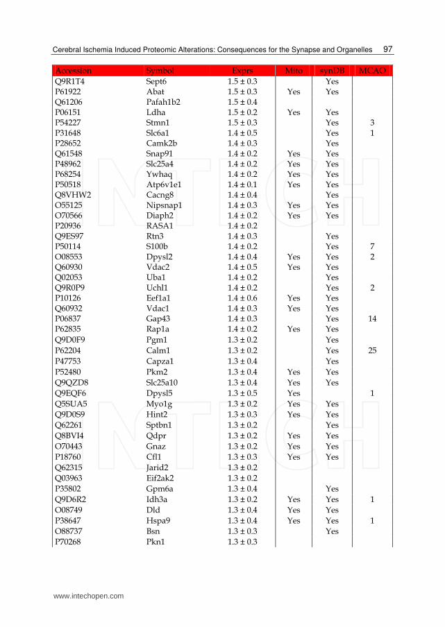
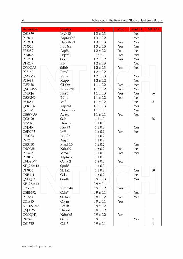
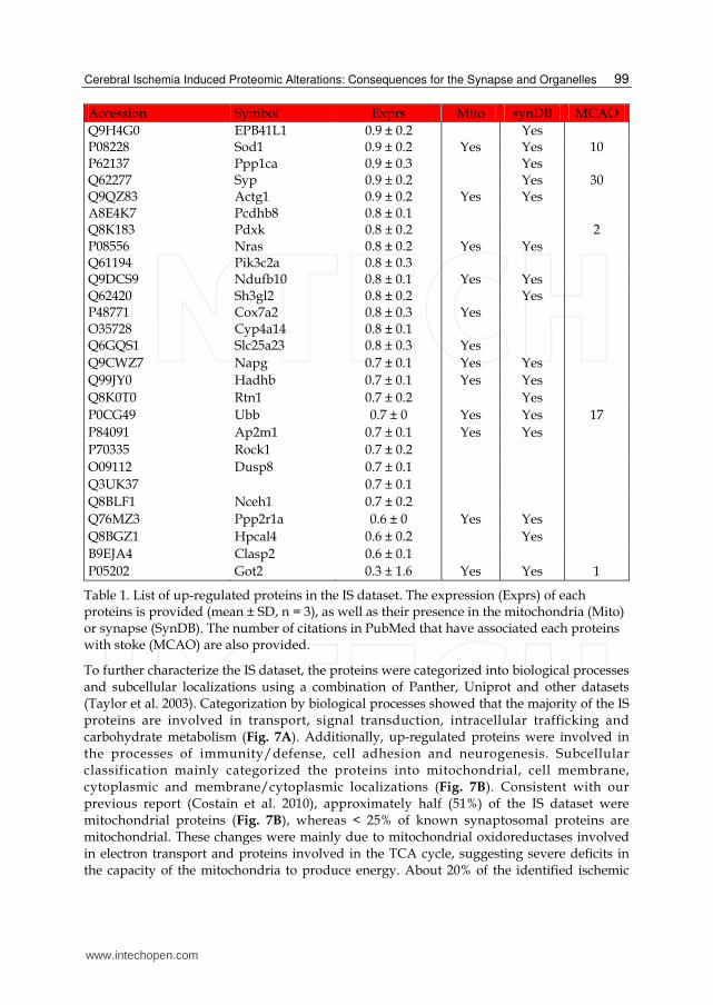
Table 1. List of up-regulated proteins in the IS dataset. The expression (Exprs) of each proteins is provided (mean ± SD, n = 3), as well as their presence in the mitochondria (Mito) or synapse (SynDB). The number of citations in PubMed that have associated each proteins with stoke (MCAO) are also provided.
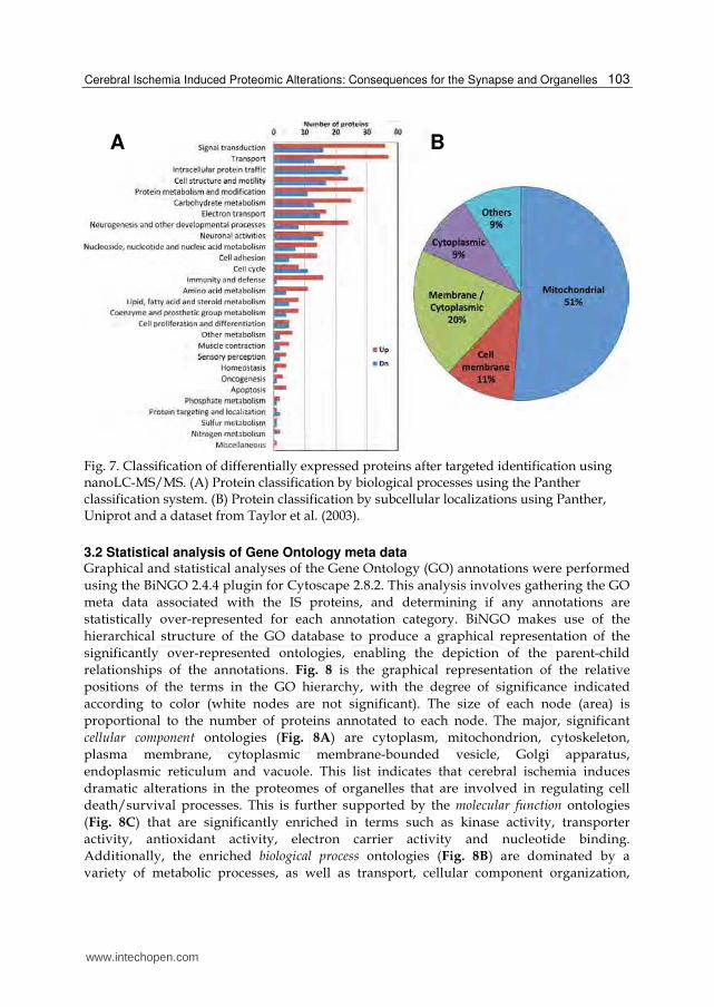
To further characterize the IS dataset, the proteins were categorized into biological processes and subcellular localizations using a combination of Panther, Uniprot and other datasets (Taylor et al. 2003). Categorization by biological processes showed that the majority of the IS proteins are involved in transport, signal transduction, intracellular trafficking and carbohydrate metabolism (Fig. 7A). Additionally, up-regulated proteins were involved in the processes of immunity/defense, cell adhesion and neurogenesis. Subcellular classification mainly categorized the proteins into mitochondrial, cell membrane, cytoplasmic and membrane/cytoplasmic localizations (Fig. 7B). Consistent with our previous report (Costain et al. 2010), approximately half (51%) of the IS dataset were mitochondrial proteins (Fig. 7B), whereas < 25% of known synaptosomal proteins are mitochondrial. These changes were mainly due to mitochondrial oxidoreductases involved in electron transport and proteins involved in the TCA cycle, suggesting severe deficits in the capacity of the mitochondria to produce energy. About 20% of the identified ischemic
www.intechopen.com
Advances in the Preclinical Study of Ischemic Stroke
100
Accession Symbol Exprs Mito SynDB MCAO
Q920I9 Wdr7 -3.3 ± 0.2 Yes Q8R1B4 Eif3c -3.3 ± 1.3 Yes P42356 PI4KA -2.9 ± 0.6 Yes Q8R071 Itpka -2.7 ± 0.5 Yes P67778 Phb -2.3 ± 0.4 Yes P19783 Cox4i1 -2.3 ± 0.8 Yes Q4U256 Ank3 -2.2 ± 0.6 Yes Q9DBG3 Ap2b1 -2.2 ± 0.5 Yes Yes P51881 Slc25a5 -2.1 ± 0.5 Yes Q3V1U8 Elmod1 -2.1 ± 0.5 Yes Yes P58281 Opa1 -2.1 ± 0.3 Yes Q64516 Gyk -2.1 ± 0.4 Yes Q99LY9 Ndufs5 -2 ± 0.6 P54285 Cacnb3 -2 ± 0.7 Yes Yes Q8VD37 Sgip1 -2 ± 0.5 Yes P49615 Cdk5 -2 ± 0.4 Yes Yes Q62159 Rhoc -2 ± 0.6 Yes Yes 2 Q9DB77 Uqcrc2 -2 ± 0.6 Yes Q9DCT2 Ndufs3 -2 ± 0.3 Yes Yes P21803 Fgfr2 -1.9 ± 0 Yes Q7TQD2 Tppp -1.9 ± 0.5 Yes Yes Q64521 Gpd2 -1.9 ± 0.6 Yes Yes P08249 Mdh2 -1.9 ± 0.2 Yes Q91WS0 Cisd1 -1.9 ± 0.7 Yes Q5DU25 Iqsec2 -1.9 ± 0.4 Q03265 Atp5a1 -1.9 ± 0.2 Yes Yes Q9CPQ1 Cox6c -1.8 ± 0.5 Yes Q04447 Ckb -1.8 ± 0.3 Yes Yes
P61161 Actr2 -1.8 ± 0.2 Yes Yes
Q9D6J6 Ndufv2 -1.8 ± 0.5 Yes
Q5NVN0 PKM2 -1.8 ± 0.4
Q6PIC6 Atp1a3 -1.7 ± 0.4
Q8QZT1 Acat1 -1.7 ± 0.6
O08539 Bin1 -1.7 ± 0.7
Q9JHU4 Dync1h1 -1.7 ± 0.3 Yes Yes
Q91VR2 Atp5c1 -1.7 ± 0.4 Yes Yes
P14618 PKM2 -1.7 ± 0.5 Yes
O88935 Syn1 -1.6 ± 0.4
Q9D0K2 Oxct1 -1.6 ± 0.5 Yes Yes
Q61330 Cntn2 -1.6 ± 0.7 Yes Yes
Q91XV3 Basp1 -1.6 ± 0.5 Yes
Q3V1L4 Nt5c2 -1.6 ± 0.3 Yes Yes
Q8BG39 Sv2b -1.6 ± 0.4
P61982 Ywhag -1.6 ± 0.3 Yes Yes
Q80ZF8 Bai3 -1.6 ± 0.4 Yes
www.intechopen.com
Cerebral Ischemia Induced Proteomic Alterations: Consequences for the Synapse and Organelles
101
Accession Symbol Exprs Mito SynDB MCAO
P17427 Ap2a2 -1.6 ± 0.3 Yes Yes
Q9DBF1 Aldh7a1 -1.6 ± 0.4 Yes Yes 8
P39053 Dnm1 -1.5 ± 0.2 Yes Yes
P70404 Idh3g -1.5 ± 0.1 Yes Yes
P99024 Tubb5 -1.5 ± 0.3 Yes Yes
P12787 Cox5a -1.5 ± 0.3 Yes Yes
Q8CI94 Pygb -1.5 ± 0.6 Yes
XP_001006010 -1.5 ± 0.1 Yes Yes
Q9QXV0 Pcsk1n -1.5 ± 0.3 Yes
P68368 Tuba4a -1.5 ± 0.3 Yes
Q2EMV9 Parp14 -1.5 ± 0.3 Yes Yes 1
P05064 Aldoa -1.5 ± 0.3 Yes NP_031573 Sirpa -1.4 ± 0.4 Yes O88342 Wdr1 -1.4 ± 0.4 Yes Yes Q9D0M3 Cyc1 -1.4 ± 0.2 Yes Q9QWI6 Srcin1 -1.4 ± 0.5 Q69ZK9 Nlgn2 -1.4 ± 0.2 Yes P56480 Atp5b -1.4 ± 0.2 Yes Q8CIV2 ORF61 -1.4 ± 0.3 Yes Yes Q3UM45 Ppp1r7 -1.4 ± 0.5 Yes Yes O55100 Syngr1 -1.4 ± 0.2 Yes Yes P97797 Sirpa -1.3 ± 0.3 Yes Yes P51174 Acadl -1.3 ± 0.3 Q9CZ13 Uqcrc1 -1.3 ± 0.4 BAE40217 Tubb5 -1.3 ± 0.3 Yes 5 AAA40509 Tubb4 -1.3 ± 0.4 Yes Q9WUM5 Suclg1 -1.3 ± 0.3 Q8CHC4 Synj1 -1.3 ± 0.4 Yes Yes P14152 Mdh1 -1.3 ± 0.2 Yes Yes P17710 Hk1 -1.2 ± 0.2 P23116 Eif3a -1.2 ± 0.1 Yes O43837 IDH3B -1.2 ± 0.4 Yes Yes Q9CZU6 Cs -1.2 ± 0.3 Yes XP_889898 -1.1 ± 0.3 Yes Yes Q6NXI6 Rprd2 -1.1 ± 0.2 Yes Yes
NP_001013813 Gm5468 -1.1 ± 0.1 Yes Yes
P11627 L1cam -1.1 ± 0.3 Yes Yes
O77784 IDH3B -1.1 ± 0.4 Yes Yes
Q9CPU4 Mgst3 -1.1 ± 0.1 Yes Yes
Q8R570 Snap47 -1.1 ± 0.3 Yes
Q8TCB6 OR51E1 -1.1 ± 0.2 Yes
NP_082221 Csl -1.1 ± 0.4 Yes Yes
Q9WUM4 Coro1c -1.1 ± 0.1 Yes Yes
P10649 Gstm1 -1.1 ± 0.2 Yes
Q9DB20 Atp5o -1 ± 0.2 Yes
www.intechopen.com
Advances in the Preclinical Study of Ischemic Stroke
102
Accession Symbol Exprs Mito SynDB MCAO
Q9R0N5 Syt5 -1 ± 0.3 Yes Yes
Q9QXY2 Srcin1 -1 ± 0.2 Yes
Q9CQY6 LOC675054 -1 ± 0.2 Yes Yes
Q3TC72 Fahd2 -1 ± 0.4 Yes Yes
Q8BK30 Ndufv3 -1 ± 0.2 Yes 1
Q9EQ20 Aldh6a1 -1 ± 0.3 Yes
P30275 Ckmt1 -1 ± 0.2 Yes Yes 1
Q8CHU3 Epn2 -1 ± 0.3 Yes Yes
O35643 Ap1b1 -0.9 ± 0.2 Yes Yes
O35874 Slc1a4 -0.9 ± 0.2 Yes
P56391 Cox6b1 -0.9 ± 0.1 Yes Yes
A2AGT5 Ckap5 -0.9 ± 0.2 Yes Yes P19536 Cox5b -0.9 ± 0.1 Yes Q60597 Ogdh -0.9 ± 0.3 Yes Q3TMW1 Ccdc102a -0.9 ± 0.2 Yes 4 NP_570954 IDH3B -0.9 ± 0.4 Yes P61205 Arf3 -0.9 ± 0.2 Yes Yes Q9DC70 Ndufs7 -0.9 ± 0.3 Yes Yes Q61124 Cln3 -0.8 ± 0.2 Yes Yes Q99LC5 Etfa -0.8 ± 0.1 P63040 Cplx1 -0.8 ± 0.1 Yes Yes Q8BH44 Coro2b -0.7 ± 0.1 Yes Yes 6 Q9CXZ1 Ndufs4 -0.7 ± 0.1 Yes P49025 Cit -0.7 ± 0.1 Yes Yes Q7TQF7 Amph -0.7 ± 0.2 Yes Yes P48678 Lmna -0.7 ± 0.1 Yes Yes O89053 Coro1a -0.6 ± 0.1 Yes Q8BUV3 Gphn -0.6 ± 0.1 Yes P35831 Ptpn12 -0.6 ± 0.1
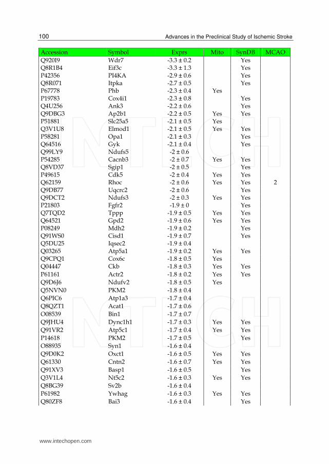
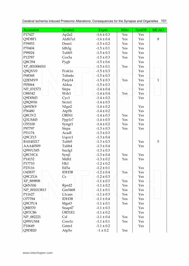
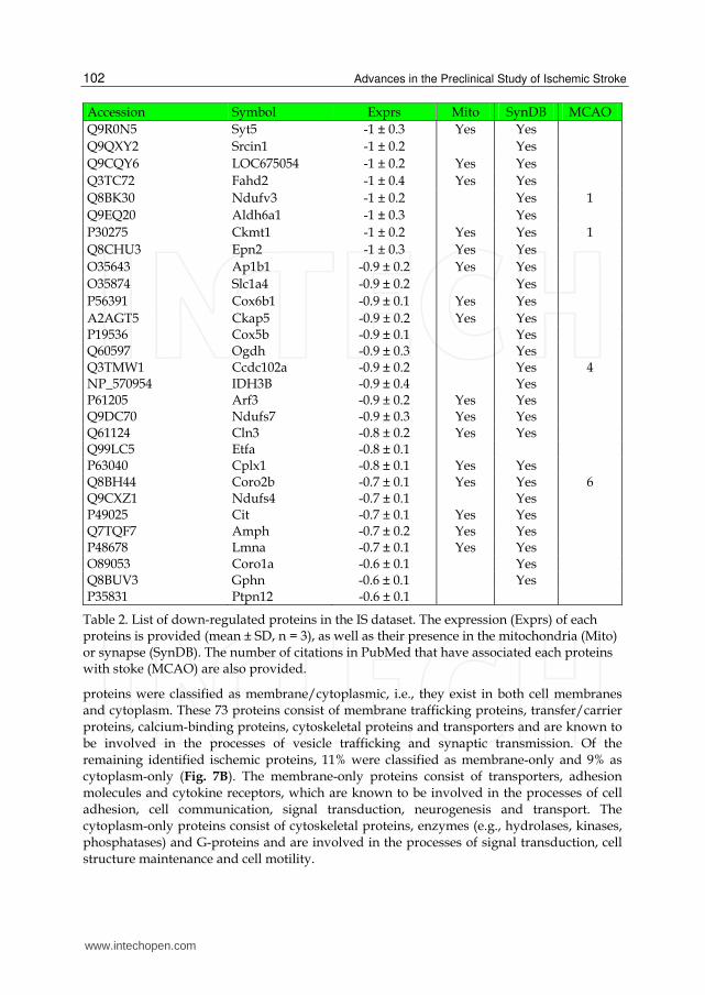
Table 2. List of down-regulated proteins in the IS dataset. The expression (Exprs) of each proteins is provided (mean ± SD, n = 3), as well as their presence in the mitochondria (Mito) or synapse (SynDB). The number of citations in PubMed that have associated each proteins with stoke (MCAO) are also provided.
proteins were classified as membrane/cytoplasmic, i.e., they exist in both cell membranes and cytoplasm. These 73 proteins consist of membrane trafficking proteins, transfer/carrier proteins, calcium-binding proteins, cytoskeletal proteins and transporters and are known to be involved in the processes of vesicle trafficking and synaptic transmission. Of the remaining identified ischemic proteins, 11% were classified as membrane-only and 9% as cytoplasm-only (Fig. 7B). The membrane-only proteins consist of transporters, adhesion molecules and cytokine receptors, which are known to be involved in the processes of cell adhesion, cell communication, signal transduction, neurogenesis and transport. The cytoplasm-only proteins consist of cytoskeletal proteins, enzymes (e.g., hydrolases, kinases, phosphatases) and G-proteins and are involved in the processes of signal transduction, cell structure maintenance and cell motility.
www.intechopen.com
Cerebral Ischemia Induced Proteomic Alterations: Consequences for the Synapse and Organelles
103
Fig. 7. Classification of differentially expressed proteins after targeted identification using nanoLC-MS/MS. (A) Protein classification by biological processes using the Panther classification system. (B) Protein classification by subcellular localizations using Panther, Uniprot and a dataset from Taylor et al. (2003).
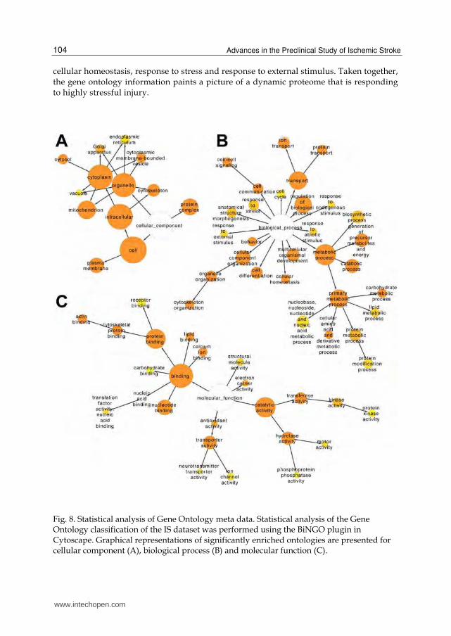
3.2 Statistical analysis of Gene Ontology meta data Graphical and statistical analyses of the Gene Ontology (GO) annotations were performed
using the BiNGO 2.4.4 plugin for Cytoscape 2.8.2. This analysis involves gathering the GO
meta data associated with the IS proteins, and determining if any annotations are
statistically over-represented for each annotation category. BiNGO makes use of the
hierarchical structure of the GO database to produce a graphical representation of the
significantly over-represented ontologies, enabling the depiction of the parent-child
relationships of the annotations. Fig. 8 is the graphical representation of the relative
positions of the terms in the GO hierarchy, with the degree of significance indicated
according to color (white nodes are not significant). The size of each node (area) is
proportional to the number of proteins annotated to each node. The major, significant
cellular component ontologies (Fig. 8A) are cytoplasm, mitochondrion, cytoskeleton,
plasma membrane, cytoplasmic membrane-bounded vesicle, Golgi apparatus,
endoplasmic reticulum and vacuole. This list indicates that cerebral ischemia induces
dramatic alterations in the proteomes of organelles that are involved in regulating cell
death/survival processes. This is further supported by the molecular function ontologies
(Fig. 8C) that are significantly enriched in terms such as kinase activity, transporter
activity, antioxidant activity, electron carrier activity and nucleotide binding.
Additionally, the enriched biological process ontologies (Fig. 8B) are dominated by a
variety of metabolic processes, as well as transport, cellular component organization,
A B
www.intechopen.com
Advances in the Preclinical Study of Ischemic Stroke
104
cellular homeostasis, response to stress and response to external stimulus. Taken together,
the gene ontology information paints a picture of a dynamic proteome that is responding
to highly stressful injury.
Fig. 8. Statistical analysis of Gene Ontology meta data. Statistical analysis of the Gene Ontology classification of the IS dataset was performed using the BiNGO plugin in Cytoscape. Graphical representations of significantly enriched ontologies are presented for cellular component (A), biological process (B) and molecular function (C).
www.intechopen.com
Cerebral Ischemia Induced Proteomic Alterations: Consequences for the Synapse and Organelles
105
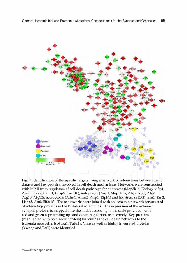
Fig. 9. Identification of therapeutic targets using a network of interactions between the IS dataset and key proteins involved in cell death mechanisms. Networks were constructed with MiMI from regulators of cell death pathways for apoptosis (Map3k14, Endog, Aifm1, Apaf1, Cycs, Capn1, Casp8, Casp10), autophagy (Aup1, Map1lc3a, Atg3, Atg5, Atg7, Atg10, Atg12), necroptosis (Aifm1, Aifm2, Parp1, Ripk1) and ER stress (ERAD; Ern1, Ern2, Hspa5, Atf6, Eif2ak3). These networks were joined with an ischemia network constructed of interacting proteins in the IS dataset (diamonds). The expression of the ischemic synaptic proteins is mapped onto the nodes according to the scale provided, with red and green representing up- and down-regulation, respectively. Key proteins (highlighted with bold node borders) for joining the cell death networks to the ischemia network (Hsp90aa1, Tuba4a, Vim) as well as highly integrated proteins (Ywhag and Taf1) were identified.
www.intechopen.com
Advances in the Preclinical Study of Ischemic Stroke
106
3.3 Comparison with ischemic-responsive proteins identified by ICAT We recently identified 41 ischemia-responsive synaptosomal proteins at 20 h using ICAT-
based nanoLC-MS/MS proteomics. While the ICAT-based method exhibited a high
quantitative reproducibility, quantitative accuracy, and a wide dynamic range (Costain et al.
2010), it did not provide a very comprehensive analysis of the proteins. A comparison
between the label-free and ICAT methods is shown in Table 3. While both methods have
high quantitative reproducibility, the label-free method identified about 5-times more
proteins and peptides in a synaptosome prep than the ICAT method. In addition, the
number of peptides per protein and proteome coverage was significantly higher using the
label-free method. The label-free method also showed higher peptide scores and identified
several cysteine-free proteins. The label-free method identified 32 of the proteins that were
also identified by ICAT (Fig. 6A) and the majority (80%) of their expressions values (i.e., fold
change in response to MCAO) were in agreement between the two methods. Thus, while
both methods are quantitatively comparable, the label-free approach provided a much more
comprehensive coverage of the proteome and addressed some of the limitations of ICAT,
including the detection of cysteine-free proteins.
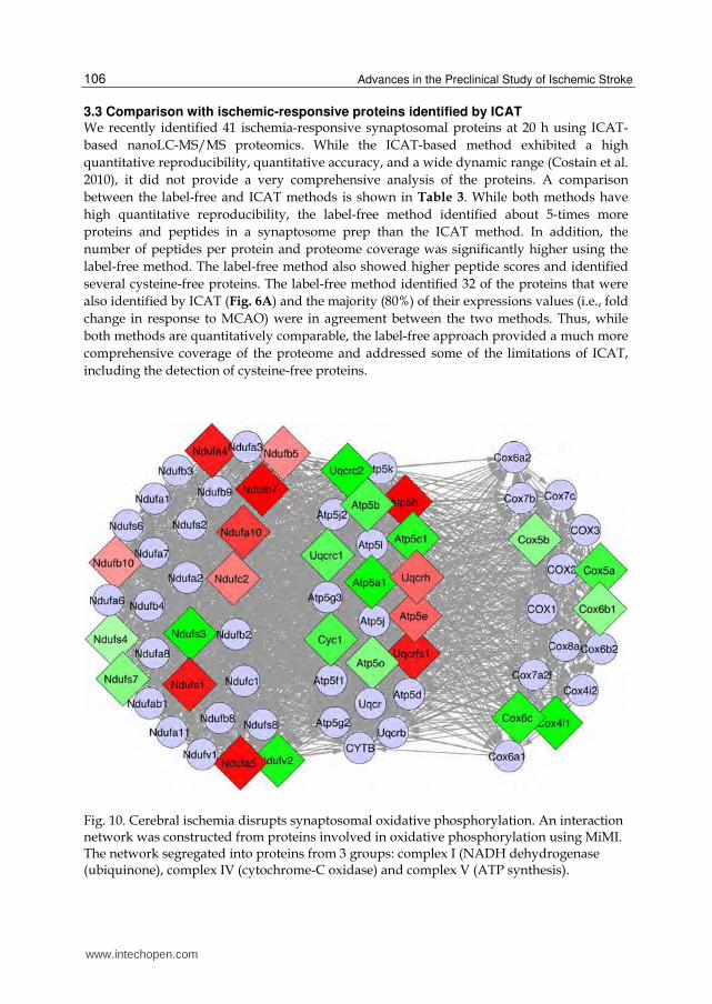
Fig. 10. Cerebral ischemia disrupts synaptosomal oxidative phosphorylation. An interaction network was constructed from proteins involved in oxidative phosphorylation using MiMI. The network segregated into proteins from 3 groups: complex I (NADH dehydrogenase (ubiquinone), complex IV (cytochrome-C oxidase) and complex V (ATP synthesis).
www.intechopen.com

Cerebral Ischemia Induced Proteomic Alterations: Consequences for the Synapse and Organelles
107
Label-free ICAT t-test
Mean number of ions detected by nanoLC-MS per sample
>12,000 <4,000 p<0.0001
Quantitative reproducibility (mean coefficient of variance) 10% 9% ns
Total unique proteins identified 371 41
Peptides per protein 3.9 1.2 p<0.0001
Protein coverage (%) 15% 4.4% p<0.0001
Peptide scores 71 32 p<0.0001
Number of cysteine-free proteins 13 N/A
Table 3. Statistical comparison of label-free and ICAT based nanoLC-MS proteomics.
3.4 Protein interaction analysis In an effort to identify proteins that are important in mediating ischemia-induced cell death,
interaction networks were constructed using the MiMI plugin in Cytoscape. Key proteins
involved in initiating the four major active cell death mechanisms were identified and
stringent interaction networks were individually constructed. Additionally, a network of
interactions among the proteins in the IS dataset was constructed, which resulted in the
generation of a network consisting of 203 proteins (IS network). The cell death networks
were merged with the IS network, resulting in the identification of a number of proteins that
are likely to be important mediators of synaptic pathology and cell death (Fig. 9). This
analysis revealed a strong degree of association between apoptosis and necroptosis, but little
direct interaction with autophagy. In comparison, ER associated degradation (ERAD) was
associated with both necroptosis and autophagy. Fig. 9 indicates that the IS network is
connected to apoptosis primarily through the vimentin (Vim) protein, whereas the
necroptosis and ERAD networks are connected to the IS network in large part due to
Hsp90aa1. In comparison, the autophagy network was connected to the IS network through
Tuba4a. Additionally, the 14-3-3 protein Ywhag was revealed to be a highly connected
protein within the IS network and the transcription factor Taf1 is a point of convergence for
the ERAD, autophagy and necroptosis pathways.
As mentioned, many of the IS proteins are involved in a group of activities that are of obvious importance to cellular metabolism and signaling. These functionalities are highly important in maintaining cellular homeostasis as well as in deciding cellular fate during injurious conditions. Fig. 10 is a graphical representation of the interactions among the proteins involved in the process of oxidative phosphorylation. The proteins segregated into three groups with related functions: cytochrome-C oxidase activity, NADH dehydrogenase (ubiquinone) activity and hydrogen ion transmembrane transporter activity. The expression of the proteins reveals that the Cox proteins were universally down-regulated, whereas proteins in the other groups exhibited more varied effects. Fig. 11A is a network of the proteins involved in glycolysis – gluconeogenesis. The figure demonstrates that there is widespread up-regulation of glycolytic proteins, with down-regulation of only two proteins. Similarly, Fig. 11B indicates that there is widespread up-regulation of proteins with anti-oxidant activity. Interestingly, two of the proteins identified in Fig. 9, Hsp90aa1 and Taf1, are also involved in the antioxidant response. The results depicted in Fig. 11 clearly demonstrate that the ischemic synaptosomal proteome is actively engaged in an attempt to counteract the effects of cerebral ischemia, namely energy depletion and oxidative damage.
www.intechopen.com

Advances in the Preclinical Study of Ischemic Stroke
108
Fig. 11. Interaction network analysis demonstrates cellular responses to energy depletion and oxidative stress. Panel A is a network of proteins involved in glycolysis - gluconeogenesis, showing the up-regulation of proteins involved in this process. Panel B is a network showing the up-regulation of proteins with anti-oxidative properties.
www.intechopen.com
Cerebral Ischemia Induced Proteomic Alterations: Consequences for the Synapse and Organelles
109
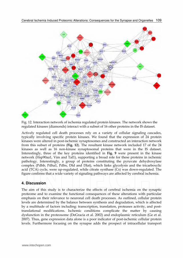
Fig. 12. Interaction network of ischemia regulated protein kinases. The network shows the regulated kinases (diamonds) interact with a subset of 16 other proteins in the IS dataset.
Actively regulated cell death processes rely on a variety of cellular signaling cascades, typically involving specific protein kinases. We found that the expression of 24 protein kinases were altered in post-ischemic synaptosomes and constructed an interaction network from this subset of proteins (Fig. 12). The resultant kinase network included 17 of the 24 kinases as well as 16 non-kinase synaptosomal proteins that were in the IS dataset. Interestingly, three of the key proteins identified in Fig. 9 were present in the kinase network (Hsp90aa1, Vim and Taf1), supporting a broad role for these proteins in ischemic pathology. Interestingly, a group of proteins constituting the pyruvate dehydroxylase complex (Pdhb, Pdha1, Pdhx, Dld and Dlat), which links glycolysis and the tricarboxylic acid (TCA) cycle, were up-regulated, while citrate synthase (Cs) was down-regulated. The figure confirms that a wide variety of signaling pathways are affected by cerebral ischemia.
4. Discussion
The aim of this study is to characterize the effects of cerebral ischemia on the synaptic proteome and to examine the functional consequences of these alterations with particular emphasis on their relevance to neuronal cell death processes. As outlined, cellular protein levels are determined by the balance between synthesis and degradation, which is affected by a multitude of factors including: transcription, translation, proteases activity, and post-translational modifications. Ischemic conditions complicate the matter by causing dysfunction in the proteosome (DeGracia et al. 2002) and endoplasmic reticulum (Ge et al. 2007). Thus, gene expression data alone is a poor indicator of post-ischemic cellular protein levels. Furthermore focusing on the synapse adds the prospect of intracellular transport
www.intechopen.com
Advances in the Preclinical Study of Ischemic Stroke
110
mechanisms and localized translation further blurring the association between transcription and synaptic protein levels (Zhao et al. 2005, Vanderklish & Bahr 2000, Havik et al. 2003). As a result of these factors, the best approach for determining post-ischemic synaptic protein levels is to perform a direct assessment using proteomic methodologies.
4.1 Cerebral ischemia-induced alterations in the synaptic proteome Here, we determined the proteomic response of the mouse brain synapse to cerebral ischemia by performing an analysis of mouse brain synaptosomes. Using a label-free nanoLC-MS/MS method, we identified 371 synaptosomal proteins that were altered 20 hrs after cerebral ischemia (Table 3), representing ≈ 27% of the total peaks detected. Linear regression analysis was used to exclude the possible influence of systemic bias on expression due to protein size and MASCOT score (Fig. 5). The purity of the synaptosomal prep was validated by the determination that > 80% of the IS dataset were previously characterized as being localized in the synapse (Fig. 6). Consistent with our previous study, the majority of the IS proteins were localized in the mitochondria (Costain et al. 2010), and the localization of the remaining proteins was consistent with the synaptic compartment. Furthermore, literature mining of PubMed indicated that only ≈ 15% of the IS proteins were associated with the MCAO model of cerebral ischemia. A statistical analysis of the ontological classification of the IS dataset revealed a number of important details (Fig. 8). Firstly, the significant enrichment of proteins located in the mitochondria, endoplasmic reticulum and Golgi apparatus indicate the importance of these structures in mediating post-ischemic neuronal function. Secondly, significantly enriched molecular function ontologies include kinase, transport, antioxidant and electron carrier activity. These functions are related to metabolism, and are consistent with tissues responding to catastrophic energy depletion. Lastly, the significantly enriched biological process ontologies were also associated with metabolism and stress responses. The present results, obtained using label-free nanoLC-MS/MS, are largely in agreement with our previous ICAT-based proteomics study (Table 3). The consistency between these datasets reflects the reproducibility and purity of synaptosomal preparations, as well as the proteomic methodologies. Furthermore, the label-free method produced a more comprehensive dataset than the ICAT method, exhibiting greater protein coverage, peptide scores and the ability to identify cysteine-free proteins.
4.2 Interaction network analysis In an effort to place the observed alterations in the IS dataset into biological context, a variety of protein interaction analyses were performed. Interaction networks can be constructed from a variety of data types, such as protein-protein, protein-DNA and genetic interactions. The value of examining such interactions is that the overall biochemical function of proteins or DNA is a product of the interactions in which they participate. Thus, analyzing functional interactions enables the construction of signaling pathways or interaction networks that are capable of modeling a system or interpreting systematic responses to a given perturbation. In the present study, we used our observed systematic alterations in synaptosomal protein expression, and protein-protein interaction analyses to aid in interpreting the net effect of ischemia on the biological system (synapses and neurons). Furthermore, we integrated our observations in IS synaptosomes with key proteins in cell death pathways that are highly predictive of cell fate. Lastly, we focused on certain subsets of proteins to produce interaction networks that provided insight into the key biological processes.
www.intechopen.com

Cerebral Ischemia Induced Proteomic Alterations: Consequences for the Synapse and Organelles
111
BiNGO and MiMI are valuable analysis tools that are integrated into the Cytoscape framework. The GO networks created by BiNGO identify the statistically overrepresented ontologies associated with a given gene or protein dataset. This enables rapid identification and characterization of ontologies (biological process, molecular function, cellular location) that are specific to a given dataset, as well as the hierarchical nature of the ontologies. MiMI, on the other hand, gathers interaction information from various public databases and constructs an interaction network based on a list of proteins of interest. In these interaction networks, lines drawn between entities (proteins) can represent a variety of interactions, such as binding, phosphorylation, or other biologically relevant modifications. Such analyses allow for the identification of intermediary proteins that are important to the network, but are not directly identified by either biochemical analysis or literature mining. Importantly, interaction networks can be used to identify highly integrated ‘hubs’, which are likely to represent key factors in a given biological process or pathology. Cell death can occur either in an unregulated or a regulated manner. Apoptosis is a well-studied regulated cell death mechanism, and awareness of regulated necrosis (necroptosis) has been increasing (Ankarcrona et al. 1995, Baines 2010, Hitomi et al. 2008). Additionally, autophagy and ER associated degradation (ERAD) are regulated processes that are vital to cell fate decisions during injurious conditions (Liu et al. 2010, Petrovski et al. 2011). Importantly, Liu et al. (2010) recently reported that cerebral ischemia induces protein aggregation, leading to multiple organelle damage that is likely to be responsible for delayed neuronal death. We constructed an IS protein interaction network that enabled the identification of five proteins that appear to be critical in linking the consequences of synaptic ischemia to regulated cell death processes (Fig. 9). Of the proteins identified, Vim appeared to provide the strongest association with apoptosis, whereas Hsp90aa1 was the protein that provided a link to necroptosis and ERAD. Although Vim is an intermediate filament protein expressed in glia that may not be expected to be found in the synapse, it is common to find proteins such as Vim and Gfap in synaptosomal preparations (Costain et al. 2008) and is likely to be due to the intimate association between glia and synaptic structures. Nonetheless, up-regulation of Vim following cerebral ischemia has been frequently reported, and is thought to represent the activation of astrocytes and the reactive gliosis process. Furthermore, genetic ablation of Vim has been shown to counteract neuronal pathology, indicating that Vim is relevant to ischemic synaptosomal function (Pekny & Pekna 2004). Similarly, up-regulation of heat shock proteins, such as Hsp27 and Hsp70, in response to cerebral ischemia is a well-documented finding (Franklin et al. 2005, Currie & Plumier 1998). The Hsp90 family of molecular chaperones are involved in a variety of cellular processes, such as signal transduction, protein folding and protein degradation. Hsp90aa1 is the inducible cytoplasmic form of Hsp90 and aids in the folding of a wide variety of proteins. While other ischemia-responsive heat shock proteins that are associated with MCAO were identified in the present study (Table 1 and 2; Hspd1, Hspa9), Hsp90aa1 has not previously been associated with cerebral ischemia and is therefore a good candidate for further examination of its role in ischemia-induced necroptosis and ERAD. The IS network analysis indicated that autophagy was associated with the IS dataset though the cytoskeletal protein Tuba4a (Fig. 9). Tuba4a has previously been identified as a synaptic protein, but has not been associated with MCAO. Alterations in cellular cytoskeletal proteins, such as Map2, are known to occur following exposure to ischemic injury (Kharlamov et al. 2009) and the observed reduction in Tuba4a expression is consistent with disruption of cytoskeletal structures. Reduced expression in other tubulin/tubulin related
www.intechopen.com

Advances in the Preclinical Study of Ischemic Stroke
112
proteins (Tubb4, Tubb5, Tppp) was also observed, indicating an autophagy-mediated failure in the microtubule system and collapse of the synaptic structure function relationship. Another interesting finding to arise from the IS network analysis is the prominence of the transcription factor Taf1 in connecting the autophagy, ERAD and necroptosis sub-networks. While Taf1 is involved in basal transcription, it has recently been found to be an important factor in certain neurodegenerative conditions (Davidson et al. 2009, Sako et al. 2011). Similarly, the down-regulated protein Ywhag was a highly integrated protein within the IS network, whereas the related protein Ywhaq was up-regulated with fewer interconnections (Fig. 9). There is growing interest in Ywhag as a mediator of neuroprotection in cerebral ischemia (Dong et al. 2010) and the observed decrease in its expression is consistent with the activation of cell death pathways. Additionally, up-regulation of Ywhaq has been observed in amyotrophic lateral sclerosis patients (Malaspina et al. 2000), and has been found to be necessary for autophagy (Wang et al. 2010). These findings suggest that the 14-3-3 proteins are likely to be playing an important role in mediating post-ischemic neuronal cell death and are good targets for therapeutic intervention in stroke. The role of mitochondria in the pathology of ischemic neuronal death (apoptotic and necrotic) is well established (Iijima 2006, Tsujimoto & Shimizu 2007). Cerebral ischemia results in a sustained increase in intracellular Ca++ that is buffered by the mitochondria. The increased Ca++ levels disrupt the mitochondrial membrane potential and induce the formation of the permeability transition pore, thereby activating the intrinsic apoptosis pathway (Tsujimoto & Shimizu 2007). In the synaptosome, hypoxia induces mitochondrial membrane potential disruption (Aldinucci et al. 2007) and Brown et al (2006) have demonstrated that synaptic mitochondria are more sensitive to Ca++ overload than non-synaptic mitochondria. Our present findings confirm our previous observation that widespread alterations in protein expression occur in post-ischemic synaptic mitochondria (Costain et al. 2010). An interaction network was constructed from the large number of IS proteins that are involved in oxidative phosphorylation (Fig. 10). This interaction network clearly demonstrates that cerebral ischemia induces an imbalance in oxidative phosphorylation, with down-regulation of complex IV components and more variable effects on complex I and V. While oxidative phosphorylation is clearly disrupted, there is evidence that the cells are attempting to compensate by up-regulating glycolytic enzymes (Fig. 11A) as well as proteins with anti-oxidative activity (Fig.
11B). Unfortunately, glycolysis cannot produce the same amount of ATP that is derived from oxidative phosphorylation. A wide variety of protein kinases are involved in mediating regulated cell death, and we identified a subset of 24 protein kinases within the IS dataset. An interaction network was constructed from the IS kinases subset (Fig. 12) and the kinase network independently identified three of the key proteins singled out in the IS network analysis (Fig. 9), supporting the contention that these proteins are involved in post-ischemic cell signaling pathways. Hsp90aa1, Taf1 and Vim were essential for connecting the four cell death mechanisms to the IS dataset, and their association with alterations in protein kinase levels further confirms their importance in mediating synaptic pathology.
5. Conclusion
In closing, this study has demonstrated that the synapse is highly responsive to cerebral ischemia and is highly informative about cerebral ischemia-induced cell death mechanisms at the organelle level. We have used interaction network analyses of the IS dataset to clarify
www.intechopen.com
Cerebral Ischemia Induced Proteomic Alterations: Consequences for the Synapse and Organelles
113
the effects cerebral ischemia on metabolic function, as well as to identify five proteins that are integral to cell death pathways and are potential targets for therapeutic intervention. This report also confirms that cerebral ischemia induces marked aberrations in synaptic mitochondria, lysosomes, endoplasmic reticulum and golgi apparatus, thereby emphasizing the interplay between organelles during oxidative damage.
6. References
Abe, K., Chisaka, O., Van Roy, F. & Takeichi, M. (2004) Stability of dendritic spines and synaptic contacts is controlled by alpha N-catenin. Nature Neuroscience, 7, 357-363.
Aldinucci, C., Carretta, A., Ciccoli, L., Leoncini, S., Signorini, C., Buonocore, G. & Pessina, G. P. (2007) Hypoxia affects the physiological behavior of rat cortical synaptosomes. Free Radical Biology and Medicine, 42, 1749-1756.
Ankarcrona, M., Dypbukt, J. M., Bonfoco, E., Zhivotovsky, B., Orrenius, S., Lipton, S. A. & Nicotera, P. (1995) Glutamate-induced neuronal death: a succession of necrosis or apoptosis depending on mitochondrial function. Neuron, 15, 961-973.
Baines, C. P. (2010) Role of the mitochondrion in programmed necrosis. Front Physiol, 1, 156. Brown, M. R., Sullivan, P. G. & Geddes, J. W. (2006) Synaptic mitochondria are more
susceptible to Ca2+overload than nonsynaptic mitochondria. Journal of Biological Chemistry, 281, 11658-11668.
Chen, X., Karnovsky, A., Sans, M. D., Andrews, P. C. & Williams, J. A. (2010) Molecular characterization of the endoplasmic reticulum: insights from proteomic studies. Proteomics, 10, 4040-4052.
Cheng, D., Hoogenraad, C. C., Rush, J. et al. (2006) Relative and absolute quantification of postsynaptic density proteome isolated from rat forebrain and cerebellum. Molecular and Cellular Proteomics, 5, 1158-1170.
Costain, W. J., Haqqani, A. S., Rasquinha, I., Giguere, M. S., Slinn, J., Zurakowski, B. & Stanimirovic, D. B. (2010) Proteomic analysis of synaptosomal protein expression reveals that cerebral ischemia alters lysosomal Psap processing. Proteomics, 10, 3272-3291.
Costain, W. J., Rasquinha, I., Sandhu, J. K., Rippstein, P., Zurakowski, B., Slinn, J., Macmanus, J. P. & Stanimirovic, D. B. (2008) Cerebral ischemia causes dysregulation of synaptic adhesion in mouse synaptosomes. Journal of Cerebral Blood Flow and Metabolism, 28, 99-110.
Couchman, J. R. (2003) Syndecans: proteoglycan regulators of cell-surface microdomains? Nature Reviews Molecular Cell Biology, 4, 926-937.
Currie, R. W. & Plumier, J. C. (1998) The heat shock response and tissue protection. In: Delayed preconditioning and adaptive cardioprotection, (G. F. Baxter and D. M. Yellon eds.), Vol. 207, pp. 135-153. Kluwer Academic Publishers, Dordrecht ; Boston.
Davidson, M. E., Kerepesi, L. A., Soto, A. & Chan, V. T. (2009) D-Serine exposure resulted in gene expression changes implicated in neurodegenerative disorders and neuronal dysfunction in male Fischer 344 rats. Archives of Toxicology, 83, 747-762.
DeGracia, D. J., Kumar, R., Owen, C. R., Krause, G. S. & White, B. C. (2002) Molecular pathways of protein synthesis inhibition during brain reperfusion: implications for neuronal survival or death. Journal of Cerebral Blood Flow and Metabolism, 22, 127-141.
www.intechopen.com
Advances in the Preclinical Study of Ischemic Stroke
114
Deisseroth, K., Mermelstein, P. G., Xia, H. & Tsien, R. W. (2003) Signaling from synapse to nucleus: the logic behind the mechanisms. Current Opinion in Neurobiology, 13, 354-365.
Dong, Y., Zhao, R., Chen, X. Q. & Yu, A. C. (2010) 14-3-3gamma and neuroglobin are new intrinsic protective factors for cerebral ischemia. Molecular Neurobiology, 41, 218-231.
Ehlers, M. D. (2002) Molecular morphogens for dendritic spines. Trends in Neurosciences, 25, 64-67.
Enright, L. E., Zhang, S. & Murphy, T. H. (2007) Fine mapping of the spatial relationship between acute ischemia and dendritic structure indicates selective vulnerability of layer V neuron dendritic tufts within single neurons in vivo. Journal of Cerebral Blood Flow and Metabolism, 27, 1185-1200.
Franklin, T. B., Krueger-Naug, A. M., Clarke, D. B., Arrigo, A. P. & Currie, R. W. (2005) The role of heat shock proteins Hsp70 and Hsp27 in cellular protection of the central nervous system. International Journal of Hyperthermia, 21, 379-392.
Gasic, G. P. & Nicotera, P. (2003) To die or to sleep, perhaps to dream. Toxicology Letters, 139, 221-227.
Ge, P., Luo, Y., Liu, C. L. & Hu, B. (2007) Protein aggregation and proteasome dysfunction after brain ischemia. Stroke, 38, 3230-3236.
Gilbert, R. W., Costain, W. J., Blanchard, M. E., Mullen, K. L., Currie, R. W. & Robertson, H. A. (2003) DNA microarray analysis of hippocampal gene expression measured twelve hours after hypoxia-ischemia in the mouse. Journal of Cerebral Blood Flow and Metabolism, 23, 1195-1211.
Ginsberg, M. D. (2008) Neuroprotection for ischemic stroke: past, present and future. Neuropharmacology, 55, 363-389.
Haqqani, A. S., Kelly, J. F. & Stanimirovic, D. B. (2008) Quantitative protein profiling by mass spectrometry using label-free proteomics. Methods in Molecular Biology, 439, 241-256.
Hasbani, M. J., Schlief, M. L., Fisher, D. A. & Goldberg, M. P. (2001) Dendritic spines lost during glutamate receptor activation reemerge at original sites of synaptic contact. Journal of Neuroscience, 21, 2393-2403.
Havik, B., Rokke, H., Bardsen, K., Davanger, S. & Bramham, C. R. (2003) Bursts of high-frequency stimulation trigger rapid delivery of pre-existing alpha-CaMKII mRNA to synapses: a mechanism in dendritic protein synthesis during long-term potentiation in adult awake rats. European Journal of Neuroscience, 17, 2679-2689.
Hirosawa, M., Hoshida, M., Ishikawa, M. & Toya, T. (1993) MASCOT: multiple alignment system for protein sequences based on three-way dynamic programming. Computer Applications in the Biosciences, 9, 161-167.
Hitomi, J., Christofferson, D. E., Ng, A., Yao, J., Degterev, A., Xavier, R. J. & Yuan, J. (2008) Identification of a molecular signaling network that regulates a cellular necrotic cell death pathway. Cell, 135, 1311-1323.
Hou, S. T. & MacManus, J. P. (2002) Molecular mechanisms of cerebral ischemia-induced neuronal death. International Review of Cytology, 221, 93-148.
Iijima, T. (2006) Mitochondrial membrane potential and ischemic neuronal death. Neuroscience Research, 55, 234-243.
Jordan, B. A., Fernholz, B. D., Boussac, M., Xu, C., Grigorean, G., Ziff, E. B. & Neubert, T. A. (2004) Identification and verification of novel rodent postsynaptic density proteins. Molecular and Cellular Proteomics, 3, 857-871.
www.intechopen.com
Cerebral Ischemia Induced Proteomic Alterations: Consequences for the Synapse and Organelles
115
Kagedal, K., Johansson, U. & Ollinger, K. (2001) The lysosomal protease cathepsin D mediates apoptosis induced by oxidative stress. FASEB Journal, 15, 1592-1594.
Kharlamov, A., LaVerde, G. C., Nemoto, E. M., Jungreis, C. A., Yushmanov, V. E., Jones, S. C. & Boada, F. E. (2009) MAP2 immunostaining in thick sections for early ischemic stroke infarct volume in non-human primate brain. Journal of Neuroscience Methods, 182, 205-210.
Li, K. W., Hornshaw, M. P., Van Der Schors, R. C. et al. (2004) Proteomics analysis of rat brain postsynaptic density. Implications of the diverse protein functional groups for the integration of synaptic physiology. Journal of Biological Chemistry, 279, 987-1002.
Liu, C., Gao, Y., Barrett, J. & Hu, B. (2010) Autophagy and protein aggregation after brain ischemia. Journal of Neurochemistry, 115, 68-78.
MacManus, J. P., Graber, T., Luebbert, C., Preston, E., Rasquinha, I., Smith, B. & Webster, J. (2004) Translation-state analysis of gene expression in mouse brain after focal ischemia. Journal of Cerebral Blood Flow and Metabolism, 24, 657-667.
Malaspina, A., Kaushik, N. & de Belleroche, J. (2000) A 14-3-3 mRNA is up-regulated in amyotrophic lateral sclerosis spinal cord. Journal of Neurochemistry, 75, 2511-2520.
Martone, M. E., Jones, Y. Z., Young, S. J., Ellisman, M. H., Zivin, J. A. & Hu, B. R. (1999) Modification of postsynaptic densities after transient cerebral ischemia: a quantitative and three-dimensional ultrastructural study. Journal of Neuroscience, 19, 1988-1997.
Mattson, M. P. & Duan, W. (1999) "Apoptotic" biochemical cascades in synaptic compartments: roles in adaptive plasticity and neurodegenerative disorders. Journal of Neuroscience Research, 58, 152-166.
Mattson, M. P. & Gleichmann, M. (2005) The neuronal death protein par-4 mediates dopaminergic synaptic plasticity. Molecular Interventions, 5, 278-281.
Mattson, M. P., Keller, J. N. & Begley, J. G. (1998) Evidence for synaptic apoptosis. Experimental Neurology, 153, 35-48.
Morciano, M., Burre, J., Corvey, C., Karas, M., Zimmermann, H. & Volknandt, W. (2005) Immunoisolation of two synaptic vesicle pools from synaptosomes: a proteomics analysis. Journal of Neurochemistry, 95, 1732-1745.
Park, J. S., Bateman, M. C. & Goldberg, M. P. (1996) Rapid alterations in dendrite morphology during sublethal hypoxia or glutamate receptor activation. Neurobiology of Disease, 3, 215-227.
Passafaro, M., Nakagawa, T., Sala, C. & Sheng, M. (2003) Induction of dendritic spines by an extracellular domain of AMPA receptor subunit GluR2. Nature, 424, 677-681.
Pastuszko, A., Wilson, D. F. & Erecinska, M. (1982) Neurotransmitter metabolism in rat brain synaptosomes: effect of anoxia and pH. Journal of Neurochemistry, 38, 1657-1667.
Pedrioli, P. G., Eng, J. K., Hubley, R. et al. (2004) A common open representation of mass spectrometry data and its application to proteomics research. Nature Biotechnology, 22, 1459-1466.
Pekny, M. & Pekna, M. (2004) Astrocyte intermediate filaments in CNS pathologies and regeneration. Journal of Pathology, 204, 428-437.
Peng, J., Elias, J. E., Thoreen, C. C., Licklider, L. J. & Gygi, S. P. (2003) Evaluation of multidimensional chromatography coupled with tandem mass spectrometry (LC/LC-MS/MS) for large-scale protein analysis: the yeast proteome. Journal of Proteome Research, 2, 43-50.
www.intechopen.com
Advances in the Preclinical Study of Ischemic Stroke
116
Peng, J., Kim, M. J., Cheng, D., Duong, D. M., Gygi, S. P. & Sheng, M. (2004) Semiquantitative proteomic analysis of rat forebrain postsynaptic density fractions by mass spectrometry. Journal of Biological Chemistry, 279, 21003-21011.
Petrovski, G., Das, S., Juhasz, B., Kertesz, A., Tosaki, A. & Das, D. K. (2011) Cardioprotection by endoplasmic reticulum stress-induced autophagy. Antioxidants & Redox Signaling, 14, 2191-2200.
Phillips, G. R., Florens, L., Tanaka, H., Khaing, Z. Z., Fidler, L., Yates, J. R., 3rd & Colman, D. R. (2005) Proteomic comparison of two fractions derived from the transsynaptic scaffold. Journal of Neuroscience Research, 81, 762-775.
Rafalowska, U., Erecinska, M. & Wilson, D. F. (1980) The effect of acute hypoxia on synaptosomes from rat brain. Journal of Neurochemistry, 34, 1160-1165.
Sako, W., Morigaki, R., Kaji, R., Tooyama, I., Okita, S., Kitazato, K., Nagahiro, S., Graybiel, A. M. & Goto, S. (2011) Identification and localization of a neuron-specific isoform of TAF1 in rat brain: implications for neuropathology of DYT3 dystonia. Neuroscience, 189, 100-107.
Schrimpf, S. P., Meskenaite, V., Brunner, E., Rutishauser, D., Walther, P., Eng, J., Aebersold, R. & Sonderegger, P. (2005) Proteomic analysis of synaptosomes using isotope-coded affinity tags and mass spectrometry. Proteomics, 5, 2531-2541.
Stevens, S. M., Jr., Zharikova, A. D. & Prokai, L. (2003) Proteomic analysis of the synaptic plasma membrane fraction isolated from rat forebrain. Brain Research. Molecular Brain Research, 117, 116-128.
Sulkowski, G., Waskiewicz, J., Walski, M., Januszewski, S. & Rafalowska, U. (2002) Synaptosomal susceptibility on global ischaemia caused by cardiac arrest correlated with early and late times after recirculation in rats. Resuscitation, 52, 203-213.
Taylor, S. W., Fahy, E., Zhang, B. et al. (2003) Characterization of the human heart mitochondrial proteome. Nature Biotechnology, 21, 281-286.
Tsujimoto, Y. & Shimizu, S. (2007) Role of the mitochondrial membrane permeability transition in cell death. Apoptosis, 12, 835-840.
Vanderklish, P. W. & Bahr, B. A. (2000) The pathogenic activation of calpain: a marker and mediator of cellular toxicity and disease states. International Journal of Experimental Pathology, 81, 323-339.
Wang, B., Ling, S. & Lin, W. C. (2010) 14-3-3Tau regulates Beclin 1 and is required for autophagy. PLoS ONE, 5, e10409.
Witzmann, F. A., Arnold, R. J., Bai, F. et al. (2005) A proteomic survey of rat cerebral cortical synaptosomes. Proteomics, 5, 2177-2201.
Yamashima, T. & Oikawa, S. (2009) The role of lysosomal rupture in neuronal death. Progress in Neurobiology, 89, 343-358.
Zhang, S. & Murphy, T. H. (2007) Imaging the impact of cortical microcirculation on synaptic structure and sensory-evoked hemodynamic responses in vivo. PLoS Biology, 5, e119.
Zhao, H., Shimohata, T., Wang, J. Q., Sun, G., Schaal, D. W., Sapolsky, R. M. & Steinberg, G. K. (2005) Akt contributes to neuroprotection by hypothermia against cerebral ischemia in rats. Journal of Neuroscience, 25, 9794-9806.
www.intechopen.com

Advances in the Preclinical Study of Ischemic StrokeEdited by Dr. Maurizio Balestrino
ISBN 978-953-51-0290-8Hard cover, 530 pagesPublisher InTechPublished online 16, March, 2012Published in print edition March, 2012
InTech EuropeUniversity Campus STeP Ri Slavka Krautzeka 83/A 51000 Rijeka, Croatia Phone: +385 (51) 770 447 Fax: +385 (51) 686 166www.intechopen.com
InTech ChinaUnit 405, Office Block, Hotel Equatorial Shanghai No.65, Yan An Road (West), Shanghai, 200040, China
Phone: +86-21-62489820 Fax: +86-21-62489821
This book reports innovations in the preclinical study of stroke, including - novel tools and findings in animalmodels of stroke, - novel biochemical mechanisms through which ischemic damage may be both generatedand limited, - novel pathways to neuroprotection. Although hypothermia has been so far the sole"neuroprotection" treatment that has survived the translation from preclinical to clinical studies, progress inboth preclinical studies and in the design of clinical trials will hopefully provide more and better treatments forischemic stroke. This book aims at providing the preclinical scientist with innovative knowledge and tools toinvestigate novel mechanisms of, and treatments for, ischemic brain damage.
How to referenceIn order to correctly reference this scholarly work, feel free to copy and paste the following:
Willard J. Costain, Arsalan S. Haqqani, Ingrid Rasquinha, Marie-Soleil Giguere and Jacqueline Slinn (2012).Cerebral Ischemia Induced Proteomic Alterations: Consequences for the Synapse and Organelles, Advancesin the Preclinical Study of Ischemic Stroke, Dr. Maurizio Balestrino (Ed.), ISBN: 978-953-51-0290-8, InTech,Available from: http://www.intechopen.com/books/advances-in-the-preclinical-study-of-ischemic-stroke/cerebral-ischemia-induced-proteomic-alterations-consequences-for-the-synapse-and-organelles-
Related Documents