中国化学会第二届全国生物物理化学会议(NCBPC2)暨国际华人生物物理化学发展论坛 中国化学会成立 80 周年 中国化学会第二届全国生物物理化学会议 暨 国际华人生物物理化学发展论坛 会议论文集 2012 年 10 月 15-18 日 武汉 主办:中国化学会生物物理化学专业委员会 承办:武汉大学 华中师范大学 湖北省化学化工学会 协办: 美国 TA 仪器

Welcome message from author
This document is posted to help you gain knowledge. Please leave a comment to let me know what you think about it! Share it to your friends and learn new things together.
Transcript

中国化学会第二届全国生物物理化学会议(NCBPC2)暨国际华人生物物理化学发展论坛
中国化学会成立 80 周年
中国化学会第二届全国生物物理化学会议
暨 国际华人生物物理化学发展论坛
会议论文集
2012 年 10 月 15-18 日 武汉
主办:中国化学会生物物理化学专业委员会
承办:武汉大学
华中师范大学
湖北省化学化工学会
协办: 美国 TA 仪器

中国化学会第二届全国生物物理化学会议(NCBPC2)暨国际华人生物物理化学发展论坛
I
目 录
前言
一、大会组织机构
二、赞助单位
三、会议论文

中国化学会第二届全国生物物理化学会议(NCBPC2)暨国际华人生物物理化学发展论坛
II
前 言
国家自然科学基金委员会最近的学科分类目录正式在“物理化学”下列出了
“生物物理化学”,标志着生物物理化学这个分支学科在国内的确立,为我国生
物物理化学的发展奠定了基础。作为一个新的交叉学科,生物物理化学在国内尚
处于发展的早期,需要给予特别的关注和扶持。
中国化学会第一届全国生物物理化学会议暨生物物理化学发展战略研讨会于
2010年7月5日-7日在北京大学化学与分子工程学院召开。会议由北京大学、清华大
学、中国科学院化学所等共同发起,负责人:赵新生教授。作为中国化学会成立
80周年纪念活动之一,中国化学会第二届生物物理化学学术会议暨国际华人生物
物理化学发展论坛由中国化学会生物物理化学专业委员会主办,武汉大学、华中
师范大学和湖北省化学化工学会联合承办,于2012年10月15日-18日在武汉大学化
学与分子科学学院召开。生物物理化学会议是为了反映我国生物物理化学领域中
的最新成果,促进各研究单位与相关研究者之间的学术交流,尤其是华人之间的
互动与合作,以更好地推动生物物理化学的研究与发展,特别是进一步明确主攻
方向,发现和培育新的生长点,期望为我国生物物理化学事业的健康快速发展提
供更多的契机和思路。
在赵新生教授和各位同仁的努力下,经中国化学会批准,于2012年1月成立了
生物物理化学专业委员会。鉴于第一届会议的成功和影响,大家希望将此会议延
续下去。本次“武汉会议”是中国化学会生物物理化学专业委员会成立后的第一
次会议,也是第二届年会和华人生物物理化学发展论坛的联合会议。本次会议邀
请本领域的国内外知名专家8位作大会报告、38位作特邀报告,注册人数达240人。
中国化学会第二届全国生物物理化学会议(NCBPC2)暨国际华人生物物理化学发展论坛
III
会议将就国内外生物物理化学前沿、最新进展和我国生物物理化学领域中的最新
成果进行交流,希望能给各位同行提供一个高水平交流的平台,也希望能给对生
物物理化学感兴趣的学生提供良好的学习机会。期待在各位同行的支持下,能够
取得圆满成功!
热忱欢迎出席此次学术会议!衷心感谢各位代表的大力支持!
中国化学会第二届生物物理化学学术会议
暨 国际华人生物物理化学发展论坛组委会
2012 年 10 月 15 日
中国化学会第二届全国生物物理化学会议(NCBPC2)暨国际华人生物物理化学发展论坛
IV
本摘要集收集的摘要源于由作者通过 E-mai 发送到会议秘书处的 Word 文件,
会议秘书处未作内容修改。摘要集目录顺序按照大会报告、分会邀请报告、分会
口头报告、墙报进行分类,除同一单位相对集中外,均为随即产生,特此声明。
编者

中国化学会第二届全国生物物理化学会议(NCBPC2)暨国际华人生物物理化学发展论坛
V
一、大会组织机构
会议学术委员会
主 任 委员:赵新生
副主任委员 (按姓氏拼音排序):
高毅勤 蒋华良 刘 义 曲晓刚 翁羽翔
委 员 (按姓氏拼音排序):
郝京诚 黄 方 黄建国 来鲁华 李峻柏 刘长林 刘冬生
刘扬中 庞代文 沈 旭 谭砚文 王江云 王任小 王 树
徐平勇 尉志武 张文科
会议组织委员会
主 席:周 翔 杨光富
副主席:刘 义 刘长林
秘书长:蒋风雷
秘书处:李东巍 郝格菲 付佳雄 赖 璐 杨文超 王 辉 许子强 章 丹
胡艳军 韩晓乐 马 林 张万举 夏彩芬 付 莉 向 晨 袁 莲
梅 洁 赵 洁 杨立云 汪 佳 樊晓阳 周志强 田方方 葛玉舒
中国化学会第二届全国生物物理化学会议(NCBPC2)暨国际华人生物物理化学发展论坛
VI
二、赞助单位(排名不分先后)
武汉大学化学与分子科学学院“111 引智计划”
通用电气医疗系统贸易发展(上海)有限公司
武汉珈源量子点技术开发有限公司
英国应用光物理公司
武汉欣申试化工科技有限公司
湖北昊博生物科技有限公司
源资信息科技(上海)有限公司
中国化学会第二届全国生物物理化学会议(NCBPC2)暨国际华人生物物理化学发展论坛
VII
三、会议论文
大会报告(Plenary Lectures)
PL1 Single molecule study on DNA/RNA modification
赵新生
PL2 Technical advances, limitations, and advantages of ITC and SPR in detecting protein-ligand
interactions
Ronan O'Brien
PL3 Progress in Single-Molecule Protein Dynamics
Haw Yang
PL4 特殊结构核酸及相关蛋白的手性识别及应用
曲晓刚
PL5 药物-受体作用中的热力学和动力学
蒋华良
PL6 Understanding E. coli chemotaxis at molecular level
来鲁华
PL7 端粒 G-四链体折叠的动力学和热力学控制及生理构象
谭铮
PL8 生命过程实时动态研究对纳米标记材料性能的苛刻要求及应对新策略
庞代文
邀请报告(Invited Lectures)
I1 Flux Network Analysis: Generalized Michaelis-Menten Equation in Enzymatic Catalysis and
Efficient Energy Transfer in Light-Harvesting Systems
吴建澜
I2 Coupling between switching regulation and torque generation in bacterial flagellar motor
Jianhua Xing
I3 Porphyrin-based Near-Infrared Emissive Lanthanide Complexes for Molecular Imaging
黄伟国
I4 DNA-Tagged Nanomaterials as Biophysical Nanoprobes
邓兆祥
I5 DNA G-Quadruplexes as Potential Anticancer Drug Targets
Danzhou Yang
I6 Two-State or Non-Two-State? A Protocol to Differentiate the Two Types of Unfolding Processes
of Proteins
中国化学会第二届全国生物物理化学会议(NCBPC2)暨国际华人生物物理化学发展论坛
VIII
尉志武
I7 Biophysical Chemistry of Alzheimer’s Disease
Liming Ying
I8 基于结构的农药分子设计研究
杨光富
I9
刘海燕
I10 微量热技术在蛋白质药物、生物及材料领域提供的重要信息
林明申
I11 The salt bridge exchange binding mechanism between streptavidin and its DNA aptamer –
Thermodynamics and spectroscopic evidences
Wen-Yih Chen
I12 Unnatural Metalloprotein Design
王江云
I13 Single-Molecule Dynamics of Metallochaperones and Metalloregulators
Peng Chen
I14 Single-Molecule Calorimetry Studies of DNA Structural Transitions during DNA Overstretching
Jie Yan
I15 Control the Molecular Interactions with DNA Nanomachine
刘冬生
I16 Conjugated Polymers as Light Harvesting Complexes for Two-photon Applications
Qinghua Xu
I17 花生四烯酸代谢产物靶点发现及生物学功能探讨
沈旭
I18 [Ni,Fe]-hydrogenase maturation pathway- a biophysical study
孙红哲
I19 Probing Allostery Through DNA
孙育杰
I20 A High-Quality Benchmark for Scoring Function Assessment
王任小
I21 Unique photoactivatable fluorescent proteins for diffraction-limited and superresolution imaging
徐平勇
I22 利用 FRET 技术研究蛋白质折叠过程中的构象变化
黄方
I23 基于石墨烯和 DNA 的生物检测体系的研究
周翔
中国化学会第二届全国生物物理化学会议(NCBPC2)暨国际华人生物物理化学发展论坛
IX
I24 单链核酸与蛋白质相互作用的定量研究
张文科
I25
李峻柏
I26 蛋白质溶解中的保护和变性作用
高毅勤
I27 Study on the abnormal aggregation of Tau protein, and the ultrasensitive / selective detection of
AD biomarker
姚天明
I28 纳米界面的生物传感与效应
樊春海
I29 Conjugated Polymers as Light Harvesting Complexes for Two-photon Applications
刘扬中
I30 多功能共轭聚合物的设计、荧光成像与疾病治疗探索研究
王树
I31 Covalent Modification of rGO via Diazonium Chemistry and Use as a Drug Delivery System
郝京诚
I32 Learning to Speak Protein -- a case study on Adenylate Kinase
谭砚文
I33 Understanding the Electronic Energy Transfer Pathways in C-phycocyanin Trimer and Hexamer
by jointly using Förster theory and TDDFT method
万坚
I34 What We Have Seen and Learned at Bio-Nano Interface
聂广军
I35 利用原子力和超分辨光学显微镜研究细胞膜结构
王宏达
I36
I37 Functional nanostructured materials based on surface modification of natural cellulose
substances
黄建国
I38 The Contrasting Effect of Macromolecular Crowding on Amyloid Fibril Formation of Prion
Proteins
梁毅
口头报告(Oral Presentations)
O1 End Effects Influence Short Model Peptide Conformation
中国化学会第二届全国生物物理化学会议(NCBPC2)暨国际华人生物物理化学发展论坛
X
石正双
O2 典型蛋白质结构形成与动力学特性
王炜
O3 Inherent Relationships among Different Prediction Methods for Intrinsically Disordered Proteins
刘志荣
O4 Novel bio-imaging techniques with optical highlighter fluorescent protein
Xinxin Zhu
O5 细菌介导的共轭聚合物多色调控及其在细胞成像方面的应用
刘礼兵
O6 微纳局域环境中水的性质研究
于安池
O7 Peptide Conformational Dynamics Probed by Ultrafast Infrared Spectroscopy
王建平
O8 Single-molecule studies on hydrophobic hydration
曹毅
O9 Dehydration Dynamics of Cell Membrane-Bound Interfacial Water Molecules Revealed by a
Dipole Molecular Knife
叶树集
O10 膜蛋白魔角旋转固体核磁共振波谱学进展
杨俊
O11 DNA Base Flipping: a Selective Integrated Tempering Sampling Study
杨立江
O12 Ag2S Quantum Dot: A Bright and Biocompatible Fluorescent Nanoprobe in the Second
Near-Infrared Window
王强斌
O13 荧光星状大分子的合成及生物特异性标记
尹梅贞
O14 自组装短肽的设计及其生物应用
徐海
O15 金属磷酸盐纳米管:P 与 N 的艺术
郭向可
O16 血红素与 Ab 相互作用机理的进一步认识及其对蛋白质酪氨酸硝化的影响
高中洪
O17 多肽链在疏水表面上自组装的分子动力学研究
穆彦
中国化学会第二届全国生物物理化学会议(NCBPC2)暨国际华人生物物理化学发展论坛
XI
O18
鄢丹
O19 核壳纳米粒子 SERS 标记物的制备及蛋白识别检测
杜学忠
O20 离子液体型表面活性剂的聚集行为及其与蛋白质相互作用研究
于丽
O21 生物热动力学方法在药物质量控制中的应用
任永申
O22 运用单分子技术和结构模型定量研究 DNA-蛋白质相互作用及其构象变化
苏晓东
O23 聚电解质微纳米马达的制备与机理研究
贺强
O24 联吡啶金配合物与朊蛋白神经肽突变体的相互作用
杜为红
O25 多巴胺在受体膜蛋白中分子通道理论透视帕金森疾病病理
徐四川
O26 光敏性脂核酸的制备及在可控释放中的应用
孙亚伟
O27 The Special Optical Properties and Application of heterocyclic compound Diethyl
6-anilino-5H-2,3-dithia-5,7-diazacyclopenta(cd)indene-1,4-dicarboxylate
杨昌英
O28 Bioapplication of polymeric layer-by-layer multilayer films
齐伟
O29 基于 RGD 短肽调控的功能半导体纳米结构的合成
何化
O30 溶菌酶在适度疏水表面吸附的热力学及分子构象
耿信鹏
O31 Molecular Interactions at surfaces of Biosensors
姜磊
O32 Molecular Dynamic Characterization of Identifying the Intermediates during the
Folding/unfolding of Protein GB1
吴晓敏
O33 In Situ Molecular-Level Insights into the Interfacial Structure Changes of
Membrane-Associated Prion Protein Fragment [118-135] Investigated by Sum Frequency
Generation Vibrational Spectroscopy
中国化学会第二届全国生物物理化学会议(NCBPC2)暨国际华人生物物理化学发展论坛
XII
李红春
O34 基于核酸适配体聚合酶链式反应的肿瘤细胞检测研究
吕凤婷
O35 Synthesis of nordihydroguaiaretic acid derivatives and their bioactivity on K562 and S. pombe
cell lines
李强国
O36 蛋白质(多肽)与生物仿生膜相互作用的盐离子催化效应及其机理
魏锋
O37 AmKn 自组装多肽的抗菌及抗肿瘤机理研究
陈翠霞
O38 Enthalpic Discrimination of Homochiral Pairwise Interactions: Enantiomers of proline and
hydroxyproline in dimethyl formamide (DMF)+H2O and dimethylsulfoxide (DMSO)+H2O
Mixtures at 298.15 K
胡新根
O39 Ultrafast Dynamics of Excitation Energy Transfer in LH2 complex from Thermochromatium
tepidum
王鹏
墙报(Poster Presentation)
P1 具有热-磁控制释放药物特性的磁性脂质体
裘丹,安学勤
P2 纳米银的可控合成及其荧光特性
李俊琳,安学勤
P3 具有热-光诱导可逆荧光特性的聚联乙炔-脂质体
严晓娟,安学勤
P4 一种具有近红外发射荧光特性的 AgNPs-AgCNs-PAA 组合体
黄文学,安学勤
P5 一种同时具有光学和磁学性质的金铁纳米合金
王冬青,安学勤
P6 荧光探针与脂质体的微观结构
王俊芝,牟尧,安学勤
P7 磷脂酶 A2 对模型胆汁的稳定性的影响
潘正凤,李素军,安学勤
P8 Enthalpic pairwise interactions of enantiomers of penicillamines in dimethyl sulfoxide + water
mixtures at 298.15K

中国化学会第二届全国生物物理化学会议(NCBPC2)暨国际华人生物物理化学发展论坛
XIII
Weina Cheng, Zhaopeng Jia, Shan Liu, Jiamin Liu, Pingliang Chen, Xingen Hu
P9 聚电解质多层支撑流动磷脂双层的制备与机理研究
邵婧鑫,贺强
P10 α-氨基丁酸对映异构体在 DMSO+水混合溶剂中的焓对作用
贾召鹏,胡新根,程维娜,刘姗
P11 甜菜碱盐酸盐在纯水及 DMSO 和 DMF 水溶液中的焓相互作用
刘嘉敏 胡新根 郭政 贾召鹏 成维娜 陈平良
P12 甘氨酸在二甲基亚砜和 N,N-二甲基甲酰胺水溶液中的焓对作用
刘嘉敏 胡新根 郭政 梁红玉
P13 乙二醇与 1,3-丙二醇在 N,N-二甲基甲酰胺和水混合溶剂中的焓对作用
刘嘉敏 胡新根 郭政 梁红玉
P14 Interaction between anti-tumor drug Carmofur and human serum albumin
Pei–Qi Li, Hai–Ying Li, Chen Liu, Yan–Jun Hu
P15 Interaction of curcumin with DNA investigated by spectroscopic methods
Xiao–Yun Li, Xiao–Ling Li, Hua–Li Yue, Yan–Jun Hu
P16 Spectrometry investigation on the interaction between naringenin and bovine hemoglobin
Chen Liu, Pei–Qi Li, Yan–Jun Hu
P17 Studies on the Interaction between Rare-earth Salts of Heteropoly EuHSiMo10W2O40•25H2O
and DNA
Ran Mi, Hua Huang, Yu Ouyang, Ai–Min Bai, Yan–Jun Hu
P18 Investigation of the interaction between the rare-earth salts of heteropoly acid and bovine serum
albumin
E Tang, Ran Mi, Yu Ouyang, Ai–Min Bai, Yan–Jun Hu
P19 Spectral studies on the interaction between Nifedipine and DNA
Hong–Ying Wang, Yan–Jun Hu
P20 Study of the interaction between Berberine and herring sperm DNA: Using Rhodamine B as a
fluorescence probe
Bing–Qiong Yu, Xiao–Ling Li, Yan–Jun Hu
P21 A novel method: Synthesis of functional Au Nanoparticles
Hua–Li Yue, Xue–Cheng Yu, Yan–Jun Hu
P22 基于蛋白质分子在天然纤维素纳米丝上的自组装制备成块层次状超顺磁材料
顾元青,刘效艳,牛韬,黄建国
P23 Nanofibrous rutile-titania/graphite composite derived from natural cellulose substance
Yan Luo, Xiaoyan Liu, Jianguo Huang
P24 Surface-functionalized cellulosic materials used for specific DNA recognition and colorimetric
cysteine detection
Wei Xiao, Haiyun Hu and Jianguo Huang

中国化学会第二届全国生物物理化学会议(NCBPC2)暨国际华人生物物理化学发展论坛
XIV
P25 银纳米颗粒负载的基于天然纤维素的二氧化钛/碳纤维材料及其抗菌性能
刘效艳 黄建国
P26 发光聚电解质复合纳米管的制备
牛韬,徐俊波,黄建国
P27 以四膜虫为模板合成聚苯胺
陈诚 刘信 熊燕 刘鹏 李曦
P28 基于铜配合物/碳纳米管修饰电极的无标记 DNA 电化学传感器研究
李小雨,雷永久,张志军,董玉林,李曦
P29 磁性介孔复合粒子的合成及药物释放研究
刘信 黄坤 刘鹏 董玉林 李曦
P30 Microcalorimetry studies on the antimicrobial actions of Aconitum Alkaloids
Lian Liu • Wei Shao • Guimei Lin
P31 螯合剂对金属-Aβ42 聚集体解离的促进作用
孟燕,陈丽媛,张勇,刘长林
P32 RGD 短肽修饰的多苯并咪唑类化合物作为基因载体的研究
张妍,刘耀璟,刘长林
P33 阴离子卟啉作为内源性锌离子探针应用于前列腺癌细胞的早期诊断
赵丹, 杨婵丽,章丹*,刘长林
P34 基于蛋白质 α-螺旋结构的荧光探针应用于癌细胞的诊断
江 南, 张勇,孙祥浪,刘长林
P35 ATSM 通过抑制胞内 SOD1 活性来调控 ROS 的研究
杨婵丽,孟燕,刘长林
P36 识别和抑制 Aβ 聚集体的多功能螯合剂
张勇,周霞,胡家尾,王继猛,舒威虎,付光学,付鹏飞,刘长林
P37 生物多聚阴离子促进野生型 SOD1 和 ALS 相关 SOD1 突变体 A4V 聚集的研究
郭晶,张世炳,孟燕,刘长林
P38 The enthalpic interaction coefficients of N, N´-hexamethylenebisacetamide with three types of
amino acids in aqueous D-mannitol solutions at 298.15 K
Lina Dong, Min Liu, Aiju Chen, Dezhi Sun
P39 Free radical scavenging activity and the thermodynamic properties: The comparison of
melatonin and other antioxidants
Xiangrong Li, Gongke Wang, Yan Lu
P40 Study On the Interaction Between Decyl-β-D-glucopyranoside and BSA in Aqueous Solution
Gongke Wang, Yun Zheng, Yan Lu
P41 A “turn-on” fluorescent aptasensor for lead (Ⅱ) detection based on graphene oxide and
中国化学会第二届全国生物物理化学会议(NCBPC2)暨国际华人生物物理化学发展论坛
XV
G-quadruplex
Xiang Li, Gongke Wang, Wen Tang, Yan Lu
P42 Synthesis, characterization and DNA binding properties of base derivatives
Maierhaba Wumaier, Tianming Yao, Shuo Shi
P43 铱金属配合物的合成及与 G-四联体的键和性质研究
吕春燕,姚天明,石硕
P44 Probing Allostery Through DNA
Sangjin Kim, Erik Broströmer, Dong Xing, Jianshi Jin, Shasha Chong,Hao Ge, Siyuan Wang,
Xiaodong Su, Yujie Sun & X. Sunney Xie
P45 利用高分辨原子力显微镜研究线粒体膜结构
田咏梅, 李佳涵, 蔡明军, 赵伟栋, 徐海娇, 刘义,王宏达
P46 利用原子力显微镜研究高尔基体膜结构
徐海娇,苏维恒,蔡明军,蒋俊光,曾宪录,王宏达
P47 利用超分辨显微镜研究红细胞膜表面的 Na+-K+ ATPase 分布
高婧,吴佳桢,蒋俊光,王宏达
P48 现场条件下研究细胞膜转运单分子的过程
潘延刚, 单玉萍, 刘姝姮, 王宏达
P49 利用超分辨显微镜研究红细胞膜表面的 Na+-K+ ATPase 分布
高婧,吴佳桢,蒋俊光,王宏达
P50 Molecular Dynamics Study of the binding of BH3-domain Peptides to the Bcl-2 Family
Proteins
Chengke Li (李成克), Yan Li (李嫣), and Renxiao Wang(王任小)
P51 A Novel Strategy for Small-Molecule Design Targeting Protein-Protein Interactions
Yan Li (李嫣), Zhihai Liu (刘志海), Zhixiong Zhao (赵志雄), Renxiao Wang (王任小)
P52 Virtual Screening of Small-Molecule Inhibitors Targeting the Protein-Protein Interactions
Involving Autophagy-Related Protein Atg5
Zhixiong Zhao (赵志雄), Yan Li (李嫣), Zhengxi Zhang (张政希), Mi Zhou (周宓) and Renxiao
Wang (王任小)
P53 Conformational Analysis of Alanine Cyclopeptide: A Molecular Dynamics Simulation Study
Han Li (韩莉), Min Chiju (闵翅驹), Wang Renxiao (王任小)
P54 磷脂溶解酶 A2 捕捉单个磷脂分子
秦姗姗 尉志武
P55 溶菌酶吸附带电磷脂脂质体的二维密度效应
罗俊杰 吴富根 郑燕珍 尉志武
P56 Deep Midgap States Decay Kinetics of Photogenerated Electrons in TiO2 Anatase
Zhu Ming, Zhu Gang-Bei, and Yu-Xiang Weng*, Wang Yun-peng
中国化学会第二届全国生物物理化学会议(NCBPC2)暨国际华人生物物理化学发展论坛
XVI
P57 原子力显微镜法研究方解石-氨基酸的相互作用
赵 康 徐 海
P58 计算机录入部分或全部国民及其它生物话语或语音,促进工作、生活、科技发展及社会安
全管理
徐汉友
P59 A sensitive and rapid methodological development for sterility testing by microcalorimetry
coupled with full-enclosed filtration-culture ampoule system
Yong-shen REN, Dan YAN, Ping ZHANG, Zhi-nan MEN, Xiao-He XIAO
P60 Quantification of IgG using Nano-Fluorescence Immunoassay
Tingting Cheng, Tianming Yao
P61 Detection of trace quantities of IgG protein use Swcnts modify glassy carbon electrode
Hao Chen, Tianming Yao
P62 偶极修饰剂与生物膜相互作用的分子水平研究
马素兰,叶树集
P63 三七皂苷 Rg1 对凝血酶活性的影响
曾伟成
P64 Analysis of MicroRNA-Induced Silencing Complex-Involved MicroRNA-Target Recognition
by Single-Molecule FRET
Ying Li, Kun Yang, Chun-yang Zhang
P65 Antibacterial and thermal properties of lanthanide complexes with 3,5-Dimethoxybenzoic acid
and 1,10-phenanthroline
Jun-Ru Zheng, Ning Ren, Jian-Jun Zhang, Da-Hai Zhang, Yuan Li
P66 聚乙烯酰亚胺与双链 DNA 相互作用的单分子力谱研究
张薇 寇晓龙 张文科
P67 Quantitative detection of Pb2+ using silver enhancement of DNAzyme-gold nanoparticle
aggregation and progressive dilution
Zhao Fang Liu, Xin Sheng Zhao
P68 The spontaneous flipping rate of single mismatched base pair in double-stranded DNA
Yandong Yin, Lijiang Yang, Guanqun Zheng, Chengqi Yi, Chuan He, Yi Qin Gao, Xin Sheng Zhao
P69 Direct Observation of the Uptake of Outer Membrane Proteins by the Periplasmic Chaperone
Skp
Zhi-Xin Lv, Qiang Shao, Yi Qin Gao, Xin Sheng Zhao
P70 Probing Knot Location and Size in Denatured Knotted Protein by Single Molecule
Spectroscopy
Peng Wang, Lijiang Yang, Pengcheng Liu, Yi Qin Gao , Xin Sheng Zhao
P71 仿生离子液体在胆固醇生物传感器中的应用
付颖懿,卓克垒,王键吉
中国化学会第二届全国生物物理化学会议(NCBPC2)暨国际华人生物物理化学发展论坛
XVII
P72 Toxicity of CdTe Quantum Dots on yeast Saccharomyces cerevisiae
Xiaole Han, Jia Wang, Lu Lai, Fangfang Tian, Yi Liu
P73 Isolated mitochondria under the effect of a novel hydrazone compound
Lian Yuan, Fang-Fang Tian, Jie Zhao, Feng-Lei Jiang, Yi Liu
P74 砷基化合物与线粒体的相互作用
樊晓阳,李佳涵,刘义
P75 Synthesis and a comparative biophysical study of a novel DLCs and its analogue
Chen Xiang, Yu-Shu Ge, Zi-Qiang Xu, Feng-Lei Jiang, Yi Liu
P76 Studies of Interaction between Dimetridazole and Human Serum Albumin by Spectroscopic
Methods
Wan-Ju Zhang, Fang Wang, Xu-Jie Xiong, Dong-Wei Li, Yi Liu
P77 CdSe 量子点的合成、表征及其生物效应
梅洁,赵洁,蒋风雷,刘义
P78 不同浓度 Gd3+对大鼠肝脏线粒体功能的影响
赵洁,梅洁,袁莲,向晨,戴捷,刘义
P79 不同浓度 Gd3+对水稻离体线粒体生理功能的研究
赵洁,金建成,夏彩芬,戴捷,刘义
P80 光谱法研究 Ce (Ш)对水稻线粒体膜渗透性的影响
夏彩芬,金建成,赵洁,戴捷,刘义
P81 CdTe 量子点的细胞毒理研究
钟慧敏,蔺晨,许子强,杨立云,赖璐,蒋风雷,刘义
P82 点击化学及其在分析检测中的应用
付莉 许子强 蒋风雷 刘义
P83 核/壳结构量子点对 DNA 的损伤作用
汪佳,韩晓乐,赖璐,刘义
P84 桦木酸与人血清白蛋白相互作用及其机理
李东巍,秦凯,林兵兵,杨礼云,刘义
P85 几种药物小分子与人血清白蛋白、小牛胸腺 DNA 相互作用机理的计算机模拟研究
李东巍,何欢,林兵兵,刘义
P86 Molecular Modeling of Interaction between Cellular Membrane and Nanoparticles
Tongtao Yue

大会报告
Plenary Lectures
中国化学会第二届全国生物物理化学会议(NCBPC2)暨国际华人生物物理化学发展论坛
PL1
Single molecule study on DNA/RNA modification Xin Sheng Zhao
Department of Chemical Biology, College of Chemistry and Molecular Engineering, Peking University, Beijing 100871, P.R. China, [email protected]
Key words: single molecule, nucleic acid, modification, dynamic mechanism
Why and how do twins develop into individuals of different identities, although they carry almost identical gene? What is the molecular mechanism of human memory and thinking? How does a tumor start and evolve? These questions are among most intriguing questions in life sciences, and they have a common feature: although an individual organism develops under a predetermined DNA sequence, its fate can be different depending on different experiences. This fact cannot be explained merely by the "central dogma". The interaction from the environment will put prints on the organism, which influences its further evolution. The study on the biological function of modification on nucleic acid is a frontier in current life sciences, and it has great significance. Due to previously limited research tools, the study on the dynamic mechanism of chemical modification of nucleic acid is still on a rudimentary stage. We have been launching an invesigation of the dynamic and molecular mechanism of chemical modification on nucleic acid by enzymes by using single molecule detection coupled with other techniques. I will talk about our work of single base-pair flipping in double-stranded DNA, which is a piece of essential information for understanding the DNA repair, and our work of uracil modification in RNA by the H/ACA RNP enzyme, which is a key step to endue rRNA special functions.
This work was supported by the National Basic Research Program of China (2010CB912302 and 2012CB917304) and the National Natural Science Foundation of China (20973015).
中国化学会第二届全国生物物理化学会议(NCBPC2)暨国际华人生物物理化学发展论坛
PL3
Progress in Single-Molecule Protein Dynamics
Haw Yang
Department of Chemistry, Princeton University, Princeton, NJ 08544, USA, [email protected]
Keywords:Förster-type resonance energy transfer (FRET), photon statistics, photon-by-photon, conformational change
On one hand, the three-dimensional organization of a protein can now be quicklyand routinely determined with atomistic precision. On the other hand, previously unknown protein functions continue to be identified and exploited at an ever increasing speed, as well new chemical-biology tools are being invented at a breathtaking pace, providing unprecedented new ways to study and engineer proteins. Yet, weaving throughthese exciting developments at both ends of protein science (structure and function) is a piece of basic science in biophysics that would prove to be a fertile land for the creative—the prediction of the functionally relevant dynamics of a protein given a snapshot of its structure. A major impediment to achieving this goal is the lack of information detailing the manner by which a protein reorganizes its structure in under the sway of thermal fluctuations. In this context, one may arguethat time-dependent single-molecule spectroscopy is the experimental means to afford a direct access to real-time molecular dynamics, and that molecular dynamics (MD) simulation the computation-theoretical tool to offer atomistic insights.This presentation will cover some examples that highlight the developments of high-precision single-molecule experiments, discuss the conceptual advances they enablein protein biophysics, and speculate on the future prospects of bridging the MD and experimental time scales.
中国化学会第二届全国生物物理化学会议(NCBPC2)暨国际华人生物物理化学发展论坛
PL4
特殊结构核酸及相关蛋白的手性识别及应用
冯凌燕,陈勇,李蒙,吴丽,赵传奇,曲晓刚*
中国科学院长春应用化学研究所化学生物学实验室/稀土资源利用国家重点实验室, 长
春,吉林 130022,[email protected]
生物大分子功能的实现与其存在形态及构象密切相关。为此,设计、合成对生物靶分子
具有特殊选择性配体具有重要意义,实现在分子水平调控重要基因及相关蛋白功能,为发展
选择性高、毒副作用小的治疗试剂奠定基础。本文将报道、总结最近我们实验室在本领域的
研究进展及正在开展的工作[1-10]
。感谢国家自然科学基金委、科技部 973项目、中国科学院百
人计划优秀入选者后续支持、中科院重大知识创新工程及吉林省对本课题的大力支持。
关键词:端粒;端粒酶;Aβ蛋白;阿尔兹海默症;分子识别。
参考文献:
1. J. Geng, M. Li, E. J. Ren, Wang, X. Qu, (Cover Article) Angew. Chem. Intl. Ed., 2011, 50: 4184. 2. C. Chen, J. Geng, F. Pu, X. Yang, J. Ren, X. Qu, Angew. Chem. Intl. Ed., 2011, 50: 882. 3. C. Zhao, K. Qu, C. Xu, J. Ren, X. Qu, Nucleic Acids Res., 2011, 39: 3939. 4. Y. Song, L. Feng, J. Ren, X. Qu, Nucleic Acids Res., 2011, 39: 6835. 5. M. Li, X. Yang, J. Ren, K. Qu, X. Qu, (Cover Article) Adv. Mater., 2012, 24: 1722. 6. L. Wu, J. Wang, L. Feng, J. Ren, W. Wei, X. Qu, Adv. Mater., 2012, 24: 2447. 7. Y. Yu, M. Li, G. Liu, J. Geng, J. Wang, J. Ren, C. Zhao, X. Qu, Chem. Sci., 2012, 3: ASAP. 8. L. Feng, Z. Huang, J. Ren, X. Qu, Nucleic Acids Res., 2012, 40: ASAP. 9.C, Zhao, J. Ren, J. Gregolinski, J. Lisowski, X. Qu, Nucleic Acids Res., 2012, 40: ASAP. 10. Y. Chen, K. Qu, C. Zhao, L. Wu, J. Ren, J. Wang, X. Qu, Nat. Comm., 2012, 3: ASAP.

中国化学会第二届全国生物物理化学会议(NCBPC2)暨国际华人生物物理化学发展论坛
PL5
药物-受体作用中的热力学和动力学
蒋华良
中国科学院上海药物研究所
药理学是一门理论性很强的学科,化学热力学、化学动力学和统计(热)力学是药理学的
基础。物理化学知识对深入进行药物作用机制研究作用很大。本报告讨论药物作用机制中的
热力学和动力学模拟,着重介绍我们最近发展的药物-受体相互作用热力学和动力学参数的精
确计算方法。
中国化学会第二届全国生物物理化学会议(NCBPC2)暨国际华人生物物理化学发展论坛
PL6
Understanding E. coli Chemotaxis at Molecular Level
Shuangyu Bi1,2, Daqi Yu1, 2, Guangwei Si2, Chunxiong Luo2, Qi Ouyang2,
Yuhai Tu2, Luhua Lai1, 2* 1 BNLMS, State Key Laboratory for Structural Chemistry of Unstable and Stable Species, College of Chemistry and Molecular Engineering, Peking University, Beijing 100871; 2 Center for Quantitative
Biology, Peking University, Beijing 100871; * [email protected] Keywords: chemotaxis; molecular mechanism; molecular docking; isothermal titration caloriometry; microfluidic device; molecular dynamics
Chemotaxis is one of the essential functions of bacteria, which is also closely related to human development, infection, cancer metastasis, etc. We have studied the molecular mechanism of E. coli chemotaxis by screening novel chemoeffectors for its Tar receptor using combined computational and experimental approaches. Tar is the major chemotaxis receptor in E. coli, which is responsible for the attractant response toward aspartate. A piston-like model was proposed for the signalling process caused by Tar. In order to understand the key interactions that cause the piston-like movement, we tried to identify molecules that can bind to Tar and cause different cell response: attracting, repelling, and no response. An antagonist is defined here as a molecule that only bind without producing biological responses. We used molecular docking to screen for molecules that may bind to the Tar receptor. Molecules from the computational screen were tested for their effects on E. coli cell movement using a specially designed microfluidic device and their binding abilities to the periplasma domain of Tar were measured using isothermal titration calorimetry (ITC). Tar binding molecules confirmed by ITC showed different effects: some of them behave as attractants, while others only showed binding without causing any chemotaxis movement. These different responses can be explained by the different conformational changes of the Tar receptor as disclosed by molecular dynamics simulations. Comparing the binding details of attractant and antagonist, the interactions were classified into two groups, one mainly contributing to the functional movement, and the other mainly contributing to binding. Based on the analysis, we successfully designed a few Tar mutants that can positively response to other types of amino acids. Our studies reveal the mechanism of chemotaxis at molecular level, which may be generally valid in bacteria two-component systems. This study provides an example of using multi-disciplinary appraoches to understand biology at moleuclar level.
中国化学会第二届全国生物物理化学会议(NCBPC2)暨国际华人生物物理化学发展论坛
PL7
端粒 G-四链体折叠的动力学和热力学控制及生理构象
谭铮
北京,中国科学院,动物研究所
北京市朝阳区北辰西路 1号院 5号,邮编:100101
富 G核酸能够折叠成四股链的 G-四链体结构。可以形成 G-四链体的序列在基因组中广泛存
在。G-四链体结构在许多生理过程,如复制、转录、翻译中起调控作用。由于这个原因,G-四链
体正在成为预防癌症和其他疾病的有价值的治疗靶点。了解 G-四链体的生理构象对揭示 G-四链体
的生物学功能十分重要。
G-四链折叠拓扑结构具有高度的多态性。通常的 G-四链体研究多是采用核酸在热力学平衡态
下最稳定的结构。然而 DNA在细胞内平时与蛋白质形成复合物,当它被释放时所形成的 G-四链体
结构是否就是在热力学平衡态下的结构呢?这一问题在以往的研究中几乎完全被忽略。
许多生物过程,如染色质重塑和转录调控,主要是受动力学控制。我们通过研究端粒 DNA 在
含有 PEG200作为分子拥挤试剂的钾离子溶液中经酶促反应释放后的折叠过程,发现它并不是折叠
成热力学平衡条件下的反平行构象,而是首先快速地形成全平行构象,然后再经过一个漫长的时
间转化成热力学平衡条件下的反平行构象。
这一个例子表明 G-四链体折叠的动力学和热力学之间的竞争,决定了 G-四链体的生理构象。
要了解 G-四链体的生物学功能,因该更多地关注 G-四链体折叠的动力学过程。
中国化学会第二届全国生物物理化学会议(NCBPC2)暨国际华人生物物理化学发展论坛
PL8
生命过程实时动态研究对纳米标记材料性能的苛刻要求及应
对新策略
崔然 1,包蕾 1,谷亦平 1,张志凌 1,田智全 1,谢志雄 2,刘茴茴 2,庞
代文 1,* 1武汉大学生物医学分析化学教育部重点实验室、化学与分子科学学院、武汉生物技术研究院,
武汉,430072 2武汉大学生命科学学院,武汉,430072
针对生命过程实时动态研究对纳米标记材料性能的苛刻要求,提出了解决生物纳米标记
材料结构-性能控制的新策略:耦合多个细胞内的反应途径,将合成体系复杂化,制造更多的
调控机会,“精确”控制纳米材料的合成。
为利用纳米材料和技术认识和解决涉及生物体内“时间”和“空间”“动态”变化的复
杂生物学问题奠定材料和方法学基础。

邀请报告
Invited Lectures
中国化学会第二届全国生物物理化学会议(NCBPC2)暨国际华人生物物理化学发展论坛
I1
Flux Network Analysis: Generalized Michaelis-Menten Equation in Enzymatic Catalysis
and Efficient Energy Transfer in Light-Harvesting Systems
Jianlan Wu 1, 2 and Jianshu Cao 2
1. Department of Physics, Zhejiang University, 2. Department of Chemistry, MIT
The single-molecule fluorescence experiments have revealed enzymatic kinetics over a broad
distribution of time scales. To reconcile this conformation-modulated kinetics with the conventional Michaelis-Menten (MM) equation, we applied the flux network to analyze catalysis of a monomeric enzyme and derived the generalized M-M equation. The recovery of the conventional MM equation depends on the detailed balanced condition. The flux network analysis method is further applied to the natural light-harvesting protein complexes, from which we can extract the major energy transfer paths and nontrivial quantum effects together with a quantum-classical comparison technique.
References: 1) "Generalized Michaelis-Menten Equation for Conformation-Modulated Monomeric Enzymes." Adv. Chem. Phys. 146, 329 (2012). 2) "Michaelis-Menten Equation and Detailed Balance in Enzymatic Networks", J. Phys. Chem. B 115, 5493 (2011). 3) "Efficient energy transfer in light-harvesting systems, I: optimal temperature, reorganization energy and spatial-temporal correlations", New J. Phys. 12, 105012 (2010). 4) "Efficient Energy Transfer in Light-Harvesting Systems, II: Quantum-Classical Comparison, Flux Network, and Robustness Analysis", arXiv, 1109.5769 (submitted to J. Chem. Phys.)
中国化学会第二届全国生物物理化学会议(NCBPC2)暨国际华人生物物理化学发展论坛
I2
Coupling between switching regulation and torque generation in
bacterial flagellar motor
Fan Bai1,2, Tohru Minamino2, Zhanghan Wu3, Keiichi Namba2, Jianhua Xing3
1 Biodynamic Optical Imaging Centre, Peking University, Beijing 100871, People’s Republic of China 2 Graduate School of Frontier Biosciences, Osaka University, 1-3 Yamadaoka, Suita, Osaka 565-0871,
Japan 3 Department of Biological Sciences, Virginia Tech, Blacksburg, Virginia, 24061-0406, USA,
The bacterial flagellar motor plays a crucial role in both bacterial locomotion and chemotaxis. Recent experiments reveal that the switching dynamics of the motor depend on the rotation speed of the motor, and thus the motor torque, non-monotonically 1,2. Here we present a unified mathematical model which treats motor torque generation based on experimental torque-speed curves and the torque-dependent switching based on the conformational spread model 3. The model successfully reproduces the observed switching rate as a function of the rotation speed, and provides a generic physical explanation independent of most details. A stator affects the switching dynamics through two mechanisms: accelerating the conformational flipping rate of individual rotor -switching units, which contributes most when the stator works at a high torque and thus a low speed; and influencing a larger number of rotor-switching units within unit time, whose contribution is the greatest when the motor rotates at a high speed. Consequently, the switching rate shows a maximum at intermediate speed, where the above two mechanisms find an optimal output. The load -switching relation may serve as a mechanism for sensing the physical environment, similar to the chemotaxis mechanism for sensing the chemical environment. It may also coordinate the switch dynamics of motors within the same cell. Reference 1. Fahrner, K. A. Ryu, W. S. and Berg, H. C. Nature 2003, 423:938. 2. Yuan, J. Fahrner, K. A. and Berg, H. C. J. Mol. Biol. 2009, 390: 394. 3. Bai, F. Minanino, T. Wu, Z. Namba, K. J. Xing, Phys Rev. Lett. (accepted).
中国化学会第二届全国生物物理化学会议(NCBPC2)暨国际华人生物物理化学发展论坛
I4
Porphyrin-based Near-Infrared Emissive Lanthanide Complexes for Molecular Imaging
Wai-Kwok Wong
Department of Chemistry, Hong Kong Baptist University, Hong Kong SAR, [email protected]
Emissive lanthanide-based imaging probes have become a potent alternative for most organic-based counterparts due to several of their characteristics such as long emissive lifetimes, large Stokes shifts and sharp fingerprint emission peaks etc. Their long emissive lifetimes also allow differentiation from short-lived autofluorescence from biological entities by time-resolved microscopy which is desired for better bioimaging quality. Furthermore, near-infrared (NIR) radiation sits within the biological optical window in which light penetration through biological tissues is maximum and hence a better efficacy in molecular imaging could be achieved. The seminar will thus cover NIR emissive lanthanide complexes (Er and Yb) with a porphyrin based chromophore designed for molecular imaging (i.e. organelles specific) as well as for photodynamic therapy.
References:
1. J.-X. Zhang, H.-G. Li, C.-F. Chan, R.-F. Lan, W.-L. Chan, G.-L. Law, W. K. Wong and K. L. Wong, “A Potential Water-Soluble Ytterbium-Based Porphyrin-Cyclen Dual Bio-Probe for Golgi apparatus Imaging and Photodynamic Therapy”, Chem. Commun. 2012, in press.
2. T. Zhang, X. Zhu, W.-M. Kwok, C. T.-L. Chan, H.-L. Tam, W.-K. Wong and K.-L. Wong, ‘Highly Water Soluble and Impressive NIR to NIR Ytterbium Emission in Mitochondria of Living Cells’, J. Am. Chem. Soc., 2011, 20120-20122.
3. W.-K. Wong, X. Zhu, W.-Y. Wong, ‘Synthesis, structure, reactivity and photoluminescence of lanthanide(III) monoporphyrinate complexes’, Coord. Chem. Rev., 2007, 251: 2386-2399.
中国化学会第二届全国生物物理化学会议(NCBPC2)暨国际华人生物物理化学发展论坛
I3
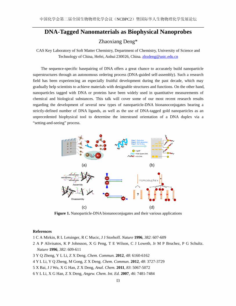
DNA-Tagged Nanomaterials as Biophysical Nanoprobes Zhaoxiang Deng*
CAS Key Laboratory of Soft Matter Chemistry, Department of Chemistry, University of Science and Technology of China, Hefei, Anhui 230026, China. [email protected]
The sequence-specific basepairing of DNA offers a great chance to accurately build nanoparticle
superstructures through an autonomous ordering process (DNA-guided self-assembly). Such a research field has been experiencing an especially fruitful development during the past decade, which may gradually help scientists to achieve materials with designable structures and functions. On the other hand, nanoparticles tagged with DNA or proteins have been widely used in quantitative measurements of chemical and biological substances. This talk will cover some of our most recent research results regarding the development of several new types of nanoparticle-DNA bionanoconjugates bearing a strictly-defined number of DNA ligands, as well as the use of DNA-tagged gold nanoparticles as an unprecedented biophysical tool to determine the interstrand orientation of a DNA duplex via a “setting-and-seeing” process.
(a) (b)
(c) (d) Figure 1. Nanoparticle-DNA bionanoconjugates and their various applications
References 1 C A Mirkin, R L Letsinger, R C Mucic, J J Storhoff. Nature 1996, 382: 607-609 2 A P Alivisatos, K P Johnsson, X G Peng, T E Wilson, C J Loweth, Jr M P Bruchez, P G Schultz.
Nature 1996, 382: 609-611 3 Y Q Zheng, Y L Li, Z X Deng. Chem. Commun. 2012, 48: 6160-6162 4 Y L Li, Y Q Zheng, M Gong, Z X Deng. Chem. Commun. 2012, 48: 3727-3729 5 X Bai, J J Wu, X G Han, Z X Deng, Anal. Chem. 2011, 83: 5067-5072 6 Y L Li, X G Han, Z X Deng, Angew. Chem. Int. Ed. 2007, 46: 7481-7484
中国化学会第二届全国生物物理化学会议(NCBPC2)暨国际华人生物物理化学发展论坛
I5
DNA G-QUADRUPLEXES AS POTENTIAL ANTICANCER DRUG TARGETS
Danzhou Yang
College of Pharmacy, University of Arizona, Tucson, AZ 85721 Dept of Chemistry and Biochemistry, University of Arizona, Tucson, AZ 85721
Comprehensive Member, Arizona Cancer Center, Tucson, AZ 85724 Faculty Member, BIO5 Institute, University of Arizona, Tucson, AZ 85721
DNA G-quadruplex secondary structures formed in specific G-rich sequences, in particular, those formed
in gene proximal promoter regions as transcriptional regulators, have recently emerged as a new class of cancer-specific molecular targets for cancer therapeutics. Specifically, c-MYC, one of the most commonly deregulated genes in human cancers, has a DNA G-quadruplex motif in the promoter Nuclease Hypersensitive Element (NHE) III1 which regulates 75–85% of its total transcription. The c-MYC promoter G-quadruplex has been shown to be amenable to small molecule drug targeting to modulate gene regulation. We have previously determined the K+ solution structure of the major c-MYC promoter G-quadruplex, which is parallel-stranded with two G3NG3 single-nt double-chain-reversal side loops. Very recently, we have determined the solution structure of a 2:1 quindoline–c-MYC G-quadruplex complex by NMR, which represents the first drug complex structure of a biologically relevant unimolecular promoter quadruplex. Our study provides important implications for the structure-based rational design of drugs that target unimolecular parallel-stranded G-quadruplexes commonly found in oncogene promoters.
中国化学会第二届全国生物物理化学会议(NCBPC2)暨国际华人生物物理化学发展论坛
I6
Two-State or Non-Two-State? A Protocol to Differentiate the Two
Types of Unfolding Processes of Proteins
Junjie Luo, Fugen Wu, Jisheng, Yu, and Zhiwu Yu* Key Laboratory of Bioorganic Phosphorous Chemistry and Chemical Biology (Ministry of Education), Department of Chemistry, Tsinghua University, Beijing 100084, P. R. China.
Key-words: Protein, Unfolding Mechanism, Scanning Calorimetry, Infrared Spectroscopy
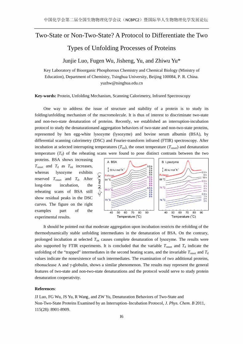
One way to address the issue of structure and stability of a protein is to study its folding/unfolding mechanism of the macromolecule. It is thus of interest to discriminate two-state and non-two-state denaturation of proteins. Recently, we established an interruption-incubation protocol to study the denaturationand aggregation behaviors of two-state and non-two-state proteins, represented by hen egg-white lysozyme (lysozyme) and bovine serum albumin (BSA), by differential scanning calorimetry (DSC) and Fourier-transform infrared (FTIR) spectroscopy. After incubation at selected interrupting temperatures (Tint), the onset temperature (Tonset) and denaturation temperature (Td) of the reheating scans were found to pose distinct contrasts between the two proteins. BSA shows increasing Tonset and Td as Tint increases, whereas lysozyme exhibits reserved Tonset and Td. After long-time incubation, the reheating scans of BSA still show residual peaks in the DSC curves. The figure on the right examples part of the experimental results.
It should be pointed out that moderate aggregation upon incubation restricts the refolding of the thermodynamically stable unfolding intermediates in the denaturation of BSA. On the contrary, prolonged incubation at selected Tint causes complete denaturation of lysozyme. The results were also supported by FTIR experiments. It is concluded that the variable Tonset and Td indicate the unfolding of the “trapped” intermediates in the second heating scans, and the invariable Tonset and Td values indicate the nonexistence of such intermediates. The examination of two additional proteins, ribonuclease A and γ-globulin, shows a similar phenomenon. The results may represent the general features of two-state and non-two-state denaturations and the protocol would serve to study protein denaturation cooperativity.
References:
JJ Luo, FG Wu, JS Yu, R Wang, and ZW Yu, Denaturation Behaviors of Two-State and Non-Two-State Proteins Examined by an Interruption–Incubation Protocol, J. Phys. Chem. B 2011, 115(28): 8901-8909.
中国化学会第二届全国生物物理化学会议(NCBPC2)暨国际华人生物物理化学发展论坛
I7
Biophysical Chemistry of Alzheimer’s Disease
Thomas Branch a, Mauricio Barahona a,b, Liming Ying a,c
a Institute of Chemical Biology
b Department of Mathematics c Molecular Medicine, National Heart and Lung Institute
Imperial College London, London SW7 2AZ, UK [email protected]
Alzheimer’s disease (AD) affects 37 million people worldwide with an estimated global cost of $600 billion in 2010. With the anticipated rapid rise in ageing population in China, AD will become a growing socioeconomic burden. Although some treatments can slow the progression of the disease, there is currently no effective therapeutics to stop or prevent it. The prevailing amyloid cascade hypothesis of AD states that the accumulation of Aβ peptides is a key event in the cause of AD. The pathology of AD involves a build-up of amyloid-β peptides, predominantly of 40 and 42 residues (Aβ40 and Aβ42, respectively). This process is thought to be due to aggregation upon interactions with metal ions such as zinc and copper, eventually leading to the deposit as fibrils and plaques. Recent high profile failures in clinical trials of AD drugs focusing on Aβ production and clearance prompt us to explore an alternative metal chaperone approach. This may prevent the formation of metal-laden oligomers and promote the return of synaptic metals from oligomers to neurons. Several fundamental biophysical chemical questions remain to be addressed. First, at what metal ion concentration and at what time do Aβ oligomers start to form in the synaptic cleft upon the release of synaptic metal ions? Secondly, how do metal protein attenuating compounds (MPACs) influence the onset of the oligomerisation and what is the optimal affinity of MPAC for metal ions? Thirdly, what are the roles of the abundant cerebrospinal fluid (SCF) metal binding proteins, such as human serum albumin (HSA), in regulating the interactions between metal ions and Aβ? These questions are extremely difficult to address in vivo, due to low nanomolar concentrations of Aβ in the CSF and the 15-20 nanometre size of the synaptic cleft. An integrated single molecule experimental and computational approach in vitro, and a fluorescence correlation spectroscopy based screening for the interaction of potential drug candidates with Aβ oligomers at physiologically relevant low nM Aβ monomer concentrations, have been developed, to tackle these questions quantitatively and to provide the potential to test intervention strategies. Keywords: Alzheimer’s disease (AD), amyloid-β peptide, oligomerisation, single molecule fluorescence, fluorescence correlation spectroscopy (FCS) References:
[1] Karran, E., Mercken, M. and De Strooper B., Nat. Rev. Drug Discovery, 2011, 10: 698-712. [2] Roberts, B. R., Ryan, T. M., Bush, A. I., Masters, and C. L., Duce, J. A., J. of Neurochem,
2012, 120 (Suppl. 1): 149-166. [3] Kenche, V. B., and Barnham, K. J., British J. of Pharmacology, 2011, 163: 211-219.

中国化学会第二届全国生物物理化学会议(NCBPC2)暨国际华人生物物理化学发展论坛
I10
微量热技术在蛋白质药物、生物及材料领域提供的重要讯息
林明申[email protected] 美国TA仪器
传统热分析技术如差式扫描量热(DSC)、热重分析(TGA)、热机械分析(TMA)、动态热机械
分析(DMA)等,在材料、高分子、食品及其他领域的发展应用已相当成熟。近年来随着科技的
发展及研究的需求,热分析仪器大幅度的更新及改善。微量热分析仪器 (Nano ITC, Nano DSC,
MCDSC, TAM Air, TAM III)即是在传统热分析仪器的基础上,加强仪器的灵敏度及稳定性使其
能检测到少量样品中微小的特性变化,特别是针对生物样品,例如:表征生物分子交互作用、生
物药物制剂配方筛选、微生物生长代谢能量研究、评估生医材料与细胞兼容性等应用。本文
将介绍利用微量热仪器在蛋白质药物、微生物以及材料领域的相关用。
关键词:微量热;蛋白质;生物分子交互作用;微生物。
中国化学会第二届全国生物物理化学会议(NCBPC2)暨国际华人生物物理化学发展论坛
I11
Salt bridge exchange binding mechanism between streptavidin and its DNA aptamer – Thermodynamics and spectroscopic
evidences
Tai-ChihKuo,1 Peng-Chen Lee,2 Ching-Wei Tsai,2,* and Wen-Yih Chen2,* 1Department of Biochemistry, Taipei Medical University, Taipei, 11031, Taiwan, China
2Department of Chemical and Materials Engineering, National Central University, Jhong-Li City,
Tao-Yuan County 32001,Taiwan, China
Wen-Yih Chen: Email: [email protected] Tel: +886-3-4227151#34222 Fax: +886-3-422-5258
Aptamers are valuable for the discovery and application of new principles and designs of
nucleic acid-ligand interactions. This study investigated the thermodynamics and conformational
changes associated with the binding between streptavidin (SA) and its DNA aptamer under various
temperatures and salt concentrations by Isothermal Titration Calorimetry (ITC) and spectroscopic
methods. The binding was enthalpy-driven with a large entropy cost. The binding association
constant (Ka) was independent of the salt concentrations; however, enthalpy increased with the salt
concentration. The binding was accompanied with substantial conformational changes, which were
insensitive to the variation of salt concentrations. These non-classical results indicate the prominent
involvement of the binding-site hydration water molecules in SA-aptamer binding. We propose a
salt-bridge exchange model to explain the salt-independent Ka values. The binding affinity is mainly
driven by the electrostatic interactions between the salt bridge within the proteins and those within
the aptamer-Na+ counter ions exchange their partners. The salt bridge exchange results in the
observed structural changes and the possible release of hydration water molecules. The negative heat
capacity (ΔCp) of ITC and spectroscopic studies supported the discussions. Furthermore, the
spectroscopic studies and ITC measurements indicated that each SA tetramer bound a maximum of 2
aptamer molecules.
Keywords:Isothermal titration calorimetry, circular dichroism, streptavidin, aptamer.
Abbreviations used: SA, streptavidin; ITC, isothermal titration calorimetry; CD, circular dichroism.
中国化学会第二届全国生物物理化学会议(NCBPC2)暨国际华人生物物理化学发展论坛
I12
Unnatural Metalloprotein Design Jiangyun Wang1
1Institute of Biophysics, Chinese Academy of Science, Beijing, China. [email protected].
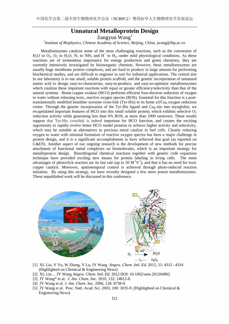
Metalloenzymes catalyze some of the most challenging reactions, such as the conversion of H2O to O2, O2 to H2O, N2 to NH3 and H+ to H2, under mild physiological conditions. As these reactions are of tremendous importance for energy production and green chemistry, they are currently intensively investigated by bioinorganic chemists. However, these metalloenzymes are usually huge membrane protein complexes, and are hard to produce in large amount for performing biochemical studies, and are difficult to engineer to suit for industrial applications. The central aim in our laboratory is to use small, soluble protein scaffold, and the genetic incorporation of unnatural amino acid to design easy-to-characterize, easy-to-produce, and easy-to-optimize metalloenzymes which catalyze these important reactions with equal or greater efficiency/selectivity than that of the natural systems. Heme-copper oxidase (HCO) performs efficient four-electron reduction of oxygen to water without releasing toxic, reactive oxygen species (ROS). Essential for this function is a post-translationally modified histidine–tyrosine cross-link (Tyr-His) in its heme a3/CuB oxygen reduction center. Through the genetic incorporation of the Tyr-His ligand and CuB site into myoglobin, we recapitulated important features of HCO into this small soluble protein, which exhibits selective O2 reduction activity while generating less than 6% ROS, at more than 1000 turnovers. These results support that Tyr-His crosslink is indeed important for HCO function, and creates the exciting opportunity to rapidly evolve better HCO model proteins to achieve higher activity and selectivity, which may be suitable as alternatives to precious metal catalyst in fuel cells. Cleanly reducing oxygen to water with minimal formation of reactive oxygen species has been a major challenge in protein design, and it is a significant accomplishment to have achieved that goal (as reported on C&EN). Another aspect of our ongoing research is the development of new methods for precise attachment of functional metal complexes on biomolecules, which is an important strategy for metalloprotein design. Bioorthogonal chemical reactions together with genetic code expansion technique have provided exciting new means for protein labeling in living cells. The main advantages of photoclick reaction are its fast rate (up to 50 M-1S-1), and that it has no need for toxic copper catalyst. Moreover, spatiotemporal control is achieved through photo-induced reaction initiation. By using this strategy, we have recenlty designed a few more potent metalloenzymes. These unpublished work will be discussed in this conference.
[1] XL Liu, Y Yu, W Zhang, Y Lu, JY Wang Angew. Chem. Intl. Ed. 2012, 51: 4312 –4316 (Highlighted on Chemical & Engineering News)
[2] XL Liu… JY Wang Angew. Chem. Intl. Ed. 2012 DOI: 10.1002/anie.201204962 [3] JY Wang* et al. J. Am. Chem. Soc. 2010, 132: 14812-8. [4] JY Wang et al. J. Am. Chem. Soc. 2006, 128: 8738-9. [5] JY Wang et al. Proc. Natl. Acad. Sci. 2003, 100: 3035-9. (Highlighted on Chemical &
Engineering News)
中国化学会第二届全国生物物理化学会议(NCBPC2)暨国际华人生物物理化学发展论坛
I13
Single-molecule dynamics of metallorgulators: new mechanisms for transcription regulation
Peng Chen
Department of Chemistry and Chemical Biology, Cornell University, Ithaca, NY 14853, USA, [email protected]
Key words: metalloregulator, transcription regulation, copper efflux regulator, single-molecule imaging
To maintain normal metal metabolism, organisms utilize dynamic cooperation of many
biomacromolecules for regulating metal ion concentrations and bioavailability. Our group has been studying the macromolecular machineries for metal regulation and transport at the singe-molecule level. I will summarize our recent progresses in developing novel single-molecule imaging approaches to study these protein machineries. In particular I will describe a new study about the novel pathways for metalloregulators to turn off transcription after activation. Metalloregulators regulate transcription in response to metal ion concentrations. Many studies have provided insights into how transcription is activated upon metal binding by MerR-family metalloregulators. In contrast, how transcription is turned off after activation is unclear. Turning off transcription promptly is important, however, as the cells would not want to continue expressing metal resistance genes and thus waste energy after metal stress is relieved. Using single-molecule FRET measurements, we studied the dynamic interactions of CueR, a Cu+-responsive MerR-family metalloregulator, with DNA. Besides quantifying its DNA binding and unbinding kinetics, we discovered that CueR spontaneously flips its binding orientation at the recognition site. CueR also has two different binding modes, corresponding to interactions with specific and nonspecific DNA sequences, which would facilitate recognition localization. Most strikingly, a CueR molecule coming from solution can directly substitute for a DNA-bound CueR or assist the dissociation of the incumbent CueR, both of which are the first such examples for any DNA-binding protein. The kinetics of the direct protein substitution and assisted dissociation reactions indicate that these two novel processes can provide efficient pathways to replace a DNA-bound holo-CueR with apo-CueR, thus turning off transcription promptly and facilely. References 1. Peng Chen, Nesha May Andoy, Jaime Benitez, Aaron Keller, Debashis Panda, Feng
Gao“Tackling Metal Regulation and Transport at the Single-Molecule Level”Natural Product Reports, 2010, 27:757-767.
2. Aaron Keller, Jaime Benitez, Derek Klarin, LinghaoZhong, Matthew Goldfogel, Feng Yang, Tai-Yen Chen, Peng Chen“Dynamic Multi-Body Protein Interactions Suggest Versatile Pathways for Copper Trafficking”Journal of the American Chemical Society, 2012, 134: 8934-8943.
3. Chandra P. Joshi, Debashis Panda, Danya J. Martell Smart, Nesha May Andoy, Tai-Yen Chen, Ahmed Gaballa, John D. Helmann, Peng Chen“Single-Molecule Analysis Suggests Novel Pathways for Turning Off Transcription by A MerR-family Metalloregulator” Submitted.
中国化学会第二届全国生物物理化学会议(NCBPC2)暨国际华人生物物理化学发展论坛
I14
Single-Molecule Calorimetry Studies of DNA Structural
Transitions during DNA Overstretching
Jie Yan
Department of Physics Mechanobiology Institute
Centre for Bioimaging Sciences National University of Singapore
Torsionally unconstrained double-stranded DNA (dsDNA) undergoes several distinct structural
transitions under large tension, including productions of (i) an ssDNA strand under tension while the other strand coils, (ii) two parallel ssDNA strands that share tension (melting bubble), and (iii) “B-to-S” transition to a novel overstretched dsDNA, termed “S-DNA”. Here we investigated the entropy and enthalpy changes associated with all the three transitions as well as the micromechanics of the resulting respective DNA structures. These results provide the first experimental evidence for the formation of DNA melting bubble driven by large tension and provide the final answer to the question of whether non-melted S-DNA exists.
E-mail: [email protected]
中国化学会第二届全国生物物理化学会议(NCBPC2)暨国际华人生物物理化学发展论坛
I15
Control the Molecular Interactions with DNA Nanomachine
Dongsheng Liu* Department of Chemistry, Tsinghua University, Beijing 100084, China.
Polyvalent interactions are characterized by the simultaneous binding of multiple ligands on one biological entity to multiple receptors on another. Comparing to corresponding monovalent interactions, polyvalent interactions often have cooperative effect, resulting in a significant enhancement of binding affinity and specificity. Better performance of polyvalent interactions strongly relies on precise control of relative spatial position of multiple ligands. 1
In the past decades, DNA has been demonstrated as an ideal material to fabricate nearly arbitrary one-, two- to three-dimensional nanostructures2. More recently, the well-established modification and accurate addressability of DNA make these nanostructures ideal scaffolds to hold ligands in position and show polyvalent effects3. Furthermore, the conformational switchability of DNA also enables the fabrication of nanoscale molecular machines, which can perform movements upon stimuli.4 It provides a promising method to study dynamic behavior at nanometer scale including small molecules5 and macromolecules.6
Here we report a DNA machine which can reversibly regulate target binding affinity based on a distance-dependent bivalent binding. It is a tweezer-like DNA machine which could tune the spatial distance between two ligands to construct or destroy the bivalent binding. The DNA machine could strongly bind to target protein when the ligands are placed at an appropriate distance, while it releases the target when the bivalent binding is disrupted by enlarging the distance between ligands. This “capture-release” cycle could be repeatedly driven by single-stranded DNA without changing the ligands and target protein.7 Keywords: DNA, self-assembly, nanomachine, polyvalency, dynamics [1]. M. Mammen, S. K.Choi & G. M.Whitesides, Angew. Chem. Int. Ed. 1998, 37: 2755-2794. [2]. A. V. Pinheiro, D. Han, W. M. Shih & H. Yan Nature Nanotechnology 2011, 6: 763-772. [3]. S. Rinker, Y. G. Ke, Y. Liu, R. Chhabra, & H. Yan Nature Nanotech. 2008, 3: 418-422. [4]. H. Liu, D. S. Liu, Chem. Commun. 2009, 2625-2636. [5]. H. Liu, Y. Zhou, Y. Yang, W. Wang, L. Qu, C. Chen, D. Liu, D. Zhang, and D. Zhu J. Phys.
Chem. B, 2008, 112(22): 6893-6896. [6]. Y. Sun, H. Liu, L. Xu, L. Wang, Q.-H. Fan, D. Liu, Langmuir 2010, 26, 12496-12499. [7]. C. Zhou, Z. Yang & D. Liu J. Am. Chem. Soc., in press.

中国化学会第二届全国生物物理化学会议(NCBPC2)暨国际华人生物物理化学发展论坛
I16
Conjugated Polymers as Light Harvesting Complexes for Two-photon Applications
Xiaoqin Shen, Ning Tian, Xinsheng Ren, Fang He, Shuang Li, and Qing-Hua
Xu*
Department of Chemistry, National University of Singapore, Singapore 117543,
Conjugated polymers display interesting optical properties such as optical amplification via energy
transfer. They also have large two-photon absorption cross sections compared to the small molecules. We have demonstrated that they can act as two-photon light harvesting complexes to enhance two-photon emission of dyes by up to 1000 times. Using this strategy, we have developed a two-photon sensor scheme for detection of mercury ions with LOD of ~6 nm. We further developed photosensitizer doped conjugated polymer nanoparticles, which showed significantly enhanced two-photon excitation singlet oxygen generation efficiency and two-photon photodynamic therapy activity. These nanoparticles display features required for ideal photosensitizers: low cytotoxicity in the dark and efficient two-photon photodynamic activity under laser radiation. These novel nanophotosensitizers allow simultaneous in vivo monitoring by two-photon fluorescence imaging during two-photon photodynamic treatment. Key Words: Conjugated polymers; Two-photon excitation; Fluorescence; Sensing; Bio-imaging; Photodynamic therapy References (1) N. Tian; Q. H. Xu Adv. Mater. 2007, 19: 1988 (2) X. S. Ren; Q. H. Xu Langmuir 2009, 25: 29 (3) C. L. Wu; Q. H. Xu Macromol. Rapid Commun.2009, 30: 504 (4) F. He, X. S. Ren; X. Q. Shen; Q. H Xu Macromolecules 2011, 44: 5373 (5) X. Q. Shen; F. He; J. H. Wu; G. Q . Xu; S. Q. Yao; Q. H. Xu Langmuir, 2011, 27: 1739 (6) X. Q. Shen; L. Li; H. Wu; S. Q. Yao; Q. H. Xu Nanoscale 2011, 3: 5140
2hv
dye
FRET
3O21O2
Two-photon excitation
Photosensitizer
PFEMO
Cell Apoptosis
中国化学会第二届全国生物物理化学会议(NCBPC2)暨国际华人生物物理化学发展论坛
I17
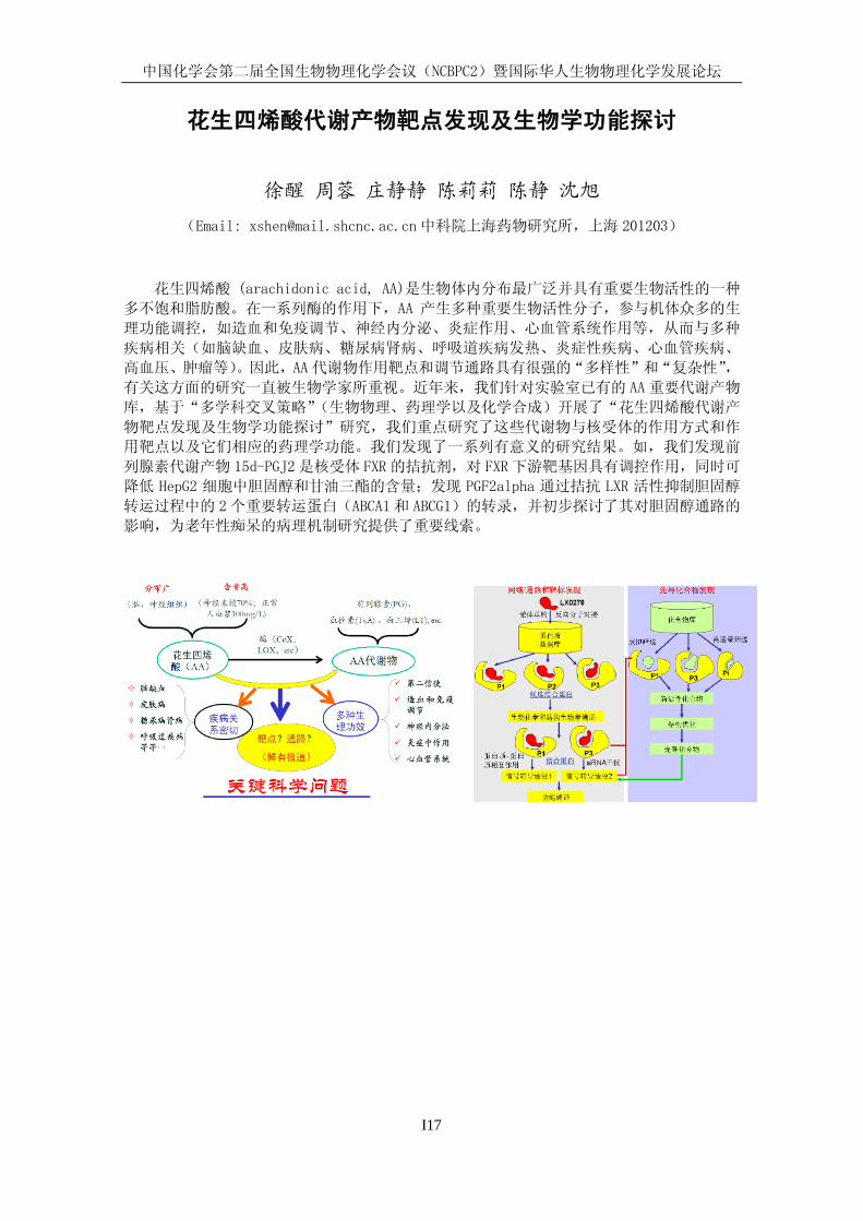
花生四烯酸代谢产物靶点发现及生物学功能探讨
徐醒 周蓉 庄静静 陈莉莉 陈静 沈旭
(Email: [email protected]中科院上海药物研究所,上海 201203)
花生四烯酸 (arachidonic acid, AA)是生物体内分布最广泛并具有重要生物活性的一种
多不饱和脂肪酸。在一系列酶的作用下,AA 产生多种重要生物活性分子,参与机体众多的生
理功能调控,如造血和免疫调节、神经内分泌、炎症作用、心血管系统作用等,从而与多种
疾病相关(如脑缺血、皮肤病、糖尿病肾病、呼吸道疾病发热、炎症性疾病、心血管疾病、
高血压、肿瘤等)。因此,AA 代谢物作用靶点和调节通路具有很强的“多样性”和“复杂性”,
有关这方面的研究一直被生物学家所重视。近年来,我们针对实验室已有的 AA重要代谢产物
库,基于“多学科交叉策略”(生物物理、药理学以及化学合成)开展了“花生四烯酸代谢产
物靶点发现及生物学功能探讨”研究,我们重点研究了这些代谢物与核受体的作用方式和作
用靶点以及它们相应的药理学功能。我们发现了一系列有意义的研究结果。如,我们发现前
列腺素代谢产物 15d-PGJ2是核受体 FXR 的拮抗剂,对 FXR下游靶基因具有调控作用,同时可
降低 HepG2 细胞中胆固醇和甘油三酯的含量;发现 PGF2alpha 通过拮抗 LXR 活性抑制胆固醇
转运过程中的 2个重要转运蛋白(ABCA1和 ABCG1)的转录,并初步探讨了其对胆固醇通路的
影响,为老年性痴呆的病理机制研究提供了重要线索。
中国化学会第二届全国生物物理化学会议(NCBPC2)暨国际华人生物物理化学发展论坛
I18
[Ni,Fe]-hydrogenase maturation pathway- a biophysical study
Hongzhe Sun (孙红哲), Wei Xia, Hongyan Li, Tianfan Cheng
Department of Chemistry, The University of Hong Kong (香港大学化学系), Pokfulam Road,Hong
Kong, P.R. China
[Ni,Fe]-hydrogenases are widely produced by many types of bacteria and involved in hydrogen
metabolism.1 The large subunit of the hydrogenase contains a dinuclear [Ni,Fe] metal center, which
is required for enzyme activity. The maturation of [Ni,Fe]-hydrogenase is highly dependent on a
battery of chaperone proteins. Among these, HypA, SlyD and HypB were proposed to exert nickel
delivery functions during the metallocenter assembly,2 although the detailed mechanism of nickel
insertion carried out by these two proteins remains unknown.3 We found that both Helicobacter pylori HypA (and SlyD) and HypB bind Zn2+ and Ni2+ with a
stoichiometry of one metal ion per protein. Both apo- and Zn2+-bound HypB have intrinsic weak GTPase activity. However, Ni2+ binding to HypB significantly enhanced its GTPase activity and induced the dimerization of the protein, indicative of an important role of Ni2+ played rather than Zn2+. The HypA·HypB complex interfaces on HypA protein were mapped based on chemical shift perturbation, and a HypA·HypB complex model was built up using HpHypA solution structure,4 homolog modeling and docking methods. Based on all the data, a GTPase regulated Ni2+ delivery mechanism was proposed to elucidate the detailed functions performed by the two Ni2+ chaperones. Very recently, we also found that HpSlyD forms a complex with UreE as well as HypB.5 These structural and functional studies provide insight into the molecular mechanism of Ni2+ delivery during [Ni,Fe]-hydrogenase (and urease) maturation.
This work was supported by RGC of Hong Kong (HKU7043/06P, HKU1/07C, HKU7042/07P,
HKU7038/08P, HKU7049/09P, N_HKU752/09), Croucher Foundation and The University of Hong
Kong.
[1] P.M. Vignais, B. Billoud, J. Meyer, FEMS Microbiol. Rev., 25, 455 (2001).
[2] J.W. Olso, N.S. Mehta, R.J. Maier, Mol. Microbiol. 39, 176 (2001); T.F. Cheng, H. Li, W. Xia, H.
Sun, 2011.
[3] A. Atanassova, D.B. Zamble, J. Bacteriol. 187, 4689 (2005).
[4] W. Xia, H.Y. Li, K.H. Sze, H. Sun, J. Am. Chem. Soc. 131, 10031 (2009).
[5] T.F. Cheng, H.Y. Li, W. Xia, H. Sun, J. Biol. Inorg. Chem. 17, 331 (2012).
中国化学会第二届全国生物物理化学会议(NCBPC2)暨国际华人生物物理化学发展论坛
I19
Probing Allostery Through DNA
Sangjin Kim1*†, Erik Broströmer1*, Dong Xing2*, Jianshi Jin2,3*, Shasha Chong1, Hao Ge2,4,5, Siyuan Wang1, Xiao-dong Su2, Yujie Sun2§ & X. Sunney Xie1,2§
1 Department of Chemistry & Chemical Biology, Harvard University, Cambridge, MA 02138, USA. 2Biodynamic Optical Imaging Center (BIOPIC), Peking University, Beijing 100871, China. 3Academy for Advanced Interdisciplinary Studies, Peking University, Beijing 100871, China 4Beijing International Center for Mathematical Research, Peking University, Beijing 100871, China. 5 School of Mathematical Sciences and Centre for Computational Systems Biology, Fudan University, Shanghai 200433, China * These authors contributed equally to this work. †Present address: Department of Molecular Cellular & Developmental Biology, Yale University, New Haven, CT 06511, USA § To whom correspondence should be addressed. E-mail: [email protected] (Y.S.); [email protected] (X.S.X)
Allostery is well-documented for proteins but less recognized for DNA-protein interactions, in which DNA has been often considered as a mere template providing recognition sequences. Here we report that DNA binding affinity of a protein is significantly affected by another protein bound nearby, oscillating as a function of their separation. The oscillation has a periodicity of ~10 base pairs, the helical pitch of the B-form DNA, and a characteristic decay length of ~15 base pairs. We prove by a hairpin experiment that this effect is not due to protein–protein interactions but the distortion of the inter-helical distance along the linker DNA.The two proteins either stabilize or destabilize each other with an oscillating total free energy of the ternary complex. We demonstrate such DNA allostery affects gene expression levels in live E. coli cells. Pertinent to eukaryotic gene expression, we show that the binding affinity of a transcription factor depends on its separation from nearby nucleosomes.

中国化学会第二届全国生物物理化学会议(NCBPC2)暨国际华人生物物理化学发展论坛
I20
A High-Quality Benchmark for Scoring Function Assessment
Yan Li (李嫣), Zhihai Liu(刘志海), Jie Li(李婕), Renxiao Wang(王
任小)*
State Key Laboratory of Bioorganic Chemistry, Shanghai Institute of Organic Chemistry, Chinese Academy of Sciences, 345 Lingling Road, Shanghai 200032, P. R. China [email protected]
Molecular docking methods rely on scoring functions to select ligand binding poses, to compute
binding affinities, or to conduct other tasks. Various scoring functions have been developed in the past, and many more are still emerging. It is certainly desirable to assess the performance of these scoring functions on suitable benchmarks. The PDBbind database, which is currently maintained in our group, is a systematic collection of the experimental binding data of the protein-ligand complexes in the Protein Data Bank. It provides an ideal starting point for compiling such a benchmark. We have developed an approach for selecting the representing ones out of the protein-ligand complexes with high-resolution crystal structures and reliable binding data in the PDBbind database. Structural diversity at both the protein side and the ligand side is also emphasized during this process. The final outcome, namely the PDBbind core set (version 2011), consists of 216 protein-ligand complexes in 72 families. Based on this data set, a total of 20 popular scoring functions from both commercial software and academic groups were assessed in three aspects, i.e. "docking power", "ranking power", and "scoring power". A number of general remarks regarding the performance of these scoring functions were derived for scoring function users and developers. Key words: scoring function, molecular docking, benchmark, PDBbind, protein-ligand complex
中国化学会第二届全国生物物理化学会议(NCBPC2)暨国际华人生物物理化学发展论坛
I21
Unique photoactivatable fluorescent proteins for
diffraction-limited and superresolution imaging
Pingyong Xu Institute of Biophysics, Chinese Academy of Sciences
The development and application of super-resolution imaging technologies enable us to define
the accurate localization of biological molecules at nanometer precision and has been hot issues in recent years in imaging field. Photoactivatable fluorescent proteins (PAFPs) are molecules that switch to a new fluorescent state in response to specific light activation, and play vital roles in super-resolution imaging. There are three classes of PAFPs: dark-to-bright photoactivators (PAFPs), irreversible photoconverters (PCFPs), and reversible highlighters (RSFP). However, compared to traditional fluorescent proteins (such as GFP or RFP), only limited PAFPs are available for super-resolution microscopy. Based on the demand of currently used super-resolution microscopy and the good photochemical property of mEos2, we first developed several novel PAFPs, mGeos, with various switching rates, photon numbers and brightness. Next,based on the crystal structure of green state mEos2, we evolved two truly monomeric and bright RSFPs, mEos3.1 and mEos3.2,with the good photochemical properties of rapid maturation rate, high photon budget and extremely high labeling density. These novel fluorescent proteinsare excellent PAFPsfor both single color and dual color PALM superresolutionimaging, and have a broad brand of applications in traditional fluorescence microscopy such as dynamic tracking and pulse chase labeling of proteins.
中国化学会第二届全国生物物理化学会议(NCBPC2)暨国际华人生物物理化学发展论坛
I22
利用 FRET 技术研究蛋白质折叠过程中的构象变化
黄方
中国石油大学(华东)生物工程与技术中心,青岛,266580,[email protected]
关键词:蛋白质折叠、FRET、构象变化、机理
一个广为接受的观点是,蛋白质必须折叠至特定的构象才具备生物活性,研究蛋白质的
折叠过程并揭示其折叠机理是蛋白质研究的核心内容之一。蛋白质折叠过程必然伴随着蛋白
质构象的变化,因此对蛋白质折叠过程中构象变化的研究,对于揭示蛋白质折叠机理具有极
其重要的作用。蛋白质折叠机理的提出往往都是以蛋白质的折叠动力学为基础的,但是缺乏
结构信息的动力学数据并不能够充分解释蛋白质的折叠机理。荧光共振能量转移技术(FRET)
基于共振能量转移效率对能量给体和受体之间距离的强烈依赖性,能够精确地测定给体和受
体之间的距离及其变化,因此被广泛应用于蛋白质折叠过程中构象变化的研究。本人将介绍
将 FRET 技术与快速动力学技术以及单分子荧光技术相结合研究蛋白质折叠机理的一些实例。
中国化学会第二届全国生物物理化学会议(NCBPC2)暨国际华人生物物理化学发展论坛
I23
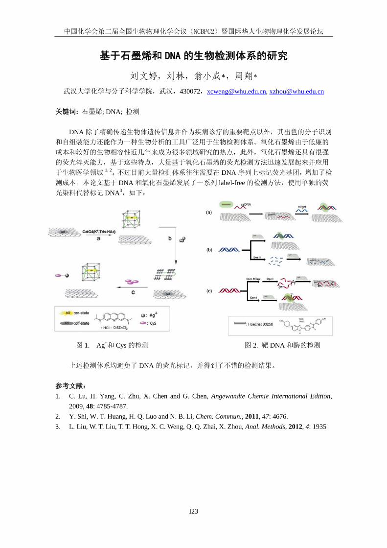
基于石墨烯和 DNA 的生物检测体系的研究
刘文婷,刘林,翁小成*,周翔*
武汉大学化学与分子科学学院,武汉,430072,[email protected], [email protected] 关键词: 石墨烯; DNA; 检测
DNA 除了精确传递生物体遗传信息并作为疾病诊疗的重要靶点以外,其出色的分子识别
和自组装能力还能作为一种生物分析的工具广泛用于生物检测体系。氧化石墨烯由于低廉的
成本和较好的生物相容性近几年来成为很多领域研究的热点,此外,氧化石墨烯还具有很强
的荧光淬灭能力,基于这些特点,大量基于氧化石墨烯的荧光检测方法迅速发展起来并应用
于生物医学领域 1, 2。不过目前大量检测体系往往需要在 DNA 序列上标记荧光基团,增加了检
测成本。本论文基于 DNA 和氧化石墨烯发展了一系列 label-free 的检测方法,使用单独的荧
光染料代替标记 DNA3,如下:
图 1. Ag+和 Cys 的检测 图 2. 靶 DNA 和酶的检测
上述检测体系均避免了 DNA 的荧光标记,并得到了不错的检测结果。
参考文献:
1. C. Lu, H. Yang, C. Zhu, X. Chen and G. Chen, Angewandte Chemie International Edition, 2009, 48: 4785-4787.
2. Y. Shi, W. T. Huang, H. Q. Luo and N. B. Li, Chem. Commun., 2011, 47: 4676. 3. L. Liu, W. T. Liu, T. T. Hong, X. C. Weng, Q. Q. Zhai, X. Zhou, Anal. Methods, 2012, 4: 1935
中国化学会第二届全国生物物理化学会议(NCBPC2)暨国际华人生物物理化学发展论坛
I24
单链核酸与蛋白质相互作用的定量研究
陈莹,张薇,刘宁宁,张文科*
吉林大学超分子结构与材料国家重点实验室,长春,130012 [email protected]
核酸与蛋白质的相互作用贯穿于分子生物学中心法则的每个环节,研究核酸与蛋白质的相
互作用对于认识诸如 DNA复制等重要生命过程并最终实现对相应过程的调控具有重要意义。传
统的分子生物学研究方法,从生物样品的提取、纯化到样品的检测、分析均涉及大量的分子,
反映大量分子的平均效应。平均效应虽然可以反映出体系的整体趋势,但体系中很多的“罕见”
现象(Rare events)则被忽略了。此外,平均效应也无法连续、精确地体现出单个事件的不同
步骤,如蛋白质折叠与解折叠,因此在研究过程中丢失了很多重要的信息。而单个生物大分子
的研究可以从单个分子水平给出一些体相平均方法不能给出的信息(如捕捉或跟踪诸如蛋白质
错误折叠等罕见事件),因此单个分子的研究成为近年来的研究热点之一。1,2
基于原子力显微镜技术(AFM)的单分子力学谱(以下简称单分子力谱)是近十年发展起来
的新型超高灵敏度的检测方法,它使得人们可以对单个大分子进行力学操纵和加工,从而对分
子结构与构象变化,分子间的相互作用以及反应历程实现单分子水平的实时-原位观测。目前
单分子力学谱已经成为生物物理,高分子科学,表面科学和细胞分子生物学等领域不可或缺的
一种重要的研究手段。3-6
我们利用蛋白质工程技术将枯草芽孢杆菌的单链DNA结合蛋白的C末端引入半胱氨酸继而
实现了该蛋白在固体基片表面的有效固定,并定量测量了单股 DNA与该蛋白的相互作用;同时
结合传统凝胶电泳实验揭示了该蛋白与单股 DNA 之间的动态结合本质。7,8另外,在前期工作基
础上,9我们进一步利用动力学力谱揭示了烟草花叶病毒 RNA 与蛋白质外壳的解组装机理。
关键词:AFM,单分子力谱,核酸-蛋白相互作用,病毒解组装
参考文献:
[1]Uppenbrink, J. and Clery, D. Science, 1999, 283: 1667-1695. [2]Vinson, V. and Chin, G. Science, 2007, 316: 1143. [3]Merkel, R. Phys. Rep., 2001, 346: 343–385. [4]Clausen-Schaumann, H.; Seitz, M.; Krautbauer, R. and Gaub, H. E. Curr. Opin. Chem. Biol., 2000,
4: 524–530. [5]Zhang, W. K. and Zhang, X. Prog. Polym. Sci., 2003, 28: 1271–1295. [6] Liu, N. N. and Zhang, W. K. ChemPhysChem, 2012, 13: 2238-2256. [7] Liu, N. N.; Bu, T. J.; Song, Y.; Zhang, W.; Li, J. J.; Zhang, W. K.; Shen, J. C. and Li, H. B.
Langmuir, 2010, 26: 9491–9496. [8] Zhang, W.; Lü, X. J.; Zhang, W. K. and Shen, J. C. Langmuir, 2011, 27: 15008–15015.
[9] Liu, N. N.; Peng, B.; Lin, Y.; Su, Z. H.; Niu, Z. W.; Wang, Q.; Zhang, W. K.; Li, H. B.; Shen, J. C. J. Am. Chem. Soc., 2010, 132: 11036–11038.
中国化学会第二届全国生物物理化学会议(NCBPC2)暨国际华人生物物理化学发展论坛
I26
蛋白质溶解中的保护和变性作用
高毅勤 1* 1 北京大学化学与分子工程学院,北京海淀区颐和园路 5 号,100871, *[email protected]
无机盐和有机小分子对于调控细胞电解质和渗透压平衡具有重要作用,对蛋白质等生物
分子的溶液结构也有重要影响。虽然相关研究已有超过百年的历史,这些小分子或离子影响
生物分子结构的分子机制还很不清楚。鉴于溶剂效应在蛋白质的溶解、结构形成,功能实现
和蛋白质制备与结晶中的重要作用,十分有必要理解小分子与蛋白质相互作用的物理图像。
通过引入并解释阴阳离子在不同界面的布局优先性的不同和阴阳离子之间成对中表现出来的
协同性,我们建立了简单的公式将阴阳离子溶解能与他们对非极性分子和多肽的溶解度的影
响联系起来,较为合理地解释了不同极性的分子在水中溶解度对不同无机盐的响应。同时,
解释了无机盐对水的界面张力的影响。另一方面,我们研究了有机共溶剂影响蛋白质结构的
分子机理,建立了一个较为完整的蛋白质主链在水溶液中溶解的物理图象。我们利用全原子
分子模拟对这些简单物理图像进行了验证。
关键词:蛋白质结构,溶剂化,蛋白质变性
参考文献
[1] Y.Q. Gao, JPCB, In press. [2] Q. Shao, JCTC, In press. [3] Y.Q. Gao, JPCB, 12466 (2011). [4] W.J. Xie, Y.Q. Gao, Faraday Discussion, accepted.
Denaturant or renaturant: effects of small molecules in protein solvation
Yi Qin Gao1,* 1College of Chemistry and Molecular Engineering, Peking University, #5 Yiheyuan Lu, Beijing,
100871 Solvation plays very important roles in the structure formation and function of proteins. Small
molecules and ions also play important roles in keeping the osmotic pressure of the cellular environment. These effects of cosolvents and cosolutes have been the subject of research for over a century. Urea denaturation and the effects of inorganic salts on protein structures have been known for more than a century. We will discuss a simple theory that takes into account differentiated preferential binding of anions/cations at interfaces of different polarity and the cooperative cation/anion association. Using this theory, we were able explain the relation between the solvation energies of the ions and their effects on the solubility of model compounds, such as benzene and peptides, as well as the salt effects on water/air surface tension. A simple model is established to explain the effects of both inorganic salts and organic cosolvents on the protein solubility and structure. We will also discuss results from all-atom molecular dynamics simulations that were used to test these models.
中国化学会第二届全国生物物理化学会议(NCBPC2)暨国际华人生物物理化学发展论坛
I27
Study on the abnormal aggregation of Tau protein, and the ultrasensitive / selective detection of AD biomarker
Yao Tian-Ming*, Cheng Ting-Ting, Shi Shuo*, Ji Liang-Nian
Department of Chemistry, Tongji University, Shanghai 200092, [email protected]
Keywords: aggregation, inhibition, detection, tau protein
The incidence of Alzheimer’s disease increases dramatically with age, however, only a small percentage is directly related to familial forms. Epidemiological studies suggest that environmental factors may be involved besides genetic risk factors. Mounting evidence is demonstrating strong associations between neuro-toxic metal exposure and AD. We, for the first time, systematically investigated the interaction of tau peptide R2, R3 with transition cations. We find that tau peptide R2,R3 show high affinity to the group IIB cations, Zn(II), Cd(II) and Hg(II). The coordination of metal cation, especially Hg(II), induces a conformational conversion on R2 peptide chain. The result suggests that the cooperative folding of R2 through cross-bridging of group IIB cation has a pronounced impact on tau aggregation. The neurotoxins of group IIB cations may stem from their strong binding to thiol group of cysteine residue on tau peptide chain.
The search for direct inhibitors of the tau aggregation process and their rational design will present an exciting challenge to reach potential drug candidates. One straightforward strategy for the discovery of new active molecules is the screening of compound libraries containing sufficient structural diversity. We found for the first time that Tannic acid and Flavonoids could inhibit fibril formation of R3 by deformation of the flexible extended structure, consequently losing its aggregation ability. The inhibitory ability is closely related to their binding modes and binding degree to R3. A structure model was built using molecular simulation to elucidate the possible docking site for Tannic acid on the tau peptide surface. Our results suggest that tau peptide recognizably interacts with Tannic acid by forming a hairpin binding motif, a key framework required for inhibiting Tau polymerization, in addition to hydrogen bond, hydrophilic/ hydrophobic interaction, and static electrical interaction, as reported earlier.
Early diagnosis of AD is crucial for the current drug treatments, which have shown to slow the progression of AD. Although ultrasensitive detection has become routine for nucleic acids, it remains challenging for proteins. Driven by the growing needs, we developed several biosensor system for protein based on aptamer and nano-particulars, targeting to ultrasensitive and selective assay for the detection of AD biomarkers such as tau protein.
References 1.Dan-Jing Yang, Shuo Shi*, Tian-Ming Yao*, Liang-Nian Ji, Biometals, 2012, 25: 361-372 2. D.J.Yang, S.Shi, L.F.Zheng, T.M.Yao*, and L.N.Ji, Biopolymers, 2010, 93: 1100-1108 3. L.F.Jiang, T.M.Yao*, Z.L.Zhu, C.Wang, L.N.Ji, BBA, 2007,1774: 1414 4. L.J.Han, S.Shi, L.F.Zheng, D.J.Yang, T.M.Yao*, L.N.Ji, Bull.Chem.Soc.Jpn, 2010, 83: 911. 5. Wenliang Sun, Tianming Yao* and Shuo Shi*, Analyst, 2012, 137: 1550–1552. 6. Shuo Shi,* Juan Zhao, Xing Gao, Chunyan Lv, Li Yang, Jian Hao, Hailiang Huang, Junliang Yao, Wenliang Sun, Tianming Yao* and Liangnian Ji, Dalton Trans., 2012, 41: 5789-5793
This work was supported by the National Natural Science Foundation of China (No20871094, No 20901060, and No81171646).
中国化学会第二届全国生物物理化学会议(NCBPC2)暨国际华人生物物理化学发展论坛
I29
核磁共振在金属蛋白与金属药物研究中的应用
刘扬中
中国科学技术大学,合肥市,230026,[email protected]
关键词: 核磁共振、金属蛋白、金属药物
核磁共振研究是在原子水平上研究蛋白质溶液性质的重要方法,如蛋白质结构解析、动力
学性质及蛋白质的识别作用等。由于金属离子的特殊性质,核磁共振在金属蛋白的研究中具有
与一般蛋白不同特殊性。 顺磁性的金属会对核磁共振信号产生显著的影响,但同时也可作为顺磁探针使用,例如对
于含 Heme 的蛋白,可以利用顺磁金属导致 dipolar shift,通过磁轴计算 Heme 获得轴向上配位
基团的空间取向。通过 NMR 的金属滴定是获得金属结合位点的有效手段,同时,可以根据
NMR 信号的分布判断蛋白质在金属离子作用下的折叠状态。利用顺磁离子的弛豫作用可以获
得金属离子与各个信号之间的距离信息,是近年来用于蛋白质结构计算的一个重要辅助手段。 金属药物(如抗癌药物顺铂)的作用机理倍受关心,由于金属药物的特异性差,其作用靶
点可能非常复杂。核磁共振为金属药物的研究提供了一个有效的方法。195Pt 的核自旋为 1/2,有较高的灵敏度,通过铂谱可以直接研究药物与靶分子的作用,同时,195Pt 的化学位移与配
位原子和配位环境相关,当顺铂与生物分子作用时,其氯原子可能被蛋白质或 DNA 中的 S 或
N 原子取代,导致 195Pt 的化学位移发生相应的变化,是判断反应产物的重要依据。此外,Pt配位导致配位原子以及周围原子的化学位移变化是探测药物配位位点的重要手段,3JPt-H 偶合1H 化学位移向低场移动 0.5-0.9 ppm,配位 N 原子的化学位移变化可达 80-100 ppm;将顺铂中
的氮原子 15N 同位素标记,可以通过 1H15N 二维核磁共振研究药物的反应产物分析以及动力学
过程跟踪。 虽然普遍认为金属药物顺铂的作用靶点是 DNA,蛋白质在药物的作用机理中起着关键的
作用,一些金属蛋白对顺铂有很高的亲和性。采用 NMR 研究金属药物与金属蛋白相互作用,
可为掌握药物在细胞内的作用方式和理解药物作用机理提供重要的理论基础。 参考文献: 1. NMR studies of metalloproteins, Hongyan Li, Hongzhe Sun, Topics in Current Chemistry, 2012,
326, 69-98. 2. Applications of heteronuclear NMR spectroscopy in biological and medicinal inorganic
chemistry, Luca Ronconia, Peter J. Sadlerb, Coordination Chemistry Reviews, 2008, 252, 2239–2277

中国化学会第二届全国生物物理化学会议(NCBPC2)暨国际华人生物物理化学发展论坛
I30
多功能共轭聚合物的设计、荧光成像与疾病治疗探索研究
王树 中国科学院化学研究所,北京市海淀区中关村北一街二号,100190
重大疾病的控制和治疗是人类面临的重大挑战。目前集识别、成像与治疗功能于一体的
多功能药物的研发越来越受到人们的重视,有望成为重大疾病治疗的新策略。我们课题组的
一个主要研究内容是设计合成新型共轭聚合物材料,并探索其在生物识别、细胞成像以及疾
病治疗领域的新应用。设计合成了系列抗光漂白能力的共轭聚合物荧光探针,研究了它们的
生物相容性,筛选了三类具有较低细胞毒性的水溶性聚合物(聚芴、聚噻吩与聚苯撑乙烯)。
通过调控红绿兰三基色聚合物在同一个改造的微生物模板上的自组装,获得了单一激发波长
下多色的共轭聚合物微米粒子,可用于荧光编码与细胞成像。设计构建了兼具细胞成像与抗
癌作用的共价连接卟啉基团的水溶性聚噻吩衍生物,光照下该体系产生活性氧,可有效的杀
伤肿瘤细胞,同时通过聚合物在细胞中成像位置的不同可实现对活细胞以及凋亡细胞的有效
识别。设计并合成了含有季铵盐与 PEG 侧链的 PPV 衍生物,特殊的结构使其具有选择性结合
细菌,而不结合细胞的特性,实现了对细菌的选择性识别与成像,同时可通过光动力机制杀
伤细菌而对细胞没有损伤作用。这些结果为设计基于共轭聚合物的识别、成像与疾病治疗多
功能体系提供了依据。
关键词:共轭聚合物;设计;成像;疾病治疗;多功能
参考文献
[1] C. Zhu, L. Liu, Q. Yang, F. Lv, S. Wang, Chem. Rev. 2012, online ASAP
[2] X. Duan, L. Liu, F. Feng, S. Wang, Acc. Chem. Res. 2010, 43: 260-270.
[3] C. Zhu, Q. Yang, L, Liu, F. Lv, S. Li, G. Yang, S. Wang, Adv. Mater. 2011, 23: 4805-4810.
[4] C. Xing, Q. Xu, H. Tang, L. Liu, S. Wang, J. Am. Chem. Soc. 2009, 131: 13117-13124.
[5] C. Zhu, Q. Yang, L. Liu, S. Wang, Angew. Chem. Int. Ed. 2011, 50: 9607-9610.
[6] F. Feng, L. Liu, S. Wang, Nature Protocols 2010, 5: 1255-1264.
[7] X. Feng, G. Yang, L. Liu, F. Lv, Q. Yang, S. Wang, D. Zhu, Adv. Mater. 2012, 24: 637-641.
[8] F. Feng, H. Wang, L. Han, S. Wang, J. Am. Chem. Soc. 2008, 130: 11338-11343.
中国化学会第二届全国生物物理化学会议(NCBPC2)暨国际华人生物物理化学发展论坛
I31
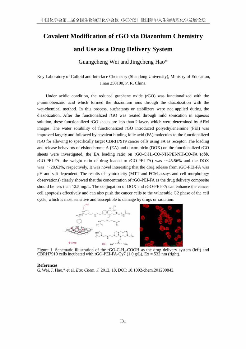
Covalent Modification of rGO via Diazonium Chemistry
and Use as a Drug Delivery System
Guangcheng Wei and Jingcheng Hao*
Key Laboratory of Colloid and Interface Chemistry (Shandong University), Ministry of Education, Jinan 250100, P. R. China.
Under acidic condition, the reduced graphene oxide (rGO) was functionalized with the p-aminobenzoic acid which formed the diazonium ions through the diazotization with the wet-chemical method. In this process, surfactants or stabilizers were not applied during the diazotization. After the functionalized rGO was treated through mild sonication in aqueous solution, these functionalized rGO sheets are less than 2 layers which were determined by AFM images. The water solubility of functionalized rGO introduced polyethyleneimine (PEI) was improved largely and followed by covalent binding folic acid (FA) molecules to the functionalized rGO for allowing to specifically target CBRH7919 cancer cells using FA as receptor. The loading and release behaviors of elsinochrome A (EA) and doxorubicin (DOX) on the functionalized rGO sheets were investigated, the EA loading ratio on rGO-C6H4-CO-NH-PEI-NH-CO-FA (abb. rGO-PEI-FA, the weight ratio of drug loaded to rGO-PEI-FA) was ~45.56% and the DOX was ~28.62%, respectively. It was novel interesting that the drug release from rGO-PEI-FA was pH and salt dependent. The results of cytotoxicity (MTT and FCM assays and cell morphology observations) clearly showed that the concentration of rGO-PEI-FA as the drug delivery composite should be less than 12.5 mg/L. The conjugation of DOX and rGO-PEI-FA can enhance the cancer cell apoptosis effectively and can also push the cancer cells to the vulnerable G2 phase of the cell cycle, which is most sensitive and susceptible to damage by drugs or radiation.
Figure 1. Schematic illustration of the rGO-C6H4-COOH as the drug delivery system (left) and CBRH7919 cells incubated with rGO-PEI-FA-Cy7 (1.0 g/L), Ex = 532 nm (right).
References G. Wei, J. Hao,* et al. Eur. Chem. J. 2012, 18, DOI: 10.1002/chem.201200843.

中国化学会第二届全国生物物理化学会议(NCBPC2)暨国际华人生物物理化学发展论坛
I32
Learning to Speak Protein --
a case study on Adenylate Kinase
Yan-Wen Tan Department of Physics, Fudan University, Shanghai, China
Enzymes are remarkable molecular machines that make many difficult biochemical reactions
possible with incredible precision and efficiency. However, our understandings of the working
principles of enzymes have not reached the level where one can readily deduce the mechanism
and the catalytic rates from an enzyme’s structure. To understand the working and ‘coding’
principles of enzymes, we focus on enzyme molecule’s properties in the time domain, namely the
conformational dynamics of proteins. We use single-molecule optical microscopy and enzymatic
assays to measure the energy landscape of a model enzyme system, adenylate kinase (AK) from
Escherichia coli. Using the high-resolution time-dependent single-molecule FRET (Förster
Resonance Energy Transfer), we have measured AK's lid movements on the millisecond scale and
map out its entire conformational distribution along the FRET coordinate without a presumed
model. Using this information, we have quantitatively recovered AK's energetic landscape and
related its stochastic lid dynamics to its catalytic function. Furthermore, bioinformatics has been
applied to the analysis of AK protein family. The relationship between AK’s genetic coding and its
catalytic function is experimentally established by introducing targeted mutation on specific AK
sites. This study provides new directions for protein engineering.
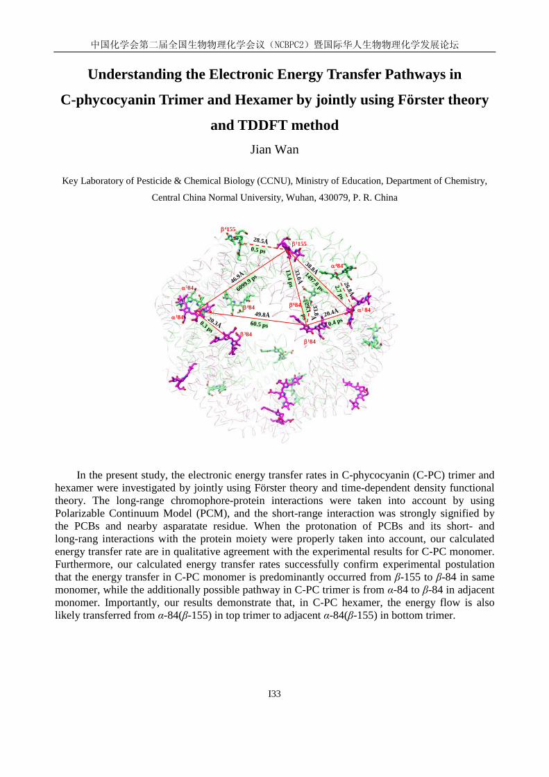
中国化学会第二届全国生物物理化学会议(NCBPC2)暨国际华人生物物理化学发展论坛
I33
Understanding the Electronic Energy Transfer Pathways in
C-phycocyanin Trimer and Hexamer by jointly using Förster theory
and TDDFT method Jian Wan
Key Laboratory of Pesticide & Chemical Biology (CCNU), Ministry of Education, Department of Chemistry,
Central China Normal University, Wuhan, 430079, P. R. China
β1155
β4155
α184
α584
β484
β384
β684
β184
α2 84
α484
0.5 ps
28.5Å
2.7 ps26.8Å
33.8Å29.1 ps
0.4 ps20.4Å
13.4 ps33.0Å
6099.9 ps46.9Å
49.8Å60.5 ps
1497.8 ps
38.8Å
0.3 ps
20.3Å
β1155
β4155
α184
α584
β484
β384
β684
β184
α2 84
α484
0.5 ps
28.5Å
2.7 ps26.8Å
33.8Å29.1 ps
0.4 ps20.4Å
13.4 ps33.0Å
6099.9 ps46.9Å
49.8Å60.5 ps
1497.8 ps
38.8Å
0.3 ps
20.3Å
In the present study, the electronic energy transfer rates in C-phycocyanin (C-PC) trimer and hexamer were investigated by jointly using Förster theory and time-dependent density functional theory. The long-range chromophore-protein interactions were taken into account by using Polarizable Continuum Model (PCM), and the short-range interaction was strongly signified by the PCBs and nearby asparatate residue. When the protonation of PCBs and its short- and long-rang interactions with the protein moiety were properly taken into account, our calculated energy transfer rate are in qualitative agreement with the experimental results for C-PC monomer. Furthermore, our calculated energy transfer rates successfully confirm experimental postulation that the energy transfer in C-PC monomer is predominantly occurred from β-155 to β-84 in same monomer, while the additionally possible pathway in C-PC trimer is from α-84 to β-84 in adjacent monomer. Importantly, our results demonstrate that, in C-PC hexamer, the energy flow is also likely transferred from α-84(β-155) in top trimer to adjacent α-84(β-155) in bottom trimer.
中国化学会第二届全国生物物理化学会议(NCBPC2)暨国际华人生物物理化学发展论坛
I34
What We Have Seen and Learned at Bio-Nano Interface
聂广军
国家纳米科学中心,100190,北京 [email protected]
随着纳米科学与技术的快速发展和纳米材料的工业化应用,特别是在生物医药、健康和环
境领域的广泛应用,人们对纳米-生物界面的生物物理化学相互作用机制和其对生物纳米材料
在生物体作用的深远影响越来越重视,期望通过对界面化学的机制研究,一方面能够指导新型
纳米药物的研发;另一方面,希望尽量把可能的毒副作用控制在最小范围。作为一个全新的研
究领域,不仅是物理、化学、材料学、生物医学和纳米科学与技术的广泛交叉的最好例证,也
是现代科学技术发展的必然趋势。可以预见,未来的具有革命性变革的科学技术最有可能产生
于传统学科的交叉点。将重点介绍课题组近 3年来在纳米生物界面方向上的一些探索。
关键词:纳米生物界面;功能纳米材料;生物屏障
参考文献:
[1] Yiye Li, Yunlong Zhou, Hai-Yan Wang, Sarah Perrett, Yuliang Zhao, Zhiyong Tang, Guangjun Nie, Chirality of Glutathione Surface Coating Affects the Toxicity of Quantum Dots, Angew Chem Int Ed, 2011, 50: 5860-5864.
[2] Wendi Zhang, Chi Wang, Zhenzhen Lu, Jun-Jie Yin, Yu-Ting Zhou, Xingfa Gao, Ying Fang, Guangjun Nie, Yuliang Zhao, Unraveling Stress-induced Toxicity Properties of Graphene Oxide and the Underlying Mechanism, Advanced Materials, in press.
[3] Cuiji Sun, Hui Yang,Yi Yuan, Xin Tian, Liming Wang, Yi Guo, Li Xu, Jianlin Lei, Ning Gao, Gregory J. Anderson, Xing-jie Liang, Chunying Chen, Yuliang Zhao, Guangjun Nie, Controlling assembly of paired gold clusters within apoferritin nanoreactor for in vivo kidney targeting and biomedical imaging, J Am Chem Soc, 2011, 133: 8617–8624
中国化学会第二届全国生物物理化学会议(NCBPC2)暨国际华人生物物理化学发展论坛
I35
利用原子力和超分辨光学显微镜研究细胞膜结构
王宏达,赵伟栋,蔡明军,田咏梅,高婧,吴佳桢,蒋俊光
中国科学院长春应用化学研究所,吉林省长春市人民大街 5625号,130022,
关键词:细胞膜,原子力显微镜,超分辨光学显微镜,单分子
细胞膜是活细胞的重要组成部分,它有许多重要的生物功能,如物质隔离、物质交换和
细胞通讯等。在分子水平研究细胞膜的结构对解释细胞膜的功能和治疗细胞膜相关疾病有重
要的指导意义。几十年来,人们已经提出各种细胞膜模型,如流动镶嵌模型、脂筏模型和蛋
白区域模型等1-2。但是,由于这些模型是基于间接或非现场的方法,细胞膜的结构至今还是非
常有争议的研究热点。这些争议可能一直持续到我们真正在单分子水平上观察到细胞膜的结
构。我们利用现场原子力显微镜和超分辨荧光显微镜(STORM)在分子水平上观察细胞膜3。结
果发现细胞膜中的蛋白在磷脂中是非对称性和区域性分布的,细胞膜中的重要功能蛋白与脂
筏区域结合4,5。
参考文献:
[1] S. J. Singer, G. L. Nicolson. Science, 1972, 175: 720-730.
[2] K. Simons, E. Ikonen. Nature, 1997, 387: 569-572.
[3] J. Jiang, X. Hao, M. Cai, Y. Shan, X. Shang, Z. Tang, H. Wang. Nano Letters, 2009, 9:
4489-4493.
[4] H. Wang, X. Hao, Y. Shan, J. Jiang, M. Cai, X. Shang. Ultramicroscopy, 2010, 110: 305-312.
[5] M. Cai, W. Zhao, X. Shang, J. Jiang, H. Ji, Z. Tang, H. Wang. Small, 2012, 8: 1243-1250.
国家自然科学基金(项目编号:20975098,21073181)、科技部重大科学研究计划(项目编号:2011CB933600)、中科院“百人计划”基金资助。
中国化学会第二届全国生物物理化学会议(NCBPC2)暨国际华人生物物理化学发展论坛
I37
Functional nanostructured materials based on surface
modification of natural cellulose substances Jianguo Huang
Department of Chemistry, Zhejiang University, Hangzhou, Zhejiang 310027
Keywords: Surface modification, Biomimetic synthesis, Nanomaterials, Cellulose
Biological organisms are produced from self-assembly of highly ordered functional units and
are inherently complex and hierarchical, possessing macro-to-nanoscale features. It is a facile,
low-cost and environmentally benign short-cut to artificial functional materials with unique
multilevel structures and morphologies employing biological substances such as natural cellulose
substances as platforms for the self-assembly of various guest substrates. Various nanostructured
materials with designed properties and functionalities were fabricated by means of self-assembly of
different guest substrates (such as metal oxide thin films, small molecules, polymers,
biomacromolecules, nanoparticles, and colloidal spheres) on the surfaces of cellulose nanofibers,
that is, surface modification, of bulk natural cellulose substances (e.g., commercial laboratory filter
paper). The combination of the specific chemical properties of the guest substrates and the unique
physical features of the natural cellulose substances sheds new light on the design and syntheses of
new functional nanomaterials
References:
1. W. Xiao, H. Hu, J. Huang, Colorimetric detection of cysteine by surface functionalization of
natural cellulose substance. Sens. Actuators, B 2012, DOI: 10.1016/j.snb.2012.05.087.
2. C.Jin, Y.Jiang, T. Niu, J. Huang, Cellulose-based material with amphiphobicity to inhibit
bacterial adhesion by surface modification. J. Mater. Chem. 2012, 22: 12562-12567.
3. W. Xiao, J. Huang, Immobilization of oligonucleotides onto zirconia-modified filter paper and
specific molecular recognition. Langmuir 2011, 27: 12284-12288.
4. C. Jin, R.Yan, J. Huang, Cellulose substance with reversible photo-responsive wettability by
surface modification. J. Mater. Chem. 2011, 21: 17519-17525.
5. T. Niu, Y.Gu, J. Huang, Luminescent cellulose sheet fabricated by facile self-assembly of
cadmium selenide nanoparticles on cellulose nanofibres. J. Mater. Chem. 2011, 21: 651-656.
6. X. Zhang, J. Huang, Functional surface modification of natural cellulose substances for
colorimetric detection and adsorption of Hg2+ in aqueous media. Chem. Commun. 2010, 46:
6042-6044.
7. X. Liu, Y. Gu, J. Huang, Hierarchical, titania-coated, carbon nanofibrous material derived from a
natural cellulosic substance. Chem. Eur. J. 2010, 16: 7730-7740.
中国化学会第二届全国生物物理化学会议(NCBPC2)暨国际华人生物物理化学发展论坛
I38
The Contrasting Effect of Macromolecular Crowding on Amyloid Fibril Formation of Prion Proteins
Qian Ma, Jun-Bao Fan, Zheng Zhou, Bing-Rui Zhou, Sheng-Rong Meng, Ji-Ying Hu, Jie Chen, and Yi Liang*
State Key Laboratory of Virology, College of Life Sciences, Wuhan University, Wuhan 430072, China
Amyloid fibrils associated with neurodegenerative diseases such as Alzheimer disease, prion
disease, and amyotrophic lateral sclerosis (ALS) can be considered biologically relevant failures of cellular quality control mechanism. It is known that in vivo human prion protein (PrP) and its pathogenic mutants, human Tau protein, and human copper, zinc superoxide dismutase (SOD1) pathogenic mutants have the tendency to form fibril deposits in a variety of tissues and they are associated with prion disease, Alzheimer disease, and ALS, respectively, while the rabbit PrP and hen egg white lysozyme do not readily form fibrils and are unlikely to cause neurodegenerative diseases. Furthermore, misfolded Tau protein accumulating in Alzheimer disease and misfolded SOD1 accumulating in ALS can cause aggregation of their native counterparts in crowded physiological environments through a mechanism similar to the infectious prion protein PrPSc causing aggregation of its cellular isoform PrPC. In this study, we have investigated the contrasting effect of macromolecular crowding on fibril formation of different proteins including prion proteins from human and rabbit. As revealed by assays based on thioflavin T binding and turbidity, human Tau fragments, when phosphorylated by glycogen synthase kinase-3β, do not form filaments in the absence of a crowding agent but do form fibrils in the presence of a crowding agent, and the presence of a strong crowding agent dramatically promotes amyloid fibril formation of human prion protein and its two pathogenic mutants E196K and D178N. Such an enhancing effect of macromolecular crowding on fibril formation is also observed for a pathological human SOD1 mutant A4V. On the other hand, rabbit prion protein and hen lysozyme do not form amyloid fibrils when a crowding agent at 300 g/l is used but do form fibrils in the absence of a crowding agent. Furthermore, aggregation of these two proteins is remarkably inhibited by Ficoll 70 and dextran 70 at 200 g/l. We suggest that proteins associated with neurodegenerative diseases are more likely to form amyloid fibrils under crowded conditions than in dilute solutions. By contrast, some of the proteins that are not neurodegenerative disease-associated are unlikely to misfold in crowded physiological environments. A possible explanation for the contrasting effect of macromolecular crowding on these two sets of proteins (amyloidogenic proteins and non-amyloidogenic proteins) has been proposed. Information obtained from the present study can enhance our understanding of the molecular mechanisms of neurodegenerative diseases, and should lead to a better understanding of how proteins misfold and how proteins avoid misfolding in crowded physiological environments.

口头报告
Oral Presentations
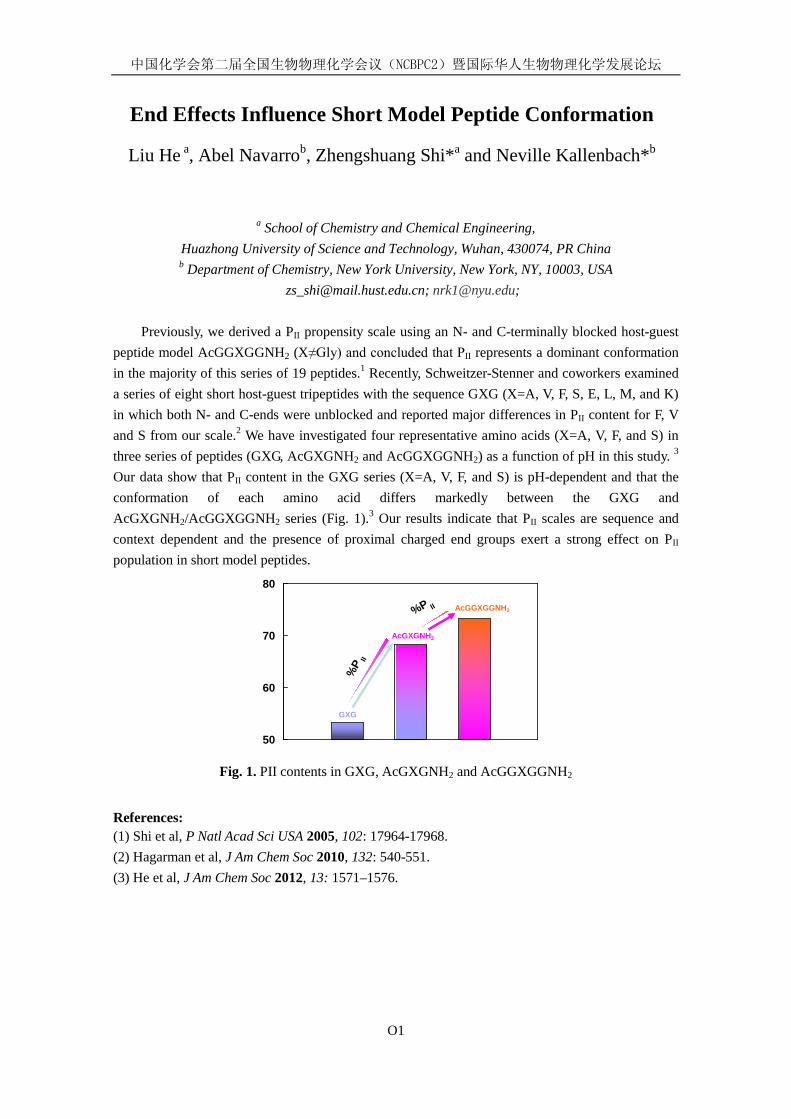
中国化学会第二届全国生物物理化学会议(NCBPC2)暨国际华人生物物理化学发展论坛
O1
End Effects Influence Short Model Peptide Conformation
Liu He a, Abel Navarrob, Zhengshuang Shi*a and Neville Kallenbach*b
a School of Chemistry and Chemical Engineering,
Huazhong University of Science and Technology, Wuhan, 430074, PR China b Department of Chemistry, New York University, New York, NY, 10003, USA
[email protected]; [email protected];
Previously, we derived a PII propensity scale using an N- and C-terminally blocked host-guest peptide model AcGGXGGNH2 (X≠Gly) and concluded that PII represents a dominant conformation in the majority of this series of 19 peptides.1 Recently, Schweitzer-Stenner and coworkers examined a series of eight short host-guest tripeptides with the sequence GXG (X=A, V, F, S, E, L, M, and K) in which both N- and C-ends were unblocked and reported major differences in PII content for F, V and S from our scale.2 We have investigated four representative amino acids (X=A, V, F, and S) in three series of peptides (GXG, AcGXGNH2 and AcGGXGGNH2) as a function of pH in this study. 3 Our data show that PII content in the GXG series (X=A, V, F, and S) is pH-dependent and that the conformation of each amino acid differs markedly between the GXG and AcGXGNH2/AcGGXGGNH2 series (Fig. 1).3 Our results indicate that PII scales are sequence and context dependent and the presence of proximal charged end groups exert a strong effect on PII population in short model peptides.
%P II
GXG
AcGXGNH2
AcGGXGGNH2
50
60
70
80
%P II
Fig. 1. PII contents in GXG, AcGXGNH2 and AcGGXGGNH2
References: (1) Shi et al, P Natl Acad Sci USA 2005, 102: 17964-17968. (2) Hagarman et al, J Am Chem Soc 2010, 132: 540-551. (3) He et al, J Am Chem Soc 2012, 13: 1571–1576.
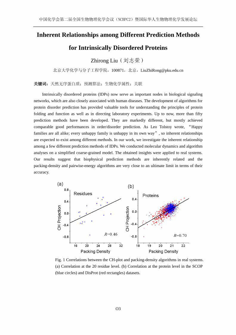
中国化学会第二届全国生物物理化学会议(NCBPC2)暨国际华人生物物理化学发展论坛
O3
Inherent Relationships among Different Prediction Methods
for Intrinsically Disordered Proteins
Zhirong Liu(刘志荣)
北京大学化学与分子工程学院,100871,北京,[email protected]
关键词:天然无序蛋白质;预测算法;生物化学属性;关联
Intrinsically disordered proteins (IDPs) now serve as important nodes in biological signaling networks, which are also closely associated with human diseases. The development of algorithms for protein disorder prediction has provided valuable tools for understanding the principles of protein folding and function as well as in directing laboratory experiments. Up to now, more than fifty prediction methods have been developed. They are markedly different, but mostly achieved comparable good performances in order/disorder prediction. As Leo Tolstoy wrote, “Happy families are all alike; every unhappy family is unhappy in its own way”, so inherent relationships are expected to exist among different methods. In our work, we investigate the inherent relationship among a few different prediction methods of IDPs. We conducted molecular dynamics and algorithm analyses on a simplified coarse-grained model. The obtained insights were applied to real systems. Our results suggest that biophysical prediction methods are inherently related and the packing-density and pairwise-energy algorithms are very close to an ultimate limit in terms of their accuracy.
Fig. 1 Correlations between the CH-plot and packing-density algorithms in real systems. (a) Correlation at the 20 residue level. (b) Correlation at the protein level in the SCOP (blue circles) and DisProt (red rectangles) datasets.
Novel bio-imaging techniques with optical highlighter
fluorescent protein
Xinxin Zhu and Wei Min Columbia University, New York, NY 10027, [email protected], [email protected] Keywords: fluorescence microscopy, microviscosity sensor, high-order nonlinearity, fluorescent timer, optical highlighter Abstract: Optical highlighters are a remarkable family of fluorescent proteins (FPs) that could change their excitation and emission spectrum upon certain wavelength activation, including photo-activatable FP, photo-convertible FP and photo-switchable FP. Optical highlighters have been extensively used in super-resolution imaging, protein dynamics, gene expression and cellular trafficking et al. Instead, we are taking advantage of optical highlighters in three different fluorescence imaging applications: genetically-encoded microviscosity sensor, multiphoton microscopy with high-order nonlinearity, and light-driven fluorescent timer. First of all, we report that the photoswitching kinetics of the chromophore inside Dronpa, a FP with the reversible light-regulated on-off switching capability, is actually slowed down by increasing medium viscosity outside Dronpa. This effect is attributed to protein-flexibility mediated coupling where the chromophore’s cis-trans isomerization during photoswitching is accompanied by conformational motion of a part of the protein β-barrel whose dynamics should be hindered by medium friction. Based on this effect, we developed a genetically encoded protein-specific micro-viscosity sensor. Secondly, we demonstrate that the population transfer kinetics between long-lifetime bright and dark quantum states of the photoinduced molecular switches could generate additional high-order nonlinear dependence of the signal on the laser intensity. Two distinctive imaging techniques, multiphoton deactivation and imaging (MPDI) and multiphoton activation and imaging (MPAI), are proposed and show more than three-order nonlinearity, which provides more localized information with superior image contrast than regular two-photon microscopy. Last but not the least, we demonstrate a novel class of fluorescent timer (FT), a light-driven FT, based on the photo-convertible fluorescent protein, mEos2. It’s a new method to image the protein “age” in live cells with a simple snap-shot measurement. Compared with previous fluorescent timer proteins, this light-driven FT could be tuned to match different time ranges, from as fast as several minutes to as slow as dozens of hours. As more FTs with varied maturation time and color are strongly desired by scientists, this tunable light-driven FT will be a valuable tool that does not only provide the spatial location information of an individual protein, but also its temporal dynamics. Reference: 1. Y.-T. Kao, X. Zhu and W. Min “Protein-flexibility mediated coupling between photoswitching
kinetics and surrounding viscosity of a photochromic fluorescent protein” Proc. Natl. Acad. Sci. USA, 109, 3220 (2012).
2. X. Zhu, Y.-T. Kao and W. Min “Molecular-switch-mediated multiphoton fluorescence microscopy with high-order nonlinearity” J. Phys. Chem. Lett. 3, 2082 (2012)
3. X. Zhu, Y.-T. Kao, F. Xu and W. Min “Light-driven fluorescent timer” (In Preparing)
中国化学会第二届全国生物物理化学会议(NCBPC2)暨国际华人生物物理化学发展论坛
O5
Bacteria-Mediated Assemblies of Multi-Spectral Conjugated
Polymer Microparticles for Cell Imaging and Barcoding
Libing Liu, Xuli Feng, Shu Wang Beijing National Laboratory for Molecular Sciences, Key Laboratory of Organic Solids, Institute of
Chemistry, Chinese Academy of Sciences, Beijing 100190, P. R. China. [email protected]
Recently, developing multiplexed bioassays and molecular imaging have become more and more
important in gene profiling, drug discovery and clinical diagnostics. Spectroscopic coding technology based on both color and intensity of the light emitted from more than one coloring materials which are embedded inside or attached on the surface of the microparticles has been developed for multiplexed bioassays. Organic dyes have widely separated absorption and excitation spectra and now QDs have been developed as an alternative way.
Four cationic conjugated polymers have been prepared with four fluorescence colors from blue, green to yellow and red just by fine tuning the backbone and side chains of the polymers. Due to the amphiphilic properties, they can easily form conjugated polymer nanoparticles (CPNs) in aqueous solution with the size of 50 to 100 nm. The multicolor CPNs-encoded microparticles were prepared based on the self-assemblies of bacteria (E.coli) and CPNs by just mixing them together, where multicolor can be regulated through fine tuning fluorescence resonance energy transfer (FRET) among blue, green and red (RGB)-emissive CPNs under a single excitation wavelength. These multicolor microparticles exhibit low toxicity toward cells and have been successfully applied for cell imaging and optical barcoding. The significance of these studies lies in that it provides a very efficient, versatile and simple method for preparing color-barcoded microparticles.
Reference 1. J. Yang,; S. R. Dave,; X. Gao, J. Am. Chem. Soc. 2008, 130: 5286-5292. 2. X. Feng,; Y.Tang,; X.Duan,; L.Liu,; S. Wang, J. Mater. Chem. 2010: 20, 1312-1316. 3. X.Feng,; G.Yang,; L.Liu,; F.Lv, Q. Yang, S.Wang, D.Zhu, Adv. Mater. 2012: 24, 637-641
中国化学会第二届全国生物物理化学会议(NCBPC2)暨国际华人生物物理化学发展论坛
O6
微纳局域环境中水的性质研究
于安池
中国人民大学化学系,100872,北京 [email protected]
关键词:光化学与光物理、荧光寿命、转动常数、粘度与极性、近红外发光探针
摘要:生物体内存在着大量的微纳米局域环境,这些特殊环境为许多生物分子提供它们执行
生理功能的场所,缺少了这些特殊环境它们便失去生理活性。为了更好地了解局域环境的性
质及局域环境对化学或生物反应的影响,我们以水溶性 IR125 分子为探针分子,通过稳态吸
收光谱、稳态荧光光谱和时间分辨荧光光谱实验研究了 AOT 反胶束中包含水的性质。我们发
现包含于反胶束体系内的水分子与自由本体水的极性和粘度有很大的不同。我们发现 w0 =
[H2O]/[AOT] = 8 是 AOT 反胶束中水的性质改变的转折点。对比探针分子 C152 在 AOT 反胶
束中的光物理性质实验结果,我们发现近红外吸收和发射的 IR125 分子可以作为一种有效的
探针分子探测反胶束中水和内界面上水的性质。以 IR125 为探针分子,我们测定了 w0 = 40 时
AOT 反胶束内水的粘度,发现在 w0 = 40 时 AOT 反胶束中水的粘度大约是自由本体水的粘度
的 12 倍。该研究为我们进一步认识和了解发生在微纳米局域环境中的反应奠定了基础。
参考文献: 1. Ruixue ZHU, Rong LU, Anchi YU, Chin. J. Chem., 2011, 29: 405-410.
2. Ruixue Zhu, Rong Lu, and Anchi Yu, Phys. Chem. Chem. Phys., 2011, 13: 20844-20854.

中国化学会第二届全国生物物理化学会议(NCBPC2)暨国际华人生物物理化学发展论坛
O7
Peptide Conformational Dynamics Probed by Ultrafast
Infrared Spectroscopy Jianping Wang
Beijing National Laboratory for Molecular Sciences, State Key Laboratory of Molecular Reaction Dynamics, Institute of Chemistry, Chinese Academy of Sciences, Beijing, 100190,
In recent years, the structures and dynamics of peptides and proteins in the condensed phases have been studied extensively by ultrafast vibrational methods, including the two-dimensional infrared (2D IR) and infrared pump-probe spectroscopies. The amide-I mode, being mostly the C=O stretching motion, is often used as the intrinsic structural probe for peptide backbone structural dynamics. 2D IR studies of the subset of the anharmonic amide-I modes can provide detailed structural informations of the peptides and proteins. In the mean while, anharmonic vibrational frequency, coupling, as well as anharmonicity of the amide-I mode, are found to become novel structural parameters. Therefore computational assessment of these parameters is also of great importance. This presentation includes our recent works in these two closely related fields: (1) the structural dynamics of amide-I mode in a model glycopeptide has been examined by IR pump-probe spectroscopy in combination of ab initio molecular dynamics simulations; (2) a molecular mechanics force field-based amide-I frequency map (the MM-map) that has been constructed for the α- and β-peptide oligomers; (3) ultrafast correlated molecular motions of peptides oligomers have been examined by broad-band 2D IR spectroscopy; and (4) ultrafast structural dynamics of a photocycle intermediate of the photoactive yellow protein has been investigated by 2D IR spectroscopy in the amide-I region. Our results demonstrate the chemical-bond level sensitivity and femtosecond time resolution of these vibrational methods, whose combination yields a very powerful structural tool for monitoring the ultrafast events in the condensed-phase biophysics. Key words: α- and β-peptides; amide-I mode; 2D IR; coupling; molecular dynamics simulations
References [1] C. Han and J. Wang, Chem. Phys. Chem. 2012, 13: 1522-1534. [2] K. Cai, C. Han, and J. Wang, Phys. Chem. Chem. Phys. 2009, 11: 9194-9159. [3] J. Wang, Phys. Chem. Chem. Phys. 2009, 11: 5310–5322. [4] J. Zhao and J. Wang, J. Chem. Phys. 2012, 136: 214112; doi: 10.1063/1.4725181. [5] J. Wang, K. Cai, and X. Ma, Chem. Phys. Chem. 2009, 10: 2242-2250.
中国化学会第二届全国生物物理化学会议(NCBPC2)暨国际华人生物物理化学发展论坛
O8
Single-molecule studies on hydrophobic hydration
高翔,秦猛,曹毅,王炜
南京大学物理学院,江苏省南京市汉口路 22 号,210093
关键词:疏水作用,单分子力谱,原子力显微镜
Hydrophobic interactions underpin many biological self-assembly processes, including protein folding and assembly, ligand-receptor interaction, and micelle and membrane formation. Recent theoretic work by Lum, Chandler and Weeks highlighted the distinct mechanisms for the hydrophobic hydration of small solutes and larger ones. Small solutes can be incorporated into surrounding water molecules by simple entropy driven fluctuations of water, whereas solvation of larger solutes requires the formation of hydrophobic-hydrophilic interface and is an enthalpy dominated process. However, experimentally verifying this theory is fraught with difficulties. Here, using atomic force microscopy (AFM) based single-molecule force spectroscopy, we quantitatively determine the contribution of entropy and enthalpy for the hydrophobic hydration of polystyrene. Polystyrene forms nanospheres of ~2 nm in diameter in water. Stretching polystyrene using single-molecule AFM leads to the shrink of the nanosphere and the hydration of polystyrene. The resulting force-extension curves show a force plateau at shorter extension and a gradual increase of force at longer extension. We are able to obtain the free energy profile for the hydrophobic hydration process from the obtained force-extension curves in a model-free fashion. The entropy and enthalpy for the hydrophobic hydration are in good agreement with the theoretic predictions. Moreover, our experimental results show the crossover length for the two distinct mechanisms of hydration of small and large particles is ~ 1 nm and can be tuned by the solvent condition. Thus, our experiments directly support the length-dependent hydrophobic hydration theory and provide a novel way for the quantification of the free energy for the hydrophobic hydration. Since hydrophobic interactions occur at different length scales in biological systems, we anticipate that our results will be helpful for the understanding of the distinct effect of hydrophobic interactions in various self-assembly process.

中国化学会第二届全国生物物理化学会议(NCBPC2)暨国际华人生物物理化学发展论坛
O9
Dehydration Dynamics of Cell Membrane-Bound Interfacial
Water Molecules Revealed by a Dipole Molecular Knife
Shuji Ye Hefei National Laboratory for Physical Sciences at Microscale,
University of Science and Technology of China, Hefei, Anhui, P.R.China 230026 [email protected]
Keywords: sum frequency generation, membrane-bound water, dehydration, dipole molecular knife
The cell membrane-bound interfacial water molecules provide a unique environment for many biological functions of the cell membrane. They govern many interactions between cells and their environments, such as the exchange of ions/molecules and information between the inside and outside of the cell. The dehydration dynamics and the water-membrane bound strength are related to the membrane fluidity, membrane assembly, and the adsorption/desorption of various biomolecules, ions, and drugs at the membrane interfaces. A molecular level elucidation of the dehydration dynamics of membrane-bound interfacial water will not only improve our current knowledge about the membrane-bound water itself but also aid in understanding the behavior and process with which cell membrane is associated. In this study, we applied sum frequency generation vibrational spectroscopy (SFG-VS) and a dipole molecular knife technique to investigate the dehydration dynamics of membrane-bound interfacial water. Two separate dehydration dynamic components were observed: a large amplitude fast component and a small amplitude slow component, which originate from the water molecules with a weak and a strong water-membrane bound strength, respectively. In addition, it was found that the water-membrane bound strength depends largely on the charge status of the lipid head group and has an order of that: neutrally-charged membrane << positively-charged membrane << negatively-charged membrane. The negatively-charged membrane-bound water reorganizes to become ordered quickly after dehydration. The ion types and concentration can increase the component ratio of the strong water-membrane bound strength.
中国化学会第二届全国生物物理化学会议(NCBPC2)暨国际华人生物物理化学发展论坛
O10
膜蛋白魔角旋转固体核磁共振波谱学进展
杨 俊
中国科学院武汉物理与数学研究所,武汉磁共振中心,波谱与原子分子物理国家重点实验室,
武汉,430071,[email protected],电话:027-87199723
关键词:固体核磁共振;膜蛋白;魔角旋转;三维结构测定;动力学;膜蛋白的功能
膜蛋白是一类非常重要的蛋白质,执行很多基本的和重要的细胞生物学功能。因难以拿
到用于X-射线晶体学研究的完美晶体,膜蛋白的结构研究是结构生物学公认的难题。魔角旋
转固体核磁共振是一种研究膜蛋白结构和功能的有力研究手段。在膜蛋白的研究中,魔角旋
转核磁共振具有以下独特和不可替代的优势:(1)魔角旋转核磁共振研究能够在磷脂双层膜
的环境下研究膜蛋白,这种磷脂双层膜的环境非常接近膜蛋白行使其功能的天然环境,从而
保证了膜蛋白的真实的结构,最大程度地避免膜蛋白结构的失真;(2)魔角旋转核磁共振信
号的线宽不受膜蛋白的分子量大小的影响,因此它能够研究分子量大的膜蛋白;(3)固体核
磁共振还具有非常丰富的研究蛋白质分子相互作用和动力学的研究手段。 最近十多年来,膜蛋白的魔角旋转核磁共振研究已经取得了非常显著的进展,用魔角旋
转核磁共振研究膜蛋白这个方向已经成为结构生物科学最活跃的前沿领域之一。具体进展有
以下方面:(1)超高场固体核磁共振谱仪(最高1GHz)和一系列高性能的探头的开发和使用
大大提高魔角旋转固体核磁共振研究膜蛋白的能力,一些实验室已经能够对30 kDa左右的膜蛋
白进行核磁共振信号的全归属和结构研究;(2)膜蛋白样品制备技术包括无细胞表达技术,
同位素标记技术以及高均一性的脂膜重建技术等显著提高膜蛋白的固体核磁共振谱的质量;
(3)一批基于偶极-偶极和J-耦合相互作用的同核和异核的化学位移相关的多维脉冲序列已经
初步建立起来;(4)超极化技术(如动态核极化技术,DNP)已经成功用于膜蛋白的研究,
导致1-2数量级的膜蛋白实际样品的固体核磁共振信号增强,显示了非常良好的发展前景。 在过去的几年里,我们发展了一系列的研究膜蛋白的结构、动力学的魔角旋转固体核磁
共振新方法。我们发展了新的脉冲序列和同位素标记方法用来研究蛋白质的界面,这种方法
提高了固态核磁共振研究蛋白质相互作用的效率;我们发展了研究在不同时间标度下的固态
蛋白质动力学的多维脉冲实验方法;我们还用2D和3D MAS NMR方法揭示膜蛋白
proteorhodopsin在磷脂双层膜环境下的动力学,这增强了我们对膜蛋白动力学的理解。在本报
告中,我将介绍膜蛋白魔角旋转核磁共振波谱学的最新进展,同时也将介绍武汉物数所在这
个研究方向上一些进展。
参考文献: [1] JJ Lacapere, et al, Trends Biochem. Sci., 2007, 32: 259. [2] A McDermott, Annu. Rev. Biophys., 2009, 38: 385. [3] J Yang, et al, J. Am. Chem. Soc., 2008, 130: 5798–5807. [4] J Yang, et al, J. Am. Chem. Soc., 2011, 133: 4874-4881. [5] J Yang, et al, J. Am. Chem. Soc., 2009, 131: 13690-13702.
中国化学会第二届全国生物物理化学会议(NCBPC2)暨国际华人生物物理化学发展论坛
O11
DNA Base Flipping: a Selective Integrated Tempering
Sampling Study
Lijiang Yang and Yi Qin Gao
Institute of Theoretical and Computational Chemistry, College of Chemistry and Molecular Engineering, Peking University, Beijing, China, 100871
It is widely known that many enzymatic DNA modifications and repair processes occur extrahelically with the target base flipped out of the duplex DNA. How exactly these bases flipped, however, is still arguable. Here, selective integrated tempering sampling (SITS) was used to calculate the individual potential of mean force (PMF) for flipping of the mismatched TT bases in a DNA dodecamer. Since SITS can selectively enhance the sampling over the configuration space of interest (e.g. the flipping T base) and keep the rest of the system largely unperturbed, it is very efficient in yielding relevant thermodynamics quantities. Our thermodynamics calculations showed that the abnormal paired bases, compared to the normal Watson-Crick base pairs, have relatively low free-energy barriers (~2-3 kcal/mol) between the flipped-in and flipped-out states. In addition, the spontaneous flipping of both tested bases was more likely to occur through the major groove.
中国化学会第二届全国生物物理化学会议(NCBPC2)暨国际华人生物物理化学发展论坛
O12
Ag2S Quantum Dot: A Bright and Biocompatible Fluorescent Nanoprobe in the Second Near-Infrared Window
Qiangbin Wang1*, Yan Zhang1, Yejun Zhang1, Guosong Hong2, Hongjie Dai2
1 Division of Nanobiomedicine and i-Lab, Suzhou Institute of Nano-Tech and Nano-Bionics, Chinese Academy of Sciences, Suzhou, 215123 China.
2 Department of Chemistry, Stanford University, Stanford, California 94305, USA.
Fluorescent imaging in the second near-infrared window (NIR-II, 1.0~1.4 μm) is appealing due to
minimal autofluorescence and negligible tissue scattering in this region, affording maximal penetration depth for deep tissue imaging with high feature fidelity. Simulations and modeling studies suggested that fluorophores with emission in the 1000-1320 nm NIR-II region could significantly improve signal-to-noise ratio compared to those emitting at 650-950 nm (NIR-I). Recent efforts have been devoted to identifying NIR-II emitting agents for in vivo imaging applications. Quantum dots (QDs) such as PbSe, PbS, and CdHgTe with NIR emission have been successfully developed. However, the highly toxic nature of Pb, Cd and Hg is of concern for in vivo applications. Therefore, highly biocompatible NIR-II fluorescent probes that do not contain Cd, Pd or Hg will facilitate biological imaging in this beneficial spectral region. Herein, we first reported a new type of NIR QDs, Ag2S QDs, with emission in the NIR-II region. For the first time, highly selective in vitro targeting and imaging of different cell lines were achieved using biocompatible NIR-II Ag2S QDs with different targeting ligand. Furthermore, in vivo imaging of early-stage tumor in mice with Ag2S QDs was also achieved. Video-rate dynamic contrast-enhanced imaging revealed deep inner organs and tumor in mice. Due to ultralow background and reduced photon scattering in NIR-II, early-stage detection of ultrasmall tumor (~0.25 mm3) and hindlimb vessel imaging with Ag2S QDs at high spatial resolution and deep tissue penetration were demonstrated. The 6PEG-Ag2S QDs afforded an unusually high tumor uptake of QDs of >10 % injected dose/gram, owing to a long circulation half-life of ~4 h. Clearance of the injected 6PEG-Ag2S QDs occurred mainly through the biliary pathway in mice. The Cd- and Pd-free nature, NIR-II emission, branched PEG coating and favorable pharmacokinetics of 6PEG-Ag2S QDs make them a promising in vivo imaging agent. [1] Y Du, et al. Near- Infrared Fluorescent Ag2S Quantum Dots from Single Source Precursor. J. Am.
Chem. Soc. 2010, 132: 1470-1471. [2] S Shen, et al. Matchstick-Shaped Ag2S-ZnS Heteronanostructures Preserving Both UV-Blue
and Near-Infrared Photoluminescence. Angew. Chem. Int. Ed. 2011, 50: 7115-7118. [3] S Shen, et al. Manganese-Doped Ag2S-ZnS Heteronanostructures. Chem. Mater. 2012, 24:
2407-2413. [4] Y Zhang, et al. Ag2S Quantum Dot: A Bright and Biocompatible Fluorescent Nanoprobe in the
Second Near-Infrared Window. ACS Nano, 2012, 6: 3695-3702.
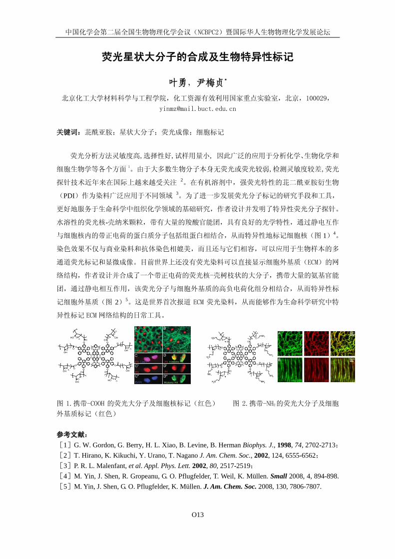
中国化学会第二届全国生物物理化学会议(NCBPC2)暨国际华人生物物理化学发展论坛
O13
荧光星状大分子的合成及生物特异性标记
叶勇,尹梅贞*
北京化工大学材料科学与工程学院,化工资源有效利用国家重点实验室,北京,100029,
关键词:苝酰亚胺;星状大分子;荧光成像;细胞标记
荧光分析方法灵敏度高,选择性好,试样用量小, 因此广泛的应用于分析化学、生物化学和
细胞生物学等各个方面1。由于大多数生物分子本身无荧光或荧光较弱,检测灵敏度较差,荧光
探针技术近年来在国际上越来越受关注 2。在有机溶剂中,强荧光特性的苝二酰亚胺衍生物
(PDI)作为染料广泛应用于不同领域 3。为了进一步发展荧光分子标记的研究手段和工具,
更好地服务于生命科学中组织化学领域的基础研究,作者设计并发明了特异性荧光分子探针。
水溶性的荧光核-壳纳米颗粒,带有大量的羧酸官能团,具有良好的光学特性,通过静电互作
与细胞核内的带正电荷的蛋白质分子包括组蛋白相结合,从而特异性地标记细胞核(图 1)4。
染色效果不仅与商业染料和抗体染色相媲美,而且还与它们相容,可以应用于生物样本的多
通道荧光标记和显微成像。目前世界上还没有荧光染料可以直接显示细胞外基质(ECM)的网
络结构,作者设计并合成了一个带正电荷的荧光核-壳树枝状的大分子,携带大量的氨基官能
团,通过静电相互作用,该荧光分子与细胞外基质的高负电荷化组分相结合,从而特异性标
记细胞外基质(图 2)5。这是世界首次报道 ECM 荧光染料,从而能够作为生命科学研究中特
异性标记 ECM 网络结构的日常工具。
图 1.携带-COOH 的荧光大分子及细胞核标记(红色) 图 2.携带-NH2的荧光大分子及细胞
外基质标记(红色) 参考文献: [1]G. W. Gordon, G. Berry, H. L. Xiao, B. Levine, B. Herman Biophys. J., 1998, 74, 2702-2713;[2]T. Hirano, K. Kikuchi, Y. Urano, T. Nagano J. Am. Chem. Soc., 2002, 124, 6555-6562; [3]P. R. L. Malenfant, et al. Appl. Phys. Lett. 2002, 80, 2517-2519; [4]M. Yin, J. Shen, R. Gropeanu, G. O. Pflugfelder, T. Weil, K. Müllen. Small 2008, 4, 894-898.[5]M. Yin, J. Shen, G. O. Pflugfelder, K. Müllen. J. Am. Chem. Soc. 2008, 130, 7806-7807.
OH OH
OH
OH
OHOH
OH
OH
On
n
O
n
O
n
O
O
n
O
OO
OO
O
OO
OO
OO
OO
O
N
N
O
OO
O
O OO O
O
On
On
O n
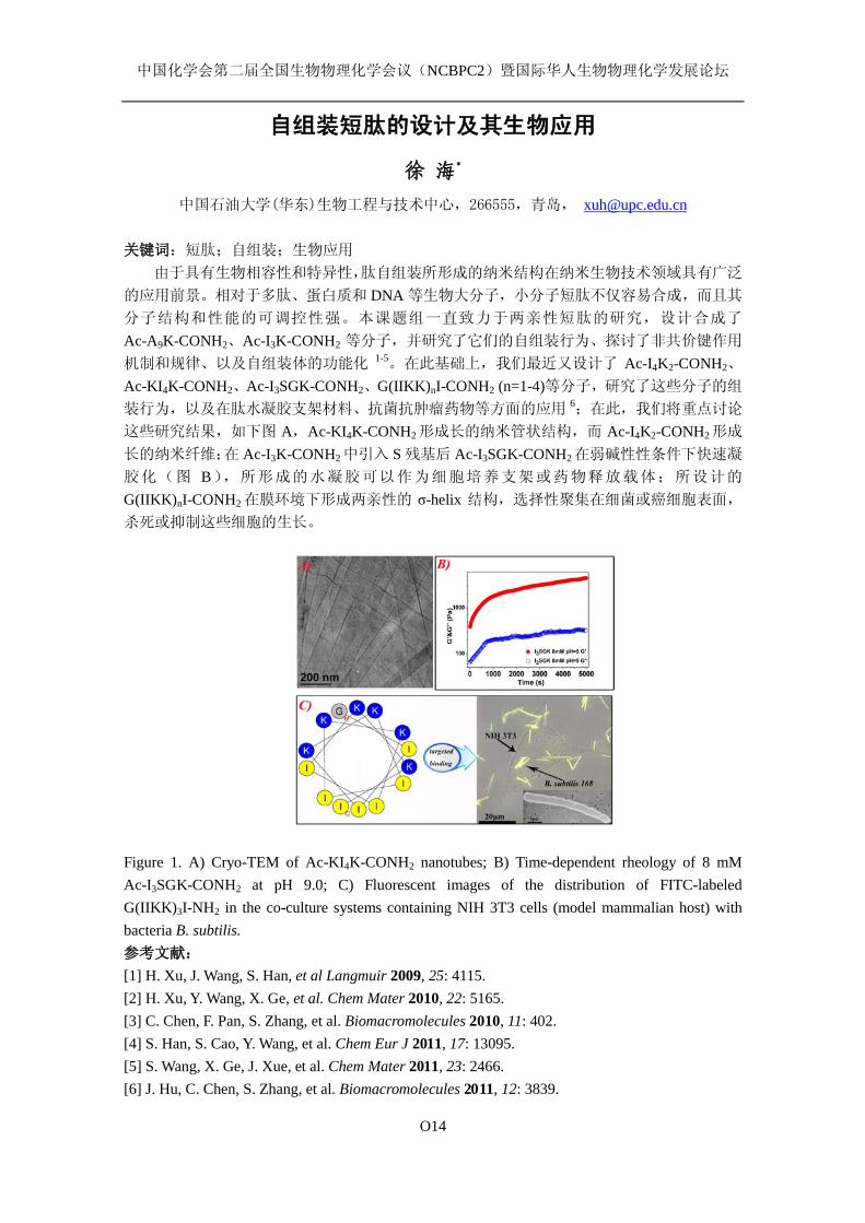
中国化学会第二届全国生物物理化学会议(NCBPC2)暨国际华人生物物理化学发展论坛
O14
自组装短肽的设计及其生物应用
徐 海*
中国石油大学(华东)生物工程与技术中心,266555,青岛, [email protected] 关键词:短肽;自组装;生物应用
由于具有生物相容性和特异性,肽自组装所形成的纳米结构在纳米生物技术领域具有广泛
的应用前景。相对于多肽、蛋白质和 DNA 等生物大分子,小分子短肽不仅容易合成,而且其
分子结构和性能的可调控性强。本课题组一直致力于两亲性短肽的研究,设计合成了
Ac-A9K-CONH2、Ac-I3K-CONH2 等分子,并研究了它们的自组装行为、探讨了非共价键作用
机制和规律、以及自组装体的功能化 1-5。在此基础上,我们最近又设计了 Ac-I4K2-CONH2、
Ac-KI4K-CONH2、Ac-I3SGK-CONH2、G(IIKK)nI-CONH2 (n=1-4)等分子,研究了这些分子的组
装行为,以及在肽水凝胶支架材料、抗菌抗肿瘤药物等方面的应用 6;在此,我们将重点讨论
这些研究结果,如下图 A,Ac-KI4K-CONH2 形成长的纳米管状结构,而 Ac-I4K2-CONH2形成
长的纳米纤维;在 Ac-I3K-CONH2 中引入 S 残基后 Ac-I3SGK-CONH2 在弱碱性性条件下快速凝
胶化(图 B),所形成的水凝胶可以作为细胞培养支架或药物释放载体;所设计的
G(IIKK)nI-CONH2 在膜环境下形成两亲性的 σ-helix 结构,选择性聚集在细菌或癌细胞表面,
杀死或抑制这些细胞的生长。
Figure 1. A) Cryo-TEM of Ac-KI4K-CONH2 nanotubes; B) Time-dependent rheology of 8 mM Ac-I3SGK-CONH2 at pH 9.0; C) Fluorescent images of the distribution of FITC-labeled G(IIKK)3I-NH2 in the co-culture systems containing NIH 3T3 cells (model mammalian host) with bacteria B. subtilis. 参考文献:
[1] H. Xu, J. Wang, S. Han, et al Langmuir 2009, 25: 4115. [2] H. Xu, Y. Wang, X. Ge, et al. Chem Mater 2010, 22: 5165. [3] C. Chen, F. Pan, S. Zhang, et al. Biomacromolecules 2010, 11: 402. [4] S. Han, S. Cao, Y. Wang, et al. Chem Eur J 2011, 17: 13095. [5] S. Wang, X. Ge, J. Xue, et al. Chem Mater 2011, 23: 2466. [6] J. Hu, C. Chen, S. Zhang, et al. Biomacromolecules 2011, 12: 3839.
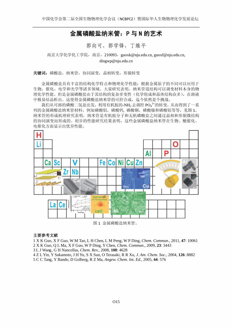
中国化学会第二届全国生物物理化学会议(NCBPC2)暨国际华人生物物理化学发展论坛
O15
金属磷酸盐纳米管:P 与 N 的艺术
郭向可,郭学锋,丁维平
南京大学化学化工学院,南京,210093,[email protected], [email protected], [email protected]
关键词:磷酸盐,纳米管,协同演变,晶相转变,形貌转变
金属磷酸盐具有丰富的结构化学特点和物理化学性能:根据金属原子的不同可以应用于
生物,催化,电学和光学等诸多领域。大量研究表明,纳米管道结构可以调变材料本身的物
理化学性能。但是金属磷酸盐由于其结构的复杂多变性(化学组成和晶体结构众多),在溶液
中极易结晶析出。这使得金属磷酸盐纳米管的可控合成,迄今依然是个挑战。 我们从可溶的磷酸二氢盐出发,利用有机胺的-NH2去调控 PO4
3+的转变,从而得到了一系
列的金属磷酸盐纳米管材料,例如磷酸铝,磷酸钙, 磷酸铜,磷酸镍和磷酸钴等等,见图 1。纳米管的形成机理研究表明,纳米管是有机胺分子和无机磷酸盐之间通过晶相和形貌微结构
的协同演变而形成的。初步的性能研究结果表明,这些金属磷酸盐纳米管在生物、酸催化、
电催化方面显示出优异性能。
图 1 金属磷酸盐纳米管。
主要参考文献
1 X K Guo, X F Guo, W M Tao, L H Chen, L M Peng, W P Ding, Chem. Commun., 2011, 47: 10061 2 X K Guo, Q L Ma, X F Guo, W P Ding, Y Chen, Chem. Commun., 2009, 23: 3443 3 L J Wang, G H Nancollas, Chem. Rev., 2008, 108: 4628 4 Z L Yin, Y Sakamoto, J H Yu, S X Sun, O Terasaki, R R Xu, J. Am. Chem. Soc., 2004, 126: 8882 5 C C Tang, Y Bando, D Golberg, R Z Ma, Angew. Chem. Int. Ed., 2005, 44: 576
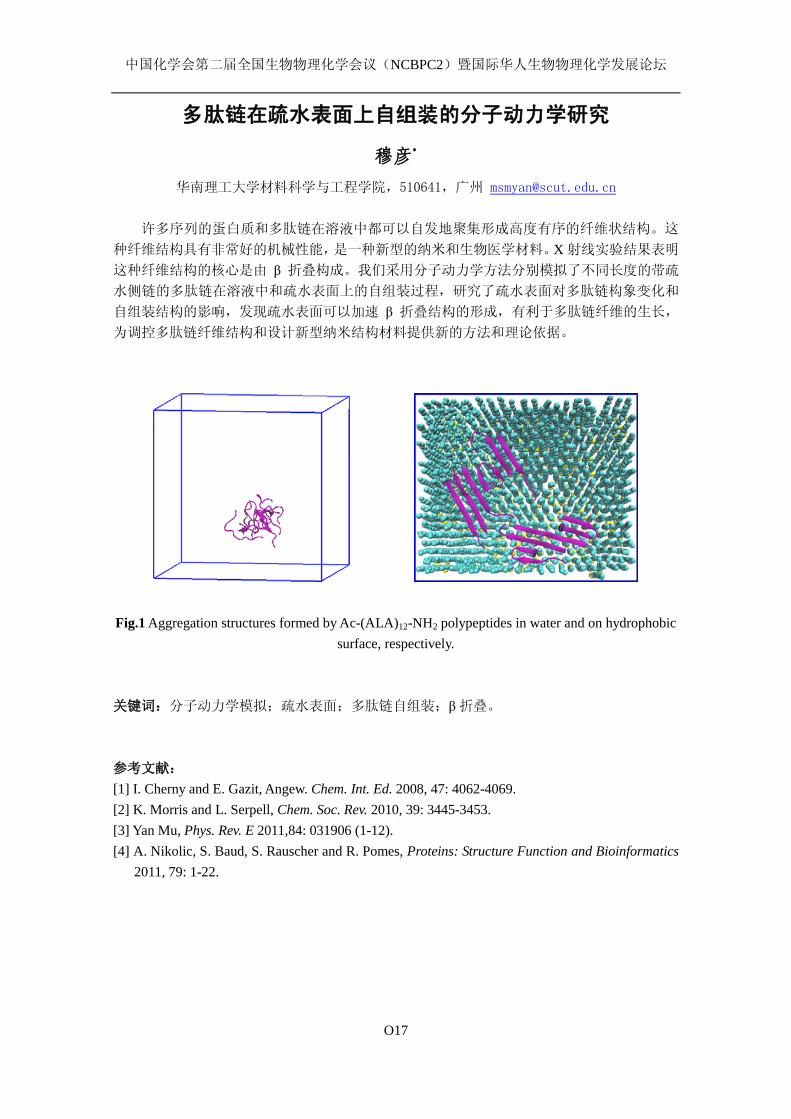
中国化学会第二届全国生物物理化学会议(NCBPC2)暨国际华人生物物理化学发展论坛
O17
多肽链在疏水表面上自组装的分子动力学研究
穆彦*
华南理工大学材料科学与工程学院,510641,广州 [email protected]
许多序列的蛋白质和多肽链在溶液中都可以自发地聚集形成高度有序的纤维状结构。这
种纤维结构具有非常好的机械性能,是一种新型的纳米和生物医学材料。X 射线实验结果表明
这种纤维结构的核心是由 β 折叠构成。我们采用分子动力学方法分别模拟了不同长度的带疏
水侧链的多肽链在溶液中和疏水表面上的自组装过程,研究了疏水表面对多肽链构象变化和
自组装结构的影响,发现疏水表面可以加速 β 折叠结构的形成,有利于多肽链纤维的生长,
为调控多肽链纤维结构和设计新型纳米结构材料提供新的方法和理论依据。
Fig.1 Aggregation structures formed by Ac-(ALA)12-NH2 polypeptides in water and on hydrophobic
surface, respectively. 关键词:分子动力学模拟;疏水表面;多肽链自组装;β折叠。 参考文献: [1] I. Cherny and E. Gazit, Angew. Chem. Int. Ed. 2008, 47: 4062-4069. [2] K. Morris and L. Serpell, Chem. Soc. Rev. 2010, 39: 3445-3453. [3] Yan Mu, Phys. Rev. E 2011,84: 031906 (1-12). [4] A. Nikolic, S. Baud, S. Rauscher and R. Pomes, Proteins: Structure Function and Bioinformatics
2011, 79: 1-22.

中国化学会第二届全国生物物理化学会议(NCBPC2)暨国际华人生物物理化学发展论坛
O19
核壳纳米粒子 SERS 标记物的制备及蛋白识别检测
孔宪明,喻倩,张现峰,杜学忠*
南京大学化学化工学院,介观化学教育部重点实验室,南京 210093,[email protected]
关键词 核壳纳米粒子;SERS;富含组氨酸蛋白;金属配位 表面增强拉曼散射(SERS)光谱是一种无损伤、高灵敏、高选择性鉴定化学和生物物质
的有力工具,甚至达到单分子检测水平。SERS 效应与金银纳米结构的局域等离子共振效应引
起的电磁场增强紧密相关。尽管金银纳米粒子聚集能够产生“热点”,导致电磁场和增强因子显
著增强[1],但纳米粒子进一步聚集将导致不可逆聚沉。银纳米粒子具有更好的 SERS 活性,但
不够稳定。为了解决这些问题,将金银纳米粒子表面包裹氧化硅形成核壳结构[2],不仅抑制金
银纳米粒子的聚集,提高金银溶胶的稳定性,而且氧化硅壳层表面更容易进行多种功能基团
修饰。由于大多被检测分子具有较弱的拉曼散射截面,拉曼探针标记是一种主要的 SERS 检测
方法。 我们发展一种简单的方法,在高醇水比的混合溶液中,无需亲玻璃硅烷试剂预先处理,
直接制备嵌入拉曼探针的 Ag@SiO2 核壳纳米粒子 SERS 标记物。制备的 Ag@SiO2纳米粒子在
高浓度盐溶液中具有很好的稳定性,且可以长期保存。利用 SERS 标记物,通过两种方法实现
蛋白高灵敏检测。方法一,将蛋白共价偶联在 Ag@SiO2纳米粒子标记物表面,通过 Cu2+配位
作用,结合到(亚胺二乙酸)IDA 自组装单层膜固体基片表面,实现蛋白 SERS 检测。方法二,
制备 Ag@SiO2 SERS 标记物与纳米金的复合纳米粒子,金纳米粒子再进行 IDA 功能化修饰。
通过 Cu2+配位作用,溶液中的蛋白在 IDA 功能化基片表面与复合纳米粒子标记物形成夹心型
结构,实现溶液蛋白 SERS 检测。肌红蛋白共含有 11 个组氨酸残基,其中 5 个位于表面[3]。
肌红蛋白通过表面组氨酸残基和 Cu2+,配位结合到 IDA 配体。 进一步研究核壳纳米粒子 SERS 标记物对蛋白识别的选择性。将胃蛋白酶和卵白蛋白分别
共价固定在核壳纳米粒子表面,在 Cu2+存在条件下,胃蛋白酶表面没有组氨酸残基,不能被
IDA 功能化基片识别捕获,没有检测到 SERS 信号,而卵白蛋白表面含有 1 个组氨酸残基,通
过 Cu2+配位桥连作用,能够特异性结合到 IDA 功能化基片表面而被检测到。Ag@SiO2 核壳纳
米粒子与纳米金的复合纳米粒子 SERS 标记物检测溶液中蛋白,由于胃蛋白酶表面不含有组氨
酸残基,卵白蛋白表面只含有 1 个组氨酸残基,导致能够发生蛋白特异性识别的夹心型结构
无法形成。结合两种方法,可以鉴定只含有 1 个表面组氨酸残基的蛋白。显然,制备的 Ag@SiO2
纳米粒子 SERS 标记物,如果选择相应的配体,同样可以用于其它种类蛋白和生物分子检测,
具有普适性。
参考文献:
[1] S. R. Emory; S. M. Nie, Science, 1997, 275, 1102. [2] L. M. Liz-Marzán; M. Giersig; P. Mulvaney Langmuir, 1996, 12, 4329. [3] H. Yim.; M. S. Kent; D. Y. Sasaki; B. D. Polizzotti; K. L. Kiick; J. Majewski; S Satija. Phys.
Rev. Lett. 2006, 96, 198101.

中国化学会第二届全国生物物理化学会议(NCBPC2)暨国际华人生物物理化学发展论坛
O20
表面活性离子液体的聚集行为及其与蛋白质相互作用研究
王晓晴,耿 斐,于 丽*
山东大学胶体与界面化学教育部重点实验室,250100,济南,[email protected]
咪唑类表面活性离子液体作为新型两亲分子而受到关注。其聚集行为研究结果表明,咪
唑类表面活性离子液体在水中可以自聚集形成胶束,碳链越长,越易形成胶束,具有优于传
统表面活性剂的表面活性。热力学参数计算结果表明其胶束化过程为熵驱动。长链烷基咪唑
溴 CnmimBr (n=12, 14, 16) 与牛血清白蛋白 (BSA)、溶菌酶及β-乳球蛋白之间相互作用的
研究结果表明,低浓度的表面活性离子液体具有保护牛血清白蛋白的二级结构而破坏溶菌酶
和β-乳球蛋白的二级结构的特点,而高浓度的表面活性离子液体能破坏这三种蛋白质的二级
结构且使其发生变性。表面活性离子液体与三种蛋白质之间的相互作用方式是有差别的:低
浓度时离子液体与 BSA及β-乳球蛋白的相互作用方式为静电相互作用,高浓度时为疏水相互
作用;而离子液体与溶菌酶的相互作用方式为疏水相互作用。
关键词:咪唑类表面活性离子液体;牛血清白蛋白;溶菌酶;β-乳球蛋白;相互作用。
Fig.1 不同温度下C16mimBr的表面张力曲线 (□) 278.15 K Fig. 2 C16mimBr/BSA体系的远紫外圆二色光谱
(○) 288.15 K (△) 298.15 K (▽) 308.15 K (◇) 318.15 K
(☆) 328.15 K
参考文献:
[1] J. Škerjanc; K. Kogej; J. Cerar Langmuir 1999, 15: 5023-5028. [2] O. Kosaka; P. Sehgal; H. Doe Food. Hydrocolloid 2008, 22: 144-149. [3] M. J. Rosen Surfactants and interfacial phenomena. New York: John Wiley & Sons, Ins., 1989.
中国化学会第二届全国生物物理化学会议(NCBPC2)暨国际华人生物物理化学发展论坛
O21
生物热热动力学方法在药物质量控制中的应用
任永申
中南民族大学 药学院
总结本课题组前期将生物热动力学方法用于药物质量控制的相关研究进展,以生物热动力
学方法在药物抗菌活性评价、抗病毒活性评价、中药注射剂质量一致性稳定性评价、注射剂
配伍相容性评价、注射剂无菌检查快速评价方法研究等为例,阐述生物热动力学方法在药物
质量控制中的研究、应用现状与发展前景,以期为生物生理学与药学的交叉学科发展提供借
鉴。
中国化学会第二届全国生物物理化学会议(NCBPC2)暨国际华人生物物理化学发展论坛
O22
运用单分子技术和结构模型定量研究DNA-蛋白质相互作用及其构象
变化
金坚石1,2,3, 谢晓亮1,2,4, 苏晓东1,2*
1北京大学生物动态光学成像中心(BIOPIC),北京100871; 2北京大学生命科学学院, 北京100871;3北京大
学前沿交叉学科研究院, 北京100871; 4 Department of Chemistry and Chemical Biology, Harvard
University, Cambridge, MA 02138, USA
别构效应(又称为变构效应)是蛋白与配基结合后改变蛋白质构象导致蛋白质活性改变的一种现
象。它是蛋白质功能调控的主要方式之一,在过去几十年中已经得到了广泛深入的研究。 但是在 DNA与蛋白质的相互作用中,DNA 一般只被考虑为被动的调控因子,即改变序列来实现对 DNA 结合蛋白
的调控。 通过结构的变化(即不改变序列,只改变结构,暂称为 DNA 别构效应)来实现对蛋白质 DNA结合的调控还研究得很少。 在真核生物中,组蛋白与 DNA 结合形成众多核小体,使得大部分 DNA 不
能完全处于理想的 B 型状态而以非 B 型 DNA 形式存在。 根据很多已经被解析出的高分辨率三维结构
的核小体复合物[1-3], 由于组蛋白对 DNA 的弯折作用,使得其构象相对于理想的 B 型 DNA 具有明显
的差异和变化。 因此,DNA 构象变化参与蛋白质 DNA 相互作用存在着结构基础。 运用单分子荧光成像技术测量糖皮质激素受体(GR)DNA 结合结构域(GRDBD)在核小体不同位
置的 DNA 结合序列(GRE)上的平均结合时间,来系统定量地研究 GRDBD 在核小体不同位置的 GRE 上
的结合稳定程度。 我们得到的结果表明,组蛋白对结合在核小体上的 DNA 结合蛋白的稳定性调控,
不仅仅是通过阻碍或者隐藏 DNA 结合序列来实现,对 DNA 结构的影响也会成为其中的一个因素。在
GRE 完全朝向外侧时(组蛋白没有任何阻碍作用),GRDBD 的平均结合时间比在自由的 DNA 上的集
合时间足足降低了 3.7 倍。 更加有趣的是,GRDBD 结合最稳定的状态不是在完全自由的 dsDNA,而
是在核小体上,在核小体的某个位置时,其结合时间与溶液中自由的 dsDNA 相比可以增加 1.5 倍。 对于这些现象,我们通过结构模型进行了模拟和分析,认为这是由于 DNA 的结构在核小体不同位置时,
具有很大差异的构像并且被固定。 这是 DNA 的构象变化参与 DNA-蛋白质相互作用比较直接的一个案
例。
[1]. Luger, K., et al., Crystal structure of the nucleosome core particle at 2.8 A resolution. Nature, 1997. 389(6648): p. 251-60.
[2]. Richmond, T.J. and C.A. Davey, The structure of DNA in the nucleosome core. Nature, 2003. 423(6936): p. 145-50.
[3]. Makde, R.D., et al., Structure of RCC1 chromatin factor bound to the nucleosome core particle. Nature, 2010. 467(7315): p. 562-6.
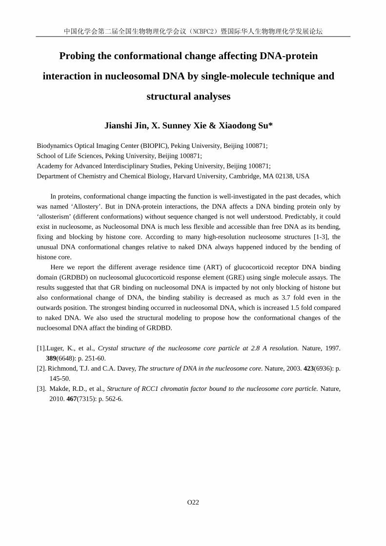
中国化学会第二届全国生物物理化学会议(NCBPC2)暨国际华人生物物理化学发展论坛
O22
Probing the conformational change affecting DNA-protein
interaction in nucleosomal DNA by single-molecule technique and
structural analyses
Jianshi Jin, X. Sunney Xie & Xiaodong Su*
Biodynamics Optical Imaging Center (BIOPIC), Peking University, Beijing 100871; School of Life Sciences, Peking University, Beijing 100871; Academy for Advanced Interdisciplinary Studies, Peking University, Beijing 100871; Department of Chemistry and Chemical Biology, Harvard University, Cambridge, MA 02138, USA
In proteins, conformational change impacting the function is well-investigated in the past decades, which was named ‘Allostery’. But in DNA-protein interactions, the DNA affects a DNA binding protein only by ‘allosterism’ (different conformations) without sequence changed is not well understood. Predictably, it could exist in nucleosome, as Nucleosomal DNA is much less flexible and accessible than free DNA as its bending, fixing and blocking by histone core. According to many high-resolution nucleosome structures [1-3], the unusual DNA conformational changes relative to naked DNA always happened induced by the bending of histone core.
Here we report the different average residence time (ART) of glucocorticoid receptor DNA binding domain (GRDBD) on nucleosomal glucocorticoid response element (GRE) using single molecule assays. The results suggested that that GR binding on nucleosomal DNA is impacted by not only blocking of histone but also conformational change of DNA, the binding stability is decreased as much as 3.7 fold even in the outwards position. The strongest binding occurred in nucleosomal DNA, which is increased 1.5 fold compared to naked DNA. We also used the structural modeling to propose how the conformational changes of the nucloesomal DNA affact the binding of GRDBD.
[1].Luger, K., et al., Crystal structure of the nucleosome core particle at 2.8 A resolution. Nature, 1997.
389(6648): p. 251-60. [2]. Richmond, T.J. and C.A. Davey, The structure of DNA in the nucleosome core. Nature, 2003. 423(6936): p.
145-50. [3]. Makde, R.D., et al., Structure of RCC1 chromatin factor bound to the nucleosome core particle. Nature,
2010. 467(7315): p. 562-6.
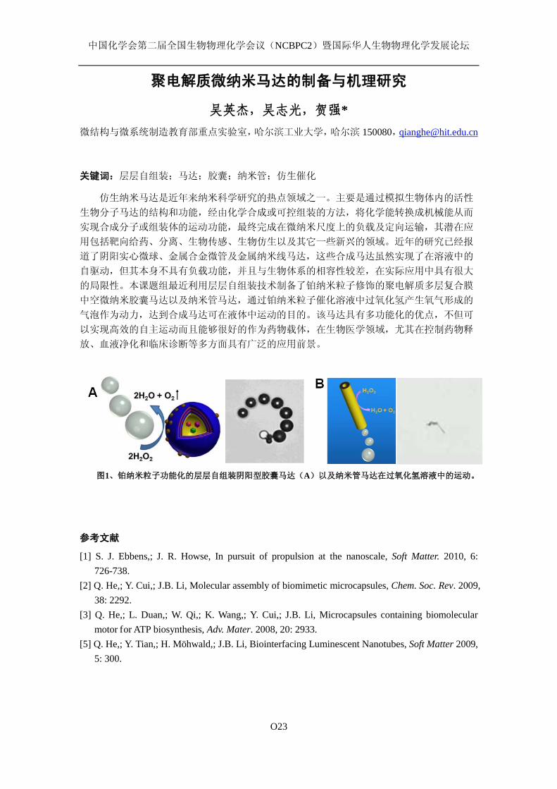
中国化学会第二届全国生物物理化学会议(NCBPC2)暨国际华人生物物理化学发展论坛
O23
聚电解质微纳米马达的制备与机理研究
吴英杰,吴志光,贺强* 微结构与微系统制造教育部重点实验室,哈尔滨工业大学,哈尔滨 150080,[email protected]
关键词:层层自组装;马达;胶囊;纳米管;仿生催化
仿生纳米马达是近年来纳米科学研究的热点领域之一。主要是通过模拟生物体内的活性
生物分子马达的结构和功能,经由化学合成或可控组装的方法,将化学能转换成机械能从而
实现合成分子或组装体的运动功能,最终完成在微纳米尺度上的负载及定向运输,其潜在应
用包括靶向给药、分离、生物传感、生物仿生以及其它一些新兴的领域。近年的研究已经报
道了阴阳实心微球、金属合金微管及金属纳米线马达,这些合成马达虽然实现了在溶液中的
自驱动,但其本身不具有负载功能,并且与生物体系的相容性较差,在实际应用中具有很大
的局限性。本课题组最近利用层层自组装技术制备了铂纳米粒子修饰的聚电解质多层复合膜
中空微纳米胶囊马达以及纳米管马达,通过铂纳米粒子催化溶液中过氧化氢产生氧气形成的
气泡作为动力,达到合成马达可在液体中运动的目的。该马达具有多功能化的优点,不但可
以实现高效的自主运动而且能够很好的作为药物载体,在生物医学领域,尤其在控制药物释
放、血液净化和临床诊断等多方面具有广泛的应用前景。
图1、铂纳米粒子功能化的层层自组装阴阳型胶囊马达(A)以及纳米管马达在过氧化氢溶液中的运动。
参考文献
[1] S. J. Ebbens,; J. R. Howse, In pursuit of propulsion at the nanoscale, Soft Matter. 2010, 6: 726-738.
[2] Q. He,; Y. Cui,; J.B. Li, Molecular assembly of biomimetic microcapsules, Chem. Soc. Rev. 2009, 38: 2292.
[3] Q. He,; L. Duan,; W. Qi,; K. Wang,; Y. Cui,; J.B. Li, Microcapsules containing biomolecular motor for ATP biosynthesis, Adv. Mater. 2008, 20: 2933.
[5] Q. He,; Y. Tian,; H. Möhwald,; J.B. Li, Biointerfacing Luminescent Nanotubes, Soft Matter 2009, 5: 300.
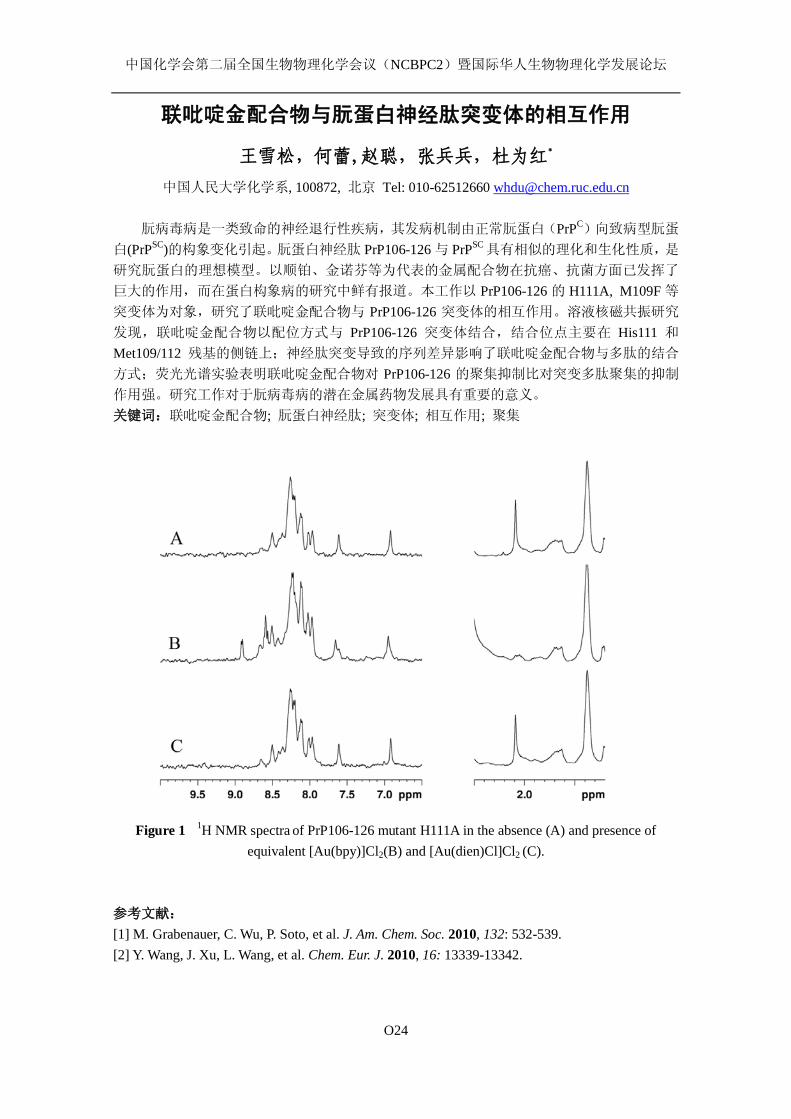
中国化学会第二届全国生物物理化学会议(NCBPC2)暨国际华人生物物理化学发展论坛
O24
联吡啶金配合物与朊蛋白神经肽突变体的相互作用
王雪松,何蕾,赵聪,张兵兵,杜为红*
中国人民大学化学系, 100872, 北京 Tel: 010-62512660 [email protected]
朊病毒病是一类致命的神经退行性疾病,其发病机制由正常朊蛋白(PrPC)向致病型朊蛋
白(PrPSC)的构象变化引起。朊蛋白神经肽 PrP106-126 与 PrPSC 具有相似的理化和生化性质,是
研究朊蛋白的理想模型。以顺铂、金诺芬等为代表的金属配合物在抗癌、抗菌方面已发挥了
巨大的作用,而在蛋白构象病的研究中鲜有报道。本工作以 PrP106-126 的 H111A, M109F 等
突变体为对象,研究了联吡啶金配合物与 PrP106-126 突变体的相互作用。溶液核磁共振研究
发现,联吡啶金配合物以配位方式与 PrP106-126 突变体结合,结合位点主要在 His111 和
Met109/112 残基的侧链上;神经肽突变导致的序列差异影响了联吡啶金配合物与多肽的结合
方式;荧光光谱实验表明联吡啶金配合物对 PrP106-126 的聚集抑制比对突变多肽聚集的抑制
作用强。研究工作对于朊病毒病的潜在金属药物发展具有重要的意义。 关键词:联吡啶金配合物; 朊蛋白神经肽; 突变体; 相互作用; 聚集
Figure 1 1H NMR spectra of PrP106-126 mutant H111A in the absence (A) and presence of
equivalent [Au(bpy)]Cl2(B) and [Au(dien)Cl]Cl2 (C). 参考文献: [1] M. Grabenauer, C. Wu, P. Soto, et al. J. Am. Chem. Soc. 2010, 132: 532-539. [2] Y. Wang, J. Xu, L. Wang, et al. Chem. Eur. J. 2010, 16: 13339-13342.
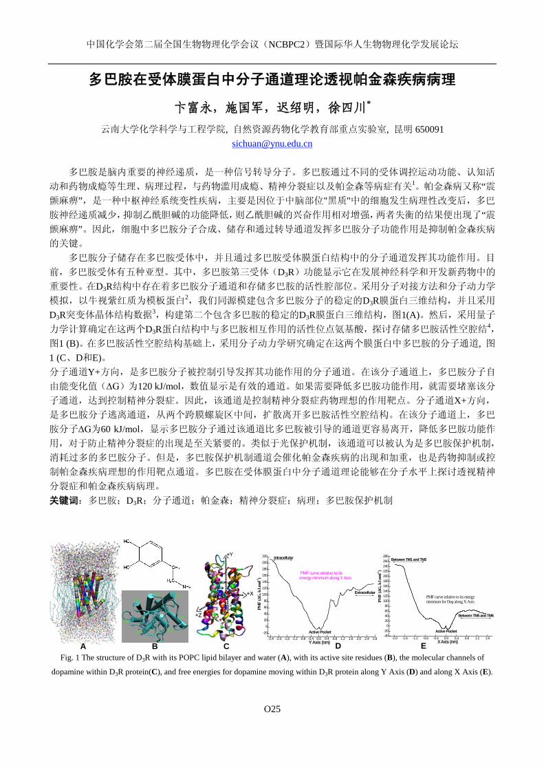
中国化学会第二届全国生物物理化学会议(NCBPC2)暨国际华人生物物理化学发展论坛
O25
多巴胺在受体膜蛋白中分子通道理论透视帕金森疾病病理
卞富永,施国军,迟绍明,徐四川* 云南大学化学科学与工程学院, 自然资源药物化学教育部重点实验室, 昆明 650091
多巴胺是脑内重要的神经递质,是一种信号转导分子。多巴胺通过不同的受体调控运动功能、认知活
动和药物成瘾等生理、病理过程,与药物滥用成瘾、精神分裂症以及帕金森等病症有关1。帕金森病又称“震颤麻痹”,是一种中枢神经系统变性疾病,主要是因位于中脑部位"黑质"中的细胞发生病理性改变后,多巴
胺神经递质减少,抑制乙酰胆碱的功能降低,则乙酰胆碱的兴奋作用相对增强,两者失衡的结果便出现了“震颤麻痹”。因此,细胞中多巴胺分子合成、储存和通过转导通道发挥多巴胺分子功能作用是抑制帕金森疾病
的关键。 多巴胺分子储存在多巴胺受体中,并且通过多巴胺受体膜蛋白结构中的分子通道发挥其功能作用。目
前,多巴胺受体有五种亚型。其中,多巴胺第三受体(D3R)功能显示它在发展神经科学和开发新药物中的
重要性。在D3R结构中存在着多巴胺分子通道和存储多巴胺的活性腔部位。采用分子对接方法和分子动力学
模拟,以牛视紫红质为模板蛋白2,我们同源模建包含多巴胺分子的稳定的D3R膜蛋白三维结构,并且采用
D3R突变体晶体结构数据3,构建第二个包含多巴胺的稳定的D3R膜蛋白三维结构,图1(A)。然后,采用量子
力学计算确定在这两个D3R蛋白结构中与多巴胺相互作用的活性位点氨基酸,探讨存储多巴胺活性空腔结4,
图1 (B)。在多巴胺活性空腔结构基础上,采用分子动力学研究确定在这两个膜蛋白中多巴胺的分子通道, 图1 (C、D和E)。 分子通道Y+方向,是多巴胺分子被控制引导发挥其功能作用的分子通道。在该分子通道上,多巴胺分子自
由能变化值(ΔG)为120 kJ/mol,数值显示是有效的通道。如果需要降低多巴胺功能作用,就需要堵塞该分
子通道,达到控制精神分裂症。因此,该通道是控制精神分裂症药物理想的作用靶点。分子通道X+方向,
是多巴胺分子逃离通道,从两个跨膜螺旋区中间,扩散离开多巴胺活性空腔结构。在该分子通道上,多巴
胺分子ΔG为60 kJ/mol,显示多巴胺分子通过该通道比多巴胺被引导的通道更容易离开,降低多巴胺功能作
用,对于防止精神分裂症的出现是至关紧要的。类似于光保护机制,该通道可以被认为是多巴胺保护机制,
消耗过多的多巴胺分子。但是,多巴胺保护机制通道会催化帕金森疾病的出现和加重,也是药物抑制或控
制帕金森疾病理想的作用靶点通道。多巴胺在受体膜蛋白中分子通道理论能够在分子水平上探讨透视精神
分裂症和帕金森疾病病理。 关键词:多巴胺;D3R;分子通道;帕金森;精神分裂症;病理;多巴胺保护机制
-2.4 -2.0 -1.6 -1.2 -0.8 -0.4 0.0 0.4 0.8 1.2 1.6 2.0 2.4 2.8-20
0
20
40
60
80
100
120
140
160
180
200
220 Intracellular
Active Pocket
Extracellular
PMF curve relative to itsenergy minimum along Y Axis
PMF
(∆G
, kJ.
mol
-1)
Y Axis (nm)-2.0 -1.6 -1.2 -0.8 -0.4 0.0 0.4 0.8 1.2 1.6
-40-20
020406080
100120140160180200220240260280
Between TM1 and TM2
Between TM5 and TM6
Active Pocket
PMF
(∆G
, kJ.
mol
-1)
X Axis (nm)
PMF curve relative to its energyminimum for Dop along X Axis
Fig. 1 The structure of D3R with its POPC lipid bilayer and water (A), with its active site residues (B), the molecular channels of
dopamine within D3R protein(C), and free energies for dopamine moving within D3R protein along Y Axis (D) and along X Axis (E).
C D A B E
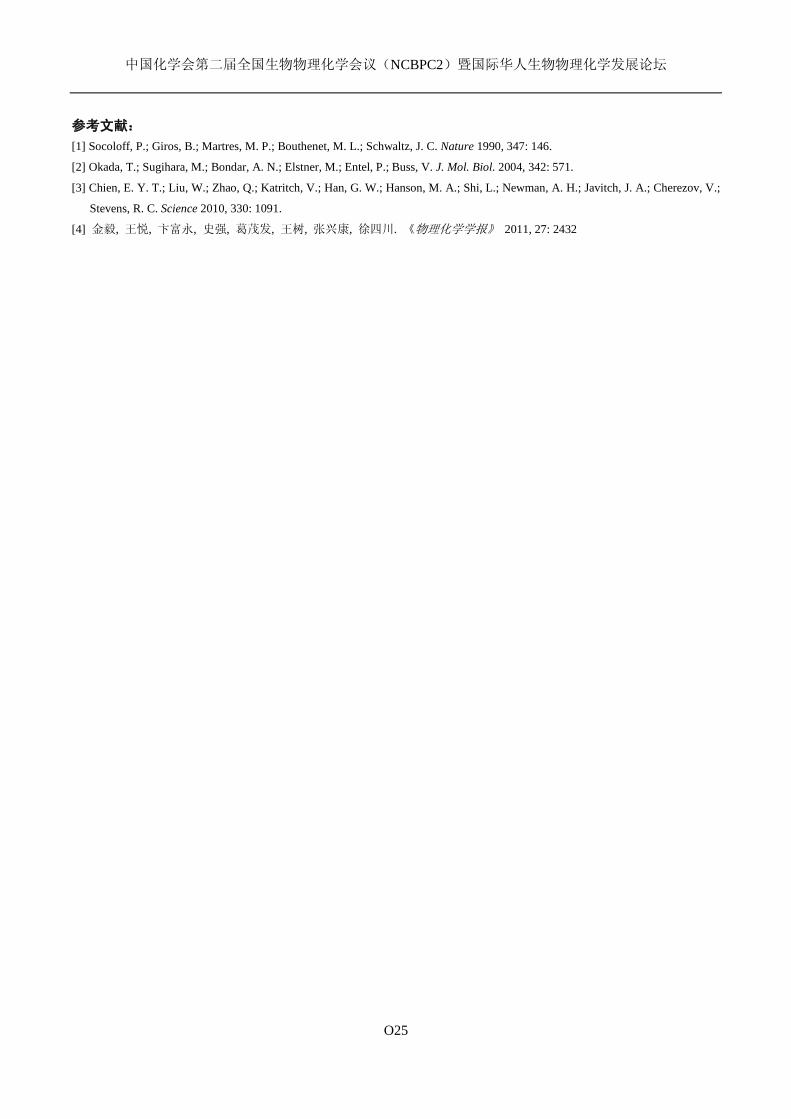
中国化学会第二届全国生物物理化学会议(NCBPC2)暨国际华人生物物理化学发展论坛
O25
参考文献:
[1] Socoloff, P.; Giros, B.; Martres, M. P.; Bouthenet, M. L.; Schwaltz, J. C. Nature 1990, 347: 146.
[2] Okada, T.; Sugihara, M.; Bondar, A. N.; Elstner, M.; Entel, P.; Buss, V. J. Mol. Biol. 2004, 342: 571.
[3] Chien, E. Y. T.; Liu, W.; Zhao, Q.; Katritch, V.; Han, G. W.; Hanson, M. A.; Shi, L.; Newman, A. H.; Javitch, J. A.; Cherezov, V.;
Stevens, R. C. Science 2010, 330: 1091.
[4] 金毅, 王悦, 卞富永, 史强, 葛茂发, 王树, 张兴康, 徐四川. 《物理化学学报》 2011, 27: 2432
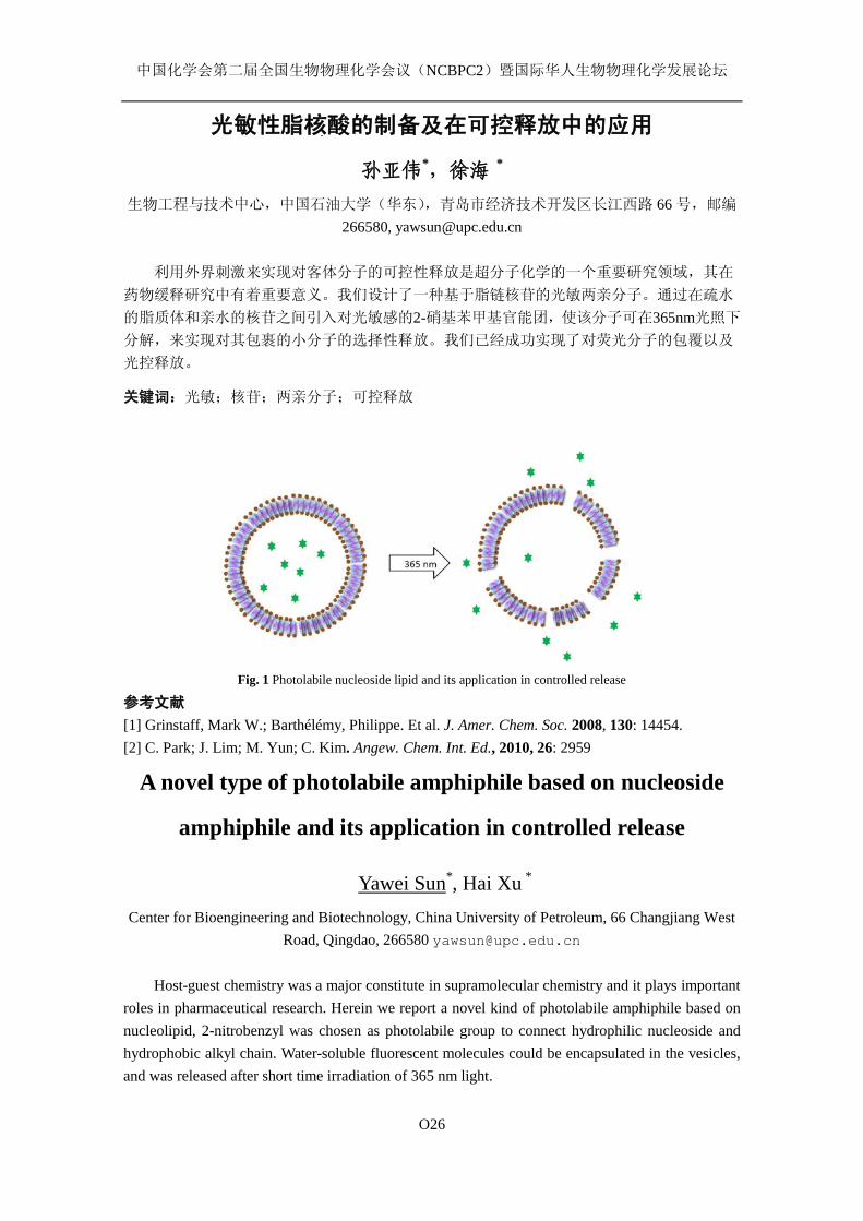
中国化学会第二届全国生物物理化学会议(NCBPC2)暨国际华人生物物理化学发展论坛
O26
光敏性脂核酸的制备及在可控释放中的应用
孙亚伟*,徐海 * 生物工程与技术中心,中国石油大学(华东),青岛市经济技术开发区长江西路 66 号,邮编
266580, [email protected]
利用外界刺激来实现对客体分子的可控性释放是超分子化学的一个重要研究领域,其在
药物缓释研究中有着重要意义。我们设计了一种基于脂链核苷的光敏两亲分子。通过在疏水
的脂质体和亲水的核苷之间引入对光敏感的2-硝基苯甲基官能团,使该分子可在365nm光照下
分解,来实现对其包裹的小分子的选择性释放。我们已经成功实现了对荧光分子的包覆以及
光控释放。
关键词:光敏;核苷;两亲分子;可控释放
Fig. 1 Photolabile nucleoside lipid and its application in controlled release
参考文献
[1] Grinstaff, Mark W.; Barthélémy, Philippe. Et al. J. Amer. Chem. Soc. 2008, 130: 14454. [2] C. Park; J. Lim; M. Yun; C. Kim. Angew. Chem. Int. Ed., 2010, 26: 2959
A novel type of photolabile amphiphile based on nucleoside
amphiphile and its application in controlled release
Yawei Sun*, Hai Xu * Center for Bioengineering and Biotechnology, China University of Petroleum, 66 Changjiang West
Road, Qingdao, 266580 [email protected]
Host-guest chemistry was a major constitute in supramolecular chemistry and it plays important roles in pharmaceutical research. Herein we report a novel kind of photolabile amphiphile based on nucleolipid, 2-nitrobenzyl was chosen as photolabile group to connect hydrophilic nucleoside and hydrophobic alkyl chain. Water-soluble fluorescent molecules could be encapsulated in the vesicles, and was released after short time irradiation of 365 nm light.

中国化学会第二届全国生物物理化学会议(NCBPC2)暨国际华人生物物理化学发展论坛
O27
The Special Optical Properties of a heterocyclic compound
5H-2,3-dithia-5,7-diaza-cyclopenta[c,d]indenes Liangrong Zhao, Mingguo Liu, Mingxun Yan, Yayun Zhu, Liangjun Pan, Changying Yang* College of Chemistry and Life Science, Three Gorges University, Yichang 443002, [email protected]
The investigation of the significant intermolecular hydrogen bonding effects on the structure and aggregate properties of the special structure of a heterocyclic compound 5H-2,3-dithia-5,7-diaza-cyclopenta[c,d]indenes (Scheme. 1)[1], in H2O/DMSO was performed in this paper through spectroscopy experiments as well as theoretical calculation. The benzene ring of the double-thiophene heterocyclic compounds is coplane with bithiophene in DMSO, which is benefit to form H-aggregation for the compound and bring down fluorescence intensity. However, it was observed that the formation of intermolecular hydrogen bonds with H2O could lead to a torsion angle (47°) between the benzene ring to the main plane. When the H2O content is 66~70% in solvent, absorption peak of the compound red shift sharply from 390 nm to 444 nm, meanwhile the fluorescence intensity strengthened (Figure 1). It is indicated that J- aggregation formed then. The theoretical study based on DFT calculation is very helpful for the understanding on the optical properties (Scheme. 2). The low temperature was benefit for the photoluminescence of compound, for the emission quenched quickly by heating, indicated that the thermo effect of J-aggregation (Figure 2).
S S
NH N
NH
O
O
CH3
O
O
CH3
1.80 Å
1.78
Å
1.80 Å
1.78
Å
Scheme 1. Structure of
5H-2,3-dithia-5,7-diaza-cyclopenta[c,d]indenes.
Scheme 2. Optimized geometric structures of
hydrogen-bonded complexes.
400 450 500 5500
1000
2000
3000
0 20 40 60 80 100
2
4
6
8
I/I0
Water volume fraction/v%
2
1
0
FL In
tens
ity
Wavelength/nm 400 450 500 5500
1000
2000
3000
388 K
353 K
313 Kback to 313 K
FL In
tens
ity
Wavelength/nm
Fig. 1 The emission spectra of compound in the
H2O/DMSO mixture with different volume percentages
of water: (0) 0% H2O; (1) 70% H2O; (2) 90% H2O. The
inset shows the change in the emission intensity of
compound versus water fractions in DMSO.
Fig. 2 The fluorescence spectra of
compound in 70% H2O/DMSO mixture at
different temperatures.
1 Ming-guo Liu, Yang-gen Hu, Ming-wu Ding. Tetrahedron.2008,64: 9052-9059.
中国化学会第二届全国生物物理化学会议(NCBPC2)暨国际华人生物物理化学发展论坛
O28
Cell adhesion properties of polymeric layer-by-layer
multilayer films
Wei Qi*, Peng Cai, Jianbo Ma, Min Sun Key Laboratory of Life-Organic Analysis, School of Chemistry and Chemical Engineering, Qufu
Normal University, Qufu Shandong 273165 China, [email protected]
Keywords: layer-by-layer assembly technique; biocompatible polymeric film; cell adhesion There is currently a growing interest in rendering biomaterial surfaces not only biocompatible but
also readily engineered and functionalized with specific properties. Among the new methods that have emerged for preparing biocompatible surfaces with controlled properties at the nanometer/micrometer scales, the layer-by-layer (LbL) assembly technique appears highly versatile and powerful1-2. LbL assembly of polymeric multilayer films represents a bottom-up approach for reengineering the molecular landscape of substrate surfaces3. Our aim is to use these films as a platform to study cellular processes, including adhesion, proliferation and motility, etc.
Here, we report behaviors of mammalian cells, such as human embryo skin fibroblast cells, mouse embryo fibroblast cells and rat aortic endothelial cells, grown on multilayered thin films of CHI/PAA to investigate biocompatibility of the artificial surface. Proliferation assay, cell shape analysis and focal adhesion study reveal that the cells grow well on the LbL assembled substrates. The present study suggests that these films hold high potential for bioapplications showing high biocompatibility and provide promising surface coating materials for implants. Acknowledgements
This work was financially supported by the National Nature Science Foundation of China (No. 21003084), and Shandong Province Promotive Research Foundation for Excellent Young and Middle-aged Scientists, China (No. BS2010CL023).
References [1] Varvara Gribova, Rachel Auzely-Velty, and Catherine Picart Chem. Mater. 2012, 24: 854-869. [2] Wei Qi, Li Duan, Xuehai Yan, Kewei Wang, Jinbo Fei, Yue Cui, and Junbai Li Biomaterials
2009, 30: 2799-2806. [3] Gero Decher Science 1997, 277: 1232-1237.
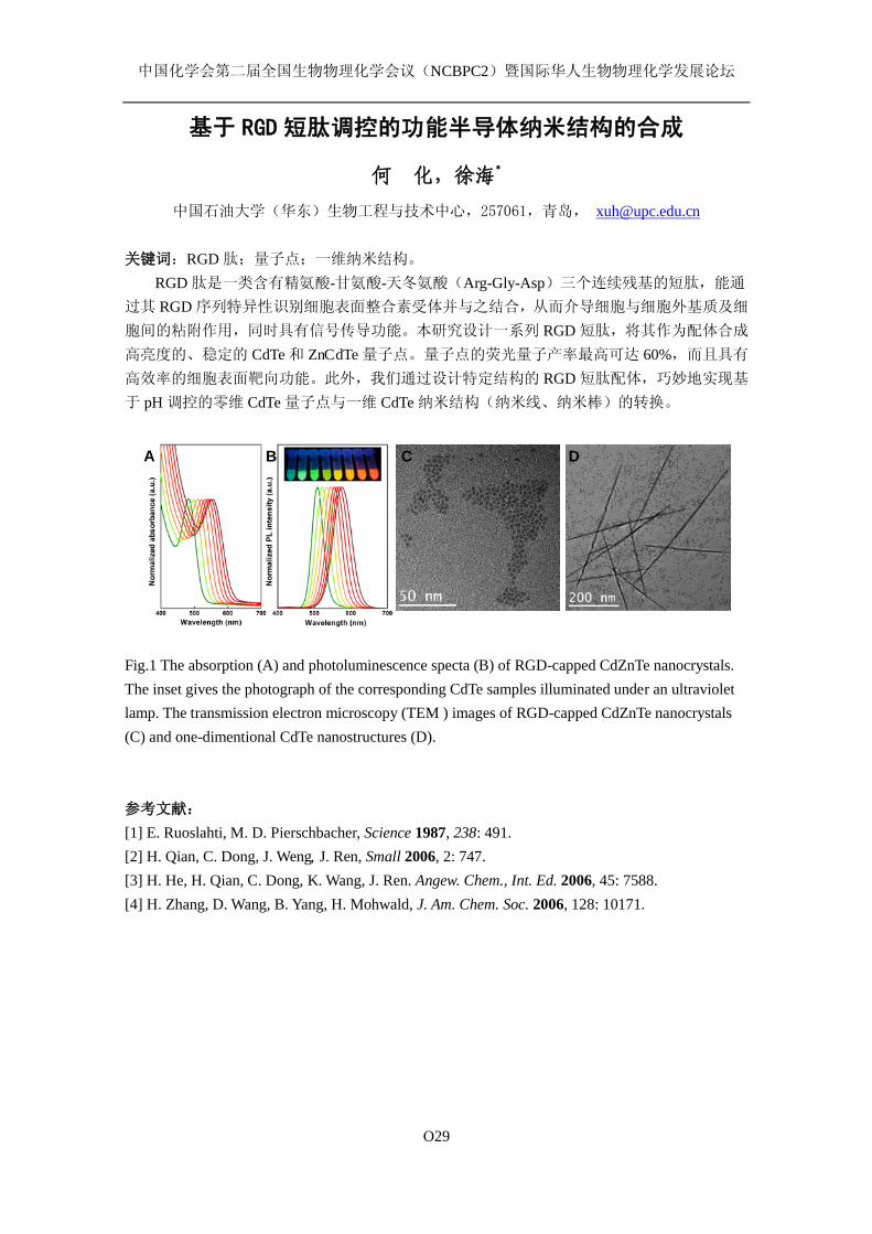
中国化学会第二届全国生物物理化学会议(NCBPC2)暨国际华人生物物理化学发展论坛
O29
基于 RGD 短肽调控的功能半导体纳米结构的合成
何 化,徐海*
中国石油大学(华东)生物工程与技术中心,257061,青岛, [email protected]
关键词:RGD 肽;量子点;一维纳米结构。
RGD 肽是一类含有精氨酸-甘氨酸-天冬氨酸(Arg-Gly-Asp)三个连续残基的短肽,能通
过其 RGD 序列特异性识别细胞表面整合素受体并与之结合,从而介导细胞与细胞外基质及细
胞间的粘附作用,同时具有信号传导功能。本研究设计一系列 RGD 短肽,将其作为配体合成
高亮度的、稳定的 CdTe 和 ZnCdTe 量子点。量子点的荧光量子产率最高可达 60%,而且具有
高效率的细胞表面靶向功能。此外,我们通过设计特定结构的 RGD 短肽配体,巧妙地实现基
于 pH 调控的零维 CdTe 量子点与一维 CdTe 纳米结构(纳米线、纳米棒)的转换。 Fig.1 The absorption (A) and photoluminescence specta (B) of RGD-capped CdZnTe nanocrystals. The inset gives the photograph of the corresponding CdTe samples illuminated under an ultraviolet lamp. The transmission electron microscopy (TEM ) images of RGD-capped CdZnTe nanocrystals (C) and one-dimentional CdTe nanostructures (D).
参考文献:
[1] E. Ruoslahti, M. D. Pierschbacher, Science 1987, 238: 491. [2] H. Qian, C. Dong, J. Weng, J. Ren, Small 2006, 2: 747. [3] H. He, H. Qian, C. Dong, K. Wang, J. Ren. Angew. Chem., Int. Ed. 2006, 45: 7588. [4] H. Zhang, D. Wang, B. Yang, H. Mohwald, J. Am. Chem. Soc. 2006, 128: 10171.
A B C D
中国化学会第二届全国生物物理化学会议(NCBPC2)暨国际华人生物物理化学发展论坛
O30
溶菌酶在适度疏水表面吸附的热力学及分子构象
耿信鹏
西安工程大学环境与化工学院 西安,710048, [email protected]
In this paper, the systematical research 1 in our laboratory is addressed related to thermodynamics and their fractions, isotherms, conformational changes and thermal stability in adsorbed state for native and denatured Lyz adsorptions upon the moderately hydrophobic surface under various conditions. The thermodynamics of stoichiometric displacement theory for adsorption (SDT-A) that subdivided the general thermodynamics of a displacement adsorption process into (net) adsorption for adsorbate and (net) desorption for solvent has successfully been used for protein adsorption systems and can elucidate the four subprocesses in protein adsorption. Furthermore, the relations between the four subprocesses and the two fractions of thermodynamics were first built up.
The results show that adsorbed amounts of native and denatured Lyz increase with (NH4)2SO4 concentrations increment because of enhancing hydrophobic interactions and that of denatured Lyz decrease with guanidine hydrochloride (GuHCl) concentrations increment due to looser molecule. The enthalpies obtained by microcalorimetric measurement show that adsorption for native Lyz is an endothermic and entropy driven process contributed mainly by orderly conformational loss of adsorbed Lyz (ordered structure loss enthalpy and entropy, ∆ Hml>0 and ∆ Sml>0), while that for denatured Lyz is complicated: an endothermic and entropy driven caused mainly by dehydration at lower GuHCl concentrations, and an exothermic and enthalpy driven induced by molecular conformational gain (∆ Hmo<0 and ∆ Smo<0) at higher ones. The moderately hydrophobic surface can provide denatured Lyz energy and make it gain more ordered structure with surface coverage or salt concentration increment. Thermodynamic analysis that which one of the four adsorption subprocesses plays a major role for contribution to the thermodynamic fractions was made in detail. The investigations on thermal stability (by DSC) and the changes in molecular conformation (by FTIR) of adsorbed native and denatured Lyz show that with both GuHCl and (NH4)2SO4 concentrations increment, the ordered secondary structures contents (sum of α-helix and β-sheet) increase and the transition temperatures corresponding to endothermic peaks in thermal program lower, indicating thermal stability and tertiary structure reduced. With surface coverage increment, the ordered secondary structures contents, the thermal stability and tertiary structures decrease. Keywords: Lysozyme adsorption; thermodynamic fraction; conformational change; thermal stability; adsorption subprocess; hydrophobic surface Acknowledgement:We would like to thank National Natural Science Foundation of China for sponsoring the projects (Grant No.20673080).
[1] X. P. Geng, J. J. Peng, J. Geng, H. Y. Hou, Adsorption behaviors of lysozyme adsorbed onto moderately hydrophobic surface, in: X. G. Maang and W. F. Cheung (Eds.), Lysozymes: Sources, Functions and Role in Disease, Nova Science Publishers Inc., Hauppauge, in press.

中国化学会第二届全国生物物理化学会议(NCBPC2)暨国际华人生物物理化学发展论坛
O31
Molecular Interactions at surfaces of Biosensors
姜磊, 杨丽敏
生物技术与工程中心,中国石油大学(华东),长江西路66号,青岛,266580,
关键词: surface forces, atomic force microscope, biosensor, interactions
The physical properties and molecular interactions of macromolecules attached at the surfaces of
biosensors were studied. The macromolecules including polyelectrolyte and proteins were physically
adsorbed or chemically grafted to the substrate surface, producing various kinds of functional layers at
the interface. The structure, orientation and interactions of such layers with the opposing analyte were
characterized using the Atomic Force Microscope (AFM), surface plasma resonance (SPR), etc. The
effects of solution conditions, including the counterion valencies, concentration and kinetics were
compared by measuring the surface forces and hence the conformation or assembly of polymers at the
solid/liquid interface could be illustrated. These understanding would not only assist the preparation of
polymer coated chips of biosensors, but also improve the surface quality of polymer-anchored silica
nanoparticles as the multivalent analyte in the High Throughput Screening (HTS) application.
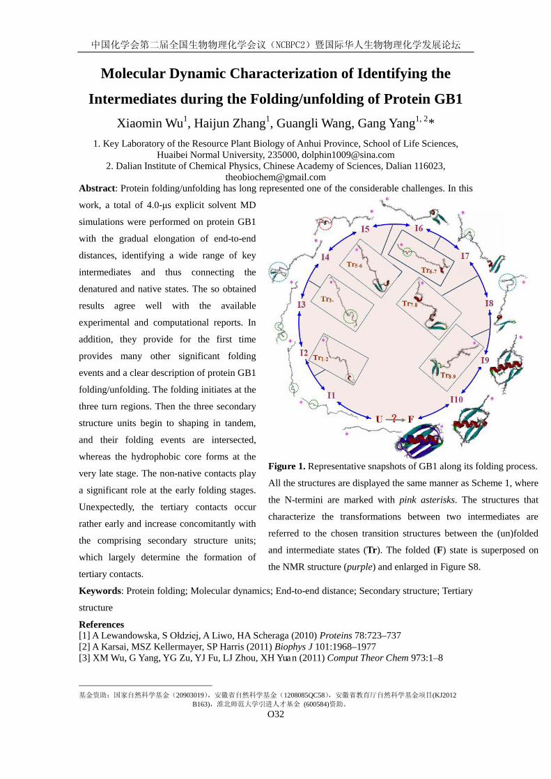
中国化学会第二届全国生物物理化学会议(NCBPC2)暨国际华人生物物理化学发展论坛
O32
Figure 1. Representative snapshots of GB1 along its folding process.
All the structures are displayed the same manner as Scheme 1, where
the N-termini are marked with pink asterisks. The structures that
characterize the transformations between two intermediates are
referred to the chosen transition structures between the (un)folded
and intermediate states (Tr). The folded (F) state is superposed on
the NMR structure (purple) and enlarged in Figure S8.
Molecular Dynamic Characterization of Identifying the
Intermediates during the Folding/unfolding of Protein GB1 Xiaomin Wu1, Haijun Zhang1, Guangli Wang, Gang Yang1, 2*
2. Dalian Institute of Chemical Physics, Chinese Academy of Sciences, Dalian 116023,
1. Key Laboratory of the Resource Plant Biology of Anhui Province, School of Life Sciences,
Huaibei Normal University, 235000, [email protected]
[email protected] Abstract: Protein folding/unfolding has long represented one of the considerable challenges. In this
work, a total of 4.0-μs explicit solvent MD
simulations were performed on protein GB1
with the gradual elongation of end-to-end
distances, identifying a wide range of key
intermediates and thus connecting the
denatured and native states. The so obtained
results agree well with the available
experimental and computational reports. In
addition, they provide for the first time
provides many other significant folding
events and a clear description of protein GB1
folding/unfolding. The folding initiates at the
three turn regions. Then the three secondary
structure units begin to shaping in tandem,
and their folding events are intersected,
whereas the hydrophobic core forms at the
very late stage. The non-native contacts play
a significant role at the early folding stages.
Unexpectedly, the tertiary contacts occur
rather early and increase concomitantly with
the comprising secondary structure units;
which largely determine the formation of
tertiary contacts.
Keywords: Protein folding; Molecular dynamics; End-to-end distance; Secondary structure; Tertiary
structure
References [1] A Lewandowska, S Ołdziej, A Liwo, HA Scheraga (2010) Proteins 78:723–737 [2] A Karsai, MSZ Kellermayer, SP Harris (2011) Biophys J 101:1968–1977 [3] XM Wu, G Yang, YG Zu, YJ Fu, LJ Zhou, XH Yuan (2011) Comput Theor Chem 973:1–8
基金资助:国家自然科学基金(20903019),安徽省自然科学基金(1208085QC58),安徽省教育厅自然科学基金项目(KJ2012 B163),淮北师范大学引进人才基金 (600584)资助。
中国化学会第二届全国生物物理化学会议(NCBPC2)暨国际华人生物物理化学发展论坛
O33
In Situ Molecular-Level Insights into the Interfacial Structure
Changes of Membrane-Associated Prion Protein Fragment
[118-135] Investigated by Sum Frequency Generation
Vibrational Spectroscopy
Hongchun Li, Shuji Ye*, Feng Wei, Sulan Ma, Yi Luo
University of Science and Technology of China Hefei National Laboratory for Physical Science at Microscale,230026,Hefei
Protein aggregation and misfolding are associated with many “protein deposition diseases”. A precise molecular detail of the conformation transitions of such membrane-associated protein structure is critical to understanding disease mechanism and developing effective treatments. One potential model peptide for studying the mechanism of protein deposition diseases is prion protein fragment [118-135] (PrP118-135), which shares homology with the C-terminal domain of the Alzheimer’s β-amyloid peptide. In this study, sum frequency generation vibrational spectroscopy (SFG-VS) has been applied to characterize interactions between PrP118-135 and 1-palmitoyl-2-oleoyl-sn- glycero-3- phospho- (1'-rac-glycerol) (POPG) lipid bilayer in situ. The conformation change and orientation of PrP118-135 in lipid bilayer have been determined using SFG spectra with different polarization combination. It was found that low-concentration PrP118-135 adopts predominantly α-helical structure but with tiny β-sheet structure. With PrP118-135 concentration increasing, the molecular number ratio of parallel β-sheet structure increases and reaches 44 % at the concentration of 0.10 mg/mL, indicating the formation of abnormally folded scrapie isoforms. The α-helical structure inserts into lipid bilayer with a tilt angle of 32o versus the surface normal, while the β-sheet structure lies down on the lipid bilayer with the tilt and twist angle both of 90o. The 3300 cm-1 N-H stretch signal in psp spectra arises from α-helical structure at low PrP concentration and from the β-sheet structure at high PrP concentration. Results from this study will provide an in-depth insight into the early events in the aggregation of PrP in cell membrane.
Keyword: prion protein, aggregation, membrane, SFG
References [1] Y. R. Shen, The Principles of Nonlinear Optics; Wiley: New York, 1984. [2] Gorbenko, G. P.; Kinnunen, P. K. J. Chemistry and Physics of Lipids 2006, 141: 72-82. [3] Elfrink, K.; Ollesch, J.; Stohr, J.; Willbold, D.; Riesner, D.; Gerwert, K. Proc. Natl. Acad. Sci.
USA 2008, 105: 10815-10819. [4] L. Fu, J. Liu, E. C . Y. Yan, J. Am. Chem. Soc. 2011, 133: 8094-8097.
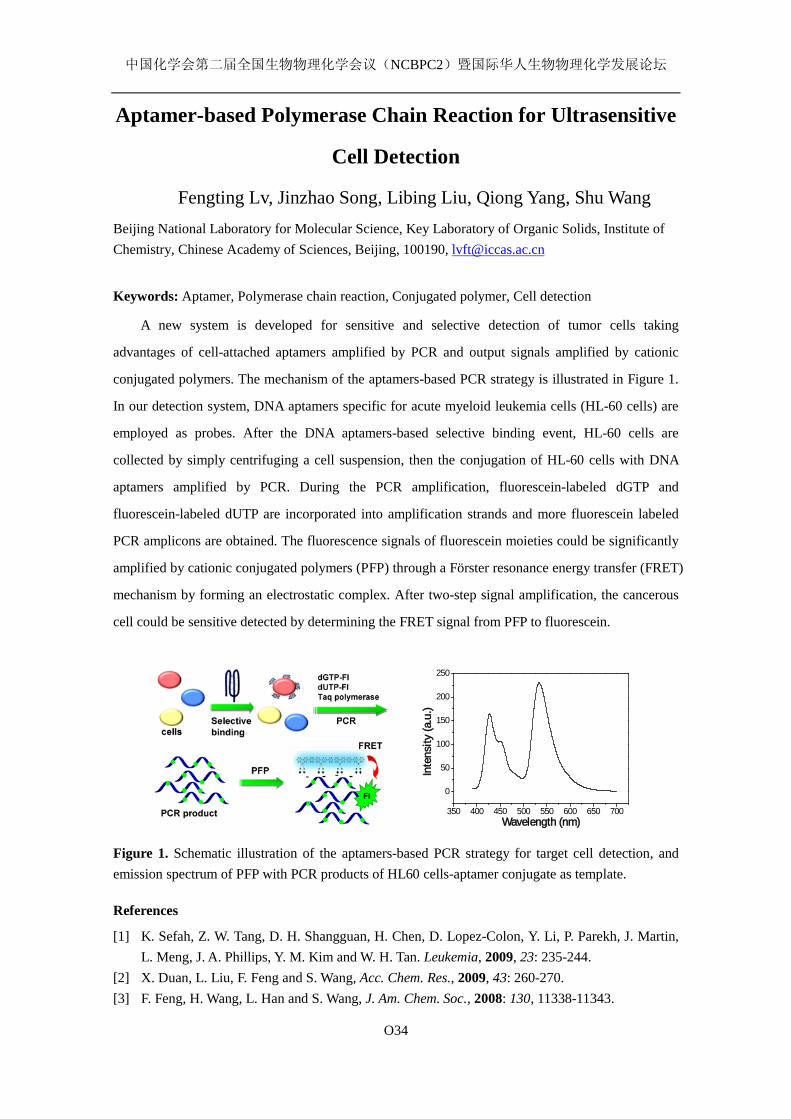
中国化学会第二届全国生物物理化学会议(NCBPC2)暨国际华人生物物理化学发展论坛
O34
Aptamer-based Polymerase Chain Reaction for Ultrasensitive
Cell Detection
Fengting Lv, Jinzhao Song, Libing Liu, Qiong Yang, Shu Wang Beijing National Laboratory for Molecular Science, Key Laboratory of Organic Solids, Institute of Chemistry, Chinese Academy of Sciences, Beijing, 100190, [email protected]
Keywords: Aptamer, Polymerase chain reaction, Conjugated polymer, Cell detection
A new system is developed for sensitive and selective detection of tumor cells taking
advantages of cell-attached aptamers amplified by PCR and output signals amplified by cationic
conjugated polymers. The mechanism of the aptamers-based PCR strategy is illustrated in Figure 1.
In our detection system, DNA aptamers specific for acute myeloid leukemia cells (HL-60 cells) are
employed as probes. After the DNA aptamers-based selective binding event, HL-60 cells are
collected by simply centrifuging a cell suspension, then the conjugation of HL-60 cells with DNA
aptamers amplified by PCR. During the PCR amplification, fluorescein-labeled dGTP and
fluorescein-labeled dUTP are incorporated into amplification strands and more fluorescein labeled
PCR amplicons are obtained. The fluorescence signals of fluorescein moieties could be significantly
amplified by cationic conjugated polymers (PFP) through a Förster resonance energy transfer (FRET)
mechanism by forming an electrostatic complex. After two-step signal amplification, the cancerous
cell could be sensitive detected by determining the FRET signal from PFP to fluorescein.
350 400 450 500 550 600 650 700
0
50
100
150
200
250
Inte
nsity
(a.u
.)
Wavelength (nm)
Figure 1. Schematic illustration of the aptamers-based PCR strategy for target cell detection, and emission spectrum of PFP with PCR products of HL60 cells-aptamer conjugate as template.
References
[1] K. Sefah, Z. W. Tang, D. H. Shangguan, H. Chen, D. Lopez-Colon, Y. Li, P. Parekh, J. Martin, L. Meng, J. A. Phillips, Y. M. Kim and W. H. Tan. Leukemia, 2009, 23: 235-244.
[2] X. Duan, L. Liu, F. Feng and S. Wang, Acc. Chem. Res., 2009, 43: 260-270. [3] F. Feng, H. Wang, L. Han and S. Wang, J. Am. Chem. Soc., 2008: 130, 11338-11343.
中国化学会第二届全国生物物理化学会议(NCBPC2)暨国际华人生物物理化学发展论坛
O35
Synthesis of nordihydroguaiaretic acid derivatives and their
bioactivity on K562 and S. pombe cell lines.
Qiangguo Li1, Xu Li1, Chuanhua Li1, Qingqi Chen2, Jilin Hu1, Jianhong
Jiang1, Huiwen Gu3
1.Hunan Provincial Key Laboratory of Xiangnan Rare-Precious Metals Compounds and Applications, Department of Chemistry and Life Science, Xiangnan University, Chenzhou 423000, Hunan Province, P.R. China. [email protected]; 2.MedKoo Biosciences, Inc. P O Pox 16222, Chapel Hill, NC 27516-6222, USA; 3. State Key Laboratory of Chemo/Biosensing & Chemometrics, College of Chemistry and Chemical Engineering, Hunan University, Changsha 410082, Hunan Province, P.R. China.
Nordihydroguaiaretic acid (NDGA) is a natural occurring lignan mainly isolated from creosote bush (Larrea divaricata Cav. Or Corillea tridentate. NDGA and its synthetic analogues are potentially useful in treating diseases related to cancers, diabetes, viral, bacterial infections, and inflammation. In this paper we report the optimized synthesis and the bioactivity of several NDGA derivative 3 and its cyclized analogue 4. The bioactivity on K563 and S. pombe cell lines was determined by microcalorimetry. The results showed that all the compounds terameprocol 2, NDGA derivative 3 and its cyclized analogue 4 possessed good inhibition on the growth metabolism of S. pombe and K562 cell lines. The values of IC50 of terameprocol 2, NDGA derivative 3 and NDGA derivative 4 on the growth metabolism of S. pombe cell lines being 3.32, 2.93 and 3.51 mg L−1, respectively. The values of IC50 of terameprocol 2, NDGA derivative 3 and NDGA derivative 4 on the growth metabolism of K562 cell lines being 5.87, 8.57 and 11.23 mg L−1, respectively. The inhibition ability of these compounds above on the growth of the S. pombe has been observed to decrease in the order NDGA derivative 3> terameprocol 2> NDGA derivative 4 and the K562 has been observed to decrease in the order terameprocol 2> NDGA derivative 3 > NDGA derivative 4. Key words: Nordihydroguaiaretic acid; NDGA derivative; microcalorimetry; anticancer; S. pombe; K562 Acknowledgment This research was financially supported by the National Natural Science Foundation of China (no. 20973145), the Hunan Provincial Key Laboratory Special Foundation (no. 2011TP4016-1) of China, and the Hunan Provincial Educational Ministry Foundation of China (no. 11A112). References [1] J.M. Lü, J. Nurko, S.M. Weakley, J. Jiang, P. Kougias, P.H. Lin, Q. Yao, C. Chen, Med Sci Monit. 2010, 16: 93-
100
[2] Q.Chen, Curr Top Med Chem. 2009, 9: 1636-1659.
[3] Q.G. Li, H. Zhang, X. Li, B. Wang, J.L. Hu, F.H. Yao, D.J. Yang, S.X. Xiao, L. J. Ye, Chin. J. Chem. 2011, 29:
2285-2292
中国化学会第二届全国生物物理化学会议(NCBPC2)暨国际华人生物物理化学发展论坛
O36
蛋白质(多肽)与生物仿生膜相互作用的盐离子催化效应及其
机理1
中国科学技术大学化学物理系 与 合肥微尺度国家实验室,合肥,230026,
魏锋, 叶树集*
蛋白质(多肽)与细胞膜的相互作用在细胞活动和生命过程中扮演着非常重要的角色。细胞
膜上的蛋白质 (多肽)结构在实现细胞生物功能例如离子传输、信息交换、基因调节、免疫应
答、细胞组装等过程中起着决定性作用。此外,人体内含有浓度约为 0.15M 的大量无机盐离
子。这些无机盐离子对蛋白质与细胞膜相互作用的影响密切关系到蛋白质在生物膜上的吸附、
稳定、折叠和凝聚途径。蛋白质与生物膜间的相互作用过程很复杂,其主要受蛋白质与生物
膜间的范德华、亲疏水和静电等作用力驱动。而无机盐离子种类和浓度则是影响这些驱动力
的重要参数。本研究以蜂毒素为模型,利用具有界面选择性的非线性和频光谱技术从分子水
平、原位实时地具体探究了无机盐离子环境下的蛋白质与细胞仿生膜的相互作用。结果表明
磷酸缓冲溶液对蜂毒素与磷脂双层膜的结合有很强的催化作用。同时,蜂毒素与带不同电荷
的的磷脂双层膜结合时的行为也各有不同,蜂毒素和负电荷膜,中性膜,正电荷膜的最佳结
合浓度分别为 2 mM ≥ cbuffer ≥ 0 mM,cbuffer≥20 mM 和 cbuffer ≥ 40 mM。
关键词: 蛋白质、细胞仿生膜、蜂毒素、盐离子催化效应、结合机理
Ion promoting the association between protein and modeling
cell membrane
Feng Wei, Shuji Ye* Hefei National Laboratory for Physical Sciences at Microscale and Department of Chemical Physics,
University of Science and Technology of China, Hefei 230026, China
The interactions between protein and cell membrane play a crucial role in many biological functions. Molecular insights into the ionic effects on such interactions will enrich current acknowledge of the driving forces and nature behind the interaction, and thus provide inportant clues to design or develop novelty biomedicine and drug delivery devices. In this research, we used mastoparans as modeling protein, and applied sum frequency generation vibrational spectroscopy to investigate the influence of ions on the interaction between protein and cell membrane. It is found that the phosphate buffer solution can strongly promote the mastroparan-membrane association. The ionic effects depend on the charge of lipid bilayer: for the negatively-charged, neutral, and positively-charged lipid bilayers, the ionic concentrations that have best promotion influence are 2 mM ≥ cbuffer ≥ 0 mM, cbuffer≥20 mM, and cbuffer≥40 mM, respectively.
Keywords: Protein, cell membrane, Mastoparan, Ion catalyzing effects, binding mechanism.
自然科学基金面上资助(21073175)
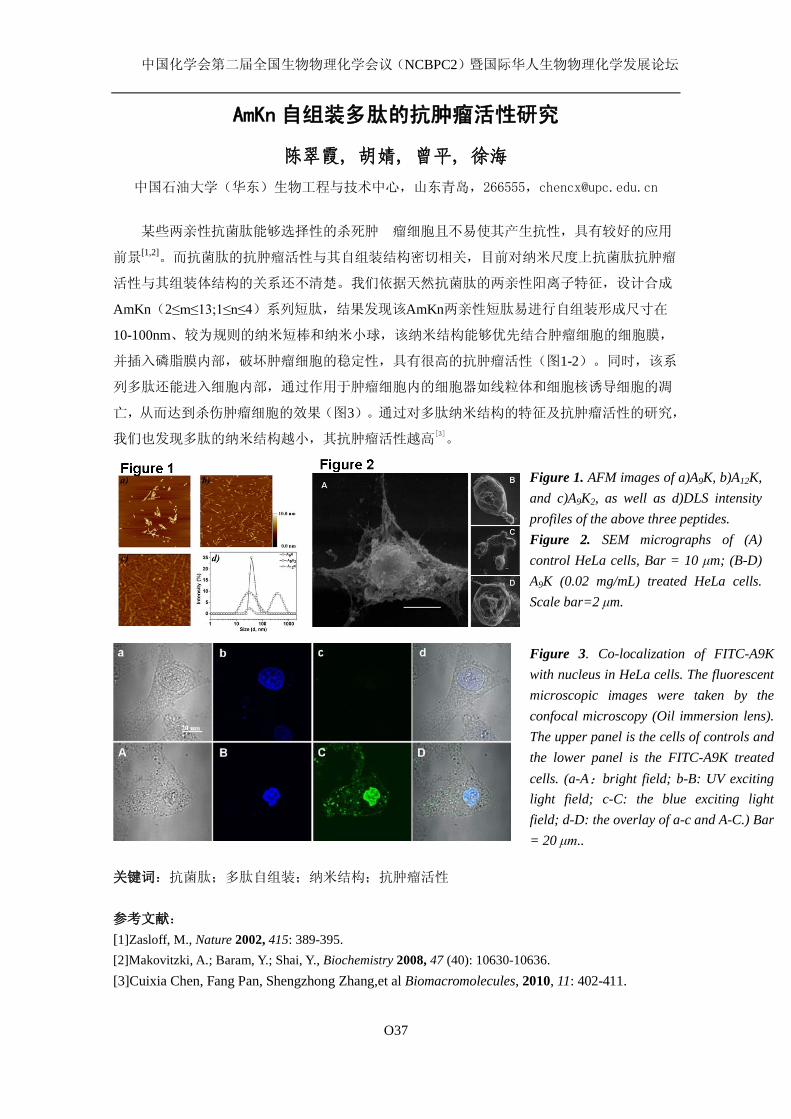
中国化学会第二届全国生物物理化学会议(NCBPC2)暨国际华人生物物理化学发展论坛
O37
AmKn 自组装多肽的抗肿瘤活性研究
陈翠霞, 胡婧, 曾平, 徐海
中国石油大学(华东)生物工程与技术中心,山东青岛,266555,[email protected]
某些两亲性抗菌肽能够选择性的杀死肿 瘤细胞且不易使其产生抗性,具有较好的应用
前景[1,2]。而抗菌肽的抗肿瘤活性与其自组装结构密切相关,目前对纳米尺度上抗菌肽抗肿瘤
活性与其组装体结构的关系还不清楚。我们依据天然抗菌肽的两亲性阳离子特征,设计合成
AmKn(2≤m≤13;1≤n≤4)系列短肽,结果发现该AmKn两亲性短肽易进行自组装形成尺寸在
10-100nm、较为规则的纳米短棒和纳米小球,该纳米结构能够优先结合肿瘤细胞的细胞膜,
并插入磷脂膜内部,破坏肿瘤细胞的稳定性,具有很高的抗肿瘤活性(图1-2)。同时,该系
列多肽还能进入细胞内部,通过作用于肿瘤细胞内的细胞器如线粒体和细胞核诱导细胞的凋
亡,从而达到杀伤肿瘤细胞的效果(图3)。通过对多肽纳米结构的特征及抗肿瘤活性的研究,
我们也发现多肽的纳米结构越小,其抗肿瘤活性越高[3]。
关键词:抗菌肽;多肽自组装;纳米结构;抗肿瘤活性 参考文献: [1]Zasloff, M., Nature 2002, 415: 389-395. [2]Makovitzki, A.; Baram, Y.; Shai, Y., Biochemistry 2008, 47 (40): 10630-10636. [3]Cuixia Chen, Fang Pan, Shengzhong Zhang,et al Biomacromolecules, 2010, 11: 402-411.
Figure 1. AFM images of a)A9K, b)A12K, and c)A9K2, as well as d)DLS intensity profiles of the above three peptides. Figure 2. SEM micrographs of (A) control HeLa cells, Bar = 10 μm; (B-D) A9K (0.02 mg/mL) treated HeLa cells. Scale bar=2 μm.
Figure 3. Co-localization of FITC-A9K with nucleus in HeLa cells. The fluorescent microscopic images were taken by the confocal microscopy (Oil immersion lens). The upper panel is the cells of controls and the lower panel is the FITC-A9K treated cells. (a-A:bright field; b-B: UV exciting light field; c-C: the blue exciting light field; d-D: the overlay of a-c and A-C.) Bar = 20 μm..
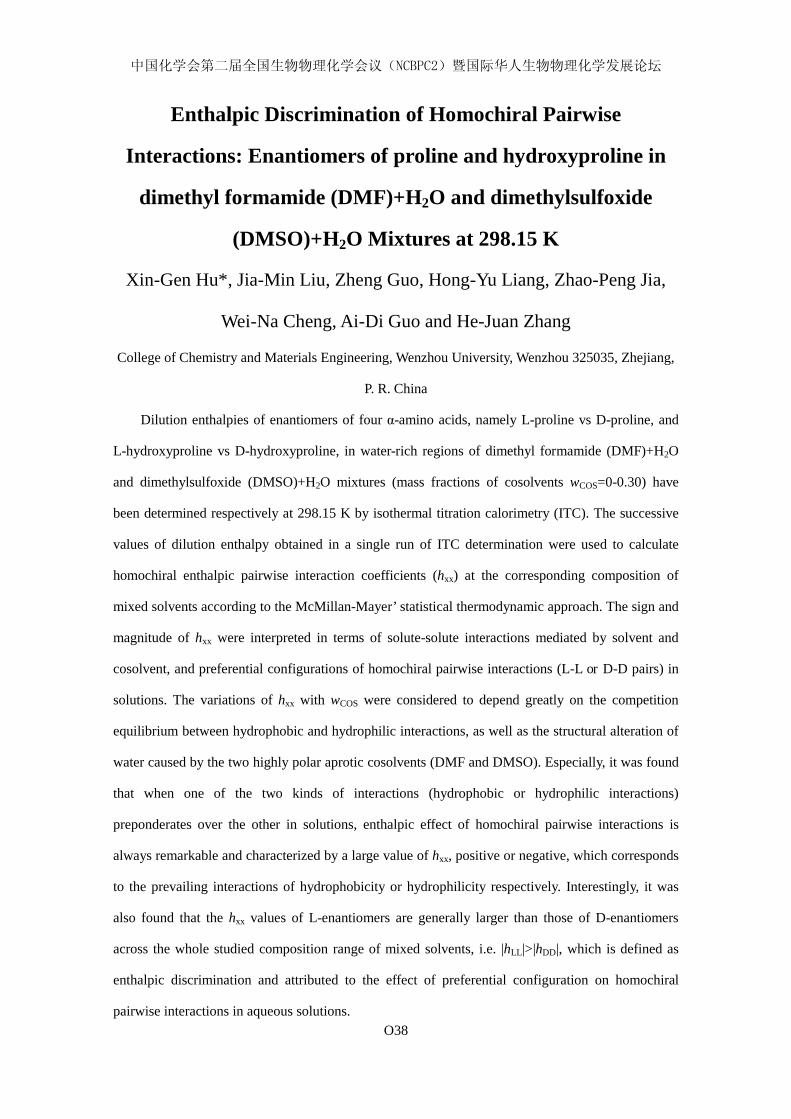
中国化学会第二届全国生物物理化学会议(NCBPC2)暨国际华人生物物理化学发展论坛
O38
Enthalpic Discrimination of Homochiral Pairwise
Interactions: Enantiomers of proline and hydroxyproline in
dimethyl formamide (DMF)+H2O and dimethylsulfoxide
(DMSO)+H2O Mixtures at 298.15 K
Xin-Gen Hu*, Jia-Min Liu, Zheng Guo, Hong-Yu Liang, Zhao-Peng Jia,
Wei-Na Cheng, Ai-Di Guo and He-Juan Zhang
College of Chemistry and Materials Engineering, Wenzhou University, Wenzhou 325035, Zhejiang,
P. R. China
Dilution enthalpies of enantiomers of four α-amino acids, namely L-proline vs D-proline, and
L-hydroxyproline vs D-hydroxyproline, in water-rich regions of dimethyl formamide (DMF)+H2O
and dimethylsulfoxide (DMSO)+H2O mixtures (mass fractions of cosolvents wCOS=0-0.30) have
been determined respectively at 298.15 K by isothermal titration calorimetry (ITC). The successive
values of dilution enthalpy obtained in a single run of ITC determination were used to calculate
homochiral enthalpic pairwise interaction coefficients (hxx) at the corresponding composition of
mixed solvents according to the McMillan-Mayer’ statistical thermodynamic approach. The sign and
magnitude of hxx were interpreted in terms of solute-solute interactions mediated by solvent and
cosolvent, and preferential configurations of homochiral pairwise interactions (L-L or D-D pairs) in
solutions. The variations of hxx with wCOS were considered to depend greatly on the competition
equilibrium between hydrophobic and hydrophilic interactions, as well as the structural alteration of
water caused by the two highly polar aprotic cosolvents (DMF and DMSO). Especially, it was found
that when one of the two kinds of interactions (hydrophobic or hydrophilic interactions)
preponderates over the other in solutions, enthalpic effect of homochiral pairwise interactions is
always remarkable and characterized by a large value of hxx, positive or negative, which corresponds
to the prevailing interactions of hydrophobicity or hydrophilicity respectively. Interestingly, it was
also found that the hxx values of L-enantiomers are generally larger than those of D-enantiomers
across the whole studied composition range of mixed solvents, i.e. |hLL|>|hDD|, which is defined as
enthalpic discrimination and attributed to the effect of preferential configuration on homochiral
pairwise interactions in aqueous solutions.
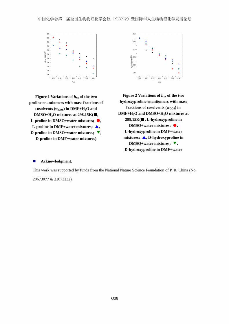
中国化学会第二届全国生物物理化学会议(NCBPC2)暨国际华人生物物理化学发展论坛
O38
0.00 0.05 0.10 0.15 0.20 0.25 0.30
100
120
140
160
180
200
220
240
260
280
300
h xx/J⋅k
g⋅m
ol-2
wCOS
0.00 0.05 0.10 0.15 0.20 0.25 0.30
-350
-300
-250
-200
-150
-100
h xx/J⋅k
g⋅m
ol-2
wCOS
C1
Acknowledgment.
This work was supported by funds from the National Nature Science Foundation of P. R. China (No.
20673077 & 21073132).
Figure 1 Variations of hxx of the two proline enantiomers with mass fractions of
cosolvents (wCOS) in DMF+H2O and DMSO+H2O mixtures at 298.15K(■,
L-proline in DMSO+water mixtures; ●, L-proline in DMF+water mixtures; ▲,
D-proline in DMSO+water mixtures; ▼, D-proline in DMF+water mixtures)
Figure 2 Variations of hxx of the two hydroxyproline enantiomers with mass
fractions of cosolvents (wCOS) in DMF+H2O and DMSO+H2O mixtures at
298.15K(■, L-hydroxyproline in DMSO+water mixtures; ●,
L-hydroxyproline in DMF+water mixtures; ▲, D-hydroxyproline in
DMSO+water mixtures; ▼, D-hydroxyproline in DMF+water
i )
中国化学会第二届全国生物物理化学会议(NCBPC2)暨国际华人生物物理化学发展论坛
O39
Ultrafast Dynamics of Excitation Energy Transfer in LH2
complex from Thermochromatium tepidum
Peng Wang, Jian-Ping Zhang
Department of Chemistry, Renmin University of China,
Beijing 100872, P. R. China,[email protected]
Key words: Ultrafast dynamics, purple photosynthetic bacteria, light-harvesting complex 2,
time-resolved spectroscopy.
Purple photosynthetic bacterium Thermochromatium (Tch.) tepidum is a moderate thermophile
growing in an optimal temperature range of 48-50 ºC. Its light-harvesting complex 2 (LH2) possesses heterogeneous compositions of apoprotein and carotenoid (Car), but the high-resolution crystallographic structure remains unknown. We have attempted an ultrafast time-resolved spectroscopic study of the isolated LH2 and complex from Tch. tepidum. The spectral dynamics and population kinetics of the DDM and the LDAO preparations of LH2 reveal efficient S2-state mediated Car-to-Car and Car-to-bacteriochlorophyll (BChl) singlet excitation energy transfer (EET) occurring in a time scale of 100 fs, as well as the Qy-state mediated B800-to-B850 singlet EET for the DDM preparation proceeding with a time constant of 1.2 ps. The relative spacial arrangement and the functions of carotenoids with various conjugation lengths were discussed based on the detailed analysis. We also carried out femtosecond magic-angle and polarized pump-probe spectroscopies for the light-harvesting complex 2 (LH2) from the same species in aqueous phase and in chromatophores. To examine the effects of LH2 aggregation on the dynamics of excitation energy transfer, dominant monodispersed and aggregated LH2s were prepared by controlling the surfactant concentrations. The B800-to-B850 intra-LH2 energy transfer time was determined to be 1.3 ps for isolated LH2, which, upon aggregation in aqueous phase or clustering in chromatophores, shortened to 1.1 or 0.9 ps, respectively. The inter-complexes EET dynamics were studied and discussed. These results are helpful in understanding the light-harvesting function of a bacterial photosynthetic membrane incorporating different types of antenna complexes.
Refs. [1] Yang Fan, et al., Acta Phys. -Chim. Sin., 2010, 26: 2021-2030 [2] Yang Fan, et al., J. Phys. Chem. B 2011, 115: 7906–7913

墙报
Poster Presentations

中国化学会第二届全国生物物理化学会议(NCBPC2)暨国际华人生物物理化学发展论坛
P1
具有热-磁控制释放药物特性的磁性脂质体
裘丹,安学勤*
华东理工大学化学与分子工程学院,上海,200237,[email protected]
关键词:温控制释放,磁控制释放,脂质体,超顺磁性,可控合成
近年来,载药体系在时间和空间上实现触发控制释放是目前亟待解决的重要问题。利用
在环境的介质条件的变化(如:温度,pH值,超声,紫外可见等)可实现非侵入性的外部控
制释放。热刺激药物释放特别感兴趣的是因为可以在不伤害细胞和组织的情况下,通过体外
温度的改变实现药物释放。传统的热敏脂质体通过加热体系的温度达到相变温度(Tm)以上
使脂质体膜相变导致药物释放。本论文采用超临界CO2方法制备了一种新型的自组装磁性脂质
体,该脂质体具有超顺磁性氧化铁纳米粒子嵌入在膜中,模型药物被包封于水核中的微观结
构。通过温度和交流电的电磁场(AMF)的触发实现模型药物从脂质体中释放(Fig.1)。采
用荧光探针法从微观结构变化的角度解释了的控制释放的机理。通过打开和关闭的AMF可以引
发的脂质体反复释放。磁控制释放的研究结果表明,在该脂质体进行的控制释放是由于磁热
效应导致脂质体相变和magnetic-impelled motions改变了双分子层的渗透,而不是脂质体结构的
破坏导致药物释放。
Fig.1 A Controllable release from thermo-sensitive magnetoliposomes by magnetic-stimulated and
thermo-stimulated
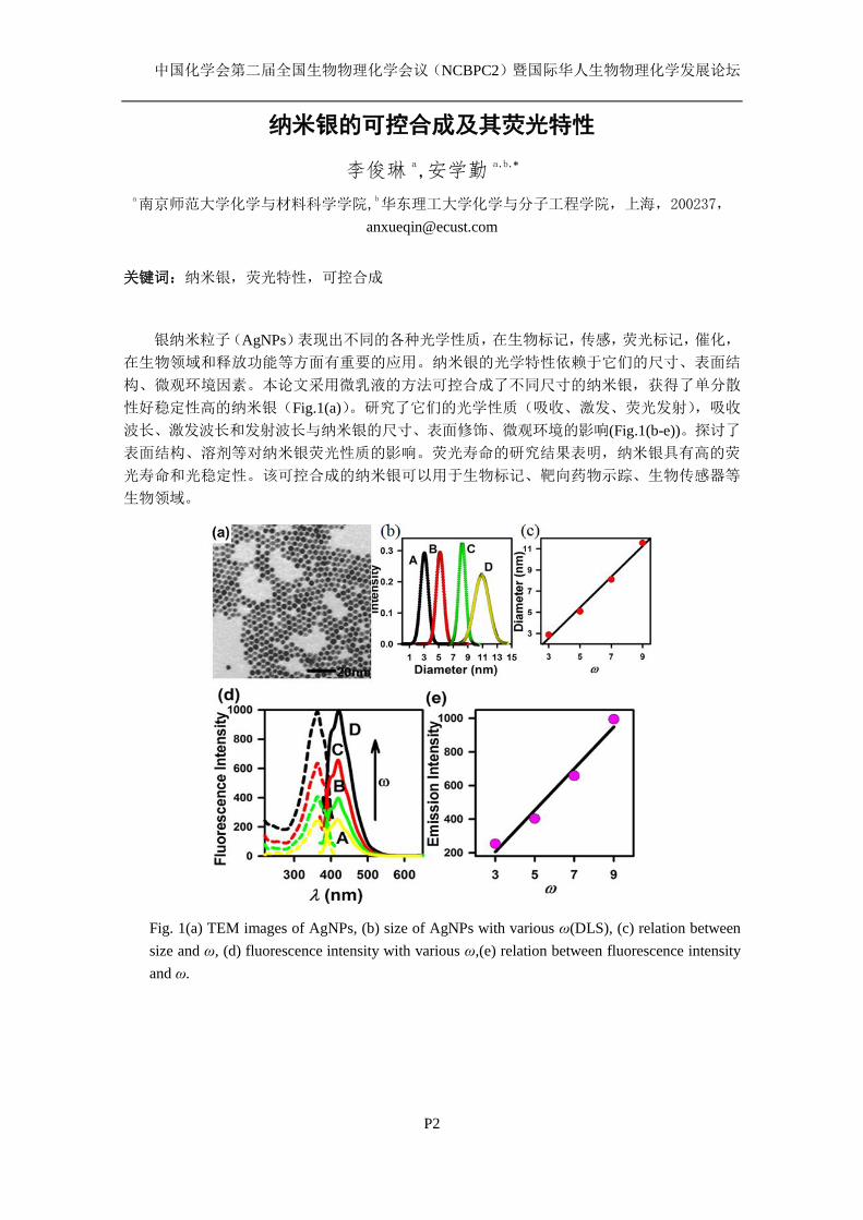
中国化学会第二届全国生物物理化学会议(NCBPC2)暨国际华人生物物理化学发展论坛
P2
纳米银的可控合成及其荧光特性
李俊琳 a,安学勤 a,b,*
a南京师范大学化学与材料科学学院,
b华东理工大学化学与分子工程学院,上海,200237,
关键词:纳米银,荧光特性,可控合成
银纳米粒子(AgNPs)表现出不同的各种光学性质,在生物标记,传感,荧光标记,催化,
在生物领域和释放功能等方面有重要的应用。纳米银的光学特性依赖于它们的尺寸、表面结
构、微观环境因素。本论文采用微乳液的方法可控合成了不同尺寸的纳米银,获得了单分散
性好稳定性高的纳米银(Fig.1(a))。研究了它们的光学性质(吸收、激发、荧光发射),吸收
波长、激发波长和发射波长与纳米银的尺寸、表面修饰、微观环境的影响(Fig.1(b-e))。探讨了
表面结构、溶剂等对纳米银荧光性质的影响。荧光寿命的研究结果表明,纳米银具有高的荧
光寿命和光稳定性。该可控合成的纳米银可以用于生物标记、靶向药物示踪、生物传感器等
生物领域。
Fig. 1(a) TEM images of AgNPs, (b) size of AgNPs with various ω(DLS), (c) relation between
size and ω, (d) fluorescence intensity with various ω,(e) relation between fluorescence intensity
and ω.
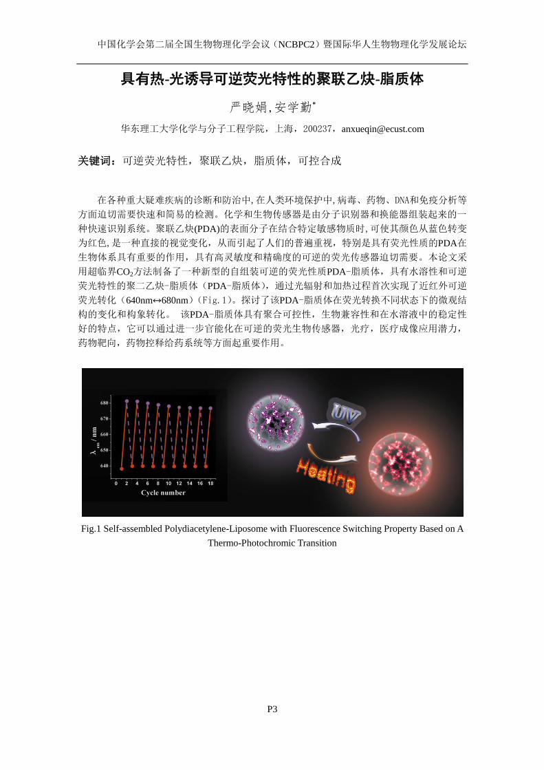
中国化学会第二届全国生物物理化学会议(NCBPC2)暨国际华人生物物理化学发展论坛
P3
具有热-光诱导可逆荧光特性的聚联乙炔-脂质体
严晓娟,安学勤*
华东理工大学化学与分子工程学院,上海,200237,[email protected]
关键词:可逆荧光特性,聚联乙炔,脂质体,可控合成
在各种重大疑难疾病的诊断和防治中,在人类环境保护中,病毒、药物、DNA和免疫分析等
方面迫切需要快速和简易的检测。化学和生物传感器是由分子识别器和换能器组装起来的一
种快速识别系统。聚联乙炔(PDA)的表面分子在结合特定敏感物质时,可使其颜色从蓝色转变
为红色,是一种直接的视觉变化,从而引起了人们的普遍重视,特别是具有荧光性质的PDA在
生物体系具有重要的作用,具有高灵敏度和精确度的可逆的荧光传感器迫切需要。本论文采
用超临界CO2方法制备了一种新型的自组装可逆的荧光性质PDA-脂质体,具有水溶性和可逆
荧光特性的聚二乙炔-脂质体(PDA-脂质体),通过光辐射和加热过程首次实现了近红外可逆
荧光转化(640nm↔680nm)(Fig.1)。探讨了该PDA-脂质体在荧光转换不同状态下的微观结
构的变化和构象转化。 该PDA-脂质体具有聚合可控性,生物兼容性和在水溶液中的稳定性
好的特点,它可以通过进一步官能化在可逆的荧光生物传感器,光疗,医疗成像应用潜力,
药物靶向,药物控释给药系统等方面起重要作用。
Fig.1 Self-assembled Polydiacetylene-Liposome with Fluorescence Switching Property Based on A
Thermo-Photochromic Transition
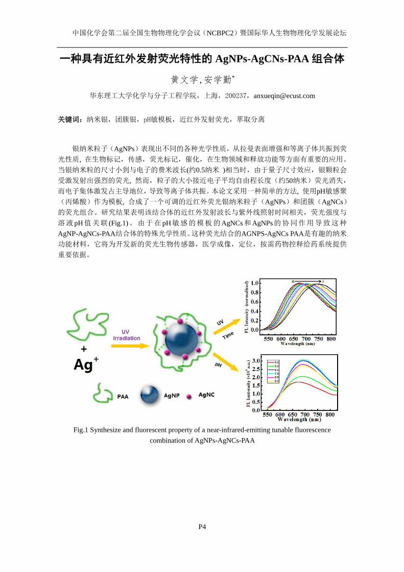
中国化学会第二届全国生物物理化学会议(NCBPC2)暨国际华人生物物理化学发展论坛
P4
一种具有近红外发射荧光特性的 AgNPs-AgCNs-PAA 组合体
黄文学,安学勤*
华东理工大学化学与分子工程学院,上海,200237,[email protected]
关键词:纳米银,团簇银,pH敏模板,近红外发射荧光,萃取分离
银纳米粒子(AgNPs)表现出不同的各种光学性质,从拉曼表面增强和等离子体共振到荧
光性质, 在生物标记,传感,荧光标记,催化,在生物领域和释放功能等方面有重要的应用。
当银纳米粒的尺寸小到与电子的费米波长(约0.5纳米 )相当时,由于量子尺寸效应,银颗粒会
受激发射出强烈的荧光, 然而,粒子的大小接近电子平均自由程长度(约50纳米)荧光消失,
而电子集体激发占主导地位,导致等离子体共振。本论文采用一种简单的方法, 使用pH敏感聚
(丙烯酸)作为模板, 合成了一个可调的近红外荧光银纳米粒子(AgNPs)和团簇(AgNCs)
的荧光组合。研究结果表明该结合体的近红外发射波长与紫外线照射时间相关,荧光强度与
溶液pH值关联 (Fig.1)。由于在pH敏感的模板的AgNCs和AgNPs的协同作用导致这种
AgNP-AgNCs-PAA结合体的特殊光学性质。这种荧光结合的AGNPS-AgNCs PAA是有趣的纳米
功能材料,它将为开发新的荧光生物传感器,医学成像,定位,按需药物控释给药系统提供
重要依据。
Fig.1 Synthesize and fluorescent property of a near-infrared-emitting tunable fluorescence
combination of AgNPs-AgNCs-PAA

中国化学会第二届全国生物物理化学会议(NCBPC2)暨国际华人生物物理化学发展论坛
P5
一种同时具有光学和磁学性质的金铁纳米合金
王冬青 a,安学勤 a,b,*
华东理工大学化学与分子工程学院,上海,200237,[email protected]
关键词:金铁纳米合金,相图,热力学平衡,物理化学性质
肿瘤热疗早已成为综合治疗癌症的一种治疗手段,但是传统的热疗效果往往不能达到满
意的效果,而且会损伤正常的组织细胞。而超顺磁性纳米粒子的出现使传统的热疗有了更深
层次的意义。近年来,肿瘤的磁靶向热疗发展成为肿瘤热疗的一种新方法。即先将磁性介质
定位于肿瘤组织,然后在外部交变磁场下,磁性介质使肿瘤部位快速形成靶向高温区,进而
高温消灭肿瘤细胞。超顺磁性纳米粒子磁热转换原理是:在交变磁场作用下,超顺磁性纳米
粒子能通过磁滞损耗来吸收大量磁场能量进而产生热量将肿瘤组织加热到有效治疗温度,从
而杀灭癌细胞以达到治疗恶性肿瘤的目的。超顺磁性纳米粒子的靶向性,使其只在肿瘤部位
产生热量,而不损伤正常细胞。国内外在肿瘤的磁靶向热疗方面已经取得了一定的成果,但
合适的交变磁场的选择等,仍是有待解决的难题。科研工作者们一直在致力研究这样一种同
时具有磁化强度和光学性质(近红外吸收),粒径小而均一,且性质比较稳定,无毒,具有良
好的生物相容性的纳米材料。具有这种特性的纳米粒子可以同时起到靶向定位,成像和靶向
治疗等多功能的作用,这将在减少正常细胞和组织的损伤的基础上大大提高疗效,从而为纳
米医学领域提供更大的发展平台。本论文采用反相微乳法合成了同时具有光学和磁学性质的
FeAu合金纳米粒子,并用十二硫醇对表面进行了修饰。通过X射线粉末衍射仪(XRD),X射线
能谱仪(EDS),透射电子显微镜(TEM),动态光散射仪(DLS),超导量子干涉仪(SQUID),
荧光光谱仪,紫外一可见分光光度计对其性能进行表征。结果表明:用十二硫醇修饰的金铁
纳米合金形貌呈球形、单分散性好、稳定性高。该金-铁纳米合金不但具有超顺磁性和可控的
高的饱和磁场强度,而且具有可控的吸收波长和荧光发射波长(Fig.1)。
Fig. 1 A novel Fe-Au nano-alloy with coupling optical and magnetic properties
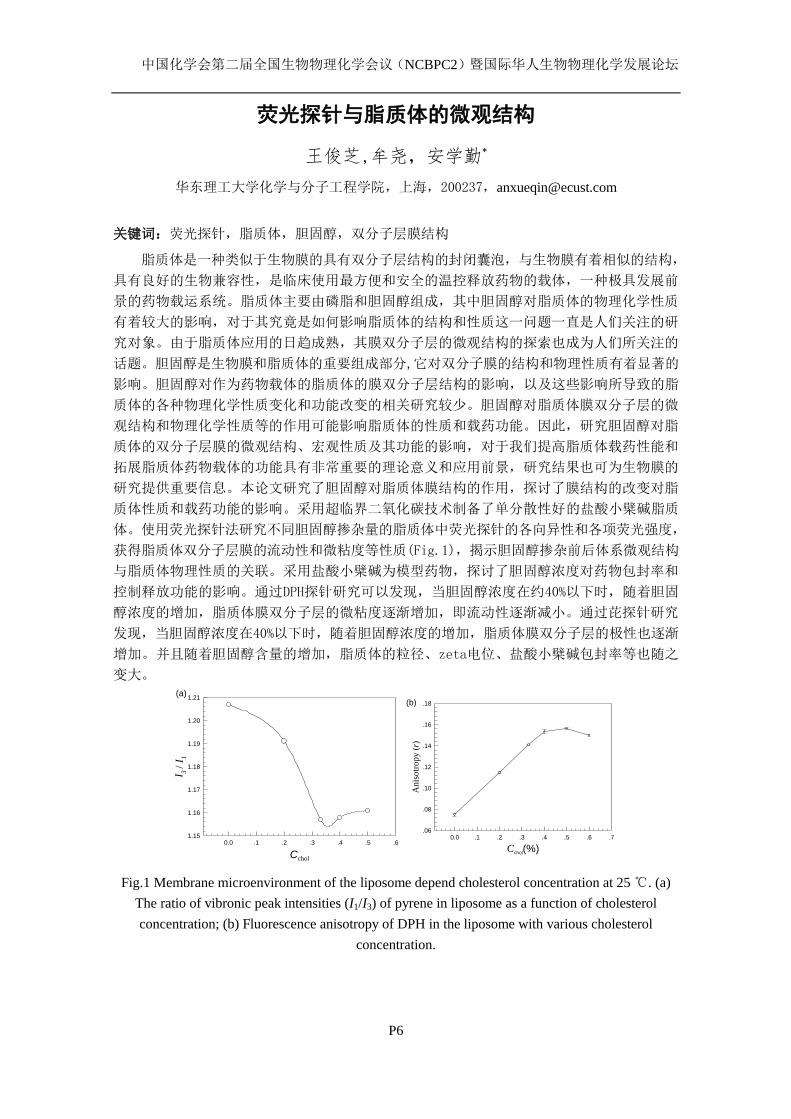
中国化学会第二届全国生物物理化学会议(NCBPC2)暨国际华人生物物理化学发展论坛
P6
荧光探针与脂质体的微观结构
王俊芝,牟尧,安学勤*
华东理工大学化学与分子工程学院,上海,200237,[email protected]
关键词:荧光探针,脂质体,胆固醇,双分子层膜结构
脂质体是一种类似于生物膜的具有双分子层结构的封闭囊泡,与生物膜有着相似的结构,
具有良好的生物兼容性,是临床使用最方便和安全的温控释放药物的载体,一种极具发展前
景的药物载运系统。脂质体主要由磷脂和胆固醇组成,其中胆固醇对脂质体的物理化学性质
有着较大的影响,对于其究竟是如何影响脂质体的结构和性质这一问题一直是人们关注的研
究对象。由于脂质体应用的日趋成熟,其膜双分子层的微观结构的探索也成为人们所关注的
话题。胆固醇是生物膜和脂质体的重要组成部分,它对双分子膜的结构和物理性质有着显著的
影响。胆固醇对作为药物载体的脂质体的膜双分子层结构的影响,以及这些影响所导致的脂
质体的各种物理化学性质变化和功能改变的相关研究较少。胆固醇对脂质体膜双分子层的微
观结构和物理化学性质等的作用可能影响脂质体的性质和载药功能。因此,研究胆固醇对脂
质体的双分子层膜的微观结构、宏观性质及其功能的影响,对于我们提高脂质体载药性能和
拓展脂质体药物载体的功能具有非常重要的理论意义和应用前景,研究结果也可为生物膜的
研究提供重要信息。本论文研究了胆固醇对脂质体膜结构的作用,探讨了膜结构的改变对脂
质体性质和载药功能的影响。采用超临界二氧化碳技术制备了单分散性好的盐酸小檗碱脂质
体。使用荧光探针法研究不同胆固醇掺杂量的脂质体中荧光探针的各向异性和各项荧光强度,
获得脂质体双分子层膜的流动性和微粘度等性质(Fig.1),揭示胆固醇掺杂前后体系微观结构
与脂质体物理性质的关联。采用盐酸小檗碱为模型药物,探讨了胆固醇浓度对药物包封率和
控制释放功能的影响。通过DPH探针研究可以发现,当胆固醇浓度在约40%以下时,随着胆固
醇浓度的增加,脂质体膜双分子层的微粘度逐渐增加,即流动性逐渐减小。通过芘探针研究
发现,当胆固醇浓度在40%以下时,随着胆固醇浓度的增加,脂质体膜双分子层的极性也逐渐
增加。并且随着胆固醇含量的增加,脂质体的粒径、zeta电位、盐酸小檗碱包封率等也随之
变大。
Cchol
0.0 .1 .2 .3 .4 .5 .6
I 3 /
I 1
1.15
1.16
1.17
1.18
1.19
1.20
1.21(a)
Cchol(%)0.0 .1 .2 .3 .4 .5 .6 .7
An
isotr
op
y (
r)
.06
.08
.10
.12
.14
.16
.18(b)
Fig.1 Membrane microenvironment of the liposome depend cholesterol concentration at 25 ℃. (a)
The ratio of vibronic peak intensities (I1/I3) of pyrene in liposome as a function of cholesterol
concentration; (b) Fluorescence anisotropy of DPH in the liposome with various cholesterol
concentration.
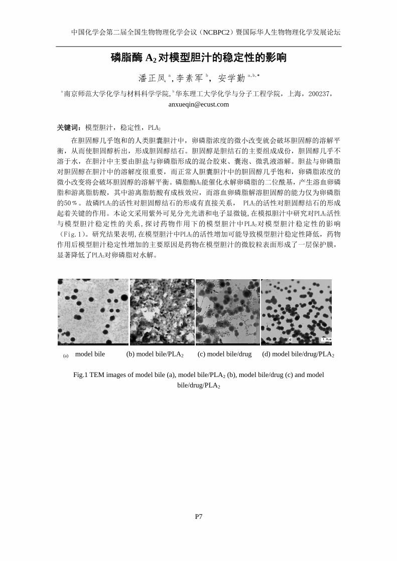
中国化学会第二届全国生物物理化学会议(NCBPC2)暨国际华人生物物理化学发展论坛
P7
磷脂酶 A2对模型胆汁的稳定性的影响
潘正凤 a,李素军 b,安学勤 a,b,*
a南京师范大学化学与材料科学学院,
b华东理工大学化学与分子工程学院,上海,200237,
关键词:模型胆汁,稳定性,PLA2
在胆固醇几乎饱和的人类胆囊胆汁中,卵磷脂浓度的微小改变就会破坏胆固醇的溶解平
衡,从而使胆固醇析出,形成胆固醇结石。胆固醇是胆结石的主要组成成份,胆固醇几乎不
溶于水,在胆汁中主要由胆盐与卵磷脂形成的混合胶束、囊泡、微乳液溶解。胆盐与卵磷脂
对胆固醇在胆汁中的溶解度很重要,而正常人胆囊胆汁中的胆固醇几乎饱和,卵磷脂浓度的
微小改变将会破坏胆固醇的溶解平衡。磷脂酶A2能催化水解卵磷脂的二位酰基,产生溶血卵磷
脂和游离脂肪酸,其中游离脂肪酸有成核效应,而溶血卵磷脂解溶胆固醇的能力仅为卵磷脂
的50﹪。故磷PLA2的活性对胆固醇结石的形成有直接关系, PLA2的活性对胆固醇结石的形成
起着关键的作用。本论文采用紫外可见分光光谱和电子显微镜,在模拟胆汁中研究对PLA2活性
与模型胆汁稳定性的关系,探讨药物作用下的模型胆汁中PLA2对模型胆汁稳定性的影响
(Fig.1)。研究结果表明,在模型胆汁中PLA2的活性增加可能导致模型胆汁稳定性降低,药物
作用后模型胆汁稳定性增加的主要原因是药物在模型胆汁的微胶粒表面形成了一层保护膜,
显著降低了PLA2对卵磷脂对水解。
(a) model bile (b) model bile/PLA2 (c) model bile/drug (d) model bile/drug/PLA2
Fig.1 TEM images of model bile (a), model bile/PLA2 (b), model bile/drug (c) and model
bile/drug/PLA2

中国化学会第二届全国生物物理化学会议(NCBPC2)暨国际华人生物物理化学发展论坛
P8
Enthalpic pairwise interactions of enantiomers of
penicillamines in dimethyl sulfoxide + water mixtures at
298.15K
Weina Cheng Zhaopeng Jia Shan Liu Jiamin Liu Pingliang Chen
Xingen Hu
College of Chemistry and Materials Engineering, Wenzhou University, Zhejiang Wenzhou
325035,[email protected]
The dilution enthalpies of isomers of penicillamines, namely D-penicillamine, L-penicillamine
in Dimethyl sulfoxide (DMSO) + water mixtures have been determined respectively, using
isothermal titration calorimeter (ITC) at 298.15 K. According to the McMillan-Mayer theory, the
homochiral enthalpic pairwise interaction coefficients (XX
h ) of the penicillamines in the DMSO+
water mixtures of various mass fractions (wDMSO= 0 to 0.3) have been calculated. The results are
discussed from the point of view of solute-solute interaction and solute-solvent interaction. That is to
say, the variations of XX
h with wDMSO for the enantiomers depend largely on the competition
equilibrium between hydrophobic-hydrophobic, hydrophilic-hydrophilic and
hydrophobic-hydrophilic interactions in DMSO + water mixtures.
KEYWORDS Isothermal titration calorimetry (ITC), enthalpic pairwise interaction coefficient,
penicillamines, DMSO+ water mixtures
(■, D-penicillamine ; ●, L-penicillamine)
References
1. McMillan, W. G.; Mayer, J. E. The Statistical Thermodynamics of Multicomponent Systems. J. Chem. Phys.
1945, 13: 276-305.
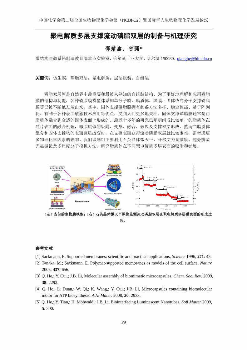
中国化学会第二届全国生物物理化学会议(NCBPC2)暨国际华人生物物理化学发展论坛
P9
聚电解质多层支撑流动磷脂双层的制备与机理研究
邵婧鑫,贺强*
微结构与微系统制造教育部重点实验室,哈尔滨工业大学,哈尔滨 150080,[email protected]
关键词:仿生膜;磷脂双层;聚电解质;层层组装;自组装
磷脂双层膜是自然界中最重要和最被人熟知的自组装结构。为了更好地理解和应用磷脂
膜的结构与功能,各种磷脂膜模型体系如单分子膜、脂质体、黑膜、固体或高分子支撑磷脂
膜等已被不断地发展出来。其中,固体支撑磷脂膜拥有制备方法多样、稳定性高、易于阵列
化、有利于各种表面敏感技术应用等优点,受到人们更多地关注。固体支撑磷脂膜通常是由
脂质体融合到合适的固体表面上形成的,最近十多年的研究已阐明组成比较单一的脂质体在
硅片表面的融合机理,即脂质体的吸附、变形、融合、破裂及支撑双层形成。然而当脂质体
组分和固体支撑物的表面性质改变时,在支撑表面获得流动磷脂双层就比较困难,需考虑更
多物理化学因素的影响。我们课题组主要利用石英晶体微天平、开尔文力显微镜、超分辨荧
光显微镜及多尺度分子模拟方法,研究脂质体在不同聚电解质多层表面的吸附和铺展。
(左)当前的生物膜模型;(右)石英晶体微天平原位监测流动磷脂双层在聚电解质多层膜表面的形成过
程。
参考文献
[1] Sackmann, E. Supported membranes: scientific and practical applications, Science 1996, 271: 43.
[2] Tanaka, M.; Sackmann, E. Polymer-supported membranes as models of the cell surface, Nature
2005, 437: 656.
[3] Q. He,; Y. Cui,; J.B. Li, Molecular assembly of biomimetic microcapsules, Chem. Soc. Rev. 2009,
38: 2292.
[4] Q. He,; L. Duan,; W. Qi,; K. Wang,; Y. Cui,; J.B. Li, Microcapsules containing biomolecular
motor for ATP biosynthesis, Adv. Mater. 2008, 20: 2933.
[5] Q. He,; Y. Tian,; H. Möhwald,; J.B. Li, Biointerfacing Luminescent Nanotubes, Soft Matter 2009,
5: 300.
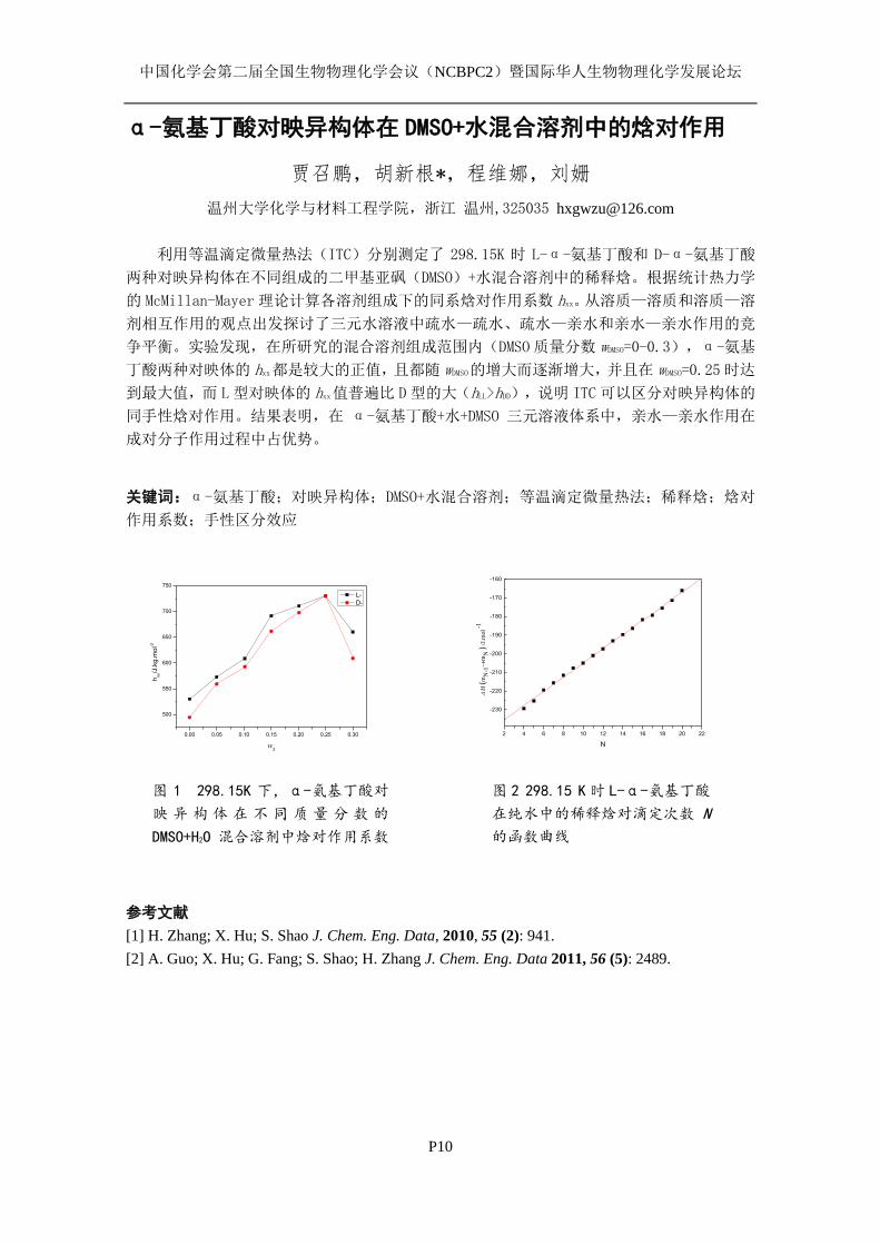
中国化学会第二届全国生物物理化学会议(NCBPC2)暨国际华人生物物理化学发展论坛
P10
α-氨基丁酸对映异构体在 DMSO+水混合溶剂中的焓对作用
贾召鹏,胡新根*,程维娜,刘姗
温州大学化学与材料工程学院,浙江 温州,325035 [email protected]
利用等温滴定微量热法(ITC)分别测定了 298.15K 时 L-α-氨基丁酸和 D-α-氨基丁酸
两种对映异构体在不同组成的二甲基亚砜(DMSO)+水混合溶剂中的稀释焓。根据统计热力学
的 McMillan-Mayer理论计算各溶剂组成下的同系焓对作用系数 hxx。从溶质—溶质和溶质—溶
剂相互作用的观点出发探讨了三元水溶液中疏水—疏水、疏水—亲水和亲水—亲水作用的竞
争平衡。实验发现,在所研究的混合溶剂组成范围内(DMSO质量分数 wDMSO=0-0.3),α-氨基
丁酸两种对映体的 hxx都是较大的正值,且都随 wDMSO的增大而逐渐增大,并且在 wDMSO=0.25时达
到最大值,而 L型对映体的 hxx值普遍比 D型的大(hLL>hDD),说明 ITC可以区分对映异构体的
同手性焓对作用。结果表明,在 α-氨基丁酸+水+DMSO 三元溶液体系中,亲水—亲水作用在
成对分子作用过程中占优势。
关键词:α-氨基丁酸;对映异构体;DMSO+水混合溶剂;等温滴定微量热法;稀释焓;焓对
作用系数;手性区分效应
0.00 0.05 0.10 0.15 0.20 0.25 0.30
500
550
600
650
700
750
hxx/J
.kg
.mo
l-2
w2
L-
D-
2 4 6 8 10 12 14 16 18 20 22
-230
-220
-210
-200
-190
-180
-170
-160
H
(m
N-1
mN
) /
J.m
ol
-1
N
参考文献
[1] H. Zhang; X. Hu; S. Shao J. Chem. Eng. Data, 2010, 55 (2): 941.
[2] A. Guo; X. Hu; G. Fang; S. Shao; H. Zhang J. Chem. Eng. Data 2011, 56 (5): 2489.
图 1 298.15K 下,α-氨基丁酸对
映 异 构 体 在 不 同 质 量 分 数 的
DMSO+H2O 混合溶剂中焓对作用系数
比较
图 2 298.15 K时 L-α-氨基丁酸
在纯水中的稀释焓对滴定次数 N
的函数曲线
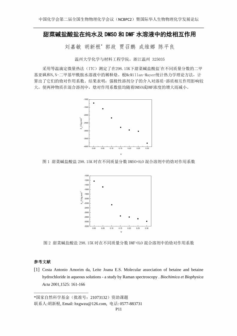
中国化学会第二届全国生物物理化学会议(NCBPC2)暨国际华人生物物理化学发展论坛
P11
甜菜碱盐酸盐在纯水及 DMSO 和 DMF 水溶液中的焓相互作用
刘嘉敏 胡新根* 郭政 贾召鹏 成维娜 陈平良
温州大学化学与材料工程学院,浙江温州 325035
采用等温滴定微量热法(ITC)测定了在298.15K下甜菜碱盐酸盐1在不同质量分数的二甲
基亚砜和N,N-二甲基甲酰胺水溶液中的稀释焓。根McMillan-Mayer统计热力学理论方法,计
算出了它们的焓对作用系数。结果表明:强极性溶剂分子的介入对溶质-溶质相互作用影响较
大,使两种物质在混合溶剂中,焓对作用系数值均随着DMSO或DMF浓度的增大而减小。
0.00 0.05 0.10 0.15 0.20 0.25 0.30-4000
-3500
-3000
-2500
-2000
-1500
-1000
w
hxx/J
.kg.m
ol-2
图 1 甜菜碱盐酸盐 298.15K时在不同质量分数 DMSO+H2O混合溶剂中的焓对作用系数
0.00 0.05 0.10 0.15 0.20 0.25 0.30-3200
-3000
-2800
-2600
-2400
-2200
-2000
-1800
-1600
-1400
-1200
-1000
w
hxx/J
.kg.m
ol-2
图 2 甜菜碱盐酸盐 298.15K时在不同质量分数 DMF+H2O混合溶剂中的焓对作用系数
参考文献
[1] Costa Antonio Amorim da, Leite Joana E.S. Molecular association of betaine and betaine
hydrochloride in aqueous solutions - a study by Raman spectroscopy . Biochimica et Biophysica
Acta 2001,1525: 161-166
__________________________
*国家自然科学基金(批准号:21073132)资助课题
联系人:胡新根, Email: [email protected], 电话: 0577-883731

中国化学会第二届全国生物物理化学会议(NCBPC2)暨国际华人生物物理化学发展论坛
P12
甘氨酸在二甲基亚砜和 N,N-二甲基甲酰胺水溶液中的焓
对作用
刘嘉敏 胡新根* 郭政 梁红玉
温州大学化学与材料工程学院,浙江温州 325035
蛋白质是生物体细胞的重要组成部分,对生物体的生长发育至关重要。蛋白质的
基本功能和结构单位是氨基酸,它具有蛋白质的一些基本性质。本文利用 ITC200测定
了甘氨酸在不同质量分数(w=0-0.3)的 DMSO+H2O和 DMF+H2O两种混合溶剂中的稀释焓,
根据 McMillan-Mayer 理论计算得到其在不同混合溶剂组成下的同系焓对作用系数 hXX。
发现甘氨酸的 hXX在所研究的两种混合溶剂中均为负值,且随共溶剂浓度 w 的增大而逐
渐减少,说明三元溶液中亲水—亲水作用占优势,DMSO 和 DMF 作为共溶剂的介入,主
要表现为“结构破坏者”。
0.05 0.10 0.15 0.20 0.25 0.30
-600
-550
-500
-450
-400
w
h xx/J.
kg.m
ol-2
图 1. 甘氨酸 298.15K 时在不同质量分数的 DMSO 水溶液中的焓对作用系数
0.05 0.10 0.15 0.20 0.25 0.30-750
-700
-650
-600
-550
-500
-450
-400
w
h xx/J.
kg.m
ol-2
图2 甘氨酸298.15K时在不同质量分数的DMF水溶液中的焓对作用系数
*国家自然科学基金(批准号:21073132)资助课题
联系人:胡新根, Email: [email protected], 电话: 0577-88373126
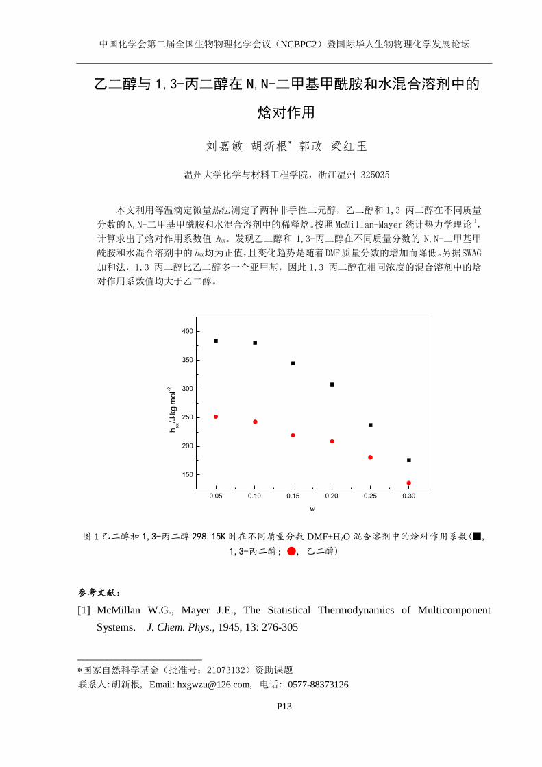
中国化学会第二届全国生物物理化学会议(NCBPC2)暨国际华人生物物理化学发展论坛
P13
乙二醇与 1,3-丙二醇在 N,N-二甲基甲酰胺和水混合溶剂中的
焓对作用
刘嘉敏 胡新根* 郭政 梁红玉
温州大学化学与材料工程学院,浙江温州 325035
本文利用等温滴定微量热法测定了两种非手性二元醇,乙二醇和 1,3-丙二醇在不同质量
分数的 N,N-二甲基甲酰胺和水混合溶剂中的稀释焓。按照 McMillan-Mayer统计热力学理论1,
计算求出了焓对作用系数值 hXX。发现乙二醇和 1,3-丙二醇在不同质量分数的 N,N-二甲基甲
酰胺和水混合溶剂中的hXX均为正值,且变化趋势是随着DMF质量分数的增加而降低。另据SWAG
加和法,1,3-丙二醇比乙二醇多一个亚甲基,因此 1,3-丙二醇在相同浓度的混合溶剂中的焓
对作用系数值均大于乙二醇。
0.05 0.10 0.15 0.20 0.25 0.30
150
200
250
300
350
400
hxx/Jk
gm
ol-2
w
图 1 乙二醇和 1,3-丙二醇 298.15K时在不同质量分数 DMF+H2O 混合溶剂中的焓对作用系数(■,
1,3-丙二醇; ●, 乙二醇)
参考文献:
[1] McMillan W.G., Mayer J.E., The Statistical Thermodynamics of Multicomponent
Systems. J. Chem. Phys., 1945, 13: 276-305
__________________________
*国家自然科学基金(批准号:21073132)资助课题
联系人:胡新根, Email: [email protected], 电话: 0577-88373126
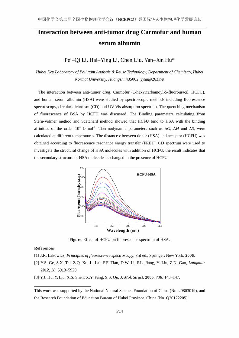
中国化学会第二届全国生物物理化学会议(NCBPC2)暨国际华人生物物理化学发展论坛
P14
Interaction between anti-tumor drug Carmofur and human
serum albumin
Pei–Qi Li, Hai–Ying Li, Chen Liu, Yan–Jun Hu*
Hubei Key Laboratory of Pollutant Analysis & Reuse Technology, Department of Chemistry, Hubei
Normal University, Huangshi 435002, [email protected]
The interaction between anti-tumor drug, Carmofur (1-hexylcarbamoyl-5-fluorouracil, HCFU),
and human serum albumin (HSA) were studied by spectroscopic methods including fluorescence
spectroscopy, circular dichroism (CD) and UV-Vis absorption spectrum. The quenching mechanism
of fluorescence of BSA by HCFU was discussed. The Binding parameters calculating from
Stern-Volmer method and Scatchard method showed that HCFU bind to HSA with the binding
affinities of the order 104 L·mol
-1. Thermodynamic parameters such as ∆G, ∆H and ∆S, were
calculated at different temperatures. The distance r between donor (HSA) and acceptor (HCFU) was
obtained according to fluorescence resonance energy transfer (FRET). CD spectrum were used to
investigate the structural change of HSA molecules with addition of HCFU, the result indicates that
the secondary structure of HSA molecules is changed in the presence of HCFU.
330 360 390 420 450
100
200
300
400
500
600
Flu
ore
sen
ce I
nte
nsi
ty (
a.u.)
Wavelength (nm)
HCFU-HSA
Figure. Effect of HCFU on fluorescence spectrum of HSA.
References
[1] J.R. Lakowicz, Principles of fluorescence spectroscopy, 3rd ed., Springer: New York, 2006.
[2] Y.S. Ge, S.X. Tai, Z.Q. Xu, L. Lai, F.F. Tian, D.W. Li, F.L. Jiang, Y. Liu, Z.N. Gao, Langmuir
2012, 28: 5913–5920.
[3] Y.J. Hu, Y. Liu, X.S. Shen, X.Y. Fang, S.S. Qu, J. Mol. Struct. 2005, 738: 143–147.
This work was supported by the National Natural Science Foundation of China (No. 20803019), and
the Research Foundation of Education Bureau of Hubei Province, China (No. Q20122205).

中国化学会第二届全国生物物理化学会议(NCBPC2)暨国际华人生物物理化学发展论坛
P15
Interaction of curcumin with DNA investigated by
spectroscopic methods
Xiao–Yun Li, Xiao–Ling Li, Hua–Li Yue, Yan–Jun Hu*
Hubei Key Laboratory of Pollutant Analysis & Reuse Technology, Department of Chemistry, Hubei
Normal University, Huangshi 435002, [email protected]
Curcumin is a polyphenolic bioactive compound found in the spice turmeric endowed with
diverse pharmacological and biological activities. In this study, fluorescence spectroscopy in
combination with UV vis absorption spectroscopy was employed to investigate the high affinity
binding of curcumin to herring sperm DNA (hs-DNA). In the mechanism discussion, the absorption
value of simply adding free DNA and free curcumin was a little greater than the absorption value of
DNA–curcumin complex as the increasing concentrations of curcumin, which indicated an
intercalative binding mode. When use acridine orange (AO) as a fluorescence probe, fluorescence
quenching of the emission peak was seen in the DNA-AO system when curcumin was added. The
effects of ionic strength, chemical denaturants, thermal denaturation and pH were studied to show
the factors of the interaction, and provided further support for the intercalative binding mode.
0 2 4 6 8 10
1.6
2.0
2.4
DNA-Cur complex
DNA+Cur
Ab
sorb
an
ce
105[Cur] (mol/L)
Figure 1. UV–Vis spectra of curcumin in the
presence of DNA.
520 540 560 580 600 620
0.0
0.2
0.4
0.6
0.8
1.0
0.0 0.5 1.0 1.5 2.0
0.0
0.4
0.8
1.2
(F
0-F)
/F
105[Cur] (mol/L)
Flu
oresc
en
ce i
nte
nsi
ty
Wavelength (nm)
A
K
Figure 2. Fluorescence spectrum of DNA–AO
with various concentrations of Cur.
References
[1] B.K. Paul, N. Guchhait, J. Phys. Chem. B 2011, 115: 11938–11949.
[2] J. Barry, M. Fritz, J.R. Brender, et al., J. Am. Chem. Soc. 2009, 131: 4490–4498.
[3] X.L. Li, Y.J. Hu, H. Wang, B.Q. Yu, H.L. Yue, Biomacromolecules 2012, 13: 873–880.
[4] J.R. Lakowicz, Principles of fluorescence spectroscopy. 3rd
ed., Springer Press, New York, 2006.
This work was supported by the National Natural Science Foundation of China (No. 20803019), and
the Research Foundation of Education Bureau of Hubei Province, China (No. Q20122205).

中国化学会第二届全国生物物理化学会议(NCBPC2)暨国际华人生物物理化学发展论坛
P16
Spectrometry investigation on the interaction between
naringenin and bovine hemoglobin
Chen Liu, Pei–Qi Li, Yan–Jun Hu*
Hubei Key Laboratory of Pollutant Analysis & Reuse Technology, Department of Chemistry, Hubei
Normal University, Huangshi 435002, [email protected]
This paper investigates the association of bovine hemoglobin (BHb) with naringenin by
fluorescence spectroscopy under the human physiological conditions. Binding parameters
calculating from Stern-Volmer method and Scatchard method showed that naringenin bind to BHb
with the binding affinities of the order 104 L·mol
-1. The thermodynamic parameters studies revealed
that the binding was characterized by negative enthalpy and positive entropy changes and the
electrostatic interactions play a major role for naringenin-BHb association. It was observed from
Figure 1 that a progressive increase in the fluorescence intensity occured with the addition of
naringenin. Figure 2 shows the effects of temperatures to the binding constants.
Keywords: naringenin, bovine hemoglobin, fluorescence spectroscopy.
330 360 3900.0
0.2
0.4
0.6
0.8
1.0
l (Naringenin only)
Flu
ore
scen
ce I
nte
nsi
ty (
a.u.)
Wavelength (nm)
a
k
Figure 1. Effect of Naringenin on fluorescence
spectrum of BHb.
0.0 0.2 0.4 0.6 0.8 1.01
2
3
4
F0/(
F0-F
)
10-5/[Q] (L mol-1
)
292K
298K
304K
Figure 2. Modified Stern-Volmer plots for the
queching of BHb by Naringenin at different
temperatures.
References
[1] Y.J. Hu, Y. Wang, Y. Ou-Yang, J. Zhou, Y. Liu, J. Lumin. 2010, 130: 1394–1399.
[2] Q. Shao, P. Wu, P. Gu, X. Xu, H. Zhang, C. Cai, J. Phys. Chem. B 2011, 115: 8627–8637.
[3] P. Mandal, T. Ganguly, J. Phys. Chem. B 2009, 113: 14904–14913.
This work was supported by the National Natural Science Foundation of China (No. 20803019), and
the Research Foundation of Education Bureau of Hubei Province, China (No. Q20122205).
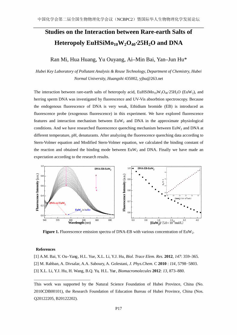
中国化学会第二届全国生物物理化学会议(NCBPC2)暨国际华人生物物理化学发展论坛
P17
Studies on the Interaction between Rare-earth Salts of
Heteropoly EuHSiMo10W2O40·25H2O and DNA
Ran Mi, Hua Huang, Yu Ouyang, Ai–Min Bai, Yan–Jun Hu*
Hubei Key Laboratory of Pollutant Analysis & Reuse Technology, Department of Chemistry, Hubei
Normal University, Huangshi 435002, [email protected]
The interaction between rare-earth salts of heteropoly acid, EuHSiMo10W2O40·25H2O (EuW2), and
herring sperm DNA was investigated by fluorescence and UV-Vis absorbtion spectroscopy. Because
the endogenous fluorescence of DNA is very weak, Ethidium bromide (EB) is introduced as
fluorescence probe (exogenous fluorescence) in this experiment. We have explored fluorescence
features and interaction mechanism between EuW2 and DNA in the approximate physiological
conditions. And we have researched fluorescence quenching mechanism between EuW2 and DNA at
different temperature, pH, denaturants. After analyzing the fluorescence quenching data according to
Stern-Volmer equation and Modified Stern-Volmer equation, we calculated the binding constant of
the reaction and obtained the binding mode between EuW2 and DNA. Finally we have made an
expectation according to the research results.
540 570 600 630 660 690
0.0
0.2
0.4
0.6
0.8
1.0
Fluo
resc
ence
Inte
nsity
(a.u
.)
Wavelength (nm)
DNA-EB-EuW2
DNA and EuW2
EuW2 in buffer
a
v
0.0 0.8 1.6 2.4 3.2 4.0
0.6
0.7
0.8
0.9
1.0
0.0 0.8 1.6 2.4 3.2 4.0
0.0
0.2
0.4
0.6
F 0 / F
- 1
[ EuW2] /(5.0×10-6mol/L)
Fluo
resc
ence
Inte
nsity
(a.u
.)
[EuW2]/(5.0×10-6mol/L)
DNA-EB-EuW2
Figure 1. Fluorescence emission spectra of DNA-EB with various concentration of EuW2.
References [1] A.M. Bai, Y. Ou–Yang, H.L. Yue, X.L. Li, Y.J. Hu, Biol. Trace Elem. Res. 2012, 147: 359–365.
[2] M. Rahban, A. Divsalar, A.A. Saboury, A. Golestani, J. Phys.Chem. C 2010 : 114, 5798−5803.
[3] X.L. Li, Y.J. Hu, H. Wang, B.Q. Yu, H.L. Yue, Biomacromolecules 2012: 13, 873–880.
This work was supported by the Natural Science Foundation of Hubei Province, China (No.
2010CDB00101), the Research Foundation of Education Bureau of Hubei Province, China (Nos.
Q20122205, B20122202).
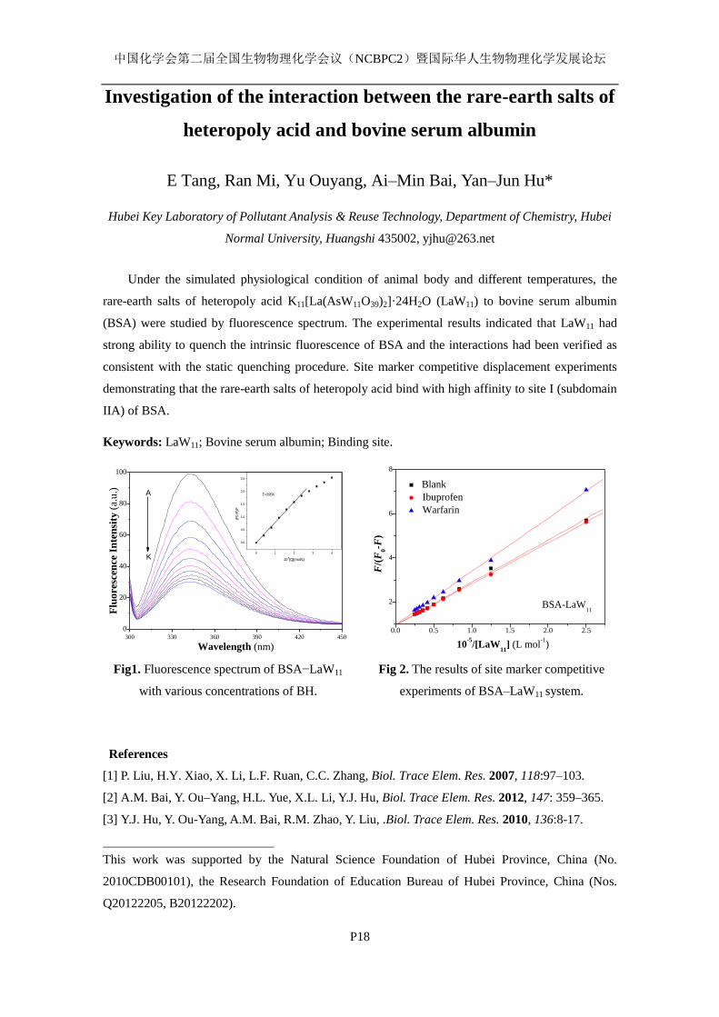
中国化学会第二届全国生物物理化学会议(NCBPC2)暨国际华人生物物理化学发展论坛
P18
Investigation of the interaction between the rare-earth salts of
heteropoly acid and bovine serum albumin
E Tang, Ran Mi, Yu Ouyang, Ai–Min Bai, Yan–Jun Hu*
Hubei Key Laboratory of Pollutant Analysis & Reuse Technology, Department of Chemistry, Hubei
Normal University, Huangshi 435002, [email protected]
Under the simulated physiological condition of animal body and different temperatures, the
rare-earth salts of heteropoly acid K11[La(AsW11O39)2]·24H2O (LaW11) to bovine serum albumin
(BSA) were studied by fluorescence spectrum. The experimental results indicated that LaW11 had
strong ability to quench the intrinsic fluorescence of BSA and the interactions had been verified as
consistent with the static quenching procedure. Site marker competitive displacement experiments
demonstrating that the rare-earth salts of heteropoly acid bind with high affinity to site I (subdomain
IIA) of BSA.
Keywords: LaW11; Bovine serum albumin; Binding site.
300 330 360 390 420 450
0
20
40
60
80
100
0 1 2 3 4
0.0
0.5
1.0
1.5
2.0
2.5
(F0-
F)/F
10-5[Q](mol/L)
T=295K
Flu
ore
scen
ce I
nte
nsi
ty (
a.u
.)
Wavelength (nm)
A
K
Fig1. Fluorescence spectrum of BSA−LaW11
with various concentrations of BH.
0.0 0.5 1.0 1.5 2.0 2.5
2
4
6
8
Blank
Ibuprofen
Warfarin
F/(
F0-F
)
10-5
/[LaW11
] (L mol-1
)
BSA-LaW11
Fig 2. The results of site marker competitive
experiments of BSA–LaW11 system.
References
[1] P. Liu, H.Y. Xiao, X. Li, L.F. Ruan, C.C. Zhang, Biol. Trace Elem. Res. 2007, 118:97–103.
[2] A.M. Bai, Y. Ou–Yang, H.L. Yue, X.L. Li, Y.J. Hu, Biol. Trace Elem. Res. 2012, 147: 359–365.
[3] Y.J. Hu, Y. Ou-Yang, A.M. Bai, R.M. Zhao, Y. Liu, .Biol. Trace Elem. Res. 2010, 136:8-17.
This work was supported by the Natural Science Foundation of Hubei Province, China (No.
2010CDB00101), the Research Foundation of Education Bureau of Hubei Province, China (Nos.
Q20122205, B20122202).

中国化学会第二届全国生物物理化学会议(NCBPC2)暨国际华人生物物理化学发展论坛
P19
Spectral studies on the interaction between Nifedipine and DNA
Hong–Ying Wang, Yan–Jun Hu*
Hubei Key Laboratory of Pollutant Analysis & Reuse Technology, Department of Chemistry, Hubei
Normal University, Huangshi 435002, [email protected]
This paper presents the study on interaction between Nifedipine and herring sperm DNA (hsDNA)
using several spectroscopic methods, including fluorescence, UV-Vis, CD-spectroscopy and so on.
The results show that Nifedipine interacts with DNA through both the phosphate groups and the
nitrogenous bases of DNA. The formation of DNA–Nifedipine complex may change the
microenvironment of double helix of DNA. At the same time, the spectroscopic changes of ethidium
bromide (EB) on its binding to DNA are utilized to study the interaction between Nifedipine and
DNA. The interaction information regarding quenching mechanisms, binding parameters,and
thermodynamic parameters are all our investigation aims. It is also proved that the formation of the
DNA–Nifedipine complex is influenced by pH value, ionic strength, temperature, and
denaturants.
400 450 500 550 600 650
0.0
0.2
0.4
0.6
0.8
1.0
Flu
oresc
en
ce I
nte
nsi
ty (
au)
Wavelength (nm)
Figure 1. Fluorescence spectrum of DNA with various concentration of Nifedipine.
References
[1] X.L. Li, Y.J. Hu, H. Wang, B.Q. Yu, H.L. Yue, Biomacromolecules 2012, 13: 873–880.
[2] Y. Shi, C. Guo, Y. Sun, Z. Liu, F. Xu, Y. Zhang, Z. Wen, Z. Li, Biomacromolecules 2011, 12:
797–803.
[3] A. Wildes, N. Theodorakopoulos, J. Valle-Orero, S. Cuesta-López, J.L. Garden, M. Peyrard,
Phys. Rev. Lett. 2011, 106: 1-4.
This work was supported by the National Natural Science Foundation of China (No. 20803019), and
the Research Foundation of Education Bureau of Hubei Province, China (No. Q20122205).
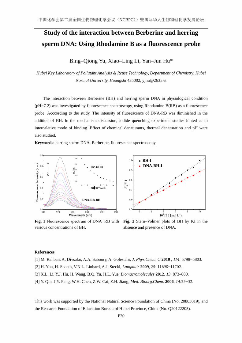
中国化学会第二届全国生物物理化学会议(NCBPC2)暨国际华人生物物理化学发展论坛
P20
Study of the interaction between Berberine and herring
sperm DNA: Using Rhodamine B as a fluorescence probe
Bing–Qiong Yu, Xiao–Ling Li, Yan–Jun Hu*
Hubei Key Laboratory of Pollutant Analysis & Reuse Technology, Department of Chemistry, Hubei
Normal University, Huangshi 435002, [email protected]
The interaction between Berberine (BH) and herring sperm DNA in physiological condition
(pH=7.2) was investigated by fluorescence spectroscopy, using Rhodamine B(RB) as a fluorescence
probe. Acccording to the study, The intensity of fluorescence of DNA-RB was diminished in the
addition of BH. In the mechanism discussion, iodide quenching experiment studies hinted at an
intercalative mode of binding. Effect of chemical denaturants, thermal denaturation and pH were
also studied.
Keywords: herring sperm DNA, Berberine, fluorescence spectroscopy
540 570 600 630 660 6900.0
0.2
0.4
0.6
0.8
1.0
0 2 4 6 8 10
0.2
0.4
0.6
0.8
1.0
DNA-RB-BH
FL
(a.u
)
[BH](10-5mol/L)
DNA-RB-BH
Wavelength (nm)
Flu
oresc
en
ce I
nte
nsi
ty (
a.u
.) A
K
0 2 4 6 8 100.5
0.6
0.7
0.8
0.9
1.0
F
0/F
-1
BH-I-
DNA-BH-I-
103 [I
-] (mol L
-1)
Fig. 1 Fluorescence spectrum of DNA−RB with
various concentrations of BH.
Fig. 2 Stern–Volmer plots of BH by KI in the
absence and presence of DNA.
References
[1] M. Rahban, A. Divsalar, A.A. Saboury, A. Golestani, J. Phys.Chem. C 2010 , 114: 5798−5803.
[2] H. You, H. Spaeth, V.N.L. Linhard, A.J. Steckl, Langmuir 2009, 25: 11698−11702.
[3] X.L. Li, Y.J. Hu, H. Wang, B.Q. Yu, H.L. Yue, Biomacromolecules 2012, 13: 873–880.
[4] Y. Qin, J.Y. Pang, W.H. Chen, Z.W. Cai, Z.H. Jiang, Med. Bioorg.Chem. 2006, 14:25−32.
This work was supported by the National Natural Science Foundation of China (No. 20803019), and
the Research Foundation of Education Bureau of Hubei Province, China (No. Q20122205).
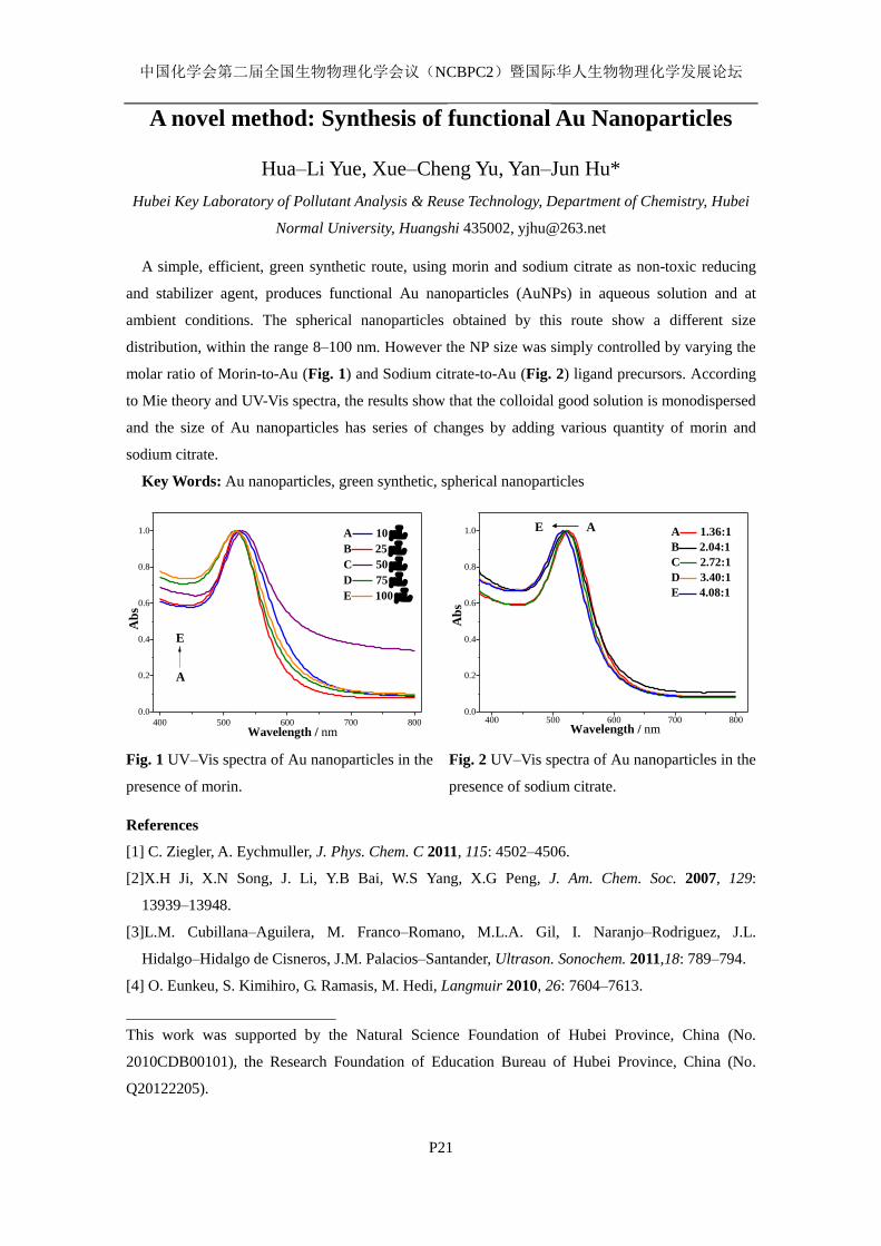
中国化学会第二届全国生物物理化学会议(NCBPC2)暨国际华人生物物理化学发展论坛
P21
A novel method: Synthesis of functional Au Nanoparticles
Hua–Li Yue, Xue–Cheng Yu, Yan–Jun Hu*
Hubei Key Laboratory of Pollutant Analysis & Reuse Technology, Department of Chemistry, Hubei
Normal University, Huangshi 435002, [email protected]
A simple, efficient, green synthetic route, using morin and sodium citrate as non-toxic reducing
and stabilizer agent, produces functional Au nanoparticles (AuNPs) in aqueous solution and at
ambient conditions. The spherical nanoparticles obtained by this route show a different size
distribution, within the range 8–100 nm. However the NP size was simply controlled by varying the
molar ratio of Morin-to-Au (Fig. 1) and Sodium citrate-to-Au (Fig. 2) ligand precursors. According
to Mie theory and UV-Vis spectra, the results show that the colloidal good solution is monodispersed
and the size of Au nanoparticles has series of changes by adding various quantity of morin and
sodium citrate.
Key Words: Au nanoparticles, green synthetic, spherical nanoparticles
400 500 600 700 800
0.0
0.2
0.4
0.6
0.8
1.0
E
Ab
s
Wavelength / nm
A 10 LB 25 LC 50LD 75 LE 100L
A
400 500 600 700 8000.0
0.2
0.4
0.6
0.8
1.0 E
Ab
s
Wavelength / nm
A 1.36:1
B 2.04:1
C 2.72:1
D 3.40:1
E 4.08:1
A
Fig. 1 UV–Vis spectra of Au nanoparticles in the
presence of morin.
Fig. 2 UV–Vis spectra of Au nanoparticles in the
presence of sodium citrate.
References
[1] C. Ziegler, A. Eychmuller, J. Phys. Chem. C 2011, 115: 4502–4506.
[2]X.H Ji, X.N Song, J. Li, Y.B Bai, W.S Yang, X.G Peng, J. Am. Chem. Soc. 2007, 129:
13939–13948.
[3]L.M. Cubillana–Aguilera, M. Franco–Romano, M.L.A. Gil, I. Naranjo–Rodriguez, J.L.
Hidalgo–Hidalgo de Cisneros, J.M. Palacios–Santander, Ultrason. Sonochem. 2011,18: 789–794.
[4] O. Eunkeu, S. Kimihiro, G. Ramasis, M. Hedi, Langmuir 2010, 26: 7604–7613.
This work was supported by the Natural Science Foundation of Hubei Province, China (No.
2010CDB00101), the Research Foundation of Education Bureau of Hubei Province, China (No.
Q20122205).
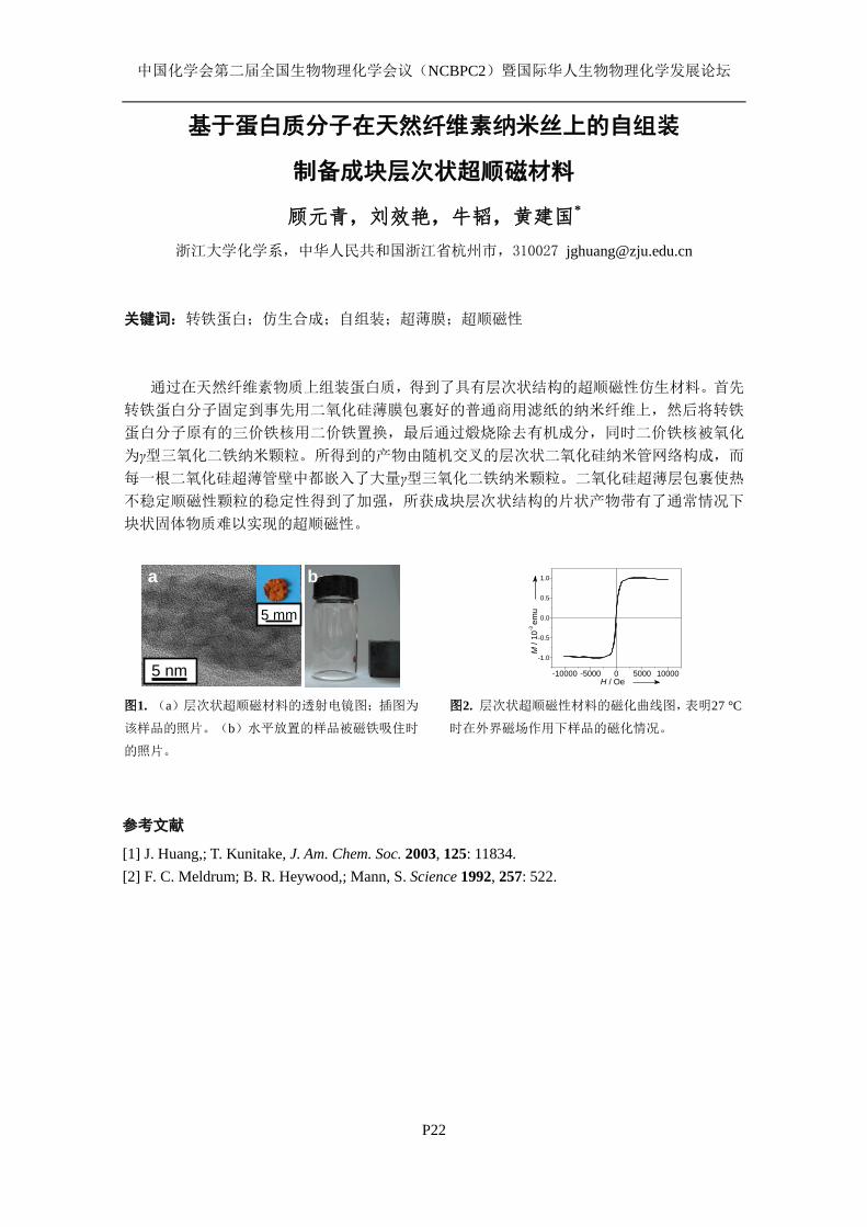
中国化学会第二届全国生物物理化学会议(NCBPC2)暨国际华人生物物理化学发展论坛
P22
基于蛋白质分子在天然纤维素纳米丝上的自组装
制备成块层次状超顺磁材料
顾元青,刘效艳,牛韬,黄建国*
浙江大学化学系,中华人民共和国浙江省杭州市,310027 [email protected]
关键词:转铁蛋白;仿生合成;自组装;超薄膜;超顺磁性
通过在天然纤维素物质上组装蛋白质,得到了具有层次状结构的超顺磁性仿生材料。首先
转铁蛋白分子固定到事先用二氧化硅薄膜包裹好的普通商用滤纸的纳米纤维上,然后将转铁
蛋白分子原有的三价铁核用二价铁置换,最后通过煅烧除去有机成分,同时二价铁核被氧化
为γ型三氧化二铁纳米颗粒。所得到的产物由随机交叉的层次状二氧化硅纳米管网络构成,而
每一根二氧化硅超薄管壁中都嵌入了大量γ型三氧化二铁纳米颗粒。二氧化硅超薄层包裹使热
不稳定顺磁性颗粒的稳定性得到了加强,所获成块层次状结构的片状产物带有了通常情况下
块状固体物质难以实现的超顺磁性。
图1. (a)层次状超顺磁材料的透射电镜图;插图为
该样品的照片。(b)水平放置的样品被磁铁吸住时
的照片。
-10000 -5000 0 5000 10000
-1.0
-0.5
0.0
0.5
1.0
M /
10-3
emu
H / Oe
图2. 层次状超顺磁性材料的磁化曲线图,表明27 °C
时在外界磁场作用下样品的磁化情况。
参考文献
[1] J. Huang,; T. Kunitake, J. Am. Chem. Soc. 2003, 125: 11834.
[2] F. C. Meldrum; B. R. Heywood,; Mann, S. Science 1992, 257: 522.
5 nm
5 mm a b
中国化学会第二届全国生物物理化学会议(NCBPC2)暨国际华人生物物理化学发展论坛
P23
Nanofibrous rutile-titania/graphite composite derived from
natural cellulose substance
Yan Luo, Xiaoyan Liu, Jianguo Huang
Department of Chemistry, Zhejiang University, Hangzhou, Zhejiang 310027, China,
Calcination and carbonization of ultrathin titania gel film pre-coated cellulose nanofibres of
ordinary laboratory filter paper at 1300 C under argon atmosphere yielded a hierarchical
nanofibrous rutile-titania/carbon composite material. The carbon fibres derived from the initial
cellulose nanofibres possess a core-shell structure with amorphous carbon at the inner part and
graphite state carbon at the outer part. And the initial titania gel films were converted into rutile
phase titania nanoparticles with sizes of 2050 nm anchored on the surface of the carbon fibres. The
existence of carbon fibres hindered the aggregation of the titania particles under high temperature.
The rutile-titania/carbon composite resulted possesses considerable electric conductivity as well as
photocatalytic activity towards photodegradation of methylene blue in water.
Keywords: cellulose, carbonization, rutile-titania, photocatalysis
Reference:
1. X. Liu, Y. Gu and J. Huang, Chem. Eur. J., 2010, 16: 7730–7740.
2. J. Huang and T. Kunitake, J. Am. Chem. Soc., 2003, 125: 11834–11835.

中国化学会第二届全国生物物理化学会议(NCBPC2)暨国际华人生物物理化学发展论坛
P24
Surface-functionalized cellulosic materials used for specific
DNA recognition and colorimetric cysteine detection
Wei Xiao, Haiyun Hu and Jianguo Huang*
Department of Chemistry, Zhejiang University, Hangzhou, Zhejiang 310027, China
Morphologically complex cellulosic substance (e.g., commercial filter paper) was employed as
solid substrates for immobilization of different guest species, which endowed the natural cellulose
substance with engineered properties, leading to the development of a series of novel
surface-functionalized cellulosic materials. A uniform ultrathin zirconia gel film was first deposited
on each cellulose nanofiber in bulk filter paper by a facile sol–gel process. Relying on the large
surface area of filter paper and the strong affinity of zirconia for the phosphate group,
terminal-phosphate probe DNA was abundantly immobilized on the zirconia-modified filter paper so
as to convert the composite to a biofunctional material for the sensitive and repetitive recognition of
the corresponding complementary target DNA on the nanomolar level. By contrast, the amount of
captured probe DNA or recognized target DNA on a zirconia-coated flat substrate (e.g., quartz plate)
was much less than that captured or recognized on filter paper, resulting in a relatively insensitive
recognition event. Moreover, Immobilization of ruthenium dye–Hg2+
complex (N719–Hg2+
complex)
monolayer on ultrathin zirconia gel film coated cellulose nanofibers of filter paper yielded a
solid-phase sensing device with high sensitivity, selectivity and reversibility for colorimetric
detection of cysteine in aqueous media. This assay relies on the specific mercury dispossession by
cysteine from the immobilized N719–Hg2+
complex. And the dissociation of N719 and Hg2+
caused
by mercury snatch behavior of cysteine engenders obvious orange-to-purple color change of the
zirconia gel layer and N719–Hg2+
complex modified filter paper, hence realizing the colorimetric
detection of cysteine. The detection limit of this assay is 20 μM by the naked eye and the selectivity
for cysteine against interference of the other 19 natural amino acids and their mixture is extremely
high.
Keywords: cellulose zirconia sol‒gel oligonucleotide colorimetric
References
1) Xiao, W.; Huang, J. Langmuir 2011, 27: 12284–12288.
2) Xiao, W.; Hu, H.; Huang, J. Sensors and Actuators B: Chemical 2012,
doi:10.1016/j.snb.2012.05.087
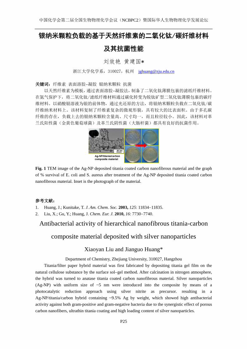
中国化学会第二届全国生物物理化学会议(NCBPC2)暨国际华人生物物理化学发展论坛
P25
银纳米颗粒负载的基于天然纤维素的二氧化钛/碳纤维材料
及其抗菌性能
刘效艳 黄建国*
浙江大学化学系,310027,杭州 [email protected]
关键词:纤维素 表面溶胶凝胶 银纳米颗粒 抗菌
以天然纤维素为模板,通过表面溶胶凝胶法,制备了二氧化钛薄膜包裹的滤纸纤维材料。
在氮气保护下,将二氧化钛/滤纸纤维材料通过碳化转变为锐钛矿型二氧化钛薄膜包裹的碳纤
维材料。以硝酸银溶液为银的前体物,通过光还原的方法,将银纳米颗粒负载在二氧化钛/碳
纤维纳米材料上。该材料复制了纤维素复杂的微观形貌,具有较大的比表面积。由于多孔碳
纤维的存在,负载上去的银纳米颗粒含量高、尺寸均一,而且粒径较小。因此,该材料对革
兰氏阳性菌(金黄色葡萄球菌)及革兰氏阴性菌(大肠杆菌)都具有良好的抗菌作用。
Fig. 1 TEM image of the Ag-NP deposited titania coated carbon nanofibrous material and the graph
of % survival of E. coli and S. aureus after treatment of the Ag-NP deposited titania coated carbon
nanofibrous material. Inset is the photograph of the material.
参考文献:
1. Huang, J.; Kunitake, T. J. Am. Chem. Soc. 2003, 125: 1183411835.
2. Liu, X.; Gu, Y.; Huang, J. Chem. Eur. J. 2010, 16: 77307740.
Antibacterial activity of hierarchical nanofibrous titania-carbon
composite material deposited with silver nanoparticles
Xiaoyan Liu and Jianguo Huang*
Department of Chemistry, Zhejiang University, 310027, Hangzhou
Titania/filter paper hybrid material was first fabricated by depositing titania gel film on the
natural cellulose substance by the surface solgel method. After calcination in nitrogen atmosphere,
the hybrid was turned to anatase titania coated carbon nanofibrous material. Silver nanoparticles
(Ag-NP) with uniform size of ~5 nm were introduced into the composite by means of a
photocatalytic reduction approach using silver nitrite as precursor. resulting in a
Ag-NP/titania/carbon hybrid containing ~9.5% Ag by weight, which showed high antibacterial
activity against both gram-positive and gram-negative bacteria due to the synergistic effect of porous
carbon nanofibers, ultrathin titania coating and high loading content of silver nanoparticles.
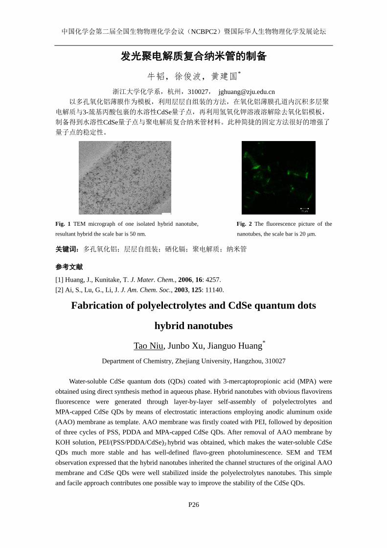
中国化学会第二届全国生物物理化学会议(NCBPC2)暨国际华人生物物理化学发展论坛
P26
发光聚电解质复合纳米管的制备
牛韬,徐俊波,黄建国*
浙江大学化学系,杭州,310027, [email protected]
以多孔氧化铝薄膜作为模板,利用层层自组装的方法,在氧化铝薄膜孔道内沉积多层聚
电解质与3-巯基丙酸包裹的水溶性CdSe量子点,再利用氢氧化钾溶液溶解除去氧化铝模板,
制备得到水溶性CdSe量子点与聚电解质复合纳米管材料。此种简捷的固定方法很好的增强了
量子点的稳定性。
Fig. 1 TEM micrograph of one isolated hybrid nanotube, Fig. 2 The fluorescence picture of the
resultant hybrid the scale bar is 50 nm. nanotubes, the scale bar is 20 μm.
关键词:多孔氧化铝;层层自组装;硒化镉;聚电解质;纳米管
参考文献
[1] Huang, J., Kunitake, T. J. Mater. Chem., 2006, 16: 4257.
[2] Ai, S., Lu, G., Li, J. J. Am. Chem. Soc., 2003, 125: 11140.
Fabrication of polyelectrolytes and CdSe quantum dots
hybrid nanotubes
Tao Niu, Junbo Xu, Jianguo Huang*
Department of Chemistry, Zhejiang University, Hangzhou, 310027
Water-soluble CdSe quantum dots (QDs) coated with 3-mercaptopropionic acid (MPA) were
obtained using direct synthesis method in aqueous phase. Hybrid nanotubes with obvious flavovirens
fluorescence were generated through layer-by-layer self-assembly of polyelectrolytes and
MPA-capped CdSe QDs by means of electrostatic interactions employing anodic aluminum oxide
(AAO) membrane as template. AAO membrane was firstly coated with PEI, followed by deposition
of three cycles of PSS, PDDA and MPA-capped CdSe QDs. After removal of AAO membrane by
KOH solution, PEI/(PSS/PDDA/CdSe)3 hybrid was obtained, which makes the water-soluble CdSe
QDs much more stable and has well-defined flavo-green photoluminescence. SEM and TEM
observation expressed that the hybrid nanotubes inherited the channel structures of the original AAO
membrane and CdSe QDs were well stabilized inside the polyelectrolytes nanotubes. This simple
and facile approach contributes one possible way to improve the stability of the CdSe QDs.
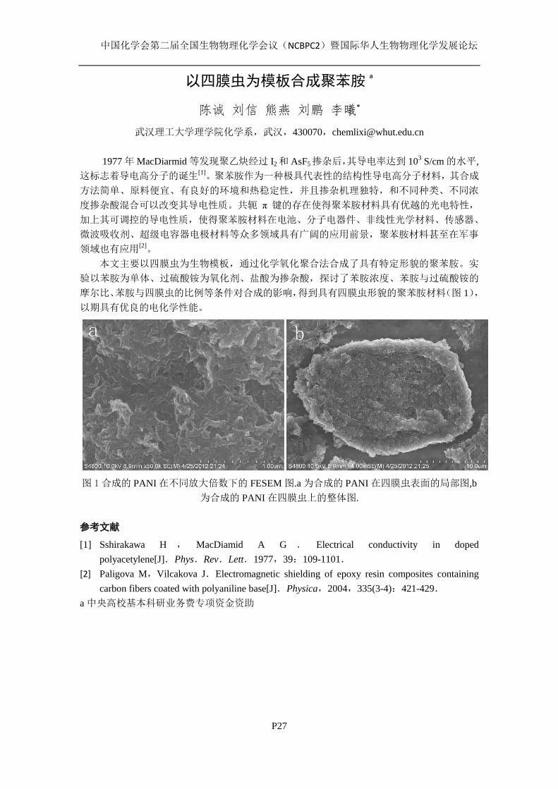
中国化学会第二届全国生物物理化学会议(NCBPC2)暨国际华人生物物理化学发展论坛
P27
以四膜虫为模板合成聚苯胺a
陈诚 刘信 熊燕 刘鹏 李曦*
武汉理工大学理学院化学系,武汉,430070,[email protected]
1977 年 MacDiarmid 等发现聚乙炔经过 I2 和 AsF5 掺杂后,其导电率达到 103 S/cm 的水平,
这标志着导电高分子的诞生[1]。聚苯胺作为一种极具代表性的结构性导电高分子材料,其合成
方法简单、原料便宜、有良好的环境和热稳定性,并且掺杂机理独特,和不同种类、不同浓
度掺杂酸混合可以改变其导电性质。共轭 π 键的存在使得聚苯胺材料具有优越的光电特性,
加上其可调控的导电性质,使得聚苯胺材料在电池、分子电器件、非线性光学材料、传感器、
微波吸收剂、超级电容器电极材料等众多领域具有广阔的应用前景,聚苯胺材料甚至在军事
领域也有应用[2]。
本文主要以四膜虫为生物模板,通过化学氧化聚合法合成了具有特定形貌的聚苯胺。实
验以苯胺为单体、过硫酸铵为氧化剂、盐酸为掺杂酸,探讨了苯胺浓度、苯胺与过硫酸铵的
摩尔比、苯胺与四膜虫的比例等条件对合成的影响,得到具有四膜虫形貌的聚苯胺材料(图 1),
以期具有优良的电化学性能。
图 1合成的 PANI 在不同放大倍数下的 FESEM 图.a 为合成的 PANI 在四膜虫表面的局部图,b
为合成的 PANI 在四膜虫上的整体图.
参考文献
[1] Sshirakawa H , MacDiamid A G . Electrical conductivity in doped
polyacetylene[J].Phys.Rev.Lett.1977,39:109-1101.
[2] Paligova M,Vilcakova J.Electromagnetic shielding of epoxy resin composites containing
carbon fibers coated with polyaniline base[J].Physica,2004,335(3-4):421-429.
a 中央高校基本科研业务费专项资金资助
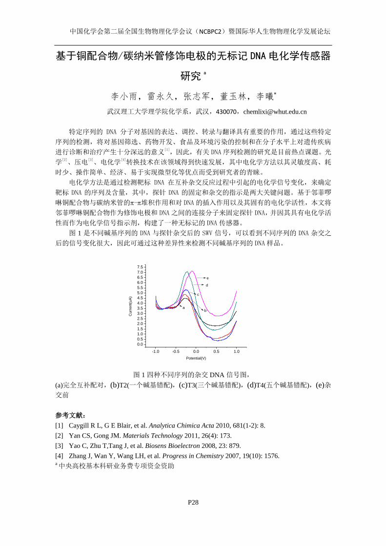
中国化学会第二届全国生物物理化学会议(NCBPC2)暨国际华人生物物理化学发展论坛
P28
基于铜配合物/碳纳米管修饰电极的无标记 DNA电化学传感器
研究a
李小雨,雷永久,张志军,董玉林,李曦*
武汉理工大学理学院化学系,武汉,430070,[email protected]
特定序列的 DNA 分子对基因的表达、调控、转录与翻译具有重要的作用,通过这些特定
序列的检测,将对基因筛选、药物开发、食品及环境污染的控制和在分子水平上对遗传疾病
进行诊断和治疗产生十分深远的意义[1],因此,有关 DNA序列检测的研究是目前热点课题。光
学[2]、压电
[3]、电化学
[4]转换技术在该领域得到快速发展,其中电化学方法以其灵敏度高、耗
时少、操作简单、经济、易于实现微型化等优点而受到研究者的青睐。
电化学方法是通过检测靶标 DNA 在互补杂交反应过程中引起的电化学信号变化,来确定
靶标 DNA 的序列及含量,其中,探针 DNA 的固定和杂交的指示是两大关键问题。基于邻菲啰
啉铜配合物与碳纳米管的-堆积作用和对 DNA的插入作用以及其固有的电化学活性,本文将
邻菲啰啉铜配合物作为修饰电极和 DNA之间的连接分子来固定探针 DNA,并因其具有电化学活
性而作为电化学信号指示剂,构建了一种无标记的 DNA传感器。
图 1 是不同碱基序列的 DNA 与探针杂交后的 SWV 信号,可以看到不同序列的 DNA 杂交之
后的信号变化很大,因此可通过这种差异性来检测不同碱基序列的 DNA样品。
-1.0 -0.5 0.0 0.5 1.0
0.00.51.01.52.02.53.03.54.04.55.05.56.06.57.07.5
Cur
rent
(uA
)
Potential(V)
ab
c
d
e
图 1 四种不同序列的杂交 DNA 信号图,
(a)完全互补配对,(b)T2(一个碱基错配),(c)T3(三个碱基错配),(d)T4(五个碱基错配),(e)杂
交前
参考文献:
[1] Caygill R L, G E Blair, et al. Analytica Chimica Acta 2010, 681(1-2): 8.
[2] Yan CS, Gong JM. Materials Technology 2011, 26(4): 173.
[3] Yao C, Zhu T,Tang J, et al. Biosens Bioelectron 2008, 23: 879.
[4] Zhang J, Wan Y, Wang LH, et al. Progress in Chemistry 2007, 19(10): 1576. a 中央高校基本科研业务费专项资金资助
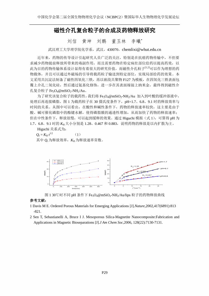
中国化学会第二届全国生物物理化学会议(NCBPC2)暨国际华人生物物理化学发展论坛
P29
磁性介孔复合粒子的合成及药物释放研究
刘信 黄坤 刘鹏 董玉林 李曦*
武汉理工大学理学院化学系,武汉,430070,[email protected]
近年来,药物的传导设计引起研究人员广泛的关注,特别是在抗癌药物传输中,不但要
求减少药物提前释放所带来的毒副作用,而且需要药物在特定病灶部位给药以提高药效,以
此为目的药物传输体系设计显得有着很大的研究价值,而磁性介孔粒子[1,2]可以作为理想的药
物载体,并且可以通过外磁场的引导将载药粒子输送到特定部位,实现局部给药的效果。本
文采用共沉淀法制备了磁性四氧化三铁,再以嵌段共聚物 F127 为模板,在四氧化三铁表面包
覆上介孔二氧化硅,然后通过氨基化修饰,进一步在其表面嫁接上纳米金,最终得到磁性介
孔复合粒子 Fe3O4@mSiO2-NH2/Au。
为了研究该复合粒子的载药性,我们将 Fe3O4@mSiO2-NH2/Au 加入到叶酸的缓冲溶液中,
处理后再连接磷脂。图 1 为载药粒子在 30 摄氏度条件下,pH=1.7、6.8、9.1 时的释放效率与
时间的关系。从图中可以看出,在酸性和碱性条件下,药物的释放速率较快,这主要是由于
酸、碱可催化磷脂中的酯键水解,使得磷脂膜的通透性增加,从而加快了药物的释放速率;
但在中性条件下,释放很慢,可以起到缓释的效果。通过 Higuchi 模拟(式 1),可算得 pH 为
1.7、6.8、9.1 时的 KH大小分别是 1.28、0.467 和 0.883,说明药物的释放是以内扩散为主。
Higuchi 关系式为:
Qt = KH t1/2
(1)
其中 Qt为释放效率,KH为释放速率常数。
图 1 30℃时不同 pH 条件下 Fe3O4@mSiO2-NH2/Au/lips 粒子的药物释放曲线
参考文献:
1 Davis M E. Ordered Porous Materials for Emerging Applications [J].Nature,2002,417(6891):813
-821.
2 Sen T, Sebastianelli A, Bruce I J. Mesoporous Silica-Magnetite Nanocomposite:Fabrication and
Applications in Magnetic Bioseparations [J].J Am Chem Soc,2006, 128(22):7130-7131.
0 100 200 300 400 500 600
0
15
30
45
rele
ase%
time(min)
pH=1.7
pH=6.8
pH=9.1
0 5 10 15 20 25
0
10
20
30
40
50
pH=9.1
pH=6.8K
H=0.467
KH=0.883
KH=1.28
rele
ase%
t1/2
(min1/2
)
pH=1.7
中国化学会第二届全国生物物理化学会议(NCBPC2)暨国际华人生物物理化学发展论坛
P30
Microcalorimetry studies on the antimicrobial actions of
Aconitum Alkaloids
Lian Liu · Wei Shao · Guimei Lin
School of Pharmaceutical Science, Shandong University, 44 West Wenhua Road, Jinan, Shandong
Province, 250012, P. R. China
The rate of heat output is one of the suitable measurements of metabolic activity of the
organism. In this paper, microcalorimetry was first applied to study the effect of Aconitum Alkaloids
on Escherichia coli (E.coli) and Staphylococcus aureus (S.aureus) growth. The power-time curves
were plotted with a TAM air isothermal microcalorimeter. The parameters such as the growth rate
constant μ, the peak-time Tp, inhibitory ratio I, enhancement ratio E were calculated. From the data,
the relationships between μ and the concentration of Aconitum Alkaloids c were established. The
results revealed that Aconitum Alkaloids had no effect on E. coli. Aconitum Alkaloids had potential
inhibitory effect on S.aureus. Results obtained from our study strongly suggest that microcalorimetry
is an ideal method to investigate the effect of drug on microorganism.
Keywords Aconitum Alkaloids · E. coli · S. aureus · microcalorimetry
(1) C Zhao; M Li; YF Luo; WK Wu. Isolation and structural characterization of an
immunostimulating polysaccharide from fuzi, Aconitum carmichaeli. Carbohydr Res 2006, 341:
485-491.
(2) JH Chen; CY Lee; BC Liau; MR Lee; TT Jong; ST Chiang. Determination of aconitine-type
alkaloids as markers in fuzi (Aconitum carmichaeli) by LC/(+)ESI/MS3. J Pharm and Biomed
Anal 2008, doi:10.1016/j.jpba.2008.08.022.
(3) JS Wang; R Heijden;G Spijksma;T Reijmers; M Wang; GW Xu; T Hankemeier; J Greef. Alkaloid
profiling of the Chinese herbal medicine Fuzi by combination of matrix-assisted laser desorption
ionization mass spectrometry with liquid chromatography–mass spectrometry. J Chromatogr A
2009, doi:10.1016/j.chroma.2008.11.0774
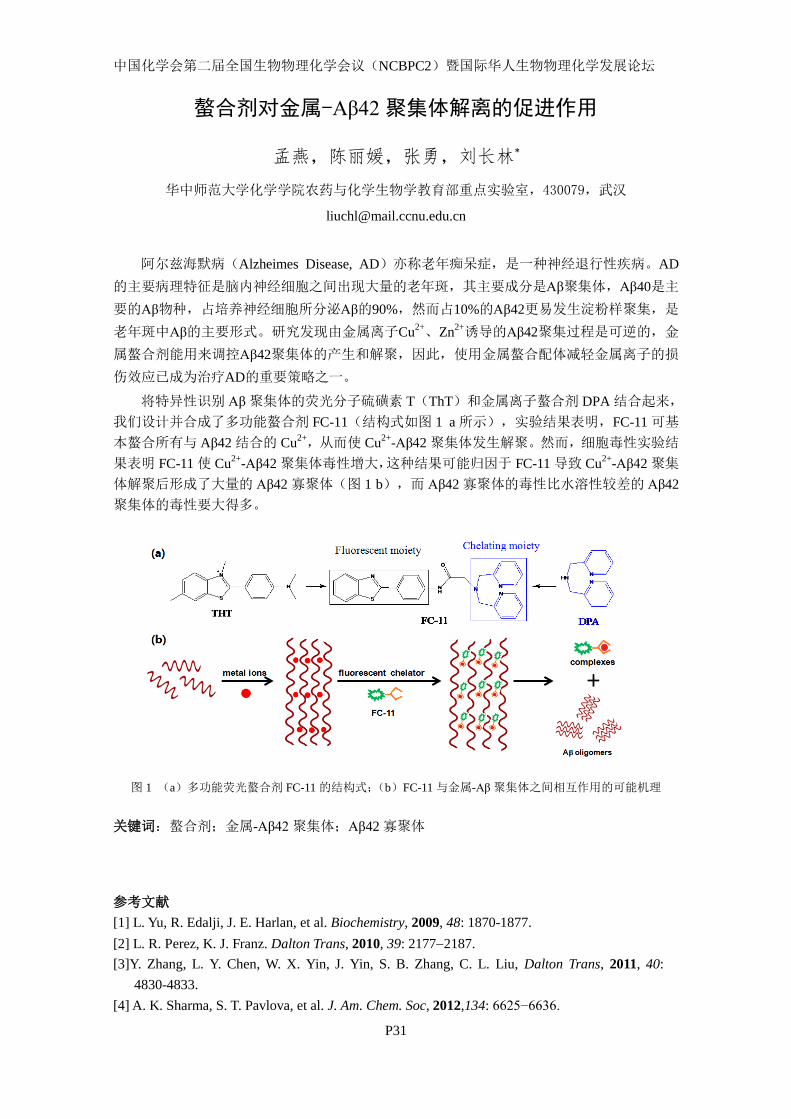
中国化学会第二届全国生物物理化学会议(NCBPC2)暨国际华人生物物理化学发展论坛
P31
螯合剂对金属-Aβ42聚集体解离的促进作用
孟燕,陈丽媛,张勇,刘长林*
华中师范大学化学学院农药与化学生物学教育部重点实验室,430079,武汉
阿尔兹海默病(Alzheimes Disease, AD)亦称老年痴呆症,是一种神经退行性疾病。AD
的主要病理特征是脑内神经细胞之间出现大量的老年斑,其主要成分是Aβ聚集体,Aβ40是主
要的Aβ物种,占培养神经细胞所分泌Aβ的90%,然而占10%的Aβ42更易发生淀粉样聚集,是
老年斑中Aβ的主要形式。研究发现由金属离子Cu2+、Zn
2+诱导的Aβ42聚集过程是可逆的,金
属螯合剂能用来调控Aβ42聚集体的产生和解聚,因此,使用金属螯合配体减轻金属离子的损
伤效应已成为治疗AD的重要策略之一。
将特异性识别 Aβ聚集体的荧光分子硫磺素 T(ThT)和金属离子螯合剂 DPA 结合起来,
我们设计并合成了多功能螯合剂 FC-11(结构式如图 1 a 所示),实验结果表明,FC-11 可基
本螯合所有与 Aβ42 结合的 Cu2+,从而使 Cu
2+-Aβ42 聚集体发生解聚。然而,细胞毒性实验结
果表明 FC-11 使 Cu2+
-Aβ42 聚集体毒性增大,这种结果可能归因于 FC-11 导致 Cu2+
-Aβ42 聚集
体解聚后形成了大量的 Aβ42 寡聚体(图 1 b),而 Aβ42 寡聚体的毒性比水溶性较差的 Aβ42
聚集体的毒性要大得多。
图 1 (a)多功能荧光螯合剂 FC-11 的结构式;(b)FC-11 与金属-Aβ聚集体之间相互作用的可能机理
关键词:螯合剂;金属-Aβ42聚集体;Aβ42 寡聚体
参考文献
[1] L. Yu, R. Edalji, J. E. Harlan, et al. Biochemistry, 2009, 48: 1870-1877.
[2] L. R. Perez, K. J. Franz. Dalton Trans, 2010, 39: 21772187.
[3]Y. Zhang, L. Y. Chen, W. X. Yin, J. Yin, S. B. Zhang, C. L. Liu, Dalton Trans, 2011, 40:
4830-4833.
[4] A. K. Sharma, S. T. Pavlova, et al. J. Am. Chem. Soc, 2012,134: 6625−6636.
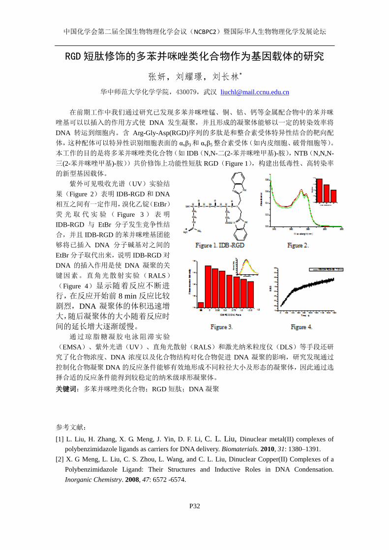
中国化学会第二届全国生物物理化学会议(NCBPC2)暨国际华人生物物理化学发展论坛
P32
RGD 短肽修饰的多苯并咪唑类化合物作为基因载体的研究
张妍,刘耀璟,刘长林*
华中师范大学化学学院,430079,武汉 [email protected]
在前期工作中我们通过研究已发现多苯并咪唑锰、铜、钴、钙等金属配合物中的苯并咪
唑基可以以插入的作用方式使 DNA 发生凝聚,并且形成的凝聚体能够以一定的转染效率将
DNA 转运到细胞内。含 Arg-Gly-Asp(RGD)序列的多肽是和整合素受体特异性结合的靶向配
体,这种配体可以特异性识别细胞表面的 αvβ3 和 αvβ5 整合素受体(如内皮细胞、破骨细胞等)。
本工作的目的是将多苯并咪唑类化合物(如 IDB(N,N-二(2-苯并咪唑甲基)-胺),NTB(N,N,N-
三(2-苯并咪唑甲基)-胺))共价修饰上功能性短肽 RGD(Figure 1),构建出低毒性、高转染率
的新型基因载体。
紫外可见吸收光谱(UV)实验结
果(Figure 2)表明 IDB-RGD 和 DNA
相互之间有一定作用,溴化乙锭(EtBr)
荧 光 取 代 实 验 ( Figure 3 ) 表 明
IDB-RGD 与 EtBr 分子发生竞争性结
合,并且 IDB-RGD 的苯并咪唑基团能
够将已插入 DNA 分子碱基对之间的
EtBr 分子取代出来,说明 IDB-RGD 对
DNA 的插入作用是使 DNA 凝聚的关
键因素。直角光散射实验(RALS)
(Figure 4)显示随着反应不断进
行,在反应开始前 8 min 反应比较
剧烈,DNA 凝聚体的体积迅速增
大,随后凝聚体的大小随着反应时
间的延长增大逐渐缓慢。
通过琼脂糖凝胶电泳阻滞实验
(EMSA)、紫外光谱(UV)、直角光散射(RALS)和激光纳米粒度仪(DLS)等手段还研
究了化合物浓度、DNA 浓度以及化合物结构对化合物促进 DNA 凝聚的影响,研究发现通过
控制化合物凝聚 DNA 的反应条件能够有效地形成不同粒径大小及形态的凝聚体,因此通过选
择合适的反应条件能得到较稳定的纳米级球形凝聚体。
关键词:多苯并咪唑类化合物;RGD 短肽;DNA 凝聚
参考文献:
[1] L. Liu, H. Zhang, X. G. Meng, J. Yin, D. F. Li, C. L. Liu, Dinuclear metal(II) complexes of
polybenzimidazole ligands as carriers for DNA delivery. Biomaterials. 2010, 31: 1380–1391.
[2] X. G Meng, L. Liu, C. S. Zhou, L. Wang, and C. L. Liu, Dinuclear Copper(II) Complexes of a
Polybenzimidazole Ligand: Their Structures and Inductive Roles in DNA Condensation.
Inorganic Chemistry. 2008, 47: 6572 -6574.
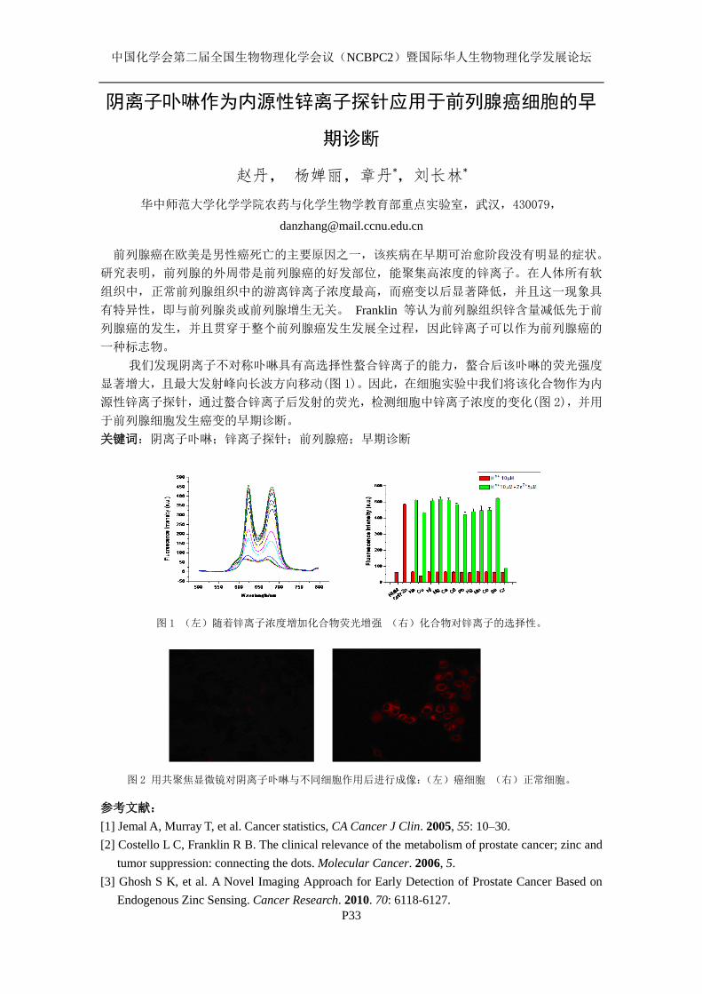
中国化学会第二届全国生物物理化学会议(NCBPC2)暨国际华人生物物理化学发展论坛
P33
阴离子卟啉作为内源性锌离子探针应用于前列腺癌细胞的早
期诊断
赵丹, 杨婵丽,章丹*,刘长林*
华中师范大学化学学院农药与化学生物学教育部重点实验室,武汉,430079,
前列腺癌在欧美是男性癌死亡的主要原因之一,该疾病在早期可治愈阶段没有明显的症状。
研究表明,前列腺的外周带是前列腺癌的好发部位,能聚集高浓度的锌离子。在人体所有软
组织中,正常前列腺组织中的游离锌离子浓度最高,而癌变以后显著降低,并且这一现象具
有特异性,即与前列腺炎或前列腺增生无关。 Franklin 等认为前列腺组织锌含量减低先于前
列腺癌的发生,并且贯穿于整个前列腺癌发生发展全过程,因此锌离子可以作为前列腺癌的
一种标志物。
我们发现阴离子不对称卟啉具有高选择性螯合锌离子的能力,螯合后该卟啉的荧光强度
显著增大,且最大发射峰向长波方向移动(图 1)。因此,在细胞实验中我们将该化合物作为内
源性锌离子探针,通过螯合锌离子后发射的荧光,检测细胞中锌离子浓度的变化(图 2),并用
于前列腺细胞发生癌变的早期诊断。
关键词:阴离子卟啉;锌离子探针;前列腺癌;早期诊断
图 1 (左)随着锌离子浓度增加化合物荧光增强 (右)化合物对锌离子的选择性。
图 2 用共聚焦显微镜对阴离子卟啉与不同细胞作用后进行成像:(左)癌细胞 (右)正常细胞。
参考文献:
[1] Jemal A, Murray T, et al. Cancer statistics, CA Cancer J Clin. 2005, 55: 10–30.
[2] Costello L C, Franklin R B. The clinical relevance of the metabolism of prostate cancer; zinc and
tumor suppression: connecting the dots. Molecular Cancer. 2006, 5.
[3] Ghosh S K, et al. A Novel Imaging Approach for Early Detection of Prostate Cancer Based on
Endogenous Zinc Sensing. Cancer Research. 2010. 70: 6118-6127.
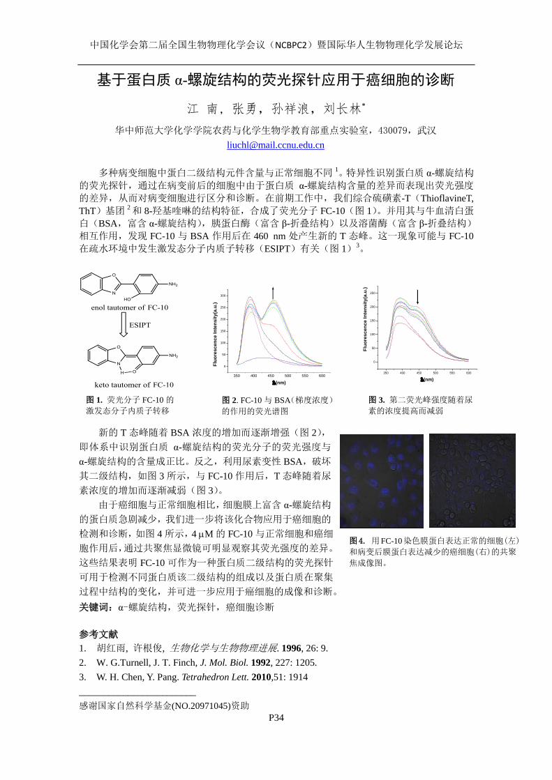
中国化学会第二届全国生物物理化学会议(NCBPC2)暨国际华人生物物理化学发展论坛
P34
基于蛋白质 α-螺旋结构的荧光探针应用于癌细胞的诊断
江 南, 张勇,孙祥浪,刘长林*
华中师范大学化学学院农药与化学生物学教育部重点实验室,430079,武汉
多种病变细胞中蛋白二级结构元件含量与正常细胞不同 1。特异性识别蛋白质 α-螺旋结构
的荧光探针,通过在病变前后的细胞中由于蛋白质 α-螺旋结构含量的差异而表现出荧光强度
的差异,从而对病变细胞进行区分和诊断。在前期工作中,我们综合硫磺素-T(ThioflavineT,
ThT)基团 2 和 8-羟基喹啉的结构特征,合成了荧光分子 FC-10(图 1)。并用其与牛血清白蛋
白(BSA,富含 α-螺旋结构),胰蛋白酶(富含 β-折叠结构)以及溶菌酶(富含 β-折叠结构)
相互作用,发现 FC-10 与 BSA 作用后在 460 nm 处产生新的 T 态峰。这一现象可能与 FC-10
在疏水环境中发生激发态分子内质子转移(ESIPT)有关(图 1)3。
新的 T 态峰随着 BSA 浓度的增加而逐渐增强(图 2),
即体系中识别蛋白质 α-螺旋结构的荧光分子的荧光强度与
α-螺旋结构的含量成正比。反之,利用尿素变性 BSA,破坏
其二级结构,如图 3 所示,与 FC-10 作用后,T 态峰随着尿
素浓度的增加而逐渐减弱(图 3)。
由于癌细胞与正常细胞相比,细胞膜上富含 α-螺旋结构
的蛋白质急剧减少,我们进一步将该化合物应用于癌细胞的
检测和诊断,如图 4 所示,4 M 的 FC-10 与正常细胞和癌细
胞作用后,通过共聚焦显微镜可明显观察其荧光强度的差异。
这些结果表明 FC-10 可作为一种蛋白质二级结构的荧光探针
可用于检测不同蛋白质该二级结构的组成以及蛋白质在聚集
过程中结构的变化,并可进一步应用于癌细胞的成像和诊断。
关键词:α-螺旋结构,荧光探针,癌细胞诊断
参考文献
1. 胡红雨, 许根俊, 生物化学与生物物理进展. 1996, 26: 9.
2. W. G.Turnell, J. T. Finch, J. Mol. Biol. 1992, 227: 1205.
3. W. H. Chen, Y. Pang. Tetrahedron Lett. 2010,51: 1914
________________________
感谢国家自然科学基金(NO.20971045)资助
图 3. 第二荧光峰强度随着尿
素的浓度提高而减弱 图 2. FC-10 与 BSA(梯度浓度)
的作用的荧光谱图
图 1. 荧光分子 FC-10 的
激发态分子内质子转移
350 400 450 500 550 600
0
50
100
150
200
250
300
Fluo
resc
ence
Inte
nsity
(a.u
.)
(nm)
350 400 450 500 550 600
0
50
100
150
200
250
Fluo
resc
ence
Inte
nsity
(a.u
.)
(nm)
图4. 用FC-10染色膜蛋白表达正常的细胞(左)
和病变后膜蛋白表达减少的癌细胞(右)的共聚
焦成像图。
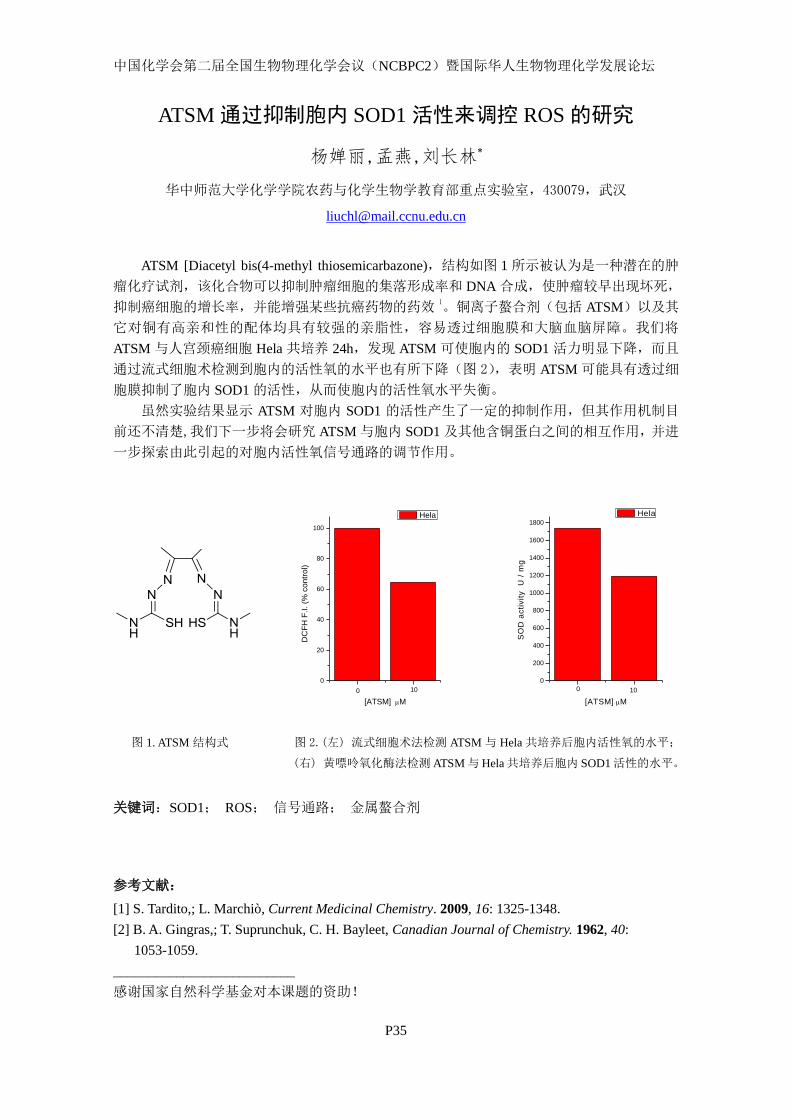
中国化学会第二届全国生物物理化学会议(NCBPC2)暨国际华人生物物理化学发展论坛
P35
ATSM 通过抑制胞内 SOD1 活性来调控 ROS 的研究
杨婵丽,孟燕,刘长林*
华中师范大学化学学院农药与化学生物学教育部重点实验室,430079,武汉
ATSM [Diacetyl bis(4-methyl thiosemicarbazone),结构如图 1 所示被认为是一种潜在的肿
瘤化疗试剂,该化合物可以抑制肿瘤细胞的集落形成率和 DNA 合成,使肿瘤较早出现坏死,
抑制癌细胞的增长率,并能增强某些抗癌药物的药效1。铜离子螯合剂(包括 ATSM)以及其
它对铜有高亲和性的配体均具有较强的亲脂性,容易透过细胞膜和大脑血脑屏障。我们将
ATSM 与人宫颈癌细胞 Hela 共培养 24h,发现 ATSM 可使胞内的 SOD1 活力明显下降,而且
通过流式细胞术检测到胞内的活性氧的水平也有所下降(图 2),表明 ATSM 可能具有透过细
胞膜抑制了胞内 SOD1 的活性,从而使胞内的活性氧水平失衡。
虽然实验结果显示 ATSM 对胞内 SOD1 的活性产生了一定的抑制作用,但其作用机制目
前还不清楚,我们下一步将会研究 ATSM 与胞内 SOD1 及其他含铜蛋白之间的相互作用,并进
一步探索由此引起的对胞内活性氧信号通路的调节作用。
0
20
40
60
80
100
DC
FH F
.I. (%
con
trol)
[ATSM] M
Hela
0 10
0
200
400
600
800
1000
1200
1400
1600
1800S
OD
act
ivity
U /
mg
[ATSM] M
Hela
0 10
图 1. ATSM 结构式 图 2.(左) 流式细胞术法检测 ATSM 与 Hela 共培养后胞内活性氧的水平;
(右) 黄嘌呤氧化酶法检测 ATSM 与 Hela 共培养后胞内 SOD1 活性的水平。
关键词:SOD1; ROS; 信号通路; 金属螯合剂
参考文献:
[1] S. Tardito,; L. Marchiò, Current Medicinal Chemistry. 2009, 16: 1325-1348.
[2] B. A. Gingras,; T. Suprunchuk, C. H. Bayleet, Canadian Journal of Chemistry. 1962, 40:
1053-1059.
__________________________
感谢国家自然科学基金对本课题的资助!
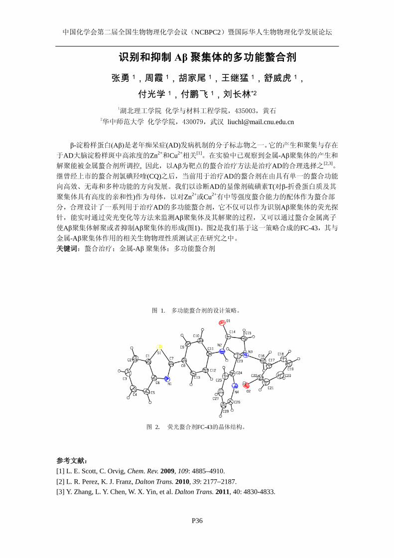
中国化学会第二届全国生物物理化学会议(NCBPC2)暨国际华人生物物理化学发展论坛
P36
识别和抑制 Aβ聚集体的多功能螯合剂
张勇 1,周霞 1,胡家尾 1,王继猛 1,舒威虎 1, 付光学 1,付鹏飞 1,刘长林*2
1湖北理工学院 化学与材料工程学院,435003,黄石
2华中师范大学 化学学院,430079,武汉 [email protected]
β-淀粉样蛋白(Aβ)是老年痴呆症(AD)发病机制的分子标志物之一。它的产生和聚集与存在
于AD大脑淀粉样斑中高浓度的Zn2+和Cu2+相关[1]。在实验中已观察到金属-Aβ聚集体的产生和
解聚能被金属螯合剂所调控, 因此,以Aβ为靶点的螯合治疗方法是治疗AD的合理选择之[2,3]。 继曾经上市的螯合剂氯碘羟喹(CQ)之后,当前用于治疗AD的螯合剂在由具有单一的螯合功能
向高效、无毒和多种功能的方向发展。我们以诊断AD的显像剂硫磺素T(对β-折叠蛋白质及其
聚集体具有高度的亲和性)作为母体,以对Zn2+或Cu2+有中等强度螯合能力的配体作为螯合部
分,合理设计了一系列用于治疗AD的多功能螯合剂,它不仅可以作为识别Aβ聚集体的荧光探
针,能实时通过荧光变化等方法来监测Aβ聚集体及其解聚的过程,又可以通过螯合金属离子
使Aβ聚集体解聚或者抑制Aβ聚集体的形成(图1)。图2是我们基于这一策略合成的FC-43,其与
金属-Aβ聚集体作用的相关生物物理性质测试正在研究之中。 关键词:螯合治疗;金属-Aβ聚集体;多功能螯合剂
图 1. 多功能螯合剂的设计策略。
图 2. 荧光螯合剂FC-43的晶体结构。
参考文献: [1] L. E. Scott, C. Orvig, Chem. Rev. 2009, 109: 4885–4910. [2] L. R. Perez, K. J. Franz, Dalton Trans. 2010, 39: 2177−2187. [3] Y. Zhang, L. Y. Chen, W. X. Yin, et al. Dalton Trans. 2011, 40: 4830-4833.
中国化学会第二届全国生物物理化学会议(NCBPC2)暨国际华人生物物理化学发展论坛
P37
生物多聚阴离子促进野生型 SOD1 和 ALS 相关 SOD1 突变体
A4V 聚集的研究
郭晶,张世炳,孟燕,刘长林* 华中师范大学农药与化学生物学教育部重点实验室,430079,武汉
在发现铜锌超氧化物歧化酶(SOD1)突变体是肌萎缩厕所硬化症(ALS)的致病原因 20
年之后,我们对其毒性机理仍不是很清楚。有很多关于 SOD1 突变体结构和功能变化的假说,
如金属化或脱辅基 SOD1 稳定性的降低,疏水性和聚集倾向的增加,以及异常化学作用。形
成蛋白质聚集体倾向的上升是所有与 ALS 相关的 SOD1 突变体所共有的毒性性质。 越来越多体内、外研究表明,作为两大类关键的生物多聚阴离子,肝素和核酸(DNA,
RNA),它们对有聚集倾向的蛋白质有很强的亲和性。很多研究发现多聚阴离子与病变蛋白质
(包括 Aβ、核突触蛋白、运甲状腺素蛋白和阮病毒蛋白 PrP)1 的聚集相关。虽然还不能肯
定富含 SOD1 的蛋白质聚集体是否含有核酸,但是我们已经报道了 DNA 在酸性条件下能加速
野生型 SOD1 转变为沉积物。而 DNA 在此所扮演的角色称为模板效应 2-5。正如在核酸调控的
运甲状腺素蛋白和阮病毒蛋白聚集的研究中所观察到的,DNA 最终会留在 SOD1 聚集体中。 生物多聚阴离子参与病变蛋白质聚集体的形成,可能成为调控聚集体细胞毒性的途径之
一。近来的体内、外研究表明,相较于包括 Aβ在内的蛋白质所形成的类淀粉斑块,其可溶性
寡聚体的神经毒性更大。本文研究了在生物多聚阴离子存在下,一种与 ALS 相关的 SOD1 突
变体 A4V 的聚集。结果证实在 A4V 聚集过程中,DNA 和肝素都起到了模板作用;而且如同
野生型 SOD1 聚集过程,这种模板效应不仅能加速 A4V 聚集体的形成,还能将其 A4V 寡聚体
转变成沉积物。这种转变显著降低了 A4V 聚集体的毒性。因此,在 SOD1 突变体聚集过程中,
多聚阴离子的模板作用有望成为一种降低 SOD1 寡聚体或聚集体的有效途径。 关键词:生物多聚阴离子;SOD1;A4V;聚集
参考文献:
1. C Liu,Y Zhang. Nucleic acid-mediated protein aggtegation and assembly. In:Donev R, editor. Advances in Protein Chemistry and Structural Biology. London: Elsevier. pp. 1-40,2011.
2. W Jiang, Y Han, R Zhou, L Zhang, C Liu. DNA is a template for accelerating the aggregation of copper, zinc superoxide dismutase. Biochemistry 2007, 46: 5911-5923.
3. W Jiang, B Zhang, J Yin, L Liu, L Wang, C Liu. Polymorphism of the SOD1-DNA aggregation species can be modulated by DNA. Biopolymers 2008, 89:1154-1169.
4. J Yin, S Hu, W Jiang, L Liu, S Lan, C Liu. DNA-triggered aggregation of copper, zinc superoxide dismutase in the presence of ascorbate. PLoS One 2010,5: e12328.
5. J Yin, R Chen, C Liu. Nucleic acid induced protein aggregation and its role in biology and pathology. Frontiers in Bioscience 2009, 14: 5084-5106.
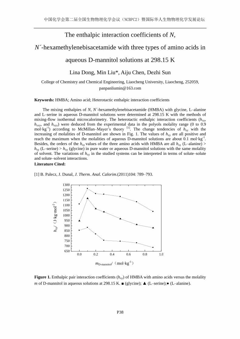
中国化学会第二届全国生物物理化学会议(NCBPC2)暨国际华人生物物理化学发展论坛
P38
The enthalpic interaction coefficients of N,
N -́hexamethylenebisacetamide with three types of amino acids in
aqueous D-mannitol solutions at 298.15 K
Lina Dong, Min Liu*, Aiju Chen, Dezhi Sun
College of Chemistry and Chemical Engineering, Liaocheng University, Liaocheng, 252059,
Keywords: HMBA; Amino acid; Heterotactic enthalpic interaction coefficients
The mixing enthalpies of N, N -́hexamethylenebisacetamide (HMBA) with glycine, L–alanine
and L–serine in aqueous D-mannitol solutions were determined at 298.15 K with the methods of
mixing-flow isothermal microcalorimetry. The heterotactic enthalpic interaction coefficients (hxy,
hxxy, and hxyy) were deduced from the experimental data in the polyols molality range (0 to 0.9
mol·kg-1
) according to McMillan–Mayer’s theory [1]
. The change tendencies of hxy with the
increasing of molalities of D-mannitol are shown in Fig. 1. The values of hxy are all positive and
reach the maximum when the molalities of aqueous D-mannitol solutions are about 0.1 mol·kg-1
.
Besides, the orders of the hxy values of the three amino acids with HMBA are all hxy (L–alanine) > hxy (L–serine) > hxy (glycine) in pure water or aqueous D-mannitol solutions with the same molality
of solvent. The variations of hxy in the studied systems can be interpreted in terms of solute–solute
and solute–solvent interactions.
Literature Cited:
[1] B. Palecz, J. Dunal, J. Therm. Anal. Calorim.(2011)104: 789–793.
Figure 1. Enthalpic pair interaction coefficients (hxy) of HMBA with amino acids versus the molality
m of D-mannitol in aqueous solutions at 298.15 K. ■ (glycine); ▲ (L–serine);● (L–alanine).
0.0 0.2 0.4 0.6 0.8 1.0650
700
750
800
850
900
950
1000
1050
1100
1150
1200
1250
1300
hx
y/(
J·kg·m
ol-2)
mD-mannitol/(mol·kg-1)
中国化学会第二届全国生物物理化学会议(NCBPC2)暨国际华人生物物理化学发展论坛
P39
Free radical scavenging activity and the thermodynamic
properties: The comparison of melatonin and other
antioxidants
Xiangrong Li1,2
, Gongke Wang1, Yan Lu
1
1School of Chemistry and Environmental Science, Henan Normal University, Xinxiang, Henan
453007, PR China; 2 Department of Chemistry, School of Basic Medicine, Xinxiang Medical
University, Xinxiang, Henan, 453003, PR China, [email protected], [email protected]
Melatonin is produced mainly by the pineal gland of mammals and it also exists in plants 1. In
this paper, we studied the ability of melatonin to scavenge several free radicals such as DPPH radical,
hydroxyl radical and superoxide anion radical using UV spectrum. And then, the binding of
melatonin to human serum albumin (HSA) has been studied using isothermal titration calorimetry
(ITC). The equilibrium constant K, standard change of enthalpyΔH0 and the number of binding site n
of HSA for melatonin have been determined by ITC at pH 7.4 and 298.15K. Based on the
thermodynamic formula, entropy change ΔS0 and Gibbs free energy change ΔG
0 of the process were
obtained. The results indicate that melatonin shows a potent antioxidant activity in the DPPH
radical-scavenging assay (IC50 = 1.682×10⁻⁴
±0.014 mol.L
-1), significantly inhibits
.OH radical (IC50
= 5.285×10⁻⁴
±0.011 mol.L
-1), and O2
.- anion radical (IC50 = 6.666×10
-3±0.003 mol
.L
-1). The binding
of melatonin to HSA is synergistically driven by enthalpy and entropy and the hydrophobic
interaction plays a major role in the binding reaction. What’s more, the free radical scavenging
activity and the thermodynamic properties of melatonin were compared with the other three
antioxidants including ascorbic acid (ASC), reduced glutathione (GSH) and
6-hydroxy-2,5,7,8-tetramethylchroman-2-carboxylic acid (trolox).
Keywords: Melatonin; Human serum albumin; Free radical scavenging activity; UV spectrum;
Isothermal titration calorimetric
References
[1] H. P. Zhu, S. M. Chen, S. D. Yan, W. F. Wang, Protective effect of melatonin on photo-damage to
lysozyme. J. Photochem Photobiol., B. 2009, 94: 125–130.
This work is supported by the National Natural Science Foundation of China (20673034, 21173071)
and the Research Fund for the Doctoral Program of Higher Education of China (20060476001)
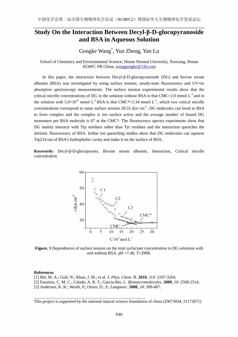
中国化学会第二届全国生物物理化学会议(NCBPC2)暨国际华人生物物理化学发展论坛
P40
Study On the Interaction Between Decyl-β-D-glucopyranoside and BSA in Aqueous Solution
Gongke Wang*, Yun Zheng, Yan Lu
School of Chemistry and Environmental Science, Henan Normal University, Xinxiang, Henan
453007, PR China, [email protected]
In this paper, the interaction between Decyl-β-D-glucopyranoside (DG) and bovine serum albumin (BSA) was investigated by using surface tension, steady-state fluorescence and UV-vis absorption spectroscopy measurements. The surface tension experimental results show that the critical micelle concentrations of DG in the solution without BSA is that CMC=2.0 mmol L-1 and in the solution with 5.0×10⁻⁶ mmol L-1 BSA is that CMC*=2.34 mmol L-1, which two critical micelle concentrations correspond to same surface tension 28.55 dyn cm-1. DG molecules can bond to BSA to form complex and the complex is not surface active and the average number of bound DG monomers per BSA molecule is 67 at the CMC*. The fluorescence spectra experiments show that DG mainly interacts with Trp residues rather than Tyr residues and the interaction quenches the intrinsic fluorescence of BSA. Iodine ion quenching studies show that DG molecules can squeeze Trp214 out of BSA’s hydrophobic cavity and make it on the surface of BSA. Keywords: Decyl-β-D-glucopyrano, Bovine serum albumin, Interaction, Critical micelle concentration
Figure. 1 Dependences of surface tension on the total surfactant concentration in DG solutions with and without BSA. pH =7.40, T=298K.
References [1] Mir, M. A.; Gull, N.; Khan, J. M.; et al. J. Phys. Chem. B. 2010, 114: 3197-3204. [2] Faustino, C. M. C.; Calado, A. R. T.; Garcia-Rio, L. Biomacromolecules. 2009, 10: 2508-2514. [3] Andersen, K. K.; Westh, P.; Otzen, D.; E. Langmuir. 2008, 24: 399-407. _____________________________________________________ This project is supported by the national natural science foundation of china (20673034, 21173071)
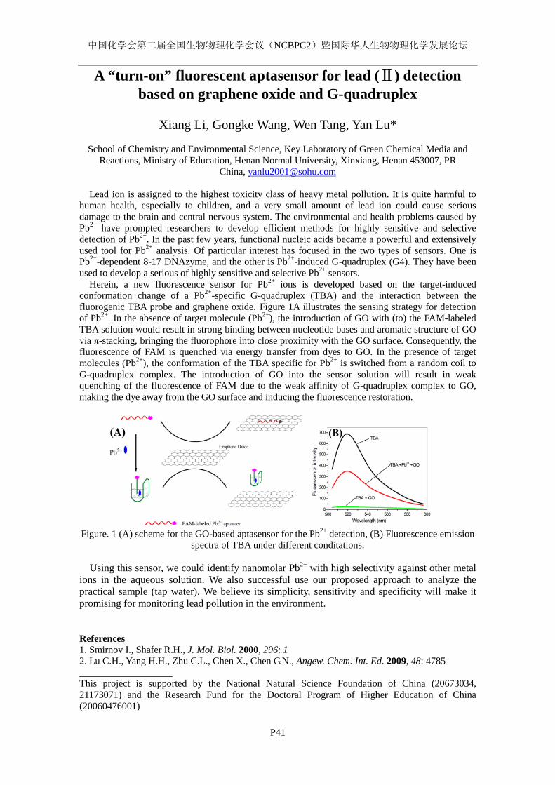
中国化学会第二届全国生物物理化学会议(NCBPC2)暨国际华人生物物理化学发展论坛
P41
A “turn-on” fluorescent aptasensor for lead (Ⅱ) detection based on graphene oxide and G-quadruplex
Xiang Li, Gongke Wang, Wen Tang, Yan Lu*
School of Chemistry and Environmental Science, Key Laboratory of Green Chemical Media and
Reactions, Ministry of Education, Henan Normal University, Xinxiang, Henan 453007, PR China, [email protected]
Lead ion is assigned to the highest toxicity class of heavy metal pollution. It is quite harmful to
human health, especially to children, and a very small amount of lead ion could cause serious damage to the brain and central nervous system. The environmental and health problems caused by Pb2+ have prompted researchers to develop efficient methods for highly sensitive and selective detection of Pb2+. In the past few years, functional nucleic acids became a powerful and extensively used tool for Pb2+ analysis. Of particular interest has focused in the two types of sensors. One is Pb2+-dependent 8-17 DNAzyme, and the other is Pb2+-induced G-quadruplex (G4). They have been used to develop a serious of highly sensitive and selective Pb2+ sensors.
Herein, a new fluorescence sensor for Pb2+ ions is developed based on the target-induced conformation change of a Pb2+-specific G-quadruplex (TBA) and the interaction between the fluorogenic TBA probe and graphene oxide. Figure 1A illustrates the sensing strategy for detection of Pb2+. In the absence of target molecule (Pb2+), the introduction of GO with (to) the FAM-labeled TBA solution would result in strong binding between nucleotide bases and aromatic structure of GO via π-stacking, bringing the fluorophore into close proximity with the GO surface. Consequently, the fluorescence of FAM is quenched via energy transfer from dyes to GO. In the presence of target molecules (Pb2+), the conformation of the TBA specific for Pb2+ is switched from a random coil to G-quadruplex complex. The introduction of GO into the sensor solution will result in weak quenching of the fluorescence of FAM due to the weak affinity of G-quadruplex complex to GO, making the dye away from the GO surface and inducing the fluorescence restoration.
Figure. 1 (A) scheme for the GO-based aptasensor for the Pb2+ detection, (B) Fluorescence emission
spectra of TBA under different conditations.
Using this sensor, we could identify nanomolar Pb2+ with high selectivity against other metal ions in the aqueous solution. We also successful use our proposed approach to analyze the practical sample (tap water). We believe its simplicity, sensitivity and specificity will make it promising for monitoring lead pollution in the environment.
References 1. Smirnov I., Shafer R.H., J. Mol. Biol. 2000, 296: 1 2. Lu C.H., Yang H.H., Zhu C.L., Chen X., Chen G.N., Angew. Chem. Int. Ed. 2009, 48: 4785 This project is supported by the National Natural Science Foundation of China (20673034, 21173071) and the Research Fund for the Doctoral Program of Higher Education of China (20060476001)
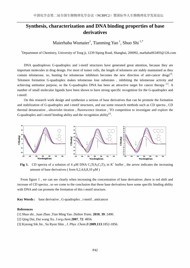
中国化学会第二届全国生物物理化学会议(NCBPC2)暨国际华人生物物理化学发展论坛
P42
Synthesis, characterization and DNA binding properties of base derivatives
Maierhaba Wumaier1, Tianming Yao 1, Shuo Shi 1,*
1Department of Chemistry, University of Tong ji, 1239 Siping Road, Shanghai, 200092, [email protected]
DNA quadruplexes G-quadruplex and i-motif structures have generated great attention, because they are
important molecules in drug design. For most of tumor cells, the length of telomeres are stably maintained as they contain telomerase, so, hunting for telomerase inhibitors becomes the new direction of anti-cancer drugs[3]. Telomere formation G-quadruplex makes telomerase lose substrates , inhibiting the telomerase activity and achieving antitumor purpose, so the G-quadruplex DNA has been an attractive target for cancer therapy [1]. A number of small molecular ligands have been shown to have strong specific recognition for the G-quadruplex and i-motif.
On this research work design and synthesize a serious of base derivatives that can be promote the formation and stabilization of G-quadruplex and i-motif structures, and use some research methods such as CD spectra , CD thermal denaturation , ultraviolet titration , fluorescence titration , TO competition to investigate and explore the G-quadruplex and i-motif binding ability and the recognition ability[2].
220 240 260 280 300 320 340
-20
-10
0
10
20
30
40
CD[m
deg]
Wavelength/nm 220 240 260 280 300 320 340
-30
-20
-10
0
10
20
30
40
50
CD[m
deg]
Wavelength/nm220 240 260 280 300 320 340
-30-25-20-15-10-505
1015202530354045
CD[m
deg]
Wavelength/nm Fig 1. CD spectra of a solution of 4 μM DNA C3T(A2C3T)3 in K+ buffer , the arrow indicates the increasing
amount of base derivatives ( from 0,2,4,6,8,10 μM ) From figure 1 , we can see clearly when increasing the concentration of base derivatives ,there is red shift and
increase of CD spectra , so we come to the conclusion that these base derivatives have some specific binding ability with DNA and can promote the formation of this i-motif structure. Key Words : base derivative , G-quadruplex , i-motif , anticancer References [1] Shuo shi , Juan Zhao ,Tian Ming Yao. Dalton Trans. 2010, 39: 2490. [2] Qing Dai, Dai wang Xu. J.org.chem.2007, 72: 4856. [3] Kyeong Sik Jin , Su Ryon Shin , J. Phys .Chem.B 2009,113:1852-1856.
N NH
HN
N
NH2
O
N NH
HN
N
NH2
O
N NH
HN
N
NH2
O
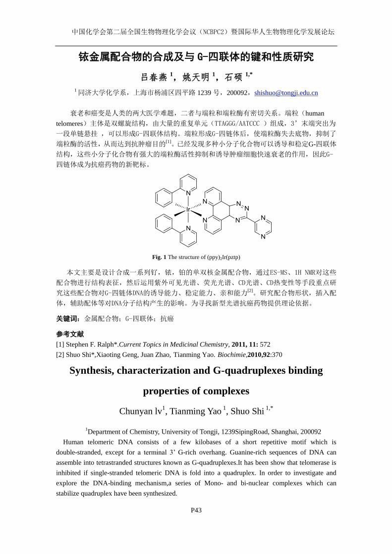
中国化学会第二届全国生物物理化学会议(NCBPC2)暨国际华人生物物理化学发展论坛
P43
铱金属配合物的合成及与 G-四联体的键和性质研究
吕春燕 1,姚天明 1,石硕 1,*
1 同济大学化学系,上海市杨浦区四平路 1239 号,200092,[email protected]
衰老和癌变是人类的两大医学难题,二者与端粒和端粒酶有密切关系。端粒(human
telomeres)主体是双螺旋结构,由大量的重复单元(TTAGGG/AATCCC )组成,3’末端突出为
一段单链悬挂 ,可以形成G-四联体结构。端粒形成G-四链体后,使端粒酶失去底物,抑制了
端粒酶的活性,从而达到抗肿瘤目的[1]。已经发现多种小分子化合物可以诱导和稳定G-四联体
结构,这些小分子化合物有强大的端粒酶活性抑制和诱导肿瘤细胞快速衰老的作用,因此G-
四链体成为抗癌药物的新靶标。
Fig. 1 The structure of (ppy)2Ir(pztp)
本文主要是设计合成一系列钌,铱,铂的单双核金属配合物,通过ES-MS、1H NMR对这些
配合物进行结构表征,然后运用紫外可见光谱、荧光光谱、CD光谱、CD热变性等手段重点研
究这些配合物对G-四链体DNA的诱导能力、稳定能力、亲和能力[2]。研究配合物形状,插入配
体,辅助配体等对DNA分子结构产生的影响。为寻找新型光谱抗癌药物提供理论依据。
关键词:金属配合物;G-四联体;抗癌
参考文献
[1] Stephen F. Ralph*.Current Topics in Medicinal Chemistry, 2011, 11: 572
[2] Shuo Shi*,Xiaoting Geng, Juan Zhao, Tianming Yao. Biochimie,2010,92:370
Synthesis, characterization and G-quadruplexes binding
properties of complexes
Chunyan lv1, Tianming Yao
1, Shuo Shi
1,*
1Department of Chemistry, University of Tongji, 1239SipingRoad, Shanghai, 200092
Human telomeric DNA consists of a few kilobases of a short repetitive motif which is
double-stranded, except for a terminal 3’ G-rich overhang. Guanine-rich sequences of DNA can
assemble into tetrastranded structures known as G-quadruplexes.It has been show that telomerase is
inhibited if single-stranded telomeric DNA is fold into a quadruplex. In order to investigate and
explore the DNA-binding mechanism,a series of Mono- and bi-nuclear complexes which can
stabilize quadruplex have been synthesized.
中国化学会第二届全国生物物理化学会议(NCBPC2)暨国际华人生物物理化学发展论坛
P44
Probing Allostery Through DNA
Sangjin Kim1*†
, Erik Broströmer1*
, Dong Xing2*
, Jianshi Jin2,3*
, Shasha
Chong1,Hao Ge
2,4,5, Siyuan Wang
1, Xiaodong Su
2, Yujie Sun
2§& X. Sunney
Xie1,2§
1 Department of Chemistry & Chemical Biology, Harvard University, Cambridge, MA 02138, USA.
2Biodynamic Optical Imaging Center, Peking University, Beijing 100871, China.
3Academy for Advanced Interdisciplinary Studies, Peking University, Beijing 100871, China
4Beijing International Center for Mathematical Research, Peking University, Beijing 100871, China.
5 School of Mathematical Sciences and Centre for Computational Systems Biology, Fudan
University, Shanghai 200433, China
* These authors contributed equally to this work.
†Present address: Department of Molecular Cellular & Developmental Biology, Yale University,
New Haven, CT 06511, USA
§ To whom correspondence should be addressed.
E-mail: [email protected] (Y.S.); [email protected] (X.S.X)
Allostery is well-documented for proteins but less recognized for DNA-protein interactions, in
which DNA has been often considered a mere template providing recognition sequences. Here we
report that when two proteins specifically bind to DNA within tens of base pairs, the binding affinity
of one protein is altered by the other. We prove that this effect is not due to protein–protein
interactions but to allostery through DNA. Asthe distance between the two proteins is varied,
thisallosteric coupling oscillates between positive and negative cooperativity with a periodicity of
~10 base pairs, the helical pitch of the B-form DNA.We providethermodynamicanalyses of the
coupling energetics as well as an experimentally validated mechanism for the allostery. We also find
that the binding affinity of a transcription factor is significantly varied in an oscillatory fashion as it
approaches a nucleosome, suggesting DNA allostery in gene regulation. This physiological
relevancewas further demonstrated by gene expression in live E. coli cells.
中国化学会第二届全国生物物理化学会议(NCBPC2)暨国际华人生物物理化学发展论坛
P45
利用高分辨原子力显微镜研究线粒体膜结构
田咏梅 1, 李佳涵 2, 蔡明军 1, 赵伟栋 1, 徐海娇 1, 刘义 2*
王宏达 1* 1中国科学院长春应用化学研究所,电分析化学国家重点实验室,130022,长春
[email protected] 2武汉大学化学与分子科学学院,病毒学国家重点实验室,430072,武汉
关键词:原子力显微镜, 线粒体膜 线粒体是细胞的能量工厂。细胞内许多复杂的生理过程都与线粒体紧密相关,例如细胞
凋亡、心磷脂保护、中间代谢、钙离子信号转导等1。线粒体功能障碍可能引发很多疾病,如
癌症、帕金森、糖尿病等2。因此,在分子水平上研究线粒体的膜结构对于设计以线粒体为靶
向的药物3、癌症治疗等具有十分重要的意义。
从经典的电子显微镜到新出现的各种超分辨荧光显微镜(STED, TALM 等)都已经被用于
研究线粒体的结构。但是这些都是间接的观察,有些甚至是破坏性的。线粒体膜的精细结构
仍然是不清楚的,并且目前还没有一种很好的方法来测量线粒体膜的厚度。我们利用原子力
显微镜在接近生理的条件下研究线粒体的膜结构,探索线粒体膜的微观特征、蛋白的分布等。
大鼠肝脏线粒体的原子力显微镜成像表明线粒体外膜表面没有明显的蛋白分布。线粒体
内膜的膜间隙一侧是非常光滑的,而基质一侧是富含蛋白颗粒的,这个结论与红细胞膜内外
两侧蛋白的分布是一致的4。本文的研究有助于认识线粒体的微观膜结构,进而了解线粒体膜
的信号转导、线粒体与细胞的物质和能量代谢等生理过程,还潜在为设计以线粒体为靶点的
药物提供参考。
参考文献:
[1] D. C. Chan, Cell, 2006, 125:1241-1252. [2] A. Szewczyk, L. Wojtczak, Pharmacol. Rev., 2002, 54: 101-127. [3] A. Sasidharan, P. Chandran, D. Menon, S. Raman, S. Nair, M. Koyakutty. Nanoscale, 2011, 3:
3657-3669. [4] H. D. Wang, X. Hao, Y. P. Shan, J. G. Jiang, M. J. Cai and X. Shang, Ultramicroscopy, 2010, 110:
305-312.
中国化学会第二届全国生物物理化学会议(NCBPC2)暨国际华人生物物理化学发展论坛
P46
利用原子力显微镜研究高尔基体膜结构
徐海娇 1,2,苏维恒 1,蔡明军 1,蒋俊光 1,曾宪录 2,王宏达 1*
1中国科学院长春应用化学研究所,吉林省长春市人民大街 5625号,邮编 130022
2东北师范大学遗传与细胞研究所,吉林省长春市人民大街 5268号,邮编 130024
关键词:原子力显微镜,高尔基体膜,脂筏
高尔基体是真核细胞内膜系统的重要细胞器,并被认为是细胞经典分泌途径的枢纽。对
高尔基体形貌结构、分子组成、以及其动态结构的深刻认识是更好地理解高尔基体重要功能
的前提。然而,由于研究方法的局限性,使得在分子水平上对高尔基体结构的认识受到了限
制。例如,光学显微镜具有较低的空间分辨率;电子显微镜虽然具有较高的分辨率,但是不
能在近生理条件下对生物样品进行实时的分析和成像。
原子力显微镜由于具有多种成像环境、高的分辨率、成像直接(无需包埋,染色等处理)
等优点1,因此已经成为生物学研究中的有力工具
2-4。我们利用原子力显微镜在接近生理条件
下从分子水平对高尔基体结构进行了研究。我们的重要发现是高尔基体膜具有不对称性:外
膜非常平整;而内膜由于分布有很多蛋白,相对比较粗糙。同时我们也利用实时原位原子力
显微镜成像分析了 MβCD 和 TX-100 处理的高尔基体膜,确定了脂筏的存在。
参考文献:
[1] H. Wang, R. Bash, Stuart. Lindsay, D. Lohr. Biophysical Journal, 2005, 89: 3386.
[2] J. Jiang, X. Hao, M. Cai, Y. Shan, X. Shang, Z. Tang, H. Wang. Nano Letters, 2009, 9: 4489-93.
[3] H. Wang, X. Hao, Y. Shan, J. Jiang, M. Cai, X. Shang. Ultramicroscopy, 2010, 110: 305-12.
[4] M. Cai, W. Zhao, X. Shang, J. Jiang, Z. Tang, H. Wang. Small, 2012, 8: 1243-1250.
中国化学会第二届全国生物物理化学会议(NCBPC2)暨国际华人生物物理化学发展论坛
P47
利用超分辨显微镜研究红细胞膜表面的 Na+-K
+ ATPase分布
高婧,吴佳桢,蒋俊光,王宏达*
中国科学院长春应用化学研究所,吉林省长春市人民大街 5625号,130022
关键词:超分辨荧光显微镜,细胞膜,Na+-K
+ ATPase
近几年来,超分辨荧光显微成像技术得到了迅猛的发展,它突破了传统光学显微镜在水
平方向上 200nm的衍射极限,实现了 20nm左右的超高影像分辨率,能清楚地观察到活细胞的
纳米级结构1。Na
+-K
+ ATPase 作为一种重要的离子通道蛋白,在跨膜运输 K
+和 Na
+的过程中发
挥着显著作用。目前,人们已经对 Na+-K
+ ATPase 结构有了深入的研究
2,然而对于其在单分
子层面上的分布状态仍了解甚少。我们利用超分辨荧光显微成像技术,即直接随机光学重建
显微镜(dSTORM),在单分子水平上展示细胞膜上 Na+-K
+ ATPase 的分布
3,4,同时表明 Na
+-K
+
ATPase是在脂筏上以聚集态的形式完成其生理功能的5。这些结果为在分子水平上研究细胞膜
蛋白结构以及结构和功能的关系提供一定的指导意义。
参考文献:
[1] M. J. Rust, M. Bates, X. Zhuang, Nat. Methods, 2006, 3: 793.
[2] J. P. Morth, , B. P. Pedersen, , M. S. Toustrup-Jensen, , T. L. M. Sorensen, , J. Petersen, , J. P.
Andersen, B. Vilsen, P. Nissen. Nature, 2007, 450: 1043.
[3] J. Jiang, X. Hao, M. Cai, Y. Shan, X. Shang, Z. Tang, H. Wang, Nano Lett., 2009, 9: 4489.
[4] M. S. Itano,, C. Steinhauer. Biophys. J., 2012, 102: 1534.
[5] M. Cai, W. Zhao, X. Shang, J. Jiang, H. Ji, Z. Tang, H. Wang. Small, 2012, 8: 1243.

中国化学会第二届全国生物物理化学会议(NCBPC2)暨国际华人生物物理化学发展论坛
P48
现场条件下研究细胞膜转运单分子的过程
潘延刚, 单玉萍, 刘姝姮, 王宏达*
中国科学院长春应用化学研究所,吉林省长春市人民大街 5625号, 130022,
关键词: 力示踪, 葡萄糖转运, 细胞膜, 单分子
细胞通过细胞膜来摄取日常的营养物质,如葡萄糖和氨基酸等1-3。在分子水平上了解这
些营养分子的跨膜机制对揭示细胞膜动态结构和治疗细胞膜的相关疾病有重要的意义。我们
通过一种基于“钓鱼原理”的新方法 - 力示踪技术,在现场条件下记录单分子跨膜转运事件。
我们将 D-葡萄糖作为诱饵通过弹性连接分子连接在原子力显微镜探针的针尖上,利用力示踪
技术直接记录单个葡萄糖分子进入 Hela细胞膜葡萄糖转运载体的力和转运时间,并测出单分
子葡萄糖跨膜运输的平均速率。该方法在单分子水平上明确描述了细胞在生理条件下的膜运
载动力学,为细胞膜易化运输过程是否需要动力提供了清晰的答案。
参考文献:
[1] H. Krishnamurthy, C. L. Piscitelli, E. Gouaux. Nature, 2009, 459: 347-355.
[2] A. L. Olson, J. E. Pessin. Annu. Rev. Nutr. 1996, 16: 235-256.
[3] G. W. Gould, G. I. Bell. Trends Biochem. Sci. 1990, 15: 18-23.
中国化学会第二届全国生物物理化学会议(NCBPC2)暨国际华人生物物理化学发展论坛
P49
利用超分辨显微镜研究红细胞膜表面的 Na+-K
+ ATPase分布
高婧,吴佳桢,蒋俊光,王宏达*
中国科学院长春应用化学研究所,电分析化学国家重点实验室,长春 130022
近几年来,超分辨显微成像技术得到了迅猛的发展,已经成为研究细胞膜表面蛋白的结
构和分布的有力工具。它突破了传统光学显微镜在水平方向上 200nm的衍射极限,实现了 20nm
左右的超高影像分辨率,能清楚地观察到活细胞的纳米级结构和功能。Na+-K
+ ATPase 作为一
种重要的离子通道蛋白,在跨膜运输 K+和 Na
+的过程中发挥着显著作用。以往人们更多的关注
于对 Na+-K
+ ATPase自身结构的研究,然而对于其在单分子层面上的分布状态仍不得知。我们
利用超分辨荧光显微成像技术,即直接随机光学重建显微镜(dSTORM),在单分子水平上展示
细胞膜上 Na+-K
+ ATPase 的分布特点,并根据其分布方式和间隔距离的差异,描绘出其分布的
模拟图示;同时,结果表明 Na+-K
+ ATPase是在脂筏上以聚集态的形式完成生理功能的,这为
进一步在分子水平上研究细胞膜蛋白的结构和功能的相互关系奠定了基础。
关键词:荧光标记 光切换染料 蛋白分布 超分辨成像
参考文献:
[1] Morth, J. P., Pedersen, B. P., Toustrup-Jensen, M. S., Sorensen,T. L. M., Petersen, J., Andersen,
J. P., Vilsen, B., Nissen, P. Nature 2007, 450 (7172): 1043–U6.
[2] Junguang Jiang, Xian Hao, Mingjun Cai, Yuping Shan, Xin Shang, Zhiyong Tang, Hongda
Wang, Nano Lett. 2009, 9: 4489.
[3] Hongda Wang, Xian Hao, Yuping Shan, Junguang Jiang, Mingjun Cai, Xin Shang,
Ultramicroscopy 2010, 110: 305.
[4] M. J. Rust, M. Bates, X. Zhuang, Nat. Methods 2006, 3: 793.
[5] Thompson, R. E., D. R. Larson, and W. W. Webb. Biophys J, 2002, 82:2775-2783.
[6] Itano, M. S., C. Steinhauer, et al. Biophysical J 2012, 102(7): 1534-1542.
中国化学会第二届全国生物物理化学会议(NCBPC2)暨国际华人生物物理化学发展论坛
P50
Molecular Dynamics Study of the binding of BH3-domain
Peptides to the Bcl-2 Family Proteins
Chengke Li (李成克), Yan Li (李嫣), and Renxiao Wang*(王任小)
State Key Laboratory of Bioorganic Chemistry, Shanghai Institute of Organic Chemistry, Shanghai,
200032, [email protected]
Key words: protein-protein interactions; cancer drug design; molecular dynamics simulation; BH3
peptide; Bcl-2 family protein; MM-GB/SA
The B-cell lymphoma 2 (Bcl-2) family of proteins regulates the intrinsic pathway of apoptosis.
Interactions between specific anti- and pro-apoptotic Bcl-2 family proteins determine the cell’s fate
to live or die. Anti-apoptotic proteins have high levels of expression in many cancers and they are
attractive targets for developing anti-cancer drugs. Peptides from the BH3 region (BCL-2
homology 3 domain) of pro-apoptotic proteins have been shown to interact with anti-apoptotic Bcl-2
proteins and induce biological activity similar to that observed in parent proteins. So designed
hign-affinity BH3 peptides can be a new kind of drugs for cancer therapy.
Bcl-2, and Bcl-xL are two anti-apoptotic Bcl-2 family proteins, and they interact potently with BH3
peptides derived from bax, bad and bak, which are all pro-apoptotic Bcl-2 family proteins. Herein
we have employed molecular dynamics simulations coupled with MM/GBSA approach to study the
interactions between the anti-apoptotic Bcl-2 family proteins and BH3 peptides, and there are three
sets of protein and peptide complexes we have explored: Bcl-2 and bax peptide complexes, Bcl-xL
and bad peptide complexes, and, Bcl-xL and bak peptide complexes. There are three linear
relationships found between computational binding free energy ΔG and experimental pKd
(Kd :dissociation constant) for these three sets of simulated peptide and protein complexes
respectively. Combining with the results of computational alanine scaning and binding free energy
decomposition, some useful guidelines for high-affinity BH3-peptide design are developed. The
computational affinity data for every designed peptides binding with the anti-apoptotic proteins can
be predicted by the linear relationships we have got. Those predicted high-affinity BH3 peptides can

中国化学会第二届全国生物物理化学会议(NCBPC2)暨国际华人生物物理化学发展论坛
P50
be tested by the experiments and can also be used as starting point for further drug discovery and
development.
References
1. Ku, B., C. Liang, et al. (2011). "Evidence that inhibition of BAX activation by BCL-2 involves
its tight and preferential interaction with the BH3 domain of BAX." Cell Res 21(4): 627-641.
2. Sattler, M., H. Liang, et al. (1997). "Structure of Bcl-x(L)-Bak peptide complex: Recognition
between regulators of apoptosis." Science 275(5302): 983-986.
3. Petros, A. M., D. G. Nettesheim, et al. (2000). "Rationale for Bcl-XL/Bad peptide complex
formation from structure, mutagenesis, and biophysical studies." Protein Science 9(12):
2528-2534.

中国化学会第二届全国生物物理化学会议(NCBPC2)暨国际华人生物物理化学发展论坛
P51
A Novel Strategy for Small-Molecule Design Targeting
Protein-Protein Interactions
Yan Li (李嫣), Zhihai Liu (刘志海), Zhixiong Zhao (赵志雄), Renxiao
Wang (王任小)*
State Key Laboratory of Bioorganic Chemistry, Shanghai Institute of Organic Chemistry, Chinese
Academy of Sciences, 345 Lingling Road, Shanghai 200032, P. R. China. [email protected]
Key Words: Protein-protein interface, Characteristic interaction pattern (CIP), Fragment-based
design
Protein-protein interactions play important roles in diverse biological processes, and are
therefore potential targets for the design of novel drugs. However, the distinct characteristics at
protein-protein interfaces make the discovery of small-molecule regulator challenging. At present,
there are still no effective approaches for this goal. It is of great significance to develop a novel
strategy to solve this problem.
In our study, the major assumption is that some important residues at the binding interface of
the target protein usually form conserved interaction patterns when the protein interacts with another
protein or a ligand. These important residues are referred as a microenvironment at the target. If we
can find a small fragment which forms similar interaction pattern with the microenvironment at the
protein-protein binding interface (for example, measured by spatial alignment and property
similarity comparison), this fragment is thought to bind with the microenvironment appropriately.
When several fragments can be found to bind with different microenvironments at the binding
interface, a novel ligand would be designed by linking them together.
The detailed strategy is illustrated as Figure 1. First, we develop an algorithm to mine the
characteristic residue-triplet interaction patterns at the binding interfaces of target proteins and their
binding peptides which reflect the important microenvironments at the target protein (highlighted in
different colors). Second, a library of fragment-triplet interaction patterns is constructed by using
around 17,353 protein-ligand complex structures from PDB.1 Then, through the pattern matching,
we can find some potential fragments which form interactions with the similar microenvironments of
the target. Therefore, they can be used to replace the residues at the peptides. Finally, these searched
fragments are assembled as an intact molecule. This strategy has been successfully tested in two
typical protein-protein interfaces: Bcl-xL/BH3-peptide and MDM2/p53-peptide in a retrospective
manner. The further applications are in the works.
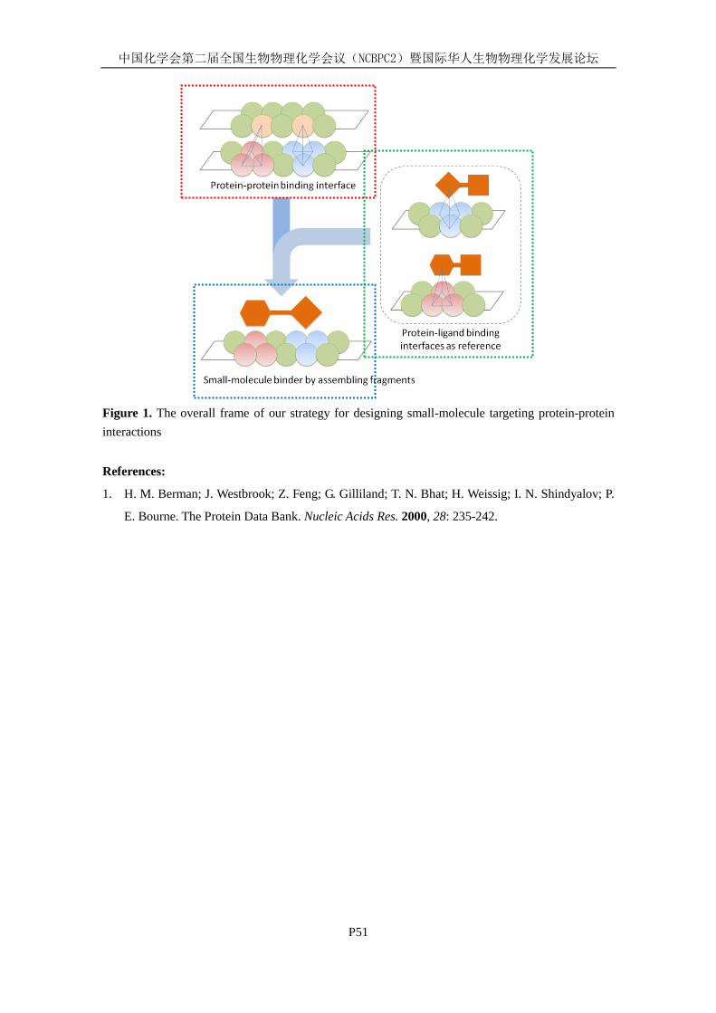
中国化学会第二届全国生物物理化学会议(NCBPC2)暨国际华人生物物理化学发展论坛
P51
Figure 1. The overall frame of our strategy for designing small-molecule targeting protein-protein
interactions
References:
1. H. M. Berman; J. Westbrook; Z. Feng; G. Gilliland; T. N. Bhat; H. Weissig; I. N. Shindyalov; P.
E. Bourne. The Protein Data Bank. Nucleic Acids Res. 2000, 28: 235-242.
中国化学会第二届全国生物物理化学会议(NCBPC2)暨国际华人生物物理化学发展论坛
P52
Virtual Screening of Small-Molecule Inhibitors Targeting the
Protein-Protein Interactions Involving Autophagy-Related Protein
Atg5
Zhixiong Zhao (赵志雄), Yan Li (李嫣), Zhengxi Zhang (张政希), Mi
Zhou (周宓) and Renxiao Wang (王任小)*
State Key Laboratory of Bioorganic Chemistry, Shanghai Institute of Organic Chemistry, Chinese
Academy of Sciences, Shanghai, P. R. China, 200032, [email protected]
Keywords: Virtual Screening, Autophagy, Protein-Protein Interaction, Atg5
Autophagy is a catabolic process involving the degradation of a cell's own components through
the lysosomal machinery[1]
. Autophagy is a tightly-regulated process that plays a normal part in cell
growth, development, and homeostasis, helping to maintain a balance between the synthesis,
degradation, and subsequent recycling of cellular products. Autophagic dysfunction is associated
with many types of diseases. Central to autophagy is the formation of autophagosomes, which
originate from phagophore assembly site (PAS) and will be further integrated with lysosomal to
degrade the cell compounents. Two unique ubiquitin-like conjugation systems, Atg8–PE and
Atg12–Atg5, are involved in the formation of PAS. The formation of Atg12-Atg5:Atg16 complex is
important in both systems[2]
. Here, we want to discover the potential small molecule inhibitors to
regulate the autophagy process through the blocking of the interaction between Atg12–Atg5 and
Atg16.
At the start point of the design, we firstly investigated the structure and protein-protein
interaction of human Atg5-Atg16. Until now, there are no report on the structure of human Atg5 and
Atg16; however, the homologous structure for ScAtg5 and ScAtg16 complex has been reported[2]
.
Thus, the HsAtg5 model was built from its homologous structure of ScAtg5 by I-TASSER server,
which is based on the best threading methods. Since the incomplete short helix structure of ScAtg16
is not suitable for modeling of HsAtg16, the QUARK server based on ab-initial method, the best
server in CASP9, was used to construct the critical segment of N-terminal of HsAtg16. The results
imply that HsAtg5 take similar structure to ScAtg5 and N-terminal of HsAtg16 contains a long helix
moiety. The two model were then superimposed to ScAtg5-ScAtg16 complex based on the reported
alignment to build the preliminary complex model. To explore the precise binding region of
HsAtg16 in the long helix moiety of N-terminal, single point mutation of different length of peptide
of HsAtg16 was synthesized and tested for their activities. Five residues were found to be important
to the HsAtg5-Atg16 interaction (I17,L21,R24,D25,Q28). The corresponding residues of ScAtg16
show obvious interaction to ScAtg5, especially R35 in ScAtg16 has been proved to be essential to
the interaction between ScAtg5 and ScAtg16.
Compared to ScAtg5-Atg16, the interaction of these residues are similar for HsAtg5-Atg16
except D25. Based on the Atg5-Atg16 interaction, pharmacophores for virtually screening were built
based on an optimized model of HsAtg5-Atg16 by 5ns MD simulation. A structure-based
pharmacophore prediction was also performed to validate the rationality of the pharmacophores.

中国化学会第二届全国生物物理化学会议(NCBPC2)暨国际华人生物物理化学发展论坛
P52
Virtual screening based on pharmacophore searching and further docking was performed for HsAtg5
on three libraries. The typical docking model show good binding mode to Atg5. 55 compounds in
in-house library have been tested and one of in-house compounds SMS-71 has been found to be
active. Its binding mode was further optimized by MD simulation and agreed well with the predicted
binding mode. Further work on testing the activities of compounds in different libraries is going on.
[1] Z. Xie; D. J. Klionsky,"Autophagosome formation: core machinery and adaptations". Nat. Cell Biol. 2007, 9:
1102-1109
[2] M. Matsushita, N. N. Suzuki, et al. "Structure of Atg5·Atg16, a Complex Essential for Autophagy." J. Biol. Chem.
2007, 282: 6763-6772
中国化学会第二届全国生物物理化学会议(NCBPC2)暨国际华人生物物理化学发展论坛
P53
Conformational Analysis of Alanine Cyclopeptide:
A Molecular Dynamics Simulation Study
Han Li (韩莉), Min Chiju (闵翅驹), Wang Renxiao (王任小)*
State Key Laboratory of Bioorganic Chemistry, Shanghai Institute of Organic Chemistry,
Chinese Academy of Sciences, 345 Lingling Road, Shanghai 200032, [email protected]
Keywords: Cyclopeptide; Molecular Dynamics Simulation; Clustering Analysis; Typical
Conformation
Polypeptide drugs cause increasing attention due to their application in clinical treatment. On the
one hand, the techniques of synthesis and application of linear peptides are quite mature. Though
many linear peptides have good stability and biological activity in vitro, they degradate rapidly in
vivo. On the other hand, cyclopeptide molecules possess relatively stable conformations. The
sensitivity of cyclopeptide to aminopeptidase and carboxypeptidase decrease because there are no
dissociative amino and carboxyl groups. That is, the metabolic stability and biological availability of
cyclopeptide are much better than those of linear peptide. Thus, the research hotspot has been
transferred to synthesis and evaluation of cyclopeptide molecules recently. In this work, we build a
series of cyclopeptide molecules (from cyclic tripeptide to nonapeptide) using alanine as the
structural units. We performance molecular dynamics simulations on these cyclopeptide molecules
both in vacuum and in solution. Conformational analysis shows that, in vacuum, excepting that
cyclic pentapeptide and hexapeptide have three typical conformations, the other cyclopeptide
molecules possess only one typical conformation. The situation is more complex in solution. Cyclic
tripeptide and pentapeptide have one typical conformation while cyclic tetrapeptide, hexapeptide and
octapeptide possess two. There are no typical conformation of cyclic heptapeptide and nonapeptide
because of their significantly flexibility. This conformational analysis provides a theoretical basis for
functional application of cyclopeptide.
Reference:
1.Y. A. Ovchinnikov; Ivanov, V., Tetrahedron 1975, 31 (18): 2177-2209.
2. J. T. Fan;, Y. S. Chen; W. Y. Xu; L. Du; G. Z. Zeng; Y. M. Zhang; J. Su; Y. Li; N. H. Tan,
Tetrahedron Lett. 2010, 51 (52): 6810-6813.
3. A. Tanka; S. Shimada; T. Hirohashi, Chem Lett. 1998, 11: 1109.
4. J. G. Beck; J. Chatterjee; B. Laufer; M. U. Kiran; A. O. Frank; S. Neubauer; O. Ovadia; S.
Greenberg; C. Gilon; A. Hoffman; H. Kessler, J. Am. Chem. Soc. 2012, 134 (29): 12125-12133.
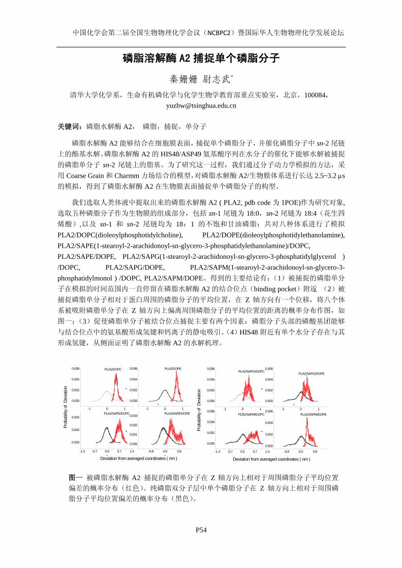
中国化学会第二届全国生物物理化学会议(NCBPC2)暨国际华人生物物理化学发展论坛
P54
磷脂溶解酶 A2 捕捉单个磷脂分子
秦姗姗 尉志武*
清华大学化学系,生命有机磷化学与化学生物学教育部重点实验室,北京,100084,[email protected]
关键词:磷脂水解酶 A2, 磷脂,捕捉,单分子
磷脂水解酶 A2 能够结合在细胞膜表面,捕捉单个磷脂分子,并催化磷脂分子中 sn-2 尾链
上的酯基水解。磷脂水解酶 A2 的 HIS48/ASP49 氨基酸序列在水分子的催化下能够水解被捕捉
的磷脂单分子 sn-2 尾链上的脂基。为了研究这一过程,我们通过分子动力学模拟的方法,采
用 Coarse Grain 和 Charmm 力场结合的模型,对磷脂水解酶 A2/生物膜体系进行长达 2.5~3.2 µs的模拟,得到了磷脂水解酶 A2 在生物膜表面捕捉单个磷脂分子的构型。
我们选取人类体液中提取出来的磷脂水解酶 A2 ( PLA2, pdb code 为 1POE)作为研究对象,选取五种磷脂分子作为生物膜的组成部分,包括 sn-1 尾链为 18:0,sn-2 尾链为 18:4(花生四
烯酸),以及 sn-1 和 sn-2 尾链均为 18:1 的不饱和甘油磷脂:共对八种体系进行了模拟
PLA2/DOPC(dioleoylphosphotidylcholine), PLA2/DOPE(dioleoylphosphotidylethanolamine), PLA2/SAPE(1-stearoyl-2-arachidonoyl-sn-glycero-3-phosphatidylethanolamine)/DOPC, PLA2/SAPE/DOPE, PLA2/SAPG(1-stearoyl-2-arachidonoyl-sn-glycero-3-phosphatidylglycerol ) /DOPC, PLA2/SAPG/DOPE, PLA2/SAPM(1-stearoyl-2-arachidonoyl-sn-glycero-3- phosphatidylmonol ) /DOPC, PLA2/SAPM/DOPE。得到的主要结论有:(1)被捕捉的磷脂单分
子在模拟的时间范围内一直停留在磷脂水解酶 A2 的结合位点(binding pocket)附近;(2)被
捕捉磷脂单分子相对于蛋白周围的磷脂分子的平均位置,在 Z 轴方向有一个位移,将八个体
系被吸附磷脂单分子在 Z 轴方向上偏离周围磷脂分子的平均位置的距离的概率分布作图,如
图一;(3)促使磷脂单分子被结合位点捕捉主要有两个因素:磷脂分子头部的磷酸基团能够
与结合位点中的氨基酸形成氢键和钙离子的静电吸引。(4)HIS48 附近有单个水分子存在与其
形成氢键,从侧面证明了磷脂水解酶 A2 的水解机理。
-1 0 1
0.000
0.002
0.004
0.006
-1 0 1
0.000
0.002
0.004
0.006
B
A
PLA2/DOPC
PLA2/SAPE/DOPC
PLA2/DOPE
PLA2/SAPE/DOPE
Deviation from averaged coordinates ( nm )
Prob
abilit
y of
Dev
iatio
n
-1.4 -0.7 0.0 0.7 1.4
0.000
0.002
0.004
A
-0.8 0.0 0.8
0.000
0.001
0.002
0.003
B
A
-1 0 1
0.000
0.002
0.004
0.006
Prob
abilit
y of
Dev
iatio
n
A
Deviation from averaged coordinates ( nm )
PLA2/SAPG/DOPC
PLA2/SAPM/DOPC
PLA2/SAPG/DOPE
PLA2/SAPM/DOPE-1 0 1
0.000
0.002
0.004
0.006
B
A
-1.4 -0.7 0.0 0.7 1.4
0.000
0.002
0.004
0.006
-0.8 0.0 0.8
0.000
0.002
0.004
0.006
B
图一 被磷脂水解酶 A2 捕捉的磷脂单分子在 Z 轴方向上相对于周围磷脂分子平均位置
偏差的概率分布(红色)。纯磷脂双分子层中单个磷脂分子在 Z 轴方向上相对于周围磷
脂分子平均位置偏差的概率分布(黑色)。
中国化学会第二届全国生物物理化学会议(NCBPC2)暨国际华人生物物理化学发展论坛
P55
溶菌酶吸附带电磷脂脂质体的二维密度效应
罗俊杰 吴富根 郑燕珍 尉志武*
清华大学化学系,生命有机磷化学与化学生物学教育部重点实验室,北京,100084,
关键词:溶菌酶,静电作用,磷脂,二维密度
蛋白质与生物膜的相互作用广泛存在于生物体的生理和病理活动中,因而一直是人们关
注的焦点之一。溶菌酶(lysozyme)的等电点(pI)为 ~11,在 pH = 7.4 的条件下,带 8 个正
电荷,是研究蛋白质与生物膜静电相互作用的模型蛋白。在前期的研究中,我们发现吸附在
带电磷脂表面的 lysozyme 能够通过自身解折叠过程诱导带电磷脂发生侧向聚集。在此基础上,
进一步探究 lysozyme 与带电磷脂相互作用机制受膜表面蛋白吸附浓度的影响,有助于我们更
深入地认识蛋白质与生物膜的相互作用规律。
在本工作中,我们构建了中性的二棕榈酰磷脂酰胆碱(DPPC)和带负电的二油酰磷脂酰
甘油(DOPG)组成的二元磷脂脂质体吸附不同浓度 lysozyme 的体系。通过圆二色性(CD)
光谱对 lysozyme 二级结构的表征,我们发现:当 lysozyme 与脂质体外侧 DOPG 摩尔比为 0.12
时,吸附在膜上的 lysozyme 二级结构解折叠程度最大;当表面吸附的 lysozyme 浓度降低时,
lysozyme 反而更接近于天然态的二级结构;当表面吸附的 lysozyme 浓度升高时,蛋白质又逐
渐回归天然态的结构。我们利用差示扫描量热(DSC)研究了结合和游离的 lysozyme 的变性,
发现出现了结合态和游离态两种 lysozyme 的变性峰。随着溶液中 lysozyme 的浓度增加,结合
态的 lysozyme 也逐渐增多,最终达到饱和(结合的 lysozyme 与外侧 DOPG 摩尔比为~0.27)。
上述结果表明,表面吸附的 lysozyme 的“二维密度”可以影响蛋白质与带电磷脂脂质体的相
互作用机制,密度较高时,蛋白质之间彼此拥挤,使其空间结构保持天然态的紧密状态。
参考文献:
[1] JJ Luo, FG Wu, JS Yu, R Wang, ZW Yu., Denaturation Behaviors of Two-State and
Non-Two-State Proteins Examined by an Interruption–Incubation Protocol, J. Phys. Chem.
B, 115 (2011): 8901–8909.
[2] FG Wu, JJ Luo, ZW Yu., Unfolding and Refolding Details of Lysozyme in the Presence of
β-Casein Micelles, Phys. Chem. Chem. Phys., 13 (2011) :3429–3436.
中国化学会第二届全国生物物理化学会议(NCBPC2)暨国际华人生物物理化学发展论坛
P56
Deep Midgap States Decay Kinetics of Photogenerated
Electrons in TiO2 Anatase
Zhu Ming, Zhu Gang-Bei, and Yu-Xiang Weng*, Wang Yun-peng
Laboratory of Soft Matter Physics, Institute of Physics, Chinese Academy of Sciences, Beijing
100080, China ,and National Laboratory of the Condensed Matter Physics, Beijing 100080, China
Electrons photogenerated in TiO2 anatase nanocrystal films were observed by FTIR absorption
spectroscopy irradiated by a 340nm-360nm LED. Transient IR absorption (2090cm-1) appeared on
TiO2 anatase nanocrystal films irradiated by a pump pulse (450nm-1423nm) which is obtained by a
OPO(optical patametric oscillator) with a 355nm pump pulse. The absorption was attributed to
optical transition of photogenerated electrons trapped in deep midgap states. From the decay kinetics
of the photogenerated electrons, there are two different deep midgap states, one is 0.87eV under the
bottom of conduction band, the other is 1.44eV under the bottom of conduction band. The decay
time of photogenerated electrons of the 1.44eV midgap state is 200us. This state appeared when the
vacuum chamber is evacuated to 5*10E-7mbar. The other 0.87eV midgap state with a much shorter
decay time which is less than 2us.
Reference
Hui Zhao, J. Phys. Chem. C 2007, 111: 3762-3769
Akira Yamakata, J. Phys. Chem. B 2001, 105: 7258-7262
Dimitar A. Panayotov, J. Phys. Chem. C 2012, 116: 4535−4544

中国化学会第二届全国生物物理化学会议(NCBPC2)暨国际华人生物物理化学发展论坛
P57
原子力显微镜法研究方解石-氨基酸的相互作用
赵 康 徐 海*
中国石油大学(华东)生物工程与技术中心,266580,青岛, [email protected]
关键词:方解石,氨基酸,AFM
利用液相原子力显微镜法研究了方解石(104)晶面在不同氨基酸(Gly、L-Glu 和 L-Lys)存在
下的生长行为。如图 1a,在碳酸钙过饱和溶液中,方解石(104)晶面呈现出金字塔型的螺旋生
长丘,四个台阶面矢量分别为[481
]+、[4
41]+、[481
]_和[4
41]_,其中 c-glide 表示滑移面在(104)
面上的投影。引入含有氨基酸(Gly、L-Glu 或 L-Lys)的过饱和生长液后,螺旋生长丘的形貌和
生长动力学发生明显变化,这些变化与氨基酸浓度、分子手性以及过饱和度密切相关(图 1b-c)。
具体而言:(1) 三种氨基酸的加入都可以使[421
]台阶方向得到表达,这可能是由于氨基酸与[42
1
]台阶边缘作用,降低了[421
]台阶边缘自由能,从而使其稳定得以表达。(2)Gly 的存在使得
螺旋生长丘的整体形貌呈六边形且关于 c-glide 对称,而 L-Glu 或 L-Lys 使得生长丘形貌呈扇
形,生长丘的对称性遭到破坏。(3) L-Glu 明显抑制方解石螺旋生长丘正/负台阶方向的生长速
率,而 Gly 和 L-Lys 都在较高浓度下对台阶生长有促进作用。
Figure 1. AFM images of calcite growth hillocks: (a) control and in the presence of (b) Gly, (c)
L-Glu, and (d) L-Lys
中国化学会第二届全国生物物理化学会议(NCBPC2)暨国际华人生物物理化学发展论坛
P58
计算机录入部分或全部国民及其它生物话语或语音,促进工
作、生活、科技发展及社会安全管理
徐汉友
主治医师,河南省南阳纺织集团有限公司职工医院内科副主任
联系地址:河南省南阳市长江路 200号,南阳纺织集团职工医院内科。邮编:473125.
电话:15518962531。[email protected]; [email protected]
背景:现代计算机技术已达到了非常先进的时代,能把纸质文字录入,能有超凡的记忆,等
等,但还没有把人类的话语或谈话的语调以数字的形式即时瞬时同步录入电脑。
目的:创新提出一项现代计算机和信息技术工程革新策略,使其能把人的话语或谈话的语调
特点瞬时同步以文字的形式录入电脑。
方法:总结现代计算机和信息技术的发展,提出这项计算机和信息技术革新策略。
结果:利用现代计算机和信息技术,把人们的的话语或谈话的语调,以数字的形式录入电脑,
人与人的语音特点各不相同,本文提出了把人们的话语或谈话的语调,以数字的形式录入电
脑的程序梗概及其用途。
结论:如果人们的的话语或谈话的语调,以数字的形式录入和存储在电脑,人与人的语音特
点各不相同,将有利于人们的工作生活,及社会安全管理和科技发展,象指纹一样,全面录
入管理,更有利于经济和社会发展。
同时,根据上述原理和方法,可以录入和储存其他生物发出的声音特点,进一步促进科技
发展和社会进步。
关键词:计算机科学;声音;话语特性;社会和科技发展;识别;言语特性处理。
Recognizing and Recording the Tone of People’ and others’
Language, to Promote Development of Work, Life and Science,
and Promote Administration of Social Security
Xu Han-You
Professor of China Academy of Management Science.
Doctor in charge of western internal medicine.
Address: Department of internal and emergency medicine, Workers hospital of Nanyang textile
corporation, Nanyang city Changjiang RD No.200, Henan province, China. Post Codes: 473125.
Telephone: 0086-15518962531. [email protected] or [email protected].
Background: As the information and computer science have been developing more and more
rapidly and broadly. Which the developments have contributed much too more to mankinds. The
computer recognizing the characters and its scanning function of the characters, the computer’s huge
storage ability are the examples. The computer science has been making the work and life more and
more easy and convenience. But there are lots of situations and problems in our life and work the

中国化学会第二届全国生物物理化学会议(NCBPC2)暨国际华人生物物理化学发展论坛
P58
computer can not do anything. For example, the computer has not recorded the tone and the words of
all people at the same time when the people are speaking.
Objective: In this paper, the author outlined a new breakthrough creation of project for information
and computer science. Which use the modern information and computer science to solve the present
problem which the computer has not recorded all the tone and the words at the same time when the
people are speaking.
Method: Summarize the modern information and computer science. Create the new breakthroughs
project of computer science.
Results: The author outlines, by developing a new technology project to recognize the people’s
speaking and their tone and characters, at the same time, transmit the recognizing into computer with
their tone and characters and store the tone and characters of the speaking information. Therefore,
any people’s speaking could be recorded into the computer with their characters of tone at the same
time.
Conclusion: This is a breakthrough creation project for information and computer science. When the
breakthrough creation came into reality. The revolution movements of science could be induced.
The life, work, and security and other active things of mankind could be more easy and wonderful.
There were much too harvesting in economical and practical gaining. And there were also much too
harvesting in social development. So there are lots of chances waiting us to make her true in the near
future when the paper of the new breakthrough creation for information and computer science is
published and accepted for application. Because recognizing and recording the tone of people’
language and his characters of the speaking information are different from each other. Like the
fingerprint, the public security and other practical gaining could be great.
The breakthrough technology project of computer science can be used for recognizing and recording
the tone of language of other biology, and their characters of the speaking or voice information, apart
from people’.
Key words: Computer science; Tone; characters of the speaking; Social and science development;
Recognize; Speaking-Character management.

中国化学会第二届全国生物物理化学会议(NCBPC2)暨国际华人生物物理化学发展论坛
P59
A sensitive and rapid methodological development for sterility
testing by microcalorimetry coupled with full-enclosed
filtration-culture ampoule system
Yong-shen REN a, b
, Dan YAN a, *
, Ping ZHANG a, Zhi-nan MEN
a,
Xiao-He XIAO a, *
a Institute of Chinese Materia Medica, 302 Military Hospital of China, Beijing 100039, China
b Pharmacy college, South-central University for Nationalities, Wuhan 430074, China
A modified full-enclosed membrane filtration-culture ampoule system coupled with
microcalorimeter was applied as an alternative method for sterility test; the detection time was
defined as Pd/| P0|≥3 when k≥0 (where Pd was the difference of sample heat power Pi with the
parallel baseline power P0, k waes the exponential growth rate). six common strains, Staphylococcus
aureus, Pseudomonas aeruginosa, Escheichia coli, Bacillus subtilis, Clostridium sporogenes and
Candida albicans were serial 10-fold diluted and cultured to evaluate the feasibility of
microcalorimetric sterility test; and the results were compared with routine observational method. It
was showed microcalorimetric method was faster than conventional observation method (p<0.05 or
p<0.01), since the detection time of the former were 0~18 h and the latter was 10~36 h at the same
inoculum of fast-growing strains; and former was more sensitive with the detection limit was even
less than 1 colony-forming unit (CFU) but latter was 10~1 CFU. Moreover, mircocalorimetry could
detect out the slow-growing microorganism (e.g. C. albicans) much earlier than observation method,
and could avoid the false negative misjudge caused by observation method. Fatherly, the
thermogenic curves recorded automatically by microcalorimeter could avoid secondary pollution by
repeated observation of conventional method, and the power-time curves were species-specific,
which could be used for bacterial contamination identification. The sterility test based on
microcalorimetry coupled with modified culture system was high sensitivity and low time
consumption, could be widely applied in sterility testing of drugs, food and surgical instruments.
Keywords: Sterility test; Microcalorimetry; Conventional method; Detection time; Sensitivity
*Corresponding author at: Institute of Chinese Materia Medica, 302 Military Hospital of China,
Beijing 100039, China; Tel.: +86 10 6693 3322; fax: +86 10 6387 9915. E-mail address: [email protected] (X.H. Xiao); Tel.: +86-10-66933322.
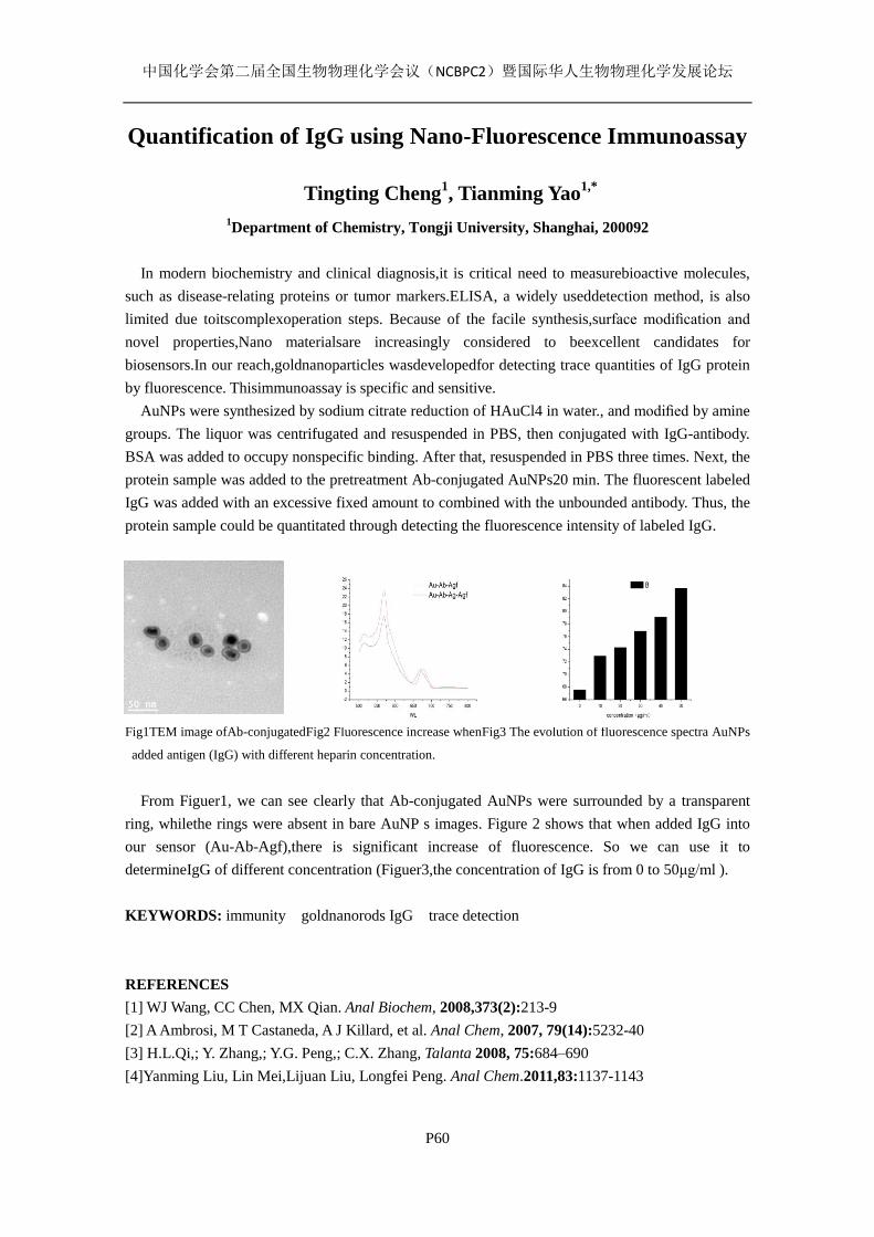
中国化学会第二届全国生物物理化学会议(NCBPC2)暨国际华人生物物理化学发展论坛
P60
Quantification of IgG using Nano-Fluorescence Immunoassay
Tingting Cheng1, Tianming Yao
1,*
1Department of Chemistry, Tongji University, Shanghai, 200092
In modern biochemistry and clinical diagnosis,it is critical need to measurebioactive molecules,
such as disease-relating proteins or tumor markers.ELISA, a widely useddetection method, is also
limited due toitscomplexoperation steps. Because of the facile synthesis,surface modification and
novel properties,Nano materialsare increasingly considered to beexcellent candidates for
biosensors.In our reach,goldnanoparticles wasdevelopedfor detecting trace quantities of IgG protein
by fluorescence. Thisimmunoassay is specific and sensitive.
AuNPs were synthesized by sodium citrate reduction of HAuCl4 in water., and modified by amine
groups. The liquor was centrifugated and resuspended in PBS, then conjugated with IgG-antibody.
BSA was added to occupy nonspecific binding. After that, resuspended in PBS three times. Next, the
protein sample was added to the pretreatment Ab-conjugated AuNPs20 min. The fluorescent labeled
IgG was added with an excessive fixed amount to combined with the unbounded antibody. Thus, the
protein sample could be quantitated through detecting the fluorescence intensity of labeled IgG.
Fig1TEM image ofAb-conjugatedFig2 Fluorescence increase whenFig3 The evolution of fluorescence spectra AuNPs
added antigen (IgG) with different heparin concentration.
From Figuer1, we can see clearly that Ab-conjugated AuNPs were surrounded by a transparent
ring, whilethe rings were absent in bare AuNP s images. Figure 2 shows that when added IgG into
our sensor (Au-Ab-Agf),there is significant increase of fluorescence. So we can use it to
determineIgG of different concentration (Figuer3,the concentration of IgG is from 0 to 50μg/ml ).
KEYWORDS: immunity goldnanorods IgG trace detection
REFERENCES
[1] WJ Wang, CC Chen, MX Qian. Anal Biochem, 2008,373(2):213-9
[2] A Ambrosi, M T Castaneda, A J Killard, et al. Anal Chem, 2007, 79(14):5232-40
[3] H.L.Qi,; Y. Zhang,; Y.G. Peng,; C.X. Zhang, Talanta 2008, 75:684–690
[4]Yanming Liu, Lin Mei,Lijuan Liu, Longfei Peng. Anal Chem.2011,83:1137-1143
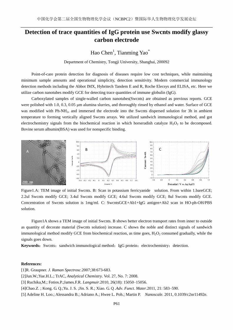
中国化学会第二届全国生物物理化学会议(NCBPC2)暨国际华人生物物理化学发展论坛
P61
Detection of trace quantities of IgG protein use Swcnts modify glassy
carbon electrode
Hao Chen1, Tianming Yao
*
Department of Chemistry, Tongji University, Shanghai, 200092
Point-of-care protein detection for diagnosis of diseases require low cost techniques, while maintaining
minimum sample amounts and operational simplicity, detection sensitivity. Modern commercial immunology
detection methods including the Abbot IMX, Hybritech Tandem E and R, Roche Elecsys and ELISA, etc. Here we
utilize carbon nanotubes modify GCE for detecting trace quantities of immune globulin (IgG).
Carboxylated samples of single-walled carbon nanotubes(Swcnts) are obtained as previous reports. GCE
were polished with 1.0, 0.3, 0.05 μm alumina slurries, and thoroughly rinsed by ethanol and water. Surface of GCE
was modified with Ph-NH2, and immersed the electrode into the Swcnts dispersed solution for 3h in ambient
temperature to forming vertically aligned Swcnts arrays. We utilized sandwich immunological method, and got
electrochemistry signals from the biochemical reaction in which horseradish catalyze H2O2 to be decomposed.
Bovine serum albumin(BSA) was used for nonspecific binding.
Figure1.A: TEM image of initial Swcnts. B: Scan in potassium ferricyanide solution. From within 1.bareGCE;
2.2ul Swcnts modify GCE; 3.4ul Swcnts modify GCE; 4.6ul Swcnts modify GCE; 8ul Swcnts modify GCE.
Concentration of Swcnts solution is 1mg/ml. C: SwcntsGCE+Ab1+IgG antigen+Ab2 scan in HO-ph-OH/PBS
solution.
Figure1A shows a TEM image of initial Swcnts. B shows better electron transport rates from inner to outside
as quantity of decorate material (Swcnts solution) increase. C shows the noble and distinct signals of sandwich
immunological method modify GCE from biochemical reaction, as time goes, H2O2 consumed gradually, while the
signals goes down.
Keywords:Swcnts;sandwich immunological method;IgG protein;electrochemistry;detection.
References:
[1]R. Graupner. J. Raman Spectrosc.2007;38:673-683.
[2]Jun.W.;Yue.H.L.; TrAC, Analytical Chemistry. Vol. 27, No. 7: 2008.
[3] Ruchika,M.; Fotios.P.;James.F.R. Langmuir.2010, 26(18): 15050–15056.
[4]Chao.Z. ; Kong. G. Q.;Yu. J. S. ;Jin. S. R.; Xiao. G. Q .Adv. Funct. Mater.2011, 21: 583–590.
[5] Adeline H. Loo.; Alessandra B.; Adriano A.; Hwee L. Poh.; Martin P. Nanoscale. 2011, 0.1039/c2nr11492e.
A C B
中国化学会第二届全国生物物理化学会议(NCBPC2)暨国际华人生物物理化学发展论坛
P62
偶极修饰剂与生物膜相互作用的分子水平研究
马素兰,叶树集*
中国科技大学合肥微尺度物质科学国家实验室, 安徽合肥,230026
关键词:和频光谱,生物膜,磷脂双分子层,偶极修饰剂,6-酮胆甾烷醇
蛋白质等生物分子与细胞膜的相互作用在生命过程中扮演着非常重要的角色。该相互作
用的一个重要驱动力是生物膜上的偶极电位,其来源于极性磷脂分子头部基团与膜界面水分
子偶极的特殊取向。添加偶极修饰剂是改变生物膜偶极电位的重要方法,然而,目前人们从
分子水平上理解偶极修饰剂与生物膜相互作用的机理甚少。本研究利用非线性和频光谱技术,
从分子水平上研究偶极修饰剂 6-酮胆甾烷醇与生物膜相互作用。结果表明,6-酮胆甾烷醇能够
有效影响磷脂双层膜上的水分子结构与有序度。但是随磷脂分子带电性质不同,其影响效果
也不同,带负电的磷脂分子与水分子结合最强,分子排布难以被打破,影响最弱;带正电的
磷脂分子次之;电中性的则较为容易受到影响。6-酮胆甾烷醇与磷脂双层膜的头部(即亲水链)
发生作用,改变磷脂分子头部的偶极电位,从而影响水分子的有序度。本研究为从分子水平
上理解偶极电位如何影响生物膜与蛋白质的相互作用驱动力机理以及进一步理解各种生物分
子、离子、药物在生物膜等界面上的吸附、解吸和传输机制提供了一些重要数据。
参考文献:
1. Simon, S.A.; Mclntosh, T.J.; Magid, A.D.; Needham, D. Biophys.J. 1992, 61: 786-798.
2. Alakoskela, J-M. I.; Kinnunen, P.K.J. Biophys. J. 2001, 80: 294–304.
3. Starke-Peterkovic, T.; Turner, N.; Vitha, M.F.; Waller. M.P.; Hibbs, D.E.; Clarke, R.J. Biophys.J.
2006, 90: 4060-4070.

中国化学会第二届全国生物物理化学会议(NCBPC2)暨国际华人生物物理化学发展论坛
P63
三七皂苷 Rg1 对凝血酶活性的影响
曾伟成
厦门市医药研究所 厦门 361003,[email protected]
关键词 三七皂苷 Rg1 凝血酶
三七总皂苷(total saponins of panax notognseng, PNS)是五加科人参属植物三七的主
要有效活性成分,含有人参皂苷 Rb1、Rb2、Rc、Rd、Rf、Rg1、Rg2、Rh1和三七皂苷 R1、R2、
R3、R4、R6 等 20 余种皂苷成分,均属于达玛烷型 (Dam2maranetype)四环三萜皂苷。其中,
人参皂苷 Rg1、人参皂苷 Rb1是三七总皂苷中含量最高的两个成分,而三七皂苷 R1则是三七
总皂苷的特征化合物[1]。本实验采用一种实用的凝血酶抑制剂抑制活性的测定方法--发色底物
分光光度法【2】
,以发色底物 S-2238与凝血酶反应,测定其得到不同三七皂苷 Rg1 浓度存在时
发色底物 S-2238和凝血酶反应的其吸光度 A值。凝血酶溶液浓度为 15 U/ml,发色底物 S-2238
浓度为 0.20mg/ml 的吸光度记为 A1,S-2238 浓度为 0.26 mg/ml 的吸光度记为 A2,测定波长
405nm在 2 min时的吸光度值。
表 1 不同三七皂苷 Rg1浓度时的凝血酶与发色底物 S-2238的吸光度 Ao
1 2 3 4 5 6 7
Rg1 浓度
(mg/ml)
0 3 6 9 12
15
18
A1 0.211 0.191 0.167 0.152 0.132 0.116 0.104
A2 0.228 0.206 0.183 0.172 0.159 0.142 0.133
实验结果表明,发色底物 S-2238在凝血酶催化作用下的显色反应随三七皂苷 Rg1浓度增
加而减弱,A1与三七皂苷 Rg1浓度关系可用下面的方程表示
y=0.2075-0.00602x
三七总皂苷具有止血或活血化瘀的双向调节功能,三七总皂苷的不同分离组分对凝
血酶活性的影响情况不同。某些组分可以增强凝血酶活性作用,某些组分可以抑制凝血
酶活性作用。本实验结果显示三七总皂苷成分 Rg1 对凝血酶的有显著抑制作用,我们的
实验亦筛选出三七皂苷部分组分对凝血酶的激活作用,这些组分相比于 Rg1 激活作用并
不是那样的显著,可以设想三七皂苷组分并不是仅仅竞争凝血酶的某个催化位点,通过
进一步的实验弄清三七皂苷各组分作用机理,将有助于对传统中医的理解。
参考文献
[1] 王兴文.水提三七总皂苷[J].云南中医学院学报,2001,24(3):1-3.
[2] 赵荣乐.一种测定凝血酶抑制剂活性的新方法[J].喀什师范学院学报,2003,24(6):52-55.
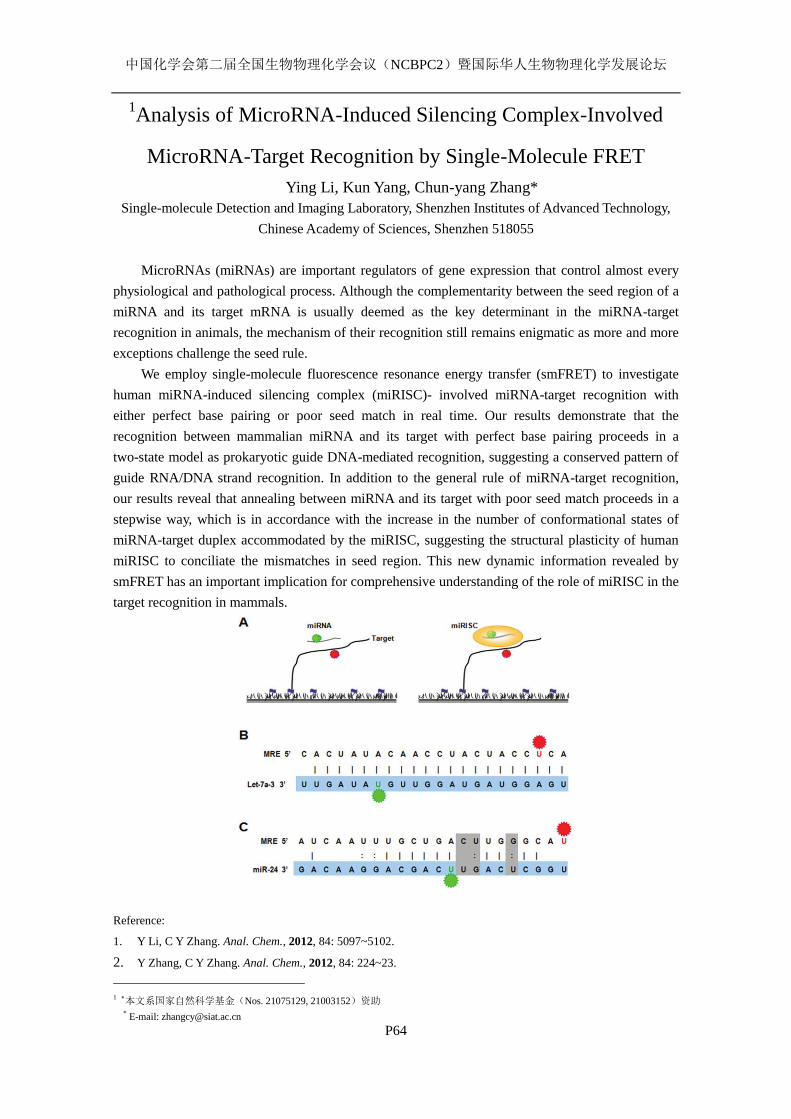
中国化学会第二届全国生物物理化学会议(NCBPC2)暨国际华人生物物理化学发展论坛
P64
1Analysis of MicroRNA-Induced Silencing Complex-Involved
MicroRNA-Target Recognition by Single-Molecule FRET
Ying Li, Kun Yang, Chun-yang Zhang*
Single-molecule Detection and Imaging Laboratory, Shenzhen Institutes of Advanced Technology,
Chinese Academy of Sciences, Shenzhen 518055
MicroRNAs (miRNAs) are important regulators of gene expression that control almost every
physiological and pathological process. Although the complementarity between the seed region of a
miRNA and its target mRNA is usually deemed as the key determinant in the miRNA-target
recognition in animals, the mechanism of their recognition still remains enigmatic as more and more
exceptions challenge the seed rule.
We employ single-molecule fluorescence resonance energy transfer (smFRET) to investigate
human miRNA-induced silencing complex (miRISC)- involved miRNA-target recognition with
either perfect base pairing or poor seed match in real time. Our results demonstrate that the
recognition between mammalian miRNA and its target with perfect base pairing proceeds in a
two-state model as prokaryotic guide DNA-mediated recognition, suggesting a conserved pattern of
guide RNA/DNA strand recognition. In addition to the general rule of miRNA-target recognition,
our results reveal that annealing between miRNA and its target with poor seed match proceeds in a
stepwise way, which is in accordance with the increase in the number of conformational states of
miRNA-target duplex accommodated by the miRISC, suggesting the structural plasticity of human
miRISC to conciliate the mismatches in seed region. This new dynamic information revealed by
smFRET has an important implication for comprehensive understanding of the role of miRISC in the
target recognition in mammals.
Reference:
1. Y Li, C Y Zhang. Anal. Chem., 2012, 84: 5097~5102.
2. Y Zhang, C Y Zhang. Anal. Chem., 2012, 84: 224~23.
1 本文系国家自然科学基金(Nos. 21075129, 21003152)资助 * E-mail: [email protected]
中国化学会第二届全国生物物理化学会议(NCBPC2)暨国际华人生物物理化学发展论坛
P65
Antibacterial and thermal properties of lanthanide complexes
with 3,5-Dimethoxybenzoic acid and 1,10-phenanthroline
Jun-Ru Zheng 1,2
, Ning Ren 3, Jian-Jun Zhang
1,2*, Da-Hai Zhang
3, Yuan Li
2
(1 Testing and Aanlysis Center, Hebei Normal University, Shijiazhuang 050024 P. R. China.)
(2College of Chemistry& Material Science, Hebei Normal University, Shijiazhuang 050024 P. R.
China)
(3Department of Chemistry, Handan College, Handan 056005 P. R. China)
Four lanthanide complexes with a general formula [Ln(3,5-DmeoxBA) 3(phen)]2(Ln =Tb(1)
Dy (2),Er (3), Yb(4); 3,5-DmeoxBA =3,5-Dimethoxybenzoic acid ;phen = 1,10-phenanthroline)were
synthesized and characterized by elemental analysis, infrared spectra(IR), and thermogravimetric ,
differential scanning calorimetry techniques combine fourier transform infrared (TG/DSC-FTIR)
technology. The thermal decomposition mechanism of the four complexes were investigated by
TG/DSC-FTIR techniques. Under the simulated atmosphere at a heating rate of 10oC·min
-1, the
thermal decomposition of the four complexes have similar thermal decomposition stages. The
antibacterial action of the four complexes on bacteria and fungus such as Escherichia coli ,
Staphylococcus aureus and Candida albicans were studied by filter paper approach. And the
antibacterial activity test indicated that these complexes exhibit better antibacterial ability than the
corresponding rare-earth chloride or ligands. Moreover, the antibacterial activity of these complexes
will increase along with the rise of their concentration in the range of tested concentrations . Heat
capacities were measured by DSC. And the values of the experimental heat capacities were fitted to
a polynomial equation with the least-squares method. Based on the fitted polynomial, the smoothed
heat capacities and thermodynamic functions (HT-H298.15K), (ST-S298.15K), and (GT-G298.15K) were
calculated. The luminescent properties of the complexes 1 and 2 were also studied.
Keywords lanthanide complexes, antibacteria, thermal properties
References
(1) Ravi, K,; Amresh K.; Chandrashekar, and Anil Kumar , K.S. Synthesis and Biological Activity
on Some Organoantimony (III) Compounds. Phosphorus Sulfur, 183(2008)1410.
(2) Guo, J. P.; Liu, B. P.; Lv, X. C.; Tan, Z. C.; Tong, B.; Shi, Q.; Wang, D. F. Molar heat capacities,
thermodynamic properties, and thermal stability oftrans-4-(Aminomethyl)cyclohexanecarboxylic
acid. J. Chem. Eng. Data 52 (2007) 1678.
(3) Song, X. Q.; Yua, Y.; Liu, W. S.; Dou, W.; Zheng, J. R.;Yao, J. N. Preparation, spectroscopic
properties of 1,4-di(N,N-diisopropylacetamido)-2,3(1H,4H)-quinoxalinedione (L) lanthanide
complexes and the supramolecular structure of [Nd2L2(NO3)6(H2O)2]·H2O. J.Solid State Chem.
180(2007)2616.
-------------------------------
This project was supported by the National Natural Science Foundation of China (No. 21073053 and
20773034) and the Natural Science Foundation of Hebei Province (No. B2012205022).

中国化学会第二届全国生物物理化学会议(NCBPC2)暨国际华人生物物理化学发展论坛
P66
聚乙烯酰亚胺与双链 DNA 相互作用的单分子力谱研究
张薇 寇晓龙 张文科*
吉林大学化学学院,长春,130012,[email protected]
关键词:聚乙烯酰亚胺 (PEI),DNA,原子力显微镜,单分子力谱
随着生物科技的发展,基因疗法成为治愈诸多顽固疾病的新希望1。而如何将外源基因植
入细胞便是该疗法的最关键环节。在各类基因载体材料中,阳离子聚合物由于易于与双链 DNA
复合、细胞毒性低、转染效率高等优点而受到广泛关注2-3。聚乙烯亚胺(PEI)则是其中具有高
转染效率的聚合物材料之一,其制备容易、价格低廉、合成效率高、改性简单等优点使之倍
受青睐4。
我们利用基于原子力显微镜(AFM)的单分子力谱技5研究了聚乙烯亚胺(PEI)在不同浓度、
pH 值下与双链 DNA 的相互作用6。研究结果表明,对 PEI-DNA 复合物拉伸所得到的无规锯
齿型力信号源于 PEI 与双链 DNA 的作用。在 PEI 浓度为 0.03 μg/ml和 0.06 μg/ml时,力信号
断裂长度分别为 231.31 nm 和 761.89 nm。氮磷比为 0.75:1 时,复合物拉伸长度较长;氮磷比
为 3:1 时,拉伸长度集中在 1.0 μm以下;氮磷比为 5.4:1 时,拉伸长度有最小值,全部集中在
0.5 μm 以下。以上结果表明 PEI 浓度越高,形成复合物结构越紧密,越不容易被破坏。此结
论在往复拉伸实验中也得到了进一步验证。通过研究不同氮磷比对 DNA 的 B-OS 转变平台的
影响,我们发现 PEI 存在与否并未导致 B-OS 转变发生明显变化,说明 PEI 主要以静电力作用
于 DNA 的磷酸骨架,并未嵌入碱基对之间,也没有与大沟、小沟发生强烈作用。溶液 pH 值
由 7.4 降至 5.0 后,力信号无明显变化,表明 PEI 与 DNA 的作用受 pH 值影响较小。
参考文献:
1. 邓洪新; 田聆; 魏于全. 基因治疗的发展现状、问题和展望. 生命科学 2005, 17: 196-199.
2. Luten, J.; van Nostruin, C. F.; De Smedt, S. C.; Hennink, W. E. Biodegradable polymers as
non-viral carriers for plasmid DNA delivery. J. Control. Release 2008, 126: 97-110.
3. Park, T. G.; Jeong, J. H.; Kim, S. W. Current status of polymeric gene delivery systems. Adv.
Drug Delivery. Rev. 2006, 58: 467-486.
4. Boussif, O.; Lezoualch, F.; Zanta, M. A.; Mergny, M. D.; Scherman, D.; Demeneix, B.; Behr, J. P.
A Versatile Vector for Gene and Oligonucleotide Transfer into Cells in Culture and in-Vivo -
Polyethylenimine. Proc. Natl. Acad. Sci. 1995, 92; 7297-7301.
5. (a) Clausen-Schaumann, H.; Seitz, M.; Krautbauer, R.; Gaub, H. E. Force spectroscopy with
single bio-molecules. Curr. Opin. Chem. Biol. 2000, 4: 524-530; (b) Zhang, W. K.; Zhang, X.
Single molecule mechanochemistry of macromolecules. Prog. Polym. Sci. 2003, 28: 1271-1295;
(c) Liu, N. N.; Zhang, W. K. Feeling Inter- or Intramolecular Interactions with the Polymer Chain
as Probe: Recent Progress in SMFS Studies on Macromolecular Interactions. ChemPhysChem
2012, 13: 2238-2256; (d) Liu, N. N.; Peng, B.; Lin, Y.; Su, Z. H.; Niu, Z. W.; Wang, Q.; Zhang,
W. K.; Li, H. B.; Shen, J. C. Pulling Genetic RNA out of Tobacco Mosaic Virus Using
Single-Molecule Force Spectroscopy. J. Am. Chem. Soc. 2010, 132: 11036-11038.
6. 张薇. 蛋白质及聚乙烯亚胺与核酸相互作用的基于 AFM 的单分子力谱研究. 吉林大学博
士学位论文. 2012.

中国化学会第二届全国生物物理化学会议(NCBPC2)暨国际华人生物物理化学发展论坛
P67
Quantitative detection of Pb2+
using silver enhancement of
DNAzyme-gold nanoparticle aggregation and progressive dilution
Zhao Fang Liu, Xin Sheng Zhao*
Beijing National Laboratory for Molecular Sciences, State Key Laboratory for Structural Chemistry
of Unstable and Stable Species, Department of Chemical Biology, College of Chemistry and
Molecular Engineering, and Biodynamic Optical Imaging Center, Peking University, Beijing 100871,
China
DNAzymes are DNA chains that catalyze chemical reactions, such as cleavage of ribonucleic
acid targets. They have emerged recently as a promising class of molecules to build sensors.
Pb2+
-specific DNAzyme contains two parts: a catalytic loop and a biding arm. Helped by Pb2+
when
the DNAzyme hybridizes with the substrate sequence and recognizes the catalytic loop, it cleaves
the substrate DNA into two pieces 1. We chose gold nanoparticles(GNPs) as the reporter group and
used silver enhancement assay to detect the Pb2+
ion (Fig. 1). A probe composed of aggregates
among the substrate-sequence-attached GNPs and DNAzyme, and partially complementary
DNA-attached GNPs. In the presence of Pb2+
, the DNAzyme activated and cleaved the substrate
strand, which caused redispersion of the aggregates. GNPs that are more dispersed have higher
catalytic capability in silver staining 2, so that a higher concentration of Pb
2+ results in increased
grayscale. In our previous work 3, we demonstrated that the progressive dilution assay (PD) can
quantitatively detect targets and requires no pretreatment or separation of real samples, since PD
effectively eliminates the interference of various sources. In this work, we combined silver
enhancement with DNAzyme-GNPs aggregation and PD assay to detect Pb2+
in real samples: water
of WeiMing Lake and human blood.
Fig. 1 A scheme of the silver enhancement of DNAzyme–gold nanoparticle aggregation and progressive dilution assay.
References:
[1] J. Liu and Y. Lu, J. Am. Chem. Soc., 2003, 125: 6642–6643.
[2] Z. Zhang, C.L. Chen, X.S. Zhao, Electroanalysis, 2009, 21:1316.
[3] Z.F. Liu, J. Ge and X.S. Zhao, Chem. Commun., 2011, 47 (17): 4956.
_________________________
*This work was supported by NKBRSF (2010CB912302, 2012CB917304) and NSFC (20973015).
Contact: Xin Sheng Zhao Email: [email protected] TEL:010-62751727
中国化学会第二届全国生物物理化学会议(NCBPC2)暨国际华人生物物理化学发展论坛
P68
The spontaneous flipping rate of single mismatched base pair in
double-stranded DNA
Yandong Yin1,2,3
, Lijiang Yang1,4
, Guanqun Zheng6, Chengqi Yi
6,
Chuan He1,3,5,6*
, Yi Qin Gao1,4*
, Xin Sheng Zhao1,2,3*
1 Beijing National Laboratory for Molecular Sciences;
2State Key Laboratory for Structural Chemistry of
Unstable and Stable Species and Biodynamic Optical Imaging Center; 3 Department of Chemical Biolo-
gy, and 4 Institute of Theoretical and Computational Chemistry, College of Chemistry and Molecular
Engineering; and 5 Synthetic and Functional Biomolecules Center; Peking University, Beijing 100871,
China. 6 Department of Chemistry, the University of Chicago, Chicago IL 60637, USA.
*Correspondence should be addressed to X. S. Z. ([email protected]), Y. Q. G. ([email protected]),
and C. H. ([email protected]).
Key Words: Dynamics, Spontaneous single base flipping, ddFCS.
Many DNA base repairs and modifications, such as DNA methylation/demethylation, deamination,
and hydroxylation, are known to occur extrahelically with the lesion base or target base flipped out of
the DNA duplex1. The original force that causes the target base outward flipping, however, is still under
debate2. To experimentally provide fundamental dynamical and kinetic information for this issue, we
directly measured the spontaneous flipping rate of the single mismatched base pair (T-C, T-G, and T-T
mismatches) in otherwise a perfectly matched double-stranded DNA (dsDNA), byapplying the newly
developed diffusion-decelerated fluorescence correlation spectroscopy (ddFCS) technique3. ddFCS
elongates the presence of molecules in the detect volume (confocal volume) to ~ 1 s, so that it allows us
tomonitor the chemical relaxation occurring in 0.01ms ~ 0.1 s time range, much wider than the time
range that conventional FCS can handle.Based on the ddFCS assay, we revealed the spontaneous flip-
ping rate of T-C, T-G, and T-T mismatched base pairs. The relatively longer lifetime (1 ~ 10 ms) of the
extrahelical state suggests that the weakened base pair may spend enough time to stay outside of the
DNA duplex and be served asa kinetic signal for protein recognition and location
This work was supported by NKBRSF (2012CB917304 to X.S.Z. and Y.Q.G. and 2010CB912302 to
X.S.Z.) and NSFC (20973015 to X.S.Z and 21125311 to Y.Q.G.). This work was also supported by Na-
tional Institutes of Health (GM071440 to C.H.).
Reference
(1) C. G. Yang, C. Yi, E. M. Duguid, C. T. Sullivan, X. Jian, P. A. Rice, C. He, Nature,2008, 452: 961.
(2) C. G. Yang, K. Garcia, C. He, ChemBioChem2009, 10: 417.
(3) Y. Yin, P. Wang, X. Yang, X. Li, C. He, X. S. Zhao, Chem. Comm.2012, 48: 7413
中国化学会第二届全国生物物理化学会议(NCBPC2)暨国际华人生物物理化学发展论坛
P69
Direct Observation of the Uptake of Outer Membrane Proteins by
the Periplasmic Chaperone Skp
Zhi-Xin Lv1,2
, Qiang Shao2,3
, Yi Qin Gao2,3
, Xin Sheng Zhao1,2
1Department of Chemical Biology, College of Chemistry and Molecular Engineering, State Key
Laboratory for Structural Chemistry of Unstable and Stable Species, and Biodynamic Optical
Imaging Center, Peking University, Beijing 100871, China. 2Beijing National Laboratory for
Molecular Sciences, Peking University, Beijing 100871, China.3Institute of Theoretical and
Computational Chemistry, College of Chemistry and Molecular Engineering, Peking University,
Beijing 100871, China.
The transportation of membrane proteins through the aqueous subcellular space is an important
and challenging process. Its molecular mechanism and the associated structural change are poorly
understood. Periplasmic chaperones, such as Skp in Escherichia coli, play key roles in the
transportation and protection of outer membrane proteins (OMPs) in Gram-negative bacteria. The
molecular mechanism through which Skp interacts with and protects OMPs remains mysterious.
Here, a combined experimental and molecular dynamics simulation study was performed to gain the
structural and dynamical information in the process of OMPs and Skp binding. Stopped-flow
experiments on site specific mutated and labeled Skp and several OMPs, namely OmpC, the
transmembrane domain of OmpA, and OmpF, allowed us to obtain the mechanism of OMP entering
the Skp cavity, and molecular dynamics simulations yielded detailed molecular interactions
responsible for this process. Both experiment and simulation show that the entrance of OMP into
Skp is a highly directional process, which is initiated by the interaction between the N-terminus of
OMP and the bottom “tentacle” domain of Skp. The opening of the more flexible tentacle of Skp, the
non-specific electrostatic interactions between OMP and Skp, and the constant formation and
breaking of salt bridges between Skp and its substrate together allow OMP to enter Skp and
gradually “climb” into its Skp cavity in the absence of an external energy supply.
_______________________
This work was supported by NKBRSF (2010CB912302, 2012CB917304) and NSFC (20973015).
Corresponding author: Xin-Sheng Zhao, Email: [email protected], TEL 86-10-62751727
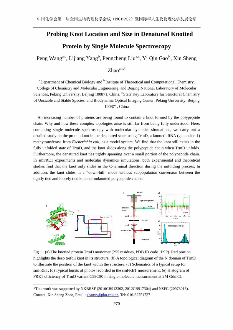
中国化学会第二届全国生物物理化学会议(NCBPC2)暨国际华人生物物理化学发展论坛
P70
Probing Knot Location and Size in Denatured Knotted
Protein by Single Molecule Spectroscopy
Peng Wanga,c
, Lijiang Yangb, Pengcheng Liu
a,c, Yi Qin Gao
b , Xin Sheng
Zhaoa,c,*
a Department of Chemical Biology and
b Institute of Theoretical and Computational Chemistry,
College of Chemistry and Molecular Engineering, and Beijing National Laboratory of Molecular
Sciences, Peking University, Beijing 100871, China; c State Key Laboratory for Structural Chemistry
of Unstable and Stable Species, and Biodynamic Optical Imaging Center, Peking University, Beijing
100871, China
An increasing number of proteins are being found to contain a knot formed by the polypeptide
chain. Why and how these complex topologies arise is still far from being fully understood. Here,
combining single molecule spectroscopy with molecular dynamics simulations, we carry out a
detailed study on the protein knot in the denatured state, using TrmD, a knotted tRNA (guanosine-1)
methytransferase from Escherichia coli, as a model system. We find that the knot still exists in the
fully unfolded state of TrmD, and the knot slides along the polypeptide chain when TrmD unfolds.
Furthermore, the denatured knot ties tightly spanning over a small portion of the polypeptide chain.
In smFRET experiments and molecular dynamics simulations, both experimental and theoretical
studies find that the knot only slides in the C-terminal direction during the unfolding process. In
addition, the knot slides in a “down-hill” mode without subpopulation conversion between the
tightly tied and loosely tied knots or unknotted polypeptide chains.
Fig. 1. (a) The knotted protein TrmD monomer (255 residues, PDB ID code 1P9P). Red portion
highlights the deep trefoil knot in its structure. (b) A topological diagram of the N domain of TrmD
to illustrate the position of the knot within the structure. (c) Schematics of a typical setup for
smFRET. (d) Typical bursts of photos recorded in the smFRET measurement. (e) Histogram of
FRET efficiency of TrmD variant C59C80 in single molecule measurement at 2M GdmCl.
__________________________
*This work was supported by NKBRSF (2010CB912302, 2012CB917304) and NSFC (20973015).
Contact: Xin Sheng Zhao, Email: [email protected], Tel: 010-62751727
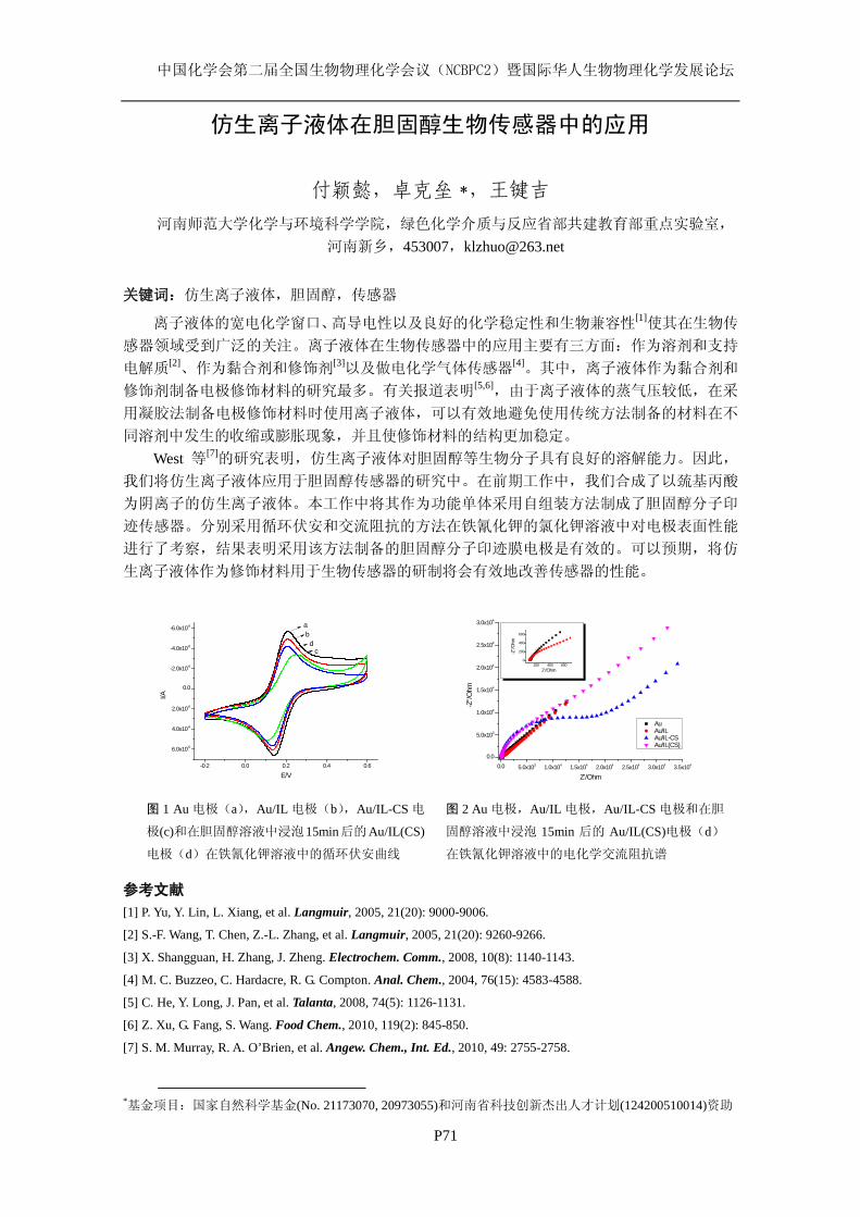
中国化学会第二届全国生物物理化学会议(NCBPC2)暨国际华人生物物理化学发展论坛
P71
-0.2 0.0 0.2 0.4 0.6
6.0x10⁻⁶
4.0x10⁻⁶
2.0x10⁻⁶
0.0
-2.0x10⁻⁶
-4.0x10⁻⁶
-6.0x10⁻⁶
I/A
E/V
ab
cd
图 1 Au 电极(a),Au/IL 电极(b),Au/IL-CS 电
极(c)和在胆固醇溶液中浸泡15min后的Au/IL(CS)
电极(d)在铁氰化钾溶液中的循环伏安曲线
0.0 5.0x10³ 1.0x10⁴ 1.5x10⁴ 2.0x10⁴ 2.5x10⁴ 3.0x10⁴ 3.5x10⁴
0.0
5.0x10³
1.0x10⁴
1.5x10⁴
2.0x10⁴
2.5x10⁴
3.0x10⁴
200 400 6000
200
400
600
-Z''/O
hm
Z'/Ohm
Au Au/IL Au/IL-CS Au/IL(CS)
-Z''/O
hm
Z'/Ohm
图 2 Au 电极,Au/IL 电极,Au/IL-CS 电极和在胆
固醇溶液中浸泡 15min 后的 Au/IL(CS)电极(d)
在铁氰化钾溶液中的电化学交流阻抗谱
仿生离子液体在胆固醇生物传感器中的应用
付颖懿,卓克垒 *
参考文献
,王键吉
河南师范大学化学与环境科学学院,绿色化学介质与反应省部共建教育部重点实验室, 河南新乡,453007,[email protected]
关键词:仿生离子液体,胆固醇,传感器
离子液体的宽电化学窗口、高导电性以及良好的化学稳定性和生物兼容性[1]使其在生物传
感器领域受到广泛的关注。离子液体在生物传感器中的应用主要有三方面:作为溶剂和支持
电解质[2]、作为黏合剂和修饰剂[3]以及做电化学气体传感器[4]。其中,离子液体作为黏合剂和
修饰剂制备电极修饰材料的研究最多。有关报道表明[5,6],由于离子液体的蒸气压较低,在采
用凝胶法制备电极修饰材料时使用离子液体,可以有效地避免使用传统方法制备的材料在不
同溶剂中发生的收缩或膨胀现象,并且使修饰材料的结构更加稳定。 West 等[7]的研究表明,仿生离子液体对胆固醇等生物分子具有良好的溶解能力。因此,
我们将仿生离子液体应用于胆固醇传感器的研究中。在前期工作中,我们合成了以巯基丙酸
为阴离子的仿生离子液体。本工作中将其作为功能单体采用自组装方法制成了胆固醇分子印
迹传感器。分别采用循环伏安和交流阻抗的方法在铁氰化钾的氯化钾溶液中对电极表面性能
进行了考察,结果表明采用该方法制备的胆固醇分子印迹膜电极是有效的。可以预期,将仿
生离子液体作为修饰材料用于生物传感器的研制将会有效地改善传感器的性能。
[1] P. Yu, Y. Lin, L. Xiang, et al. Langmuir, 2005, 21(20): 9000-9006.
[2] S.-F. Wang, T. Chen, Z.-L. Zhang, et al. Langmuir, 2005, 21(20): 9260-9266.
[3] X. Shangguan, H. Zhang, J. Zheng. Electrochem. Comm., 2008, 10(8): 1140-1143.
[4] M. C. Buzzeo, C. Hardacre, R. G. Compton. Anal. Chem., 2004, 76(15): 4583-4588.
[5] C. He, Y. Long, J. Pan, et al. Talanta, 2008, 74(5): 1126-1131.
[6] Z. Xu, G. Fang, S. Wang. Food Chem., 2010, 119(2): 845-850.
[7] S. M. Murray, R. A. O’Brien, et al. Angew. Chem., Int. Ed., 2010, 49: 2755-2758.
*基金项目:国家自然科学基金(No. 21173070, 20973055)和河南省科技创新杰出人才计划(124200510014)资助
中国化学会第二届全国生物物理化学会议(NCBPC2)暨国际华人生物物理化学发展论坛
P72
Toxicity of CdTe Quantum Dots on yeast Saccharomyces
cerevisiae
Xiaole Han, Jia Wang, Lu Lai, Fangfang Tian, Yi Liu
State key laboratory of Virology & Key laboratory of Analytical Chemistry for Biology and
Medicine (Ministry of Education), College of Chemistry and Molecular Sciences, Wuhan University,
Wuhan 430072, P. R. China. [email protected] (Y. Liu)
Along with the widespread development of the bioapplications, concerns about the biosafety of
quantum dots (QDs) have increasingly attracted intensive attentions. This study examines the toxic
effect and subcellular location of cadmium telluride (CdTe) QDs with different sizes against yeast
Saccharomyces cerevisiae (S. cerevisiae). Our innovative approach is based on the combination of
microcalorimetric, spectroscopic, electrochemistrical and microscopic methods, which allows us to
analyze clearly the toxic effect of CdTe QDs on S. cerevisiae and its mechanism. According to the
values of the half inhibitory concentration (IC50), CdTe QDs exhibit marked cytotoxicity in yeast
cells at concentrations as low as 80.81 nmol/L for green-emiting CdTe QDs (G-CdTe) and 17.07
nmol/L for orange-emiting CdTe QDs (O-CdTe). QD-induced cell death is characterized by the cell
wall breakage and the cytoplasm blebbing. These findings suggest that QDs with sizes ranging from
4.1 nm to 5.8 nm can be internalized into yeast cells, which then leads to QD-induced cytotoxicity.
Our studies provide valuable information for the design and development of aqueous QDs for
biological applications.
Keywords: CdTe quantum dots, Yeast, Microcalorimetry, Toxicity, Confocal microscopic imaging
References
[1] Xiaole Han, Lu Lai, Fangfang Tian, et al. Small, 2012, DOI: 10.1002/smll.201200591.
[2] A. Nel, T. Xia, L. Mädler, N. Li. Science, 2006, 311: 622-627.
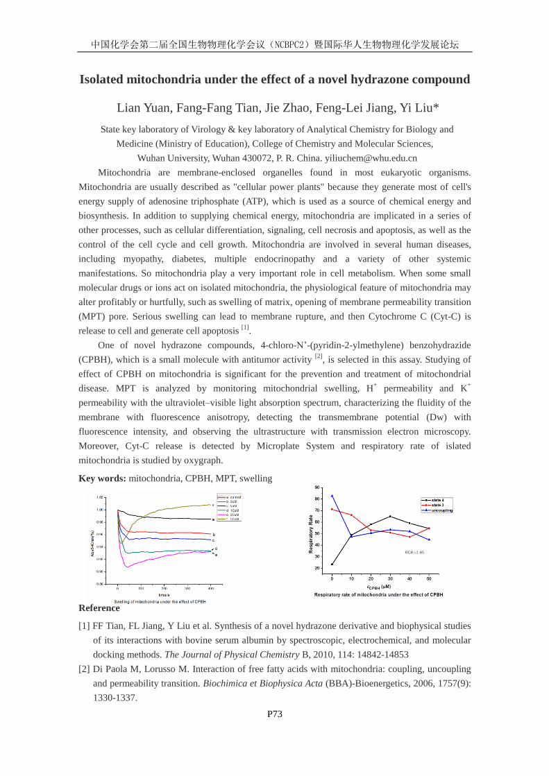
中国化学会第二届全国生物物理化学会议(NCBPC2)暨国际华人生物物理化学发展论坛
P73
Isolated mitochondria under the effect of a novel hydrazone compound
Lian Yuan, Fang-Fang Tian, Jie Zhao, Feng-Lei Jiang, Yi Liu*
State key laboratory of Virology & key laboratory of Analytical Chemistry for Biology and
Medicine (Ministry of Education), College of Chemistry and Molecular Sciences,
Wuhan University, Wuhan 430072, P. R. China. [email protected]
Mitochondria are membrane-enclosed organelles found in most eukaryotic organisms.
Mitochondria are usually described as "cellular power plants" because they generate most of cell's
energy supply of adenosine triphosphate (ATP), which is used as a source of chemical energy and
biosynthesis. In addition to supplying chemical energy, mitochondria are implicated in a series of
other processes, such as cellular differentiation, signaling, cell necrosis and apoptosis, as well as the
control of the cell cycle and cell growth. Mitochondria are involved in several human diseases,
including myopathy, diabetes, multiple endocrinopathy and a variety of other systemic
manifestations. So mitochondria play a very important role in cell metabolism. When some small
molecular drugs or ions act on isolated mitochondria, the physiological feature of mitochondria may
alter profitably or hurtfully, such as swelling of matrix, opening of membrane permeability transition
(MPT) pore. Serious swelling can lead to membrane rupture, and then Cytochrome C (Cyt-C) is
release to cell and generate cell apoptosis [1]
.
One of novel hydrazone compounds, 4-chloro-N’-(pyridin-2-ylmethylene) benzohydrazide
(CPBH), which is a small molecule with antitumor activity [2]
, is selected in this assay. Studying of
effect of CPBH on mitochondria is significant for the prevention and treatment of mitochondrial
disease. MPT is analyzed by monitoring mitochondrial swelling, H+ permeability and K
+
permeability with the ultraviolet–visible light absorption spectrum, characterizing the fluidity of the
membrane with fluorescence anisotropy, detecting the transmembrane potential (Dw) with
fluorescence intensity, and observing the ultrastructure with transmission electron microscopy.
Moreover, Cyt-C release is detected by Microplate System and respiratory rate of islated
mitochondria is studied by oxygraph.
Key words: mitochondria, CPBH, MPT, swelling
Reference
[1] FF Tian, FL Jiang, Y Liu et al. Synthesis of a novel hydrazone derivative and biophysical studies
of its interactions with bovine serum albumin by spectroscopic, electrochemical, and molecular
docking methods. The Journal of Physical Chemistry B, 2010, 114: 14842-14853
[2] Di Paola M, Lorusso M. Interaction of free fatty acids with mitochondria: coupling, uncoupling
and permeability transition. Biochimica et Biophysica Acta (BBA)-Bioenergetics, 2006, 1757(9):
1330-1337.
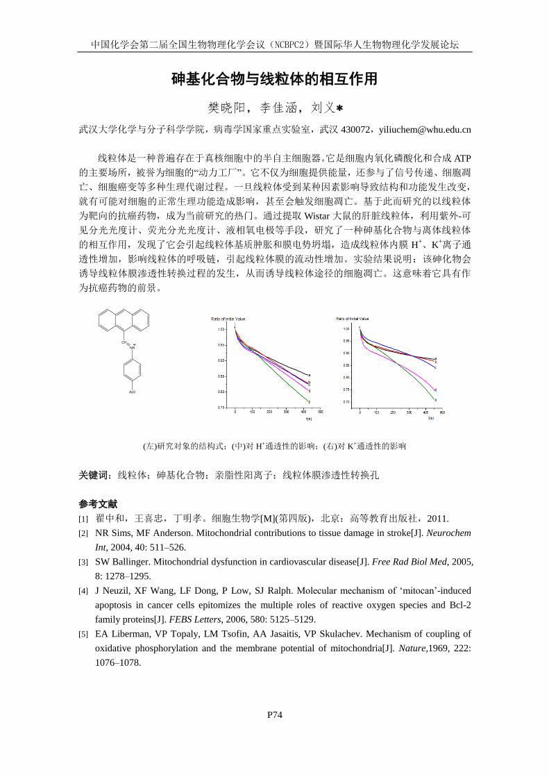
中国化学会第二届全国生物物理化学会议(NCBPC2)暨国际华人生物物理化学发展论坛
P74
砷基化合物与线粒体的相互作用
樊晓阳,李佳涵,刘义*
武汉大学化学与分子科学学院,病毒学国家重点实验室,武汉 430072,[email protected]
线粒体是一种普遍存在于真核细胞中的半自主细胞器。它是细胞内氧化磷酸化和合成 ATP
的主要场所,被誉为细胞的“动力工厂”。它不仅为细胞提供能量,还参与了信号传递、细胞凋
亡、细胞癌变等多种生理代谢过程。一旦线粒体受到某种因素影响导致结构和功能发生改变,
就有可能对细胞的正常生理功能造成影响,甚至会触发细胞凋亡。基于此而研究的以线粒体
为靶向的抗癌药物,成为当前研究的热门。通过提取 Wistar 大鼠的肝脏线粒体,利用紫外-可
见分光光度计、荧光分光光度计、液相氧电极等手段,研究了一种砷基化合物与离体线粒体
的相互作用,发现了它会引起线粒体基质肿胀和膜电势坍塌,造成线粒体内膜 H+、K
+离子通
透性增加,影响线粒体的呼吸链,引起线粒体膜的流动性增加。实验结果说明:该砷化物会
诱导线粒体膜渗透性转换过程的发生,从而诱导线粒体途径的细胞凋亡。这意味着它具有作
为抗癌药物的前景。
(左)研究对象的结构式;(中)对 H+通透性的影响;(右)对 K+通透性的影响
关键词:线粒体;砷基化合物;亲脂性阳离子;线粒体膜渗透性转换孔
参考文献
[1] 翟中和,王喜忠,丁明孝。细胞生物学[M](第四版),北京:高等教育出版社,2011.
[2] NR Sims, MF Anderson. Mitochondrial contributions to tissue damage in stroke[J]. Neurochem
Int, 2004, 40: 511–526.
[3] SW Ballinger. Mitochondrial dysfunction in cardiovascular disease[J]. Free Rad Biol Med, 2005,
8: 1278–1295.
[4] J Neuzil, XF Wang, LF Dong, P Low, SJ Ralph. Molecular mechanism of ‘mitocan’-induced
apoptosis in cancer cells epitomizes the multiple roles of reactive oxygen species and Bcl-2
family proteins[J]. FEBS Letters, 2006, 580: 5125–5129.
[5] EA Liberman, VP Topaly, LM Tsofin, AA Jasaitis, VP Skulachev. Mechanism of coupling of
oxidative phosphorylation and the membrane potential of mitochondria[J]. Nature,1969, 222:
1076–1078.
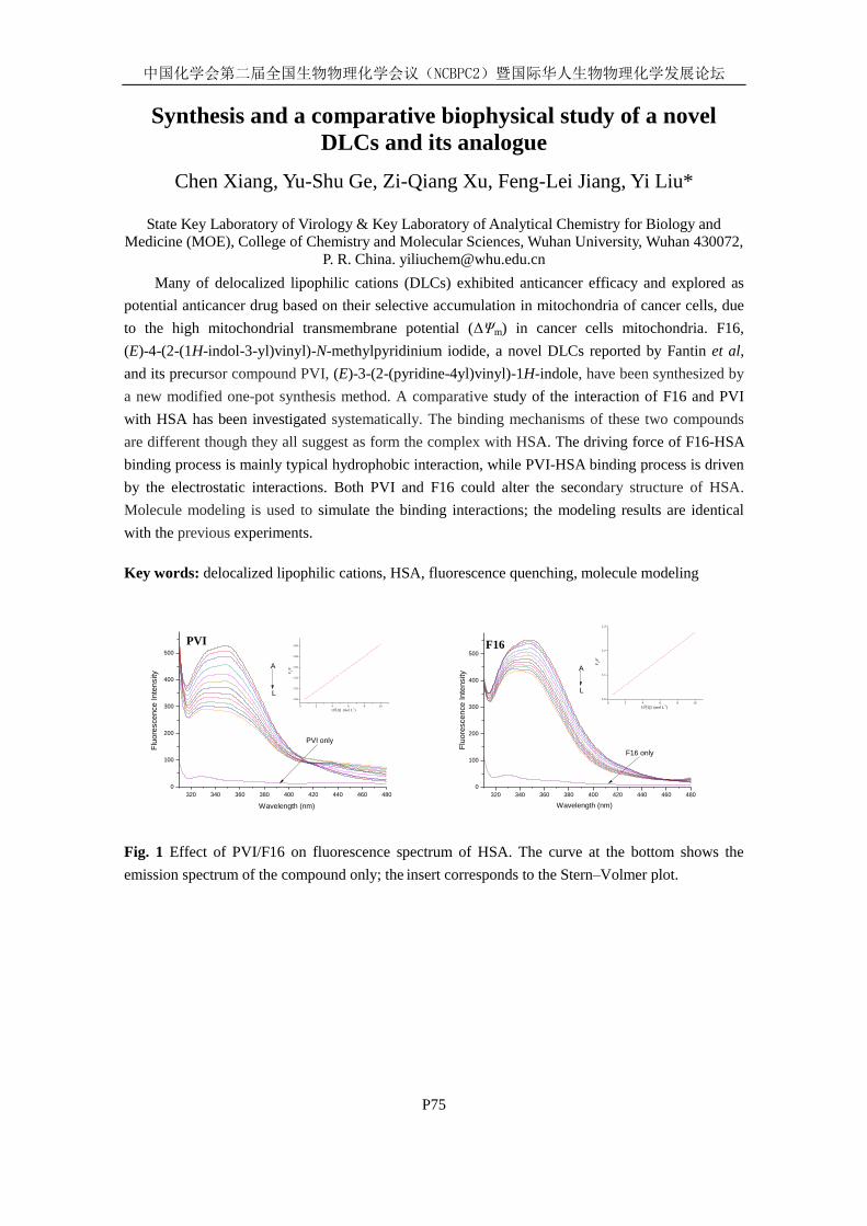
中国化学会第二届全国生物物理化学会议(NCBPC2)暨国际华人生物物理化学发展论坛
P75
Synthesis and a comparative biophysical study of a novel
DLCs and its analogue
Chen Xiang, Yu-Shu Ge, Zi-Qiang Xu, Feng-Lei Jiang, Yi Liu*
State Key Laboratory of Virology & Key Laboratory of Analytical Chemistry for Biology and
Medicine (MOE), College of Chemistry and Molecular Sciences, Wuhan University, Wuhan 430072,
P. R. China. [email protected]
Many of delocalized lipophilic cations (DLCs) exhibited anticancer efficacy and explored as
potential anticancer drug based on their selective accumulation in mitochondria of cancer cells, due
to the high mitochondrial transmembrane potential (ΔΨm) in cancer cells mitochondria. F16,
(E)-4-(2-(1H-indol-3-yl)vinyl)-N-methylpyridinium iodide, a novel DLCs reported by Fantin et al,
and its precursor compound PVI, (E)-3-(2-(pyridine-4yl)vinyl)-1H-indole, have been synthesized by
a new modified one-pot synthesis method. A comparative study of the interaction of F16 and PVI
with HSA has been investigated systematically. The binding mechanisms of these two compounds
are different though they all suggest as form the complex with HSA. The driving force of F16-HSA
binding process is mainly typical hydrophobic interaction, while PVI-HSA binding process is driven
by the electrostatic interactions. Both PVI and F16 could alter the secondary structure of HSA.
Molecule modeling is used to simulate the binding interactions; the modeling results are identical
with the previous experiments.
Key words: delocalized lipophilic cations, HSA, fluorescence quenching, molecule modeling
320 340 360 380 400 420 440 460 4800
100
200
300
400
500
0 2 4 6 8 10
1.05
1.20
1.35
1.50
1.65
1.80
106[Q] (mol L
-1)
F0/F
A
PVI
Fluo
resc
ence
Inte
nsity
Wavelength (nm)
PVI only
L
320 340 360 380 400 420 440 460 4800
100
200
300
400
500
0 2 4 6 8 101.0
1.1
1.2
1.3
106[Q] (mol L
-1)
F0/F
A
F16 only
Wavelength (nm)
Fluo
resc
ence
Inte
nsity
F16
L
Fig. 1 Effect of PVI/F16 on fluorescence spectrum of HSA. The curve at the bottom shows the
emission spectrum of the compound only; the insert corresponds to the Stern–Volmer plot.
中国化学会第二届全国生物物理化学会议(NCBPC2)暨国际华人生物物理化学发展论坛
P76
Studies of Interaction between Dimetridazole and Human
Serum Albumin by Spectroscopic Methods
Wan-Ju Zhang
1,2, Fang Wang
1, Xu-Jie Xiong
1, Dong-Wei Li
2, Yi Liu
2
1 School of Chemical Engineering, Huanggang Normal University, Huanggang 438000, P. R. China.
2 State Key Laboratory of Virology & Key Laboratory of Analytical Chemistry for Biolgy and
Medicine (MOE), College of Chemistry and Molecular Sciences, Wuhan University, Wuhan 430072,
P. R. China.
Dimetridazole (DMZ) is widely used for the therapeutic treatment of farmed animals and is
suspected of being human carcinogens and mutagens. The interaction between DMZ and human
serum albumin (HSA) was investigated systematically by fluorescence spectroscopy, synchronous
fluorescence, three-dimensional fluorescence, CD spectroscopy, UV–vis absorption spectroscopy
and molecular docking study. The results indicated that the probable quenching mechanism of HSA
by DMZ was dynamic quenching. The corresponding thermodynamic parameters, such as ΔH, ΔS
and ΔG etc., were calculated according to Van’t Hoff equation. The results (ΔH = 65.10 kJ mol-1
and
ΔS = 295.65 J mol-1
K-1
) indicated that the driving force between DMZ and HSA was mainly typical
hydrophobic interaction. The conformation changes in the interaction were studied by synchronous
fluorescence, CD spectroscopy and three-dimensional fluorescence spectra. The results revealed that
the microenvironment and conformation of HSA has been changed. Molecular modeling study
further confirmed the binding mode obtained by experimental study.
Keywords: dimetridazole, human serum albumin, fluorescence quenching, interaction
References
[1] E. Daeseleire, H.D. Ruyck, R.V. Renterghem, Analyst., 125 (2000): 1533.
[2] M.K. Tolbert, J. Gookin, Compend. Contin. Educ. Vet., 31 (2009): 374.
[3] T.W. Rosado, A. Specht, S.L. Marks, J. Vet. Intern. Med., 21 (2007): 328.
[4] Y. Sun, Y.Y. Zhao, G. B. Li, J. Chin. Chim. Soc., 58 (2011): 602.
[5] E. Froehlich, J.S. Mandeville, C.J. Jennings, R. Sedaghat-Herati, H.A. Tajmir-Riahi, J. Phys.
Chem. B, 113 (2009): 6986.
[6] J.N. Miller, Proc. Anal. Div. Chem. Soc., 16 (1979): 203.
[7] J. Ghuman, P.A. Zunszain, I. Petitpas, A.A. Bhattacharya, M. Otagiri, S. Curry, J. Mol. Biol., 353
(2005): 38.
[8] J.X. Fu, Y.S. Ge, F. L. Jiang, X.H. Sun, Y. Liu, Y. Liu, Biol. Trace. Elem. Res., 54 (2011): 788.
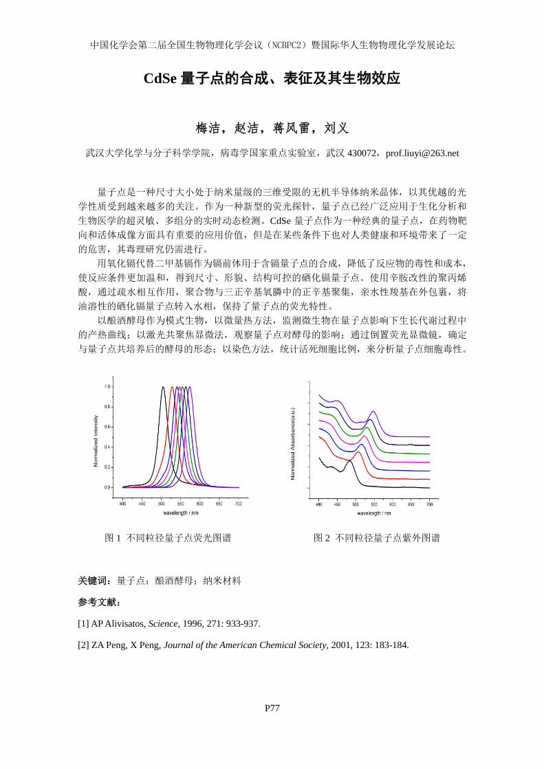
中国化学会第二届全国生物物理化学会议(NCBPC2)暨国际华人生物物理化学发展论坛
P77
CdSe 量子点的合成、表征及其生物效应
梅洁,赵洁,蒋风雷,刘义
武汉大学化学与分子科学学院,病毒学国家重点实验室,武汉 430072,[email protected]
量子点是一种尺寸大小处于纳米量级的三维受限的无机半导体纳米晶体,以其优越的光
学性质受到越来越多的关注。作为一种新型的荧光探针,量子点已经广泛应用于生化分析和
生物医学的超灵敏、多组分的实时动态检测。CdSe 量子点作为一种经典的量子点,在药物靶
向和活体成像方面具有重要的应用价值,但是在某些条件下也对人类健康和环境带来了一定
的危害,其毒理研究仍需进行。
用氧化镉代替二甲基镉作为镉前体用于含镉量子点的合成,降低了反应物的毒性和成本,
使反应条件更加温和,得到尺寸、形貌、结构可控的硒化镉量子点。使用辛胺改性的聚丙烯
酸,通过疏水相互作用,聚合物与三正辛基氧膦中的正辛基聚集,亲水性羧基在外包裹,将
油溶性的硒化镉量子点转入水相,保持了量子点的荧光特性。
以酿酒酵母作为模式生物,以微量热方法,监测微生物在量子点影响下生长代谢过程中
的产热曲线;以激光共聚焦显微法,观察量子点对酵母的影响;通过倒置荧光显微镜,确定
与量子点共培养后的酵母的形态;以染色方法,统计活死细胞比例,来分析量子点细胞毒性。
图 1 不同粒径量子点荧光图谱 图 2 不同粒径量子点紫外图谱
关键词:量子点;酿酒酵母;纳米材料
参考文献:
[1] AP Alivisatos, Science, 1996, 271: 933-937.
[2] ZA Peng, X Peng, Journal of the American Chemical Society, 2001, 123: 183-184.

中国化学会第二届全国生物物理化学会议(NCBPC2)暨国际华人生物物理化学发展论坛
P78
不同浓度 Gd3+对大鼠肝脏线粒体功能的影响
赵洁,梅洁,袁莲,向晨,戴捷,刘义*
武汉大学化学与分子科学学院,病毒学国家重点实验室,生物医学分析化学教育部重点实验
室,武汉 430072。[email protected]
线粒体是真核细胞的重要细胞器,其合成 ATP,进行三羧酸循环,完成氧化磷酸化反应,
调节膜电位,控制细胞凋亡等重要功能,对维持正常的生命过程起着不可替代的作用。采用
Francesco 及 Giorgio 的方法,分离纯化 Wistar 大鼠肝脏线粒体。运用紫外-可见吸收光谱、荧
光光谱等方法,研究不同浓度 Gd3+与大鼠肝脏线粒体相互作用时,观察其膜肿胀、膜电势以
及膜流动性的变化;同时,运用 Clark 溶氧电极,研究 Gd3+对线粒体不同呼吸状态活性及脂质
过氧化的影响;通过透射电镜,观察离体线粒体形貌的变化。实验结果表明:在实验的浓度
范围内,随着 Gd3+浓度增大,线粒体膜肿胀程度增大﹑膜流动性下降越明显,不同呼吸状态
的活性也受到严重的制约,电镜的结果也可以看出对其结构破坏程度增大。
关键词:线粒体,Gd3+,膜肿胀,膜流动性,呼吸状态,显微结构
图 1 不同浓度 Gd3+对线粒体 H
+ 渗透性 图 2 不同浓度 Gd
3+对线粒体脂质过
的影响 氧化的影响
参考文献:
[1] Paul S. Brookes, et al, Free Radical Biol. Med., 2005, 38: 12-23
[2] David M. Rhoads, Chalivendra C. Subbaiah, Mitochondrion, 2007, 7: 177-194
[3] Klimova, T. Chandel, N.S. Cell Death Differ., 2008, 15: 660-666
国家自然科学基金(201173026)资助课题
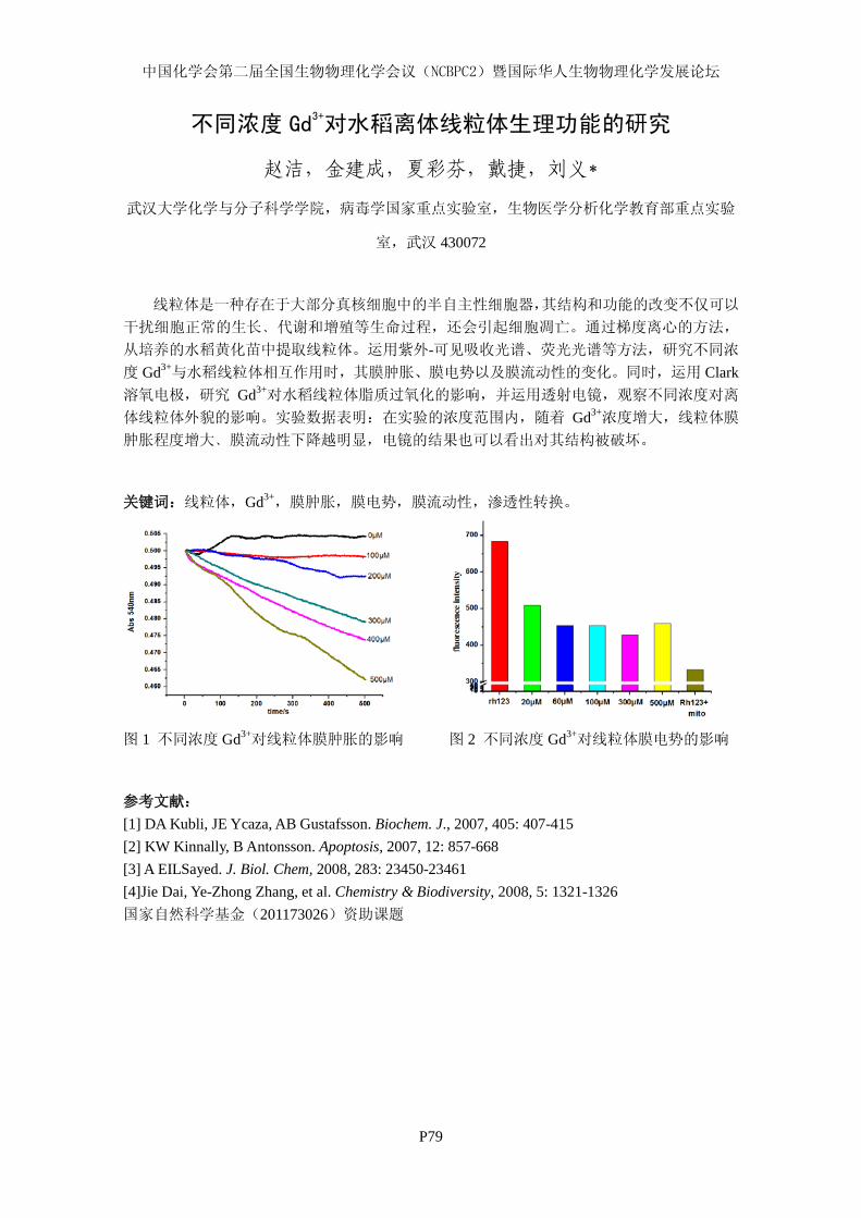
中国化学会第二届全国生物物理化学会议(NCBPC2)暨国际华人生物物理化学发展论坛
P79
不同浓度 Gd3+对水稻离体线粒体生理功能的研究
赵洁,金建成,夏彩芬,戴捷,刘义*
武汉大学化学与分子科学学院,病毒学国家重点实验室,生物医学分析化学教育部重点实验
室,武汉 430072
线粒体是一种存在于大部分真核细胞中的半自主性细胞器,其结构和功能的改变不仅可以
干扰细胞正常的生长﹑代谢和增殖等生命过程,还会引起细胞凋亡。通过梯度离心的方法,
从培养的水稻黄化苗中提取线粒体。运用紫外-可见吸收光谱、荧光光谱等方法,研究不同浓
度 Gd3+与水稻线粒体相互作用时,其膜肿胀、膜电势以及膜流动性的变化。同时,运用 Clark溶氧电极,研究 Gd3+对水稻线粒体脂质过氧化的影响,并运用透射电镜,观察不同浓度对离
体线粒体外貌的影响。实验数据表明:在实验的浓度范围内,随着 Gd3+浓度增大,线粒体膜
肿胀程度增大﹑膜流动性下降越明显,电镜的结果也可以看出对其结构被破坏。
关键词:线粒体,Gd3+,膜肿胀,膜电势,膜流动性,渗透性转换。
图 1 不同浓度 Gd3+对线粒体膜肿胀的影响 图 2 不同浓度 Gd3+对线粒体膜电势的影响
参考文献: [1] DA Kubli, JE Ycaza, AB Gustafsson. Biochem. J., 2007, 405: 407-415 [2] KW Kinnally, B Antonsson. Apoptosis, 2007, 12: 857-668 [3] A EILSayed. J. Biol. Chem, 2008, 283: 23450-23461 [4]Jie Dai, Ye-Zhong Zhang, et al. Chemistry & Biodiversity, 2008, 5: 1321-1326 国家自然科学基金(201173026)资助课题
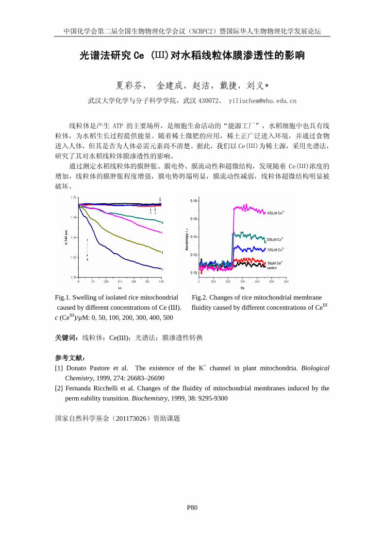
中国化学会第二届全国生物物理化学会议(NCBPC2)暨国际华人生物物理化学发展论坛
P80
光谱法研究 Ce (Ш)对水稻线粒体膜渗透性的影响
夏彩芬, 金建成,赵洁,戴捷,刘义*
武汉大学化学与分子科学学院,武汉 430072。 [email protected]
线粒体是产生 ATP 的主要场所,是细胞生命活动的“能源工厂”,水稻细胞中也具有线
粒体,为水稻生长过程提供能量。随着稀土微肥的应用,稀土正广泛进入环境,并通过食物
进入人体,但其是否为人体必需元素尚不清楚。据此,我们以 Ce(Ш)为稀土源,采用光谱法,
研究了其对水稻线粒体膜渗透性的影响。
通过测定水稻线粒体的膜肿胀、膜电势、膜流动性和超微结构,发现随着 Ce(Ш)浓度的
增加,线粒体的膜肿胀程度增强,膜电势坍塌明显,膜流动性减弱,线粒体超微结构明显被
破坏。
Fig.1. Swelling of isolated rice mitochondrial Fig.2. Changes of rice mitochondrial membrane caused by different concentrations of Ce (Ш). fluidity caused by different concentrations of CeШ
c (CeШ)/µM: 0, 50, 100, 200, 300, 400, 500 关键词:线粒体;Ce(Ш);光谱法;膜渗透性转换 参考文献: [1] Donato Pastore et al. The existence of the K+ channel in plant mitochondria. Biological
Chemistry, 1999, 274: 26683–26690 [2] Fernanda Ricchelli et al. Changes of the fluidity of mitochondrial membranes induced by the
perm eability transition. Biochemistry, 1999, 38: 9295-9300 国家自然科学基金(201173026)资助课题

中国化学会第二届全国生物物理化学会议(NCBPC2)暨国际华人生物物理化学发展论坛
P81
CdTe 量子点的细胞毒理研究
钟慧敏,蔺晨,许子强,杨立云,赖璐,蒋风雷,刘义*
武汉大学化学与分子科学学院,武汉 430072, [email protected]
由于量子尺寸效应和介电限域效应的影响,量子点具有独特的光电性质,这使得量子点
在生物化学、细胞生物学等学科中被广泛研究并显示出广阔的应用前景[1]。近几年来,量子
点等纳米材料的生物效应及其毒理学研究,已经引起了广泛的关注[2]。
本文采用 MTT 方法研究了巯基丙酸(MPA) 修饰的 CdTe QDs 对 HEK 293 和 Hela 细胞活性
的影响,得到 IC50值分别为 109.4 nmol∙L-1和 70.90 nmol∙L-1
。在显微镜下观察到量子点的加
入会破坏细胞贴壁,使细胞的形态发生变化。通过流式细胞仪测定 PI(碘化丙碇)活性可得,
加入量子点后使得细胞膜通透性增加,量子点导致细胞坏死。研究了量子点对细胞周期的影
响,加入 25、50 nmol∙L-1的 MPA-CdTe QDs对 HEK 293和 Hela细胞周期的影响并不明显(图 1)。
图 1. MPA-CdTe QDs 对 Hela 细胞周期的影响.(a)为阴性对照, (b)加入 50nM MPA-CdTe QDs
关键词:量子点;细胞毒性;细胞周期;细胞坏死
参考文献:
[1] C.Y. Zhang, H.C. Yeh, M.T. Kuroki, T.H. Wang. Nat. Mater., 2005, 4: 826. [2] Z.S. Lu, C.M. Li, H.F. Bao, Y. Qiao, Y.H. Toh, X. Yang. Langmuir, 2008, 24: 5445.
中国化学会第二届全国生物物理化学会议(NCBPC2)暨国际华人生物物理化学发展论坛
P82
点击化学及其在分析检测中的应用
付莉 许子强 蒋风雷 刘义*
武汉大学化学与分子科学学院,病毒学国家重点实验室,生物医学分析化学教育部重点实验
室,武汉 430072。[email protected]
点击化学(click chemistry)作为一种低成本快速的合成方法,通过可靠的、高效的、高选择
性的并具模块化的化学反应来实现碳杂原子连接[1]。点击反应主要有四种类型[2]:(1).环加成反
应;(2).张力环亲电试剂的亲核开环反应;(3).羰基化合物温和的缩合反应;(4).碳碳多键的加
成反应,其中研究和应用最为广泛的是铜催化的端基炔和叠氮化物生成的 1,3-偶极环加成反应
(CuAAC),它能够将两种不同物质通过五元环共价结合起来。点击化学由于其所用原料易得、
反应条件温和、副产物少、产率高、立体选择性好、分离提纯简单和产物稳定性好等特点,
在材料科学、药物筛选、高分子化学和生物医学等领域均被广泛应用。 本研究工作特别针对点击化学在分析检测中的应用[3],由于 Cu(I)常作为叠氮化物与炔基
化合物反应的催化剂,而 Cu(I)可由 Cu(II)在抗坏血酸钠溶液中还原获得,因此可以通过点击
化学反应来检测 Cu(II)。相关的研究工作正在进行之中。 关键词:点击化学; Cu(I)催化; 偶极环加成反应; 分析检测 参考文献: 1 HC Kolb, MG Finn, KB Sharpless. Angew. Chem. Int. Ed. 2001, 40 : 2004-2021. 2 MG Finn, VV Fokin. Chem. Soc. Rev. 2010, 39: 1231-1232. 3 CL Droumaguet, C Wang, Q Wang. Chem. Soc. Rev. 2010, 39: 1233-1239.
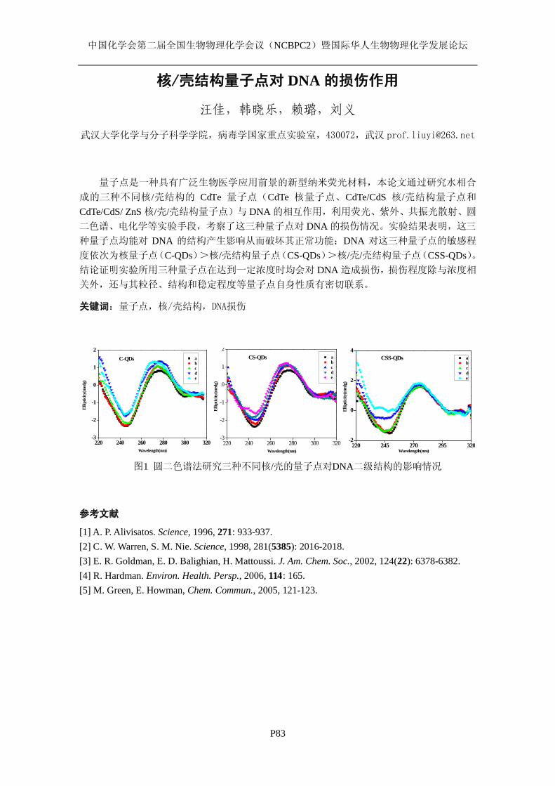
中国化学会第二届全国生物物理化学会议(NCBPC2)暨国际华人生物物理化学发展论坛
P83
核/壳结构量子点对 DNA 的损伤作用
汪佳,韩晓乐,赖璐,刘义
武汉大学化学与分子科学学院,病毒学国家重点实验室,430072,武汉 [email protected]
量子点是一种具有广泛生物医学应用前景的新型纳米荧光材料,本论文通过研究水相合
成的三种不同核/壳结构的 CdTe 量子点(CdTe 核量子点、CdTe/CdS 核/壳结构量子点和
CdTe/CdS/ ZnS 核/壳/壳结构量子点)与 DNA 的相互作用,利用荧光、紫外、共振光散射、圆
二色谱、电化学等实验手段,考察了这三种量子点对 DNA 的损伤情况。实验结果表明,这三
种量子点均能对 DNA 的结构产生影响从而破坏其正常功能;DNA 对这三种量子点的敏感程
度依次为核量子点(C-QDs)>核/壳结构量子点(CS-QDs)>核/壳/壳结构量子点(CSS-QDs)。结论证明实验所用三种量子点在达到一定浓度时均会对 DNA 造成损伤,损伤程度除与浓度相
关外,还与其粒径、结构和稳定程度等量子点自身性质有密切联系。
关键词:量子点,核/壳结构,DNA损伤
220 240 260 280 300 320-3
-2
-1
0
1
2 a b c d e
Ellip
ticity
(med
g)
Wavelength(nm)
C-QDs
220 240 260 280 300 320-3
-2
-1
0
1
2 a b c d e
Ellip
ticity
(med
g)
Wavelength(nm)
CS-QDs
220 245 270 295 320-2
0
2
4El
liptic
ity(m
edg)
Wavelength(nm)
a b c d e
CSS-QDs
图1 圆二色谱法研究三种不同核/壳的量子点对DNA二级结构的影响情况
参考文献
[1] A. P. Alivisatos. Science, 1996, 271: 933-937. [2] C. W. Warren, S. M. Nie. Science, 1998, 281(5385): 2016-2018. [3] E. R. Goldman, E. D. Balighian, H. Mattoussi. J. Am. Chem. Soc., 2002, 124(22): 6378-6382. [4] R. Hardman. Environ. Health. Persp., 2006, 114: 165. [5] M. Green, E. Howman, Chem. Commun., 2005, 121-123.
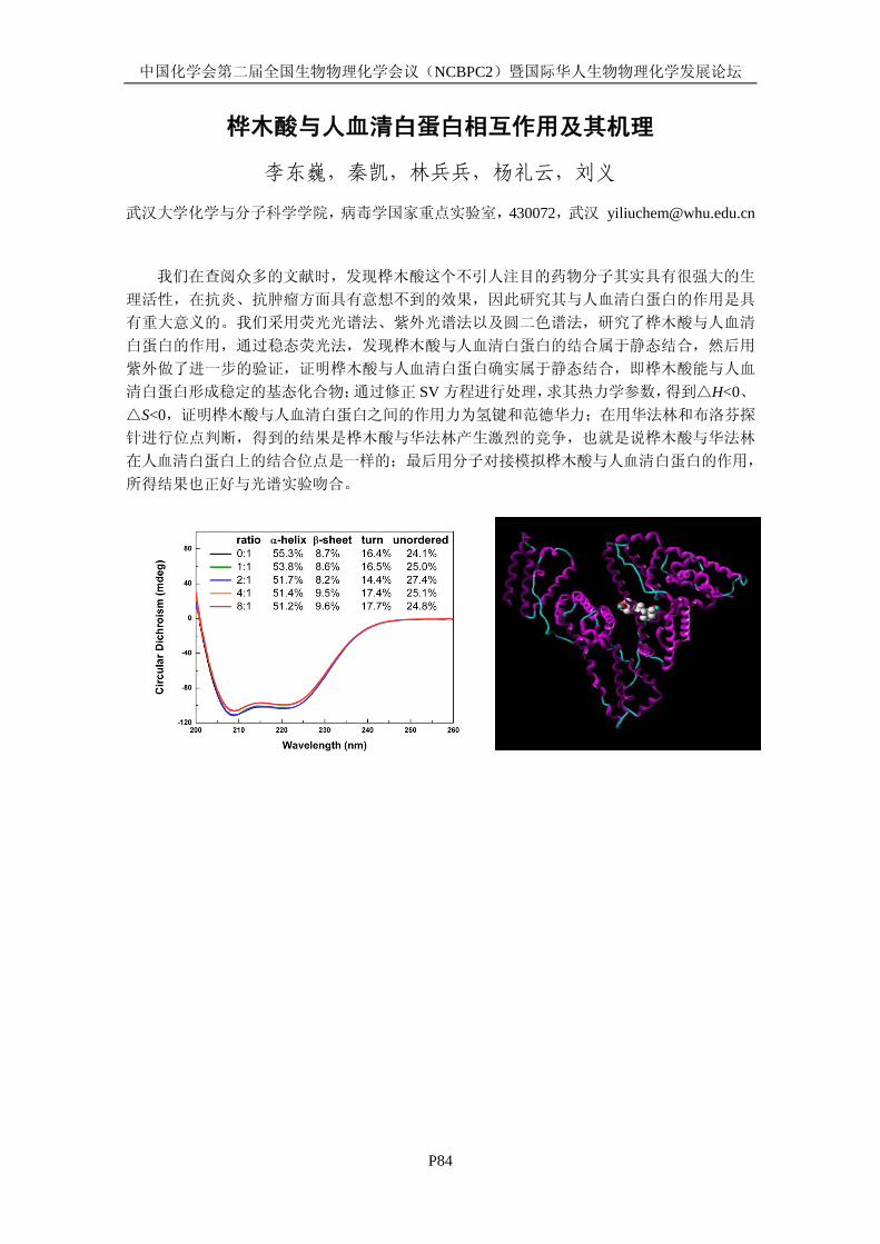
中国化学会第二届全国生物物理化学会议(NCBPC2)暨国际华人生物物理化学发展论坛
P84
桦木酸与人血清白蛋白相互作用及其机理
李东巍,秦凯,林兵兵,杨礼云,刘义
武汉大学化学与分子科学学院,病毒学国家重点实验室,430072,武汉 [email protected]
我们在查阅众多的文献时,发现桦木酸这个不引人注目的药物分子其实具有很强大的生
理活性,在抗炎、抗肿瘤方面具有意想不到的效果,因此研究其与人血清白蛋白的作用是具
有重大意义的。我们采用荧光光谱法、紫外光谱法以及圆二色谱法,研究了桦木酸与人血清
白蛋白的作用,通过稳态荧光法,发现桦木酸与人血清白蛋白的结合属于静态结合,然后用
紫外做了进一步的验证,证明桦木酸与人血清白蛋白确实属于静态结合,即桦木酸能与人血
清白蛋白形成稳定的基态化合物;通过修正 SV 方程进行处理,求其热力学参数,得到△H<0、△S<0,证明桦木酸与人血清白蛋白之间的作用力为氢键和范德华力;在用华法林和布洛芬探
针进行位点判断,得到的结果是桦木酸与华法林产生激烈的竞争,也就是说桦木酸与华法林
在人血清白蛋白上的结合位点是一样的;最后用分子对接模拟桦木酸与人血清白蛋白的作用,
所得结果也正好与光谱实验吻合。
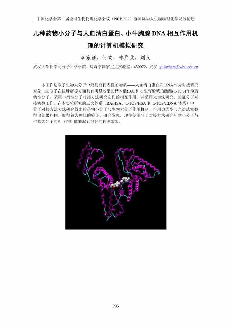
中国化学会第二届全国生物物理化学会议(NCBPC2)暨国际华人生物物理化学发展论坛
P85
几种药物小分子与人血清白蛋白、小牛胸腺 DNA 相互作用机
理的计算机模拟研究
李东巍,何欢,林兵兵,刘义 武汉大学化学与分子科学学院,病毒学国家重点实验室,430072,武汉 [email protected]
本工作选取了生物大分子中最具有代表性的物质——人血清白蛋白和DNA作为对接研究
对象,选取了在抗肿瘤等方面具有明显效果的桦木酸(BA)和 α生育酚琥珀酸酯(α-TOS)作为药
物小分子,采用半柔性分子对接方法研究它们的相互作用,并采用光谱法研究、验证分子对
接实验工作。在本实验研究的三大体系(BA/HSA、α-TOS/HSA 和 α-TOS/ctDNA 体系)中,
分子对接方法方法研究得出的药物小分子与生物大分子作用机制、作用力类型与光谱法实验
得出结果相同,取得较为理想的验证。研究发现,理性使用分子对接方法研究药物小分子与
生物大分子的相互作用能够起到很好的预测效果。
中国化学会第二届全国生物物理化学会议(NCBPC2)暨国际华人生物物理化学发展论坛
Molecular Modeling of Interaction between Cellular
Membrane and Nanoparticles
Tongtao Yue Centre for Bioengineering and Biotechnology, China University of Petroleum (East China), 66
Changjiang West Road, Qingdao Economic Development Zone, Qingdao 266555, People’s Republic of China, Email: [email protected]
Keywords: nanoparticles, lipid membrane, dissipative particle dynamics.
In recent years, bionanotechnology has opened new avenues for clinical diagnostics and therapeutics. In particular, nanoparticles (NPs) with different sizes, shapes, and chemical properties have been designed for drug delivery. Therefore, understanding how NPs with different properties interact with cellular membrane is important for the design of drug nanocarriers. For single spherical NP, four different membrane responses, including membrane rupture, NP adhesion, NP penetration, and receptor-mediated endocytosis have been observed in our work [1]. Besides, our simulation results indicate that NPs size, ligand density, and membrane surface tension are the main determinant in NP-membrane interaction. For single nonspherical NP, we found that the rotation of NP regulates the competition between ligande receptor binding and membrane deformation, and the whole internalization process can be divided into the invagination and wrapping stage [2]. Moreover, while taking multiple NPs into account, the internalization of these multiple NPs is, in fact, a cooperative process, and there exist four cooperative internalization pathways: synchronous internalization, asynchronous internalization, pinocytosis-like internalization, and independent internalization [3]. Furthermore, when a soft elastic vesicle is adsorbed on the lipid membrane, five distinct phenomena were observed: vesicle fusion, vesicle hemi-fusion, vesicle adhesion, vesicle rupture, and vesicle endocytosis [4]. References: [1] Soft Matter, 2011, 7, 9104. [2] Biomaterials, 2012, 33, 4965. [3] ACS Nano, 2012, 6, 3196. [4] Soft Matter, 2012, DOI: 10.1039/C2SM26940F.
www.tainstruments.com.cn
探索全新
NANOITC &DSCNano ITC&DSC是强而有力的测试工具,拥有以下功能:小分子蛋白质交互作用蛋白质间交互作用药物与标靶交互作用生物分子结构与稳定性

Discovery��TGA������� ���������������������� !"##$%TA�&'
�()
���*DISCOVERY
www.tainstruments.com.cn

MicroCalTM
ITC systemsAffi nity - Specificity - Stoichiometry
MicroCalTM
VP-Capillary DSCGet it right from the start
• Confi dent decision-making
• In-solution stability
• Faster results
A new level of productivity for stability studies
Deeper insights into binding mechanism
for confident conclusions
• Direct and automated biochemical technique
• Enable increased understanding of low and high affi nity interactions
• Provide deeper insight of biological activity for a wide range of research applications
Make confi dent decisions sooner, visit www.gelifesciences.com/microcal
更多详情请拨打服务热线:800-810-9118 400-810-9118 或登录 www.gelifesciences.com.cn
武汉欣申试化工科技有限公司
武汉欣申试化工科技有限公司 武大订购电话:027-87314198、87363782、68786241,转分机 601 传真号码:027-87717850
武汉欣申试化工科技有限公司简介 武汉申试创建于 2000 年,现具备一般纳税人资格。主要经营产品
有:常规试剂、医药中间体、进口试剂、实验常用仪器、实验耗材、
玻璃仪器。另外本公司具有易制毒、危险品等经营资格!
其中试剂方面为上海国药集团武汉代理、上海强顺化学试剂有限
公司武汉代理、江苏永华化学试剂有限公司特约经销商、天津富宇化
学试剂公司湖北代理、天津科密欧试剂有限公司特约经销商。进口试
剂方面,本公司为 Fisher、alading、Acrose、Alfa 武汉代理,另外
经销 Sigma、Merck、TDI、J&K 等进口公司产品。仪器与上海精宏、
上海司乐、上海顾村、上海亚荣、江苏国华、赛多利斯、日本岛津等
国内外众品牌厂家密切合作!
最具特色的玻璃标准磨口类烧器是本公司生产“申试”品牌!本
公司设有玻璃加工工厂。所有毛坯均挑选蜀玻、北玻等大型玻璃厂优
质毛坯。磨口是有专业灯工按企业标准操作。并长期与国内知名玻璃
厂合作。(蜀玻集团、天津友丰、北京欣维尔、北玻、天玻等)。
现公司客户对象为湖北省内各大高校、研究所、研发公司、制药
厂等。
公司理念:质量第一,服务为上,价格公道。以人为本,服务客
户,做百年企业!
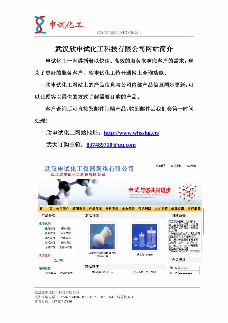
武汉欣申试化工科技有限公司
武汉欣申试化工科技有限公司 武大订购电话:027-87314198、87363782、68786241,转分机 601 传真号码:027-87717850
武汉欣申试化工科技有限公司网站简介 申试化工一直遵循着以快速、高效的服务来响应客户的需求;现
为了更好的服务客户,欣申试化工特开通网上查询功能。
欣申试化工网站上的产品信息与公司内部产品信息同步更新,可
以让顾客以最快的方式了解需要订购的产品。
客户查询后可直接发邮件订购产品,收到邮件后我们会第一时间
处理!
欣申试化工网站地址:http://www.whsshg.cn/
武大订购邮箱:[email protected]
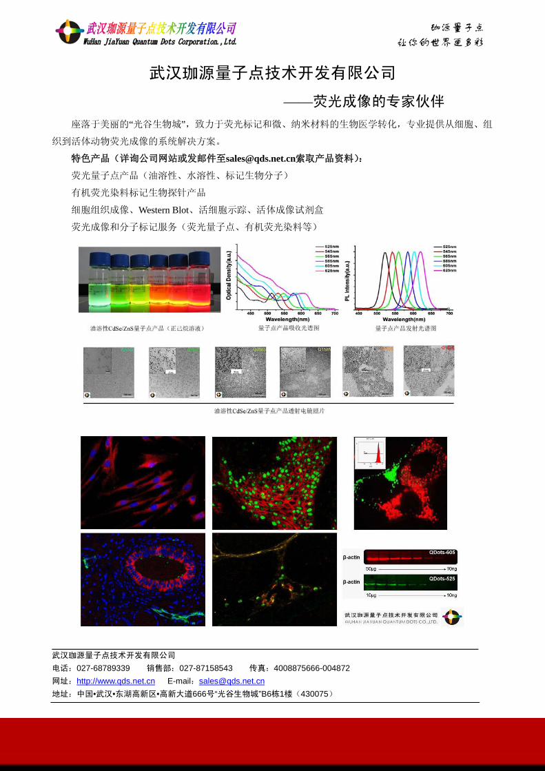
诚诚诚诚!
珈源量子点
让你的世界更多彩
武汉珈源量子点技术开发有限公司
——荧光成像的专家伙伴 座落于美丽的“光谷生物城”,致力于荧光标记和微、纳米材料的生物医学转化,专业提供从细胞、组
织到活体动物荧光成像的系统解决方案。
特色产品(详询公司网站或发邮件至[email protected]索取产品资料):
荧光量子点产品(油溶性、水溶性、标记生物分子)
有机荧光染料标记生物探针产品
细胞组织成像、Western Blot、活细胞示踪、活体成像试剂盒
荧光成像和分子标记服务(荧光量子点、有机荧光染料等)
武汉珈源量子点技术开发有限公司 电话:027-68789339 销售部:027-87158543 传真:4008875666-004872 网址:http://www.qds.net.cn E-mail:[email protected] 地址:中国•武汉•东湖高新区•高新大道666号“光谷生物城”B6栋1楼(430075)

Chirascan 圆二色光谱仪 For the most demanding research applications
Chirascan为静态圆二色光谱分析树立了新的标准, 它采用多种新技术、新设计, 使到达样品的光强度达到
最大, 特别是远紫外区(Far UV)。 它的先进的数字式数据采集原理可实现快速、准确的光谱数据采集。
可配置多种扩展功能模块(吸收、荧光、全荧光谱、荧光偏振(FP)/各向异性谱、磁CD、ORD (旋光色散)、低
温检测附件、滴定系统、停流Stopped flow 系统等),以使各种研究目的都能得到高质量的、可靠的结果数
据。有大量高水平的科研论文和基础研究成果的数据出自本系列仪器如 Chirascan , Chirascan-plus , Auto-Chirascan等。 广泛应用于分子结构、分子间相互作用等领域的研究,特别是生物大分子之间、生物大分子与小分子如药物、各类手性
化合物等的绝对构型与相互作用的研究.
Stopped Flow 停流光谱仪 (SX20系列)
停流(stopped flow)光谱技术,用于研究溶液体系的非常快速的反应的机理,如反应时间一般在 0.001 秒到
0.01 秒之间。通过检测各种光属性(如吸收、荧光、双路荧光、荧光偏振、圆二色性等)随时间的变化,研究
瞬时动力学现象,可反映反应的速度、机理、短时间存在过的反应中间体、过渡态等基础信息。
英国 Applied Photophysics ( 应用光物理公司) 生产的型号为 SX20 的停流光谱仪,数据引用率占可检
索到的已发表的文献的八成以上。可选择配置的功能模块为:时间分辨的吸收分析、二极管阵列检测、荧光和双
荧光检测、荧光偏振检测,多通道序列混合系统、淬灭停流检测 , 及 CD 圆二色停流分析系统等。 广泛应用于分子间相互作用等领域的动力学研究, 如蛋白质—蛋白质、蛋白质—配体的结合,电子转移、蛋白质折叠、酶
反应、各种协同作用及其它化学反应等。
英国应用光物理公司 Applied Photophysics Limited 21 Mole Business Park , Leatherhead, KT22 7BA, United Kingdom. T:+44 (0) 1372 386537 · F: +44 (0) 1372 386477 E: [email protected] · Web: www.photophysics.com
上海代表处 上海市恒丰路218号现代交通大厦1604室
Tel: +86-21-51801906 Fax:+86-21-51603567 Email: [email protected] Web: www.photophysics.com
Applied Photophysics was established in 1971 by the Royal Institution of Great Britain
Related Documents