Deep Neck Infections: Clinical Considerations in Aggressive Disease John F. Caccamese Jr., DMD, MD, FACS a,b, * , Domenick P. Coletti, DDS, MD a a Department of Oral and Maxillofacial Surgery, University of Maryland Medical System, 650 West Baltimore Street, Suite 1401, Baltimore, MD 21204, USA b Department of Pediatrics, University of Maryland Medical System, 22 South Greene Street, Baltimore, MD 21204, USA Deep neck infections (DNIs) are common and occur as a consequence of several etiologies. The result is either an abscess or cellulitis in the potential spaces of the neck. Countless reports exist in the literature detailing the management of infections arising from odontogenic sources, con- genital lesions, cervical adenitis, malignancies, surgical treatment, upper respiratory infection, and penetrating trauma. Life-threatening compli- cations that have been associated with DNI include airway obstruction, sepsis, septic emboli, Lemierre syndrome, descending mediastinitus, and pericarditis. Additionally, extremely aggres- sive forms of DNI exist, including necrotizing fasciitis (Fig. 1A, B and Fig. 2A, B, C). Despite numerous reports, little information exists regarding the true incidence of these in- fections. As is true in many areas of medicine, limited prospective data are available to guide therapy. Treatment over the years has been based mostly on surgical principle, airway management, and an understanding of the microbiologic flora of these infections. Antibiotic therapy, interven- tional radiology, and patient support modalities have become more sophisticated, although sur- gery continues to be the mainstay of treatment for most patients. Today, neck infections are rarely life threatening when sound and timely manage- ment is applied. Etiology DNI can arise from many sources. Dental sources are widely held to be the most common in adults and give rise to some of the more aggressive infections [1–5]. Aside from odonto- genic infections, the etiology varies significantly by report. In some of the larger series, upper respi- ratory tract infection, intravenous drug abuse, and penetrating neck trauma have all been major contributors to this disease process [1–5]; how- ever, one should consider all possibilities when evaluating a patient for DNI. Branchial sinuses, thyroglossal duct cysts, and malignancies can all masquerade as infections or present secondarily infected [5–11]. A careful history, examination, and imaging will help to differentiate the causes (Fig. 3). DNI in the pediatric patient can present more of a diagnostic dilemma. The origin of infection is more varied in the pediatric population, and the location and microbial flora can differ substan- tially as well. Although sinusitis, pharyngitis, and tonsillitis can all have important roles in pediatric DNI, patients and their families frequently pres- ent with no history of a precipitating event or illness. Even within the pediatric population, the age of the patient results in an altered distribution of the site and etiology of infection [12]. Several studies have noted a higher incidence of staphylo- coccal species in pediatric DNI, as well as * Corresponding author. Department of Oral and Maxillofacial Surgery, University of Maryland Medical System, 650 West Baltimore Street, Suite 1401, Balti- more, MD 21204 E-mail address: [email protected] (J.F. Caccamese) 1042-3699/08/$ - see front matter Ó 2008 Elsevier Inc. All rights reserved. doi:10.1016/j.coms.2008.03.001 oralmaxsurgery.theclinics.com Oral Maxillofacial Surg Clin N Am 20 (2008) 367–380

Welcome message from author
This document is posted to help you gain knowledge. Please leave a comment to let me know what you think about it! Share it to your friends and learn new things together.
Transcript
Oral Maxillofacial Surg Clin N Am 20 (2008) 367–380
Deep Neck Infections: Clinical Considerationsin Aggressive Disease
John F. Caccamese Jr., DMD, MD, FACSa,b,*,Domenick P. Coletti, DDS, MDa
aDepartment of Oral and Maxillofacial Surgery, University of Maryland Medical System,
650 West Baltimore Street, Suite 1401, Baltimore, MD 21204, USAbDepartment of Pediatrics, University of Maryland Medical System,
22 South Greene Street, Baltimore, MD 21204, USA
Deep neck infections (DNIs) are common andoccur as a consequence of several etiologies. Theresult is either an abscess or cellulitis in the
potential spaces of the neck. Countless reportsexist in the literature detailing the management ofinfections arising from odontogenic sources, con-genital lesions, cervical adenitis, malignancies,
surgical treatment, upper respiratory infection,and penetrating trauma. Life-threatening compli-cations that have been associated with DNI
include airway obstruction, sepsis, septic emboli,Lemierre syndrome, descending mediastinitus,and pericarditis. Additionally, extremely aggres-
sive forms of DNI exist, including necrotizingfasciitis (Fig. 1A, B and Fig. 2A, B, C).
Despite numerous reports, little informationexists regarding the true incidence of these in-
fections. As is true in many areas of medicine,limited prospective data are available to guidetherapy. Treatment over the years has been based
mostly on surgical principle, airway management,and an understanding of the microbiologic floraof these infections. Antibiotic therapy, interven-
tional radiology, and patient support modalitieshave become more sophisticated, although sur-gery continues to be the mainstay of treatment for
most patients. Today, neck infections are rarely
* Corresponding author. Department of Oral and
Maxillofacial Surgery, University of Maryland Medical
System, 650 West Baltimore Street, Suite 1401, Balti-
more, MD 21204
E-mail address: [email protected]
(J.F. Caccamese)
1042-3699/08/$ - see front matter � 2008 Elsevier Inc. All righ
doi:10.1016/j.coms.2008.03.001
life threatening when sound and timely manage-ment is applied.
Etiology
DNI can arise from many sources. Dentalsources are widely held to be the most common
in adults and give rise to some of the moreaggressive infections [1–5]. Aside from odonto-genic infections, the etiology varies significantly
by report. In some of the larger series, upper respi-ratory tract infection, intravenous drug abuse,and penetrating neck trauma have all been majorcontributors to this disease process [1–5]; how-
ever, one should consider all possibilities whenevaluating a patient for DNI. Branchial sinuses,thyroglossal duct cysts, and malignancies can all
masquerade as infections or present secondarilyinfected [5–11]. A careful history, examination,and imaging will help to differentiate the causes
(Fig. 3).DNI in the pediatric patient can present more
of a diagnostic dilemma. The origin of infection is
more varied in the pediatric population, and thelocation and microbial flora can differ substan-tially as well. Although sinusitis, pharyngitis, andtonsillitis can all have important roles in pediatric
DNI, patients and their families frequently pres-ent with no history of a precipitating event orillness. Even within the pediatric population, the
age of the patient results in an altered distributionof the site and etiology of infection [12]. Severalstudies have noted a higher incidence of staphylo-
coccal species in pediatric DNI, as well as
ts reserved.
oralmaxsurgery.theclinics.com
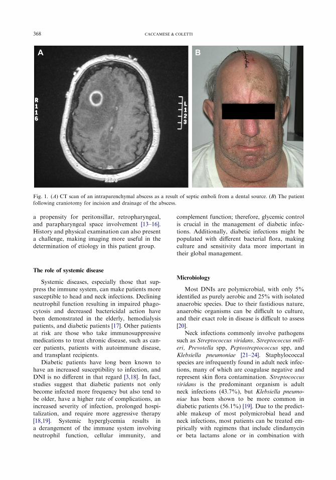
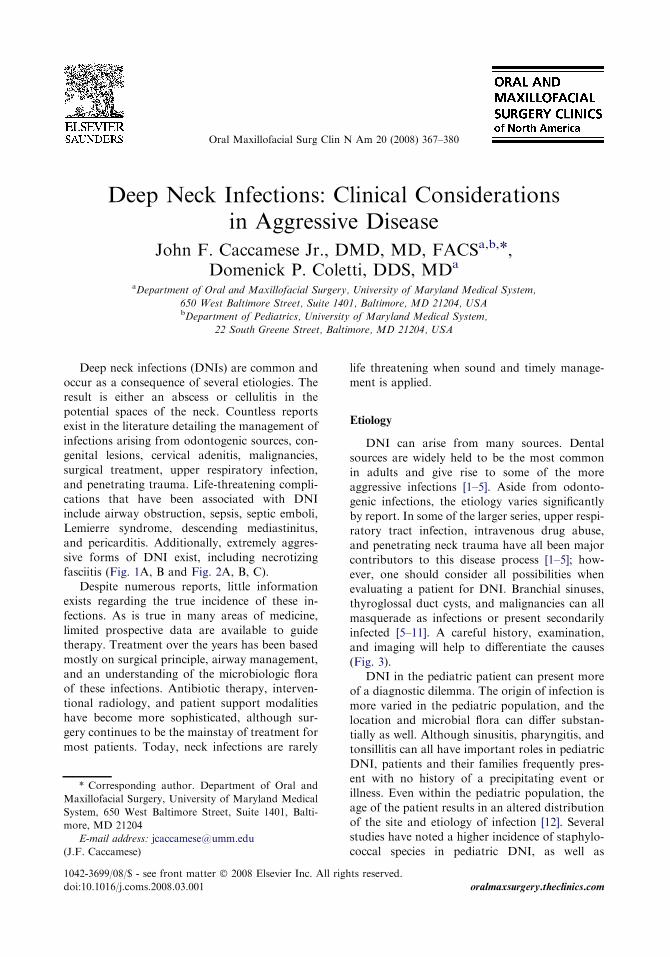
Fig. 1. (A) CT scan of an intraparenchymal abscess as a result of septic emboli from a dental source. (B) The patient
following craniotomy for incision and drainage of the abscess.
368 CACCAMESE & COLETTI
a propensity for peritonsillar, retropharyngeal,and parapharyngeal space involvement [13–16].
History and physical examination can also presenta challenge, making imaging more useful in thedetermination of etiology in this patient group.
The role of systemic disease
Systemic diseases, especially those that sup-press the immune system, can make patients moresusceptible to head and neck infections. Declining
neutrophil function resulting in impaired phago-cytosis and decreased bactericidal action havebeen demonstrated in the elderly, hemodialysis
patients, and diabetic patients [17]. Other patientsat risk are those who take immunosuppressivemedications to treat chronic disease, such as can-
cer patients, patients with autoimmune disease,and transplant recipients.
Diabetic patients have long been known tohave an increased susceptibility to infection, and
DNI is no different in that regard [3,18]. In fact,studies suggest that diabetic patients not onlybecome infected more frequency but also tend to
be older, have a higher rate of complications, anincreased severity of infection, prolonged hospi-talization, and require more aggressive therapy
[18,19]. Systemic hyperglycemia results ina derangement of the immune system involvingneutrophil function, cellular immunity, and
complement function; therefore, glycemic controlis crucial in the management of diabetic infec-
tions. Additionally, diabetic infections might bepopulated with different bacterial flora, makingculture and sensitivity data more important intheir global management.
Microbiology
Most DNIs are polymicrobial, with only 5%identified as purely aerobic and 25% with isolated
anaerobic species. Due to their fastidious nature,anaerobic organisms can be difficult to culture,and their exact role in disease is difficult to assess
[20].Neck infections commonly involve pathogens
such as Streptococcus viridans, Streptococcus mill-
eri, Prevotella spp, Peptostreptococcus spp, andKlebsiella pneumoniae [21–24]. Staphylococcalspecies are infrequently found in adult neck infec-tions, many of which are coagulase negative and
represent skin flora contamination. Streptococcusviridans is the predominant organism is adultneck infections (43.7%), but Klebsiella pneumo-
niae has been shown to be more common indiabetic patients (56.1%) [19]. Due to the predict-able makeup of most polymicrobial head and
neck infections, most patients can be treated em-pirically with regimens that include clindamycinor beta lactams alone or in combination with

Fig. 2. (A) Retropharyngeal abscess from a dental source in an otherwise healthy young patient. (B) Extension of this
abscess into the mediastinum and pericardium. The patient required thoracotomy. (C) An extended neck incision at the
time of drainage. All neck spaces were drained.
369CLINICAL CONSIDERATIONS IN AGGRESSIVE DISEASE
metronidazole [25,26]. In a recent study of odon-
togenic infections by Flynn and colleagues [25],19% of isolated species were found to be penicillinresistant, with only 4% of species with clindamy-
cin resistance. These resistant strains accountedfor 54% and 17% of cases with sensitivity data,respectively.
DNIs in children are also likely to be poly-
microbial, with beta-lactamase producing organ-isms becoming more common. Group Astreptococci represent the major pathogen, with
Prevotella being the most common anaerobe;however, many studies have demonstrated an in-creased incidence of Staphylococcus aureus in
pediatric neck infections [15,27–29]. This observa-tion is most likely a result of fewer odontogenicinfections in children and a higher rate of upper
respiratory tract–related infections. Ever concern-ing is the increasing rate of methicillin-resistantS aureus (MRSA), which according to Ossowskiand colleagues [29] has increased in their patient
cohort of neck infections from 0% to 64% over
6 years.The utility of cultures has been questioned,
with some reports indicating that culture and
sensitivity data do not lead to a change inantibiotic selection or treatment [26]. Othershave recommended cultures for extensive orrapidly spreading infections, necrotizing and
gas-forming infections, nosocomial or recurrentinfections, and those that occur in an immuno-compromised host [30]. Given the ever increasing
possibility of drug-resistant organisms, perhapsthis issue should be revisited.
In necrotizing fasciitis, the causative organisms
are classically group A b-hemolytic streptococciand Staphylococcus aureus, alone or in synergism.Other aerobic and anaerobic pathogens can also
be present, such as Bacteroides, Clostridium, Pep-tostreptococcus, Proteus, Pseudomonas, and Kleb-siella [31]. Triple antibiotic therapy is usuallyinstituted empirically, with regimens including
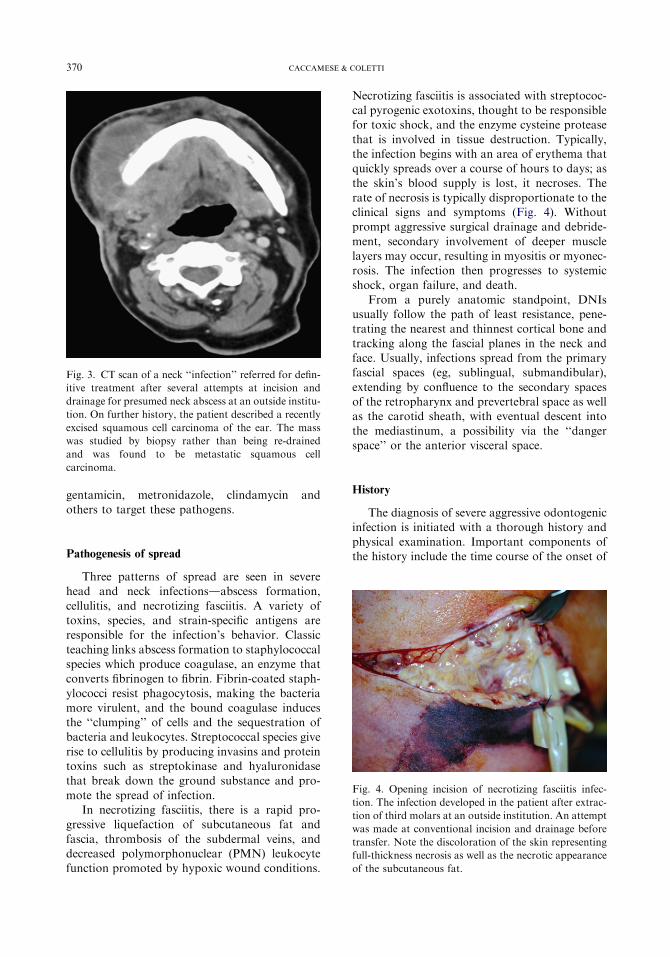
Fig. 3. CT scan of a neck ‘‘infection’’ referred for defin-
itive treatment after several attempts at incision and
drainage for presumed neck abscess at an outside institu-
tion. On further history, the patient described a recently
excised squamous cell carcinoma of the ear. The mass
was studied by biopsy rather than being re-drained
and was found to be metastatic squamous cell
carcinoma.
370 CACCAMESE & COLETTI
gentamicin, metronidazole, clindamycin andothers to target these pathogens.
Fig. 4. Opening incision of necrotizing fasciitis infec-
tion. The infection developed in the patient after extrac-
tion of third molars at an outside institution. An attempt
was made at conventional incision and drainage before
transfer. Note the discoloration of the skin representing
full-thickness necrosis as well as the necrotic appearance
of the subcutaneous fat.
Pathogenesis of spread
Three patterns of spread are seen in severehead and neck infectionsdabscess formation,
cellulitis, and necrotizing fasciitis. A variety oftoxins, species, and strain-specific antigens areresponsible for the infection’s behavior. Classic
teaching links abscess formation to staphylococcalspecies which produce coagulase, an enzyme thatconverts fibrinogen to fibrin. Fibrin-coated staph-
ylococci resist phagocytosis, making the bacteriamore virulent, and the bound coagulase inducesthe ‘‘clumping’’ of cells and the sequestration ofbacteria and leukocytes. Streptococcal species give
rise to cellulitis by producing invasins and proteintoxins such as streptokinase and hyaluronidasethat break down the ground substance and pro-
mote the spread of infection.In necrotizing fasciitis, there is a rapid pro-
gressive liquefaction of subcutaneous fat and
fascia, thrombosis of the subdermal veins, anddecreased polymorphonuclear (PMN) leukocytefunction promoted by hypoxic wound conditions.
Necrotizing fasciitis is associated with streptococ-cal pyrogenic exotoxins, thought to be responsiblefor toxic shock, and the enzyme cysteine protease
that is involved in tissue destruction. Typically,the infection begins with an area of erythema thatquickly spreads over a course of hours to days; asthe skin’s blood supply is lost, it necroses. The
rate of necrosis is typically disproportionate to theclinical signs and symptoms (Fig. 4). Withoutprompt aggressive surgical drainage and debride-
ment, secondary involvement of deeper musclelayers may occur, resulting in myositis or myonec-rosis. The infection then progresses to systemic
shock, organ failure, and death.From a purely anatomic standpoint, DNIs
usually follow the path of least resistance, pene-trating the nearest and thinnest cortical bone and
tracking along the fascial planes in the neck andface. Usually, infections spread from the primaryfascial spaces (eg, sublingual, submandibular),
extending by confluence to the secondary spacesof the retropharynx and prevertebral space as wellas the carotid sheath, with eventual descent into
the mediastinum, a possibility via the ‘‘dangerspace’’ or the anterior visceral space.
History
The diagnosis of severe aggressive odontogenicinfection is initiated with a thorough history andphysical examination. Important components ofthe history include the time course of the onset of
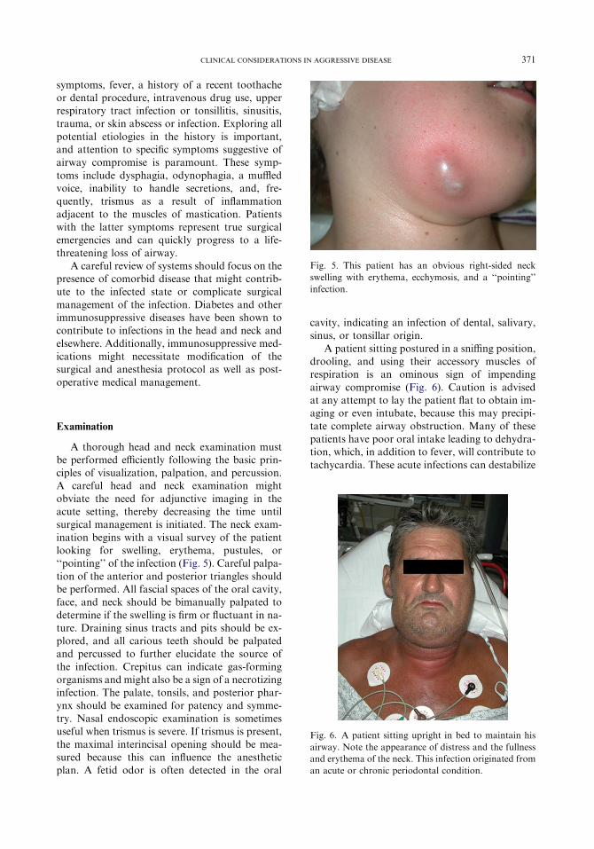
Fig. 5. This patient has an obvious right-sided neck
swelling with erythema, ecchymosis, and a ‘‘pointing’’
infection.
371CLINICAL CONSIDERATIONS IN AGGRESSIVE DISEASE
symptoms, fever, a history of a recent toothacheor dental procedure, intravenous drug use, upperrespiratory tract infection or tonsillitis, sinusitis,trauma, or skin abscess or infection. Exploring all
potential etiologies in the history is important,and attention to specific symptoms suggestive ofairway compromise is paramount. These symp-
toms include dysphagia, odynophagia, a muffledvoice, inability to handle secretions, and, fre-quently, trismus as a result of inflammation
adjacent to the muscles of mastication. Patientswith the latter symptoms represent true surgicalemergencies and can quickly progress to a life-
threatening loss of airway.A careful review of systems should focus on the
presence of comorbid disease that might contrib-ute to the infected state or complicate surgical
management of the infection. Diabetes and otherimmunosuppressive diseases have been shown tocontribute to infections in the head and neck and
elsewhere. Additionally, immunosuppressive med-ications might necessitate modification of thesurgical and anesthesia protocol as well as post-
operative medical management.
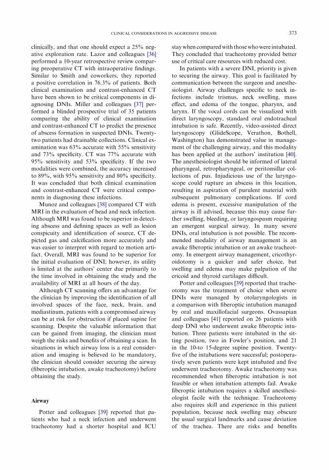
Fig. 6. A patient sitting upright in bed to maintain his
airway. Note the appearance of distress and the fullness
and erythema of the neck. This infection originated from
an acute or chronic periodontal condition.
Examination
A thorough head and neck examination mustbe performed efficiently following the basic prin-ciples of visualization, palpation, and percussion.A careful head and neck examination might
obviate the need for adjunctive imaging in theacute setting, thereby decreasing the time untilsurgical management is initiated. The neck exam-
ination begins with a visual survey of the patientlooking for swelling, erythema, pustules, or‘‘pointing’’ of the infection (Fig. 5). Careful palpa-
tion of the anterior and posterior triangles shouldbe performed. All fascial spaces of the oral cavity,face, and neck should be bimanually palpated to
determine if the swelling is firm or fluctuant in na-ture. Draining sinus tracts and pits should be ex-plored, and all carious teeth should be palpatedand percussed to further elucidate the source of
the infection. Crepitus can indicate gas-formingorganisms and might also be a sign of a necrotizinginfection. The palate, tonsils, and posterior phar-
ynx should be examined for patency and symme-try. Nasal endoscopic examination is sometimesuseful when trismus is severe. If trismus is present,
the maximal interincisal opening should be mea-sured because this can influence the anestheticplan. A fetid odor is often detected in the oral
cavity, indicating an infection of dental, salivary,sinus, or tonsillar origin.
A patient sitting postured in a sniffing position,drooling, and using their accessory muscles ofrespiration is an ominous sign of impending
airway compromise (Fig. 6). Caution is advisedat any attempt to lay the patient flat to obtain im-aging or even intubate, because this may precipi-
tate complete airway obstruction. Many of thesepatients have poor oral intake leading to dehydra-tion, which, in addition to fever, will contribute to
tachycardia. These acute infections can destabilize
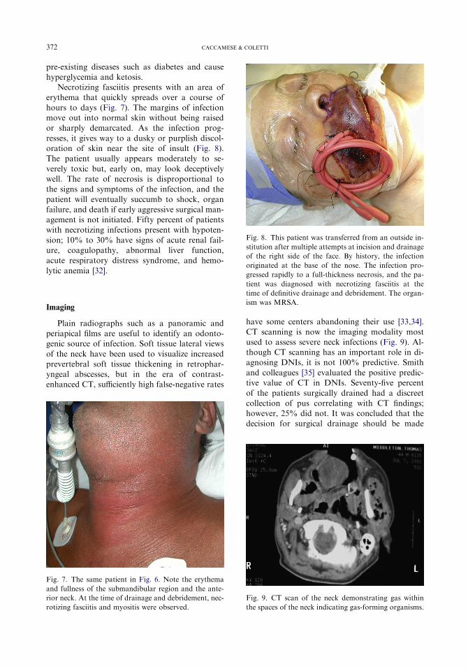
Fig. 8. This patient was transferred from an outside in-
stitution after multiple attempts at incision and drainage
of the right side of the face. By history, the infection
originated at the base of the nose. The infection pro-
gressed rapidly to a full-thickness necrosis, and the pa-
372 CACCAMESE & COLETTI
pre-existing diseases such as diabetes and causehyperglycemia and ketosis.
Necrotizing fasciitis presents with an area of
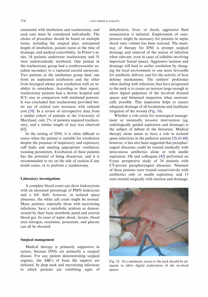
erythema that quickly spreads over a course ofhours to days (Fig. 7). The margins of infectionmove out into normal skin without being raisedor sharply demarcated. As the infection prog-
resses, it gives way to a dusky or purplish discol-oration of skin near the site of insult (Fig. 8).The patient usually appears moderately to se-
verely toxic but, early on, may look deceptivelywell. The rate of necrosis is disproportional tothe signs and symptoms of the infection, and the
patient will eventually succumb to shock, organfailure, and death if early aggressive surgical man-agement is not initiated. Fifty percent of patientswith necrotizing infections present with hypoten-
sion; 10% to 30% have signs of acute renal fail-ure, coagulopathy, abnormal liver function,acute respiratory distress syndrome, and hemo-
lytic anemia [32].
tient was diagnosed with necrotizing fasciitis at the
time of definitive drainage and debridement. The organ-
ism was MRSA.
ImagingPlain radiographs such as a panoramic andperiapical films are useful to identify an odonto-genic source of infection. Soft tissue lateral views
of the neck have been used to visualize increasedprevertebral soft tissue thickening in retrophar-yngeal abscesses, but in the era of contrast-enhanced CT, sufficiently high false-negative rates
Fig. 7. The same patient in Fig. 6. Note the erythema
and fullness of the submandibular region and the ante-
rior neck. At the time of drainage and debridement, nec-
rotizing fasciitis and myositis were observed.
have some centers abandoning their use [33,34].
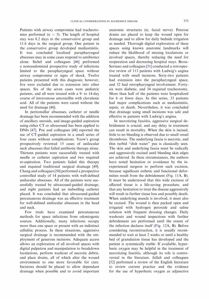
CT scanning is now the imaging modality mostused to assess severe neck infections (Fig. 9). Al-though CT scanning has an important role in di-agnosing DNIs, it is not 100% predictive. Smith
and colleagues [35] evaluated the positive predic-tive value of CT in DNIs. Seventy-five percentof the patients surgically drained had a discreet
collection of pus correlating with CT findings;however, 25% did not. It was concluded that thedecision for surgical drainage should be made
Fig. 9. CT scan of the neck demonstrating gas within
the spaces of the neck indicating gas-forming organisms.
373CLINICAL CONSIDERATIONS IN AGGRESSIVE DISEASE
clinically, and that one should expect a 25% neg-ative exploration rate. Lazor and colleagues [36]performed a 10-year retrospective review compar-ing preoperative CT with intraoperative findings.
Similar to Smith and coworkers, they reporteda positive correlation in 76.3% of patients. Bothclinical examination and contrast-enhanced CT
have been shown to be critical components in di-agnosing DNIs. Miller and colleagues [37] per-formed a blinded prospective trial of 35 patients
comparing the ability of clinical examinationand contrast-enhanced CT to predict the presenceof abscess formation in suspected DNIs. Twenty-
two patients had drainable collections. Clinical ex-amination was 63% accurate with 55% sensitivityand 73% specificity. CT was 77% accurate with95% sensitivity and 53% specificity. If the two
modalities were combined, the accuracy increasedto 89%, with 95% sensitivity and 80% specificity.It was concluded that both clinical examination
and contrast-enhanced CT were critical compo-nents in diagnosing these infections.
Munoz and colleagues [38] compared CT with
MRI in the evaluation of head and neck infection.Although MRI was found to be superior in detect-ing abscess and defining spaces as well as lesion
conspicuity and identification of source, CT de-picted gas and calcification more accurately andwas easier to interpret with regard to motion arti-fact. Overall, MRI was found to be superior for
the initial evaluation of DNI; however, its utilityis limited at the authors’ center due primarily tothe time involved in obtaining the study and the
availability of MRI at all hours of the day.Although CT scanning offers an advantage for
the clinician by improving the identification of all
involved spaces of the face, neck, brain, andmediastinum, patients with a compromised airwaycan be at risk for obstruction if placed supine forscanning. Despite the valuable information that
can be gained from imaging, the clinician mustweigh the risks and benefits of obtaining a scan. Insituations in which airway loss is a real consider-
ation and imaging is believed to be mandatory,the clinician should consider securing the airway(fiberoptic intubation, awake tracheotomy) before
obtaining the study.
Airway
Potter and colleagues [39] reported that pa-tients who had a neck infection and underwenttracheotomy had a shorter hospital and ICU
staywhencomparedwith thosewhowere intubated.They concluded that tracheotomy provided betteruse of critical care resources with reduced cost.
In patients with a severe DNI, priority is given
to securing the airway. This goal is facilitated bycommunication between the surgeon and anesthe-siologist. Airway challenges specific to neck in-
fections include trismus, neck swelling, masseffect, and edema of the tongue, pharynx, andlarynx. If the vocal cords can be visualized with
direct laryngoscopy, standard oral endotrachealintubation is safe. Recently, video-assisted directlaryngoscopy (GlideScope, Verathon, Bothell,
Washington) has demonstrated value in manage-ment of the challenging airway, and this modalityhas been applied at the authors’ institution [40].The anesthesiologist should be informed of lateral
pharyngeal, retropharyngeal, or peritonsillar col-lections of pus. Injudicious use of the laryngo-scope could rupture an abscess in this location,
resulting in aspiration of purulent material withsubsequent pulmonary complications. If cordedema is present, excessive manipulation of the
airway is ill advised, because this may cause fur-ther swelling, bleeding, or laryngospasm requiringan emergent surgical airway. In many severe
DNIs, oral intubation is not possible. The recom-mended modality of airway management is anawake fiberoptic intubation or an awake tracheot-omy. In emergent airway management, cricothyr-
oidotomy is a quicker and safer choice, butswelling and edema may make palpation of thecricoid and thyroid cartilages difficult.
Potter and colleagues [39] reported that trache-otomy was the treatment of choice when severeDNIs were managed by otolaryngologists in
a comparison with fiberoptic intubation managedby oral and maxillofacial surgeons. Ovassapianand colleagues [41] reported on 26 patients withdeep DNI who underwent awake fiberoptic intu-
bation. Three patients were intubated in the sit-ting position, two in Fowler’s position, and 21in the 10-to 15-degree supine position. Twenty-
five of the intubations were successful; postopera-tively seven patients were kept intubated and fiveunderwent tracheotomy. Awake tracheotomy was
recommended when fiberoptic intubation is notfeasible or when intubation attempts fail. Awakefiberoptic intubation requires a skilled anesthesi-
ologist facile with the technique. Tracheotomyalso requires skill and experience in this patientpopulation, because neck swelling may obscurethe usual surgical landmarks and cause deviation
of the trachea. There are risks and benefits
374 CACCAMESE & COLETTI
associated with intubation and tracheotomy, andeach case must be considered individually. Thechoice of procedure should be based on multiple
issues, including the surgical plan, anticipatedlength of intubation, patient status at the time ofdrainage, and medical comorbidity. In Potter’s se-ries, 34 patients underwent tracheotomy and 51
were endotracheally intubated. One patient inthe tracheotomy group had a cerebrovascular ac-cident secondary to a ruptured carotid aneurysm.
Two patients in the intubation group died, onefrom an unplanned extubation and the otherfrom laryngeal edema post extubation with an in-
ability to reintubate. According to their report,tracheotomy patients had a shorter hospital andICU stay in comparison with intubated patients.It was concluded that tracheotomy provided bet-
ter use of critical care resources with reducedcost [39]. In a review of retrospective data froma similar cohort of patients at the University of
Maryland, only 2% of patients required tracheot-omy, and a similar length of stay was observed[42].
In the setting of DNI, it is often difficult toassess when the patient is suitable for extubationdespite the presence of inspiratory and expiratory
cuff leaks and meeting appropriate ventilatoryweaning parameters. Extubation of these patientshas the potential of being disastrous, and it isrecommended to err on the side of caution if any
doubt exists, or to perform a tracheotomy.
Laboratory investigations
A complete blood count can show leukocytosiswith an increased percentage of PMN leukocytesand a left shift; however, in isolated space
abscesses, the white cell count might be normal.Many patients, especially those with necrotizinginfections, have a metabolic acidosis as demon-
strated by their basic metabolic panel and arterialblood gas. In cases of septic shock, lactate, bloodurea nitrogen, creatinine, potassium, and glucose
can all be elevated.
Fig. 10. At a minimum, access to the neck should be ad-
equate to allow digital exploration of the involved
spaces.
Surgical management
Medical therapy is primarily supportive innature, because DNIs are primarily a surgicaldisease. For any patient demonstrating surgical
urgency, the ABCs of basic life support areinitiated. In deep neck and necrotizing infectionsin which patients are exhibiting signs of
dehydration, fever, or shock, aggressive fluidresuscitation is initiated. Employment of vaso-pressors might be necessary for patients in septic
shock once volume has been restored. The main-stay of therapy for DNI is prompt surgicaldrainage and removal of the source of infectionwhen relevant, even in cases of cellulitis involving
important fascial spaces. Aggressive incision anddrainage will lead to earlier resolution by chang-ing the local environment to one more favorable
for antibiotic delivery and for the activity of hostdefense mechanisms. The authors’ preferencewhen dealing with infections that have progressed
to the neck is to create an incision large enough toallow digital palpation of the involved drainedspaces and bimanual inspection when anatomi-cally possible. This inspection helps to ensure
adequate drainage of all loculations and facilitatesirrigation of the wound (Fig. 10).
Whether a role exists for nonsurgical manage-
ment or minimally invasive intervention (eg,radiologically guided aspiration and drainage) isthe subject of debate in the literature. Medical
therapy alone seems to have a role in isolatedspace infections in the pediatric patient [28,43,44];however, it has also been suggested that paraphar-
yngeal abscesses could be treated medically withintravenous antibiotics alone or with needleaspiration. Oh and colleagues [45] performed an8-year prospective study of 34 patients with
CT-proven parapharyngeal abscesses. Nineteenof these patients were treated conservatively withantibiotics only or needle aspiration, and 15
were treated surgically with incision and drainage.
375CLINICAL CONSIDERATIONS IN AGGRESSIVE DISEASE
Patients with airway compromise had tracheoto-mies performed (n ¼ 5). The length of hospitalstay was 8.2 days in the conservative group and11.6 days in the surgical group. One patient in
the conservative group developed mediastinitis.It was concluded localized parapharyngealabscesses may in some cases respond to antibiotics
alone. Sichel and colleagues [46] performeda nonrandomized prospective study of infectionslimited to the parapharyngeal space without
airway compromise or signs of shock. Twelvepatients presented with this diagnosis; however,five were excluded due to extension into other
spaces. Six of the seven cases were pediatricpatients, and all were treated with a 9- to 14-daycourse of intravenous amoxicillin with clavulanicacid. All of the patients were cured without the
need for drainage [46].In peritonsillar abscesses, catheter or needle
drainage has been recommended with the addition
of ancillary steroids, and image-guided aspirationusing either CT or ultrasound has been applied toDNIs [47]. Poe and colleagues [48] reported the
use of CT-guided aspiration in a small series offour cases without complications. Yeow’s groupprospectively reviewed 15 cases of unilocular
neck abscesses that failed antibiotic therapy alone.Thirteen patients were successfully treated withneedle or catheter aspiration and two requiredre-aspiration. Two patients failed this therapy
and required traditional surgical drainage [49].Chang and colleagues [50] performed a prospectivecontrolled study of 14 patients with well-defined
unilocular abscesses. All of the patients were suc-cessfully treated by ultrasound-guided drainage,and eight patients had an indwelling catheter
placed. It was concluded that ultrasound-guidedpercutaneous drainage was an effective treatmentfor well-defined unilocular abscesses in the headand neck.
Few trials have examined percutaneousmethods for space infections from odontogenicsources. Additionally, many infections occupy
more than one space or present with an indistinctcellulitic process. In these situations, aggressivesurgical drainage is recommended with the em-
ployment of generous incisions. Adequate accessallows an exploration of all involved spaces withdigital palpation and manipulation to breakdown
loculations, perform washout of necrotic debris,and place drains, all of which alter the woundenvironment to one more favorable for cure.Incisions should be placed to allow dependant
drainage when possible and to avoid important
anatomic structures (ie, facial nerve). Penrosedrains are placed to keep the wound open fordrainage and to allow for daily bedside irrigationas needed. Thorough digital exploration of these
spaces using known anatomic landmarks willreduce the likelihood of missing loculations orinvolved spaces, thereby reducing the need for
reoperation and decreasing hospital stays. Bross-Soriano and colleagues [51] conducted a retrospec-tive review of 113 patients with Ludwig’s angina
treated with small incisions. Sixty-two patientshad extension into the parapharyngeal space,and 32 had retropharyngeal involvement. Forty-
six were diabetic, and 34 required tracheotomy.More than half of the patients were hospitalizedfor 6 or fewer days. In this series, 33 patientshad major complications such as mediastinitis,
sepsis, or death. Nevertheless, it was concludedthat drainage using small incisions was safe andeffective in patients with Ludwig’s angina.
In necrotizing fasciitis, aggressive surgical de-bridement is crucial, and any delay in treatmentcan result in mortality. When the skin is incised,
little to no bleeding is observed due to small vesselthrombosis. The underlying fascia is necrotic, andthin turbid ‘‘dish water’’ pus is classically seen.
The skin and underlying fascia must be radicallyand aggressively excised until bleeding skin edgesare achieved. In these circumstances, the authorshave noted hesitation or avoidance by the in-
experienced surgeon to perform such excision,because significant esthetic and functional defor-mities result from the debridement (Fig. 11A, B).
It must be understood that this wide excision ofaffected tissue is a life-saving procedure, andthat any hesitation to treat the disease aggressively
will result in further tissue loss and possibly death.When underlying muscle is involved, it must alsobe excised. The wound is then packed open andirrigated with hydrogen peroxide and saline
solution with frequent dressing changes. Dailywashouts and wound inspections with furtherdebridement are performed until the extent of
the infection declares itself (Fig. 12A, B). Beforeconsidering reconstruction, it is usually recom-mended to wait at least 2 weeks or until a healthy
bed of granulation tissue has developed and thepatient is systemically stable. If available, hyper-baric oxygen may be helpful in the treatment of
necrotizing fasciitis, although its role is contro-versial in the literature. Jallali and colleagues[52] performed a review of the English literatureto review current practice and the evidence
for the use of hyperbaric oxygen as adjunctive

Fig. 11. (A) The patient shown in Fig. 8 with staphylococcal necrotizing fasciitis of the right side of the face. (B) The
same patient after first stage drainage and debridement. She was eventually reconstructed with a radial forearm free flap.
376 CACCAMESE & COLETTI
therapy in necrotizing fasciitis. They concludedthat the results are currently inconsistent, andthat more robust evidence by way of prospectiverandomized trials is necessary before routine use
of hyperbaric oxygen for necrotizing fasciitis canbe recommended.
Reconstruction is not considered until after the
wounds are stabilized with signs of a healthygranulation tissue bed (Fig. 13A, B, C). Further-more, these patients are often profoundly cata-
bolic as a result of sepsis and poor nutritionand have a diminished immunologic response.
Fig. 12. (A) The patient shown in Fig. 4 after first stage debrid
extraction. (B) The same patient at the time of final debridemen
spread along the relatively avascular fascial planes took this in
tually skin grafted and is alive and well.
Sequencing reconstructive strategies for patientswith necrotizing fasciitis can be a monumentalchallenge. Extensive tissue loss following aggres-sive debridement may require composite (eg,
skin, muscle, bone) tissue replacement. The chal-lenges of reconstruction for these destructivehead and neck infections are similar to those in
oncologic head and neck surgery. They includea compromised airway, impaired sensory and mo-tor function, esthetic facial deformity, an inability
to control secretions, and impaired speech. Thegoals of reconstruction are the restoration of
ement of necrotizing fasciitis resultant from third molar
t once the infection had declared itself completely. Rapid
fection down onto the chest wall. The patient was even-
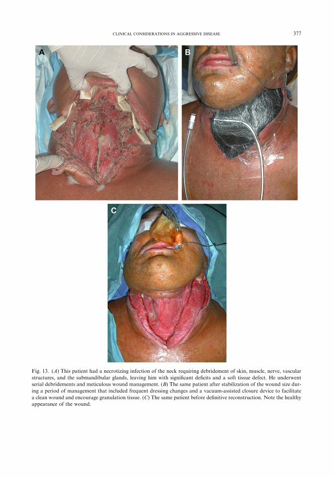
Fig. 13. (A) This patient had a necrotizing infection of the neck requiring debridement of skin, muscle, nerve, vascular
structures, and the submandibular glands, leaving him with significant deficits and a soft tissue defect. He underwent
serial debridements and meticulous wound management. (B) The same patient after stabilization of the wound size dur-
ing a period of management that included frequent dressing changes and a vacuum-assisted closure device to facilitate
a clean wound and encourage granulation tissue. (C) The same patient before definitive reconstruction. Note the healthy
appearance of the wound.
377CLINICAL CONSIDERATIONS IN AGGRESSIVE DISEASE
378 CACCAMESE & COLETTI
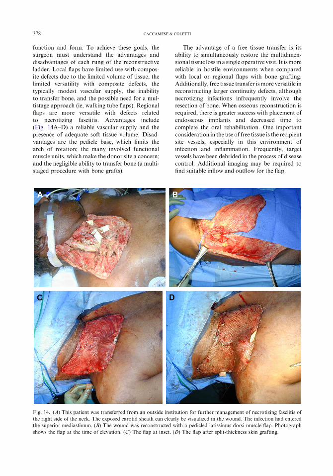
function and form. To achieve these goals, thesurgeon must understand the advantages anddisadvantages of each rung of the reconstructive
ladder. Local flaps have limited use with compos-ite defects due to the limited volume of tissue, thelimited versatility with composite defects, thetypically modest vascular supply, the inability
to transfer bone, and the possible need for a mul-tistage approach (ie, walking tube flaps). Regionalflaps are more versatile with defects related
to necrotizing fasciitis. Advantages include(Fig. 14A–D) a reliable vascular supply and thepresence of adequate soft tissue volume. Disad-
vantages are the pedicle base, which limits thearch of rotation; the many involved functionalmuscle units, which make the donor site a concern;and the negligible ability to transfer bone (a multi-
staged procedure with bone grafts).
Fig. 14. (A) This patient was transferred from an outside insti
the right side of the neck. The exposed carotid sheath can clear
the superior mediastinum. (B) The wound was reconstructed w
shows the flap at the time of elevation. (C) The flap at inset. (
The advantage of a free tissue transfer is itsability to simultaneously restore the multidimen-sional tissue loss in a single operative visit. It ismore
reliable in hostile environments when comparedwith local or regional flaps with bone grafting.Additionally, free tissue transfer ismore versatile inreconstructing larger continuity defects, although
necrotizing infections infrequently involve theresection of bone. When osseous reconstruction isrequired, there is greater success with placement of
endosseous implants and decreased time tocomplete the oral rehabilitation. One importantconsideration in the use of free tissue is the recipient
site vessels, especially in this environment ofinfection and inflammation. Frequently, targetvessels have been debrided in the process of diseasecontrol. Additional imaging may be required to
find suitable inflow and outflow for the flap.
tution for further management of necrotizing fasciitis of
ly be visualized in the wound. The infection had entered
ith a pedicled latissimus dorsi muscle flap. Photograph
D) The flap after split-thickness skin grafting.
379CLINICAL CONSIDERATIONS IN AGGRESSIVE DISEASE
A true comprehension of the architectural hardand soft tissue defects facilitates a strategicapproach to these difficult reconstructions. Theinitial sequencing of reconstruction is focused on
restoring oral lining, followed by soft tissue cover-age of the face and vital structures and then skeletalsupport. Before implementing any of the recon-
structive options available, the patient should bemedically stabilized, and all necrotic and infectedtissue must be debrided with no signs of pro-
gression. Because necrotizing fasciitis can result inmassive surface area skin loss beyond the limits ofcoverage that a single (or multiple) regional or free
flaps can provide, the defects are reconstructedanalogous to a burn patient with multiple skingrafting procedures, with flaps reserved for largeareas of composite tissue loss. A population of
patients is unsuitable for free flap surgery due tomedical comorbidities; these patients are moresuitable for regional flaps. Because each option
has its own inherent advantages anddisadvantages,the surgeon can employ a combination of each toachieve the best reconstruction possible.
Summary
The management of neck infections can bechallenging. Careful attention to the source ofinfection and airway requirements yields the best
medical and surgical results when it comes totreatment. Although there might be a role forcarefully observed inpatient medical management
in single space infections of non-odontogenicorigin and in pediatric neck infections, it is theauthors’ opinion and that of the literature thatneck infections are usually best managed with
prompt surgical treatment and supportive medicalcare. When these methods are employed, compli-cations are rarely encountered.
References
[1] Huang TT, Liu TC, Chen PR, et al. Deep neck infec-
tion: analysis of 185 cases. Head Neck 2004;26(10):
854–60.
[2] Har-El G, Aroesty JH, Shaha A, et al. Changing
trends in deep neck abscess: a retrospective study
of 110 patients. Oral Surg Oral Med Oral Pathol
1994;77(5):446–50.
[3] Parhiscar A, Har-El G. Deep neck abscess: a retro-
spective review of 210 cases. Ann Otol Rhinol
Laryngol 2001;110(11):1051–4.
[4] Sethi DS, Stanley RE. Deep neck abscesses–chang-
ing trends. J Laryngol Otol 1994;108(2):138–43.
[5] Lee JK, Kim HD, Lim SC. Predisposing factors of
complicated deep neck infection: an analysis of 158
cases. Yonsei Med J 2007;48(1):55–62.
[6] Brook I. Microbiology and management of
peritonsillar, retropharyngeal, and parapharyngeal
abscesses. J Oral Maxillofac Surg 2004;62(12):
1545–50.
[7] Brook I. Microbiology and management of infected
solid tumours. Eur J Cancer Care (Engl) 2007;16(1):
12–6.
[8] Nusbaum AO, Som PM, Rothschild MA, et al. Re-
currence of a deep neck infection: a clinical indica-
tion of an underlying congenital lesion. Arch
Otolaryngol HeadNeck Surg 1999;125(12):1379–82.
[9] Ostlie DJ, Burjonrappa SC, Snyder CL, et al. Thyro-
glossal duct infections and surgical outcomes. J
Pediatr Surg 2004;39(3):396–9 [discussion: 396–9].
[10] Park SW, HanMH, Sung MH, et al. Neck infection
associated with pyriform sinus fistula: imaging find-
ings. AJNR Am J Neuroradiol 2000;21(5):817–22.
[11] Wang CP, Ko JY, Lou PJ. Deep neck infection as
the main initial presentation of primary head and
neck cancer. J Laryngol Otol 2006;120(4):305–9.
[12] Schweinfurth JM. Demographics of pediatric head
and neck infections in a tertiary care hospital. Laryn-
goscope 2006;116(6):887–9.
[13] Choi SS, Vezina LG, Grundfast KM. Relative
incidence and alternative approaches for surgical
drainage of different types of deep neck abscesses
in children. Arch Otolaryngol Head Neck Surg
1997;123(12):1271–5.
[14] Coticchia JM, Getnick GS, Yun RD, et al. Age-,
site-, and time-specific differences in pediatric deep
neck abscesses. Arch Otolaryngol Head Neck Surg
2004;130(2):201–7.
[15] Tan PT, Chang LY, Huang YC, et al. Deep neck in-
fections in children. J Microbiol Immunol Infect
2001;34(4):287–92.
[16] Ungkanont K, Yellon RF,Weissman JL, et al. Head
and neck space infections in infants and children.
Otolaryngol Head Neck Surg 1995;112(3):375–82.
[17] MarioniG,CastegnaroE, StaffieriC, et al.Deepneck
infection in elderly patients: a single institution expe-
rience (2000–2004). Aging Clin Exp Res 2006;18(2):
127–32.
[18] Chen MK, Wen YS, Chang CC, et al. Deep neck in-
fections in diabetic patients. Am JOtolaryngol 2000;
21(3):169–73.
[19] Huang TT, Tseng FY, Liu TC, et al. Deep neck in-
fection in diabetic patients: comparison of clinical
picture and outcomes with nondiabetic patients.
Otolaryngol Head Neck Surg 2005;132(6):943–7.
[20] Brook I. Microbiology and principles of antimicro-
bial therapy for head and neck infections. Infect
Dis Clin North Am 2007;21(2):355–91.
[21] Flynn TR, Shanti RM, Levi MH, et al. Severe odon-
togenic infections. Part 1. Prospective report. J Oral
Maxillofac Surg 2006;64(7):1093–103.

380 CACCAMESE & COLETTI
[22] Kuriyama T, Karasawa T, Nakagawa K, et al. Bac-
teriologic features and antimicrobial susceptibility in
isolates from orofacial odontogenic infections. Oral
Surg Oral Med Oral Pathol Oral Radiol Endod
2000;90(5):600–8.
[23] Sakamoto H, Kato H, Sato T, et al. Semiquantita-
tive bacteriology of closed odontogenic abscesses.
Bull Tokyo Dent Coll 1998;39(2):103–7.
[24] Stefanopoulos PK, Kolokotronis AE. The clinical
significance of anaerobic bacteria in acute orofacial
odontogenic infections. Oral Surg Oral Med Oral
Pathol Oral Radiol Endod 2004;98(4):398–408.
[25] Flynn TR, Shanti RM,Hayes C. Severe odontogenic
infections. Part 2. Prospective outcomes study.
J Oral Maxillofac Surg 2006;64(7):1104–13.
[26] Wang J, Ahani A, Pogrel MA. A five-year retrospec-
tive study of odontogenic maxillofacial infections in
a large urban public hospital. Int J Oral Maxillofac
Surg 2005;34(6):646–9.
[27] Cabrera CE, Deutsch ES, Eppes S, et al. Increased
incidence of head and neck abscesses in children.
Otolaryngol Head Neck Surg 2007;136(2):176–81.
[28] Craig FW, Schunk JE. Retropharyngeal abscess in
children: clinical presentation, utility of imaging, and
current management. Pediatrics 2003;111(6 Pt 1):
1394–8.
[29] Ossowski K, Chun RH, Suskind D, et al. Increased
isolation of methicillin-resistant Staphylococcus au-
reus in pediatric head and neck abscesses. Arch Oto-
laryngol Head Neck Surg 2006;132(11):1176–81.
[30] Jones JL, Candelaria LM.Head and neck infections.
In: Fonseca RJ, Williams TP, Stewart JCB, editors.
Oral and maxillofacial surgery, vol. 5. Philadelphia:
WB Saunders; 2000. p. 77–117.
[31] Brook I, Frazier EH. Clinical and microbiological
features of necrotizing fasciitis. J Clin Microbiol
1995;33(9):2382–7.
[32] McGurk M. Diagnosis and treatment of necrotizing
fasciitis in the head and neck region. OralMaxillofac
Surg Clin North Am 2003;15(1):59–67.
[33] NagyM, Backstrom J. Comparison of the sensitivity
of lateral neck radiographs and computed tomo-
graphy scanning in pediatric deep neck infections.
Laryngoscope 1999;109(5):775–9.
[34] Haug RH, Wible RT, Lieberman J. Measurement
standards for the prevertebral region in the lateral
soft-tissue radiograph of the neck. J Oral Maxillofac
Surg 1991;49(11):1149–51.
[35] Smith JL II, Hsu JM, Chang J. Predicting deep neck
space abscess using computed tomography. Am J
Otolaryngol 2006;27(4):244–7.
[36] Lazor JB, CunninghamMJ, Eavey RD, et al. Com-
parison of computed tomography and surgical find-
ings in deep neck infections. Otolaryngol HeadNeck
Surg 1994;111(6):746–50.
[37] Miller WD, Furst IM, Sandor GK, et al. A prospec-
tive, blinded comparison of clinical examination and
computed tomography in deep neck infections. La-
ryngoscope 1999;109(11):1873–9.
[38] Munoz A, Castillo M, Meichor MA, et al. Acute
neck infections: prospective comparison between
CT and MRI in 47 patients. J Comput Assist
Tomogr 2001;25(5):733–41.
[39] Potter JK,HerfordAS, Ellis E III. Tracheotomy ver-
sus endotracheal intubation for airway management
in deep neck space infections. J OralMaxillofac Surg
2002;60(4):349–54 [discussion: 354–5].
[40] Marrel J, Blanc C, Frascarolo P, et al. Videolaryngo-
scopy improves intubation condition in morbidly
obese patients. Eur J Anaesthesiol 2007;24(12):
1045–9.
[41] Ovassapian A, Tuncbilek M, Weitzel EK, et al. Air-
waymanagement in adult patients with deep neck in-
fections: a case series and review of the literature.
Anesth Analg 2005;100(2):585–9.
[42] Coletti DP, Caccamese JF, Hartman MJ, et al. The
impact of substance abuse on the occurrence and
outcome of odontogenic deep space infections. In:
Proceedings of the Annual Scientific Meeting, Brit-
ish Association of Oral and Maxillofacial Surgeons.
Eastbourne (U.K.): Elsevier; June 28-30, 2006.
[43] Al-Sabah B, Bin Salleen H, Hagr A, et al. Retro-
pharyngeal abscess in children: 10-year study. J Oto-
laryngol 2004;33(6):352–5.
[44] McClay JE, Murray AD, Booth T. Intravenous
antibiotic therapy for deep neck abscesses defined
by computed tomography. Arch Otolaryngol Head
Neck Surg 2003;129(11):1207–12.
[45] Oh JH, Kim Y, KY, Kim CH. Parapharyngeal ab-
scess: comprehensive management protocol. ORL
J Otorhinolaryngol Relat Spec 2007;69(1):37–42.
[46] Sichel JY, Dano I, Hocwald E, et al. Nonsurgical
management of parapharyngeal space infections:
a prospective study. Laryngoscope 2002;112(5):
906–10.
[47] Herzon FS, Martin AD. Medical and surgical
treatment of peritonsillar, retropharyngeal, and
parapharyngeal abscesses. Curr Infect Dis Rep
2006;8(3):196–202.
[48] Poe LB, Petro GR, Matta I. Percutaneous
CT-guided aspiration of deep neck abscesses.
AJNR Am J Neuroradiol 1996;17(7):1359–63.
[49] Yeow KM, Liao CT, Hao SP. US-guided needle
aspiration and catheter drainage as an alterna-
tive to open surgical drainage for uniloculated
neck abscesses. J Vasc Interv Radiol 2001;
12(5):589–94.
[50] Chang KP, Chen YL, Hao SP, et al. Ultrasound-
guided closed drainage for abscesses of the head
and neck. Otolaryngol Head Neck Surg 2005;
132(1):119–24.
[51] Bross-Soriano D, Arrieta-Gomez JR, Prado-Call-
eros H, et al. Management of Ludwig’s angina
with small neck incisions: 18 years experience. Oto-
laryngol Head Neck Surg 2004;130(6):712–7.
[52] Jallali N,Withey S, Butler PE. Hyperbaric oxygen as
adjuvant therapy in the management of necrotizing
fasciitis. Am J Surg 2005;189(4):462–6.
Related Documents


![December 21, 2015 - Wisconsin Supreme Court · RB-1 (2015) [?\^]`_ acbedgfhbeij[ ahik[ l 1. mon#p qsrHt`rvuxwnzye{E|}ux~)r 'p n#w )rv|}ux~x 7 7 7 7 7 7 7 7 7 7 7 7 7 7 7 7 7 7 7 7](https://static.cupdf.com/doc/110x72/5fb3422fccf05f68ab3a22e4/december-21-2015-wisconsin-supreme-court-rb-1-2015-acbedgfhbeij-ahik.jpg)