CYTOLOGY OF BODY FLUID

7- Cytology of Body Fluid.ppt
Nov 29, 2015
7- Cytology of Body Fluid.ppt
Welcome message from author
This document is posted to help you gain knowledge. Please leave a comment to let me know what you think about it! Share it to your friends and learn new things together.
Transcript
CYTOLOGY OF BODY FLUID
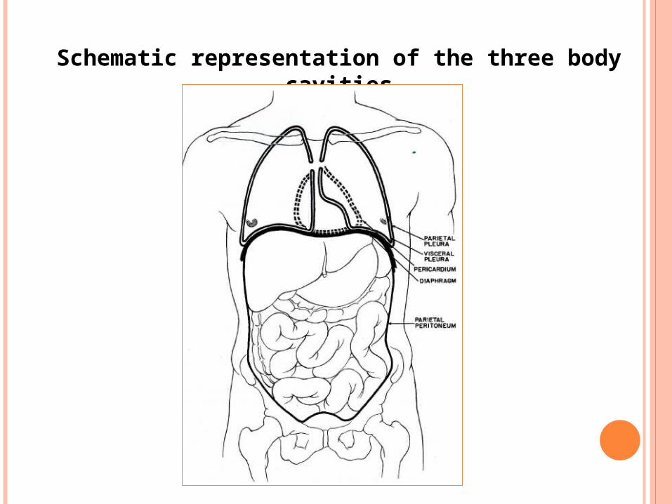
Schematic representation of the three body cavities
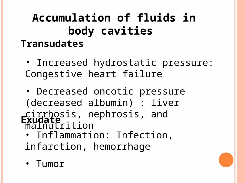
Accumulation of fluids in body cavities
Transudates
• Increased hydrostatic pressure: Congestive heart failure
• Decreased oncotic pressure (decreased albumin) : liver cirrhosis, nephrosis, and malnutrition
Exudate
• Inflammation: Infection, infarction, hemorrhage
• Tumor
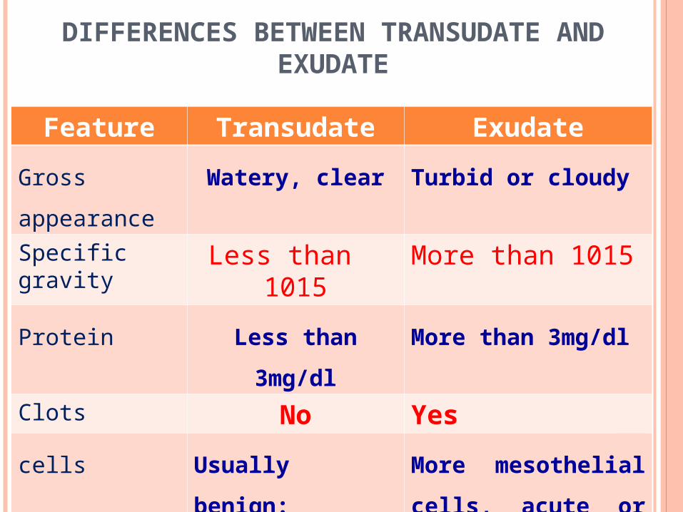
DIFFERENCES BETWEEN TRANSUDATE AND EXUDATE
Feature Transudate Exudate
Gross appearance Watery, clear Turbid or cloudy
Specific gravity Less than 1015 More than 1015
Protein Less than 3mg/dl More than 3mg/dl
Clots No Yes
cells Usually benign:
Few mesothelial
cells, few histocytes
and lymphocytes
More mesothelial cells,
acute or chronic
inflammatory cells,
RBCs, malignant cells
DIAGNOSTIC ROLE OF EFFUSION CYTOLOGY
It is very useful for diagnosis of premalignant and
malignant tumors, especially metastatic tumors.
It is very useful for diagnosis of inflammatory
conditions (septic effusion, or chronic specific
inflammation e.g. TB
Respiratory Tract
Urinary Tract
Oral Cavity
Gastrointestinal Tract
Effusions (pleural, pericardial, joint)
Cerebral Spinal Fluid
Amniotic fluid
Many other body sites
Non-Gynecological Specimen Collection
EXAMINATION OF BODY FLUID
Gross exam
Total cell count
Microscopic exam
Any other special test (Chemistry, Microbiology,
cytology (
Test are performed in various areas of lab based on what
the physician orders.
Body fluids sterile vs. non-sterile
SAMPLE COLLECTION
FNA of effusion fluids
Tapping
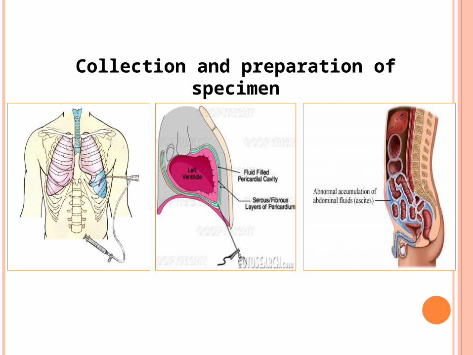
Collection and preparation of specimen
FIXATION
1ml of heparin + 100ml of effusion fluid to prevent
clotting
N.B.: do not use alcohol in fixation of fluid before
spread cytological smear on glass slides
TYPES OF STAINING SMEARS
PAP
Gram Stain
Hx & E
Cell block for remnant sediment and histopathological
examination.
Other special stains for the most suspected diseases, to
confirm diagnosis.
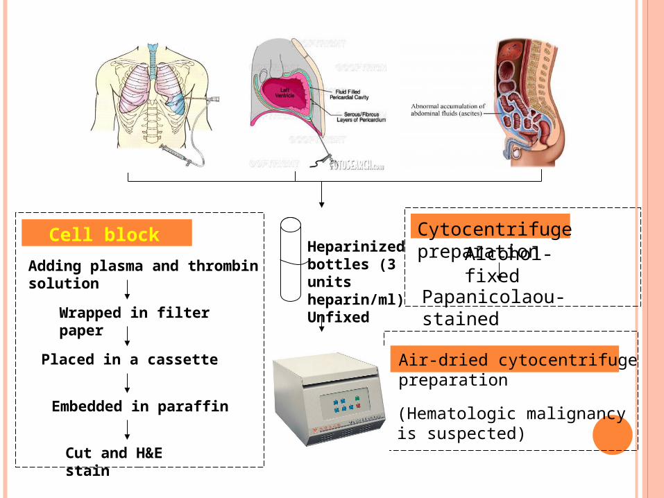
Heparinized bottles (3 units heparin/ml) Unfixed
Alcohol-fixed
Papanicolaou-stained
Cytocentrifuge preparationCell block
Adding plasma and thrombin solution
Wrapped in filter paper
Placed in a cassette
Embedded in paraffin
Cut and H&E stain
Air-dried cytocentrifuge preparation
(Hematologic malignancy is suspected)
1- CEREBROSPINAL FLUID
Fluid surrounding brain and spinal cord
Sterile
Specimen collection: by Lumbar puncture
Collect 3-5 vials, each tube has a designated department.
Gross exam: Turbidity, Color, microscopic exam, cell
count
CSF CELL DIFFERENTIAL
Numerate and differentiate cells seen
Lymphocytes: usually are few; increased with viral,
fungal, bacterial meningitis, or nervous system disease
Monocytes: Less than 2% of normal CSF, increased
with TB meningitis, viral encephalitis, subarachnoid
hemorrhage.
PMN: are few, associated with Viral and acute bacterial
inflammation.
Macrophages: are few in number associated with malignancy,
hemorrhage, inflammation
Eosinophils/Basophils: not normally seen in CSF
Plasma cells: not normally present; associated with viral disorders,
and Hodgkin's diseases.
Red Blood Cells: Few to none present
Mesothelial cells: not present
Malignant cells: will see with malignant disease and infiltrate.

• Effusion:
• Transudate
• Exudates
• Lab analysis: Gross exam, cell count, etc.
• Differential: PMN, Lymph, Mono, etc.
2- Pleural Fluid: Lung fluid
• Cells unique to the lungs: Mesothelial cells
• RBCs and WBCs: are limited, if increased without
traumatic tap ----- indicates infarction
• Cytology exam: useful in identifying malignancy or
abnormal morphological cells.
3- PERITONEAL FLUID
Abnormal accumulation of fluid (effusion) in peritoneal
cavity: Ascites
Ascites: a condition in which fluid accumulates within
the peritoneal space.
Must have an accumulation of > 100ml (several 100) before effusion
can be detected on physical exam.
Removal procedure- paracentesis
Lab analysis: distinguish between transudate and exudates,
gross exam, cell count, sedimentation, chemical analysis
PHYSICAL CHARACTERISTICS
Peritoneal Fluid Appearance: Color and clarity.
Color and clarity can indicate certain infections and diseases.
Total Cell Count: Assist in diagnosis of certain
diseases by determining total RBC and WBC number.
Lymphocytes: CHF, liver cirrhosis, nephrotic syndrome
Mesothelial Cells: Associated with TB effusions
Malignant cells: seen with malignancy
Pericardial Fluid: accumulation of fluid of the lining of
the heart (effusion)
Cause: neoplasm, infections, collagen disease, renal
disease, Cardiovascular disease.
Gross Exam: Report appearance (bloody, clear, cloudy)
4- Pericardial Fluid
Measure pH: pH less than 7.0 associated with infection or
rheumatoid disorder.
Cell count: see limited RBCs and WBCs
Evaluate sedimentation
• Examine physical, chemical and microscopic detail
• Count number of sperm, report morphology and
motility
• Specimen must be a fresh collection-clean, sterile
container.
• Gross Exam: Color, pH, Volume, and viscosity.
• Agglutination study
5- Seminal Fluid
• Joint Fluid: normally clear, viscous
• Functions as a lubricate and transports nutrient
• Arthrocentesis: aspirate of the joint fluid, aseptic
technique
• Lab Assay: Gross exam, microscopic exam, Gram
stain, cultures,...
6- Synovial Fluid:
• Appearance: clear, transparent, viscous
• Viscosity test
• Mucin Clot test
• Note crystals (intracellular vs. extra cellular)
• Slide exam: usually performed on concentration of the fluid
using Giemsa or Papnicolaou

THANK YOU
Related Documents