Bajaj P et al. Erythema Multiforme. 40 Review Article Erythema Multiforme: Classification and Immunopathogenesis Puneet Bajaj, Robin Sabharwal, Rajesh Mohammed PK, Deepti Garg, Charu Kapoor Department of Oral Pathology and Microbiology, Bhojia Dental College and Hospital, Solan Corresponding Author: Dr. Puneet Bajaj Professor & HOD, Department of Oral Pathology Bhojia Dental College and Hospital, Solan, Himachal Pardesh, India. Contact Number: +91 9814278407 Email: [email protected] Received: 01-09-2013 Revised: 01-10-2013 Accepted: 12-10-2013 This article may be cited as: Bajaj P, Sabharwal R, PK Mohammed R, Garg D, Kapoor C. Erythema Multiforme: Classification and Immunopathogenesis. J Adv Med Dent Scie 2013;1(2):40-47. Introduction: Erythema multiforme (EM) is a typically mild, self-limiting, and recurring mucocutaneous reaction characterized by target or iris lesions of the skin and mucous membranes. 1 EM usually affects apparently healthy young adults and the peak age at presentation is 20–40 years although as many as 20% of cases are children. 2 Erythema multiforme is a reactive mucocutaneous disorder that comprises variants ranging from a self-limited, mild, exanthematous, cutaneous variant with minimal oral involvement (EM minor) to a progressive, fulminating, severe variant with extensive mucocutaneous epithelial necrosis (Stevens-Johnson syndrome: SJS; and toxic epidermal necrolysis: TEN). All variants share two common features: typical or less typical cutaneous target lesions and satellite cell or more widespread necrosis of the epithelium. These features are considered to be sequelae of a cytotoxic immunologic Abstract: Erythema multiforme is a skin condition considered to be a hypersensitivity reaction to infections or drugs. It consists of a polymorphous eruption of macules, papules, and characteristic “target” lesions that are symmetrically distributed with a propensity for the distal extremities. There is minimal mucosal involvement. Erythema multiforme can be triggered by a range of factors, but more commonly it is associated with herpes simplex virus (HSV). Most other cases are initiated by drugs. The clinical classification of these disorders has often been variable, thus making definitive diagnosis sometimes difficult. The present article reviews the classification and highlights the associated potential etiologic agents, pathogenic mechanisms and treatment of Erythema multiforme. Keywords: Erythema multiforme, Target lesions, TEN

Welcome message from author
This document is posted to help you gain knowledge. Please leave a comment to let me know what you think about it! Share it to your friends and learn new things together.
Transcript

Bajaj P et al. Erythema Multiforme.
40
Review Article
Erythema Multiforme: Classification and Immunopathogenesis
Puneet Bajaj, Robin Sabharwal, Rajesh Mohammed PK, Deepti Garg, Charu Kapoor
Department of Oral Pathology and Microbiology, Bhojia Dental College and Hospital, Solan
Corresponding Author:
Dr. Puneet Bajaj
Professor & HOD,
Department of Oral Pathology
Bhojia Dental College and Hospital,
Solan, Himachal Pardesh, India.
Contact Number: +91 9814278407
Email: [email protected]
Received: 01-09-2013
Revised: 01-10-2013
Accepted: 12-10-2013
This article may be cited as: Bajaj P, Sabharwal R, PK Mohammed R, Garg D, Kapoor C. Erythema Multiforme: Classification and Immunopathogenesis. J Adv Med Dent Scie 2013;1(2):40-47.
Introduction: Erythema multiforme (EM) is a typically mild, self-limiting, and recurring mucocutaneous reaction characterized by target or iris lesions of the skin and mucous membranes.1
EM usually affects apparently healthy young adults and the peak age at presentation is 20–40 years although as many as 20% of cases are children.2
Erythema multiforme is a reactive mucocutaneous disorder that comprises variants ranging from a self-limited, mild,
exanthematous, cutaneous variant with minimal oral involvement (EM minor) to a progressive, fulminating, severe variant with extensive mucocutaneous epithelial necrosis (Stevens-Johnson syndrome: SJS; and toxic epidermal necrolysis: TEN). All variants share two common features: typical or less typical cutaneous target lesions and satellite cell or more widespread necrosis of the epithelium. These features are considered to be sequelae of a cytotoxic immunologic
Abstract: Erythema multiforme is a skin condition considered to be a hypersensitivity reaction to infections or drugs. It consists of a polymorphous eruption of macules, papules, and characteristic “target” lesions that are symmetrically distributed with a propensity for the distal extremities. There is minimal mucosal involvement. Erythema multiforme can be triggered by a range of factors, but more commonly it is associated with herpes simplex virus (HSV). Most other cases are initiated by drugs. The clinical classification of these disorders has often been variable, thus making definitive diagnosis sometimes difficult. The present article reviews the classification and highlights the associated potential etiologic agents, pathogenic mechanisms and treatment of Erythema multiforme.
Keywords: Erythema multiforme, Target lesions, TEN

Bajaj P et al. Erythema Multiforme.
41
attack on keratinocytes expressing non-self-antigens.3
Immunopathogenesis: Erythema multiforme is probably an immunologically mediated process. It is considered to be a hypersensitivity reaction associated with certain infections and medications. • Infections
� Herpes simplex virus 1 and 2 Herpes simplex virus (HSV) is the most commonly identified etiology of this hypersensitivity reaction.
� Mycoplasma pneumoniae � Fungal infections
• Medications � Barbiturates � Hydantoins � Non-steroidal anti-inflammatory
drugs � Penicillins � Phenothiazines � Sulfonamides
Erythema multiforme is also associated with Vaccines (diphtheria-tetanus, hepatitis B,14 smallpox) and appears to be the result of a
cell mediated immune reaction to the precipitating agent. The pathogenesis of herpes-associated Erythema multiforme is consistent with a delayed-type hypersensitivity reaction. The disease begins with the transport of viral DNA fragments to distant skin sites by peripheral blood mononuclear cells. HSV genes within DNA fragments are expressed on keratinocytes, leading to the recruitment of HSV-specific CD4+ TH1 cells (helper T cells involved in cell-mediated immunity). The CD4+ cells respond to viral antigens with production of interferon-γ, initiating an inflammatory cascade. This cytokine then amplifies the immune response and stimulates the production of additional cytokines and chemokines, which aids the recruitment of further reactive T cells to the area. These cytotoxic T cells, NK cells or chemokines can all induce epithelial damage (Figure I). Drug-associated erythema multiforme lesions test positive for tumor necrosis factor α and not interferon-γ as in herpes associated erythema multiforme lesions, suggesting a varying mechanism.1
Figure I: Pathogenesis of Erythema Multiforme
Much of the tissue damage in drug-induced lesions appears to be due to apoptosis and, because of the paucity of the inflammatory reaction. However, particularly in TEN and
SJS, there is some evidence for a Fas–FasL interaction. FasL mediates apoptotic cell death by binding to Fas on cells and inducing the formation of caspases. Fas is
Bajaj P et al. Erythema Multiforme.
42
present on keratinocytes and FasL is found on activated T cells and NK cells and thus binding of keratinocytes to T cells or NK cells can induce apoptosis.4,5
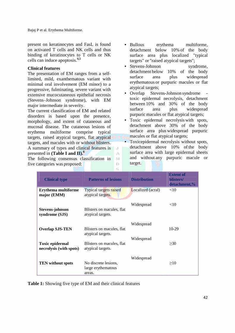
Clinical features The presentation of EM ranges from a self-limited, mild, exanthematous variant with minimal oral involvement (EM minor) to a progressive, fulminating, severe variant with extensive mucocutaneous epithelial necrosis (Stevens–Johnson syndrome), with EM major intermediate in severity. The current classification of EM and related disorders is based upon the presence, morphology, and extent of cutaneous and mucosal disease. The cutaneous lesions of erythema multiforme comprise typical targets, raised atypical targets, flat atypical targets, and macules with or without blisters. A summary of types and clinical features is presented in (Table I and II).6
The following consensus classification in five categories was proposed:
• Bullous erythema multiforme, detachment below 10% of the body surface area plus localized "typical targets" or "raised atypical targets";
• Stevens-Johnson syndrome, detachment below 10% of the body surface area plus widespread erythematous or purpuric macules or flat atypical targets;
• Overlap Stevens-Johnson syndrome - toxic epidermal necrolysis, detachment between 10% and 30% of the body surface area plus widespread purpuric macules or flat atypical targets;
• Toxic epidermal necrolysis with spots, detachment above 30% of the body surface area plus widespread purpuric macules or flat atypical targets;
• Toxicepidermal necrolysis without spots, detachment above 10% of the body surface area with large epidermal sheets and without any purpuric macule or target.
Table 1: Showing five type of EM and their clinical features
Clinical type
Patterns of lesions
Distribution
Extent of blisters/ detachment,%
Erythema multiforme major (EMM) Stevens-johnson syndrome (SJS) Overlap SJS-TEN Toxic epidermal necrolysis (with spots) TEN without spots
Typical targets raised atypical targets.
Blisters on macules, flat atypical targets.
Blisters on macules, flat atypical targets.
Blisters on macules, flat atypical targets.
No discrete lesions, large erythematous areas.
Localized (acral)
Widespread
Widespread
Widespread
Widespread
<10
<10
10-29
≥30
≥10

Bajaj P et al. Erythema Multiforme.
43
Table II: Showing clinical features that distinguish SJS, SJS-TEN overlap, and TEN
Erythema Multiforme is a self-limited eruption that usually has mild or no prodromal symptoms. All lesions typically present within approximately 3 days of onset. There may be hundreds of lesions, but less than 10% of the body surface area is usually involved. The lesions are in a fixed position with a symmetric distribution.7
They present as circular erythematous plaques in a concentric array with lesion size ranging from 2 to 20 mm. The individual lesions begin acutely as numerous sharply demarcated red or pink macules that then become popular. The papules may enlarge gradually into plaques several centimeters in diameter. The central portion of the papules or plaques gradually becomes darker red, brown, dusky, or purpuric. Crusting or blistering sometimes occurs in the center of
the lesions. The characteristic “target” or “iris” lesion has a regular round shape and three concentric zones: a central dusky or darker red area, a paler pink or edematous zone and a peripheral red ring. Some target lesions have only two zones, the dusky or darker red center and a pink or lighter red border.8
Target lesions may not be apparent until several days after the onset, when lesions of various clinical morphology usually are present, hence the name erythema “multiforme.” Initially the lesions are seen acrally (dorsal surfaces of hands, feet, elbows, and knees). The face may also be involved. Less commonly, lesions may also be seen on the palms, soles, thighs, and buttocks. Lesions
Clinical entity SJS SJS-TEN overlap TEN
Primary lesions Dusky red lesions, flat atypical targets
Dusky red lesions, flat atypical targets
Poorly delineated erythematous plaques, epidermal detachment, dusky red lesions, flat atypical targets
Distribution
Isolated lesions, confluence (+) on face and trunk.
Isolated lesions, confluence (++) on face and trunk.
Isolated lesions, confluence(+++) on face and trunk and elsewhere.
Mucosal involvement
Yes Yes Yes
Systemic symptoms
Usually Always Always
Detachment (%body surface area)
<10 10-30 >30

Bajaj P et al. Erythema Multiforme.
44
may appear at sites of trauma or physical irritation and at sites of sun exposure. Oral manifestations Oral involvement is seen in some 70% of patients with EM. Mucosal vesicles or bullae occur which rupture and leave surfaces covered with a thick white or yellow exudates. The lips may exhibit ulceration with bloody crusting and are
painful (Figure II). The oral lesions may be mistaken for acute necrotizing ulcerative ginigvostomatitis. The mucosal involvement is more severe in Steven Johnson Syndrome than in erythema multiforme major. Sometimes extensive hemorrhagic sloughing tissue extends to whole oral cavity, larynx, esophagus and respiratory tract. Erosions of the pharynx are also common.3
Figure II: Showing lip lesions in a patient with Erythema Multiforme
Histopathological features: Cutaneous or mucosal lesions exhibit intercellular edema of the spinous layer of epithelium and edema of the superficial connective tissue which may actually produce a subepidermal vesicle (Figure III). There is a zone of severe liquefaction degeneration in the upper layers of epithelium, intraepithelial vesicle formation and thinning with frequent absence of the basement membrane. Blister formation in erythema multiforme involves hydropic degeneration and mononuclear cell infiltration in the epidermis, associated with degenerative changes within the basal cell layer and in keratinocytes.9 When basal cell degeneration is marked, a subepidermal blister is produced, while a spongiotic multilocular intradermal blister may result when intercellular edema and degeneration
of keratinocytes predominate. A second histologic pattern of blister formation is characterized by a predominant infiltrate of mononuclear cells around superficial dermal vessels and marked edema of the papillary dermis. A subepidermal blister and less necrosis of epidermal cells is seen.10, 11, 12 Diagnosis: Erythema multiforme is diagnosed clinically. In SJS/TEN there is an elevation in the blood sedimentation rate. Moderate leukocytosis, fluid and electrolyte imbalances, microalbuminuria, hyponatremia, elevated liver transaminase, hypoproteinuria and anemia also may be present. A transient decline in CD4+ T-lymphocyte counts may also be seen during the acute phase of TEN.

Bajaj P et al. Erythema Multiforme.
Figure III: Showing inflammation and intraepithelial vesicle formation in the basilar portion ofthe epithelium. Numerous necrotic eosinophilic keratlnoc
Histological examination and immunostaining often show intraepithelial oedema and spongiosis early on, with satellite cell necrosis (individual eosinophilic necrotic keratinocytes surrounded by lymphocytes), degeneration of the basement membrane zone and severe papillary oedema with subepithelial or intra-epithelial vesiculation. There is intense lymphocytic infiltration at the basement membrane zone and perivascularly and non-specdeposits of IgM, C3 and fibrin at these sites. However, signs can be variable and immunostaining is not specific for EM.13,14,15 In patients who have target lesions with a preceding or coexisting HSV infection, the diagnosis can be made easily Laboratory tests (e.g., HSVimmunoglobulin M and G) may confirm a suspected history of HSV infection
Differential Diagnosis • Autoimmune bullous diseases• Drug eruption • Figurate erythema • Lupus erythematosus • Pityriasis rosea • Polymorphic light eruption
Multiforme.
howing inflammation and intraepithelial vesicle formation in the basilar portion ofthe epithelium. Numerous necrotic eosinophilic keratlnocytes are present in the blister
Histological examination and immunostaining often show intraepithelial oedema and spongiosis early on, with satellite cell necrosis (individual eosinophilic necrotic keratinocytes surrounded by lymphocytes), vacuolar
n of the basement membrane and severe papillary oedema with sub-
epithelial vesiculation. There is intense lymphocytic infiltration at
embrane zone and specific immune
and fibrin at these sites. However, signs can be variable and immunostaining is not specific for
In patients who have target lesions with a preceding or coexisting HSV infection, the diagnosis can be made easily
.g., HSV-1 & 2, M and G) may confirm a of HSV infection.16
Autoimmune bullous diseases
Polymorphic light eruption
• Stevens-Johnson syndrome• Toxic epidermal necrolysis• Urticaria • Urticarial vasculitis• Vasculitis • Viral exanthems • Other hypersensitivity reactions
Treatment: Management of erythema multiforme involves determining the etiology when possible. The first step is to treat the suspected infectious disease or to discontinue the casualerythema multiforme do not require treatment. Some of the drugs which caused in its treatment are• Oral antihistamines
symptom relief. • Topical steroids
symptom relief. • Antiviral drugs
(Zovirax), topical acyclovir, Valacyclovir, famciclovir
• Prednisone • Oral antacid may be helpful for discrete
oral ulcers. • Liquid antiseptics, such as 0.05%
chlorhexidine.
45
howing inflammation and intraepithelial vesicle formation in the basilar portion of ytes are present in the blister area.
syndrome Toxic epidermal necrolysis
Urticarial vasculitis
Other hypersensitivity reactions
Management of erythema multiforme involves determining the etiology when possible. The first step is to treat the suspected infectious disease or to
casual drug. Mild cases of erythema multiforme do not require treatment. Some of the drugs which can be
are17, 18
Oral antihistamines - used to provide
steroids - used to provide
Antiviral drugs -oral acyclovir (Zovirax), topical acyclovir, Valacyclovir, famciclovir
Oral antacid may be helpful for discrete
Liquid antiseptics, such as 0.05%

Bajaj P et al. Erythema Multiforme.
46
Prognosis: Depending on the severity, the clinical course of SJS and TEN may last up to a few weeks. A SCORTEN prognostic scoring
system has been developed to correlate mortality with selected parameters (Table III). 19
Table III: Showing SCORTEN prognostic scoring system
Prognostic factors Points SCORTEN Mortality Rate
Age > 40
1 0-1
3.2%
Heart rate >120/min
1 2
12.1%
Cancer or haematologic malignancy
1 3 35.8%
>10% body surface area
1 4 58.3%
Serum urea >10mm/L
1 >5 90%
Serum bicarbonate<20mm/L
1
Serum glucose >14mm/L 1
References:
1.Farthing P, Bagan JV, Scully C. Mucosal disease series. Number IV. Erythema multiforme. Oral Dis 2005; 11: 261–7.
2.Williams PM, Conklin RJ. Erythema multiforme: a review and contrast from Stevens–Johnson syndrome/toxic epidermal necrolysis. Dent Clin North Am 2005; 49: 67–76.
3.Ayangco L and Rogers RS III. Oral manifestations of erythema multiforme. Dermatol Clin 2003; 21: 195–205.
4.Iwai K, Miyawaki T, Takizawa T et al. Differential expression of bcl-2 and susceptibility to anti-Fas-mediated cell death in peripheral blood lymphocytes, monocytes, and neutrophils. Blood 1994; 84: 1201–8.
5.Sayama K, Yonehara S, Watanabe Y, Miki Y. Expression of Fas antigen on
6.keratinocytes in vivo and induction of
apoptosis in cultured keratinocytes. J Invest Dermatol 1994; 103: 330–4.
7.Al-Johani et al. Erythema multiforme and related disorders. OOOE 2007;103: 642-54.
8.John A. Kazmierowski and Kirk D. Wuepper. Erythema Multiforme: Clinical Spectrum and Immunopathogenesis. Springer Semin. Immunopathol. 1981; 4:45~-53.
9.Michele R. Lamoreux, Marna R. Sternbach, and W. Teresa Hsu. Erythema Multiforme. American Family Physician 2006; 74: 65-8.
10. Ackerman AB, Penneys NS, Clark WH, Jr. Erythema multiforme exudativum: Distinctive pathological process. Br J Dermatio 1971; 84:554.
11. MacVicar DN, Graham JH, Burgoon CF. Dermatitis herpetiformis, erythema
Bajaj P et al. Erythema Multiforme.
47
multiforme and butlous pemphigoid: A comparative histological and histochemical study. J Invest Dermatol 1963; 41:289.
12. Pierard J, Whimster t. The histopathological diagnosis of dermatitis herpetiformis, bullous pemphigoid and erythema multiforme. Br J Dermatol 1961: 73:253.
13. Orfanos CE, Schamburg-Lever G, Lever WF. Dermal and epidermal types of erythema multiforme. Arch Dermatol 1974; 109:682.
14. Bagot M, Charue D, Heslan M, Wechsler J, Roujeau JC, Revuz J, et al. Impaired antigen presentation in toxic epidermal necrolysis. Arch Dermatol 1993; 129:721–7.
15. Crispian Scully, Jose Baganb. Oral mucosal diseases: Erythema multiforme. British Journal of Oral and Maxillofacial Surgery2008;46: 90–5.
16. Huff JC, Weston WL, Tonnesen MG. Erythema multiforme: a critical review of
characteristics, diagnostic criteria, and causes. J Am Acad Dermatol 1983; 8:763–75.
17. Aslanzadeh J, Helm KF, Espy MJ, Muller SA, Smith TF. Detection of HSV-specific DNA in biopsy tissue of patients with erythema multiforme by polymerase chain reaction. Br J Dermatol 1992; 126:19–23.
18. Goldberg LH, Sperber J. Erythema multiforme due to herpes simplex: treatment with oral acyclovir. South Med J 1986; 79:757-9.
19. Engelhardt SL, Schurr MJ, Helgerson RB. Toxic epidermal necrolysis: an analysis of referral patterns and steroid usage. J Burn Care Rehabil 1997; 18:520–4.
20. Bastuji-Garin S, Fouchard N et al: SCORTEN: a severity-of-illness score for toxic epidermal necrolysis. J Invest Dermatol 2000; 115:149-153.
Source of support: Nil
Conflict of interest: None declared
Related Documents