ϱ ƚŚ /ŶƚĞƌŶĂƟŽŶĂů ŽŶĨĞƌĞŶĐĞ ŽŶ /ŶŶŽǀĂƟŽŶ ŝŶ ƌƚ ZĞƐĞĂƌĐŚ ĂŶĚ dĞĐŚŶŽůŽŐLJ Ϯϴ ƚŚ :ƵŶĞ - ϭ Ɛƚ :ƵůLJ ϮϬϮϮ WĂƌŝƐ ŚƩƉƐŝŶĂƌƚϮϬϮϮƐĐŝĞŶĐĞƐĐŽŶĨŽƌŐ

Welcome message from author
This document is posted to help you gain knowledge. Please leave a comment to let me know what you think about it! Share it to your friends and learn new things together.
Transcript
ϱƚŚ�/ŶƚĞƌŶĂƟŽŶĂů��ŽŶĨĞƌĞŶĐĞ�ŽŶ �/ŶŶŽǀĂƟŽŶ�ŝŶ��ƌƚ�ZĞƐĞĂƌĐŚ�ĂŶĚ�dĞĐŚŶŽůŽŐLJ
ϮϴƚŚ�:ƵŶĞ�-�ϭƐƚ�:ƵůLJ�ϮϬϮϮ WĂƌŝƐ�
ŚƩƉƐ͗ͬͬŝŶĂƌƚϮϬϮϮ͘ƐĐŝĞŶĐĞƐĐŽŶĨ͘ŽƌŐͬ
ORGANISATION COMMITEE
Ludovic BELLOT-GURLET, MONARIS, Sorbonne Université / CNRS Delphine NEFF, LAPA, IRAMAT/NIMBE, CEA / CNRS Anne-Solenn LE HÔ, C2RMF, Ministère de la Culture, IRCP, PSL / CNRS Céline PARIS, MONARIS, Sorbonne Université / CNRS Laurianne ROBINET, CRC, MNHN / CNRS / Ministère de la Culture Aurélie TOURNIÉ, CRC, MNHN / CNRS / Ministère de la Culture SCIENTIFIC COMMITTEE
Danilo BERSANI, Department of Mathematical, Physical and Computer Sciences, University of Parma, Parma, Italy António CANDEIAS, Department of Chemistry, Universidade de Evora, Evora, Portugal Maria Perla COLOMBINI, Department of Chemistry and Industrial Chemistry, University of Pisa, Pisa, Italy Kenza DUFOURMANTELLE, The Canadian Conservation Institute, Ottawa, Canada Terje GRØNTOFT, Urban Environment and Industry Department, Norwegian Institute for Air Research, Oslo, Norway Katarina KREISLOVA, National Research Institute for Materials Protection - SVUOM, Prague, Czech Republic Federica POZZI, Center for Conservation and Restoration of Cultural Heritage "La Venaria Reale", Italy Manfred SCHREINER, Institute of Science and Technology in Art, Academy of Fine Arts, Vienna, Austria Marcela SEPULVEDA, Pontificia Universidad Católica, Chile, and UMR8220 LAMS / UMR8096 ArchAm, France Su-Fen YEN, Department of Registration and Conservation, National Palace Museum, Taiwan Peter VANDENABEELE, Department of Archaeology, Ghent University, Ghent, Belgium SUPPORT
Ministère de la Culture DIM Matériaux anciens et patrimoniaux – Région Ile de France OPUS Observatoire des patrimoinesde l’Alliance Sorbonne Université Graduate School « Humanités et Sciences du Patrimoine », Université Paris Saclay Labex MiChem Sorbonne Université Fondation des Sciences du Patrimoine Centre de Recherche et de Restauration des Musées de France ACKNOWLEDGMENTS
The organizing committee has much to be thankful for their support to their authorities (Sorbonne univ, MNHN, Ministère Culture-DG2TDC and DGPatA and CEA), their institutions (MONARIS, CRC, C2RMF, LAPA), and all their colleagues to work collectively and with enthusiasm to prepare a successful and safe inArt 2022 conference. We also thank you the Scientific Committee for the constructive recommandations all over these long months.
Welcome introduction
The inArt 2020 conference should have taken place in April 2020 in Paris and be the 4th International Conference on Innovation in Art Research and Technology and about 200 participants were registered. The event could not be held due to the sanitary situation. However, the special issue was maintained in the EPJ+ “focus point on Scientific Research in Cultural Heritage” and 25 papers were published in this issue making this conference in any case a successful scientific event. Previously inArt conferences took place in Evora - Portugal in 2013, in Ghent - Belgium in 2016 and in Parma - Italy in 2018. The 2022 year is a new step in the InArt conferences, and the 5th InArt (inArt 2022) takes place in Paris from Tuesday 28 June to Friday 1st July 2022 and marks the return to a friendly face-to-face conference.
The conference inArt 2022 aims to gather professionals from all the disciplines concerned by the study and the preservation of cultural heritage materials: chemists, physicists, geologists, biologists, conservation scientists, conservators, historians, archaeologists, etc. Ancient materials require interdisciplinary approaches and the development of specific analytical methodologies due to their complexity and heterogeneity, the need for non-invasive analyses and limited sampling, or to simulate alteration processes. The conference wish to stimulate discussions between the participants around three main topics related to the scientific analysis of ancient artefacts: knowledge of the manufacturing techniques and materials; understanding of their degradation processes and the use of innovative conservation strategies; and the development of new methodologies and data treatments for their study.
The topics to be addressed within the conference sessions can be related to the following 3 main thematic sessions with various sub-themes (amongst others):
Comprehension of materials and techniques involved in Cultural Heritage ; Identification approaches, circulation of materials and manufacturing techniques, dating and chronological approaches
Degradation mechanisms and conservation strategies ; Characterisation of degradation products, impact of the environment on the degradation or protection of the objects, experimental aging simulation and modelling, diagnosis of conservation states, documentation of objects (including numerical approaches), cleaning, stabilisation and protection
Technological developments and data analysis ; In situ experiments and mobile instrumentation, imaging techniques, coupling of analytical methods and data fusion
Our heartfelt thanks to all the authors who submitted abstracts for the conference. With more than 220 submitted abstracts, this conference promises to be particularly stimulating and promising and to produce a new rich special issue depicting the scientific advances in cultural heritage.
The organising comittee

VENUE
The conference will be held in the Campus Pierre et Marie Curie of Sorbonne University, 4 Place Jussieu, 75005 Paris (France) in the auditorium of the campus international conference centre.
Rue
des
Foss
és S
t Ber
nard
Rue
Cuvi
er
Campus entrance 7 Quai Saint Bernard
Campus entrance 4 Place Jussieu
Subway station Jussieu (M7/10)
Auditorium ENTRANCE
VISITS
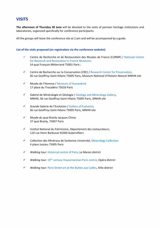
The afternoon of Thursday 30 June will be devoted to the visits of parisian heritage institutions and laboratories, organized specifically for conference participants. All the groups will leave the conference site at 2 pm and will be accompanied by a guide. List of the visits proposed (on registration via the conference website):
Centre de Recherche et de Restauration des Musées de France (C2RMF) / National Centre for Research and Restoration in French Museums 14 quai François Mitterrand 75001 Paris ;
Centre de Recherche sur la Conservation (CRC) / Research Center for Preservation, 36 rue Geoffroy-Saint-Hilaire 75005 Paris, Museum National d’Histoire Naturel MNHN site
Musée de l’Homme / Museum of Humankind 17 place du Trocadéro 75016 Paris
Galerie de Minéralogie et Géologie / Geology and Mineralogy Gallery, MNHN, 36 rue Geoffroy-Saint-Hilaire 75005 Paris, MNHN site
Grande Galerie de l’Evolution / Gallery of Evolution, 36 rue Geoffroy-Saint-Hilaire 75005 Paris, MNHN site
Musée du quai Branly-Jacques Chirac
37 quai Branly, 75007 Paris
Institut National du Patrimoine, Département des restaurateurs, 124 rue Henri Barbusse 93300 Aubervilliers
Collection des Minéraux de Sorbonne Université, Mineralogy Collection 4 place Jussieu 75005 Paris
Walking tour: Historical centre of Paris, Le Marais district
Walking tour: 19th century Haussmannian Paris centre, Opéra district
Walking tour: Paris Street art at the Buttes aux Cailles, XIIIe district

CONFERENCE DINER The conference dinner will be organised in the hotel The Westin Paris - Vendôme in the centre of Paris facing the Tuileries Gardens, the restaurant invites you to taste an authentic and inventive cuisine, in an elegant setting.
The Westin Paris – Vendôme
3 Rue de Castiglione, 75001 Paris
Nearest metro stations : line 1: Concorde station or
Tuileries station
We will be pleased to welcome you at 7 pm for a cocktail followed by dinner at 8 pm.
Cocktail reception
Diner reception

CONFERENCE PROGRAM
Tuesday 28th June
8 h Registration
9 h 15 Opening
9 h 30 KN1 - Analysis for understanding heritage and its degradation: insights from wall paintings, easel paintings and archeological materials - Nevin Austin
10 h O1 - Time-resolved hyperspectral imaging for the mapping of weakly luminescent pigments in paintings - Comelli Daniela
10 h 25 O2 - Non-destructive exploration of Late Gothic panel painting using dual-energy X-ray microtomography - Vavřík Daniel
10 h 50 O3 - The contribution of archaeometry in characterization of decorative materials from the site of Villa di Teodorico in Galeata (Italy) – Saviane Luciana
11 h 15 Coffee/Tea break
11 h 45 O4 - The Dagulf Psalter: an interdisciplinary approach to study inks, dyes and pigments of this early Carolingian manuscript - Jembrih-Simbürgerr Dubravka
12 h 10 O5 - Micro- and sub-µ X-ray CT scanning of Congolese heritage objects for wood identification and conservation - Genbrugge Siska
12 h 35 O6 -Arena scriptoria from the Portuguese Inquisition documents - occurrence, properties and chemical aspects of this writing tool - Nunes Margarida
13 h Lunch
14 h POSTER SESSION 1
15 h O7 - Reconstitution of historical recipes: understanding the influence of oil treatment on the properties of artistic oil paint - Laporte Lucie
15 h 25 O8 - SuPerStAr - Sustainable Preservation Strategies for Street Art: an Italian research project for the knowledge and the safeguard of public art in urban contexts - Modugno Francesca
15 h 50 O9 - Chasing an Alligator: an integrated approach for the study of severe paint defects in a 19th century oil painting - Marques Raquel
16 h 15 Coffee/Tea break
16 h 45 P110 – Displaying Russian space suits based on conservation research at the Deutsches Museum – Holzer Charlotte
17 h 10 O11 - Asbestos cement panel paintings - Degradation issues - Pheulpin Elise
17h 35 O12 - Metal soaps formation in painted miniatures: non-invasive evidence and experimental study - Garrappa Silvia
18 h O13 - Evidence of metal soaps in commercial paints containing ferrous pigments: yellow ocher - Costa Thiago
18 h 25 End
18 h 30 – 21 h 30 WELCOME COCKTAIL
Wednesday 29th June
9 h KN2 – Archaeometallurgy or iron. Some tentative global approches - Dillmann Philippe
9 h 30 O14 - Understanding the manufacturing techniques of Renaissance amour: a coupling between metallographic and SR-XRD investigations - Bérard Emilie
9 h 55 O15 - Trace element analysis of silver coins from XVI -XVII centuries and fire-gilding thickness of a Romanesque crucifix by X-Ray spectroscopy - Gillon Alexandre
10 h 20 O16 - The work of gold at Abydos in the Middle Kingdom: analysis of gold jewellery and leaf - Guerra Maria Filomena
10 h 45 Coffee/Tea break
11 h 15 O17 - The use of in-situ Fourier Transform hyperspectral imaging in the infrared to characterise artist's materials and paintings - Sherwood Alice
11 h 40 O18 - A multi-technical study of Native North American objects dating from the 17th to 19th centuries - Daher Céline
12 h 05 O19 - A multi-technique study of historical natural dyes - De Ferri Lavinia
12 h 30 O20 - New insights into the chemistry of Justicia spicigera dyestuff from Central America - Arberet Lucie
12 h 55 Lunch
14 h POSTER SESSION 2
15 h O21 - Vibrations and cultural heritage preservation: a new approach to protect objects Forma Loïc
15 h 25 O22 - Violin varnish technology developments of the 18th century: Radical conversion from classical oil resin mixtures to shellac-based spiritus varnishes - Zumbühl Stefan
15 h 50 O23 - Investigating the effect of oil binders on the paper supports: modeling deterioration via FTIR and VOC analysis using GC-MS - Banou Penelope
16h15 Coffee/Tea break
16 h 45 O24 - Macro-Raman-mapping: a novel tool to study the pigment distribution of art Vandenabeele Peter
17 h 10 O25 - Data fusion of Py-GC-MS and FT-IR data for the evaluation of degradation patterns in modern paints due to ozone and humidity exposure - Pagnin Laura
17h35 O26 - Studying "Justice" a scientific approach to Violeta Parra´s studio practice - Godoy Valeria
18 h O27 - Tel père, tels fils: a technical study of seven paintings by Camille, Lucien, and Georges Manzana Pissarro - Chipkin Alexandra
18 h 25 End
Thursday 30th June 9 h KN3 - Life in a large national museum: scientific research at the
Victoria and Albert Museum - Burgio Lucia
9 h 30 O28 - The use of madder lake in the production of the "Fayum" portraits - Brunel-Duverger Lucile
9 h 55 P109 - Painted metals of industrial heritage: characterization and conservation – Gordon Julie
10 h 20 O30 - Funerary inscriptions in the Siracusa catacombs: white marbles, decorative stones and painted plaster - Coccato Alessia
10 h 45 Coffee/Tea break
11 h 15 O31 - Corrosion protection of copper statuary by carboxylates-doped sol-gel coatings Lob Silvia
11 h 40 O32 - MiCorr, a transdisciplinary tool for the documentation and the diagnosis of corrosion forms on heritage metal artefacts: building bridges between conservation professionals and material scientists - Gutknecht Naima
12 h 05 O33 - Studying ancient glass to bring to light new insights into the mechanism of glass corrosion - Zanini Roberta
12 h 30 O34 - Application of Hyperspectral Imaging for characterizing VOC-induced historical glass corrosion - Sharma Deepshikha
12 h 55 Lunch
14 h – 17 h VISITS
19 h – 23 h 30 CONFERENCE DINNER
Friday 1st July 9 h KN4 - Conservation Science: a Human-Centered Approach -
Dufourmantelle Kenza 9 h 30 O35 - The right material for the right application - characterization
of the physico-mechanical properties of animal glues in different environments - Bridarolli Alexandra
9 h 55 O36 - Better preserving the archives of Nature for the future: effectiveness of historical and modern sealants in fluid collections - Zuber Baptiste
10 h 20 O37 - Gecko-inspired dry adhesives - a case study in testing methodology - Olender Jacek
10 h 45 Coffee/Tea break
11 h 15 O38 - s-SNOM characterisation of carboxylates growth in a time-depending model Stani Chiaramaria
11 h 40 O39 - New nano-Mg(OH)2 modified siloxane coating for the preservation of gypsum and gypsum-based plasters artifacts - Bergamonti Laura
12 h 05 O40 - Treating a missing part on cast plaster artefacts: a multidisciplinary methodology adapted to the characterization of filling materials - Robin Dupire Juliette
12 h 30 O41 - Influence of physicochemical properties of different limestone on microbial colonization and on biodeterioration - Reboah Paloma
13 h Lunch
14 h POSTER SESSION 3
15 h O42 - Transmission Kikuchi Diffraction, a powerful imaging technique for nanoscale structural characterisation of cultural heritage materials - Holé Clément
15 h 25 O43 - Combining LE-XRF and SR-FTIR microscopy for residue analysis of lithic artefacts - Dominici Clarissa

15 h 50 O44 - A mobile instrument for joint X-ray fluorescence and diffraction measurements on complex-shape Cultural Heritage objects - Poline Victor
16 h 15 Coffee/Tea break
16 h 45 O45 - Identification and manufacturing technology of a Late Bronze Age IA shellfish purple pigment from Ialysos, Rhodes - Facorellis Yorgos
17 h 10 O46 - Impact of pH conditions in the SERS analysis of synthetic colorants: case study of monoazo dyes - Cañamares Arribas María Vega
17h35 O47 - Identification of plant fibers from Central Africa used for the creation of textiles and the creation of a reference database in framework of the CAPTex project - De Paepe Anoek
18 h Closing Remarks

ORAL ABSTRACTS & KEYNOTES
KN1
Analysis for understanding heritage and its degradation: insights from wall paintings, easel paintings and
archeological materials
Austin Nevin
Department of Conservation, Courtauld Institute of Art, London, UK
Binding media, metals and pigments in works of art are material history - and are evidence of technology, artist practise, exchange and trade. Through the study and identification of materials, crucial data can be collected regarding physical and chemical stability providing critical information for conservation decisions. Today we have a plethora of analytical methods available to study works of art – some are portable for in situ, and others require sampling. In this talk I will highlight how we can employ analytical methods synergistically to understand the origin and behaviour of materials [1]. Case studies of works of art and archaeological materials will draw on research using portable instrumentation and cutting-edge analytical methods ranging from the study of ancient polychromy to 20th C. paintings. Investigations on wall painting fragments from the ancient Canannite capital Tel Kabri allowed the identification of degraded binding media from the Aegean style wall paintings that date to the 18th C. B.C.E. The discovery of traces of organic media in the characteristic blue paint is significant for the conservation and treatment of the paintings, for understanding of the sophistication of painting practise and the use of egg-based binding media in the Eastern Mediterranean, and more broadly also questions the presence of domestic animals in the region [2]. A second case study focuses on Tutenkhamun’s dagger that was analyzed using portable instrumentation at the Egyptian Museum in Cairo. New data established conclusively that the well-conserved ornamental blade was fashioned from finely worked meteoritic iron. The identification was possible though the comparison of data acquired from the dagger with known meteor samples, and the calculation of ratios of Nickel and Cobalt [3]. Pigments are the focus of the third case study. Analysis demonstrates how deep crimson pigments from European insects were adopted by Leonardo in the Last Supper, and how, by contrast, Veronese adopted newly introduced Mexican pigments from cochineal insects [4]. The molecular characterization of cross-sections demonstrates the use of similar kermes-based lakes in paintings by Leonardo and Masolino, and carmine-based reds in paintings by Tintoretto and Veronese, while also revealing soluble uncomplexed dyes in samples that has direct implications for conservation, cleaning and lighting. Further examples of the study of pigments include work on cadmium yellows and their degradation [5]. Research will ultimately demonstrate the benefits of synergistic collaborative studies across disciplines. [1] Y. Song et al, Heritage 2021, 4(4), 2599-2622; doi.org/10.3390/heritage4040147 [2] R. Linn, et al, Angew. Chem. Int. Ed. 2018, 57, 13257. doi.org/10.1002/anie.201806520 [3] I. Osticioli, et al, Spectrochimica Acta Part A: Molecular and Biomolecular Spectroscopy, 2019, doi.org/10.1016/j.saa.2019.117273. [4] D. Comelli, et al, Meteoritics and Planetary Science, 2016, doi.org/10.1111/maps.12664 [5] A. Jambon, Journal of Archaeological Science, 2019, doi.org/10.1016/j.jas.2017.09.008 [6] D. Comelli, et al, Analytical Chemistry 2019 91 (5), 3421-3428, doi.org/10.1021/acs.analchem.8b04914
O1
Time-resolved hyperspectral imaging for the mapping of weakly luminescent pigments in paintings
Marta Ghirardello1, Alessia Candeo1, Benedetto Ardini1, Gianluca Valentini1, Cristian Manzoni2, Thomas
Calligaro3, Laurent Pichon3, Xueshi Bai3, and Daniela Comelli1
1 Department of Physics, Politecnico di Milano, Milano, Italy 2 CNR-IFN, Milan, Italy 3 Centre de recherche et de restauration des musées de France, C2RMF, Palais du Louvre, Paris, France
The identification of the materials used in artworks is fundamental for the understanding of the artistic techniques, for dating purposes and for identifying retouching and restoration procedures. In the last decades the demand of non-invasive and non-destructive techniques for the complete characterization of artistic materials has been clearly assessed, motivating the development of new instrumentations and the definition of non-invasive analysis protocols. In this context, photoluminescence measurements are effective tools for the identification and mapping of luminescent materials through their characteristic emissions [1-2], supporting the classification obtained with other elemental and molecular analyses in a completely non-invasive way. Indeed, many materials commonly found in historical paintings are luminescent: these include organic pigments, few inorganic pigments, binders, waxes and finishing varnishes [3]. All these materials exhibit emissions characterised by different intensities, spectral and lifetime properties. However, when using continuous excitation and detection schemes, only the most intensely emitting materials can be clearly detected (typically protective paints and organic binders). Instead, other strategies are required to detect the superimposed emission of weakly emitting materials. Within this framework we propose the use of time-resolved imaging methods to detect and identify low-emitting materials in paintings. The novel approach involves the sequential use of a lifetime imaging camera and a time-gated hyperspectral camera. First, lifetime imaging is performed at different timescales – nanosecond, microsecond, and millisecond timescales – to identify the order of magnitude of the lifetimes of the emitters present in the analysed painted surface. In a second step, time-gated hyperspectral imaging is used to reconstruct the time-gated emission spectrum of the emitting materials in proper temporal windows [4]. Illustrative examples of the proposed approach will be presented, including laboratory model paint samples and modern and Renaissance paintings from the C2RMF collection in Paris. Therein, we will demonstrate how the time-resolved imaging approach is highly effective for the identification and mapping of faint fluorescent species in artworks. In particular, for the first time we will show the clear detection of the faint emission from lead white paints in paintings, thanks to the detection of its long-living emission (characterized by a lifetime of hundreds of microsecond) despite the presence of other strongly fluorescent materials, such as varnish and binders. [1] Nevin, A. et al. Time-Resolved Photoluminescence Spectroscopy and Imaging: New Approaches to the Analysis of Cultural Heritage and Its Degradation. Sensors 14, 6338–6355 (2014).[2] Dooley, K. A. et al. Molecular Fluorescence Imaging Spectroscopy for Mapping Low Concentrations of Red Lake Pigments: Van Gogh’s Painting The Olive Orchard. Angew. Chemie - Int. Ed. 59, 6046–6053 (2020). [3] Nevin, A. et al, Laser spectroscopies for elemental and molecular analysis in art and archaeology. Applied Physics A: Materials Science and Processing, 106(2): 339–361 (2012)[4] Ghirardello, M., et al. A novel photoluminescence hyperspectral camera for the study of artworks European Physical Journal Plus, 136(10), 1052 (2021)
O2
Non-destructive exploration of Late Gothic panel painting using dual-energy X-ray microtomography
Daniel Vavřík1, Václava Antušková2, Štepánka Chlumská2, Ivana Kumpová1,
Radka Šefců2, and Michal Vopálenský1
1 Czech Academy of Sciences, Institute of Theoretical and Applied Mechanics, Prague Czech Republic 2 National Gallery Prague, Prague, Czech Republic Presented results were obtained by an interdisciplinary survey of medieval Bohemian panel painting Lanna’s Virgin and Child on a Crescent Moon [1] (ca 1450, inv. no O 495, 41.5 ×31.5 cm) from the collection of the National Gallery Prague. Central theme of the painting is Virgin Mary supplemented by paintings of saints and donors on the frame and backside imitation of a stone slab. Analysing pigments, lead white, calcium carbonate, lead tin, yellow, vermilion and azurite were identified using non-invasive X-ray fluorescence spectroscopy (XRF) and mobile Raman spectroscopy. The presence of gold was confirmed by XRF on the background of the painting and on the frame. To obtain further information about structure of the wooden panel covered by polychromy, X‑ray computed tomography (CT) and dual-energy computed tomography (DECT) method [2] were utilized. CT provides spatial 3D model of the surface layers and the inner structure of the investigated object - reconstructed CT density can help to resolve material composition of the investigated object. In addition, DECT brings further information helping to distinguish materials with similar density but different chemical composition. It will be demonstrated that CT and DECT information can help to analyse polychrome composition (as well as structure of the panel wood). Thus for instance, some colours looking similar in visible light may have different CT reconstructed density, it signs places for which further detailed XRF and Raman spectroscopy analysis is desirable. In the internal structure on CT images, attachment of grab handles, as well as secondary interventions and joints (mainly metal pins connecting single wooden parts – panel and frame), are visible. Textile base canvas and application of creasing for better cohesion of individual layers and their bonding (presence of glue) can be viewed. The extent of damage caused by wood-destroying insects was also observed. Particular attention was paid to the decorative techniques – punching and impressed pattern – on the golden background. Areas, where pigments containing heavy elements (lead white, vermilion) were identified, are recognizable on CT images as well. Lead white is used in thick compact layers on the flesh of the figures on the right side of the frame. Compared to that, subtler white layers are applied to the flesh of other figures and the Virgin Mary with the Child in the centre of the painting. This difference may indicate that more authors participated in the painting. The survey allowed for non-destructive evaluation of the current state of panel painting, including defects in its internal structure, and complete the knowledge of production technology. It provided documentation for dendrochronological dating and helped to identify areas of interest suitable for further analyses. Obtained results will be compared with findings from material survey on Assumpta from Deštná (ca 1450, inv. No. O 724, 144 × 111 cm) and Lanna's Madonna (ca 1450, inv. no. O 494, 50.5 × 38.5 cm) to verify their origin in the same workshop that was assumed based on stylistic and paint technique analysis carried out by the art historian. This contribution has been financially supported by the project of the Ministry of Culture of the Czech Republic: Mobile device devoted to imaging and analysis of the layered paintings and polychromy of the works of old art (DG18P02OVV006). [1] J. Fajt, Š. Chlumská, Bohemia and Central Europe 1200–1550. The PermanentExhibition of the Collection of Old Masters of the National Gallery in Prague at the Convent of St Agnes of Bohemia, The National Gallery in Prague, Prague,2014. [2] Daniel Vavrik, et. al., Dual energy CT inspection of a carbon fibre reinforced plastic composite combined with metal components, Nondestructive Testing and Evaluation 6, 2016, pp. 47-55, ISSN 2214-6571, https://doi.org/10.1016/j.csndt.2016.05.001
O3
The contribution of archaeometry in characterization of decorative materials from the site of Villa di Teodorico in Galeata (Italy)
Luciana Saviane1, Maurizio Aceto2, Laura Fornasini3, Luciana Mantovani4, Alessia Morigi1,
Riccardo Villicich1, Danilo Bersani5
1 Dipartimento di Discipline Umanistiche, Sociali e delle Imprese Culturali, Università degli Studi di Parma, Parma, Italy. 2 Dipartimento di Scienze e Innovazione Tecnologica, Università degli Studi del Piemonte Orientale, Alessandria, Italia; Centro Interdisciplinare per lo Studio e la Conservazione del Beni Culturali, Università degli Studi del Piemonte Orientale, Vercelli, Italy. 3 ICCOM-CNR, Istituto di Chimica dei Composti Organometallici, Pisa, Italy. 4 Dipartimento di Scienze Chimiche, della Vita e della Sostenibilità Ambientale, Università degli Studi di Parma, Parma, Italy. 5 Dipartimento di Scienze Matematiche, Fisiche e Informatiche, Università degli Studi di Parma, Parma, Italy.
Villa di Teodorico in Galeata (Forlì-Cesena, Emilia Romagna) is an important archaeological site in the north of Italy. It is a multi-layered site due to 17 centuries of occupation, from 6th century BC to 12th century AD. The most interesting results concern the Roman age, when a large villa was built, and the late antiquity, when the Ostrogothic king Theodoric decided to build in this area his palatium (early sixth century AD), a pavilions villa with long corridors and wide-open spaces. A big octagonal room and neighbouring areas, paved with mosaics, belonged to the most prestigious pavilion of the Villa were recently investigated. In particular, the polygonal room was covered by a dome and decorated inside with wall mosaics, as proved by the discovery of a lot of glass mosaic tesserae in the collapse layers. The archaeometric investigation was performed on Roman wall paintings fragments and on several glass mosaic tesserae and glass sectilia fragments belonging to Palazzo di Teodorico by using a multi-technique approach that included micro-Raman spectroscopy, field emission scanning electron microscopy with energy-dispersive X-ray spectroscopy (SEM-EDS), X-ray powder diffraction (XRPD), UV–visible–NIR diffuse reflectance spectrophotometry with optic fibres (FORS) and optical stereo-microscopy. This analytical approach allowed the identification of all components, collecting molecular, elemental, microscopic, morphological and chromatic data. The characterization of samples supplied essential archaeological, historical and technological information. The production techniques and the rich materials employed suggests the importance of the site in different periods. The evolution of the manufacturing technologies and the possible trade routes mainly during late antique period are witnessed by the change in the raw materials. For example, the identification of antimony-based opacifiers in few tiles, common during imperial age, and of cassiterite (SnO2), an opacifying agent attested from late antiquity, present in many tiles, indicates than in Villa di Teodorico the adoption of more recent production techniques was starting. Morever, the use of tin-based phases in place of antimony-based phases, could suggest a shortage of the supply of antimony or the starting of closer relations with India, a producer of tin.
O4
The Dagulf Psalter: An interdisciplinary approach to study inks, dyes and pigments of this early Carolingian manuscript
Jembrih-Simbürger Dubravka1, Hofmann Christa2, Aceto Maurizio3, Vetter Wilfried1, Sterflinger Katja1,
Rainer Thomas4
1 Institute for Natural Sciences and Technology in the Arts, Academy of Fine Arts Vienna, Vienna, Austria 2 Conservation Department, Austrian National Library, Vienna, Austria 3 Dipartimento di Scienze e Innovazione Tecnologica, Università degli Studi del Piemonte Orientale, Alessandria, Italy 4 Institute of Art History, University of Zurich, Zurich, Switzerland
The Dagulf Psalter, due to its golden script also called “Golden Psalter” is a Carolingian manuscript on calf parchment, which contains Old Testament Psalms, a selection of Canticles and a collection of creeds and prefaces. It is believed that the scribe Dagulf created this codex between 793-795 as a gift from Charlemagne to Pope Adrian I. The Dagulf Psalter is preserved at the Austrian National Library in Vienna (Cod. 1861), while the ivory plates that decorated the book covers were separated in the past and are kept at the Louvre Museum in Paris.
The “Golden Psalter” is impressive not only for its golden and silver letters in various sizes, but also for the rich ornamentation and the combination of colours used for the painted initial folios. Thus, it contains three initial folios with ornamental frames and coloured grounds in shades ranging from dark purple red to bluish black and lighter blue.
As part of a larger project of the research group "Textures of Sacred Scripture" at the University of Zurich, funded by the Swiss National Science Foundation, to catalogue the various shades of purple in Carolingian and Ottonian manuscripts, the materials of the Dagulf Psalter were analysed to gain new insights into the different materials used to create these shading effects and diverse nuances of purple in Carolingian manuscripts. Further, the knowledge of inks, dyes, and pigments compositions is essential for the conservation of the manuscript. Due to the unique and high historical value of the manuscript, a non-invasive analytical approach was applied to identify materials used for paints and inks on 19 selected folios. Point analysis by Fibre Optics Reflectance Spectroscopy (FORS) identified mainly paint materials (dyes and pigments), X-ray fluorescence analysis (XRF) was used for the inks and pigments. Subsequently, Hyper Spectral Imaging (HSI) was applied to visualize the pigments as well as blue and purple dyes distribution on folios or areas of interest. Purple parchment samples dyed with orchil (Roccella tinctoria, Lasallia pustulata), folium (Chrozophora tinctoria), and shellfish purple (Hexaplex trunculus) served as references for FORS and HSI. Orchil, indigo, and ultramarine have been identified as backgrounds on the initial folios. In initials in the text orchil, indigo, and ultramarine were used alone and together. The rich decoration was created with these three main tones in combination with red lead, red ochre, cinnabar, orpiment, and lead white. The gold ink could be identified as a highly pure gold ink containing only small amounts of copper. The silver ink used in letters on the initial folios seems to be a mixture of gold and silver inks. Microscopic observations showed variations in the application of silver ink. The subtle differences are obscured by the corrosion of silver particles in the ink. Dark shades of silver inked areas are visible on the back sides of the parchment folios, without mechanical damage of the parchment. A primer layer of lead white might have protected the parchment. Depending on details painted different stratigraphies were used by the scribe/illuminator of the manuscript. The use of purple dye and silver inks in the 8th-century Dagulf psalter is further compared with a 6th-century manuscript, the Vienna Genesis. As orchil is a very light sensitive dye and silver inks can be very corrosive to the parchment, an understanding of its alteration factors influences preservation of these unique manuscripts. [1] K. Holter, Der Goldene Psalter „Dagulf-Psalter“, Vollständige Faksimile Ausgabe, Graz 1980.[2] B. Reudenbach, Der Dagulf-Psalter und sein Einband, in: Dittscheid/ Gerstl/ Hespers ed., Kunst-Kontexte, Petersberg 2016, 242-249.[3] C. Denoël et al., Illuminating the Carolingian era: New discoveries as a result of scientific analyses, Heritage Science (2018), 6-28. [4] C. Hofmann (ed.), The Vienna Genesis: Material analysis and conservation of a Late Antique illuminated manuscript on purple parchment. Wien, Köln, Weimar 2020.
O5
Micro- and sub-µ X-ray CT scanning of Congolese heritage objects for wood identification and conservation
Genbrugge Siska1, Dierickx Sofie 1,2, Beeckman Hans2,3, Hubau Wannes3, Van den Bulcke Jan2
1 Royal Museum for Central Africa, Archives and collection management, Conservation Lab 2 UGent-Woodlab, Laboratory of Wood Technology, Department of Environment, Faculty of Bioscience Engineering, Ghent University, Gent, Belgium, UGCT, UGent Centre for X-ray Tomography, Gent, Belgium 3 Royal Museum for Central Africa, Department of Biology, Wood biology
The Belgian Royal Museum for Central Africa (RMCA) houses over 120.000 ethnographic objects of which 55.000 sculptures, musical instruments, furniture and tools are made of wood or containing wooden elements. Most of these artefacts are made from unknown tropical wood with only 6% of the wood species identified. The majority of the collection is understudied and entered the museum with little or no contextual information. A correct identification however is crucial for curatorial and conservation practices in the museum, as discovering more about the wood species represented in the collection can provide insights into the processes surrounding the making of them and knowledge about their provenance. Furthermore, knowledge of the wood species of an object can aid the conservators of the museum in determining the best treatment, considering the specific characteristics of the wood species and its ageing properties. It enables conservators to optimize preservation conditions, and can facilitate international travel of the objects conform the CITES guidelines.
Unfortunately, identifying African wood species remains destructive, during which a sample of wood is removed and subsequently studied microscopically for its anatomical key features. Therefore, a sample of the object between 5 mm³ to 2 cm³ is permanently removed. These large sized losses are accepted for identifying tree samples within the field of wood biology, but are undesirable for the identification of smaller wooden artworks. The TOCOWO project (Tomography of Congolese Wooden Objects) is a collaboration between the RMCA and the UGent-Woodlab. It takes advantage of RMCA's existing expertise of microscopic wood identification, the large reference collection of more than 61.000 wood biology specimen at the RMCA, and the expertise in wood identification through computed tomography at Ghent University. Building on this expertise, the project aims to explore the possibility of micron and sub-micron computed tomography (CT) as a non-destructive alternative for the microscopic identification of wood species in the ethnographic collection. This technique has shown promise in the field of wood biology and is now being applied to a large selection of ethnographical museum objects in the RMCA. X-ray computed tomography allows for a visualization, not only of the object's surface, but also of its interior. By taking many so-called projections or radiographies while the object turns 360°, a 3-dimensional reconstruction can be obtained. Consequently, any coating the object has been treated with will have little impact on the image obtained with CT scanning and even the most deteriorated and fragile wood can be digitally sampled on a micron and sub-micron scale. The preliminary results within this project, based on more than 84 Congolese objects scanned in 2021, are very promising. An important prerequisite to a successful wood species identification is the achieved resolution, which in turn is determined by the object's dimensions, positioning, and added materials. The final objective of this 2-year project is to culminate the results in a reference database of positive identifications of Congolese wood species, as well as the establishment of a comprehensive protocol for the use of computed tomography for the purpose of systematically identifying wooden objects. Apart from its promising applications for wood identification, the CT technique has also yielded some additional and important conclusions from a conservator's and curator's point of view. Thanks to the CT images of collection objects, new insights could be gained with regards to the manufacturing process of an object, as well as (before undetected) insect activity, the structural stability of the wood, and old conservation treatments (such as consolidations or additions).The TOCOWO research project is the start of a thorough investigation into the wood collection and the project has already identified that further research projects are needed to investigate the digital CT reconstructions in order to gain more knowledge about the history, structures, techniques and deterioration of the wood to further contextualize the wood collection. Several case studies of scanned objects are presented to illustrate the protocol drafted in the framework of the project. The advantages and limitations of the CT as alternative identification technique are discussed and the ethical concerns of sampling and scanning ethnographic objects are considered.
O6
Arena scriptoria from the Portuguese Inquisition documents - occurrence, properties, and chemical aspects of this writing tool
Nunes, Margarida1*, Wanzeller Martins, Gláucia1, Sarraguça, Jorge2, Olival, Fernanda3,4, Claro, Ana5,
Moita, Patrícia1,6, Ferreira, Teresa1,7**
1 University of Évora - HERCULES Laboratory, University of Évora, Évora, Portugal 2 NOVA University Lisbon - LAQV-REQUIMTE, Department of Chemistry, NOVA School of Science and Technology, Caparica, Portugal 3 University of Évora - CIDEHUS, University of Évora, Évora, Portugal 4 University of Évora - History Department, Social Sciences School, University of Évora, Colégio do Espírito Santo, Évora, Portugal 5 NOVA School of Social Sciences and Humanities - CHAM, NOVA School of Social Sciences and Humanities, Lisboa, Portugal 6 University of Évora - Geosciences Department, Science and Technology School, University of Évora, Évora, Portugal 7 University of Évora - Chemistry Department, Science and Technology School, University of Évora, Évora, Portugal **Corresponding author: Ferreira, Teresa, [email protected] During the 14th century, the Western world witnessed a surge of interest in a range of writing tools, including arena scriptoria, commonly known as blotting sands [1, 2]. These materials were mainly used to speed up the drying time of the writing inks. Generally composed of a wide variety of minerals, they could also include other substances as painted glass or organic materials (gums, wood, or bones) and were usually kept in sanders [3]. These sand containers, which presented a variety of shapes, were covered with pierced lid to sprinkle the sand particles over the writings. Once completed the task and the ink dried, most of the blotting sands were collected back to the sanders. Nevertheless, part of them remained attached to the writing areas with a less positive impact on the manuscripts. Abrasion of the writing layers and paper supports, disrupt of the binding medium, and accumulation in the spine-folds and sewing structure with long-term impact are some examples [4]. Despite being intimately related to the historical writing procedures, blotting sands is an almost unknown topic in scientific research studies. The work here presented is a pioneering investigation on blotting sands used on the historical documents of the Portuguese Inquisition from the Courts of Coimbra, Lisbon and Évora. A total of 154 samples were collected from these documents covering a time period from the 16th and 19th centuries. Physical characteristics, including colour, grain-size distribution, and shape were primarily investigated by optical microscopy, followed by a methodological approach based on imaging analysis (ImageJ ® software). Samples were chemically characterized by scanning electron microscopy coupled with energy dispersive spectroscopy (VP-SEM/EDS), µ-Raman spectroscopy and X-ray diffraction. Heavy minerals species were identified consisting mainly in iron-oxide ores as ilmenite and hematite along with almandine-rich garnet. Other minerals as rutile, anatase and quartz were occasionally identified. Elemental analysis showed the presence of rare earth elements phases found in ilmenite hosts. The predominance of these iron-oxide ores points out the existence of mineral processing technologies probably based on gravity concentration techniques. Principal component analysis (PCA) was used to investigate a possible correlation between chemical composition and morphological features of these materials. This study aimed to make an in-depth characterization of the blotting sands used and establish relationships between the materials themselves and the criteria followed for selection, regarding their main function. Furthermore, the uncovered information will complement the forthcoming studies in the historical context of the manuscripts. Portugal has some of the best inquisitorial archives in Europe and preserving them is an essential investment.
[1] L. Blake, New College Notes, 10 (2018) 1. [2] B. Reissland, I. Joosten, E. Eis, A. Schubert, in: Pre-Conference Proceedings (Extended Abstracts), 2nd Iron Gall Ink Meeting and Final European Thematic Framework Meeting for Transitional Metals in Paper (MIP), Newcastle (2006) 31. [3] R. Milke, European Journal of Mineralogy, 24 (2012) 759 [4] R. Blanco, in: Biblioteca Virtual Miguel de Cervantes, Digital edition based on the 3rd ed., (Madrid), Imp. and Lit. de J.Palacios (1902) The authors acknowledge the ANTT team for providing access to the samples and FCT for funding (IRONIC project PTDC/ART-HIS/32327/2017, UIDB/04449/2020 and UIDP/04449/2020). G. Wanzeller thank the support by FCT for BI PTDC/ART-HIS/32327/2017 scholarship in the scope of IRONIC project and M. Nunes also thanks FCT for a PhD scholarship (SFRH/BD/147528/2019).
O7
Reconstitution of historical recipes: understanding the influence of oil treatments on artistic oil paints’ properties
Laporte Lucie 1, Ducouret Guylaine 2, Gobeaux Frédéric 3, Touboul David 4, de Viguerie Laurence 5
1 LAMS, CNRS UMR 8220, Sorbonne Université, Paris, France 2 Laboratoire SIMM, CNRS UMR 7615, ESPCI Paris, PSL Research University, Paris, France 3 LIONS – NIMBE, UMR 3685 CEA/CNRS, CEA Saclay, Gif sur Yvette, France 4 ICSN, CNRS UPR 2301, Gif-sur-Yvette Cedex France
Since the 15th century, artistic painters have prepared paint mixtures by adding oil binders to various pigments. Oils commonly used in historical paintings are called drying oils: they exhibit natural hardening properties after a long period of exposure to air. To accelerate the oil drying process, numerous historical recipes mention the use of inorganic drying compounds (or “driers”), such as lead oxide (PbO). The driers are ground, added to the oil and the mixture is heated, possibly with water [1]. This treatment induces changes of the paint properties [2], [3]. The study of liquid treated oils (before drying) is a key step to understand the influence of oil pre-treatment on oil paint properties and degradation during ageing. The present work thus aims to characterize the modifications induced by the formulation of the paint mixture. More specifically, the flow properties of treated oils at a macromolecular scale will be correlated to their organization at the supramolecular scale and to their chemical composition. We have formulated oils based on recipes from the 17th to the 19th century: our systems consist of linseed oil, widely used by painters, and lead oxide. During heating, the saponification of triglycerides contained in oil was monitored by FTIR (Fourier-Transform Infrared Spectroscopy): the formation of lead soaps modifies the physicochemical properties of the oils. First, the rheological properties are strongly modified. By increasing the initial concentration of lead oxide, the sample evolves from a Newtonian fluid to a shear-thinning fluid. The shear-thinning sample (linseed oil + 20% PbO) also exhibits viscoelastic properties. At the supramolecular scale, SAXS (Small Angle X-ray Scattering) measurements and cryo-TEM imaging revealed the presence of lamellar phases with a characteristic distance of 50 Å, corresponding to the size of a lead soap [4]. The complementary use of SFC (Supercritical Fluid Chromatography), GC-MS (Gas Chromatography coupled with Mass Spectrometry) and NMR (Nuclear Magnetic Resonance) gives an in-depth view of the modifications induced at the molecular scale. Surprisingly, we observed that saturated soaps are formed in priority during saponification, followed by the formation of unsaturated lead soaps. Moreover, a fraction of the unsaturated chains, initially in cis conformation, are isomerized to the trans conformation during heating. Saturated lead soaps, as well as unsaturated soaps in trans conformation, are more likely to organize into lamellar phases due to the absence of kinks in the fatty chain. The reconstitution of linseed oil heated with lead oxide and the use of different analytical techniques lead to a detailed understanding of the materials used by painters and their practices. [1] M. Faidutti and C. Versini, Le Manuscript de Turquet de Mayerne présenté par M. Faidutti et C. Versini, Pictoria Sculptoria et quae subalternarum artium, 1620, Audin Imprimeurs. Lyon, 1967. [2] I. Kneepkens, ‘Understanding historical recipes for the modification of linseed oil’, University of Amsterdam, Department of Art History, 2012. [3] L. De Viguerie, G. Ducouret, M. Cotte, F. Lequeux, and P. Walter, ‘New insights on the glaze technique through reconstruction of old glaze medium formulations’, Colloids and Surfaces: A Physicochemical and Engineering Aspects, vol. 331, no. 1–2, pp. 119–125, Dec. 2008. [4] F. J. Martínez-Casado et al., ‘Lead soaps: crystal structures, polymorphism, and solid and liquid mesophases’, Phys. Chem. Chem. Phys., vol. 19, no. 26, pp. 17009–17018, 2017.
O8
SuPerStAr - Sustainable Preservation Strategies for Street Art: an Italian research project for the knowledge and the
safeguard of public art in urban contexts
Modugno Francesca1, La Nasa Jacopo1, Scalarone Dominique2, Toniolo Lucia3, Cartechini Laura4, Prati Silvia5 , Izzo Francesca6, Calvano Cosima Damiana7
1 Department of Chemistry and Industrial Chemistry, University of Pisa, Italy 2 Department of Chemistry, University of Turin, Italy 3 Department of Chemistry, Materials and Chemical Engineering "Giulio Natta", Politecnico of Milan, Italy 4 National Research Council SCITEC Institute Perugia, Italy 5 Department of Chemistry “Giacomo Ciamician”, University of Bologna, Italy 6 Dipartimento Scienze Ambientali, Informatiche e Statistiche, Università Ca' Foscari Venezia, Italy. 7 Department of Chemistry, University of Bari, Italy
Street art has been recognized part of our cultural heritage only in the latest years. The ephemeral character, free access, and exposure to the environment and anthropic actions, make public paintings vulnerable to neglect, removal, vandalism, and degradation. Beyond that, the strategies aimed at their preservation and fruition are rather unclear or lacking. The project PRIN-2020 SUPERSTAR Sustainable Preservation Strategies for Street Art sets as a goal the definition of innovative guidelines for the preservation strategy of street art, aimed at safeguarding its powerful social and cultural message in the urban context. A combination of non-invasive and micro-invasive techniques are optimized to shed light on the chemical-physical properties and vulnerability aspects of modern paint materials that constitute street artworks. The studies performed in the laboratory on reference materials are supported by research performed on relevant case studies, located in different environmental urban contexts. The focus is on the materials used by the artist and the environmental risks and anthropic stresses [1,2]. Thanks to the strong collaboration between the partners with complementary expertise and with conservation institutions, the project will provide the following outputs: optimized innovative cleaning procedures for the restoration of outdoor murals and for the removal of vandalistic graffiti; selected protective coating materials with particular attention to durability aspects; and an integrated protocol for long-term sustainable monitoring and conservation. The outputs will support institutions and entities engaged in safeguarding public urban art and in establishing preservation guidelines. [1] La Nasa, Campanella, Legnaioli, Modugno et al, 60 years of street art: A comparative study of the artists’ materials through spectroscopic and mass spectrometric approaches, Journal of Cultural Heritage 48, 2021, 129–140 [2] Bertasa, Scalarone et al, Overcoming challenges in street art murals conservation: A comparative study on cleaning approach and methodology, Coatings 10, 2020, 1
O9
Chasing an Alligator: an integrated approach for the study of severe paint defects in a 19th century oil painting
Marques Raquel1, Carlyle Leslie1, Pombo Cardoso Isabel1, De Viguerie Laurence2
1 Department of Conservation and Restoration and LAQV-REQUIMTE, Faculdade de Ciências e Tecnologia, Universidade NOVA de Lisboa, Campus da Caparica, 2829-516 Caparica, Portugal 2 Laboratoire d'archéologie moléculaire et structurale, Sorbonne Universités, UPMC Univ. Paris 06, CNRS, UMR 8220, Paris, France
This paper will report on the results from a multi-analytical approach that aims to relate the formation of extreme film-forming defects commonly known as “Alligatoring” to the use of Bitumen oil paint.
A significant number of oil paintings produced in Europe during the mid-18th and 19th centuries exhibit paint failure in the form of severely disfiguring drying cracks and surface distortions often referred to as “Alligatoring” or “Bitumen Cracking”. This problem is reported to develop some years after completion of the work and has been associated with the use of bitumen/asphalt paint [1], with no clear understanding to date of the materials and mechanisms which contribute to this phenomenon.
This is the case for the oil painting O Cardeal D. Henrique recebendo a notícia da morte de D. Sebastião (Figure 1), by Marciano Henriques da Silva (1831-1873), painted in Rome in 1861 which suffers from such a severe case of Alligatoring that it has not been exhibited for over 100 years. The visual and chemical study of O Cardeal D. Henrique offers specific challenges due to its complex and highly disrupted paint layer stratigraphy coupled with the uncertainty introduced by analytical detection limits, which explains the need to use a multi-analytical approach. Analyses were carried out using Optical Microscopy (OM), micro Raman Spectroscopy (µ-Raman), micro Fourier Transform Infrared Spectroscopy (µ-FTIR) and Fourier Transform Infrared Microscopy, Scanning Electron Microscopy with Energy Dispersive X-ray Spectrometry (SEM-EDS), X-ray Fluorescence (XRF) and Mass Spectrometry techniques (such as GC-MS, Py-TMAH-GCMS and TOF-SIMS).
To improve the analytical methodology and better interpret the results obtained, reconstructions were made. Access to a 19th century colourman’s archive (Winsor & Newton) enabled the study of their “Bitumen” for oil tube paint formulations. While there are a substantial number of published recipes in 19th century artist’s manuals for the use of bitumen/asphaltum, W&N’s records offer a unique source of detailed information on the commercially prepared product which differs substantially in ingredients used and method of preparation. Based on a critical analysis of W&N’s production records for “Bitumen” a recipe from 1858 was selected and reconstructed, using historically appropriate materials. W&N’s formulation for bitumen oil paint (which calls for asphaltum as its primary ingredient) was prepared by melting and cooking together at a high heat (280-300ºC) asphaltum with lead treated oil, followed by the addition of mastic varnish, lake pigment, lead acetate and the gelled medium Megilp (lead treated oil plus mastic varnish). Production records for the individual ingredients, e.g. lead treated oil, purple lake etc, are also available in W&N’s archive, so that the ingredients and production methods for each component could also be identified and followed in the reconstruction [2,3].
In addition to clarifying the analytical results obtained from the painting, these reference samples from the reconstructions illustrate the strengths and weaknesses of organic analysis on highly processed complex mixtures. [1] Carlyle, L., & Southall, A. (1993). No Short Mechanical Road to Fame: The Implications of Certain Artists’ Materials for the Durability of British Painting: 1770-1840. In R. Hamlyn (Ed.), Robert Vernon’s Gift, British Art for the Nation 1847 (pp. 21–26). London: Tate Gallery Publications.[2] Marques, R., Carlyle, L., De Viguerie, L., Pombo Cardoso, I., Boon, J. (in press) "Winsor & Newton’s 19th-century bitumen brown oil paint. Part 1: A critical analysis of W&N production records." In ICOM-CC Art Technological Source Research Working Group, 8th Interim Meeting 26-27 Sept. 2019, Cologne.[3] Marques, R., Carlyle, L., De Viguerie, L., Cardoso, I. P., Boon, J. (in press) "Winsor & Newton’s 19th-century bitumen brown oil paint. Part 2: the reconstruction." In ICOM-CC Art Technological Source Research Working Group, 8th Interim Meeting 26-27 Sept. 2019, Cologne.

O10
X-ray and IR spectroscopies study on ancient rock art found in northern Thailand
C. Boonruang1,2 K Won-in3* K Thumanu4 U Tippawan1 C. Thongleurm5 and P Dararutana6
1 Department of Physics and Materials Science, Faculty of Science, Chiang Mai University Thailand 2 Center of Excellence in Materials Science and Technology, Chiang Mai University, Chiang Mai, Thailand 3 Department of Earth Sciences, Faculty of Science, Kasetsart University Bangkok Thailand 4 Synchrotron Light Research Institute, Nakhon Ratchasima,Thailand 5 Science and Technology Research Institute, Chiang Mai University, Chiang Mai Thailand 6 Independent Researcher, Retired Army Officer at the Royal Thai Army, Lopburi, Thailand
A great number of rock art have been found in many regions of Thailand, especially in the northern region. The ancient rock art samples from Pratu Pha Valley (Lampang province, northern Thailand) represent a piece of important cultural information. In this work, 8 samples excavated from different areas in this site have been studied. The design and materials used in the production of arts have been examined. A scanning electron microscopy coupled with an energy dispersive X-ray spectrometry (SEM-EDS) has been carried out to analyze the elemental composition and structure of the sample. Proton-induced X-ray emission spectroscopy (PIXE) has been applied to confirm the presence of trace elements. Infrared absorption spectroscopy based on synchrotron radiation (SR-IR) has been used to examine the functional group. It has been found that C, O, Al, Si, Ca, and Fe are present in all samples, while Mg, K, S, and Mn have been detected in some samples. PIXE results indicate a presence of Ti, Cr, Ni, and As. The constituents of functional groups corresponding to the wavenumber have been identified by the IR spectra. These results indicate the source of samples. Some complex chemicals for conservation have been found in these archaeological pictures.
Displaying the Sokol KV2 Space Suit at the Deutsches Museum
Holzer, Charlotte1, Pamplona, Marisa 1, Lescop, Benoit2, Rioual, Stéphane2
1 Deutsches Museum, Museumsinsel 1, Munich, Germany 2 Univ Brest, Lab-STICC, CNRS, UMR 6285, Brest, France
The Deutsches Museum in Munich holds a collection of space suits made by the Russian company NPP Zvezda, that are put on display in one show case. The Sokol KV-2 pressure suit works in combination with a Soyuz capsule and was worn by the German cosmonaut Klaus-Dietrich Flade in 1991 on the mission MIR-92. It had been on display at the Deutsches Museum for about 20 years and during this time, the colour of the originally white textiles changed to orange, brown, pink or violet. The conservation science department started research on the space suits in 2019 on the request of the space curator, who had observed significant visual change on the Sokol KV-2. The main causes for its colour change are connected to the use of light-sensitive polyamide in the outer flexible parts (elbows, pockets) and off-gazing from the rubber, present in the inner airtight layers and in the adhesives. Volatiles deriving from rubber based materials accumulated inside the suit due to lack of air movement, prevented by the layered construction of the suit and its presentation on a mannequin in a seated posture. The materials used for the mannequin were unsuitable for long-term display, which was shown by an Oddy test that revealed the emission of further pollutants. Silver, copper and lead coupons were positioned inside the suit, after removing the mannequin and they tarnished at room conditions already after a month. Analysis of the metal coupons with XPS, XRD and SEM-EDX showed the accumulation of organic species, Sulfur and Chloride on the metal surfaces. The measured compounds are explained by the presence of pollutants emitted from the suit materials. The results highlight the suitability of the silver, copper and lead coupons in the test due to their cross-sensitivity. In 2022, the Deutsches Museum will re-open 19 exhibitions, including the space collection, after a large refurbishment project, that started in 2015. Within that scope the building was insulated, heating / cooling system and LED-lighting were installed. The space suits will be presented in a new display case that is equipped with ventilation to circulate air around the objects and through a system of drawers, for passive climate control with silica gel and pollutant adsorption with charcoal textiles. New mannequins shall replace the old ones, using only conservation grade materials and carrying pollutant absorbers as well. The effectiveness of the measures taken to slow down the degradation processes in the space suits will be assessed in the new display case by a corrosion monitoring system with further integrated sensors for temperature, relative humidity and air pressure. The aim is to react to damaging environmental factors, like humidity or accumulation of pollutants, in a timely manner, for example by changing or improving the absorbers or checking the ventilation system. This approach is exemplary in the Deutsches Museum, because the suits are of utmost importance to the collection. However, this display case equipped with the corrosion monitoring (Purafil Onguard Smart) also presents the opportunity to test and compare if simpler and cheaper systems (e.g. RFID corrosion sensor tags), might have the potential to be used to a larger extent to monitor further objects sensitive to pollutants in the collection. NPP Zvezda equipped cosmonauts with the life support systems needed to survive in space from the beginning of the space age until today. Literature research, technical examination and material analysis of different types of suits, showed consistency in their way of development and production by using common materials and binding techniques. On the one hand, this reflects a fascinating tradition in research and workmanship at NPP Zvezda. On the other hand, it means that specific degradation mechanisms connected to the sensitivity and combination of materials are very similar in Russian space suits. In this sense, the preventive conservation strategy of our case study is useful to collections holding space suits from NPP Zvezda.
P110
O11
Asbestos cement panel paintings – Degradation Issues
Pheulpin Élise1, Soppa Karolina1, Küffner Markus1, Scherrer Nadim1, Zumbühl Stefan1, Gerdes Andreas2
1 Bern University of Applied Sciences - Switzerland 2 Karlsruher Institut für Technologie - Germany
Eternit ® (asbestos cement panels), originally intended for the construction industry, became the object of interest for swiss painters at the beginning of the 20th century, such as Cuno Amiet or Giovanni Giacometti. However, despite the advantages of these panels in terms format and above all price, their use was limited to a decade: the promised properties of Eternit ®, sold as the inevitable replacement for wood panels, were the subject of many disappointments, especially in terms of the mechanical and climatic resistance. Some of the works have been copied onto canvas, as the condition of the original support had deteriorated rapidly. Today, these works show notable differences in terms of conservation: some paintings have almost no damage and many have severe damages. These are protrusions and, in various stages, the paint layer standing up, up to many missing parts. Background research was carried out, in particular through the study of the epistolary exchanges of the artists mentioned above. Valuable information on the use and purchase of the plates was uncovered, as well as on the disappointed expectations of the painters. In order to determine whether these damages, were due to the painting technique or to the new substrates, five paintings of Cuno Amiet from various Swiss museums were examined with analytical methods on micro-samples and stratigraphic sections via SEM and FTIR-FPA. Technological and elemental comparison of the paint layers and degradation products have determined that the paint layer plays a minor role in the occurrence of micro-concretions. Elemental studies of the cement composing the substrates and research into the chemistry of cement allowed the formation processes of the cement to be targeted as the predominant element. The micro-concretions measured on the surface are the result of the carbonation/crystallization of calcium hydroxide in the form of calcium carbonate crystals. These protrusions are completely absent from the reverse side of the works: in fact, the pressure of the pictorial layer, which varies according to the thickness and density of the layers, does not allow the elimination of the calcium carbonate naturally formed on the surface of the cement, and favours the grouping of these crystals in the form of conglomerates. The issue of metal soaps is also found but only in subordinate numbers in the paintings by Cuno Amiet. These products show cross-reactions between the formation of calcium carbonate crystals and the saponification of the paint layers via the very high alkalinity (pH 12.3 - 13) of the calcium hydroxide in soluble form within cement. This study highlights for the first time the extreme sensitivity of these works to climatic variations, as oscillations in relative humidity can reactivate this detrimental chemical reaction of the cement-based material liberating alkaline compounds at any time. Preventive measures such as monitoring of the works and their climatic environment, as well as the strict implementation of relative humidity and temperature tolerances in the storage areas are thus strongly recommended. Climate control must be implemented also in the context of loans or exhibitions. The question of restoration, and more specifically consolidation of the numerous strongly damaged works, represents a real challenge. Consolidation actions must respect the matt appearance of the works, as purposely chosen by the post-impressionist artists of the early 20th century, while being aware of the sensitivity of the cement-based materials to the various solvents, since there currently still is a lack of knowledge on their long-term interaction.
O12
Metal soaps formation in painted miniatures: non-invasive evidence and experimental study
Garrappa Silvia1, Hradil David 1,2, Hradilová Janka 2, Bezdička Petr1, Kočí Eva1, Švarcová Silvie1
1Institute of Inorganic Chemistry of the Czech Academy of Sciences, ALMA Laboratory, 250 68 Husinec-Řež, Czech Republic
2Academy of Fine Arts in Prague, ALMA Laboratory, U Akademie 4, 170 22 Prague 7, Czech Republic
Portrait miniatures from the late sixteenth to the nineteenth century represent a highly specific and significant field of European fine art [1]. However, there is still a lack of an appropriate methodology for their investigation and conservation. This in turn makes numerous important issues related to miniature paintings still unresolved. Fragility, variability of the employed materials and detailed execution make their analysis highly challenging—since no sampling is usually allowed and any change on their surface is immediately noticeable. Within a representative set of miniature portraits on various supports (ivory, metal, glass), this study shows a fully non-invasive multi-analytical approach to describe degradation processes resulting from the interaction of metal-based pigments with oils. For the first time, metal carboxylates (metal soaps) have been evidenced not only in oil-based miniatures, but surprisingly also in watercolours, where the oil was only specifically admixed and where the Arabic gum dominated [2]. Their distribution and crystallinity was described by a combination of X-ray powder diffraction (XRPD) and micro-spectroscopic methods (i.e., Fourier transform infrared spectroscopy - FTIR, and Raman spectroscopy). At the same time, new information about the employed painting technique and pigments together with the formation of metal soaps were obtained. Although the degradation is at an advanced stage, it has not been manifested yet by any visible symptoms that might warn restorers and curators. Therefore, without targeted analysis, it would remain overlooked and cannot be taken into consideration during any treatment, storage, or exhibition of miniature portraits. To better understand this dangerous process, simplified mock-ups simulating the composition of saponified miniature areas were prepared involving both pure oil and mixed media (gum + oil) mixtures together with specific pigments such as lead white, cinnabar or Prussian blue. The saponification reactions were monitored non-destructively by FTIR and XRPD. The monitoring was performed regularly, involving (i) day by day measurements for the description of immediate processes during the early stages of drying and (ii) the long-term monitoring for the evaluation of secondary changes during the hardening of the paints up to several months. The research was funded by the Ministry of Culture of the Czech Republic (programme NAKI II, project no. DG18P02OVV034). [1] Pointon M. “Surrounded with Brilliants”: Miniature Portraits in Eighteenth-Century England. Art Bull JSTOR. 2001; 83:48. [2] Garrappa, S., et al. “Non-Invasive Identification of Lead Soaps in Painted Miniatures.” Analytical and Bioanalytical Chemistry. 2021; 413; 263–278 doi:10.1007/s00216-020-02998-7.
O13
Evidence of metal soaps in commercial paints containing ferrous pigment: yellow ochre
Costa Thiago G.1, Barbosa Rafaela da Silva1, Mangrich Antonio S.2, Fornari Mayara R.2
Matos Tassya T. da Silva2 1 Santa Catarina State Culture Foundation, Florianópolis – SC, Brazil 2 Federal University of Paraná, Curitiba – PR, Brazil
In oil paintings, the interaction between metallic species present in pigments with fatty acids resulting from the hydrolysis of oils leads to the formation of products called metallic soaps, which are a cause of degradation of historical paintings [1]. In this study, the accelerated aging (700 years) of an ochre yellow commercial oil paint was performed using the method of Seves et al. [2] applied over a linen canvas without prior treatment. The paint before and after the aging process was analyzed by FTIR, SEM-EDS and EPR spectroscopy. SEM-EDS analysis identified changes in the surface of the paint after aging (5000x magnification): before aging, a surface varying between smooth and rough was observed, while the aged sample had microwires without spatial uniformity. The EDS technique identified the following elements in commercial paint: Fe, Ba, S, Al, Si and O, which could be a mixture of ferrous pigments characteristic of the yellow color with a mixture with a filler such as BaSO4. Preliminary GC-MS analyzes characterized the oil as linseed. The FTIR technique showed a shift in the carbonyl band (-C=O) from 1750cm-1 in the unaged sample to (-C=O) from 1737cm-1 in the aged sample. In addition, in the aged sample, the carbonyl band presents a wider format, showing the presence of different groups in the sample, characteristic of the hydrolysis of oils. Furthermore, the passage of aluminosilicates present in the pigment from the crystalline form to the amorphous form is visible by the absence of bands at 3696 and 3619cm-1 in the aged oil. Finally, small bands at 1556 and 1537cm-1 (-C=O) in the aged sample evidence the interaction of the carboxylic group with metallic centers – symmetrical and antisymmetric stretches of the carboxylate group in soap [3]. The spectra obtained by EPR for unaged and aged, show characteristic Fe3+ signal in concentrated domain (~300 mT) related to the interaction between Fe3+ ions of iron oxides and hydroxides [4]. Regarding this sign, a factor g = 2.22557 was observed for the non-aged and g = 2.17193 for the aged. It is supposed that the difference between the factors may be associated with changes in the coordination environment, such as symmetry distortions, with the aging of the paint, which may be coordinated with carboxylic groups arising from the degradation of oils. Through the accelerated thermal aging method and different instrumental methods, it was possible to identify an interaction of the degradation products of the oils with the yellow ochre pigment. This was the first related to the formation of metal soaps involving this commercial ferrous pigment in oils. [1] CASADIO, F. et al. Metal Soaps in Art: Conservation and Research. Springer, 2019. [2] SEVES, A. M. et al. Effect of thermal accelerated ageing on the properties of model canvas paintings. Journal of Cultural Heritage, v.1, n.3, p.315–322, 2000. [3] BAIJ, L. et. al. Time-Dependent ATR-FTIR Spectroscopic Studies on Fatty Acid Diffusion and the Formation of Metal Soaps in Oil Paint Model Systems. Angew. Chem. Int. Ed. 2018, 57, 7351 –7354. [4] SAAB, S.C. et. al. Characterization Of Oil Shale Residue And Rejects From Irati Formation By Electron Paramagnetic Resonance. Brazilian Journal of Physics, vol. 39, no. 1, March, 2009.
KN2
Archaeometallurgy of iron. Some tentative global approaches
Dillmann Philippe1
1 LAPA, IRAMAT, NIMBE, CEA, CNRS, Université Paris-Saclay, CEA Saclay 91191 Gif-sur-Yvette France
In recent years, several analytical methodologies have been developed to study iron-based alloys. These methodologies now make it possible to consider various aspects of materials and artefacts in the spirit of a history of technique and economy. The first is the nature and quality of the materials (iron, steel, phosphorus iron) which can be studied by classical metallographic approaches, nowadays with powerful digital treatments. Secondly, the iron artefacts can be dated by extracting carbon from the steel zones and carrying out radiocarbon measurements [1]. Finally, trace element analysis of slag inclusions by La-ICP-MS allows the determination of chemical signatures that can be compared through statistical inferences with those of other artefacts or slags found on production sites [2]. These signatures are stored in databases that will be open to the scientific community. Some examples will be presented during this conference for different historical-technical contexts (Gothic cathedral construction sites, including recent studies at Notre-Dame de Paris or the iron trade in the Western Roman Empire) [3]. We will show how the intersection of considerations on material quality, dating and provenance brings new perspectives in these two contexts. [1] Leroy, S.; L’Héritier, M.; Delqué-Kolic, E.; Dumoulin, J.-P.; Moreau, C.; Dillmann, P. Consolidation or Initial Design? Radiocarbon Dating of Ancient Iron Alloys Sheds Light on the Reinforcements of French Gothic Cathedrals. Journal of Archaeological Science 2015, 53, 190–201, doi:http://dx.doi.org/10.1016/j.jas.2014.10.016. [2] L’Héritier, M.; Dillmann, P.; Sarah, G. Deciphering the Iron Provenance on a Medieval Building Yard: The Case of Bourges Cathedral. Minerals 2020, 10, 1131, doi:10.3390/min10121131. [3] Pagès, G.; Dillmann, P.; Vega, E.; Berranger, M.; Bauvais, S.; Long, L.; Fluzin, P. Vice-Versa: The Iron Trade in the Western Roman Empire between Gaul and the Mediterranean. PLoS ONE sous presse.

O14
Understanding the manufacturing techniques of Renaissance armour: a coupling between metallographic and SR-XRD investigations
Bérard Emilie1, Dillmann Philippe1, Réguer Solenn 2, Foy Eddy1, Vega Enrique1, Verna Catherine3,
Toureille Valérie4, Guillot Ivan5
1 Laboratoire Archéomatériaux et Prévision de l'Altération : IRAMAT UMR7065 CNRS et NIMBE UMR3685 CEA/CNRS, Université Paris-Saclay, CEA Saclay, Gif-sur-Yvette, France. 2 DiffAbs beamline, Synchrotron Soleil, Gif sur Yvette, France. 3 ArScAn UMR 7041, Université Paris 8 Vincennes-Saint-Denis, Saint-Denis, France. 4 HERITAGE UMR 9022, CY Cergy Paris Université, Cergy-Pontoise, France. 5 ICMPE, (UMR 7182), CNRS, UPEC, Université Paris Est, Thiais, France.
From the late Middle Ages onwards, complete suit of armour is a highly manufactured product, composed of a multitude of ferrous alloys plates skilfully articulated to protect its wearer. Studying the nature of metal used to make such pieces is of great interest for historical research, especially for the understanding of manufacturing techniques, including how materials were produced and traded by ancient societies. To decipher the structure of alloys, metallography is a very efficient technique but required to take samples on the artefacts. Furthermore, to be representative of the ancient ferrous metals that are very heterogeneous, it is in many cases necessary to get several samples on the same piece. As sampling possibilities are limited in case of museum artefact, non-invasive techniques as X-ray diffraction (XRD) are well suited to identify the different phases, characteristic of ancient ferrous alloys, such as ferrite and cementite (Fe3C). Nevertheless, such experiment presents analytical challenge due to the complex shape of armours. A specific methodology was therefore developed to conduct XRD measurements under synchrotron radiation at the SOLEIL-DiffAbs beamline to study fifteen pieces of armour, all stamped with the mark of Valentin Siebenbürger's workshop, armourer in Nuremberg at the beginning of the 16th c. This was a unique opportunity to assess the nature of the metal of artefacts made in a same workshop, thanks to the collaboration of the Musée de l'Armée (Paris, France) and the Musée des Beaux-Arts of Rennes (Rennes, France). The study presents and discusses the methodology developed for XRD measurements to determine the spatial distribution of mineral phases in ferrous alloys directly on large and complete museum amour pieces. Thanks to the very high brightness of the synchrotron source and the fast acquisition time using the pixel-hybrid 2D detector CIRPAD, it was possible to multiply the analyses on the same object in order to estimate the variability of the alloys used (carbon content, heat treatments...) at the scale of separate plates. Heterogeneities related to the nature of the metal, revealed by variations in the intensity and shape of certain peaks (as for cementite and ferrite phases), were highlighted. These results were coupled and discussed with metallographic investigations performed on a few samples and raise new questions about the manufacturing processes of the armours.
O15
Trace element analysis of silver coins from XVI-XVII centuries and fire-gilding thickness of a Romanesque crucifix by X-Ray spectroscopy
Gillon Alexandre 1, Broucke Camille 2, Haddad Ferid 1,3, Koumeir Charbel 1,3, Louarn Guy
4, Metivier Vincent 1, Mouchard Quentin 1, Pelé-Meziani Charlène 2, Raimon Aymeric 2, Salaün Gildas 2, Servagent Noel 1
1 Laboratoire de physique subatomique et des technologies associées – Université de Nantes - UFR des Sciences et des Techniques, Centre National de la Recherche Scientifique : UMR6457, Institut National de Physique Nucléaire et de Physique des Particules du CNRS, IMT Atlantique Bretagne-Pays de la Loire - IMT Atlantique - Campus de Nantes - La Chantrerie - Nantes, France 2 Grand Patrimoine de Loire-Atlantique, Laboratoire Arc'Antique et Pôle conservation du musée Dobrée, Département de Loire-Atlantique, Nantes, France 3 GIP ARRONAX, Saint-Herblain, France 4 Institut des Matériaux Jean Rouxel (IMN), CNRS, Université de Nantes, Nantes, France – Unité mixte de recherche CNRS 6502
Qualitative and quantitative analysis of ancient artefacts bring clues for curators, conservators and heritage specialists. Several techniques can be necessary to collect all the information of interest and answer questions about material identification and circulation and about manufacturing techniques. Within our multidisciplinary collaboration Arronax cyclotron, Subatech laboratory, laboratory Arc'Antique and the Nantes Material science Institute, we focus on cultural heritage artefact analysis using X-Rays emission based techniques, Particle Induced X-Ray Emission (PIXE) [1,2,3] and X-Ray Fluorescence (XRF) [4]. Studies are undergone on objects mainly come from the Dobrée museum (Grand Patrimoine de Loire-Atlantique, conservation unit).
A set of silver coins from XVI-XVII centuries is given by a numismatist who studies the silver trade during Hispanic colonisation of south America. Mine of Potosi, in actual Bolivia, was exploited for the silver which was spread in Europe from Seville to Anvers passing through Nantes where money was minted [5]. The presence of indium (ppm level) indicates that the silver comes from Potosi while gold (hundreds of ppm) is the signature of European silver. Our preliminary results on a set of coins from 1550 to 1600, with PIXE at high energy (68MeV alpha particles), shows an evolution of the gold and indium content with the years.
The second object studied is a gilded brass crucifix from the XII century. It is known as a rare local example of liturgical Romanesque art [6] and is composed of a brass Christ figure fixed on its copper cross, both are fire-gilded. Despite the high quality of the workmanship of the crucifix, tool marks are visible on the metal from which the gilding has now disappeared. Furthermore, from as far back as Antiquity, writings have warned of the difficulty of gilding non-pure copper with mercury, the decrease in purity leading to an increase in the thickness of the gilding layer [7]. Therefore, by studying the thickness of the gilding we could work on these two points. Estimation of the coating thickness is performed by PIXE and XRF. Preliminary results show that the gold thickness is different from Christ figure and its cross. Works is still in progress to infer the significance of these findings and to compare the results of those tow techniques.
Obtained results with X-rays spectroscopy methods used in this work are of potential great interest and are helpful to better understand a collection and its history. The interdisciplinary approach of this project stimulates cross fertilization and the development of specific analytical methodologies to study cultural heritage objects.
[1] Santra, S. et al (2005). Analysis of some coins by energy dispersive X-ray fluorescence (EDXRF) and high energy particle induced X-ray emission (PIXE) techniques. NIM B, 229 (3-4), 465-470. [2] Hrnjic, M. et al (2020). Multi-technical study of silver denars from medieval Poland for an improved understanding of their archaeological context and provenance. Archaeometry, 63 (3), 609-626. [3] Subercaze, A. et al. (2018). High energy PIXE: A tool to characterize multi-layer thick samples. NIM B: Beam Interactions with Materials and Atoms, 417, 41-45. [4] Dumont. L et al (2020). The Protohistoric sword from Le Gué-de-Velluire (Vendée, France): a pasticcio's history unveiled by archaeometrical research. Journal of Archaeological Science: Reports Volume 34, Part A, December 2020, 102645 [5] Ladurie, E. L. R. et al (1990). Sur les traces de l'argent du Potosi. Annales. Histoire, Sciences Sociales, 45 (2), 483-505. [6] Gaborit Chopin, D. et al (2008). Un Christ de bronze roman, don de la Société des Amis du Louvre, La revue du Louvre et des musées de France, 4, pp. 62-69 (ISSN 0035-2608) [7] Anheuser, K. (1997). The practice and characterization of historic fire gilding techniques. JOM 49, 58-62.
O16
The work of gold at Abydos in the Middle Kingdom: analysis of gold jewellery and leaf
Filomena Guerra Maria1, Ponting Matthew2, Shaw Ian3, Troalen Lore4
1 De la Molécule aux Nanos-objets : Réactivité, Interactions et Spectroscopies MONARIS, UMR8233 Sorbonne Université/CNRS, Paris, France 2 Department of Archaeology, Classics and Egyptology, University of Liverpool, United Kingdom 3 Archaeology, Classics and Egyptology, University of Liverpool, United Kingdom 4 Department of Collections Services, National Museums Scotland, Edinburgh, Edinburgh, United Kingdom The process of hammering gold to leaf for the decoration of various supports requires expertise in this technique, but no knowledge of other techniques practised by goldsmiths, such as chasing, engraving and soldering. In Egypt, specific artisans, the gold beaters, might have accomplished this work, perhaps in separated workshops using particular gold alloys. Aiming to understand the work of gold in the Middle Kingdom, we analysed by XRF and SEM-EDS gilded objects, gold foils and a few jewellery items from tombs containing the burials of individuals. The analysed items, today in the Garstang Museum of Archaeology at the University of Liverpool, were selected among the grave goods from tombs excavated by John Garstang in the North Cemetery at Abydos, in Upper Egypt, between 1906 and 1909. The wooden gilded objects and gold foils were found in tombs 381 and 533. The jewellery items are a gold mount for a heart-scarab, a cylindrical pendant amulet with gold caps and a string of ball, ring, drop and barrel beads, which were found inside tombs 405, 459 and 492, respectively.
All the gold beads contained in the restrung set of beads from tomb 492 are made from sheet gold. The gold mount for a heart-scarab found in tomb 405 consists of a roughly cut gold sheet to which was hard-soldered a gold rim forming an opened box to set the scarab. The tarnished gold sheet was roughly chased with Book of the Dead spell 30b. The cylindrical pendant amulet with gold sheet caps at each ends contains six gold strips bent over a core, pre-marked for cutting.
Data obtained by XRF and EDS shows that the jewellery items are made from whitish gold alloys containing about 60 wt% Ag, which could result from a search for gold polychromy. Other objects from the excavations of Abydos also contain high Ag amounts [1]. The use of Ag-rich electrum alloys was observed for other Middle Kingdom objects, such as those from tomb 124 excavated at Riqqeh [2] and tomb 72 excavated at Harageh [3]. The presence of PGE inclusions in the objects analysed in this work indicates the expected use of alluvial gold [4]. In addition, and such as in the case of the objects from the cited tombs excavated at Riqqeh and Harageh, the joining of the objects from Abydos was obtained using the hard-soldering process.
The gold foils and leafs from Abydos are of quite good quality. The alloys employed in the tomb decoration contain Ag amounts below 20 wt% and the gold leaf applied on the gilded objects are made using gold alloys containing even lower Ag contents. The analysis of a few foils from Middle Kingdom tombs excavated at Harageh and Lahun shows the use of similar alloys. Despite the absence of PGE inclusions, the presence of low amounts of Pt in the gold alloys from Abydos could be determined by D2XRF at the synchrotron BESSY II [5, 6]. The same type of gold seems to have been used to produce both the foils and the objects. However, the distinct alloys observed for the objects and the foils might indicate that the use of whitish gold alloys could be a Middle Kingdom workshop practice at Abydos and other sites, but for the production of objects only.
[1] Gale N.H., Stós-Gale Z.A., Ancient Egyptian Silver. The Journal of Egyptian Archaeology 6 (1981) 103-115. [2] Troalen L., Guerra M.F., Maitland M., Ponting M., Price C., Analytical study of the Middle Kingdom group of gold jewellery from tomb 124 at Riqqa. X-Ray Spectrometry 48 (2019) 586-596. [3] Troalen L., Tissot I., Maitland M., Guerra M.F., Jewellery of a young Egyptian girl: Middle Kingdom goldwork from Haraga tomb 72. Historical Metallurgy 49,2 (2015) 75-86. [4] Guerra M.F., Pagès-Camagna S., On the way to the New Kingdom. Analytical study of Queen Ahhotep's gold jewellery, Journal of Cultural Heritage 36 (2019) 143-152. [5] Tissot I., Troalen L.G., Manso M., Ponting M., Radtke M., Reinholz U., Barreiros M.A., Shaw I., Carvalho M.L., Guerra M.F., A multi-analytical approach to gold in ancient Egypt: studies on provenance and corrosion. Spectrochimica Acta Part B 108 (2015) 75-82. [6] Radtke M., Buzanich G., Reinholz U., Riesemeier H., Scharf O., Guerra M.F., Double Dispersive X-Ray Fluorescence (D²XRF) Based on an Energy Dispersive pnCCD Detector for the Detection of Platinum in Gold. Microchemical Journal 125 (2016) 56-61.
O17
The use of in-situ Fourier Transform hyperspectral imaging in the infrared to characterise artist's materials and paintings
Nevin Austin, Richardson Clare, Sherwood Alice1, Zhou Kunpeng 2
1 Courtauld Institute of Art (presenting author underlined) 2 Beijing University of Civil Engineering and Architecture
The Courtauld Institute has recently acquired a number of imaging and mapping instruments to characterise artists materials. Amongst these is the HERA Extended SWIR camera (900 nm – 2300 nm) (supplied by NIREOS), a Fourier Transform hyperspectral instrument capable of rapid acquisition of images in low lighting conditions allowing its use as a diagnostic tool that can suggest further fruitful analysis or imaging.[1] The Courtauld trains conservators in both easel painting and wall painting conservation, and the capability to use transportable analytical instruments on site is of particular importance for the latter field. However, in easel painting conservation, the ability to undertake research in situ is also increasingly important, as it removes barriers to research presented by the costs and risks associated with moving fragile works of art. HERA imaging can be used to identify areas of spectral similarity based on simple Spectral Angle Mapping mode, that can plot the distribution of materials across the painting. Data acquired can guide the selection of sites for XRF spot analysis to gain maximum information about pigment distribution. This is of particular usefulness in a time-limited appointment on-site. The low lighting conditions required allow the confident imaging of works of art on paper as well as being compatible with variable and uneven lighting available on site. Additionally, hyperspectral imaging can be used to select filters to record variations in reflectance with high spatial resolution NIR imaging using the OSIRIS camera. Typically hyperspectral imaging to identify artist’s materials combines analytical techniques in the visible spectrum with those operating in the infrared. However, working in the infrared region alone may still allow for the identification of pigments and media. As has been reported by others [2] this method can be used for the mapping of blue pigments, and we were able to identify cobalt blue pigment both alone and mixed with red lake on paintings using this method. Through imaging of works of art and prepared paint samples of known composition, we will extend the capabilities of the instrument for the assessment of other pigments and mixtures. There are other notable features visible in HERA imaging which appear to relate to surface gloss and saturation – suggesting that it may be possible to distinguish differences in paint media using this technique. Examples of the application of imaging to paintings insitu will be shown. [1] Antonio Perri, Cristian Manzoni, Daniela Comelli, Giulio N. Cerullo, Dario Polli, and Fabrizio Preda "A new compact and rugged hyperspectral camera with broad spectral coverage and resolution for low-light applications in cultural heritage", Proc. SPIE 11784, Optics for Arts, Architecture, and Archaeology VIII, 117841D (21 June 2021); https://doi.org/10.1117/12.2593642 [2] John K. Delaney, Elizabeth Walmsley, Barbara H. Berrie, and Colin F. Fletcher “Multispectral Imaging of Paintings in the Infrared to Detect and Map Blue Pigments”, in Scientific Examination of Art: Modern Techniques in Conservation and Analysis (2005), Multispectral Imaging of Paintings in the Infrared to Detect and Map Blue Pigments--John K. Delaney, Elizabeth Walmsley, Barbara H. Berrie, and Colin F. Fletcher | Scientific Examination of Art: Modern Techniques in Conservation and Analysis | The National Academies Press (nap.edu), accessed Jan 2022
O18
A multi-technical study of Native North American objects dating from the 17th to 19th centuries
Daher Céline1, Stolle Nikolaus1, Kissel Éléonore1, Sauvagnargues Fabrice 1, Robinet Laurianne 2, Heu
Sylvie 2, Percot Aline3, Paris Céline 3, Cuisin Jacques 4, Núñez-Regueiro Paz 1 1 Musée du quai Branly – Jacques Chirac, Paris, France 2 Centre de Recherche sur la Conservation (USR3224, MNHN-MC-CNRS), Paris, France 3 De la Molécule aux Nano-objets : Réactivité, Interactions et Spectroscopies (UMR 8233 Sorbonne-Université, CNRS), Paris, France 4 Muséum national d’Histoire naturelle, Paris, France
The musée du quai Branly - Jacques Chirac preserves an important set of objects collected in the 17th-19th centuries from the current territories of Canada and the United States. These objects form part of the "royal collections" belongings to the French monarchy, the aristocracy and the Church. During the French Revolution they were integrated into the newly founded French national institutions. This is an exceptional collection which enables to increase the knowledge about Native American material culture, and which can help provide better understandings of their relations with Europeans. An interdisciplinary project [1] was implemented to study this extraordinary set (CRoyAN Project – Collections Royales d’Amérique du Nord, French Royal Collections from North America). It combines the study of the contemporary written and pictorial sources, the material analysis of the objects, and collaborative exchanges with their Native American descendants. The corpus is composed of more than 200 objects comprising clothing, moccasins and pouches, boxes, jewelry, and weapons, and a beautiful set of painted bison and deer hides. These objects are made of diverse materials, such as hides, feathers, porcupine quills, moose hair, metals and shell beads, wood and stone. All are worked and processed by specific traditional techniques: tanning, sewing, dyeing, painting, weaving, embroidering, etc. For the analytical approach, three domains were investigated. Firstly, for the hides, the main material encountered in the corpus, the aim was to identify the animal species by looking at biometric data (i.e. dimensions) and morphological features (e.g. presence of hair), the tanning process from texture and color observation, combined with XRF (X-Ray Fluorescence) and FTIR (Fourier Transform Infrared) spectroscopies for the characterization of mineral and organic preparation respectively, as well as their conservation state using FTIR spectroscopy. Secondly, the coloring materials present in most objects were investigated, with the aim to distinguish the ones that were locally sourced from the ones that were traded with the Europeans. XRF spectroscopy was used to characterize pigments, whilst SERS (Surface Enhanced Raman Spectroscopy) was used to identify the dyes of porcupine quills and moose hair, and some tannins on leather clothing. Lastly, from a conservation perspective, the aim was to assess the object sensitivity to light using a microfading tester in order to define the optimum lighting conditions for its exhibition in a museum space. This paper will present the non-destructive methodological approach employed for the analysis of such unique objects as well as a selection of results that brings into light some of the technologies used in Native North America from the 17th to the 19th centuries. [1] https://croyan.quaibranly.fr/fr/
O19
A multitechnique study of historical natural dyes
Campanella Beatrice1, de Ferri Lavinia2, Legnaioli Stefano1, Vallotto Davide3, Martignon Alice3, Tomaini Benedetta3, Pojana Giulio3
1 Applied and Laser Spectroscopy Laboratory, Institute of Chemistry of Organometallic Compounds, Research Area of CNR, Pisa, Italy 2 Department of Collection Management-Museum of Cultural History, University of Oslo, Oslo, Norway. 3 Department of Philosophy and Cultural Heritage, Ca’ Foscari University of Venice, Venice, Italy
A set of textile fragments from the Guggenheim collection has been studied through a multi analytical approach to identify the dyestuff and check their compatibility with both declared historical period and the origin. For most of them, an Italian manufacture was hypothesized on the bases of stylistic and technical characteristics and they were mostly dated to the XVI century. The use of two spectroscopic techniques (Reflectance spectroscopy in the VIS interval- VISRS- and Surface Enhanced Raman Spectroscopy-SERS), together with UV reflected-false colour imaging (UVFCI) allowed for a preliminary assessment of the dying materials. Spectroscopic data were cross-checked in order to compare information obtainable when working in both non-invasive (VIS-Reflectance) and micro invasive (SERS) mode. All the presented methodologies have well known limitations but they often compensate each other, as already demonstrated in past studies [1], allowing for the identification of different components co-existing in the same sample. This is particularly true when working on dyes used on historical textiles. All of tested techniques have issues with mixtures: in VISRS bands of each compound tend to overlap or to generate new bands [2]. On the other hand, SERS spectra often show only one of the existing dyes [3]. Finally, in UVFCI colours deriving from many compounds can interfere creating new shades difficultly referable to any particular dyestuff. In some cases, equivalent results were obtained for the three tested techniques, while in others more complex situations emerged, highlighting the importance of applying a multi technique approach. Raman mainly showed cochineal in red-based artefacts and indigo in blue ones. Other interesting dyes such as orcein or annatto were identified, integrating the information previously obtained by VISRS. At the same way, VISRS allowed for the acquisition of spectra mainly affected by the presence of indigotin on green textile fragments, while in some cases SERS gave information about the yellow dye. However, yellow dyes are particularly challenging and despite the application of a multi-technique approach, in some of the fragments belonging to the Guggenheim collection it was not possible to recognize any specific compound. “M. Guggenheim” Art High School of Venice for allowing the current study on their fabrics collection and Mr. Isam Al Salem of the “Istituto d’Istruzione Superiore Bruno Franchetti” (Venice) for helping in acquisition of the UVR images during his school-work alternation period spent at LCM (Laboratory of Materials Characterization of the University Ca’ Foscari of Venice Department of Philosophy and Cultural Heritage).
[1] F.C. Agnoletto, L. de Ferri, D. Bersani, G. Pojana, The Jacopo Tintoretto “Wedding Feast at Cana”: A non-invasive and multi-technique analytical approach for studying painting materials, Spectrochimica Acta Part A: Molecular and Biomolecular Spectroscopy, 229, 2020, 117954. [2] M. Gulmini, A. Idone, E. Diana, D. Gastaldi, D. Vaudan, M. Aceto, Identification of dyestuffs in historical textiles: strong and weak points of a non-invasive approach, Dyes Pigments, 98,2013,136–145. [3] M. Shahid, J. Wertz, I. Degano, M. Aceto, M. I. Khan, A. Quye, Analytical methods for determination of anthraquinone dyes in historical textiles: A review, Analytica Chimica Acta, 1083, 2019, 58-87.

O20
New insights into the chemistry of Justicia spicigera dyestuff from Central America
Arberet Lucie1,2, Michelin Anne1, Nowik Witold1,3, Tchapla Alain2, Héron Sylvie2, Andraud Christine1
1 Centre de Recherche sur la Conservation, Muséum National d'Histoire Naturelle, Ministère de la Culture, CNRS (UAR3224), Paris, France 2 Institut de Chimie Physique, Université Paris-Saclay, CNRS (UMR8000) IUT d'Orsay, Gif-sur-Yvette, France 3 Laboratoire de recherche des monuments historiques, Centre de Recherche sur la Conservation, Ministère de la Culture, Champs sur Marne, France
The characterisation study of the colouring materials of the Codex Borbonicus, a 16th century Aztec manuscript, was recently carried out to contribute to the understanding of its creation process. [1] In this context, the use of a traditional Mesoamerican textile dye to produce brown paint layers was revealed: Justicia spicigera Schltdl., an endemic plant from Central America, was tentatively identified by comparison of the Raman signal obtained in situ on the manuscript with data from the literature [2] and supported by additional laboratory analyses carried out on model paint layers produced with fresh leaves. Besides, a similar fluorescence emission signal was recorded both for the brown pictorial layers of the manuscript and for the Justicia spicigera extracts. [3] The unique record of this dye in a Mesoamerican codex, to the authors' knowledge, and the scarcity of information regarding the chemical composition of the coloured extract motivated its analysis in laboratory. Preliminary chromatographic characterisation of the aqueous extract of J. spicigera pointed out the existence of two major compounds responsible for the colour of the extract. These compounds both contributed to the Raman signal characteristic of the brown layers while only one of them was mainly responsible for the fluorescence recorded on the Codex Borbonicus. [4] As no information regarding the identity of these compounds was found in the literature, investigating their molecular structure turns out to be needed to broaden the knowledge regarding this traditional dye and to allow its unambiguous identification in other Mesoamerican artefacts. The work presented herein aimed to extensively characterise the compounds behind the colour of the extract of J. spicigera and the signals recorded on the manuscript. First, a dedicated liquid chromatographic separation method was developed and optimised at the analytical scale, and transferred to preparative chromatography set-up to collect the two target compounds, purified and in large amount. A multi-analytical techniques approach (mass spectrometry, IR, Raman and NMR spectroscopies...) was then carried out to gather information regarding the molecular structure of the purified compounds. Additionally, considering the weak fastness of the dye, accelerated ageing experiments on model samples, coupled to joint spectroscopic and chromatographic monitoring were considered to investigate the potential correlation between the evolution of the spectral responses and of the molecular composition. In these experiments, the comparison of the results for the purified compounds and for the raw plant extract allowed to evaluate the influence of the complex composition of the extract on the degradation of the two target compounds. [1] F. Pottier, Etude des matières picturales du Codex Borbonicus - Apport des spectroscopies non-invasives à la codicologie, PhD thesis, University of Cergy-Pontoise, 2017. [2] E. Casanova-González, A. García-Bucio, J. L. Ruvalcaba-Sil, V. Santos-Vasquez, B. Esquivel,T. Falcón, E. Arroyo, S. Zetina, M. L. Roldán, C. Domingo, J. Raman Spectrosc. 2012, 43,1551-1559. [3] F. Pottier, A. Michelin, C. Andraud, F. Goubard, B. Lavédrine, Appl. Spectrosc. 2018, 72, 573-583. [4] L. Arberet, F. Pottier, A. Michelin, W. Nowik, L. Bellot-Gurlet, C. Andraud, Analyst, 2021,146, 2520-2530.
O21
Vibrations and cultural heritage preservation: a new approach to protect objects
Forma Loïc1, Wilkie-Chancellier Nicolas 1, LeConte Sandie 2, Jossic Marguerite 3,4, Boutin Henri 5
1. Laboratoire Systèmes et Applications des Technologies de l'Information et de l'Energie (SATIE - UMR 8029), CY Cergy-Paris Université, Neuville-sur-Oise, France. 2. Laboratoire de recherche, Institut National du patrimoine (INP), Aubervilliers, France 3. Équipe Conservation Recherche, Musée de la Musique, Cité de la Musique - Philharmonie de Paris, Paris, France. 4. Centre de Recherche sur la Conservation, CNRS-UAR3224, Muséum National d’Histoire Naturelle, Ministère de la Culture, Paris, France. 5. Sciences et Technologies de la Musique et du Son (UMR9912), Sorbonne Université, Ircam, CNRS, Paris, France.
Cultural heritage objects are often exposed to sources of vibrations. These sources can be due to visitors walking in the museum, nearby working constructions or trucks during transportation. For a long time, vibrations were considered as harmful only if their intensity was strong enough to be noticeable. However, even low intensity vibrations can be prejudicial, because of fatigue effect which accelerates the deterioration process of the objects [1]. Surprisingly, few documentations have been produced on the impact of vibrations on cultural heritage objects [2]. First, the physical parameters at work in the fatigue effect and the threshold associated to them are unclear. Closely linked to this point is the assessment of an object's cumulative fatigue state. There is no method to evaluate the amount of vibration an object can withstand and how close it is to breaking up. Finally, despite some recent studies [3] there is a lack of in situ measurements to characterize the vibrations objects are exposed to. The first approach to protect cultural heritage objects from vibrations has been mainly empirical and is based on tests and failures [4]. Curators and researchers in engineering started to work together a few decades ago to propose less uncertain methods. Over time, protection systems with more and more complex engineering design have been set up [5][6]. These systems, even if they proved to be efficient under certain conditions, are still suffering from limitations, especially a lack of adaptability: some of them become ineffective when they are used in a situation they were not designed for. As a consequence, every new situation requires a long and complex work to design a new system adapted to the situation. Finally, all the systems designed so far are based on physical laws that prevent them from being effective against very low frequency sources. This work proposes to tackle most of the issues exposed, with a particular focus on the development of a new kind of protection device. To address the lack of documentation in the field of cultural heritage, vibration measurements were carried out in situ, in exposition rooms and in trucks during transportation. Another part of the work consists in understanding the effect of fatigue, in order to set precise criterion for the protection device we are designing. This device, designed to overcome the lack of adaptability of the current protection systems, is equipped with electronics components that allow it to adapt to the source of vibration. This technique is called “Active Control” [7] and has been successfully used in other areas such as audio control [8] and anti-earthquake building protections [9]. The main challenge of our project is to design a system respecting cultural heritage ethics: because it is obviously not possible to fix electronic devices to cultural objects, our solution needs to control the vibration remotely. In this work, we propose to directly instrument the object's support (e.g. shelf, pedestal, case...). An experimental demonstration bench, consisting in a reproduction of a museum shelf, is used to reproduce in laboratory conditions the vibrations previously measured in situ and to test different configurations of the system (position and kind of electronics components, control algorithm...). The results show promising results for cultural heritage vibration control and the creation of a first protection system prototype. [1] Hertzberg and al. Deformation and Fracture Mechanics of Engineering Materials, 1976. [2] Wei, Design of a vibration damping system for sculpture pedestals: an integral object-based approach, ICOM-CC 16th Triemnial Conference Preprints, Lisbon, 2011. [3] Läuchli and al., Packing systems for paintings: Damping capacity in relation to transport-induced shock and vibration, Melbourne, 2014. [4] Mecklenburg and al. Art in Transit: Studies in the Transport of Paintings, London, 1991. [5] Génevaux, Le Dantec, Optimized anti-vibratory system for stretched canvas artwork hanging in a museum, J. of Cultural Heritage, 2014. [6] Fatuzzo, An Integrated approach to customize the packaging of heritage artefacts, Advances on Mechanics, Design Engineering and Manufacturing, 2017. [7] Nelson and Elliott, Active Control of Sound, Academic Press, 1992 [8] Hull and al. Global active noise control of a one-dimensional acoustic duct using a feedback controller, Journal of Dynamic Systems, Measurement, and Control, 1993 [9] Casciati and al. Active and semi-active control of structures – theory and applications: A review of recent advances, Journal of Intelligent Material Systems and Structures, 2012
O22
Violin varnish technology developments of the 18th century: Radical conversion from classical oil-resin mixtures to
shellac-based spiritus varnishes
Balthazar Soulier, Stefan Zumbühl, Christophe Zindel Historical stringed instrument varnishes have enjoyed a growing interest in the field of conservation science over the last decades. While some studies have primarily focused on the application and improvement of the investigation methods, others have set the focus on relatively small groups of instruments [1-3]. No study has so far provided a general overview of the technological evolution of violin varnishes. Within the context of a 3-year research project funded by the Karolina Blaberg Stiftung, collaboration of the art technological laboratory of the Bern University of Applied Sciences (BFH) with the Atelier Cels in Paris, offers the opportunity to examine several hundreds of well-sourced instruments from the 16. to the 19. century using the same analytical protocol.
One of the main analytical challenges is to be able to distinguish original material from contamination of later treatments. In-situ multispectral examination using a stereo microscope is a prerequisite, but not in itself sufficient to ensure the sampled material is representative. Collection of micro-samples on each instrument from multiple locations considerably improves the reliability of the results. Such a strategy requires the miniaturisation of sample sizes. Infrared spectroscopy µ-FTIR in transmission mode is capable of delivering a clear response on micro-samples. Combined with the specific sample pre-treatment method developed at BFH using gaseous sulfur tetrafluoride SF4, the analytical enhancement achieved on these microsamples makes it possible to obtain highly distinctive information on the organic composition [3]. This selective fluorination technique allows the spectral separation of otherwise overlapping carbonyl bands into distinct signals of different carbonyl-containing functional groups such as saturated ketones, unsaturated ketones, esters, primary acids, tertiary acids, peroxy acids as well as unsaturated acids. This is of particular use for the characterisation of natural resins in varnishes. Furthermore, the detection of characteristic and unique functional groups used as material-specific markers for oil, shellac and different diterpene resins like colophony, sandarac, copal and amber, as well as the triterpene resin mastic, is possible with µ-FTIR upon SF4 derivatisation. The separation and clarity of otherwise hidden bands permits to quantify the main components of a varnish. This information in turn is used to reconstruct the recipes used. Complementary elemental composition of the samples was investigated by SEM-EDS.
Within the first year of this project, the focus was set on Italian masters of the main violin-making centers from the 17th and 18th century (Milan, Cremona, Brescia, Venice, Florence, Naples...). About 70 instruments from the major masters (such as Amati, Guarneri, Stradivari, Guadagnini, Goffriller, Montagnana, Rogeri, Gagliano...) have so far been analysed. The analytical findings will be compared and extended with the historical information from written sources. Both, the material composition, as well as the characteristic degradation products detected allow conclusions to be drawn with respect to the historical preparation techniques of the varnishes. Up to the first half of the 18th century, the varnish technology was mainly defined how the raw materials were processed. Analytical data collected so far suggests that the genealogy of Italian varnishes matches historical information from northern Europe. Notable developments occurred during the second third of the 18th century, that is the gradual introduction of shellac-based varnishes throughout Italy. Thanks to the comprehensiveness of the data set, this technological change can be described both temporally and regionally.
1 Echard et al. 2010, The Nature of the Extraordinary Finish of Stradivari’s Instruments, Angewandte Chemie International Edition 49 (2010) 197-201 2 Soulier et al. 2012 Resonanzen vergessener Oberflächen: Lautenfirnisse der Renaissance Teil II. in: Zeitschrift für Kunsttechnologie und Konservierung, 26/2 (2012) 462-469. 3 Zumbühl et al. 2017, Fluorination Technique to Identify the Type of Resin in Aged Vanishes and Lacquers using Infrared Spectroscopy, Microchemical Journal 134 (2017) 317-326.
O23
Investigating the effect of oil binders on the paper supports: modeling deterioration via FTIR and VOC analysis using GC-MS
Banou Penelope1,2, Choulis Konstantinos1, Boyatzis Stamatis1, Tsakanika Areti-Lamprini3, Tsimogiannis
Dimitris3, Giannou Virginia3, Tzia Constantina3, Alexopoulou Athena-Georgia1
1 Department of Conservation of Antiquities and Works of Art, University of West Attica, Egaleo, Greece [email protected] 2 Department of conservation and preservation, General State Archives, Athens, Greece 3 School of Chemical Engineering, National Technical University of Athens, Athens, Greece
Oil paintings, oil sketches and prints on paper, books and graphic material present problems associated with the effect of oil binders contained in oil colours and traditional oil-based printing inks on paper support. Decrease of pH, discolouration, changes in opacity and embrittlement of the support, as the result of absorption and diffusion of the oil medium on paper, have been recorded by professional paper conservators worldwide [1,2]. Although previous research efforts have indicated that oil mediums oxidise paper, the irregular occurrence of these problems and the variation in the extent of damage raise a complex matter of research with multi-parameter aspects [3,4]. However, only a limited systematic study has yet been undertaken. Experimental work on artificially aged mock-ups has been employed for the investigation of the effect of drying oils on paper supports and in particular that of linseed oil as the most representative drying oil that has been widely used in painting and printing. Three types of paper that differ in fibre and paper pulp content impregnated with three types of linseed oil (cold-pressed linseed oil, refined linseed oil and stand oil) have been used for the preparation of the mock-ups. These were subjected to artificial ageing in airtight vessels, in controlled conditions of relative humidity (77%) and temperature (80oC), for 2, 4, 7, 14, 21 and 28 days. Results have confirmed that linseed oil causes changes in the optical, morphological, and mechanical properties of paper. Although linseed oils provide a common trend in the changes recorded on the mock-ups upon ageing, it appears that paper and linseed oil types influence the overall behaviour of the system paper-oil. Application of Fourier-transform infrared spectroscopy (FTIR) and analysis of volatile organic compounds (VOC) using solid-phase micro-extraction (SPME) and Gas Chromatography combined with Mass Spectroscopy (GC-MS) have been employed to record the chemical changes in the paper-linseed oil system upon ageing. FTIR analysis of all types of mock-ups upon ageing has so far displayed moderate changes in specific bands of the spectrum that are associated both with oxidation of cellulose and degradation of linseed oil. There are worth-noted changes to the bands that correspond to the production of carbonyl-containing species, hydroperoxides, carboxyl acids and VOC, but also to those that derive from the oxidative polymerization of linseed oil. The results are in accordance with the changes observed in the mock-ups with other methods, such as the gradual increase of discolouration and loss of mechanical strength. FTIR results could also provide a hypothesis for the conditions that might lead to the deterioration of the oil-impregnated paper support. In addition, FTIR results have indicated differences in chemical changes among the mock-ups impregnated with different types of linseed oil. The study of VOCs and their kinetics upon ageing aims to the determination of stages of deterioration of the oiled areas on the paper support based on the profile of VOC emissions. A set of volatile organic compounds associated with paper degradation and linseed oil polymerisation and oxidation have been detected, such as alcohols, aldehydes, ketones, carboxylic acids, lactones and furan derivatives. These compounds could be divided into categories according to the trend of emission upon ageing, but also to the quantities emitted. There are compounds that comparatively emit in small or medium quantities, peaking up at the first stages of ageing and then decreasing at a very low level, while others, after an initial decrease, their emission remains stable upon ageing. It is worth mentioning that the emission of acids remains higher than the rest of the compounds at all stages of ageing. In addition, the trend of emissions is influenced both by paper and linseed oil types. The conducted research could be the basis for the formulation of a useful non-invasive “tool” for condition assessment and the determination of stages of deterioration, which is significant for conservation decision-making for oil media on paper. [1] P. Banou, A. Alexopoulou and B.W. Singer, Journal of Paper Conservation, 16/1 (2015) 29-36. [2] J. Kosek and L. R. Green, IPC: Conference papers Manchester, (1992) 96-102. [3] V. Daniels, Free Radical Research Communication, 5/4-5 (1989) 213-220. [4] P. Banou, A. Alexopoulou, C. Chranioti, D. Tsimogiannis, A.V. Terlixi, S. Zervos and B.W. Singer, Journal of Cultural Heritage, 20 (2016) 589-598.

O24
Macro-Raman-mapping: a novel tool to study the pigment distribution of art
Vandenabeele Peter1,2, Rousaki Anastasia2
1 Ghent University, Department of Archaeology, Archaeometry Research Group, Ghent, Belgium 2 Ghent University, Department of Chemistry, Raman Spectroscopy Research Group, Ghent, Belgium
Raman spectroscopy is well appreciated in art research, as it has several advantageous features. This non-destructive method accounts for a fast analysis of inorganic as well as organic molecules and the method can be used for in situ investigation of a broad range of artworks. Fibre optics probe heads can be positioned in front of the artworks and also small handheld instruments find their way to this research field. Moreover, Raman spectroscopy allows to study the molecular composition of artworks with an excellent spatial resolution, down to the micrometre range for Raman microscopes. When spectra of a sample are recorded on predefined positions of an array, the recorded spectra can be processed to obtain an image, thus combining molecular information with spatial information on the distribution of these molecules. Raman mapping is typically performed by using benchtop instruments, as mechanical stability of the microscope stage is crucial. However, the spatial limitations of the microscope restrict the size of the object that can be investigated. Typically, these small maps are few mm² large. When upscaling the Raman mapping approach to study larger objects, such as paintings, macro-Raman mapping is needed. This novel technique allows us to map large areas of an artwork (typically tens or hundreds cm²). Therefore, a large macro-mapping stage was developed [1], using a commercially available mobile Raman spectrometer and fast translation stages. Soft- and hardware work together for precise positioning. A calibration module was designed to allow for spectral calibration and aligning all components of the probe. As accurate focussing of the laser beam is critical in Raman spectroscopy the probe is equipped with a triangulator to measure the distance to the surface of the artwork. The laser beam is automatically focussed on the surface of the artwork by means of triangulation. Next to developing this new set-up of hard- and software, also adequate data processing techniques are needed, to extract valuable information out of the recorded spectra and to represent this information in adequate images. These data processing techniques need to be optimised to quickly process thousands of Raman spectra. The most straightforward approach is simply to plot the Raman band intensities. However, corrections for background fluorescence need to be taken into account. Moreover, pigments have different intrinsic Raman cross sections, which has to be considered. However, more advanced approaches can also be used to process the multi-dimensional data cubes, for instance, by using chemometrical techniques. In this presentation, we will discuss the experimental set-up of the novel macro-Raman mapping stage in great detail, and show some results of mapping experiments will be shown, to illustrate different data processing techniques. [1] Vandenabeele, P., Rousaki, A. Developing Macro-Raman Mapping as a Tool for Studying the Pigment Distribution of Art Objects Analytical Chemistry 2021, Vol. 93, 15390−15400.
O25
Data fusion of Py-GC-MS and FT-IR data for the evaluation of degradation patterns in modern paints due to
ozone and humidity exposure
Pagnin Laura1, Rosalba Calvini 2, Katja Sterflinger1, Francesca Caterina Izzo3
1 Academy of Fine Arts Vienna, Institute of Science and Technology in Art, Vienna, Austria 2 University of Modena and Reggio Emilia, Department of Life Sciences, Reggio Emilia, Italy 3 Ca’ Foscari University of Venice, Department of Environmental Sciences, Informatics and Statistics, Venice, Italy The knowledge of the atmospheric degradation affecting the stability of modern painting materials is still of current interest. In fact, environmental parameters, such as humidity (RH), temperature, and the presence of pollutants are not stable, and they vary according to natural or anthropogenic factors [1]. This study focuses on evaluating analytical and statistical strategies to investigate the degradation processes of acrylic and styrene-acrylic paints after exposure to ozone in different RH conditions. A first comparison of the results by FT-IR and Py-GC-MS allowed to obtain qualitative information on the identified degradation products and to understand the influence of the various pigments in the mixture [2]. Nevertheless, the combination of the results obtained from two different complementary analytical techniques represents a significant potential and advantage of using data fusion methods. Specifically, the datasets obtained by FT-IR spectroscopy and Py-GC-MS analysis were combined using a low-level data fusion approach with subsequent employment of multivariate data exploration by Principal Components Analysis (PCA) [3]. This kind of approach allowed to evaluate the different chemical impact of the variables in the FT-IR spectra and the chromatograms for the characterization of the unaged samples from the aged ones, to understand which molecular compounds in the polymeric binders are more prone to ozone degradation, and which aging variables most compromise the stability of the paint materials. The advantage of applying data fusion techniques in combination with a multivariate statistical approach is the possibility of simultaneously considering all the variables resulting from different analytical methods, allowing for describing common degradation patterns of the considered data blocks. The FT-IR and Py-GC-MS data fusion approach achieved much specific information to set up conservation practices suitable for modern and contemporary artworks [4]. [1] Hamilton R, et al. (2009). The Effects of Air Pollution on Cultural Heritage. [2] Schwolow S, et al. (2019). Anal. Bioanal. Chem. 411(23):6005-6019. [3] Silvestri, M., et al. (2014). Chemometrics and Intelligent Laboratory Systems, 137:181-189. [4] Pagnin, L., et al. (2021). Microchemical Journal, 164, 106087.

O26
Studying “Justice” A Scientific Approach to Violeta Parra´s Studio Practice
Godoy Valeria1, Espinosa F1, Leiva, K1, Anselmo A2, Amaya MI1
1 Conservation Science Unit, Centro Nacional de Conservación y Restauración (National Center for Conservation and Restoration - CNCR), Santiago, Chile. 2 Visual Arts Heritage Unit, Centro Nacional de Conservación y Restauración (National Center for Conservation and Restoration - CNCR), Santiago, Chile.
Violeta Parra was a multifaceted artist (1917-1967) committed to recovering and valorizing Chilean and Latin American popular culture. Even though her legacy to poetry and traditional music has largely consolidated and continues to be the subject of research and creation, her trajectory as a Modern visual artist has been continuously understudied. Even after the positive reception of Parra´s solo exhibition at the “Musée des Arts Décoratifs” (Museum of Decorative Arts) at the Louvre Palace in 1964, the Chilean academy classifying Parra´s work as Folk or Naïve art [1]. Parra´s intensive work as a visual artist started creating “arpilleras” (embroideries on burlap) and continued by adding a wide range of media, including papier-mâché and oil painting. The culmination of her career was the project “La Carpa de la Reina”, a space of co-creation and interdisciplinary performance. Although her visual oeuvre was geographically dispersed, since the opening of the Museo Violeta Parra (Violeta Parra Museum - MVP), there has been a growing interest in Parra´s visual narrative resulting in a new core of recently published research. “Justice” is an oil painting on burlap canvas (1964, 149,5 x 109,5 cm) donated from a particular owner. In 2017, as part of a collaboration with the MVP, a team of conservators and scientists from the Centro Nacional de Conservación y Restauración (National Center for Conservation and Restoration - CNCR) examined the painting to establish its preservation status and later manage the conservation and restoration treatments. This research aimed to study the color palette and understand the studio practice used by the artist in the oil painting Justice. The scientific approach involved the complete visual documentation, non-invasive X-Ray Fluorescence (XRF) spectroscopy to determine the elemental composition, and stratigraphic analysis of the painting cross-sections to establish the technique used to create “Justice”. The XRF study was conducted at the painting composition’s most relevant color tones and motifs, which included 24 “in situ” measurements. The elemental composition of the painting showed the presence of primarily modern inorganic materials. Due to the predominance of blue colors, the XRF analysis focused on studying the blue specific color palette, which led to the identification of three characteristic elements, cobalt (Co), manganese (Mn), and copper (Cu). Eleven micro-samples were removed from the oil painting surface adjacent to the previous XRF lectures for the stratigraphic study. The main findings showed the artist made a general application of a ground layer, used a block distribution of underlying colors, and applied a heterogeneous protection layer. Results also revealed the specific attention given by the artist to the facial expression of the motifs that coincide with the application of more layers of paint al fresco. These experimental findings mostly agreed with the recently published artist’s artwork results [2-3]. Nevertheless, due to the extensive lack of scientific studies about her visual artistic facet, this research represents the first precedent about her technique and use of materials. Acknowledgments: This work was supported by Project SGP-A-17-CONS. The authors would like to thank T. Aguayo for conducting in situ XRF, the Museo Violeta Parra, and the Department of Technical Photography and Visual Arts Heritage from the CNCR. [1] [1] Yalkin, S. (2017). Folk, the Naïve and Indigeneity: Defining Strategies in Violeta Parra’s Visual Art. [2] Dillon, L. (2020). Violeta Parra’s Visual Art. Painting songs. Springer International [3] Hormazabal, V. (2013). La obra visual de Violeta Parra. Un acercamiento a sus innovaciones conceptuales y visuales a través del análisis iconográfico de arpilleras y óleos.
O27
Tel père, tels fils: A Technical Study of Seven Paintings by Camille, Lucien, and Georges Manzana Pissarro
Chipkin Alexandra1
1 Kress Fellow at Newfields Indianapolis Museum of Art, Indianapolis, United States
Camille Pissarro is widely acknowledged as the father of impressionism, who influenced the course of the entire movement. Camille’s letters also document that his own painting techniques were influenced by other artists throughout his career. Prior technical studies of the Pissarro family have compared Lucien and Camille to one another [1]. This current study examines how seven paintings by three members of the Pissarro family in the Indianapolis Museum of Art collection relate to each other, both stylistically and materially. Importantly, this project brings Georges Pissarro into the picture. The Pissarro paintings in the IMA’s collection range over a 40-year period, from the mid-19th to early-20th centuries and hence illustrate the artists’ stylistic changes. The scientific methods used to analyse these paintings include SEM-EDX and macro-XRF to understand the artists’ palettes, as well as FTIR and GCMS for binding media and varnishes. These technical results are guided by and checked against past studies and historical documents. This research will culminate in the development of an online catalogue describing the technical and historical findings. The catalogue will make these results widely accessible to scholars and the public. It has been almost a decade since the last technical study of the Pissarro family members was conducted, and since then there have been new developments and wider applications of non-invasive analytical techniques. Collaboration with Professor Don H. Johnson from Rice University will find whether the Pissarros’ supports come from one bolt of fabric. Johnson uses a thread count software he wrote to analyse canvas weaves from x-ray images of the paintings [2]. Two Camille Pissarro paintings at the Metropolitan Museum of Art were also x-rayed for thread counting analysis for comparison with the IMA paintings. Working with Senior Conservation Scientist Greg Smith at the IMA, full macro-XRF maps of the Pissarro collection add more granular data to existing knowledge of the painters’ palettes. The maps also serve as accessible visual aids for the catalogue. This project serves as a model for a small, collected technical study of paintings with a public access component and interdisciplinary collaboration. The use of non-invasive, cutting-edge scientific analysis will add new information to current scholarship on the Pissarro family. By the time of the conference, the project will be two-thirds complete, which is why it would be a fitting moment to present my findings thus far and receive feedback. Presenting to the multidisciplinary audience at the InArt 2022 conference would foster conversations that could lead my work into new and insightful directions. [1] L. Gutierrez and A. Burnstock. Technical Examination of Works by Camille and Lucien Pissarro from the Courtauld Gallery. ArtMatters, 2013 Reference(s) (if any) [2] D.H. Johnson, C.R. Johnson, Jr., R.G. Erdmann. Weave analysis of paintings on canvas from radiographs. Signal Processing, 93: 527–540, 2013

KN3
Life in a large national museum: scientific research at the Victoria and Albert Museum
Burgio Lucia
Conservation Department, Victoria and Albert Museum, Cromwell Road, South Kensington, London, United Kingdom
Over the past two centuries, the rich collections of the Victoria and Albert Museum in London have swelled to several millions of objects. While in principle this provides countless opportunities for social, curatorial, conservation and scientific research (and more), in practice capacity and access have historically been a bottleneck. The recently refurbished Science Laboratory and a refreshed strategy are allowing us to rise to the challenge and are providing new opportunities to investigate the V&A collections, garnering new insight into our objects. New, state-of-the art scientific equipment, acquired through the generous support of the Arts and Humanities Research Council, is now available not only to V&A staff, but also to visiting researchers who can apply to come and use it themselves, or ask for the assistance of V&A scientists. This has established the perfect foundation for a new flurry of fruitful collaborative projects. A few examples of these collaborations will be given here, the results of which include new discoveries about our own collections but also about similar collections worldwide; the support of the research and development of new technologies and novel materials to be applied to the conservation of fragile objects; the non-destructive investigation of the provenance of objects made of wild silk; the study of early technological developments of English porcelain; and the support provided to small regional institutions with no in-house scientific expertise and infrastructure.
O28
The use of madder lake in the production of The « Fayum » portraits
Brunel-Duverger Lucile1,2,3, Andraud Christine4, Calligaro Thomas2,3, Le Hô Anne-Solenn2,3, Michelin
Anne4, Moreau Raphaël5, Pichon Laurent2, Thomas Caroline6, Tournié Aurélie4
1 Fondation des Sciences du Patrimoine, ANR-17-EURE-0021, Cergy-Pontoise, France 2 Centre de Recherche et de Restauration des Musées de France (C2RMF), Palais du Louvre, Paris, France 3 Chimie ParisTech, PSL Research University, CNRS, Institut de Recherche de Chimie (IRCP), Paris, France 4 Centre de Recherche sur la Conservation (CRC), Muséum national d'Histoire naturelle, CNRS, Ministère de la Culture, Paris, France 5 Cyprus Institute, Aglantzia, Cyprus 6 Musée du Louvre, Paris, France
The Louvre Museum and the Centre de Recherche et de Restauration des Musées de France (C2RMF) in collaboration with the Centre de Recherche sur la Conservation (CRC) have conducted together a large study of 30 antic painted panels. This work was carried out on all the so-called “Fayum” portraits preserved at the Egyptian antiquities department of the Louvre museum, except, due to the difficulty of their handling, the two that remains on their mummy. Indeed, these portraits made between the 1st and the 4th century A.D. are found fixed on the mummy and thus perpetuate the Egyptian funerary mask tradition by following the criteria of the Greco-Roman portrait. The corpus has been completed with two representations of deities from the same chronological and geographical context. The aim of this work was to identify the material choices of the painters to better understand this specific production that present material characteristics at the crossroads between three civilizations: Egyptian, Greek and Roman. The best known being the use of beeswax as binder for the encaustic painting technique coming from the Greek civilization, but we can also mention the use of lead white and green earth imported from the Roman Empire. However, another striking novelty is the emergence of a new hue thanks to a dye obtained by a plant from another part of the Mediterranean: madder lake. Almost absent from pharaonic times, this organic pink material plays an important role in the painter’s palette during Roman period [1]. We here will focus on its use on the portraits that actually can be divided into two categories: representation of specific parts of the face and dyed textiles. On a same portrait can be found a diversity of pictorials techniques (encaustic, egg or glue tempera) for the application of various coloured materials (pigments and dyes; antic and modern) used pure or mixed. To characterize binders, pigments and dyes in a non-invasive and non-destructive way, we have selected scientific imaging methods able to collect, on the UV-SWIR range, chemical and optical information. Multi-spectral imaging (MSI), hyperspectral imaging (HSI) in the Vis-NIR (400-1000 nm) and SWIR (1000-2500 nm) have been coupled to XRF mapping which is combined with photoluminescence (PL) induced in the UV (250 nm, 365 nm) and the visible (655 nm). Regarding the madder lake, UV-MSI provides its location, whereas the VNIR-HSI and the UV-PL allow to get the precise spectral identification of the lake and its distribution. In some cases, SWIR-HSI combined to XRF data have given information regarding the nature of the white material on which the dye is fixed. Besides the fact that the madder lake seems reserved for the representation of specific patterns on portraits, the analytical methodology has shown that in the case of depicting cloths, the pink dye has often been used with other coloured materials (indigo, Egyptian blue or minium), mixed together or applied in layers, to improve or modify its hue. In this way, craftsmen were able to cover a large range of shades for representing textiles, going from bright red to bluish purple, which confers a striking realistic aspect to this production. Apart the chronological and geographical information, the results will be put in perspective with the use of this dye in the Greco-Roman Egyptian society, particularly in the making of cosmetics and dyed textiles [2] to determine how far goes the transposition of the living in the painting process of the portraits.
[1] SCOTT David. A, “A review of ancient Egyptian pigments and cosmetics”, Studies in Conservation 61, 4, 2016, p. 185-202. [2] CARDON Dominique, NOWIK Witold, BULOW-JACOBSEN Adam, MARCINOWSKA Renata, KUSYK Katarzyna and TROJANOWICZ Marek, “La pourpre en Egypte Romaine. Récentes découvertes, implications techniques, économiques et sociales”, In: Les arts de la couleur en Grèce ancienne… et ailleurs, P. JOCKEY, ed. École Française d’Athènes, 2018, p. 49-79.
O30
Funerary inscriptions in the Siracusa catacombs: white marbles, decorative stones and painted plaster
Coccato Alessia1, Gradante Ilenia 1, Barone Germana 2, Mazzoleni Paolo 2, Prag Jonathan1,3
1 University of Oxford, Faculty of Classics, Centre for the Study of Ancient Documents (CSAD), Ioannou Centre of Classical & Byzantine Studies, Oxford 2 Università di Catania, Dipartimento di Scienze Biologiche, Geologiche e Ambientali, Catania 3 Merton College, Oxford
Inscriptions constitute a significant part of the archaeological repertoire: created with diverse techniques, they are found on a great variety of material supports (on stone – frequently marble –, but also on metal, ceramic, plaster, glass, etc.), and they pertain to almost all the purposes conveyed by the written word in antiquity. This contribution focusses on funerary inscriptions in the Siracusa catacombs. “Crossreads: Text, materiality, and multiculturalism at the crossroads of the ancient Mediterranean” is a five-year ERC project funded under European Union's Horizon 2020 research and innovation programme (https://crossreads.web.ox.ac.uk/). This interdisciplinary project will offer the first coherent account of the interactions and interplay of linguistic and textual material culture in ancient Sicily over a period of 1500 years. Approximately 4,500 inscriptions, mostly on stone, have been already recorded in the I. Sicily database (http://sicily.classics.ox.ac.uk/), illustrating how Sicily was a multilingual, multicultural region at the crossroads of the ancient Mediterranean, colonised and invaded repeatedly by Phoenicians, Greeks, and Romans. In this framework, minero-petrographic and chemical analyses are being carried out on Greek and Latin inscriptions on different supports, recovered from the Siracusa catacombs (Sicily, end of 3rd to 6th century AD), based on autoptic observations, optical digital microscopy (10-70×, also with ultraviolet and infrared sources of illumination), and portable X-ray fluorescence (pXRF).
A variety of conservation states can be assessed, probably linked to the catacomb microclimate and structural changes over time, sometimes hampering the observation of the grain size and of the colour of the stones. Different tool marks can be recognized for the writing, as well as on the back and sides of the plaques. Rubricatura, meaning the presence of pigment in the engraving/incision is also often observed, sometimes preserved only as small specks of red-coloured paste. In other cases, traces of the preliminary sketch for the text are visible, either incised or painted. White marble accounts for the vast majority of the samples. Although the observation of the grain size and colour is not diagnostic per se, it allows preliminary grouping of the marble samples. Additionally, stones such as local calcarenites and slate can be found, as well as decorative ones such as cipollino, yellow limestone, and breccia of various appearance. pXRF analyses are being carried out with the purpose of multi-variate statistical analyses (as for example PCA). Comparisons across the whole dataset of selected Sicilian inscriptions, as well as with reference materials from known quarries are a long-term aim of this project and are based on on-going fieldwork.
The observed variety of materials testifies to the use, and re-use, of both local (Sicilian) and imported stones in this funerary context, as well as cheaper solutions such as painting on plaster or engraving in fresh mortar. Thanks to the high-magnification images, more knowledge has also been obtained on the practical process of epigraphic production. The chemical data will undergo multivariate analyses and comparison with reference materials to assess provenance. These diverse data convey additional information to that obtained from textual, palaeographic and linguistic studies, promoting a truly interdisciplinary approach where the relationship between materials and techniques, content of the inscription, location of the inscription, are all considered for the purpose of historical considerations. The project “Crossreads” (Horizon 2020 - ERC grant agreement No. 885040) is acknowledged for funding. The authors are grateful to A. M. Manenti, R. M. Lanteri, G. T. Ricciardi for fruitful discussions on the materials and to the staff of the Museo Archeologico Regionale Paolo Orsi in Siracusa, Sicily for practical support during the on site campaign.
Study of two types of restoration treatments for painted metals: their efficiency and mechanism of protection
Julie Gordon 1,2, Anne Genachte-Le Bail3, Aurélie Verney-Carron4, Marie-Anne Loeper Attia5, Céline Paris2, Sigrid Mirabaud6, Julie Schroter7, Remi Catillon8, Anne-Laure Carré8, Lionel Dufaux8, Ludovic
Bellot-Gurlet2, Solenn Reguer1
1 Synchrotron SOLEIL, L'Orme des Merisiers Saint-Aubin, BP 48 91192 Gif-sur-Yvette Cedex - France 2 MONARIS, CNRS UMR8233 Sorbonne-Université, 4, place Jussieu 75252 PARIS Cedex 05, France 3 DRAC Occitanie, Service Régional de l’Archéologie, 5, rue de la Salle-l'Évêque, CS 49020, 34967 Montpellier Cedex 2 4 LISA, CNRS UMR7583, INSU, Université Paris VII - Paris Diderot, Université Paris-Est Créteil Val-de-Marne, 61 Av du général de Gaulle, 94010 Créteil CEDEX, France. 5 Institut national du patrimoine, Ministère de la Culture et de la Communication, 2, rue Vivienne, 75002 Paris, France 6 INHA, Ministère de la Culture et de la Communication, France 7 Haute Ecole Arc, Saint-Imier CH-2610, Switzerland 8 Musée des arts et métiers, Conservatoire National des Arts et Métiers (CNAM), 270 rue Saint-Martin, 75141 Paris Cedex, France
Painted metal objects are often found in museum collections and especially as part of technical an industrial collection, which represent a timeline of the progress and innovation that brought the world to the modern technological state it is in today. The composite nature of these objects imposes a challenge for their conservation and restoration. These artifacts can present various states of alteration that are dependent on the usage and environments that the object has encountered during its lifetime. Before entering a museum collection, the object might have been used or it may have been produced as a model and therefore never subjected to its actual purpose. The object may have been conserved completely or with minimal intervention. It may have been stored outside and exposed to environmental conditions or kept under more stable atmospheric conditions. All of these factors play into the artifact’s current condition. The most common forms of alteration are the degradation of the paint layer (cracking, flaking, blistering) and/or corrosion of the underlying metal support. Conventional restoration methods include stabilising the corrosion products or applying a temporary protection layer on the surface that blocks exchanges with the environment. In order to properly preserve these artifacts, the origin of the alteration must be considered: does the instability of the metal cause the paint to degrade or vice versa? Understanding the reactivity of the system will allow for the proper choice of restoration treatment. As part of the CoPaiM (Conservation of Painted Metals) project, an in-depth study of ancient, degraded objects from the Musée des Arts et Métiers (Paris) collection was led [1]. A diagnosis of the state of conservation of the objects was completed by a characterization or the original materials and degradation products. The conclusions of this study will be presented. In parallel, model samples were prepared to represent the simplified form of a painted-metal system. Steel coupons were prepared and painted with either chrome green or lead white pigments mixed with a linseed oil binder. Two different conservation treatments were selected: (1) a mixture of Regalrez 1126 resin and Cosmolloid H80 wax (30%/10% w/w) and (2) NaC14, a long-chain carboxylate acting as a corrosion inhibitor. These treatments were applied on painted coupons. The samples then underwent artificial ageing by either photo-ageing or wet/dry cycles. Changes in the system were monitored using an analytical methodology adapted to the painted-metal system, as for ancient objects: organic components were studied using FTIR and GC-MS, while inorganic components were analysed using Raman spectroscopy and synchrotron X-ray based techniques. The analytical observations of the evolution of the system will be presented. The efficiency of the two treatments for painted metals will be discussed with respect to the different parameters tested. Finally, while the mechanism of protection of these treatments is generally known for either the metal or the paint on their own, we will explore how their behaviour changes in the presence of the composite system. [1] Gordon, J. et al. New strategies for the conservation of paintings on metal. In METAL 2019: Proceedings of the Interim Meeting of the ICOM-CC Metals Working Group, Neuchatel, Switzerland, Sept 2-6, 2019, pp. 369-377.
P109
O31
Corrosion protection of copper and bronze statuary by carboxylates-doped sol-gel coatings
Silvia Lob1, Thu-Hoa Tran Thi2, Aurélia Azéma3, Christine Richter4, Delphine Neff1, Philippe Dillmann2
1 LAPA, NIMBE-IRAMAT, CEA/CNRS/U Paris Saclay, France 2 LEDNA, NIMBE-IRAMAT, CEA/CNRS/U Paris Saclay, France 3 Laboratoire de Recherche des Monuments Historiques, Centre de Recherche sur la Conservation, Ministère de la Culture/CNRS, France 4 LPMS, CY Cergy Paris University
This study investigates innovative non-toxic carboxylate protection treatments against corrosion of statuary bronze artefacts. Bronzes and copper alloys have been widely used for centuries as modeling materials for statues and various artefacts. They are part of our cultural heritage and so are crucial to understanding our history. Over the past decades, research has focused on conserving these objects exposed to the atmosphere with special attention on the preservation of corrosion layers (CL). Recent studies have reported the efficiency of long chain carboxylates in protecting copper patina from subsequent deterioration1. Indeed, carboxylates appear to be very effective corrosion inhibitors, especially when applied by immersion which allows their penetration into the corrosion layer2. However, this penetration process becomes more difficult when applied by brush on site and remains a main issue. Therefore, we present a new method to address this matter. In this study, the statuary surface layer is coated with an additional silicon layer produced via the sol-gel process3 using a dip-coating technique, with a tetra-alkoxysilane (TMOS) precursor and containing high carboxylate concentration. The carboxylic acids used display short hydrocarbon chains with 7, 8 and 10 carbons. Experiments are conducted on century old copper samples exposed to weathering interactions. A multi-scale analytical approach is undertaken to better understand the mechanisms and physicochemical interactions between the organic treatment and the CL formed on the surface of the historical copper. Hydrophobic properties, coloration and chemical composition (thermogravimetric TGA, SEM-EDS and Raman spectroscopy) of the surfaces are evaluated first at a macro scale. Then, the porosity and distribution of the acids in the porous network of the silicon layer are investigated using N2 adsorption and desorption isotherms at liquid nitrogen temperature (BET porosity) and TGA. Finally, the penetration of the sol-gel treatment into the CL is investigated at a micro scale using SEM-EDS and Raman spectroscopy on the CL’s cross-sections.
In this work, we demonstrate the efficiency of silicon layer doped with short chain carboxylates as an efficient barrier layer to protect copper from corrosion. The results show that all carboxylic acids allow the formation of an organometallic complex at the surface of the CL with a surface coverage dependent on the number of carbons in the acid. All the silicon layers doped with carboxylic acids are transparent, with a thickness up to 4 µm, and have no impact on the colour of the CL’s surfaces. Moreover, the SEM-EDS and Raman studies of the CL’s cross sections show the penetration of both the sol-gel TMOS and the carboxylic acid/copper carboxylate mix deep into the external CL of brochantite. This penetration occurs up to the interface with the internal CL of cuprite. In addition, we establish the application ease
and the protection strength of such doped layers on statuary artefacts. [1] Rocca, E. & Steinmetz, J. Inhibition of lead corrosion with saturated linear aliphatic chain monocarboxylates of sodium. Corros. Sci. 43, 891–902 (2001). [2] Apchain, E. Apport des traitements carboxylates à la protection des alliages cuivreux. (2018). [3] Wang, D. & Bierwagen, G. P. Sol-gel coatings on metals for corrosion protection. Prog. Org. Coatings 64, 327–338 (2009).
O32
MiCorr, a transdisciplinary tool for the documentation and the diagnosis of corrosion forms on heritage metal artefacts: building bridges between
conservation professionals and material scientists
Gutknecht Naima1, Valbi Valentina 2, Letourmy Bernard3, Neff Delphine4, Berranger Marion2, Gaspoz Cedric3, Dillmann Philippe2 4, Degrigny Christian1
1 Haute Ecole Arc Conservation-Restauration, HES-SO University of Applied Sciences and Arts Western Switzerland, Neuchâtel, Switzerland 2 Laboratoire métallurgies et cultures IRAMAT (LMC) - UMR5060 – CNRS, Université Technologique de Belfort-Montbéliard, Belfort, France 3 Haute Ecole Arc Gestion, HES-SO University of Applied Sciences and Arts Western Switzerland, Neuchâtel, Switzerland 4 Laboratoire archéomatériaux et prévision de l’altération (LAPA) – Université Paris-Saclay, Centre du Commissariat à l’Energie Atomique et aux Energies Alternatives de Paris-Saclay, Gif-sur-Yvette, France Heritage objects are one of the main sources of data for understanding the past. The long-term goal of the institutions preserving them is to read those data from the object, exploit them, and preserve the object and the data already extracted for future research. Thus, metal objects are a rich source of information about the material supply, technological skills, habits and customs of our predecessors. Over their many lives, metal objects have preserved invaluable information about how they were created, used, reused, and, once discarded, buried - important information for understanding material degradation. Collecting and reading those data is an ambitious goal, which requires the collaboration of many professionals who must be able to benefit from them. Collection managers (historians and archaeologists) provide historical, technological, and contextual information, while conservators approach objects non-invasively or micro-invasively by observing their conservation condition under binocular microscope. Conservation scientists, through invasive or micro-invasive analysis, provide physicochemical information about the metal and the corrosion forms developed. The most important data to collect before any conservation interventions are the composition of the metal, the nature of the corrosion products and the way they are distributed. With this in mind, Bertholon [1] developed a method to serve as a common basis for documenting corrosion and establishing a common ground between professions. This method has later been translated into a comprehensive and didactic freely accessible application, MiCorr (micorr.org). Different categories of data (metadata, micrographs, analyses, etc.) collected by collection managers, conservators, and material scientists are compiled in “artefact sheets” which constitute MiCorr database. In addition, they are synthetized on stratigraphies reflecting the observation of the corrosion structures at two different scales, binocular and cross-sectional view. The binocular observation mode is non-invasive and relies on the expertise of conservators: it provides information such as texture, brightness, and compactness of the strata. The second mode, based on cross-sectional observation of micro-samples taken from the objects, allows material scientists to provide physicochemical information on the metal and its corrosion products. The MiCorr application is equipped with search engines (by keywords and stratigraphy representation) which allow the user studying an unknown object developing specific corrosion structures to find matches in the database. From there, the user has access to a set of detailed analytical data contained in the database that are usually not easily shared between professionals and that help the user to make a more relevant diagnosis of the unknown object.
In the framework of the MetalPAT project of the European Interreg France-Switzerland programme (2014-2020), data collection has been improved, as well as the possibility to easily switch from one mode of observation to the other, facilitating interdisciplinary collaboration while maintaining the specificity of approach of each field involved. A new corpus of objects has been added to enrich the MiCorr database and artefacts are studied jointly by conservators and conservation scientists macroscopically and microscopically.
With MiCorr, all the information collected at different scales by different professionals working on metal heritage at an international level becomes available. As MiCorr is freely accessible, it bypasses the lengthy publication process perceived as a barrier to knowledge for most conservation professionals, while providing access to shareable data that would otherwise remain within museums, cultural institutions and research laboratories. [1] Bertholon R., Characterization and location of the original surface of corroded archaeological objects. Surface Engineering, 17 (3), 2001.

O33
Studying ancient glass to bring to light new insights into the mechanism of glass corrosion
Zanini Roberta1,3, Roman Marco 2,Cattaruzza Elti1,3, Traviglia Arianna1
1 Center for Cultural Heritage Technology, Italian Institute of Technology, Venezia-Mestre, Italy 2 Department of Environmental Sciences Informatics and Statistics, Venezia-Mestre, Italy 3 Department of Molecular Sciences and Nanosystems, Ca’ Foscari University of Venice, Venezia-Mestre, Italy
This work reports new insight into the mechanism of natural and long-term glass corrosion acquired by laser ablation inductively coupled plasma mass spectrometry (LA-ICP-MS) analysis, an innovative technique for collecting high-resolution elemental maps of corroded archaeological glass. The theory that explains the mechanisms ruling glass corrosion has been revisited multiple times across the years thanks to the increasing availability of more advanced analytical techniques. However, that of glass corrosion remains a not completely disclosed question because of the multiple factors involved in the phenomenon: the glass composition, temperature, relative humidity, and the pH of the environment. In addition, glass alteration is a process that lasts for extremely long periods in natural conditions making its artificial simulation strikingly complicated [1]. For this reason, concrete evidence of the transformation of vitreous structures can be obtained only by the characterisation of altered ancient glass: such data can unlock new records on the nature of glass alteration layers and make the possible comparison with the results of laboratory tests. The 2D elemental maps of the cracked sample show a sharp boundary between the composition of the glass and that of the cracks that seem to be filled with mineralized material that acts as cement among the fragments of the cracked glass. The presence of Si, Ca, and high levels of Al and K into the cracks suggests that this filling is probably part of the soil where the sample was found and aged for centuries, that has penetrated the cracks during the process of their formation. Assessment of the LA-ICP-MS 3D elemental maps obtained in this work by the analysis of corroded glass fragments has established that de-alkalinization is the main phenomenon occurring to glass surface and shown the changes in glass composition from the samples’ surface to the bulk, highlighting differences in lateral and in-depth distribution of glass network formers and modifiers. The images reveal enrichment of silicon in the first nm of the glass surface and a lower concentration of modifier ions. In particular, the concentration of modifier ions increases at different depths depending on the ion nature: examining the surface from the outside layers toward the bulk, it is possible to observe the increase of Ca content first, then that of Fe, Co, and Mn, and only after a micron and a half that of Na. These results indicate the different mobility of ions into the silica network and their different exchange capability during the alteration process providing additional information about the kinetics of the leaching process, a situation rather different from the currently accepted glass dissolution mechanism theories [2]. Moreover, the silica gel layer formed on the surface of the glass seems to block the ions migration outside the silica network since the 3D images show the presence of Na in the surface where the iridescent patina is not present. These results are the starting point to change the analytical approach for investigating the glass corrosion process by focusing attention on the study of archaeological glass as unique evidence of natural and long-term glass alteration and as fundamental method to gain new insight into the not yet clarified glass corrosion mechanism. [1] Majérus, O., Lehuédé, P., Biron, I. et al., npj Mater Degrad 4, 27 (2020). https://doi.org/10.1038/s41529-020-00130-9. [2] Hellmann, R., Cotte, S., Cadel, E. et al., Nature Mater 14, 307–311 (2015). https://doi.org/10.1038/nmat4172.
O34
Application of Hyperspectral Imaging for characterizing VOC-induced historical glass corrosion
Sharma Deepshikha¹,³*, Schmidt-Ott Katharina¹, Rothenhaeusler Ulrike¹, George Sony², Joseph Edith³,4,
Lombardo Tiziana¹
1 Collection Centre, Swiss National Museum, Switzerland 2 Norwegian University of Science and technology, Norway 3 University of Neuchatel, Switzerland 4 Haute Ecole Arc Conservation Restauration, HES-SO, Switzerland *[email protected] Hyperspectral imaging (HSI), is an optical technique based on the high-resolution recording of the spectral information at every spatial point (pixels) of an object throughout a contiguous range of wavelengths across the sensitive region of the camera. Having been applied in many research areas, HSI is relatively new in the field of cultural heritage and has been mostly used for non-invasive documentation and analysis of paintings, artworks, and historical manuscripts [1]. This paper reports an attempt at exploring the application of HSI in transmittance mode for the identification and characterization of corrosion in transparent historical glass, which is difficult to identify just by visual inspection.
Glass, as a material, has not yet been extensively analysed by HSI. Due to specular reflections, gloss and transparency, glass poses many challenges to perform digital imaging. Apart from a few works published on the application of this method to stained glass [3] [4] [5], there is not much research reported on the application of HSI for the characterization of glass corrosion. One of the most crucial challenges in the preservation of historical glass is the documentation and monitoring of the alteration of glass surfaces [2]. The application of spectral imaging to record both spectral and spatial information of glass will be an advantage to perform better documentation. The resulting spectral data represents the physical as well as chemical information about the material being analysed. Thus, study of a fragile and challenging material like historical glass can benefit from a non-destructive, non-invasive and fast acquisition technique such as HSI for documentation and monitoring purposes to provide timely intervention.
The samples used in this study are model glasses mimicking potash-lime silicate composition of a selection of severely corroded glasses from the Collection Centre of the Swiss National Museum, which were exposed to pollutant emissions in the past (i.e. volatile organic compounds from chipwood). The model glasses were manufactured by traditional glass blowing technique and were subjected to artificial aging to replicate the conditions of inappropriate storage (fluctuating or fixed relative humidity with or without the presence of formic and acetic acid). These samples were withdrawn at regular intervals and were characterized to assess changes in chemical structure and composition as well as their physical appearance.
Hyperspectral images of unaged and aged glass samples were recorded with two cameras covering visible and near infrared (VNIR, 400-1000 nm, 186 bands), and short-wave infrared range (SWIR, 1000-2500 nm, 288 bands) using a custom-made HSI setup in the transmission mode. HSI data was validated using Fibre Optic Reflectance Spectroscopy (FORS), which has been successful in categorizing historical glass corrosion [6]. The HSI data was further processed using principal component analyses to classify and visualise corrosion in different aging environments and aging periods. The results show that HSI can be employed as a valuable tool to assess glass corrosion at early stages, especially by analysing the SWIR spectral region.
This research is carried out within the Marie Sklodowska-Curie Innovative Training Networks – ITN CHANGE Programme (www.change-itn.eu, grant agreement no. 813789).
[1] Fischer, C., & Kakoulli, I. (2006). Multispectral and hyperspectral imaging technologies in conservation: current research and potential applications. In Studies in Conservation, 51(sup1). [2] Kunicki-Goldfinger, J., et al. (2009). Characterization of glass surface morphology by optical coherence tomography. Studies in Conservation, 54(2). [3] Cortelazzo, G.M., Poletto, L.,and Bertoncello, R. (2011). New trends in imaging spectroscopy: the non-invasive study of the Scrovegni Chapel stained glass windows. In Proc. SPIE 8084. [4] Palomar, T., Grazia, C., Cardos, I.P., Vilarigues M., Miliani C., and Romani, A. (2019). Analysis of chromophores in stained-glass windows using Visible Hyperspectral Imaging in-situ. In Spectrochim Acta A Mol Biomol Spectrosc, 223. [5] Babini, A., George, S., & Hardeberg, J. Y. (2021). Hyperspectral imaging workflow for the acquisition and analysis of stained-glass panels. In Optics for Arts, Architecture, and Archaeology VIII (Vol. 11784, p. 117841F). International Society for Optics and Photonics. [6] Zaleski, S., Montagnino, E., Brostoff, L., Muller, I., Buechele, A., Lynn Ward-Bamford, C., France, F. & Loew, M. (2020). Application of fiber optic reflectance spectroscopy for the detection of historical glass deterioration. In Journal of the American Ceramic Society, 103(1).
KN4
Conservation Science: a Human-Centered Approach
Dufourmantelle, Kenza1
1 The Canadian Conservation Institute, Ottawa, Canada
The role of conservation science in heritage conservation has been examined and discussed at length over the history of the relatively young profession. Such discussions most often framed the conversation around cultural objects and their permanence. As the broader conservation profession comes to terms with its active role in defining and shaping our collective histories, it is important for conservation scientists to question their role as widely accepted impartial and objective sources of scientific information. How do we reconcile the humanity of our field with the scientific rigor that marks our success? How would the conservation science profession change if we were to design our research for people instead of for material conservation? This paper will attempt an exploration of conservation science through the lens of human values. Conservation science will not be presented as a derivative “behind the scenes” profession but one that can and should address current issues such as social justice, sustainability, digital transformation and war. Reflections and actions will be shared from the Canadian Conservation Institute as humble examples of the role that we may play in addressing wider societal priorities.
O35
The right material for the right application - characterization of the physico-mechanical properties of animal glues in different environments
Bridarolli Alexandra1, Ashley Amanda Freeman2, Naoki Fujisawa1, Michał Łukomski1
1 Getty Conservation Institute, Science Department, 1200 Getty Center Drive, Los Angeles, CA, USA 2 Norwegian University of Science and Technology, Department of Mechanical and Industrial Engineering, Gløshaugen, Trondheim, Norway
Animal glues have extensively been used since the ancient times in the manufacturing of art as well as everyday objects. Their use ranges from pigment binder in paint or gesso, sizing material for painting canvases, furniture joint, adhesive for manuscripts and coating materials. Conservation practices and artisans’ workshops have kept the tradition of using these materials alive due to their compatibility with the original materials used by artists, and their excellent workability, stability, and retreatibility. One of the main limitations in the use of animal glues, however, is their high sensitivity to moisture which can also defer to a certain degree from one glue to another. In this work, seven animal glues commercially available for conservation and which differ in composition (mammalian and fish origin) and manufacturing are compared. Using dynamic and quasi-static mechanical tests through dynamic mechanical analysis (DMA) and tensile testing (UTM), this study investigates the mechanical response of cast glue films to a wide range of relative humidities (5 to 85%RH) and temperatures (10, 20, 30 and 40°C) to which historic objects may be subjected. In particular, the use by DMA of isothermal scans at 0.1% RH/min enabled the fine probing of the viscoelastic properties of the films and allowed to define two phase transitions for all the glues in the range of 5-85% RH. Measurements done using UTM allowed to better understand how these transitions (especially in the glassy region) influence the performance of glues in terms of stiffness, strength, and elongation at break. The hygroscopic and thermal properties of the different glues were further evaluated by differential vapor sorption (DVS) and by dynamic scanning calorimetry (DSC) and thermal gravimetric analysis (TGA), respectively. Information about the degree of crystallinity of the cast films, highlighting for example the amorphous structure of one of the fish glues tested, as well as their moisture content at the different environmental conditions used, could be drawn up from the tests. All the results put together help demonstrate correlations between crystalline structure and mechanical performance of the glues. They also provided a catalogue of physical and mechanical properties of these materials at different environmental conditions thus defining windows in T and RH at which the good performance of each glue is insured. Overall, rabbit skin glue (RSG) is shown to be the strongest and most ductile of all the glues tested. In comparison, two of the four fish glues tested are the weakest and most brittle materials whereas another fish glue by Kremer presents similar properties to RSG. Expanding on these results and using the example of ground layer (gesso), a mixture of animal glue and chalk, it is shown how the information obtained on the animal glue films can be translated to other materials whose response to the environment is highly governed by the behavior of the glue of which they are made. As a final step of this study, the long-term mechanical behavior of the glues was investigated through DMA master curves. The results from this study will not only support practitioners in their selection of materials for conservation projects but will also facilitate more systematic evaluations of adhesives and support the modeling and prediction of risks of damage in historic objects under environmental stresses.
O36
Better preserving the archives of Nature for the future: effectiveness of historical and modern sealants in fluid collections
B. Zuber1, M. Sablier1, V. Rouchon1, M. Herbin2, S. Cersoy1
1 Centre de Recherche sur la Conservation (CRC, USR 3224), Sorbonne Universités, Muséum national d'Histoire naturelle, Ministère de la Culture, CNRS, Paris, France 2 UMR 7179 CNRS/MNHN, Bâtiment d'Anatomie Comparée, Muséum National d'Histoire Naturelle, Paris, France
Natural history collections are highly valuable and unique archives of biodiversity. Among them, fluid collections, established since the XVIIth century and enriched since then by major scientific expeditions, are diverse, often poorly documented and meet specific conservation issues1. Fluid specimens, both animal and plants, are preserved in conservative fluids such as ethanol or formalin within containers supposed to be airtight. To support current and future biochemical investigations, the closure tightness should be insured to preserve information on the species down to the molecular scale. Indeed, two main issues are frequently encountered: the fluid loss due to its evaporation, and the fluid acidification due to the oxidative nature of atmospheric oxygen. Both of them are deeply related to the airtight sealing of the jar2. A large number of sealing techniques and materials were used, corresponding to several recipes and implementation3. However, their efficiency has never been deeply investigated. Our project aims to characterize historical sealants formulations first4 and then to reproduce them in order to evaluate their efficiency. The aim is to get a better understanding of the way these ancient materials act as sealants, as little is known about their composition and ageing behaviour. To this end, we developed an experimental methodology to access the permeation dynamics of several sealants and gaskets commonly found in historic and modern wet collections. Modelling the resulting concentration curve allowed us to define key parameters to evaluate and compare the effectiveness of the preliminarily identified materials 5,6. Since several material fluxes need to be considered in long-term conservation (evaporation of conservation fluids and atmospheric oxygen inputs), several experiments were devised. The study focusses on ethanol and oxygen permeation through a selection of sealants used in wet collection (notably silicone-based sealants, bail jar gaskets and beeswax-rosin mixture). The results obtained highlight the different behaviours of sealants depending on the type of flow. However, effectiveness alone is not enough to guarantee the suitability of a material as a sealant, and another part of the project evaluates their durability, particularly when exposed to the most common conservation fluids (including fluid not yet considered for permeation study such as formalin or Kaiserling III solution). This work provides key elements for the care of wet collections allowing identification of problematic seals and providing recommendations to collection managers on the best sealants for the restoration of historical pieces or the preservation of new specimens. 1. Simmons, J. Fluid Preservation: A Comprehensive Reference. (Rowman & Littlefield, 2014). 2. Herbin, M. La conservation des collections en fluide : Approche historique et conservatoire. Ceroart (2013) doi:10.4000/ceroart.3432. 3. Pequignot, A. Le lutage des collections en fluide : histoire d’une technique entre pratique de cabinet et expériences de terrain. ephaistos (2019) doi:10.4000/ephaistos.4563. 4. Zuber, B., Cersoy, S., Herbin, M., Sablier, M. & Rouchon, V. Beeswax-rosin mixtures in historical wet collection sealants: Qualitative analysis of their composition by DSC and ATR-FTIR spectroscopy. Vibrational Spectroscopy 117, 103310 (2021). 5. Keenan, C. P., Gözükara, M. Y., Christie, G. B. Y. & Heyes, D. N. Oxygen permeability of macrocrystalline paraffin wax and relevance to wax coatings on natural corks used as wine bottle closures. Australian Journal of Grape and Wine Research 5, 66–70 (1999). 6. van Dam, A.-J. The interactions of preservative fluid, specimen container, and sealant in a fluid collection. Collection forum 14, 78–92 (2000).
O37
Gecko adhesives – a case study in testing methodology
Olender Jacek, Young Christina
Kelvin Centre for Conservation and Cultural Heritage Research, School of Culture and Creative Arts, University of Glasgow, Glasgow, UK
Geckos are widely known for their ability to adhere with their feet to any surface they walk on. Studies suggest that this is due to the micro- and nanostructures on their feet which allow them to attach to surfaces via van der Waals forces [1]. This phenomenon has led to creation of gecko-inspired dry adhesives (GDAs), which are polymer tapes with micropatterns that results in similar adhesive properties: strong normal and shear adhesion with relatively easy peel-off [2]. Thus, they present a potentially very useful and easily reversible adhesive for heritage conservation that does not require heat or solvents and do not migrate or leave a residue when removed [3]. However, experimental studies aimed at comprehensively assessing this potential are sparse, especially considering the very broad range of available designs of GDAs [4], [5]. This research is aimed at assessment of GDAs’ properties with mechanical testing of adhesive GDA joints, as well as physical and chemical characterisation of GDAs properties before adaptation by the conservation field. In this research, several types of GDAs that can be classified into two classes (with and without mushroom caps on the micropillar tips) have been tested for their performance on photographic prints. It has been found that the GDAs can perform very well as an adhesive patch on rectos of gelatine-based photographs, achieving in shear tests between 0.8 and 48.1 N of load out of 8 cm2 lap joints (depending on the type of the adhesive). At the same time, all GDAs had peel force between 0.20 and 0.47 N in 2 cm of peel front (lower than standard pressure-sensitive tapes). It shows that GDAs do have the potential of a strong, yet easily removable adhesive that works well on materials present in museum collections. The research is also supplemented by a museum case study and a two-years-long natural ageing test. The research shows the potential of GDA applications in meeting the anticipated performance that could provide an additional option for the conservation profession. It also shows the limitations of the technology at its current stage of development and suggests future directions of research for developing conservation specific GDAs. The empirical research presented in this talk is used to discuss challenges in adapting new technologies for the field of conservation. For example, the lack of standardised descriptions and examination methods of some phenomena and vaguely formulated requirements for the new materials. Furthermore, what are the safety limits for using long-lasting and short-lasting polymers and what precautions need to be applied when establishing safe working times for new materials – can these be based solely on literature research or natural ageing is necessary? Is there a minimum sample size for effective assessment of an adhesive, especially if the technology has a potential for scaling problems – as it commonly is understood to be the case with the GDAs. There are many unknowns inherent in every new technology, in this case the crucial ones are: what are the critical factors for the technology effective application, what are the thresholds for each such factor and what methods are the best suited for assessing it [6]. Issues surrounding the new materials are discussed here together with the properties of heritage materials that can potentially have the new technology applied to them. In the case of this research, this predominantly means surface energies of different heritage objects and their surface roughness. These are discussed as examples to start the conversation about the known unknowns for introducing new adhesives in conservation.
[1] K. Autumn et al., “Adhesive force of a single gecko foot-hair,” Nature, vol. 405, no. 6787, pp. 681–685, Jun. 2000, doi: 10.1038/35015073. [2] A. K. Geim, S. V Dubonos, I. V Grigorieva, K. S. Novoselov, A. A. Zhukov, and S. Y. Shapoval, “Microfabricated adhesive mimicking gecko foot-hair.,” Nat. Mater., vol. 2, no. 7, pp. 461–3, Jul. 2003, doi: 10.1038/nmat917. [3] J. Olender, C. Young, and A. Taylor, “The applicability of gecko-inspired dry adhesives to the conservation of photographic prints,” in ICOM-CC 18th Triennial Conference Preprints, Copenhagen, 4-8 September 2017, 2017, p. 0913. [4] D. Brodoceanu, C. T. Bauer, E. Kroner, E. Arzt, and T. Kraus, “Hierarchical bioinspired adhesive surfaces—a review,” Bioinspir. Biomim., vol. 11, no. 5, p. 051001, Aug. 2016, doi: 10.1088/1748-3190/11/5/051001. [5] J. Eisenhaure and S. Kim, “A review of the state of dry adhesives: Biomimetic structures and the alternative designs they inspire,” Micromachines, vol. 8, no. 4, pp. 1–38, 2017, doi: 10.3390/mi8040125. [6] G. A. Berger and H. I. Zeliger, “The procedure of developing an adhesive for paintings: the importance of valid tests,” Stud. Conserv., vol. 29, no. sup1, pp. 13–17, Jan. 1984, doi: 10.1179/sic.1984.29.Supplement-1.13.
O38
s-SNOM characterisation of carboxylates growth in natural and artificial aging painting models
Stani Chiaramaria1, Vaccari Lisa2, Catelli Emilio3, Prati Silvia3, Mazzeo Rocco3, Sciutto Giorgia3
1 CERIC-ERIC, AREA Science Park, Trieste, Italy 2 Elettra Sincrotrone Trieste, AREA Science Park, , Trieste, Italy 3 University of Bologna, Department of Chemistry "Giacomo Ciamician", Ravenna Campus, Ravenna, Italy
Thanks to their aesthetics and physical properties, drying oils and egg have historically been used as binders in artworks and paintings for spreading and protecting pigment layers. However, the reaction between free fatty acids, resulting from the hydrolysis of triglycerides, and the metal ions, present in some pigments, can produce metal carboxylates. Metal carboxylates can lead to the formation of cracks and delamination on the painting surfaces, making them unstable. In fact, the size of carboxylates increases over time and may lead to the formation of protrusions, which induce dangerous deteriorations of the paint layers [1]. Fourier Transform Infrared microscopy has been widely employed to study the oil polymerization and metal ions migration pathway leading to carboxylates formation and crystallization [2]. Unordered carboxylates in oil paintings present a broad band around 1590 cm-1, due to the COO- stretching mode. This band strongly decrease in intensity together with the carbonyl band at around 1735 cm-1, while sharp peaks at around 1540, 1460 and 1398 cm-1 emerge, as the crystallisation proceeds. Even though the carboxylates formation process is widely studied in oil paintings, less information is available in the literature about the development of these metal-soaps in egg tempera paintings [2]. In this work painting model for oils and egg-forming carboxylates from zinc white pigment (ZnO) will be presented, providing for the first time a glance at the nano-scale and thus, overcoming the limit of diffraction imposed by far-field FTIR microscopy. Time-zero, naturally aged and artificially aged zinc white paint layers have been embedded in epoxy resin and analysed by an infrared scattering-type Scanning Near-field Optical Microscope (s-SNOM) (NeaSNOM, Attocube GmbH, Munich-Harr, Germany). Thanks to this cutting-edge technique, which combines an AFM microscope with an Infrared interferometer, it was possible to simultaneously acquire high resolution AFM images and to collect infrared spectral information on single-crystal carboxylates and their surrounding environment with a spatial resolution of 20 nm (around the radius of the AFM probing tip). This advanced analytical method allowed us to monitor the morphological growth of zinc-carboxylates and to correlate these results with the chemical information about carboxylates crystallinity, obtaining an interesting overview on the complex mechanism of formation and development of these products. Moreover, significant differences in shape and dimension of the crystals and aggregation/conglomeration behaviour have been revealed, according with the different type of binder used. [1] Hermans,J.J., Keune,K., van Loon, A. and Iedema,P.D., (2017) Metal Soaps in Art: Conservation & Research, F. Casadio et al. (eds.), Springer. [2] Mazzeo, R., Prati, S., Quaranta, M., Joseph, E., Kendix, E., & Galeotti, M. (2008). Attenuated total reflection micro-FTIR characterisation of pigment-binder interaction in reconstructed paint, Analytical and Bioanalytical Chemistry, 392(1), 65-76.
O39
New nano-Mg(OH)2 modified siloxane coating for the preservation of gypsum and gypsum-based plasters artifacts
Bergamonti Laura1, Di Virgilio Gilberto1, Lazzarini Laura2, Michelini Elena.3, Ferretti Daniele3,
Potenza Marianna1, Bersani Danilo4, Lottici Pier Paolo4, Graiff Claudia1
1 University of Parma, Department of Chemistry, Life Sciences and Environmental Sustainability, Parma, Italy 2 Istituto dei Materiali per l’Elettronica ed il Magnetismo, IMEM-CNR, Parma, Italy 3. University of Parma, Department of Engineering and Architecture, Parma, Italy 4 University of Parma, Department of Mathematical, Physical and Computer Sciences, Parma, Italy
Gypsum is, today, mainly used for decorative or finishing purposes, but historically it has also been applied as a structural material in many constructions. From the Middle Ages to the Baroque age, gypsum was the preferred binder for masonry mortars and decorative plasters but also as blocks for structural elements. Many examples of great architectural richness made with plaster are found in the Mediterranean areas, where plaster has played an important role in traditional and monumental architecture [1]. Due to its high brittleness, low water resistance and low mechanical strength, gypsum may be subject to physical, chemical and biological degradation. This work discusses the synthesis of a new hydrophobic product based on organically modified siloxane and nanosized magnesium hydroxide, and its possible application in the field of conservation of ancient and contemporary architectures. The hydrophobic product was tested as coating applied on gypsum and gypsum-based plasters, and as consolidant added to building materials as mortars and plasters. The organically modified siloxane was synthesized by acid catalyzed sol-gel process using tetra-ethoxysilane (TEOS) and hydroxyl-terminated poly-dimethylsiloxane (PDMS) as precursors while nano-Mg(OH)2 was prepared by precipitation method in alkaline medium NaOH, using MgCl2 as precursor and urea as templating agent. The new hydrophobic product was obtained by mixing the two components in 1:1 volume ratio. Raman, TEM and XRD analyses confirm the crystalline nature of Mg(OH)2 with crystallite size of about 15-20 nm. The hydrolysis of TEOS and the copolymerization of TEOS/PDMS was assessed by FT-IR analysis. The effectiveness as protective coating of the hydrophobic nano-composite was investigated by capillary water absorption, and static contact angle measurements on gypsum: the water capillary coefficient decreased more than 50%, and contact angle values higher than 150° were observed. The total color differences within acceptable limits and the small decrease of the water vapor permeability confirmed the harmlessness of the treatment. The influence of the new hybrid hydrophobic product on the carbonation resistance and on the mechanical properties of mortars and plasters was demonstrated by accelerated carbonation tests and measurements of flexural and compressive strength. The mechanical properties significantly improved, showing an increase in flexural and compressive strength of about 25% and 30%, respectively, compared to rough plaster, and about 10% and 15%, respectively, compared to reference mortar samples. The depth of carbonation (measured after 84 days) in the treated material was a few millimeters, while in the untreated ones it was evident in almost the entire cross section (20 mm). The new nano-composite hydrophobic product, both as coating and as additive, is therefore a promising material for protection and consolidation of monuments and buildings of cultural heritage interest. [1] Bel-Anzué, P., Elert, K. Changes in traditional building materials: the case of gypsum in Northern Spain. Archaeol Anthropol Sci 13, 177 (2021). https://doi.org/10.1007/s12520-021-01438-6
O40
Treating a missing part on cast plaster artefacts: a multidisciplinary methodology adapted to the characterization of filling materials
Robin Dupire Juliette1, 2, 3, 4, 5, Mélinge Yannick5, Le Hô Anne-Solenn4,6, Mirabaud Sigrid7, Chastel-
Rousseau Charlotte8, Le Breton Elisabeth8, Pernot François3
1 EUR Humanités, Création, et Patrimoine ; Institut national du patrimoine 2 Fondation des sciences du patrimoine 3 Héritages : Culture(s), Patrimoine(s), Création(s), Centre National de la Recherche Scientifique, Ministère de la culture, CY Cergy Paris Université : EA4113, Cergy, France 4 Centre de Recherche et de Restauration des Musées de France, CNRS (UMR8247) Ministère de la culture, PSL Research University, Chimie ParisTech – CNRS, Institut de Recherche de Chimie Paris, 75005, Paris, France 5 Laboratoire de recherche des monuments historiques, Centre de Recherche sur la Conservation, Champs-sur-Marne – France 6 Chimie ParisTech, PSL Research University, CNRS, Institut de Recherche de Chimie (IRCP), Paris, France 7 Institut National d'Histoire de l'Art, 75002 Paris, France 8 Musée du Louvre, Paris, France Plaster collections are frequently exhibited in museums, including various typologies of artefacts such as statues, moulds, decorative arts, architectural casts, coins and medallions. It is now estimated that more than 50% of public sculpted collections are in plaster [1]. In the past decades, conservation of plaster artefacts benefited from several research targeting precise interventions such as cleaning and bonding, but many topics are yet to be studied. Plaster artefacts present very often severe degradations such as losses, cracks, desquamation, or abrasion [2], as they were not always well stored (including climate conditions) or handled with care because of their devaluation throughout the XXth century [3]. Filling a loss, from a small amount of matter to full ornamental or figurative parts, is one of the most frequent interventions proceeded to regain structural stability and satisfactory aesthetic. Methods and materials have been selected by conservators over the past decades through tests and practices [4], following deontological criteria such as stability, legibility, or innocuity. In order to refine our knowledge of gypsum-based materials and to further conservation treatments, a multidisciplinary methodology was applied to assess filling methods and materials and to determine behavior and stability of filled plaster artefacts. A selection of most frequent filler materials was analyzed, including gypsum at different W/P mass ratios and additives such as calcium carbonate, animal glue, and titanium white pigment. Multiscale structural visualizations and analyses were performed by optical microscope and SEM-EDS to identify elements and to examine structure, crystallization state, and porosity. Mechanical characterization was carried out at mesoscopic scale for both fluid and then hardened materials: (i) A rheological study has been done to characterize the formulated materials at the time of use. Complex fluids are well represented by visco-plastic behavior and structuration mechanisms have been studied. This part of the project allows us to well adapt the rheological properties of the repair fluid to the original support either by casting or by other types of applications. (ii) A mechanical study has been conducted by means of flexural, compressive and splitting tests for each formulation. Elastic properties and ultimate strength of conservation materials were identified and compared to the original treated material. Efficiency of the methodologies have been finally tested at real scale. Two collections of cast artefacts from the Louvre museum (the “gypsothèque” and the XIXth century frames collection) illustrate the research. A frame was equipped with displacement sensors over few months to illustrate the relation between the conservation context and the mechanical behavior of a composite artefact. A monitoring of three recently restored artefacts was set up to evaluate fillings behavior linked to the context of conservation such as climate and hanging. Digital images of artefact parts have been obtained using a high definition 3D scanner during time. Cross correlation analyses are then done to reveal the possible changes near the filling parts. The research programme not only offers a precise feed-back on recent interventions and behavior of filling materials, but can also help to determine a preferable context in term of preventive conservation.
[1] H. Payrault, Conservation et restauration des objets en plâtre des collections des musées français (Ecole du Louvre, Paris, 2012) [2] R. Beemen et al., in Recent Advances in Glass, Stained-Glass and Ceramics Conservation 2013, ed. Hannelore Roemich and Kate van Lookeren Campagne (SPA Uitgevers, Netherlands, 2013), p. 77 [3] C. Pinatel, (1999) https://doi.org/10.3406/bsnaf.1999.10115 [4] D. Biesel, A. Cascio, Restitution de deux sculptures en plâtre de Rembrandt Bugatti (IFROA, Paris, 1984)
O41
Influence of physicochemical properties of different limestone on microbial colonization and on biodeterioration
Reboah Paloma 1,2, Verney-Carron Aurélie 2, Lauret Olivier 2, Nowak Sophie 3, Livet Alexandre 1, Chabas
Anne 2, Balland-Bolou-bi Clarisse 1
1 Laboratoire Eau Environnement et Systèmes Urbains – LEESU UMR-MA102, Créteil, France 2 Laboratoire Interuniversitaire des Systèmes Atmosphériques – UMR7583 Créteil, France 3 Interfaces Traitements Organisation et DYnamique des Systèmes – ITODYS, UMR7086, Paris, France
Limestone monuments represents more than 50 % of historical monument in France. With time, climate (temperature, rain, wind…), air pollution (particulate matter and SO2, CO2, NH4…) and microorganisms are known to contribute to limestone deterioration. Especially, microorganisms can cause aesthetic damages with the formation of colored biofilms [3], chemical damages caused by organic acid production [2] or physical damages induced by rhizoid penetration in rocks porous network [1]. Due to the climate change and pollution, the development of microorganisms and the interactions between species are evolving [5]. It is therefore necessary to identify the microorganisms present on monuments, the influence of the substrate for their development and to understand the bioalteration processes. In this context, the site of the Père Lachaise cemetery (Paris 20ème – France) was chosen. As it has opened in 1804, the cemetery hosts unrestored steles resulting from 1 or 2 centuries weathering in urban area. Number of old steles are built in limestone because of the stone production proximity (Parisian basin). A large type of limestone is visible and each of them has different petrological and physicochemical properties leading to different alteration patterns. In this study, we focus on the influence of limestone type on biological et chemical deterioration. For this, three samples of limestone (lutetian, crinoidal and sub-lithographic limestone) were collected on steles located in division 95 of Père Lachaise cemetery. Then, biological analyses have been performed: identification of the microbial communities with NGS on region V3-V5 for bacteria and region ITS1 for fungi, analysis of the total enzymatic activity [4] and counting of bacteria and fungi with the Most Probable Number (MNP) method. The composition of the crystalline phases of limestone was determined by X-ray diffraction (DRX) and the soluble fraction by Ion Chromatography (IC) and Inductively Coupled Plasma Atomic Emission Spectroscopy (ICP-AES). The results show that according to the porosity and chemical composition of these limestone types, the microbial communities are not the same, as well as the different facies of alteration. [1] Altieri, A., Ricci, S., 1997. Calcium uptake in mosses and its role in stone biodeterioration. Int. Biodeterior. Biodegrad. 40, 201–204. https://doi.org/10.1016/S0964-8305(97)00047-4 [2] Balland-Bolou-Bi, C., Saheb, M., Bousserhine, N., Abbad-Andaloussi, S., Alphonse, V., Nowak, S., Chabas, A., Desboeufs, K., Verney-Carron, A., 2016. Effect of microorganism activities in a polluted area on the alteration of limestone used in historical buildings, in: ICDCS 2016 Science and Art : A Future for Stone_vol.1. pp. 25–32. [3] González-Gómez, W.S., Quintana, P., Gómez-Cornelio, S., García-Solis, C., Sierra-Fernandez, A., Ortega-Morales, O., De la Rosa-García, S.C., 2018. Calcium oxalates in biofilms on limestone walls of Maya buildings in Chichén Itzá, Mexico. Environ. Earth Sci. 77, 1–12. https://doi.org/10.1007/s12665-018-7406-6 [4] Green, V.S., Stott, D.E., Diack, M., 2006. Assay for fluorescein diacetate hydrolytic activity: Optimization for soil samples. Soil Biol. Biochem. 38, 693–701. https://doi.org/10.1016/j.soilbio.2005.06.020 [5] Warscheid, T., Braams, J., 2000. Biodeterioration of stone: A review. Int. Biodeterior. Biodegrad. 46, 343–368. https://doi.org/10.1016/S0964-8305(00)00109-8
O42
Transmission Kikuchi Diffraction, a powerful imaging technique for nanoscale structural characterisation of cultural heritage materials
Holé Clément1, Brunet Magali1, Joulié Sébastien1, Gilles Wallez2, Sciau Philippe1
1 CEMES - CNRS, Toulouse, France 2- Chimie ParisTech, Institut de recherche de Chimie Paris (IRCP), CNRS, PSL University, France
TKD (Transmission Kikuchi Diffraction), also know as t-EBSD (Transmission Electron Back-Scattered Diffraction) is a structural characterisation technique that has received increasing interest in recent years [1–3]. This technique, based on the indexation of net-like patterns, first mentioned in 1928 [4], enables to identify crystalline phases and to determine their crystallographic orientation. It has since been used to analyse a variety of samples ranging from nano-crystalline materials, strained materials, corrosion of alloys to geological samples [1].
Although TKD was still never applied in the particular field of Cultural Heritage materials, the technique is completely suited to these materials, which often display complex and small crystalline structures. This study is a first demonstration of the possibilities of TKD investigation on historical materials through a study on Chinese glazed ceramics.
Iron-coloured ceramics represent a major part of Chinese ceramics, from the sky-blue or light-green celadons to silver-spotted black glazed Jian ware. This element was probably most creatively used during the Song Dynasty (960-1279), which is often seen as a high-peak in ceramic manufacturing [5]. Brown-to-black glazed ceramics from the Song Dynasty exhibit specific dendritic structures that have been identified as ε-Fe2O3 [6], a rare metastable ferric oxide first discovered in 1934 [7]. The growth of these dendrites is a key point for the understanding of the manufacturing of these ceramics. Precise micro- and nano-characterisation to identify the morphology as well as the chemical and structural nature of the crystalline phases is thus a necessity for the study of their growth.
This work aims to analyse finely dendritic structures found at the surface of traditional brown-glazed Chinese ceramics. To achieve this, preliminary experiments were implemented using a combination of Raman spectroscopy and XRD to have an overview of the structural characteristics of the sample. TKD cartography has then been implemented on Focused Ion Beam-prepared thin sections of the samples. TKD results were compared with other chemical and structural imaging techniques implemented on the same thin sections with a Scanning Transmission Electron Microscope, such as Energy Dispersive x-ray Spectroscopy (STEM-EDS) and Automated Crystal Orientation Mapping (STEM-ACOM)
The results of the preliminary analyses showed that the crystals were mainly ε-Fe2O3, with small amount of hematite crystals at the surface. However TKD and STEM-EDS analyses implemented on the thin sections at the nano-scale revealed the dendrites to be much more complex than expected. They were found to contain a mixture of ε-Fe2O3, hematite (α-Fe2O3) and spinel (MgAl2O4). Grain oriented maps were extracted from the TKD cartographies and are compared with STEM-ACOM to further understand the microstructure of the dendrites and discuss their growth. [1] Sneddon, G. C., Trimby, P. W. & Cairney, J. M. Transmission Kikuchi diffraction in a scanning electron microscope: A review. Mater. Sci. Eng. R Rep. 110, 1–12 (2016). [2] Sha, G. et al. Strength, grain refinement and solute nanostructures of an Al–Mg–Si alloy (AA6060) processed by high-pressure torsion. Acta Mater. 63, 169–179 (2014). [3] Trimby, P. W. Orientation mapping of nanostructured materials using transmission Kikuchi diffraction in the scanning electron microscope. Ultramicroscopy 120, 16–24 (2012). [4] Nishikawa, S. & Kikuchi, S. Diffraction of Cathode Rays by Mica. Nature 121, 1019–1020 (1928). [5] Beurdeley, M. La céramique Chinoise. (2005). [6] Dejoie, C. et al. Learning from the past: Rare ε-Fe2O3 in the ancient black-glazed Jian (Tenmoku) wares. Sci. Rep. 4, 4941 (2015). [7] Forestier, H. & Guiot-Guillain, G. Une nouvelle variété ferromagnétique de sesquioxyde de fer. Comptes-Rendus Séances Académie Sci. 199, 720–724 (1934).
O43
Combining LE-XRF and SR-FTIR microscopy for residue analysis of lithic artefacts
Dominici Clarissa1, Stani Chiaramaria 2, Bonanni Valentina3, Rossini Matteo1, Boschin Francesco1,
Moroni Adriana1, Gianoncelli Alessandra3, Vaccari Lisa3
1 Dipartimento di Scienze Fisiche, della Terra e dell'Ambiente, UR Preistoria e Antropologia, Università degli Studi di Siena, Siena, Italy 2 CERIC-ERIC, Area Science Park, Basovizza, Italy 3 Elettra-Sincrotrone Trieste S.C.p.A., Basovizza,Italy
Residue analysis in the study of Palaeolithic represents both a well-developed field of research and a discipline with a great deal of potential yet to be explored and understood. As proven by previous works [1] [2], the use of techniques based on Synchrotron Radiation (SR) in dealing with degraded traces of organic and inorganic substances revealed advantages not achievable with conventional sources. In this contribution, we propose for the first time a new methodological protocol integrating SR-FTIR (Fourier-Transform Infrared) microscopy and LE-XRF (Low Energy X-Ray Fluorescence) microscopy for the analysis of Palaeolithic residues. The usefulness of SR-FTIR microscopy for residue studies has been well documented, and has its main advantages in: i) the scarce quantity of sample needed, being therefore microinvasive for the residue and non-destructive for the sample; ii) the capability to identify organic and inorganic molecules; iii) a spatial resolution close to the diffraction limit, allowing the discrimination between regions of different colour within the sample and the acquisition of the corresponding measurements; iv) a higher signal-to-noise ratio, for results of higher quality and readability. Synchrotron-based LE-XRF, in turn, provides a rapid identification of elements in relation to spatial distribution with a resolution of a few micrometres, thus representing a complementary and useful tool to SR-FTIR data. In our work, SR-FTIR and LE-XRF measurements were performed at the Chemical and Life Sciences branch of the SISSI beamline [3] and at the TwinMic beamline [4], respectively, at Elettra Sincrotrone Trieste (Italy). For SR-FTIR experiments, the micro-sampling and the analyses of the residues were performed in transmission mode by a diamond compression cell (DAC), according to the already set methodology [2]. For LE-XRF experiments, a selection of the samples already analysed with SR-FTIR microscopy were collected from the DAC using a Kapton tape, which for its adhesiveness permitted the removal of the samples from the cell maintaining their original flipped form. The recovered samples were placed onto a specifically dedicated sample holder suitable for TwinMic measurements, allowing the transmission alignment and the confinement of the area to be analysed [5]. With this set up, both average fluorescence spectra and LE-XRF maps of the elements of interest were acquired at two different energies to get their optimal excitation. The use of Kapton, which was tested as sample support for the first time, represented a challenge for its non-transparency to X-rays, but turned out to be functional for the analysis and a reliable solution for a precise correlation between SR-FTIR data and LE-XRF maps, thus permitting to achieve the effective spatial distribution of molecular and elemental information for the samples at a micrometric scale. The combined application of SISSI and TwinMic techniques carried out in this study is the first example of dialogue between these two beamlines in the field of Cultural Heritage, and more specifically for the analysis of residues coming from Prehistoric lithic artefacts. The microinvasiveness of this approach must be highlighted, together with the fact that measured samples remain available for further analyses after both SR-FTIR and LE-XRF experiments. Although other protocols for residue analysis are available and well established, the approach discussed here represents a feasible way to obtain the final chemical information on residues too scarce to be investigated by any other technique. The discussion of some case studies from the Upper Palaeolithic of southern Italy concerning proteins and adipocere signals is proposed to show the main aspects of the information obtainable with the two techniques. [1] Sano et al., 2019. The earliest evidence for mechanically delivered projectile weapons in Europe. Nat Ecol Evol 3: 1409-14. [2] Dominici et al., SR-FTIR microscopy for the study of residues on Palaeolithic stone tools: looking for a methodological protocol. JPCS, in press. [3] Lupi et al., 2007. Performance of SISSI, the infrared beamline of the ELETTRA storage ring. J Opt Soc Am B 24: 959-64. [4] Gianoncelli et al., 2016. Current status of the TwinMic beamline at Elettra: a soft X-ray transmission and emission microscopy station. J Synchrotron Radiat 23: 1526-37. [5] Raneri et al., 2019. Inspecting adhesion and cohesion of protectives and consolidants in sandstones of architectural heritage by X-ray microscopy methods. Mater Charact 156: 109853.
O44
A mobile instrument for joint X-ray fluorescence and diffraction measurements on complex-shape Cultural Heritage objects
Poline Victor1, Martinetto Pauline1, Bordet Pierre1, Leynaud Olivier1, Blanc Nils1
1 Institut Néel, CNRS, Univ. Grenoble Alpes, France
Investigations in Cultural Heritage (CH) research have always been facing the major issue of the fragility of the objects studied. There is an utter need for developments in mobile non-invasive and non-destructive analysis. We will show the performances of a new mobile instrument using X-ray powder diffraction (XRPD) and fluorescence (XRF) at the same point in reflection geometry. These two techniques are well-known in the CH field allowing the identification of the elements and the resulting crystalline structures without disrupting the objects’ integrity [1, 2]. Moreover, the joint use of a linear and 2D detector for XRPD allows to gain insight into the microstructure of the analysed phases. We first present the investigations of four late medieval polychrome sculptures with sophisticate relief decoration called "applied-brocade" [3]. The instrument allowed to detect/confirm the presence of these multilayered decorations, to identify the associated phases and the main differences between their stratigraphy [4, 5]. In addition, we present the analysis of an unusual metallic cover found around the neck of a burried abbot recently found in the Saint-Médard-de-Soissons abbey (13th century) [6]. The complex-shape cover is made of several lead foils with areas showing evidences of soldered joints. 2D-XRPD investigations revealed the surface carbonatation of the lead foils and two different microstructures of lead carbonate between the foils and the soldered joints with a brazing filler material made of Pb-Sn. These results show the benefits of the combined use of XRPD (1D-2D) and XRF and the importance of well-designed degrees of freedom for in situ measurements of complex-shape objects. [1] A. Gianoncelli, J. Castaing, L. Ortega, E. Dooryhée, J. Salomon, P. Walter, J.-L. Hodeau, and P. Bordet, “A portable instrument for in situ determination of the chemical and phase compositions of cultural heritage objects,”X-Ray Spectrometry, vol. 37, no. 4, pp. 418–423, 2008. [2] S. D. Meyer, F. Vanmeert, R. Vertongen, A. V. Loon, V. Gonzalez, J. Delaney, K. Dooley, J. Dik, G. V. der Snickt, A. Vandivere, and K. Janssens, “Macroscopic x-ray powder diffraction imaging reveals vermeer's discriminating use of lead white pigments inGirl with a Pearl Earring,” Science Advances, vol. 5, no. 8, p. eaax1975, 2019. [3] I. Geelen and D. Steyaert, Imitation and illusion: applied brocade in the art of the Low Countries in the fifteenth and sixteenth centuries. KIK-IRPA Royal Institute for Cultural Heritage, 2011. Publication Title: Scientia Artis 6. [4] P. Martinetto, N. Blanc, P. Bordet, S. Champdavoine, F. Fabre, T. Guiblain, J. L. Hodeau, F. Lelong, O. Leynaud, A. Prat, E. Pouyet, E. Uher, and P. Walter, “Non-invasive X-ray investigations of medieval sculptures: New insights on “applied tin-relief brocade” technique,” Journal of Cultural Heritage, vol. 47, pp. 89–99, Jan. 2021. [5] F. Lelong, E. Pouyet, S. Champdavoine, T. Guiblain, P. Martinetto, P. Walter, H. Rousselière, and M. Cotte, “Des « brocarts appliqués » dans la sculpture savoyarde, vers une caractérisation interdisciplinaire,”CeROArt, 2021. [6] ARC-Nucléart, “Prélèvement d'une sépulture à Soissons. ”https://www.arc-nucleart.fr/Pages/Actualites/2020/Alb%C3%A9ric.aspx, Apr. 2021. Accessed: 2021-01-03.
O45
Identification and manufacturing technology of a Late Bronze Age IA shellfish purple pigment from Ialysos, Rhodes
Facorellis Yorgos1, Boyatzis Stamatis1, Marketou Toula2, Kostomitsopoulou-Marketou Ariadne3, and Tsatsaroni Euphoria3
1 Department of Antiquities and Works of Art Conservation, Faculty of Fine Arts, Technological Educational Institute of Athens, Aghiou Spyridonos St., Egaleo, Athens, Greece. Email: [email protected] 2 Ephorate of Antiquities of the Dodecanese, Hippoton str., Rhodes, Greece 3 Laboratory of Organic Chemical Technology, Faculty of Chemistry, Aristotle University of Thessaloniki, Greece
A large amount of violet colored pigment lumps was found at Paraskeva’s plot, in the southwestern sector of the Late Bronze Age settlement of Ialysos, on Rhodes, during the 1988 excavation campaign. The excavation brought also to light two monumental and impressive buildings of ashlar masonry occupying two distinctive insulae, which were divided by a stone paved street. Each building included a room with pier-and-doors partitions (polythyron) [1]. The pigment was collected in a depth of 3.40 m below the surface of the modern street, found among characteristic LB IA/ LM IA pottery [2]. A fresco fragment bearing a running spiral was found above the pigment, within a layer of the earthquake debris that preceded the eruption of Thera, outside the Northwest monumental building. The building, which is partially revealed in max. dimensions 16.10 x 13.80 m, recalls in mind the well-known ashlar masonry luxurious houses of Akrotiti on Thera, known as Xestae. The pigment was most probably placed in a fabric or container which was eroded and lost. A small portion of this pigment catalogued no. AU 698, was sampled in order to perform physicochemical analyses. The aim of this study is the identification and the understanding of the manufacturing technology used for this pigment. For this purpose optical and electron microscopy (SEM-EDX), X-rays diffraction (XRD), Fourier Transform infra-red spectroscopy (FT-IR) and high performance liquid chromatography (HPLC) with ultraviolet detector were performed. Our results show that the violet pigment is a shellfish purple, which was manufactured in the form of lacquer by mixing crashed murex shells with bones and a natural resin, possibly Chios mastic. A thick layer consisting of intact and broken pieces of mollusks of murex trunculus, found in a Middle Bronze Age layer further South/Southeast of the above excavation, provided evidence for early purple dye processes in the prehistoric settlement of Ialysos dated to the beginning of the second millennium BC [3]. [1] Marketou T., 1998, Excavations at Trianda (Ialysos) on Rhodes: New Evidence for the Late Bronze Age I Period, Rend. Mor. Acc. Lincei, s. 9, v. 9, 39-82. [2] Μarketou T., Karantzali E., Mommsen H., Zacharias N., Kilikoglou V. and Schwedt A., 2006, Pottery Wares from the prehistoric settlement at Ialysos (Trianda), Rhodes, BSA, 101, 1-55. [3] Marketou T., Facorellis, Y. and Maniatis. Y., 2001, New Late Bronze Age Chronology from the Ialysos Region, Rhodes, Mediterranean Archaeology and Archaeometry, 1, 19-29.
O46
Impact of pH conditions in the SERS analysis of synthetic colorants: case study of monoazo dyes
Cañamares María Vega1, Vannucci Giulia2, Sanchez-Cortes Santiago1
1 Instituto de Estructura de la Materia, IEM-CSIC, Madrid, Spain 2 Rathgen Research Laboratory, Berlin State Museums, Berlin, Germany
With the revolution of the color market in the XIX and XX centuries, thousands of new synthetic dyestuffs became available also for artists, leading to a change in the traditional color palettes so far employed. Contrarily to natural dyes, the today’s knowledge about synthetic colorants is still incomplete or at least superficial and controversial in most cases. The investigation of these modern materials is of great interest for many reasons: it provides a chronological information about the artwork, it allows to study the artistic techniques and identify non-original materials like possible retouches or forgeries. In addition, the knowledge of such compounds helps the conservators to develop long-terms preservation interventions.
Acidic dyes are the most important and widespread family of synthetic colorants and are mainly employed on wool fabrics [1]. The most common classes of acidic dyes are part of the monoazo and anthraquinone compounds [2,3], usually containing between one and three sulphonic groups per molecule. In particular, monoazo dyes are characterized by the presence of one azo group (R-N=N-R’). Most frequently, the R and R’ residues are aromatic moieties more or less functionalized, which have stabilization effects and are accounted for providing strong vivid colors, as their presence generates an extended delocalized system. The introduction of Surface-Enhanced Raman Spectroscopy (SERS) [4] to the field of conservation science has made the study of organic colorants possible and more straightforward. Most analysis have been performed in the dyes in neutral pH conditions. Amongst the different classes of colorants that can be analyzed by SERS, acidic dyes typically require much of an effort to be investigated, as they get electrostatically repulsed by the net negative charge nanoparticles interphase, which prevents the interaction analyte-substrate.
In this study, three monoazo dyes, namely Acid Orange 20 (AO20), Acid Red 18 (AR18) and Acid Red 26 (AR26) were investigated by SERS. Given the structural nature of phenylazonaphtols (AO20, AR26) and azonaphtols (AR18), they undergo an acid-base tautomeric equilibrium. Being part of the same family of monoazo dyes, the three colorants differ on the number, nature and position of substituents in the aromatic moieties. The analyses of the dye solutions were performed at different pH conditions, in order to evaluate the preponderance of one tautomer over the other as a function of the pH, and to evaluate the optimal conditions for the SERS analysis of these synthetic dyes.
SERS spectra of the three dyes were registered at different excitation lines (442, 532 and 633 nm) and at pH values above and below 5. Ag nanostars were used as SERS substrates, which were prepared by two-steps reduction of AgNO3 with neutral hydroxylamine first, followed by a second reducing stage with trisodium citrate dehydrate [5]. Depending on the pH and the excitation line used, it was possible to detect the hydro- or keto- tautomers or none of them. This is due to the neutral or anionic nature of the dye molecule at the specific pH conditions and its resonant Raman effect at the excitation line employed. Theoretical DFT calculation of the two tautomers of each synthetic dye were also performed, and used for the spectra interpretation. [1] G. Booth, “Dyes, General Survey”, in Ulmann’s Encyclopedia of Industrial Chemistry, Weinheim: Wiley VCH, 2000. [2] R. M. Christie, Colour chemistry, RSC Paperbacks, London, 2001. [3] D. P. Chattopadhyay. Chemistry of dyeing, in M. Clark (ed), Handbook of textile and industrial dyeing, vol. 1: Principles, processes and types of dyes, Woodhead Publishing, 2011. [4] M. Moskovits, Reviews of Modern Physics, 57(3), 1985. [5] A. Garcia-Leis, J. V. Garcia-Ramos, S. Sanchez-Cortes, J. Phys. Chem. C, 117, 2013.
O47
Identification of plant fibres from Central Africa used for the creation of textiles and the creation of a reference
database in framework of the CAPTex project
De Paepe Anoek
1 Research Assistant, Royal Museum for Central Africa, Tervuren, Belgium
The Royal Museum for Central Africa in Tervuren holds approximately 5000 textile objects made from plant fibres that are originated from the Democratic Republic of Congo and neighbouring countries. This collection has been undervalued for a long time and little to no research has been conducted on this part of the collection of the museum. The project CAPTex, Towards a better appreciation of Central African textile masterpieces: Understanding the craftwork and preserving the collection of Textiles, researches a selection of the textiles based on their materials and manufacturing techniques in order to create a reference database of plant fibres used to make textiles and to preserve the textile making craftmanship for stakeholders, researchers, students, artists, and the broader public. Two main objectives of the project which be the focus on this paper are the identification of the materials that are used for creating the textiles and the creation overview of the fibres in an online database in collaboration with the Botanical Garden in Meise. The materials and techniques of African textiles are understudied, with exception of Kuba textiles, and no reference database of plant fibres used in Central-African textiles is currently available. This database will be the main source for identifying the plant fibres in RMCA's collection. The identification of the plant fibres and their aging properties is valuable information to the conservator to respond to the degradation of the fibres and intervene in the storage conditions to extend the lifespan of the textiles. Furthermore, the plant fibre database will be a useful tool for the identification of textiles made from endangered plant species according to the Convention on International Trade in Endangered Species of Wild Fauna and Flora (CITES) in case of travelling requests of the textile objects. The museum holds a reference collection of 120 raw plant fibres that will is used as the base for the reference database. These fibres were mainly collected from the Eala Botanical Garden during the 20th century. But their provenance and identification are not verified. The fibre bundles still have an old label with the name of the species. According to the old tags, there are 15 different families and 57 different species in the plant fibre collection, but this information needs to be verified. Every fibre string has been sampled and photographed. Whenever possible the samples are compared with known samples from the same species or samples from Herbaria from the collection of the Meise Botanical Garden. The samples are currently examined using Polarized Light Microscopy, Scanning Electron microscopy, and Raman Spectrometry to detect the key identification features of each sample. The different analysing techniques will be compared to each other to see which one gives the best results and to create an identification protocol. Furthermore, a selection of 42 objects is made from the textile collection, these textiles have diverse techniques that are studied. Four case studies are be discussed as an example of the research methodology for research on the identification of the textile's plant fibres and manufacturing techniques is combined. The combination of identified plant fibres and the overview of manufacturing techniques of Central African techniques is the start of a larger effort to disclose the collection, valorise the textiles and preserve the textile making craftmanship.

POSTER ABSTRACTS Session 1
Tuesday 28th of June 2022

P1
Nonlinear acoustic characterization of the degradations of Carrara Gioia marble under thermo-hydric cycles for
heritage conservation
Chavazas Marie-Laure1,2, Bromblet Philippe 2, Berthonneau Jérémie2, Payan Cédric1
1 Aix Marseille Univ, CNRS, Centrale Marseille, LMA UMR 7031, Marseille, France 2 CICRP, Marseille, France
Some buildings and sculptures made in marble and exposed to environmental conditions can develop deteriorations over time, such as bowing, cracks, expansion, reduction of mechanical strength. Previous studies [1] have shown that these degradations can result from exposure to variations of temperature. Even cycles at low temperatures (40-60 °C) can lead to significant damage. Some of the above mentioned deteriorations can be enhanced by the combination of variations of temperature and relative humidity [2]. This study is focused on the characterization and the understanding of these degradation mechanisms from laboratory experiments on calibrated samples of Carrara Gioia marble which undergo controlled temperature cycles at various temperatures between 40 and 500 °C. Some samples also undergo cycles of adsorption/desorption. The evolution of the mechanical parameters of the samples is followed by a non-destructive nonlinear acoustic technique : Nonlinear Resonant Ultrasound Spectroscopy (NRUS) [3]. A NRUS test consists in measuring the resonance peak and the deformation of the studied sample for several increasing amplitudes of excitation in order to measure the resonance frequency, the quality factor and the nonlinearity parameter. The latter relates to damage and to heterogeneity of the sample. The variation of the NRUS parameters in marble samples with temperature is compared to the evolution of the microstructure of these samples (opening of grain boundaries, microcracks, etc) through observations at the microscopic scale, such as thin sections or X-ray microtomography scans. [1] Schouenborg et al., Testing and Assessment of Marble and Limestone (TEAM) – Important Results from a Large European Research Project on Cladding Panels, Journal of ASTM International, vol. 4, no. 5, 2007 [2] Koch et al., The combined effect of moisture and temperature on the anomalous expansion behaviour of marble, Environmental Geology (2004), 46:350-363 [3] Payan et al., Quantitative linear and nonlinear resonance inspection techniques and analysis for material characterization: Application to concrete thermal damage, The Journal of the Acoustical Society of America 136, 537 (2014)
P2
First investigation of cadmium soaps formation in light aged modern oil mock-up paints: a multi-analytical study
Pintus Valentina1,2, Garrappa Silvia3, Baragona Anthony J.4, Hadril David5, Szabo Ferenc6,
Sterflinger Katja1
1 Institute of Science and Technology in Art, Academy of Fine Arts Vienna, Augasse 2-6, 1090 Vienna, Austria 2 Institute for Conservation-Restoration, Modern-Contemporary Art, Academy of Fine Arts Vienna, Augasse 2-6, 1090 Vienna, Austria 3 Institute of Inorganic Chemistry of the Czech Academy of Sciences, ALMA Laboratory, 250 68 Husinec-Řež, Czech Republic 4 Freelance, Affiliated with the University of Applied Arts, Vienna, Salzgries 12, 1013 Vienna, Austria 5 Academy of Fine Arts in Prague, ALMA Laboratory, U Akademie 4, 170 22 Prague 7, Czech Republic 6 Light and Colour Science Research Laboratory, Faculty of Information Technology, Department of Electrical Engineering and Information Systems, University of Pannonia, Egyeptem Str., Veszprem, Hungary
A huge topic of research in heritage science involving many different institutions worldwide focuses on the issue of metal soaps in oil paints; heretofore most research is on paint containing certain inorganic pigments, such as lead- and zinc- based ones. This research documents and investigates for the first time by using a multi-analytical approach, the formation of cadmium soaps in light-aged modern oil mock-up paints. The paint samples for this study were self-prepared by mixing linseed oil as a binder with cadmium red PR108 (CdS, xCdSe) and cadmium yellow PY37 (CdS, ZnS) in known concentrations, and then exposing them to Light Emitting Diode (LED)-based lighting thereby reproducing indoor museum conditions applied that modern and contemporary art materials are typically exposed to. A halogen lamp representing a traditional light source for museum lighting was also considered thus providing a comparison to the LED. For this purpose, Optical Microscopy (OM) in reflected Visible (Vis) and Ultraviolet light (UV) light and Scanning Electron Microscopy (SEM) / Energy Dispersive X-Ray (SEM/EDX) were used for visualizing, documenting, and analyzing the formation of cadmium soaps and their different shapes in the considered samples, on both unaged and aged samples, both on the paint surface and in cross-section. A micro-Fourier Transform Infrared Spectroscope (µ-FTIR) in Attenuated Total Reflection (ATR) mode equipped with both Mercury-Cadmium-Telluride (MCT) and Focal Plane Array (FPA) detectors was employed for the chemical mapping of the cadmium carboxylates possibly formed. Furthermore, X-Ray Powder Diffraction (XRPD) analysis allowed the detection of the crystallized cadmium soaps complementing the spectroscopic data, which also show information related to the presence of crystal structure cadmium soaps. Finally, Thermally-assisted Hydrolysis and Methylation of GC/MS (THM-GC/MS) analyses were carried out for evaluating the variation in chemical percentage composition of the linseed oil binder. The results obtained within this research highlighted the different shapes, sizes, and average distribution of metal soaps between the aged cadmium yellow PY37 and cadmium red PR108 oil-based specimens. The cadmium yellow-based samples metal soaps appeared in a bigger size and in a lamellar form, while the cadmium red PR108 they had predominantly a round shape and smaller size. Moreover, differences related to the average distribution of cadmium soaps in light aged samples were also observed. In particular, both yellow and red mixtures aged under LED light showed a larger averaged distribution and protrusions comparing to those aged under halogen lamp and natural light. This study provides new useful insights related to factors affecting the formation of cadmium soaps in modern paint systems necessary for the developing of new strategies for the conservation of such artworks.
P3
Non-invasive on-site Raman and XRF study of the 17th-18th century painted enamelled Chinese metal wares:
Comparison with French enamelling technology
Colomban Philippe1, Kırmızı Burcu 2, Zhao Bing3, Clais Jean-Baptiste4, Droguet Vincent5
1 Sorbonne Université, CNRS, MONARIS UMR8233, 4 Place Jussieu, 75005 Paris, France, [email protected] 2 Yıldız Technical University Faculty of Architecture, Department of Conservation and Restoration of Cultural Property, Yıldız Yerleşkesi B Blok, Beşiktaş 34349 İstanbul, Turkey 3 CRCA, CNRS, Collège de France, 75005, Paris, France 4 Musée du Louvre, Département des objets d’art, quai F. Mitterand, 75001 Paris, France 5 Musée de Fontainebleau, Place Charles de Gaulle, 77300 Fontainebleau, France
A selection of twelve painted enamelled metal wares from the 17th -19th centuries (Qing Dynasty) was analysed on-site (Musée du Louvre, Paris and Musée Chinois de Fontainebleau, France) by mobile Raman and XRF microspectroscopy. Some of these wares both display cloisonné and painted enamelling techniques. Pigments (Naples Yellow pyrochlore solid solution, hematite, carbon etc.), opacifiers (fluorite, lead arsenate) and corresponding lead-based glassy matrices were identified. In two artefacts, cassiterite was unpredictably used as an opacifier in some parts of the decor. Lead arsenate apatite detected in some of the 17th century blue enamels is related to the use of arsenic-rich European cobalt ores, as also characterized in French soft-paste porcelain and glass decors and high-quality Limoges enamels for the same period [1-3]. However, lead arsenate pigment was also deliberately used for white opacification. Based on the shape of the Raman scattering background, the presence of colloidal gold (Au° nanoparticles) in red-related enamels was identified. Different types of Naples Yellow pigments were identified with Sb-rich, Sn-rich and mixed Sb-Sn-(Zn, Fe?) compositions [4]. The results are compared to previous data obtained on Chinese cloisonné and Limoges enamels as well as French enamelled watches [1-6]. [1] Ph. Colomban, L. Arberet, B. Kırmızı, On-site Raman analysis of 17th and18th century Limoges enamels: Implications on the European cobalt sources and the technological relationship between Limoges and Chinese enamels, Ceram. Int. 43 [13] (2017) 10158-10165. [2] Ph. Colomban, T.-A. Lu, V. Milande, Non-invasive on-site Raman study of blue-decorated early soft-paste porcelain: the use of arsenic-rich (European) cobalt ores – Comparison with huafalang Chinese porcelains, Ceram. Int. 44 [8] (2018) 9018-9026. [3] Ph. Colomban, B. Kırmızı, Non-invasive on-site Raman study of polychrome and white enamelled glass artefacts in imitation of porcelain assigned to Bernard Perrot and his followers, J. Raman Spectrosc. 51 [1] (2020) 133-146. [4] Ph. Colomban, Y. Zhang, B. Zhao, Non-invasive Raman analyses of Chinese huafalang and related porcelain wares. Searching for evidence for innovative pigment technologies, Ceram. Int. 43 [15] (2017) 12079-12088. [5] Ph. Colomban, B. Zhao, J.-B. Clais, B. Kırmızı V. Droguet, Non-invasive on-site Raman study of pigments and glassy matrix of the 17th-18th century painted enamelled Chinese metal wares: Comparison with French enamelling technology, Coatings, submitted. [6] Ph. Colomban, B. Kırmızı, C. Gougeon, M. Gironda, C. Cardinal, Pigments and glassy matrix of the 17th-18th century enamelled French watches: A non-invasive on-site Raman and pXRF study, J. Cult. Herit. (2020) in press.

P4
Material and technical analysis as a support for art-historical characterization of selected mural paintings in Austria around 1400
Kriznar, Anabelle1,2
1 Department of Sculpture and History of Arts, Faculty of Fine Arts, University of Seville (Spain) 2 Department of Art History, Faculty of Arts, University of Ljubljana (Slovenia) Around 1400, the area of the Middle East Europe was an important crossroad between political, economic, and social ways, as well as cultural and artistic ones, which were coming from the Northern and Southern Europe (especially Italy). This can be well observed through a complex style of artworks, but also in their technical execution, in the use of materials and procedures, having in mind that they differ between the North and the South of Europe. The area is today divided between Austria, Slovakia, Hungary, Slovenia and Croatia and is reach with numerous gothic monuments that reflect the encounter of both stylistic currents, clearly seen in mural paintings of the time. In this research, several mural cycles from Austria have been selected, that can be found in churches of: Rust (“Fischerkirche”), Marz (Virgin’s Coronation parish church), Kobenz (St. Ruprecht parish church), Ofenbach (St. Veid parish church) and St. Johann in Steinfeld (St. John parish church). They all show a mixture of Czech, Viennese and Italian Trecento style. They have all been studied from the art-historical point of view, which indicates several artist or workshop connection between them. Since some of these connections are doubtful, a precise material analysis was carried out, in order to offer a precise information on materials and techniques applied that can also trace an artist’s hand based on its way of constructing a painting (painting procedure) and following its brushstroke. One of the questions that we try to find an answer for, is also – can we trace this North-South influence also in the material and technical execution of these artworks? The present material and technical study can tell us much more about the artist/ workshop and is a necessary addition to an art-historical investigation. Firstly, all paintings were precisely studied in situ, with the help of normal and ranking light, and by digital microscope, which offered basic information on materials used and painting procedures, several pigment changes, and their state of conservation. They were also digitally documented for later study. For material analysis, small samples of pigments and plasters were extracted and studied directly or prepared as cross-sections in laboratory, by different analytical techniques such as optical microscopy, SEM-EDX, FTIR, XRF and in case of plasters, XRD. The results are still being worked on, but they already show some discrepancies regarding the previous hypothesis about artists hands, while on the other side, some can be confirmed. The plaster was in all cases prepared as a mixture of lime and sand, but its cuality varies depending on the cleanliness of the sand (variable quantity of impurities detected by XRD). The pigments used are mostly natural inorganic, suitable for a fresco painting: lime white, yellow and red earths, azurite, malachite, in some cases also green earth was detected, and an organic black pigment. In some areas, a lead-based pigment was used, which is still under investigation and was detected due to its darkening. Also, a degradation of blue azurite to green paratacamite due to the humidity was observed in some of the murals. The principal technique was fresco buono, however all these mural cycles were finished a secco. The proportion of a secco part is differing between the paintings and can point toward different artist’s hand or a larger participation of a workshop. For a complete table of results, some of them still need to be processed and compared, in order to efficiently add this knowledge to the art-historian one. In any case, the encounter between the North and the South can be confirmed also from the technical point of view. [1] Elga Lanc: Die mittelalterlichen Wandmalereien in Wien und Niederösterreich, Corpus der mittelalterlichen Wandmalereien Österreichs, Band I (Verlag der Österreichischen Akademie der Wissenschaften, Dunaj 1983) [2] Elga Lanc: Die mittelalterlichen Wandmalereien in der Steiermark, Corpus der mittelalterlichen Wandmalereien Österreichs, Band II (Verlag der Österreichischen Akademie der Wissenschaften, Dunaj 2002) [3] P. Mora, L. Mora, P. Philippot: La conservazione delle Pitture Murali (Editrice Compositori, Bologna 2001) [4] M. Matteini, A. Moles: Scienza e restauro. Metodi d´indagine (Nardini, Firenze 1994) [5] A. Cocciato, L. Moens, P. Vandenabeele: On the stability of mediaeval inorganic pigments: a literature review of the effect of climate, material selection, biological activity, analysis and conservation treatments, Heritage Science 5/12, pp. 2-25, 2017.

P5
Painting between art and craft: a multi-analytical characterization of 15th-century ceiling panels in Cremona (Italy)
Tartaglia Camilla1
1 Politecnico di Milano, Department of Architecture and Urban Studies, Milano, Italy It is a well-known fact that ancient wooden ceilings often had a decorative function added to their structural role, which led to the practice of covering one or more of their elements with painting layers. Painted wooden ceiling panels, in particular, filled the empty intersections between beams and joists and were a characteristic decorative element in public and private buildings of the Mediterranean area from the late Middle Ages to the Renaissance. In the area of Cremona (Lombardy) they became particularly popular during the second half of the 15th century, to the point that they are nowadays considered a distinctive feature and a “cultural phenomenon” of the territory [1]. In terms of manufacturing, they were produced in series by local workshops: they are therefore unique pictorial objects, on the border between art and handicraft. Despite this, contemporary studies have paid little attention to their investigation, especially from an analytical point of view, and only in recent years some diagnostic analyses have allowed to acquire a better knowledge of some of their materials and manufacturing procedures [2]. Investigating painted ceiling panels nowadays therefore means broadening the fragmented knowledge of these very peculiar objects. In this work, two painted ceiling panels from Cremona were analysed by means of a multi-analytical approach, in order to characterize the painting components and delve into the painting technique. Analyses were performed on samples, and involved mainly laboratory micro-Raman spectroscopy (473, 532 and 633 nm excitation wavelengths) integrated by micro-FTIR spectroscopy and optical microscopy [3]. The panels, belonging to two different palaces of the city’s historical centre, both date back to the second half of the 15th century and are attributed to workshops connected to the famous painter Bonifacio Bembo. They are part of two sets of ceiling panels which are hereby analysed for the very first time. The painting layers of the ceiling panels display four most recurrent colours: red and blue above all, followed by white and black. Investigations showed that the prevailing pigments in these layers are, respectively, cinnabar (used also in incarnates), indigo, gypsum/lead white and a carbon black. Despite this rather simple framework, analyses revealed that these layers often conceal minor components that were added to “refine” the colours, such as ochres, orpiment, etc. Preparations of the substrate, on the contrary, are mostly just a protein-based primer with a little gypsum. The studied ceiling panels therefore display a “borderline” situation between some simple decorative procedures due to serial productions, and the rather elaborate choice of certain painting materials and techniques. Particular insight is given into the use of indigo, since this organic pigment is usually considered to have been quite rare in Lombardy at the time. The integration with historical data suggests that this colour was derived from woad, the “European indigo” plant, and that its adoption in these painting layers was linked to the use of the woad plant in local textile-dyeing industries. The detection of indigo blue mirrors the analytical data available for other painted ceiling panels of the Mediterranean area [2] and provides proof that well-known azurite was not the only blue pigment to be commonly used in wooden ceiling decorations. Overall, the studied objects share several characteristics with other painted ceiling panels in the Mediterranean area, suggesting a common decorative practice. Nevertheless, they also display some distinctive features which allow to get insight into local availability of materials and into the peculiar activity of Cremonese workshops. [1] Aglio, R. (2020). Funzione propria e significante del colore nelle tavole da soffitto rinascimentali padane. In Colore e Colorimetria. Contributi multidisciplinari. Vol. XVI A, Atti della XVI Conferenza del Colore, Gruppo del Colore – Associazione Italiana Colore, Bergamo 3-4 settembre 2020, 321-328. [2] Boularand, S. (2016). Usage des pigments sur les décors polychromes des plafonds peints médiévaux. Un inventaire en zone méditerranéenne. In Plafonds peints médiévaux en Europe, Actes des 9e Rencontres de la RCCPM, Marseille-Fréjus 29 septembre – 1er octobre 2016, 140-154. [3] Stanzani, E., Bersani, D., Lottici, P. P., & Colomban, P. (2016). Analysis of artist’s palette on a 16th century wood panel painting by portable and laboratory Raman instruments. Vibrational Spectroscopy, 85, 62-70.
P6
The potential of employing portable 3D Scanners and 3D Printers as in-situ measurements systems: Case studies on archaeological
artefacts and materials
Antreas Kantaros*, Theodore Ganetsos, Dimitrios Tseles
* Department of Industrial and Product Design Engineering, University of West Attica, Greece [email protected]
Background: In the last years, the continuous evolution of 3D imaging and fabrication technologies (3D Scanning and 3D Printing respectively) has led to numerous novel applications in a number of scientific fields. The field of Cultural Heritage is, undoubtedly, one such sector where the potential of the aforementioned technologies has started to make an impact. In this context, 3D Scanning technology offers the ability to digitize artefacts and archaeological materials serving the dual purpose of digital preservation and on the other hand to reveal a great degree of crucial details that are unrecognizable with the bare eye. Also, by employing 3D Printing Technology in the Cultural Heritage field, conservators and archaeologists can benefit from fabricating precise, highly detailed, replicas of separate components or the whole artifact. Fabricating an exact replica of an artifact, especially when certain components are missing, can help conservators and archaeologists to compile a complete reconstruction. Objective: To present three relevant case studies where portable 3D Scanners and 3D Printers were used. Methods: The use of price-effective, commercially available 3D Scanners and 3D Printers, that are portable and can be used in the field [3], in order to test the quality and speed of reproduction and its applicability to hands-on education and artifact dissemination purposes. Results: The first case [1] has to do with the Limestone Sculptures known as “The Kouroi of Atalanti” which were 3D scanned with a EinScan Pro 2X Plus (Shining 3D Tech. Co., Ltd.) using Shining Software. The second case [2] has to do with a Homo naledi cranium which was obtained from an excavation site, was 3D scanned and a scaled-down replica was 3D printed.
The third case has to do with measurements in an excavation site in Delphi, Greece, where in cooperation with conservators and archaeologists, 3D Scanning in findings and artisans is performed along with 3D Printing in order to produce exact replicas. Conclusion: Case studies prove that the employment of portable 3D Scanning and 3D Printing equipment has great potential and can produce results of great value in the Cultural Heritage field.
1. Maria I. Papageorgiou, Theodore Ganetsos, The “Kouroi of Atalanti”: Limestone Funerary Statues and Grave Stele from the Cemetery of Ancient Opous (Atalanti): Preliminary Study, Analysis and Investigation of the Composition, Variety and Possible Sources of Limestone of a New Locrian Sculpture Workshop, Archaeology, Vol. 9 No. 1, 2021, pp. 41-46. doi: 10.5923/j.archaeology.20210901.08.
2. Konstantinos Naseb, Petros I. Stavroulakis, Karim Sadr, Theodore Ganetsos, Nikolaos Laskaris, Digital 3D Preservation of a Rare Homo Naledi Skull, Archaeology, Vol. 9 No. 1, 2021, pp. 54-55. doi: 10.5923/j.archaeology.20210901.10.
3. Kantaros Antreas, Dimitrios Piromalis, Employing a Low-Cost Desktop 3D Printer: Challenges, and How to Overcome Them by Tuning Key Process Parameters, International Journal of Mechanics and Applications, Vol. 10 No. 1, 2021, pp. 11-19. doi: 10.5923/j.mechanics.20211001.02.
P7
Authentication and dating of a painting by XRF elemental mapping, pigment characterization and
comparative stylistic analysis
Zsófia Végvári1, Orsolya Kárpáti1 and Imre Varga2
1 Art-Forensic Laboratory Budapest, Hungary; [email protected] 2 Eötvös University, Institute of Chemistry, Budapest, Hungary; [email protected] The subject of our study was an oil painting on canvas coming from a private collection. The surface of the artwork was in overall good condition cleaned and very little repaired, but its craquelure remained apparently intact. The artwork was preliminary attributed to Ivan K. Aivazovsky (1817-1900) based on comparative stylistic analysis. The theme of the investigated painting was a stormy sea landscape like many other pieces in the painter’s oeuvre. Elemental composition of inorganic pigments was measured in seven points by using a handheld XRF spectrometer. Based on the result of pigment analysis and the particle size range of pigments the artwork can be dated to the time interval of 1850-1880. The signature in the lower left corner appeared to be fragmentary, thus it was difficult to recognize. This area was noticeably damaged presumably due to chemical and mechanical intervention in contrast to the virtually unmodified surface of the painting. Microscopic examination showed that the aging cracks were continuous and intact over the entire surface. The remnants of the signature appeared to be the same age as the whole artefact considering that the craquelure was coherent and uniform even on the damaged, abraded part and its vicinity. Recently, some application of non-invasive scanning XRF technique for characterization of spatial distribution of elements in painting were reported [1-3]. In our work a Bruker M6 JETSTREAM large area micro XRF analyser was used for elemental mapping at the area of signature. Single element and combined maps were evaluated. Especially the elemental map based on Ca K line intensities resulted in significant improvement of signature visualisation. Applicability of XRF elemental mapping for discovery of barely visible signature details of the investigated painting was proved and authorship of Ivan Aivazovsky was successfully confirmed. Keywords: Paintings, elemental mapping, micro-XRF, handheld XRF, inorganic pigments, authenticity, Bruker M6 Jetstream, Aivazovsky, Ca K line, revealed signature, technical art history [1] Saverwyns, S., Currie, C., Lamas-Delgado, E. Microchem. J.137 (2018) 139-147 [2] Pereira, M.O. et al. Spectrochim. Acta A 246 (2021) 118925 [3] Caliri, C., Bicciheri, M., Biocca, P., Romano, F.P. X-ray Spectrom. 50 (2021) 332-340
P8
Investigation of iron oxides pigments from the UNESCO World Heritage site of Røros (Norway) conservation works
Caggiani Maria Cristina1, de Ferri Lavinia2, Andriulo Fabrizio2, Bertino Antonella1, Fugazzotto Maura1,3,
Barone Germana1, Mazzoleni Paolo1 1 Department of Biological, Geological and Environmental Sciences, University of Catania, Italy 2 Department of Collection Management - Museum of Cultural History, University of Oslo, Norway 3 Department of Humanities, University of Catania, Italy
“Røros Mining Town and the Circumference” is included in the UNESCO World Heritage List, comprising the town itself, its industrial-rural cultural landscapes, a smelter and the Winter Transport Route. The copper mines, established in the 17th century, were in use until 1977, but the site had to be completely reconstructed after its destruction by Swedish troops in 1679. Nowadays, about 2000 wooden one- and two-storey houses are visible in Røros. The great part of these buildings is externally painted in yellow, brown-black and above all red colour in different shades. In time, the walls had to be repainted several times, up to the most recent conservation interventions, which took place starting from 2012. Among the pigments employed for the repainting, there are commercial yellowish, red and brown ones mainly based on iron oxides, and a smaller group of ochres. These pigments, locally available, are among the most common materials of the Norwegian painting tradition and could have been sampled in the Røros surroundings. It is also known that ochres were used mixed to linseed oil as binder also to paint the external facades. A collection of 17 powdered natural and commercial pigments to be employed in the conservation interventions of the wood-based historical houses was mainly studied by means of Diffuse Reflectance Infrared Spectroscopy (DRIFTS), taking the opportunity to start the establishing of a DRIFT spectra database of ochres and iron-oxide-based pigments in general. Very few DRIFTS references for pigments are in fact present in the literature [1-4], which is a limit of an otherwise useful, portable non-invasive technique to be employed for in situ analysis of cultural heritage [5]; furthermore, it seems particularly challenging to find well-discussed ochres DRIFT spectra. In order to better study these spectra and to associate their features with the pigments composition, X-ray Diffractometry (XRD) and Raman spectroscopy were also performed on the same powders. Further investigations are currently planned on samples from the historical structures of Røros, in order to compare them with results obtained on the pure pigments. Dr. Jon Brænne (Norsk institutt for kulturminneforskning) is thanked for providing materials. This work is supported by found of the “Programma Ricerca di Ateneo UNICT 2020-22 linea 2” of the Department of Biological, Geological and Environmental Sciences, University of Catania.
[1] I. Arrizabalaga, O. Gómez-Laserna, J.A. Carrero, J. Bustamante, A. Rodríguez, G. Arana, J.M. Madariaga, Diffuse reflectance FTIR database for the interpretation of the spectra obtained with a handheld device on built heritage materials, Analytical Methods, 7(3), 2015, 1061–70. [2] C. Miliani, F. Rosi, A. Daveri, B.G. Brunetti, Reflection infrared spectroscopy for the non-invasive in situ study of artists’ pigments, Applied Physics A, 106(2), 2012, 295–307. [3] F. Izzo, C. Germinario, C. Grifa, A. Langella, M. Mercurio, External reflectance FTIR dataset (4000–400 cm−1) for the iden fica on of relevant mineralogical phases forming Cultural Heritage materials, Infrared Physics Technology 106, 2020, 103266. [4] M. Manfredi, E. Barberis, M. Aceto, E. Marengo, Non-invasive characterization of colorants by portable diffuse reflectance infrared Fourier transform (DRIFT) spectroscopy and chemometrics. Spectrochimica Acta Part A 181, 2017, 171–179. [5] M.C. Caggiani, A. Coccato, P. Mazzoleni, A. D’Alessio, A. Russo, G. Barone, Integrated analytical approach to unveil the secrets of the recently discovered “Sphinx Room”: a new piece of Domus Aurea puzzle, Heritage Science 8, 2020, 124.
P9
Pleistocene pygmy hippos from Aghia Napa: oxygen isotope analysis on fossil skeletal material for
paleoenvironmental reconstruction
Nakasi Maria Anna1, Stathopoulou Elizabeth1, Maria Tassi2, Karalis Petros2, Dotsika Elissavet2, Theodorou Georgios1 and Tsiolakis Efthymios3
1 Department of Historical Geology & Palaeontology, Faculty of Geology & Geoenvironment, National and Kapodistrian University of Athens, Panepistimiopolis, 15784 Zographou, Greece 2 Stable Isotope Unit, Institute of Nanoscience and Nanotechnology, National Center of Scientific Research “Demokritos”, GR15310 Ag. Paraskevi Attikis, Greece 3 Cyprus Geological Survery Department, 2064 Strovolos, Cyprus
Dwarf elephants, pygmy hippos and some species of micromammals are some of the animals that lived in Cyprus, 11.000 to 13.000 years ago (Theodorou et al., 2007). Fossil skeletal material of the pygmy hippopotamus Phanourios minor have been excavated from the fossiliferous site in Aghia Napa (Theodorou et al., 2004) and is considered as the smallest hippopotamus of all known insular hippopotamuses. The animal is estimated to have measured about 76 cm (2.5 ft) tall and 121 cm (4.0 ft) long (Hadjisterkotis et al., 2008). During our study, material from Aghia Napa was used in order to attempt to reconstruct the palaeoenvironmental conditions at the time this species lived on the island. The method used to determine the living conditions of this extinct species is the isotopic analysis of carbonate bioapatite of bones and tooth samples. The O18 / O16 (δ18O) ratios used function as a paleothermometer. The analyses were realized at the Stable Isotope Unit, at the NCSR 'Demokritos'. In the case of skeletal material, the study of stable isotopes can be based on the extracted collagen or the material’s bioapatite. In our case, all attempts to extract collagen failed as the material was totally dissolved during purification. Thus, the analysis continued based on the study of the phosphates and carbonates of the bone and tooth bioapatite. In the case of teeth, tooth enamel was preferred. Furthermore, several possible parameters that may have affected the isotopic composition of apatite were investigated, including age, sex, tooth type and diagenesis (Clemenz, 2012; Dotsika et al, 2011). The detection of diagenetic alteration is critical for palaeoclimatic reconstructions based on the oxygen isotope composition of fossil bones and teeth, and so all data from our study was evaluated taking under consideration the diagenetic profile of the skeletal material at the site (Stathopoulou, 2005).
Clemenz, M. T., 2012. New insight from old bones: stable isotope analysis of fossil mammals, Journal of Mammalogy, 93(2), 368- 380. Dotsika, E., Zisi, N., Tsoukala, E., Poutoukis, D., Lykoudis, S. and Giannakopoulos, A., 2011. Palaeoclimatic information from isotopic signatures of Late Pleistocene Ursus ingressus bone and teeth apatite (Loutra Arideas Cave, Macedonia, Greece), Quaternary International, 245 (2), 291-301. Hadjisterkotis E. and Reese S.D. 2008. Considerations on the potential use of cliffs and caves by the extinct endemic late Pleistocene hippopotami and elephants of Cyprus, European Journal of Wildlife Research, 54: 122-133 Theodorou G., Panayides I., Stathopoulou E., Papaspyropoulos C., Agiadi K. and Tsolakis E., 2004. Remarks on the endemic fossil Hippopotamus from Aghia Napa (Cyprus), Proceedings of the 5th International Symposium on Eastern Mediterranean Geology, 1, 355-358. Theodorou, G., Roussiakis, S., Athanassiou, A., Giaourtsakis, I., and Panayides, I., 2007. A late Pleistocene endemic Genet (Carnivora, Viverridae) from Aghia Napa, Cyprus, Bulletin of the Geological Society of Greece, 40(1), 201-208.

P12
Panorama of black writing inks: from Antiquity to the Middle Ages
Rabin Ira, Hahn Oliver
BAM Bundesanstalt für Materialforschung u. -prüfungUnter den Eichen 87, Berlin; University of Hambug, Hamburg, Germany
Tracing the transition from writing inks based on soot, common in Antiquity to the iron-gall inks commonly used in the Middle Ages builds a focus of our investigative work at the BAM and the Centre for the Studies of Manuscript Cultures (CSMC) [1-4]. With the aim of creating a detailed history of writing black inks, we worked out a non-invasive protocol to collect statistically relevant ink data from dated and localized manuscripts covering a large time span and different geographic areas. The first step of our protocol consists of the screening carried out by means of the NIR & IRR reflectography. The optical differences between carbon, tannin, and iron-gall inks are best recognized by comparing their response to the infrared light: carbon ink has a deep black color, iron-gall ink becomes transparent above 1400 nm, and tannin ink disappears at about 750 nm, we have simplified the analysis using a small USB microscope with built-in NIR (940 nm) and UV (395 nm) LED in addition to an external white light source [1]. Comparing the images under white and near infrared illumination, we determine the ink typology by observing the changes in the opacity of the ink. Here, carbon-based inks show no change in their opacity when illuminated with NIR wavelength, while the opacity of iron-gall inks changes considerably, and tannin inks become transparent. Our own recent finding that mixed carbon / iron-gall inks were quite popular in the late Antiquity and early Middle Ages suggested to return to the conventional IRR method.
[1] Rabin, I. (2014). Instrumental analysis in manuscript studies. in Comparative Oriental Manuscript Studies. An Introduction, Eds.: A.
Bausi et al. Online and print-on-demand edition, 27-30. [2] Ghigo, T., I. Rabin, P. Buzi (2020), Black Egyptian Inks in Late Antiquity: New Insights on Their Manufacture and Use. Archaeol
Anthropol Sci, 12, 70-84. [3] Hahn, O., G. Nehring, R. Freisitzer, I. Rabin (2021), A Study of Early European Inks from St. Paul in Lavanttal, Gazette medieval, 65,
58-81. [4] Nehring, G., O. Bonnerot, N. Gordon, I. Rabin (2021) Writing and Correcting a Torah Scroll in Germany of the 13th–14th centuries,
COMSt Bulletin 7, https://doi.org/10.25592/uhhfdm.9620.
P13
No-destructive provenance study of Mediterranean obsidian artefacts: Insights on the combined use of
portable XRF and LIBS systems
Ilaria Costantini1, Nagore Prieto-Taboada1, Marco Veneranda2, Silvia Fdez Ortiz de Vallejuelo1, Anna Maria De Francesco3, Kepa Castro1, Gorka Arana1, Juan Manuel Madariaga1,4
1 Department of Analytical Chemistry, University of the Baque Country UPV/EHU, PO Box 644, 48080 Bilbao, Spain 2 Department of Condensed Matter Physics, Crystallography and Mineralogy, University of Valladolid, Spain 3 Department of Earth Sciences, University of Calabria, Italy 4 Unesco Chair on Cultural Landscape and Heritage, University of the Basque Country UPV/EHU, Vitoria-Gasteiz, Spain.
Since 2009, the IBeA group (University of the Basque Country UPV/EHU) make use of no-destructive analytical techniques to study the cultural heritage preserved at the Archaeological Park of Pompeii (PAP). According to the collaboration agreement signed in 2018, one of the scientific objectives established between the two institutions consists in determining the provenance of the obsidian-wall mirrors preserved in the house of Gilded Cupids [1]. Numerous studies prove that, taking advantage of the different elemental composition of ancient obsidian sources, quantitative laboratory techniques (LA-ICP-MS, PIXE, XRF) can be effectively used to perform provenance studies [2]. In recent years, the reliability of obsidian sourcing methods based on the use of portable techniques has been also demonstrated [3]
However, unlike objects preserved in optimal conditions (i.e. in museums), the numerous alteration processes suffered by Pompeian mirrors (which are continuously exposed to the acidic atmosphere of the Naples bay) make that the correct determination of the obsidians source is extremely difficult to achieve by interpreting data gathered from only one portable technique. To overcome this issue, the present work aims to develop a chemometric model that, taking into account the complementary information provided by LIBS and XRF elementary techniques, is based on the combined use of both techniques.
To achieve this goal, the experimental work proposed in this study has been organized in three phases: at first, geological samples proceeding from the main obsidian sources exploited during antiquity (Lipari, Palmarola, Pantelleria, Milos and Sardegna) were digested and analysed by ICP-MS. After determining the set of elements that are useful to discriminate the considered sources, state of the art laboratory micro-ED-XRF and LIBS systems were used to build targeted chemometric models. The laboratory models were finally compared to those created from the interpretation of spectroscopic data gathered from the use of portable LIBS and XRF instruments.
As suggested by preliminary results, model’s comparison confirms that different obsidian samples can be discriminated by using portable spectroscopic systems. Furthermore, by taking into consideration the discriminant elements detected by both portable XRF and LIBS, it was also proved that a more solid chemometric model can be obtained than those based on the use of single spectroscopic techniques. After optimization, the developed method will be used to discriminate the provenance of the wall mirrors preserved in the house of the Gilded Cupids. For this purpose, a new campaign of analysis will be soon performed.
This work has been funded by the DEMORA (Grant No. PID2020-113391GB-I00) projects funded by the Spanish Agency for Research AEI (MICINN/FEDER-UE) [1] M. Veneranda, et al. (2018) Heritage Science, 6, 40. [2] A.M. De Francesco, et al. (2018) Quaternary International, 468, 134-140. [3] S.C. Lynch, et al. (2016) Journal of Radioanalytical and Nuclear Chemistry, 309, 257-265.

P14
Rheological study of complex paints based on red lakes and glass particles
Mathilde Tiennot1,5, Alexandre Pierre2, Gilles Bastian1, Myriam Eveno1, Odile Majérus3, Daniel Caurant3, Christine Andraud4, Aurélie Tournié4, Yannick Mélinge5, Elisabeth Ravaud1
1 Centre de Recherche et de Restauration des Musées de France, C2RMF, Paris 2 CY Cergy Paris University, Laboratoire de Mécanique et Matériaux du Génie Civil, L2MGC Cergy-Pontoise 3 Physico-Chimie des Matériaux Témoins de l’Histoire (IRCP-PCMTH), Institut de Recherche de Chimie Paris (UMR 8247) PSL – Chimie ParisTech, Paris 4 Centre de Recherche sur la Conservation des Collections, CRCC, Muséum National d’Histoire Naturelle, Paris 5 Laboratoire de Recherche des Monuments Historiques, LRMH, Champs-sur-Marne
Red lake pigments come from organic dyestuffs extracted from vegetal or animals and fixed on a substrate, generally alum. Mixed in oil, they allow preparing paint layers combining a transparent aspect, a glowing effect and a deep colour. The final appearance and colour of these layers strongly depend on their thickness, the application method, and the amount of pigment mixed in oil. These parameters are related to the paint’s flow behaviour, involved throughout the painting process when the brush is dipped into the paint, during the transfer from the brush, and during the deposition and application of the paint layer on the surface. Thus, this research focuses on the evaluation of the rheological properties of red lake paint layers, with respect to their pigment to binder ratio. We also evaluated the influence of the amount of powdered glass particles on the rheological properties of glazes, added during the preparation in order to modify their texture and to their application [1, 2]. For the present study, fresh red lake paint layers are prepared with cochineal bound in linseed oil, at various pigment concentration, around a reference proportion determined to provide a suitable glaze. Various amounts of glass particles were also added, using a first set of particles ranging from 25 μm to 32 μm in equivalent diameter, and the second one of particles around 10 μm in equivalent diameter [3]. When submitted to simple shearing through restructuration/destructuration experimental protocol, such suspensions exhibit a visco-plastic rheological behaviour [4]. The effect of the pigment added and its combination with the glass particles added is then analysed for both the viscosity and the limit shear stress. The flow properties of the suspensions can be then deduced [5]. Rheological tests have been carried out using a rotational rheometer, with a plate-plate geometry. Measurements are done through fixed shear stresses and the values are ranged from 1 to 200 Pa during 30 seconds. Both the viscosity and the yield stress of the raw cochineal glazes increase with the increase of the pigment concentration. The red lake paint layers exhibit a shear thinning behavior, specifically influenced by the pigment concentration at low shear rates. The addition of glass particles slightly increases the viscosity and yield stress of all the prepared suspensions, with respect to the volume fraction of the pigment. Moreover, no significant differences in flow behaviour are observed between the two sizes of glass particles.
[1] Arroyo et al., 2012, The Influence of Glass in the Color of Red Lakes Layers in Oil Painting: A Case Study in a Pictorial Series Attributed to Murillo Located in Guadalajara, Mexico. MRS Proceedings, 1374:61-72 [2] Spring, 2012, Colourless powdered glass as an additive in fifteenth- and sixteenth- century European paintings, Natl Gall Tech Bull, 33:4–26 [3] Cooper et al., 2019, Projet VIP : Influence de l’addition de verre broyé sur les propriétés physico-chimiques des couleurs employées en peinture à l’huile (artistique), Unpublished report, Fondation des Sciences du Patrimoine [4] Coussot, 2005, Rheometry of Pastes, Suspensions, and Granular Materials [5] Blair, 1969, Rheology and Painting, Leonardo, 2(1) :51–53
P15
Classification of Archaeological Adhesives from Eastern Europe and Urals by ATR-FT-IR Spectroscopy and Chemometric Analysis
Chen Shidong1, Vahur Signe 1, Teearu Anu1, Juus Taisi2, Zhilin Mikhail3, Savchenko Svetlana4, Oshibkina
Svetlana3, Asheichyk Vitali5, Vashanau Aliaksandr6, Lychagina Evgeniia7, Kashina Ekaterina8, German Konstantin9, Dubovtseva Ekaterina10, Kriiska Aivar11, Leito Ivo1, Oras Ester1,2
1 Analytical Chemistry, University of Tartu, Estonia 2 Department of Archaeology, University of Tartu, Estonia 3 Russian Academy of Sciences, Institute of Archaeology, Russian Federation 4 Department of Archaeology and Ethnology, Sverdlovsk Regional Museum, Russian Federation 5I ndependent researcher, Belarus 6 National Academy of Sciences of Belarus, Institute of History, Belarus 7 Perm State Humanitarian Pedagogical University, Perm State University, Russian Federation 8 State Historical Museum, Russian Federation 9 FSBIS Karelian Research Centre Russian Academy of Sciences, Russian Federation 10 FSBI Ural Branch of the Russian Academy of Sciences, Institute of History and Archaeology, Russian Federation 11 University of Tartu, Institute of History and Archaeology, Estonia The use of resins and tars has a long history going back to the Paleolithic with functions ranging from hafting, decoration, inner surface lining of different containers and repairing aid. Archaeological adhesives can be either pure resin/tar or mixtures of tar, resin with some other materials in order to enhance the adhesive strength. By the analysis of adhesives, we could investigate the techniques and skills of tool hafting and adhesive producing. Gas chromatography-mass spectrometry (GC-MS) has been the most widely employed technique for analyzing different resinous materials, but this method is laborious, complex and costly, and needs somewhat larger sample size. We propose a useful approach for the identification and classification of archaeological adhesives based on Attenuated Total Reflection Fourier Transform Infrared spectroscopy (ATR-FT-IR) coupled with principal component analysis (PCA) based discriminant analysis (DA) [1]. It is a fast and simple technique, which allows to analyze very small samples and causes minimal/no destruction to the objects. Although FT-IR does not allow substance identification on a molecular level, it is fully capable of identifying major molecular classes and distinguish materials with different structures. Developed ATR-FT-IR-DA classification method was applied for the analysis of 100 adhesive samples from different prehistoric composite artefacts, pottery and amorphous lumps across Eastern Europe and Urals. The adhesives were classified into 3 groups: birch bark tar without major additives (72), birch bark tar with additives (13) and minor/non-birch bark tar samples (15). Birch bark tar containing adhesives were separated from minor/non-birch bark tar samples. Samples identified as birch bark tar without major additives were further classified possibly by their location, age or cultural specific manufacturing practices. To further confirm the classification results, GC-MS analysis was conducted on nine samples selected from three compositional groups. This study demonstrates that ATR-FT-IR-DA classification is a minimally-destructive, rapid and reliable pre-scanning method for analyzing archaeological adhesives, especially suitable for small samples. DA classification can also help further distinguish samples with different backgrounds such as initial production, environmental conditions and site-specific preservation.
[1] Chen, S. et al. (2021) ‘Classification of archaeological adhesives from Eastern Europe and Urals by ATR-FT-IR spectroscopy and chemometric analysis’, Archaeometry. John Wiley & Sons, Ltd. doi: 10.1111/ARCM.12686.
P16
Different shades of lead antimonate pigments: inside the productions of yellow-orange glazes
from Khorsabad and Susa sites
Beauvoit Emmie 1,2, Majérus Odile 1, 2, Caurant Daniel 1, 2, Wallez Gilles 1, 2, 3, Cuny Julien 4, Thomas Ariane 4, Bouquillon Anne 1, 2
1 Centre de Recherche et de Restauration des Musées de France (C2RMF), Palais du Louvre, 75001 Paris, FRANCE 2 Chimie ParisTech, CNRS, Institut de Recherche de Chimie Paris (IRCP), PSL Research University, 75005 Paris, FRANCE 3 Sorbonne Université, UFR 926, 75005 Paris, France 4 Musée du Louvre, Département des Antiquités Orientales, 75001 Paris, FRANCE Between the 14th and 4th centuries BC, glazed and colored bricks were produced in huge quantities to decorate monumental architecture in cities of Middle East [1]. From the point of view of archaeology and the history of techniques, these glazed decorations are of great interest. As a matter of fact, their production required thorough knowledge and important resources, in societal context favorable to invention and exchange. The regions of production of these bricks (Assyria, Babylonia, Susiana) moved throughout history between the Mesopotamian alluvial plain and the Iranian world, from the middle of the 2nd millennium BC to the Achaemenid Persian period. However, the knowledge transfers and local innovations that took place in this geographical space is not clearly defined. This study aims at gathering information on the brick manufacturing processes by investigating colored glazes. Among the numerous colors of these decorations (different shades of blue, white, black, yellow, orange, green), we focused on the yellow-orange range, colored by lead antimonates. This type of pigment was widespread in Antiquity from Egypt to Rome and used in the Renaissance under the name of "Naples yellow" [2][3]. In this work, a double approach was conducted in parallel. First, multi-scale characterizations were performed on the well-preserved yellow-orange glazes of 15 colored bricks representative of the decoration of the palaces of Sargon (Khorsabad, 8th century BC) and Darius (Susa, 6th century BC). Secondly, and on the basis of the latter analyses, replicas of glazes colored by addition of lead antimonates, were synthesized in laboratory under controlled conditions (firing time, cooling rate). By comparing the microstructure and final composition of the synthesized colored glazes to those of archaeological samples, we can get some indications on technical elements of the production (recipes, firing temperature…). A combination of analytical techniques that included optical microscopy, SEM-EDX, X-Ray diffraction and µ-Raman spectroscopy are used to characterize the microstructure of both archaeological and replica yellow-orange glazes. The analysis of ancient pigments present in the glazes of bricks allowed us to compare the recipes of yellow-orange colored glazes employed in the Neo-Assyrian and Achaemenid periods. The analyses showed that the samples of the two corpuses present some differences especially with regard to the nature and the quantity of lead antimonates present in the glaze. The results suggested that the pigment was prepared ex-situ and then mixed with an alkali glass frit or with initial glass-forming raw materials. The chemical diversity of the identified antimonates, thanks to the numerous substitutions permitted by their pyrochlore structure (especially antimony with iron and lead with calcium), make them sensitive markers of the chemical, thermal and kinetic conditions in the glazing process. Understanding reactivity and transformations of these pigments during firing gives some precious information in order to reconstruct the history of craftsmanship and the transfer of know-how between the ancient civilizations of the Middle East in Antiquity. [1] A. Fügert, H. Gries, 2020, Chapter 1: ‘I had baked bricks glazed in lapis lazuli color' ‒ A Brief History of Glazed Bricks in the Ancient Near East, in Glazed Brick Decoration in the Ancient Near East - Proceedings of a Workshop at the 11th International Congress of the Archaeology of the Ancient Near East (Munich) in April 2018, Oxford, Archaeopress Publishing Ltd, pp. 1-15.[2] S. Lahlil, M. Cotte, I. Biron, J. Szlachetko, N. Menguy, J. Susini, 2011, Synthesizing lead antimonate in ancient and modern opaque glass. Journal of Analytical Atomic Spectrometry, pp. 1040-1050. [3] P. Holakooei, 2013, A multi-spectroscopic approach to the characterization of early glaze opacifiers: Studies on an Achaemenid glazed brick found at Susa, south-western Iran (mid-first millennium BC). Spectrochimica Acta Part A: Molecular and Biomolecular Spectroscopy, pp. 49-56.
P18
Vitriol manufacture in the middle Ages: variety and homogeneity of possible products
Clément Vuillard1,2, Céline Remazeilles2, Philippe Refait2, Florian Tereygeol3, Véronique Rouchon1
1 Centre de Recherche sur la Conservation (CRC, UAR3224), Museum national d’Histoire naturelle, Ministère de la Culture, CNRS, Paris 2 Laboratoire des Science de l’Ingénieur et de l’Environnement (LaSIE, UMR7356), La Rochelle Université, CNRS, La Rochelle, France 3 Institut de Recherche sur les ArchéoMATériaux (IRAMAT, UMR7065), NIMBE CEA/CNRS, Université de Paris Saclay, CEA Saclay, Gif sur Yvette In the Middle Ages, metallic sulphates have been named with several appellations including the term “Vitriol”. They are formed in Acid Mine Drainage contexts and constitute a large family of minerals. Their detailed description is only recent as many of them were identified in the 19th or even the 20th c. Since Antiquity, the sulphates were of great interest in pharmacopoeia because of their astringent properties. They were also used to tan leather, dye fabrics and make black inks. X-ray fluorescence elemental analyses of ancient manuscripts show that inks were often prepared with sulphates that include, beside iron, variable proportions of zinc and copper. Since the black colour of the ink involves iron (it corresponds to the formation of an iron gallate precipitate), the vitriol used for ink making is often considered to be melanterite (FeSO4·7H2O). However, this view oversimplifies the reality as it ignores the complexity of solid systems combining sulphates and metals. Solid solutions combining several phases of iron, zinc and copper sulphates may exist, as for instance melanterite, boothite (CuSO4·7H2O) and goslarite (ZnSO4·7H2O). More precisely, copper or zinc ions may substitute melanterite iron sites. Similarly, iron ions may substitute copper in the chalcanthite (CuSO4·5H2O) network.
Native vitriol concretions can be found in mining but these products were also manufactured since antiquity. Pliny the elder (Historia Naturalis) briefly mentions how to make atramentum sutorium (the term “Vitriol” was not used at his time). Much later (XVIth c.), Georgius Agricola provides a more detailed description in his De Re Metallica. These authors account for the use of sulphate and metal bearing waters obtained from the metal sulphide ores weathering. These solutions are evaporated by the heating of the water. Their concentrations in sulphates and metals increase so, approaching the saturation threshold. The enriched solutions are then cooled down allowing vitriol precipitation after a few days. When initial ores correspond to chalcopyrite (FeCuS2), re-emerging waters probably contain both iron and copper in significant amounts. As melanterite and chalcanthite have similar precipitation thresholds, it is relatively difficult to anticipate which of these two minerals is more likely to precipitate. Moreover, these two types of crystals may be easily confused as their appearance is similar: they bear resemblance to green-blue glass pieces, the hue depending upon the amount of exogenous cations.
The present work aims at a better characterization of possible mineral phases obtained through these processes. It intends to provide answers to basic questions, such as: what is the maximum amount of copper that can be included in melanterite? Does this amount depend on the cooling speed? On the initial copper concentration? Is it possible to obtain chalcanthite crystals when synthetizing melanterite with a solution that contains high amounts of copper? Etc. The manufacture of the crystals was carried out both in the laboratory and in the field of the experimental archaeology platform of Melle (France) that provides more historical heating conditions. Concentrated solutions with different amounts of iron and copper sulphates were prepared and heated up to reduce the volume to half of the initial value. Then they were left to cool at ambient temperature. Crystals formed after one day. Their colour varied from light green to deep blue. The resulting mineral phases were characterized by X-ray diffraction. As melanterite may easily dehydrate under the X-ray beam, it appeared necessary to analyse samples in a capillary with their corresponding saturated solutions to avoid any evolution during measurements. Raman spectroscopy was also used as a complementary technique to distinguish melanterite crystals from chalcanthite ones. The relative amounts of iron and copper incorporated in the precipitated crystals, were quantified by X-ray fluorescence measurements using a specific calibration protocol. Simultaneously, the distribution of iron and copper was mapped with a spatial resolution of 100 µm by X-ray fluorescence imaging (Jetstream, Bruker).
The combination of these different techniques made it possible to evaluate the proportions of iron and copper incorporated in the different mineral phases. It showed that melanterite formation prevails, even when the mixtures contain equal amounts of iron and copper. However, when the amount of copper is significantly greater than that of iron (molar ratio Cu:Fe=3), formation of chalcanthite with some proportion of iron was also observed in addition to that of melanterite.
P19
Counterfeit or authentic coins: can a dedicated analytical approach contribute to solving this still open issue?
Ghiara Giorgia1, Rossino Elisa1, Bonizzoni Letizia1, Chiari Massimo2, Gargano Marco1, Gatta Giacomo
Diego1, Massardo Sara3, Mazzinghi Anna2, Trasatti Stefano Pierpaolo Marcello1
1 Università degli studi di Milano, Milan, ITALY 2 INFN, Florence & Department of Physics and Astronomy, Florence, ITALY 3 Università degli Studi di Genova, Genoa, ITALY
Ancient coins can be a source of invaluable information from both an artistic and cultural point of view, contributing to the reconstruction of the technological skills of specific cultures and populations [1]. However, coins are also the easiest metallic artefacts to be counterfeited, thus making sometimes hard to distinguish a forgery from an authentic object. Excellent examples of falsification are in fact difficult to detect, also by authenticators, as modern metallurgical techniques can easily reproduce stylistic and physical features. Dedicated analytical approaches are therefore required to distinguish a forgery from an authentic object. It is common knowledge that some specific markers can be identified from either the corrosion patina or the chemical characterization of coins [2]. The evolution of a natural patina follows a thermodynamic process that bears specific kinetics, and the alteration of such kinetics can be detected either by the degree of crystallinity of the patina or by the presence of specific corrosion products. From a chemical point of view, some alloying or trace elements are typical of modern times since they derive from modern metallurgical process [3]. This study deals with a non-destructive characterization of forged roman coins found in Northern Italy in the last decades of the 20th century and belonging to a private collector. The characterization was conducted with the aim of preserving the integrity of the objects, by carrying out non-invasive and non-destructive analyses. The multi-analytical approach, applied to collect information on the superficial alteration products and the type of alloy, involved a series of techniques: i) X-Ray diffraction (XRD) and X-Ray Fluorescence (XRF); ii) Particle Induced X-Ray Emission (PIXE); iii) Raman spectroscopy; iv) Reflectance transformation imaging (RTI). Chemical results of the surface indicated that coins are made of brass and display a high concentration of elements like Pb, Mn and Ni, typical of modern brasses. In addition, the presence of corrosion products such as CuO and CuS, forming a homogeneous layer on the surface, could lead to possible markers for a reproduction of ancient patinas.
[1] A.M. Mezzasalma, G. Mondio, T. Serafino, G. De Fulvio, M. Romeo, A. Salici, Ancient coins and their modern fakes: an attempt of physico-chemical unmasking, Mediterr. Archaeol. Archaeometry, 9 (2), 2009, 15-28 [2] G.W. Sayles, Classical Deception Counterfeits Forgeries and Reproduction of Ancient Coins, Krause Publication, USA (2000) [3] Y. Salem, E. H. Mohamed, The role of archaeometallurgical characterization of ancient coins in forgery detection, Nucl. Instrum. Methods Phys. Res. B: Beam Interact. Mater. Atoms, 461, 2019, 247-255
P20
On the footstep of ceramists and painters in Archaic age: SR-µ-XRF mapping and SR-µ-XANES on painted architectural terracottas
Alessandra Gianoncelli1, Sebastian Schoeder2, Germana Barone3, Alfonsina Russo4, Paolo Mazzoleni3, Simona Raneri5
1 Elettra Sincrotrone Trieste, Basovizza-Trieste, Italy 2 Synchrotron SOLEIL, PUMA beamline, Gif-sur-Yvette Cedex, France, Geological, Biological and Environmental Sciences, University of Catania, Catania Italy, 4 Parco archeologico del Colosseo, Roma, Italy 5 ICCOM-CNR, Institute of Chemistry of Organometallic Compounds, National Research Council, Italy, [email protected] In archaeological studies, the identification of production centres and the definition of technological routines employed by artisans usually need the support of archaeometric analyses. Decorated ceramics constitute a peculiar class of archeological artifacts, for which both decorative and functional aspects have to be investigated. Among them, architectural terracottas represent highly valuable materials, making it possible to follow, in some cases, transitions in architectural rules of templar structures, including innovations in manufacture technology and decorative procedures, as well mobility of knowledge and artisans (especially painters). However, they are not often well preserved, due to the frequent remake of roofs in ancient templar structures and the substitution of decorative apparatus. Moreover, the high relevance of the artifacts prevents in some cases sampling procedures, while sometime only micro-samples can be obtained. In this perspective, non-destructive analyses on micro-scale can greatly improve the quality of archaeometric research finalized to investigate aspects as processes used for the manufacturing, identification of decoration phases in respect to the firing of the piece, possible alteration processes of pigments influencing color of the decoration, etc. [1-3]. In addition, material characterization is particularly relevant when the doubtful provenance of artifacts makes a proper archeological classification difficult. Structural and chemical analytics were combined at PUMA beamline of SOLEIL taking advantage from the brilliance of synchrotron radiation source and the high resolution of synchrotron-based spectroscopies to characterize pigments and painting techniques on two sets of selected very unique artifacts., namely decorated slabs from Cerveteri and Palatino hill. The first group consists in Cerveteri painted architectural terracotta samples recovered from a requisition lead in 2016 by the Italian authorities during one of the most significant investigations on the illegal trade of antiquities in the last decades [4]. Exploratory studies using non-destructive methods (i.e. portable XRF and Raman Spectroscopy) allowed to obtain preliminary information and support the authenticity of the slabs [5]. The second set is composed by polychrome painted architectural terracottas found in Palatine and Roman Foro in Rome, nowadays preserved at the Colosseo Archaeological Park. They represent unique pieces datable to the late archaic age (V cent. B.C.), whose manufacture is possibly related to both Etruscan and Roman workshops [6], being characterized by polychrome decorated surfaces, with tones and motifs resembling the Etruscan roof revetments slabs from Cerveteri [7-9]. Beside the possible link with Etruscan context, some archaeological interpretation would relate these roof revetments to the presence of the famous painters Damofilo and Gorgaso (from Greece or from Greek Sicilian colonies) who moved to Rome during V cent., when they performed the decoration of Cerere temple in Rome (in 495 B.C.), as well founded several workshops in the capital. The investigation of color palette aimed supporting questions related to production technique, firing phases and the possible application of pigments as overpaintings. The identification of pigments and the manufacture technology related to decorated surface (firing phases employed to obtain both ceramic paste and decorated layers) can therefore clarify the hypothesized connections with Greek, Sicilian or Etruscan contexts, unveiling the mobility of man and goods during the investigated period.The present study aimed at resolving the open question related to the identification of some pigments and the painting technique and at giving a contribution to the debate the connection between Etruscan and Roman artisans.The presentation will show the historical and artistic outcomes of the spectroscopic results obtained on micro-chips sampled from the artifacts, highlighting the potentialities and the advantages of Cultural Heritages dedicated beamlines like PUMA [10-11]. [1] A. Duran, M. C. Jimenez De Haro, J. L. Perez-Rodriguez, M. L. Franquelo, L. K. Herrera, A. Justo, Determination of pigments and binders in Pompeian wall paintings using synchrotron radiation – high-resolution X-ray powder diffraction and conventional spectroscopy – chromatography, Archaeometry, 52, 2010, 286–307[2] C. Calza, M.J. Anjos S.M.F. Mendonça de Souza, A. Brancaglion Jr., R.T. Lopes, X-ray microfluorescence with synchrotron radiation applied in the analysis of pigments from ancient Egypt, Appl. Phys. A, 90, 2008, 75-79.[3] K. Janssens, M. Alfeld, G. Van der Snickt, W. De Nolf, F. Vanmeert, M. Radepont, L. Monico, J. Dik, M. Cotte, G. Falkenberg, C. Miliani, B. G. Brunetti, The Use of Synchrotron Radiation for the Characterization of Artists' Pigments and Paintings, Annual Review of Analytical Chemistry, 6, 2013, 399-425[4] A. Russo, R. Cosentino, R. Zaccagnini (Eds). Pittura di terracotta. Mito e immagine nelle lastre dipinte di Cerveteri. Gangemi Editore, 2018.[5] G. Barone, M. Fugazzotto, P. Mazzoleni, S. Raneri, A. Russo, Color and painting techniques in Etruscan architectural slabs, Dyes and Pigments, 171, 2019, 107766,https://doi.org/10.1016/j.dyepig.2019.107766.[6] Fugazzotto, M., Stroscio, A., Mazzoleni, P., Panella, C., Russo, A., Raneri, S., and Barone, G. (2021) Ceramic technology and paintings of archaic architectural slabs, louteria and antefixes from the Palatine Hill in Rome (Italy). Archaeometry, https://doi.org/10.1111/arcm.12684[7] F. Bordignon, P. Postorino, P. Dore, G. Trojsi, Raman identification of green and blue pigments in Etruscan polychromes on architectural terracotta panels, J. Raman Spectrosc. 38, 2007, 255–259[8] G. Barone, P. Mazzoleni, A. Cecchini, A. Russo, In situ Raman and pXRF spectroscopic study on the wall paintings of Etruscan Tarquinia tombs, Dyes and Pigments, 150, 2018, 390-403[9] G. Barone, M. Fugazzotto, P. Mazzoleni, S. Raneri, A. Russo, Color and painting techniques in Etruscan architectural slabs, Dyes and Pigments, 171, 2019, 107766.[10] A. Gianoncelli, S. Raneri, S. Schoeder, T. Okbinoglu, G. Barone, A. Santostefano, P. Mazzoleni, Synchrotron µ-XRF imaging and µ-XANES of black-glazed wares at the PUMA beamline: Insights on technological markers for colonial productions, Microchemical Journal, 154, 2020, 104629.[11] A. Gianoncelli, G. Kourousias, S. Schoeder, A. Santostefano, M. L’Héronde, G. Barone, P. Mazzoleni, S. Raneri, Synchrotron X-ray Microprobes: An Application on Ancient Ceramics, Applied Sciences, 11 - 17, 2021, 8052.
P21
Micro-Raman spectroscopy investigation on corals and shells collections of Cultural Heritage interest
Fornasini Laura1, Bersani Danilo2, Csermely Davide3, Potenza Marianna3 Graiff Claudia3, Cerrano Carlo4,
Bo Marzia5, Bavestrello Giorgio5, Savini Alessandra6, Lottici Pier Paolo2 and Bergamonti Laura3
1. ICCOM-CNR, Institute of Chemistry of Organometallic Compounds, National Research Council, 56124 Pisa, Italy 2. University of Parma, Department of Mathematical, Physical and Computer Sciences, 43124 Parma, Italy 3. University of Parma, Department of Chemistry, Life Sciences and Environmental Sustainability, 43124 Parma, Italy 4. Polytechnic University of Marche, Department of Life and Environmental Sciences, 60131 Ancona, Italy 5. University of Genova, Department of Earth, Environmental and Life Sciences, 16132 Genova, Italy 6. Milano-Bicocca University, Department of Geological Sciences and Geotechnologies, 20126, Milan, Italy Many marine organisms such as corals, pearls and shells have been used since ancient times to create artworks (i.e. jewelry, sculptures, cameos) for their bright and spectacular natural colors. Considered as "organic precious stones", they are therefore studied as important objects of the Cultural Heritage, both for their use in jewelry and as materials to be properly preserved and exhibited in museums or scientific collections. Corals, pearls and mollusk shells are widely studied to investigate the complex organic-inorganic structure of the biogenic carbonates and the nature and origin of the pigments in marine biogenic carbonates. Raman spectroscopy is a powerful, selective and non-destructive tool, that has proven to be instrumental both in investigating the spatial distribution of the inorganic phases (calcite, aragonite or vaterite) and in defining the nature of the organic pigments in corals, shells and pearls [1].
Here, we report on Raman measurements on calcitic and aragonitic marine shells and corals, from white to pink to red, in order to clarify the structure of the skeleton and the nature of the pigments. The investigated samples belong to academic institutions, Natural History museums and private collections. For the study were considered both soft and hard corals, and marine gastropods shells. Many of the studied organisms are endangered species. The sub-fossil Sciacca Red coral, in Sicily Channel, is the most extensive sub-fossil deposit found in the Mediterranean basin. The formation of the Red coral banks of Sciacca is an extraordinary event in Red coral bio-history [2].
The resonance condition of the laser excitation enables the observation of overtones and combinations of four main vibrational modes of the polyenic or carotenoid pigment up to > 5000 cm-1. In red and pink Corallium rubrum (calcite skeleton) the vibrational modes ν1 and ν2 at ≈ 1131 and 1522 cm-1 correspond to C-C and C=C stretching motion in polyenic (partially demethylated or unmethylated) structures. It is interesting to note that the Red coral from different areas shows small shifts in the wavenumbers of the vibrational modes ν1 and ν2 and in the intensity of the signals, which could help in provenance and authentication studies of precious objects. The Stylaster sp. coral has an aragonite-based skeleton (702-706 cm-1 characteristic Raman doublet). The pigments belong to the carotenoid family: the C-C and C=C stretching modes at 1159 cm-1 and at 1516 cm-1 are shifted compared to the corresponding features in (calcite-based) Corallium rubrum.
The cameo has an aragonite-based skeleton like the Cassis sp. shell. The interior and external side of shell and the pink base and white sculpted figures of the cameo show the ν1 and ν2 vibrational modes at the characteristic wavenumbers of polyene dyes. In the investigated marine shells, where both calcite and aragonite are found, the colors are due to a mixture of pigments belonging to unmethylated polyene molecules. The spectra acquired on the skeleton of hard corals (fig.1, E-H) show the typical doublet of the aragonite phase. In haermatipic scleractinians the colors are mainly due to the symbiotic algae in the tissues: in the Raman spectra of the investigated museum samples there is no evidence of residual colors.
Raman spectroscopy proves to be a powerful tool to investigate the spatial distribution of the inorganic phases (calcite, aragonite or vaterite) and to determine the biomineralized structure. Being a sensitive, non-invasive and non-destructive technique, also applicable in situ, Raman spectroscopy can be useful in the authentication of art objects and in provenance studies. Furthermore, working in Raman resonance conditions is helpful to reveal even small quantities of pigments involved in the coral or shells coloration and enables important tests for the vibrational properties of the polyenic or carotenoid molecules. [1] Bergamonti L., Bersani D., Mantovan S., Lottici P.P., Micro-Raman investigation of pigments and carbonate phases in corals and molluscan shells, European Journal of Mineralogy 25, 845-853 (2013) [2] Bavestrello, G., Bo, M., Calcagnile, L. et al. The sub-fossil red coral of Sciacca (Sicily Channel, Mediterranean Sea): colony size and age estimates. Facies 67, 13 (2021)
P22
Moonmilk and associated microorganisms found in ornated caves in the Vézère Valley (Dordogne, France)
Bhattacharya Sriradha 1,2, Chapoulie Rémy 1, Ferrier Catherine 4, Mirao José 2,5, Lacanette Delphine 6,
Bassel Léna 4, Salvador Catia 2, Caldeira Ana Teresa 2,3
1 Archéosciences Bordeaux UMR 6034—CNRS-Université Bordeaux Montaigne, Maison de l’archéologie, Pessac, France 2 HERCULES Laboratório, Universidade de Évora – Évora, Portugal 3 Department of Chemistry, School of Science and Technology, Colégio Luís António Verney, Évora, Portugal 4 PACEA UMR 5199—CNRS-Université de Bordeaux, Pessac, France 5 Department of Geosciences, School of Science and Technology, Évora, Portugal 6 I2M UMR 5295—Bordeaux INP, Bordeaux, Pessac, France
Hypogenic environments are unique for the development of specific microbial communities that need to be studied. Caves with rock art pose an additional challenge due to the fragility of the paintings and engravings and to microbial colonization which may induce chemical, mechanical and aesthetic alterations. Therefore, it is essential to recognise the communities that thrive in these environments and to monitor the activity and effects on the host rock in order to better preserve and safeguard these ancestral art forms. In prehistorical caves, one of the phenomena that may affect the wall paintings is known under the name of moonmilk. It is a secondary deposit that is found in limestone caves. It can show a large variety of morphologies and can also be a host to numerous microbial communities [1]. The origin of moonmilk has been a highly debated topic with scholars attributing its growth either to inorganic or to biological process [2]. This study deals with the identification of the microorganisms sampled in tw0 ornated caves (named Font-de-Gaume and Combarelles I) in the Vézère Valley (Dordogne, France). These caves were chosen because of their proximity to Lascaux which was listed as a UNESCO World Heritage Site in October 1979. Microbial cultivation methods combined with High Throughput techniques, help us to better understand the microbial biodiversity and biodeteriogenic activity within the hypogenic environment of these two important caves. This study is part of a long-term monitoring program envisaged to comprehend the effect of this biocolonisation and to understand the population dynamics that thrive in this hypogean environment. [1] Mauran G., Bassel L., Ferrier C., Lacanette D., Bousquet B., Chapoulie R., (2019), Variability and sampling strategy of cave wall concretion: Case study of the moonmilk found in Leye cave (Dordogne), Archaeometry, Wiley, 61 (2), 327-341. [2] Cailleau G., Verrecchia E. P., Braissant O., Emmanuel L. (2009), The Biogenic Origin of Needle Fibre Calcite. The Journal of the International Association of Sedimentology, 56, 6, 1858-1875. This project has received funding from the European Union’s Horizon 2020 research and innovation programme ED-ARCHMAT under the Marie Skłodowska-Curie grant agreement, N° 766311.
P23
Ecce Agnus Dei material influences and techniques in 17th century Iberian world studied through complementary techniques
Antunes Vanessa1,2*,Valadas Sara3, Candeias António3, Mirão José3,Cardoso Ana3, Pessanha Sofia2,
Manso Marta2,4, Bruquetas-Galán Rocio5, Carvalho Maria L.2
1 ARTIS-Instituto História da Arte, Faculdade de Letras, Universidade de Lisboa (ARTIS-FLUL), Lisboa, Portugal 2 LIBPhys-UNL, Laboratório de Instrumentação, Engenharia Biomédica e Física da Radiação, Departamento de Física, Faculdade de Ciências e Tecnologia, Universidade Nova de Lisboa, Caparica, Portugal 3 Laboratório HERCULES, Escola de Ciências e Tecnologia, Universidade de Évora, Évora, Portugal 4 Faculdade de Belas-Artes, Universidade de Lisboa, Largo da Academia Nacional de Belas-Artes, Lisboa, Portugal 5 Museo de America, Madrid, Spain *Corresponding author: [email protected] The scope of this work covers the study of 3 paintings of Agnus Dei, one by Zurbaran (Royal Academy of Fine Arts of San Fernando, Spain), another one from Baltazar Gomes Figueira and the third one by his daughter Josefa d’Obidos, both in Évora Museum, Portugal. The main goal is to define the materials used and the particular techniques employed, from ground to priming and painting layers in order to compare the materials and the used techniques and establish influences among them. The major scientific issue through this study will be achieved by comparing both Portuguese paintings from the artistic and material points of view and conclude about the influences on father and daughter. Furthermore we intend to compare Portuguese paintings with Zurbaran’s work. The artistic study of these paintings reaches historical, iconographic and iconological dimensions. The stratigraphic study, will allow to compare ground layers, pigments and binders using complementary techniques: X-ray Fluorescence spectrometry (XRF), Infrared Photography (IRP), Macro Photography (MP), micro-X-ray Diffraction (µ-XRD), Scanning Electron Microscopy with Energy Dispersive Spectroscopy (SEM-EDS), Raman micro-spectroscopy (µ-Raman) and Fourier Transform Infrared micro-spectroscopy (µ-FTIR). This systematical investigation brings a new insight on the techniques and materials used in these Masterpieces and will highlight the conclusions on Iberian productions and their artistic, technical and material influences.

P24
Painting technique and state of conservation of post-Byzantine wall paintings in Saint Nicholas church in Zagori-north western Greece
Facorellis Yorgos1, Tziamourani Eleni1, Ioakimoglou Eleni1, Boyatzis Stamatis1, Terlixi Agni2 and
Karabotsos Athanasios1
1 Department of Antiquities and Works of Art Conservation, Faculty of Applied Arts and Culture, University of West Attica, Greece, Email: [email protected] 2 National Gallery of Athens – Alexandros Soutzos Museum, Athens, Greece The church of Saint Nicholas is located in a small village named Vitsa in the Zagori area, a mountain near the city of Ioannina at Epirus (North Western Greece). Epirus is famous for its ecclesiastic monuments [1]. Painted churches can be found in all villages of Zagori, depicting the typical painting techniques of the era. Some of those churches are restored, but in most of them, the mural paintings found are suffering from considerable degradation from humidity, salts and microorganisms. A multianalytical approach combining VLM, FLM, SEM-EDS, FTIR, microscopy-FTIR and HPLC has allowed us to identify the pigments, the nature of the organic binding media used and the state of conservation of the post-Byzantine wall paintings of Saint Nicholas church. Our data indicate that the pigments used were cinnabar, iron oxides, malachite, green earth, lazurite, carbon black and calcite applied on a limestone plaster. Egg yolk has been the main medium for paint layers throughout the detected samples, while animal glue is basically present in plaster indicating that the painters used the secco technique. The overall information gained from this study consists an important contribution to historical knowledge concerning the post-byzantine mural paintings materials and techniques used by the local artists of that period. Scientific documentation of the determination of the high degradation degree of the binding media involving loss of cohesion (powdering of the paint layers), records and underlines the necessity for immediate restorative action of this ecclesiastic monument. In addition, this study can be a valuable asset to the conservators for the proper decision-making towards achieving efficient treatments of the mural paintings.
[1] Chouliaras, I., 2009, The religious mural paintings of the 16th and 17th century Western Zagori, Rizario Foundation, Athens, (in Greek).
P25
Integrated study and conservation of 15th century masterpiece St. Vincent Panels: the contributions of nowadays technology
VALADAS Sara1,2,3, CANDEIAS António 1,2,3, CARDOSO Ana1,2,MANHITA Ana1, MIGUEL Catarina1,2,3,
GIRONDA Michelle4, CAETANO Joaquim5, FRANCO Anísio5, MENDES José6, OLIVEIRA Rita6, CAMPOS Susana5, Serra e MOURA Teresa5, 6 e 7, NASCIMENTO Virgínia Glória2,6,8
1 HERCULES Laboratory, University of Évora, Palácio do Vimioso, Évora, Portugal. 2 City University of Macau Chair in Sustainable Heritage, University of Évora, Casa Cordovil, Évora, Portugal. 3 Associated Laboratory IN2PAST 4 Global Market Segment Manager Art & Conservation, Bruker Nano GmbH 5 MNAA – Museu Nacional de Arte Antiga, Lisbon, Portugal 6 The Project of Study, Conservation and Restoration of St. Vincent Panels, MNAA – Museu Nacional de Arte Antiga, , Lisbon, Portugal 7 Universidad Complutense de Madrid, Facultad de Bellas Artes, Estudos de Doctorado, Madrid, Spain. 8 Universidade de Lisboa, Faculdade de Belas-Artes, Centro de Investigação e de Estudos em Belas-Artes (CIEBA), Lisbon, Portugal. St. Vincent Panels, attribute to Nuno Gonçalves (1450 – before 1491), is a unique paint set with a highly symbolic importance in Portuguese culture. The six paintings originally formed part of the altarpiece of St. Vincent in the chancel of Lisbon cathedral and present a group of 58 people, that represent the Portuguese society in the 15th century, around the double figuration of St. Vicente, being a unique “group portrait” in the history of European painting. The Project of Study, Conservation and Restoration of St. Vincent Panels, ongoing in the MNAA – Museu Nacional de Arte Antiga and sponsored by Millenium BCP Foundation, proposes an interdisciplinary approach, combining the high-level skills and infrastructure for conservation science of MNAA – Museu Nacional de Arte Antiga, HERCULES Laboratory of the University of Évora and José de Figueiredo Laboratory, three Portuguese institutions recognized in the cultural heritage field. The integrated study of this 15th century masterpiece, allow us to explore the application of new technologies of examination and analysis, as well as digital tools for the documentation and exhibition of the panels, using the scientific research and the technological innovation to support the intervention of conservation and restoration. This multidisciplinary approach combines the existing knowledge of historical documentary sources with a vast and comprehensive set of techniques for surface analysis and material characterization of the panels (support, underdrawing and paint layer), to identify previous restorations and to assist in the choice of compatible solutions on the ongoing intervention. The research plan has two main goals: providing scientific support during the conservation and restoration intervention and to increase the state-of-the-art knowledge about this Polyptych, using imaging techniques, in-situ micro-analysis and laboratory techniques, namely: (1) Analysis of the surface of the paintings through detailed examination by visible and standard light photography, macrophotography and UV fluorescence photography, X-ray radiography, infrared reflectography, IR macrophotography and analysis of the support. The adopted methodology will allow to obtain information on the execution techniques and to evaluate the integrity and state of conservation of the paintings; (2) Material characterization of the paintings combining in-situ analytical techniques (MA- XRF) with microscopy analysis and highly sensitive laboratory analytical techniques (LC-MS, SEM-EDS, micro-Raman and micro FTIR), allowing the study of the materials used in the creation of the paintings. Furthermore, this is a unique opportunity to study and to ask new questions that will allow us to increase the knowledge about the work of Nuno Gonçalves, bringing to light new data about this iconic paint set. Considering the scarce documentary records and the small set of remaining paintings from this period, this study assumes a great importance to better understand the profound artistic changes that took place in Portugal in the 15th and 16th centuries. The study was performed under the framework of The Project of Study, Conservation and Restoration of St. Vincent Panels, sponsored by Millenium BCP Foundation. The authors wish to thank all previous collaborations on examination of these panels as well as all research team members in ongoing research. The authors would like to acknowledge to José de Figueiredo Laboratory - DGPC, MNAA, Ghent University, HERCULES Laboratory of Evora University and the Portuguese platform of the European Research Infrastructure for Heritage Science (ERIHS.pt) for the availability of the equipment to carry out the analyses. [1] FIGUEIREDO, José de (1910). O pintor Nuno Gonçalves - José de Figueiredo. Lisboa [s.n.]. [2] GONÇALVES, António Manuel (1960). Do restauro dos painéis de São Vicente de Fora. Amigos do Museu de Arte Antiga, Lisboa. [3] ABRANTES, Ana Paula et al (1994). Nuno Gonçalves: novos documentos: estudo da pintura portuguesa do séc. XV. Instituto Português de Museus, Lisboa. [4] MENDES, José (2012). A obra de Nuno Gonçalves: estudo técnico. PhD theses on easel painting conservation. Universidade Católica Portuguesa, Porto. http://hdl.handle.net/10400.14/13434.
P26
East Asian Surface Treatment, Nakdong Technique: a Comparative Study Through Mock-ups and Koto Musical Instruments
Lee Chaehoon1,2, Rizzo Adriana 3, Chung Yongjae4, Malagodi Marco2,5, Liccelli Maurizio1,
Frederickx Manu6, *
1. Department of Chemistry, University of Pavia, Pavia, Italy 2. Arvedi Laboratory of non-Invasive Diagnostics, CISRiC, University of Pavia, Cremona, Italy 3. Department of Scientific Research, Metropolitan Museum of Art, New York, USA 4. Department of Heritage Conservation and Restoration, Korea National University of Cultural Heritage, Buyeo, Republic of Korea 5. Department of Musicology and Cultural Heritage, University of Pavia, Cremona, Italy 6. Department of Object Conservation, Metropolitan Museum of Art, New York, USA
Surface treatment is an essential and final procedure performed on most musical instruments. The treatment itself protects the object from certain environmental factors and, particularly for string musical instruments, protects its surface from getting scratched directly by the player [1,2]. On East Asian historical string musical instruments, surface treatments include natural pigments or organic pigments mixed with animal glue, lacquer, or traditional heat treatment, referred to as the nakdong technique. The latter is a scorching method applied to the surface of the paulownia wood by pressing the surface of the wood with a hot iron. The Nakdong technique is conventionally used in most East Asian string musical instruments (e.g., gayageum in South Korea, koto in Japan, and guzheng in China) to preserve humidity and improve sound quality [3-5]. More recently, some of the makers have used a gas-torch directly on the surface of paulownia wood to darken the surface. In this study, mock-ups were prepared using two types of nakdong techniques. Also, some of the mock-ups were aged and then compared with the naturally aged surfaces of two koto instruments at the Metropolitan Museum of Art (the MET) to understand the degradation characteristics. For the mock-up, a traditional iron tool specifically used for the treatment was custom-made and heated inside the hot kiln stove to 1000 °C. After that, the iron tool was pressed with one stroke for one to three seconds along the direction of the wood grain. Lastly, each burnt surface was brushed off with a brush [3]. Alternatively, according to modern methods, a gas-torch was applied to scorch the surface of the wood directly. Mock-ups were aged in a QUV Accelerated Tester on a condition of UV-A 340 nm, 60 °C, 0.77 W/m2 for four weeks. The two 19th century, koto musical instruments from Japan housed at the MET were selected as representative comparative case studies, as they feature characteristic surfaces: dark brown for acc. # 46.34.36 and light brown for acc. # 89.4.109. Stereo microscope and colorimetry helped evaluate the surface characteristics imparted through the traditional iron tool and modern tool, torch. For anatomical property change of the wood after treatment of nakdong technique, a cross-section of the mock-up samples was observed by scanning electron microscopy (SEM), which helped to understand its features. External reflection-Fourier transform infrared spectroscopy (ER-FTIR) showed changes related to cellulose and lignin of the wood substrate. Consequently, the mock-ups helped to understand the physical and chemical characteristics of the wooden surface after treatment by the nakdong technique and highlighted the difference between the usage of the traditional and modern tools. Additionally, the mock-ups assisted in interpreting some of the characteristics of the surface of historical musical instruments.
[1] Wilder, T. The conservation, restoration, and repair of stringed instruments and their bows; IPCI-Canada: Montreal, 2010; Vol. 2. [2] Pollens, S. The Manual of Musical Instrument Conservation; Cambridge university press, 2015. [3] Lee, C.; Jung, H.; Chung, Y. Functional characteristics of nakdong technique treated on paulownia wood surface. J. Korean Wood Sci. Technol. 2021, 49, 82–92, doi:10.5658/WOOD.2021.49.1.82. [4] Kim, B.G. A study on the improvement and manufacture method of traditional musical instrument on the basis of Akhakgwebeom. Soc. Kangwon Prov. Folk. 2010, 339–378. [5] Obataya, E. Effects of natural and artificial ageing on the physical and acoustic properties of wood in musical instruments. J. Cult. Herit. 2017, 27, S63–S69, doi:10.1016/j.culher.2016.02.011.
P28
Use and re-use of medieval parchments: a non-invasive multianalytical approach applied to the rare case of a polyphonic fragment from Pavia
Volpi Francesca1,2, Fiocco Giacomo 2, Rovetta Tommaso 2, Albano Michela 1,2, Lee Chaehoon2, Gargano Marco 3, Comelli Daniela 4, Manzoni Cristian 5, Saviotti Federico 6, Calvia Antonio 1, Malagodi Marco 1,2
1 Department of Musicology and Cultural Heritage, University of Pavia, Cremona, Italy 2 Arvedi Laboratory of non-Invasive Diagnostics, CISRiC, University of Pavia, Cremona, Italy 3 Department of Physics, University of Milan, Milan, Italy 4 Department of Physics, Politecnic of Milan, Milan, Italy 5 Institute for Photonics and Nanotechnologies CNR, Milan, Italy 6 Department of Humanistic studies, University of Pavia, Pavia, Italy
A scientific investigation was carried out on an extraordinary fragment of an ancient parchment, dated back to the Middle Ages, and containing rare polyphonic musical notes and lyrics. The polyphonic parchment was found in the prestigious Biblioteca Universitaria in Pavia, Italy, as a cover of a 17th century book. Indeed, the reuse of well-done and resistant parchments to cover paper books was a widespread practice in Europe from the 15th to the 17th centuries. The finding of this ancient parchment is of great importance of for musicologists, not only in relation to presence of polyphonic notes and musical lyrics in French and Latin language belonging to the Lombard tradition between 13th and 15th centuries, but also in relation to the circulation of ancient medieval texts. The parchment was investigated with a non-invasive approach based on the combination of UV-Vis-IR multiband and hyperspectral imaging, X-ray Fluorescence (XRF), and portable reflection infrared spectroscopy (FTIR-R) [1-3]. Furthermore, reflectance transformation imaging (RTI) was performed to characterize the object surface. The final aim is to achieve information on the parchment state of conservation and on the composition of the inks to attempt a temporal reconstruction of the manufact life, from its use as a musical parchment to its re-use as a book cover. Analyses allowed to distinguish mainly four inks, one red and three black. Both multispectral and hyperspectral imaging well identified the red ink as cinnabar, also confirmed by XRF data. Conversely, only XRF analyses permitted to distinguish the black inks into three possible different recipes of iron-gall ink, as supported by FTIR-R spectra showing the presence of carboxylic acids from the ink. Furthermore, the copious number of XRF points of analysis allowed a data statistical treatment, verifying the presence of different inks not readily distinguishable by other non-invasive analysis. Finally, FTIR-R highlighted a marked difference in composition between the polyphonic ancient parchment and another fragment of parchment used to reinforce the latter when it was used as a book cover, implying a change in the manufacturing process. By combining results from four non-invasive and portable techniques, beside to study the conservation conditions we were able to address important research questions about the manufacture processes and composition of this extraordinary finding, to suggest a possible reading about which part was successively added to the original text, and to help reconstructing missed or lost parts of the musical text. This study would benefit from the comparison with similar manuscripts made on parchment to assess the evolution of the materials and the crafting techniques adopted in Northern Italy during the 14th and 15th centuries and their spread in Europe. [1] L. Hajji et al., Characterization of natural degradation of historical Moroccan Jewish parchments by complementary spectroscopic techniques, Microchemical Journal 139 (2018) 250–259. [2] L. Pronti et al., Characterization and Digital Restauration of XIV-XV Centuries Written Parchments by Means of Nondestructive Techniques : Three Case Studies, Journal of Spectroscopy, Article ID 2081548 (2018) 1-14. [3] S. Boyatzis et al., A study of the deterioration of aged parchment marked with laboratory iron gall inks using FTIR-ATR spectroscopy and micro hot table, Heritage Science 4:13 (2016) 1-17.
P29
19th - 20th century zinc white paints: a multi-dimensional approach
Palladino Nicoletta1, 2, Salvant Johanna 1, Etgens Victor1
1 Centre de Recherche et Restauration des Musées de France (C2RMF) 2 Laboratoire Institut photonique d'analyse non-destructive européen des matériaux anciens (IPANEMA) Zinc white (ZnO) is a white pigment used in Fine Arts since the middle of the 19th century, as an alternative to the traditional but toxic lead white. Its use spread more and more until the introduction of titanium white in the 1920s. Its properties were and are particularly appreciated by certain artists and colourmen (e.g., coolness, brilliance, transparency, lower covering power), and are not replaceable by other whites, especially to create colour shades. Moreover, it is an essential white for today’s market, due to the toxicity of lead white and the recent suspects of carcinogenicity of titanium dioxide (TiO2) powders, a component of titanium white. The properties of zinc white (e.g., particle size, morphology, composition, luminescence) depend on production method (i.e., French or American) and synthesis conditions, which varied over time and around the world [1]. The pigment can cause the formation of metal soaps provoking damage in artworks (e.g., delamination and painting losses in Krøyer [2], white surface spots in Fontana [3], flaking paint and cleavage in Pollock [4]). However, the phenomenon does not seem to be systematic and detrimental for all of the artworks containing the pigment. Ongoing research has shed light on the interaction of zinc white with oils and zinc soaps formation, focusing on the influence of moisture [5]. Moreover, photoluminescence seems to identify different types of ZnO [6] (e.g., due to different crystalline defects, dependent on synthesis method, as observed since the 1950s [7]). Our work aims at connecting ZnO structure and properties, as well as contributing to the development of the recent field of Technical Art History, by connecting the materiality of the pigment with its history of production and use. We are developing a holistic methodology including scientific and historical-artistic perspectives through the analysis of samples of historical zinc white-based materials, and a selection of artworks containing the pigment, in collaboration with the Musée d’Orsay and the Art Institute of Chicago. The corpus of historical samples is unique of its kind: a reference database of European and US brands of different ages, including samples of the Vieille Montagne, the main European manufacturer of ZnO between the 19th and 20th century, and paint tubes, pigment powders, pastels and colour charts of the main colour manufacturers (e.g., Lefranc, Winsor&Newton, Talens, Maimeri). Moreover, new approaches for the characterization of the interaction pigment-binder are tested on oil paint mockups prepared with both commercial and synthesized ZnO powders (e.g., XPS, ATR-FTIR including FIR). The analytical strategy includes: Non-invasive techniques for paintings (e.g., XRF, p-XRD, PL) ; Multi-dimensional techniques for the analysis of historical materials, paint mockups and micro-fragments from artworks (e.g., optical and electronic microscopy, XRD), Novel methods for Cultural Heritage (e.g., cathodoluminescence, XPS), Large facilities (e.g., PIXE/IBIL at AGLAE, HA-XRD at the ESRF). The analytical work is supported by a review of artists using zinc white and mentions of the use of the pigment in the 19th - 20th centuryThe poster will include a presentation of the project and its methodology, as well as preliminary insights from the characterization of the historical materials and the review on the use of zinc white. [1] Feller R. L., Artists’ Pigments. A Handbook of Their History and Characteristics. 1986[2] Andersen C. K., et al., Zinc, Paint loss and Harmony in blue. Perspective. 2016 [3] Izzo F. C., 20th century artists’ oil paints: a chemical-physical survey. Università Ca’ Foscari Venezia. 2010 [4] Rogala D., et al., Condition Problems Related to Zinc Oxide Underlayers: Examination of Selected Abstract Expressionist Paintings from the Collection of the Hirshhorn Museum and Sculpture Garden, Smithsonian Institution. J Am Inst Conserv. 2010 [5] Baij L., et al., The concentration and origins of carboxylic acid groups in oil paint. RSC Adv. 2019 [6] Artesani A., Time-Resolved Photoluminescence in conservation science: study of crystal defects as markers of modern semiconductor pigments and of their degradation. Politecnico di Milano; 2019 [7] Morley-Smith C.T., Zinc Oxide - A Reactive Pigment. J Oil Col. 1958

P30
A Combined Analytical Approach to Identify the Construction Technique of the Katholikon Mural Paintings of the Holy Great
Monastery of Vatopedi (Mount Athos, Greece)
Kouloumpi Eleni 1, Oikonomou Artemios 2, Papaioannou Eleftherios 3, Karydis Christos 3, Terlixi Agni-Vasileia 1, Vermeersch Eva 4, Vandenabeele Peter 4,5, Rousaki Anastasia 4 1. Laboratory of Physicochemical Research, The National Gallery - Alexandros Soutzos Museum, Athens, Greece. 2. Science and Technology in Archaeology Research Center, The Cyprus Institute, Nicosia, Cyprus. 3. Ionian University, Department of Environment, Program Conservation of Antiquities & Works of Art, Zakynthos, Greece. 4. Raman Spectroscopy Research Group, Department of Chemistry, Ghent University, Ghent, Belgium. 5. Archaeometry Research Group, Department of Archaeology, Ghent University, Ghent, Belgium.
Keywords: Mount Athos; Holy Great Monastery of Vatopedi; wall paintings; micro-Raman spectroscopy; optical microscopy The Holy Great Monastery of Vatopedi has a prominent position among the other foundations of Mount Athos since the beginning of the 11th century. The first reference of the Vatopedi Monastery was in a document of 985 AD. Today, it is ranked second in the hierarchy of Mount Athos monasteries and holds the largest population of monks of all the monasteries of Mount Athos. Taking into consideration the year of creation, in combination with the data that emerged from the opening of the tomb of the founders, the construction of the katholikon can be placed at the end of the 10th beginning of the 11th century. However, the mural paintings of the Katholikon were produced three centuries later. According to the iconography, the mural decoration of the katholikon has been completed and restored several times by different artists and workshops, and in different chronological phases, throughout the millennial operation of the monument. Given the importance of the monument, the aim of the research is to document the construction technique, the different phases and to identify selected pigments in specific murals of the monument in order to be included in the wider list of pigments that have been used in all its painting phases. For the purposes of the completion of the aforementioned research objectives, a combination of elemental, molecular and optical techniques were employed. X-ray fluorescence, (portable) Fourier-transform infrared spectroscopy and micro-Raman spectroscopy were chosen for physicochemically characterizing the pigments and components used (primary and conservation materials) and conclude on the construction technique (layer by layer identification) applied on the mural paintings of the katholikon. Optical and fluorescence microscopy shed light on the stratigraphy of the complex cross sections that were collected, by differentiating restoration layers or post-treatments from the initial ones and by visualizing the construction sequence. Acknowledgments A. Rousaki acknowledges Ghent University for its financial support through the Special Research Fund-BOF (BOF - postdoctoral fellowship). The authors greatly acknowledge the Abbot of the monastery Archimandrite Efraim and all the brotherhood, especially to Father Arsenios, Father Joseph and Father Daniel.

P31
A non-invasive on-site Raman and pXRF study of the pigments and glassy matrix of 17th-18th century enamelled French watches
Colomban Philippe1, Kırmızı Burcu2, Gougeon Catherine3, Gironda Michele4,
Cardinal Catherine5
1 Sorbonne Université, CNRS, MONARIS UMR8233, Paris, France, [email protected] 2 Yıldız Technical University Faculty of Architecture, Department of Conservation and Restoration of Cultural Property, Yıldız
Yerleşkesi B Blok, Beşiktaş İstanbul, Turkey 3 Musée du Louvre, Département des objets d’artParis, France. 4 XGLab S.R.L – Bruker, Milan, Italy 5 Université Clermont-Auvergne, CHEC, MSH, Clermont-Ferrand, France
Painted enamelling technique on watches as a new form of fine art emerged circa 1630 in France. This study reports the on-site characterization of twelve enamelled watches dating to the 17th and 18th centuries from the collections of Musée du Louvre in Paris. Due to the rareness and high quality of the artefacts, analyses were carried out with a non-invasive approach by mobile Raman microspectroscopy and partially by pXRF. The enamels were found to contain pigments such as Naples Yellow pyrochlore, hematite, carbon, lapis lazuli, arsenic sulphide, manganese oxides and opacifiers such as cassiterite and lead arsenate. Lead-rich silicate compositions were identified for the corresponding glassy matrix of the enamels. An interesting outcome is that different hues of the enamels had been obtained by mixing many colouring agents, rather than using pure pigments as in the case of Limoges enamelled objects. The characteristic Raman signature of lead arsenate apatite detected in some of the 17th century blue enamels is related to the use of arsenic-rich European cobalt ores, as also characterized in the blue areas of French (soft-paste) porcelain decors and high quality Limoges enamels for the same period [1-3]. The presence of colloidal gold (Au° nanoparticles) was also indirectly detected by Raman analysis in the red-related areas of the 18th century watches. At least, three types of Naples Yellow pigment were identified with Sb-rich, Sn-rich and mixed compositions [4]. [1] Ph. Colomban, L. Arberet, B. Kırmızı, On-site Raman analysis of 17th and18th century Limoges enamels: Implications on the
European cobalt sources and the technological relationship between Limoges and Chinese enamels, Ceram. Int. 43 [13] (2017) 10158-10165.
[2] Ph. Colomban, T.-A. Lu, V. Milande, Non-invasive on-site Raman study of blue-decorated early soft-paste porcelain: the use of arsenic-rich (European) cobalt ores – Comparison with huafalang Chinese porcelains, Ceram. Int. 44 [8] (2018) 9018-9026.
[3] Ph. Colomban, M. Maggetti, A. d’Albis, Non-invasive Raman identification of crystalline and glassy phases in a 1781 Sèvres Royal Factory soft paste porcelain plate, J. Eur. Ceram. Soc. 38 [15] (2018) 5228-5233.
[4] Ph. Colomban, B. Kırmızı, C. Gougeon, M. Gironda, C. Cardinal, Pigments and glassy matrix of the 17th-18th century enamelled French watches: A non-invasive on-site Raman and pXRF study, J. Cult. Herit. (2020) in press.
P32
Analysis of Early Modern Age medicine found in a shipwreck from the Baltic Sea
Teearu Anu1, Tammekivi Eliise1, Vahur Signe1
1 Institute of Chemistry, Faculty of Science and Technology, University of Tartu, Ravila 14a, 50411, Tartu, Estonia
In the summer of 2015, a shipwreck was found near the Island of Naissaar in Tallinn Bay (part of the Baltic Sea, Northern Estonia). The overall condition of the shipwreck was very poor, but the numerous artefacts that were found scattered on the sea bed in and around the wreck helped to date the ship around the end of the 16th century. The objects found near the shipwreck included common household pottery (e.g., a very specific German tankard that was used for the dating of the ship) and globular vessels with bungholes [1]. In addition, numerous smaller cylindrical jars [1, 2] dating to the Early Modern Age and syringes [1] were found on the site suggesting that the ship had carried cargo for a pharmacy. Inside these small cylindrical jars, a dark brown translucent and sticky material was found. These kinds of findings are rare in the region, especially given that the contents of the jars had been preserved under wet conditions. In order to determine the composition (and purpose) of the material, several analytical techniques were used - optical microscopy, ATR-FT-IR, SEM-EDS, FT-ICR-MS with ESI and MALDI sources, NP-HPLC-MS, and GC-MS. In addition, for the analysis of the sample, methodological development for some of the used techniques was carried out:
novel internal standards were applied for the analysis with positive and negative ion mode MALDI-FT-ICR-MS [3];
new matrix materials: 1-, 2-, 3-, and 4-aminoacridine were tested for the analysis of resinous materials in negative ion mode MALDI-FT-ICR-MS, among them 3- and 4- aminoacridine were shown to give the best results in case of the sample found in the jars [4];
NP-HPLC was coupled with ESI(+)-FT-ICR-MS and a methodology suitable for the analysis of aged resinous samples was developed.
The combined different analytical techniques gave valuable information about the sample and helped to determine that the material found inside the jars was probably a medical ointment. For example, components related to pine resin (main component probably pine tar), natural essential oils, and other natural and aromatic compounds were identified in the sample. Based on these results, it might be possible to conclude if this medical ointment had been made by a local (Estonian) pharmacist or supplied from a foreign country. [1] Mäss, V.; Russow, E. A Delivery for a Pharmacy? Exceptional Collection of Early Modern Age Finds from the Sea Bed of Tallinn Bay.
E. AVE 2015. 2016, 211-224 [2] Teearu, A. Development of MALDI-FT-ICR-MS Methodology for the Analysis of Resinous Materials. PhD thesis, University of Tartu,
2017 [3] Teearu, A.; Vahur, S.; Rodima, T.; Herodes, K.; Bonrath, W.; Netscher, T.; Tshepelevitsh, S.; Trummal, A.; Lõkov, M.; Leito, I. Method
Development for the Analysis of Resinous Materials with MALDI-FT-ICR-MS: Novel Internal Standards and a New Matrix Material for Negative Ion Mode. J Mass Spectrom. 2017, 52 (9), 603-617
[4] Tammekivi, E.; Ghiami-Shomami, A.; Tshepelevitsh, S.; Trummal, A.; Ilisson, M.; Selberg, S.; Vahur, S.; Teearu, A.; Lõkov, M.; Peets, P.; Pagano, T.; Leito, I. Experimental and Computational Study of Aminoacridines as MALDI(-)-MS Matrix Materials for the Analysis of Complex Samples. J Am Soc Mass Spectrom. 2021, 32 (4), 1080-1095
P33
Archaeological textiles – efficiency of different cleaning methods
Drábková Klára, Bureš Víchová Jana, Krejčí Jan, Škrdlantová Markéta
Department of Chemical Technology of Monument Conservation, University of Chemistry and Technology, Prague, Czech Republic
Textile objects frequently occur in archaeological findings. These findings are often in a bad state of conservation and therefore the basic conservation procedure as a cleaning must be performed with a high carefulness. This work is concerned with studying the efficiency of different cleaning methods. Model samples were made of silk, flax and wool that are the most common textile fibres among the archaeological findings. The moisture/water content is usually crucial factor for choice of cleaning method; therefore the wet and dry model samples were prepared. Both types of samples were buried in wet soil for 3 weeks. The wet samples were cleaned immediately after taking out form the soil; the dry samples were let dry at laboratory for one year. The wet samples were cleaned in bath as well as on vacuum table. Water, Syntapon L (anionic surfactant) and Tinovetin JUN HC (non-ionic surfactant) were used as a cleaning agents. Silk and wool are the most stable in their isoelectric point; therefore, the pH of surfactant solution was adjusted to 5 in case of cleaning of proteinaceous fibres. The influence of carboxymethyl cellulose addition (prevention of soil redeposition) on cleaning efficiency was also tested. Over-dried samples were cleaned by mechanical cleaning (vacuum cleaner or surgical suction pump), by moistened tampons and by wet cleaning (the most effective procedure according to wet samples testing). The efficiency of cleaning was studied by colorimetric measurement and by microscopy. There is a polemic about suitability of wet cleaning in case of over dried textiles. Therefore, the silk model samples with properties resembling state of conservation of archaeological textiles were prepared. The silk was buried in soil for nine moth (from autumn to end of spring) and subsequently dried at the laboratory for 1.5 year. Samples properties were very close to the real archaeological findings (the silk was fragile, fragmented, holey and its mechanical properties were very low). These over-dried and fragile silk samples were soaked in water for one hour and then dried in the freezer or on the sieve. Properties of the textiles were studied by means of microscopy, tensile strength measurement and limiting viscosity number measurement. This work was supported by project of Ministry of Culture Czech Republic NAKI II (DG20P02OVV009).

P34
Burning down the painting: in situ and micro-invasive investigations on a seriously injured Schifano’s painting
Nodari Luca1, La Nasa Jacopo2, Modugno Francesca2, Biale Greta2, Tufano Maria Kstia3, Legnaioli
Stefano4, Campanella Beatrice4 and Tomasin Patrizia1
1 ICMATE - CNR and INSTM Padova Research Unit, Research Area of Padova, Padova, Italy; [email protected]; [email protected] 2 Department of Chemistry and Industrial Chemistry, University of Pisa, Pisa, Italy; [email protected]; [email protected]; [email protected] 3 MARIAKATIATUFANO –Conservazione Arte Contemporanea- Bologna, Italy; [email protected] 4 ICCOM -CNR, Research Area of Pisa, Pisa, Italy; [email protected], [email protected]. The present work deals with the characterization, by means of in situ and ex situ techniques, of the materials used by Mario Schifano (1934-1998), an Italian artist, in the making of a canvas painting (≈3mt x 2.4mt). The artwork, belonging to a private collector, was severely injured by the effects of a domestic fire and, in summer 2021, a restoration intervention was planned. Accordingly, before the restoration, a measurement campaign, by means of in situ contactless and ex situ micro-destructive techniques, was conducted. The obtained information will be useful not only to understand the painting technique but also to select the proper products for the restoration intervention. The results of the in-situ IR analysis have shown the compositional complexity of the Schifano’s artwork, i.e., the simultaneous use of different commercial paints. In detail, acryl binders seem to be prominent in magenta and red hues, while alkyd-based paints [1] were used in the green, blue, and yellow tones. Surprisingly, in all the analyzed areas of the painting, signals ascribable to a vinyl binder [2] were detected. Moreover, ER-FTIR has suggested the presence of other components, characterized by the AI, AII and triazene ring absorptions. To achieve details of the molecular composition of the materials, non-invasive characterization was integrated with laboratory analysis: small fragments were sampled and analyzed by micro-FTIR and analytical pyrolysis coupled with gas chromatography/mass spectrometry (double-shot Py-GC/MS). Micro-destructive analyses confirmed the results obtained by the in situ survey, that is the use of both acryl and vinyl resins, and also permitted to expand the range of the details of the materials used by the artist. In particular double-shot Py-GC/MS analysis has shown even the use of an alkyd modified melamine resin,: double-shot Py-GC/MS allowed us to detect and separate different fractions of the samples on the basis of their thermal degradation temperature achieving the identification of plasticizers and of the most thermally labile portion of the paint binders in the first shot (350° C), and of the polymeric resin in the second shot (600 °C) [3]. Moreover, the analysis highlighted the presence of pyrolysis products of the acryl resin in the first shot suggesting that the painting was exposed at temperature higher than 400 °C.
1. J. La Nasa, L. Nodari, F. Nardella, F. Sabatini, I. Degano, F. Modugno, S. Legnaioli, B. Campanella, M.K. Tufano, M. Zuena, P. Tomasin, Chemistry of modern paint media: The strained and collapsed painting by Alexis Harding, Microchemical Journal, 155, 2020, 104659. 2. M. Zuena, S. Legnaioli, B. Campanella, V. Palleschi, P. Tomasin, M. K. Tufano, F. Modugno, J. La Nasa, L. Nodari, Landing on the moon 50 years later: A multi-analytical investigation on Superficie Lunare (1969) by Giulio Turcato, Microchemical Journal, 157, 2020, 105045. 3. J. La Nasa, G. Biale, F. Sabatini, I. Degano, M.P. Colombini, F. Modugno, Synthetic materials in art: a new comprehensive approach for the characterization of multi-material artworks by analytical pyrolysis, Heritage Science, 7, 2019, 8
P35
Ceramic Busts of Italian Origin: Comparative Material Investigation
Kateřina Hricková1, Alexandra Kloužková2, Radka Šefců1, Martina Bajeux Kmoníčková1, Martina Kohoutková2
1 National Gallery Prague, Prague 1, Czech Republic 2 University of Chemistry and Technology Prague, Prague 6 – Dejvice, Czech Republic This paper deals with the results of the comparative material survey of three ceramic polychrome sculptures of Italian origin from 2th half of the 15th century or beginning of the 16th century, which are stored and displayed in National Gallery Prague: the busts Portrait of a man with a cap (Florentine master, Inv. no. P 5510), Lorenzo de Medici (Florentine master, Inv. no. P 5473) and Man of Sorrows (Matteo di Giovanni Civitali, 1436–1501, Inv. no. P 181). Complex chemical-technological investigation started with non-destructive analysis in order to select sampling places: macro-photo documentation of the surface and its state as well as of polychromy residues in visible and UV light, elemental analysis using portable XRF and molecular analysis using portable Raman spectroscope. From taken samples of preserved polychromy, polished sections were prepared on which an individual stratigraphy was observed. Then, the pigments present in the individual colour layers were examined by optical microscopy and by means of specific micro-chemical reactions. To confirm these investigation, a micro-Raman spectroscopy was used; the spectra were measured on the individual pigment grains and in mapping mode. In addition, elemental analysis using a Scanning Electron Microscopy coupled with an energy-dispersive detector was also performed. Finally, binding media were determined by means of Infrared Spectroscopy with ATR technique. From the obtained results, it was possible to compare the materials of ceramic mass of these or identify original polychromy and later supplements. Thus, the natural science survey is indispensable for interdisciplinary evaluation of the nature of used materials and their damage and for monitoring of particular details of workshop practice. This work has been financially supported by the project of the Ministry of Culture Czech Republic: Technology of Treatment and Identification of Degradation Processes of Ceramic Finds from Hradčany Palaces – Methods of Restoration and Conservation of Porous and Dense Ceramics and Porcelain (DG18P02OVV028).
P36
Consolidating Agents for Archaeological Textiles
Krejčí Jan1, Škrdlantová Markéta1, Drábková Klára1, Bureš Víchová Jana1
1 Department of Chemical Technology of Monument Conservation, University of Chemistry and Technology, Prague, Technická 5, Prague 6, Czech Republic
Archaeological excavations also include textile objects usually made of proteins (silk, wool). Cellulose-based materials are less common because they are usually more easily degraded. The archaeological textiles are commonly in a very bad condition, consolidation of them is one of the necessary steps of the conservation intervention. Model samples of silk, wool and flax textiles were prepared. First, it was necessary to simulate the fragility and surface fraying of the real archaeological findings. The samples were therefore exposed to oxidative damage to simulate heavily damaged real archaeological textiles by immersion in 5% NaClO solution buffered at pH 6 for 30 minutes and following washing in distilled water and drying at 105 °C for 24 hours. This procedure ensured a significant decrease in the mechanical properties similarly to archaeological findings. Five consolidating agents were chosen for application to the model samples - polyethylene glycol (PEG 400), hydroxypropyl cellulose (Klucel G), methyl hydroxyethyl cellulose (Tylose MH300) and Lascaux 498 HV acrylic dispersion. The application was carried out by immersion of samples in aqueous solutions of the mentioned consolidating agents in various concentrations for 10 minutes. Subsequently, the excess of solutions was removed by soaking in filter paper. Then the samples were dried at room temperature on a sieve. In addition to the influence of consolidation itself, it was necessary to determine the long-term stability of the consolidation effect. The samples were therefore further exposed to artificial aging. A total of three different types of artificial aging were used - dry heat (65 °C, 7% RH, 8 weeks), moist heat (55 °C, 65% RH, 8 weeks) and UV-enriched artificial daylight (5, 2 klx, 13 W/m2, 38 ± 2 °C, 16% RH, 2 weeks). The consolidation effect and its long-term stability were evaluated by measurements of color changes, flexural rigidity and tensile strength of model samples. A peeling test of model samples evaluated by image analysis was also performed. This work was supported by project of Ministry of Culture Czech Republic NAKI II (DG20P02OVV009).
P37
Coteau’s jewelled porcelain, a summit of ceramic art: An on-site Raman and pXRF study
Colomban Philippe1, Kırmızı Burcu2, Clais Jean-Baptiste3, Gironda Michele4
1 Sorbonne Université, CNRS, MONARIS UMR8233, Paris, France, [email protected] 2 Yıldız Technical University, Faculty of Architecture, Department of Conservation and Restoration of Cultural Property, Yıldız
Yerleşkesi B Blok, Beşiktaş İstanbul, Turkey 3 Musée du Louvre, Département des objets d’art, Paris, France 4 XGLab S.R.L – Bruker, Milan, Italy
Gilt-decorated porcelains of Joseph Coteau and Philippe Parpette, unifying metal and ceramic enamel techniques, had been produced from ~1770 to 1800 but this production was rare and poorly documented [1,2]. Enamelled decorations of these artefacts display gold foils (paillon in French) coated with lead-based transparent and opaque enamels fired on the porcelain glaze. Rare items were being produced at different factories (Sèvres Royal Factory, Duc d’Artois Factory) and it is reported that some copies were made during the 19th century. This study presents the first (non-destructive) Raman and pXRF study of four cups and two pairs of vases. In these artefacts, decorations made of enamelled gold foil were applied both on soft-paste (Parpette and Coteau production) and hard-paste (Coteau production) porcelain body. The analyses showed the differences in the glazing technology used. The bonding between the gold foil and the glaze was obtained by addition of arsenic and possibly some other fluxes (boron, bismuth?). The use of cassiterite opacifier seems to be characteristic of Coteau’s technique. [1] R. de Plinval de Guillebon, Un phénomène périodique : la mode du décor d’or appliqué et d’émail sur la porcelaine française
(XVIIe-XIXe siècles), Rev. Soc. Amis Sèvres 1 (1992) 7-22. [2] T. Preaud, Sèvres enamelled porcelain: Eight dies (and a quarrel) rediscovered, The Burlington Magazine 128 [999] (1986) 391-
397.

P38
Following Pt in gold Byzantine tesserae: towards provenance and chronology with synchrotron radiation
Guerra Maria Filomena1, Neri Elisabetta2, Radtke Martin3, Reinholz Uwe3
1 De la Molécule aux Nanos-objets : Réactivité, Interactions et Spectroscopies MONARIS, UMR8233 Sorbonne Université / CNRS, PARIS, France 2 Spectroscopie atomique et nucléaire, archéométrie Art, Archéologie et Patrimoine (AAP), Université de Liège, Belgium 3 Federal Institute for Materials Research and Testing - Bundesanstalt für Materialforschung und-prüfung – BAM Headquarters - Berlin, Germany
The analytical study by optical microscopy, SEM-EDS, PIXE-PIGE and Raman microspectroscopy of more than eighty gold leaf tesserae from archaeological sites located in France, Germany, Italy, Albania, Turkey, Syria and Israel, provided information on the composition of the gold alloys and on the nature of the glass pigments and opacifiers [1-3]. For the concerned period, spaning the 4th to 10th centuries AD, the use of different types of raw glass and of different opacification recipes suggested that several workshops were producing tesserae in the Eastern and Western parts of the Mediterranean. In addition, the compositions of the tesserae gold leaf and the monetary alloys in circulation match well. This provided valuable criteria that were used to suggest dates of production as well as to confirm the separation between new productions and reused tesserae. However, to confirm the suggested producing dates, it is necessary to obtain information about the origin of the gold used to manufacture the tesserae, more specifically its origin. Among thechemical elements that were used to determine the provenance of Byzantine gold, Pt has proven to be a valuable tool [4]. Despite the thinness of the tesserae gold leaf (0.2-0.4 µm) inserted between two layers of glass, we were able to determine the amounts of Pt conatined in the gold alloys by D²XRF (Double Dispersive X-Ray Fluorescence) at the synchrotron BESSY II, in Berlin. The analyses were carried out using the setup developed at the BAMline, which combines a crystal for wavelength dispersion and an energy resolving single-photon counting pnCCD. With this system and under optimal conditions, a MDL of 1 μg/g was reached for the determination of Pt [5]. The analysis by D²XRF of forty-one samples from ten archaeological sites (dated from the 4th to the 9th century AD) confirmed the relationship between the composition of gold coins and gold leaf in the Byzantine Empire, therefore supporting the suggested use of monetary alloys in the production of gold tesserae. In addition, the Pt contents were used to separate the samples in three chemical groups. The group formed by those containing the highest Pt contents can be related to the use of Oriental supplies. The group containing the samples with the lowest Pt contents suggests the use of recycling.
[1] Neri E., Verità M., Glass and metal analyses of gold leaf tesserae from 1st to 9th century mosaics. A contribution to technological and chronological knowledge. J. Archaeol. Sci. 40 (2013) 4596-4606. [2] Neri E., Morvana C., Colomban P., Guerra M.F., Prigent V., Late Roman and Byzantine mosaic opaque “glass-ceramics” tesserae (5th – 9th century). Ceramics International 42 (2016) 18859-18869. [3] Neri E., Verità M., Biron I., Guerra M.F., Glass and gold: Analyses of 4th - 12th c. Levantine mosaic tesserae. A contribution to technological and chronological knowledge. J. Archaeol. Sci. 70 (2016) 158–171. [4] Morrisson C., Brenot C., Barrandon J.N., Callu J.P., Poirier J., Halleux R., L'or monnayé I : Purification et altérations de Rome à Byzance. Cahiers Ernest-Babelon 2, éditions du CNRS, Paris, 1985. [5] Radtke M., Buzanich G., Reinholz U., Riesemeier H., Scharf O., Guerra M.F., Double Dispersive X-Ray Fluorescence (D²XRF) based on an energy dispersive pnCCD detector for the detection of platinum in gold. Microchemical Journal 125 (2016) 56-61.
P39
Technological study of gold votive figurines from the Colombian Eastern Cordillera
Filomena Guerra Maria1, Nuñez-Regueiro Paz2, Vartanian Emmanuel3, Moulhérat Christophe2,
Robcis Dominique4
1 De la Molécule aux Nanos-objets : Réactivité, Interactions et Spectroscopies MONARIS, UMR8233 Sorbonne Université / CNRS PARIS, France 2 Musée du quai Branly - Jacques Chirac, Paris cedex 07, France 3 : Re.S.Artes, Bordeaux, France 4 : Centre de recherche et de restauration des musées de France, Ministère de la Culture et de la Communication, Palais du Louvre, Paris, France
The Muisca chiefdoms covered a large extension of the Eastern Cordillera of Colombia, when the Spanish conquered the region in the 16th century. Unlike many other Northern Andean societies, the Muisca did not produce large quantities of personal adornments in gold. Instead, they favoured the production of an extraordinary range of votive objects, the tunjos, intended for ritual offerings [1]. Indeed, the latter played a central role in Muisca religious practices, as did human sacrifice and the ingestion of psychoactive substances [2]. These votive figurines, which represent, among others, animals, male and female individuals, babies and sometimes more complex life scenes, have recently been the subject of several analytical studies. These studies revealed little finishing, the use of both gold and tumbaga alloys (as already suggested by Créqui-Monfort, Rivet and Arsendaux in 1919 [3]), and the use of alloys of quite variable composition [4, 5]. The aim of this work is to go further in the technological description of these votive objects by carrying out the analytical study of a group of tunjos in the collection of the musée du Quai Branly - Jacques Chirac. On the basis of the data obtained by µPIXE and by using portable LIBS and XRF, we were able to obtain the composition of the gold and tumbaga alloys at different penetration depths and therefore confirm the use of depletion gilding to enhance the surface of some of the analysed objects. By additionally using different techniques of exam based on optical microscopy, SEM and X-radiography, we were able to provide a complete technological description of the studied tunjos. For most of the objects, we could observe the expected one-piece fabrication by the lost wax casting process. The use of this technique is confirmed by the presence of metal flow channels visible under the stereomicroscope and the SEM. As expected, the objects have, in general, as-cast surfaces, which sometimes contain remnants of the moulding materials. However, by bringing together the information obtained at different scales and depths, we were able to identify for some objects a mounting process in at least two steps. The use of distinct alloys to make the different small parts of these figurines was confirmed by µPIXE analysis. This mounting process might be related either to a different manufacturing technique (perhaps associated with other workshop practices) or to a possible (ancient) repair or modification of the objects. [1] Lleras-Pérez R., Prehispanic metallurgy and votive offerings in the Eastern Cordillera Colombia. BAR International Series 778, 1999, Oxford. [2] Torres C.M., The use of psychoactive plants by ancient indigenous populations of the North Andes. J. Psychedelic Studies 3/2 (2019) 198-211. [3] Créqui-Montfort G., Rivet P., Arsandaux H., Contribution à l'étude de l'archéologie et de la métallurgie colombiennes. J. Société des Américanistes 11 (1919) 525-591. [4] Uribe-Villegas M.A., Martinón-Torres M., Composition, colour and context in Muisca votive metalwork (Colombia, AD 600-1800). Antiquity 86 (2012) 772-791. [5] Martinón-Torres M., Uribe-Villegas M.A., The prehistoric individual, connoisseurship and archaeological science: The Muisca goldwork of Colombia. J. Archaeol. Sci. 63 (2015) 136-166.
P40
Identification of red pigments on Csontváry’s paintings
Zsófia Végvári1), Orsolya Kárpáti1), László Előd Aradi2), Imre Varga3)
1) Art-Forensic Laboratory H-1123 Budapest, Hungary; [email protected] 2) Eötvös University, Institute of Geography and Earth sciences, Department of Petrology and Geochemistry, Lithosphere Fluid Research Lab, Budapest, Hungary; [email protected] 3) Eötvös University, Institute of Chemistry, Budapest, Hungary; [email protected] Keywords: Raman spectroscopy, opticalmicroscopy, textile, dye, Hungarian painting, pigment, mordant, 1890s, Csontváry, BASF, colourant Since the renaissance, art canvas or panel is commonly defined as ‘made with oil’ – it is a wide-spread, evident definition on painting technique. For centuries, old masters were preparing their own paints in their workshops. Pre-prepared oil paint tubes started to spread slowly in the second half of the 19th century, to this time many original paint recipes had been forgotten. Although tube paints guaranteed a simpler painting process, their fastness and mixing abilities were often inefficient, and their acquisition was limited. In our research, we raised the question: what kind of materials were available in this age, which were primarily considered ‘non-artistic’, but could be used in art as well? Could colours have other sources than art suppliers? After thorough researching, we have placed Tivadar Csontváry Kosztka and his painting materials of his early art (1893 The heron, 1900 Old woman praying, 1890s Mediterranean landscape) to the centre of this paper. He was an independent pioneer of modern Hungarian art. Paintings of Csontváry have bright colours which do not fade or darken, their surfaces are impastoed, plaster-like – but their paint coats are flaking off due to the wrong choice of binder. Earlier unfinished researches in the 1960s attempted to shed light on Csontváry’s unusual use of materials [1]. Csontváry used a uniquely mixed tempera, which is not bound by eggs and casein; his goal with this was to reach an oil-free, pastel-like glowing effect on his paintings – as it was concluded.Csontváry was closely related to chemistry and photography as a pharmacologist; but before higher education he gained practice with dyed silk as well. In 1884 he started running a pharmacy in Gács (Halič) which was internationally famous about its textile industry and baize mill. He referred to himself as a painter only from 1893.We have carried out analyses on recognized Csontváry paintings. In this paper we are focusing on the examination of red colours. Our goal was to reveal what kind of colouring materials Csontváry used which could have been available in Gács at the time.With the aid of optical microscopy and Raman spectroscopy, cinnabar, hematite (iron oxide) and alizarin as red colours have been identified on the paintings. While cinnabar and hematite were used as pharmaceutical materials and dyeing mordants besides being traditional art pigments, alizarin was primarily a colourant in textile dyeing. After understanding the characteristics of Csontváry’s painting media, we have investigated the work phases and sections of Gács baize mill, and systematized the textile dyeing and mordanting bases and processes of the age. Raman spectra of alizarin red from the paintings were recorded and compared to alizarin red textile samples from an 1880s BASF sample book [2], since BASF materials were possibly used in Gács baize mill. They showed satisfactory agreement , which leads us to a hypothesis that Csontváry could have obtained certain “colours” from the dyeing department of Gács baize mill. Changes in the recipes of textile dyeing over time may provide an opportunity to trace the appearance of similar pigments on paintings using modern analytical techniques. Oil paint tubes were becoming popular and well-spread, but a cheaper and easier method for creating colours was possible in light-industrial areas. Further usage of Csontváry’s favourite dyes resulted in strong colours and almost infinite possibilities of combining colours. Due to the applied materials, Csontváry’s paintings will remain as they are: bright and rich in colours. [1] Lajos Németh: Catalogues of Csontváry’s exhibitions. In: Selection from the texts of Tivadar Csontváry Kosztka and the Csontváry literature, ed. G. Gerlóczy, L. Németh, 1976; p.159 [2] BASF Badische Anilin & Soda-Fabrik, Ludwigshafen s/Rhin. Couleurs d’Aniline sur tissus laine et mi-laine, 1880s, pp.32
P41
Imagery of Contemporary Art Materials by X-ray Raman Spectroscopy and Artificial Intelligence
Dalecky Lauren1, Bertrand Loïc1, Desolneux Agnès2, Chevalier Aurélia3, Rueff Jean-Pascal4, and
Cazals Laure1
1 Laboratoire de Photophysique et Photochimie Supramoléculaires et Macromoléculaires UMR CNRS 8531, École Normale Supérieure Paris-Saclay, Gif-sur-Yvette, FR 2 Centre Borelli, École Normale Supérieure Paris-SaclayGif-sur-Yvette, FR 3 GALAXIES Beamline, Synchrotron SOLEIL, Saint-Aubin, France 4 Conservation of Cultural Heritage Aurélia Chevalier SàrlLancy, Switzerland Inelastic X-ray Scattering (IXS) and X-ray Raman Scattering (XRS) have shown their capacity for the bulk-speciation of complex heterogeneous systems, like those found in cultural heritage materials. Energies in the hard X-ray range used in this method have shown their capacity to identify organic components on the micro-scale by characterization of adsorption features in the carbon K-edge spectra [1,2]. Development in the XRS method has broadened its use as a 3D X-ray imaging method for direct tomography [3,4] of multi-layered, organic samples, and applied to palaeontological samples providing insights into the 3D carbon speciation of these precious materials [2]. In this study, we broaden the application of IXS methods for the study of heritage materials to include chemically sensitive, inorganic-organic systems, such as those found in contemporary and modern paint materials. The sample corpus is around an exceptional collection of painting materials from the contemporary painter Simon Hantaï (1922–2008), an influential artist of the 20th-century post-war era in Paris [5], most known for his abstract paintings dating from 1962-early 1980s. Paints (Lefebvre-Foinet, Lefranc & Bourgeois, Matrifa, Rowney) used by Hantaï date back to the 1950s and 1960s during the industrial surge for the fabrication of artists’ materials, and many of these products were used by his contemporaries (Riopelle, de Staël, Matisse). Physico-chemical characterization of samples from Hantaï’s paint tubes provides rare knowledge for the documentation and alteration mechanisms of these materials. The advantage of IXS/XRS as an imaging method is achieved through the ability of the crystal-based analyzer coupled with a 2D pixelated detector to allow "direct tomography" by simply scanning the surface of the object, thus a suitable technique for the study of layered samples such as artists' paintings. However, we have encountered limitations to this technique, mainly the deposited dose of high-energy X-rays, which must be reduced to limit irradiation that causes alteration of the sample during the analysis. We present the first results showing the potential of IXS/XRS analysis to identify artists' pigments in contemporary art imaging. This project is funded by the Ecole Normale Supérieure Paris-Saclay. We would like to thank the ESRF and SOLEIL synchrotrons for previous beamtimes. We also thank Uwe Bergmann (SLAC Stanford/University of Wisconsin) and Simo Huotari (University of Helsinki) for discussions about the development of the XRS methodology, and the members of the IPANEMA laboratory for their collaboration.
[1] Gueriau et al., Noninvasive synchrotron-based x-ray Raman scattering discriminates carbonaceous compounds in ancient and historical materials. Analytical Chemistry, 89(20), 10819-10826, 2017. [2] Georgiou et al. Carbon speciation in organic fossils using 2D to 3D x-ray Raman multispectral imaging. Science Advances, 5.8: eaaw5019, 2019. [3] S. Huotari et al., Direct tomography with chemical-bond contrast. Nat. Mater. 10, 489–493 (2011). [4] Sahle et al., Improving the spatial and statistical accuracy in x-ray Raman scattering based direct tomography. J. Synchrotron. Radiat. 24, 476–481 (2017). [5] Warnock, Molly, and Patrick Hersant. Penser La Peinture: Simon Hantaï. Gallimard, 2012.
P42
Improvement of flexibility of archaeological textiles
Škrdlantová Markéta, Drábková Klára, Krejčí Jan, Bureš Víchová Jana
Department of Chemical Technology of Monument Conservation, University of Chemistry and Technology, Prague, Technická 5, Prague 6, Czech Republic
Archaeological textiles are characterized by their stiffness and fragility, their condition after conservation significantly depends on the method of cleaning and drying. Undesirable stiffness (which significantly negatively affects their long-term stability) can occur especially during drying. Therefore, it is important to choose the optimal drying method during the conservation or use suitable softening agents. In this work, the effect of different methods of drying and the use of softening agents on the properties of archaeological textiles were studied. Silk, wool, and flax were chosen as representatives of archeological textiles. The textile samples were buried in wet soil for 3 weeks. Afterward the samples were washed three times for 10 minutes in distilled water and then placed into a bath with softening agents, resp. into a bath of distilled water. Polyethylene glycol (PEG 400) and two solutions of glycerol with different concentration were used as softening agents. The samples were dried by the following methods: air drying on a hard support (glass), air drying on a soft support (glass covered with cotton textiles), air drying on a sieve, drying by a gradual decrease of relative humidity, drying using organic solvents and freeze-drying (in the freezer or in the lyophilizer). The influence of different drying methods and the addition of softening agents on the properties of samples were evaluated by measurements of flexural rigidity, microscopic observation, and organoleptic properties. The samples were left for 14 days at room temperature and 50% relative humidity before measurements. The effect of softening agents on long-term stability of silk, wool, and flax was also studied. The textile samples were immersed in 6% PEG 400 solution and 4% glycerol solution for 10 minutes and dried on a sieve. Properties of textiles were measured before and after artificial ageing (moist heat, dry heat, UV-enriched artificial daylight) for treated and untreated samples. Properties changes were determined by means of viscometry (average degree of polymerization or limiting viscosity number), colorimetry (colour changes) and tensile strength measurement. This work was supported by a project of Ministry of Culture Czech Republic NAKI II (DG20P02OVV009).
P43
Investigation of paper fillers in Japanese woodcuts from the Taishō period
del Hoyo-Meléndez Julio M.1, Ryguła Anna1, Sobiczewska Ewa1, Stępień Aldona1
1 National Museum in Krakow, Poland
The main aim of this research was to determine the physico-chemical characteristics of papers by identifying mineral and organic fillers found in Japanese woodcuts made in the early Taishō period. In order to understand the changes in paper production methods used for printing ukiyo-e, a comparative analysis of woodcuts was carried out using Jens Wiebel's collection created in the above-mentioned period and originals from the Feliks Jasieński collection both owned by the National Museum in Krakow. The techniques employed include Fourier transform infrared spectroscopy (FTIR), X-ray fluorescence spectrometry (XRF), Raman spectroscopy (RS), optical microscopy (OM) and scanning electron microscopy (SEM). These techniques allowed the identification of chemical compounds and elements present in the samples. In order to develop the best measurement methodology, an analysis was also performed for contemporary Japanese handmade paper standards with a known filler composition. The conducted research allowed to characterize the paper substrates and identify the following compounds: silica, talc, mica, chalk, gypsum, starch, cellulose and protein, as well as elements, i.e. calcium (Ca), silicon (Si), magnesium (Mg), aluminum (Al), potassium (K), sulfur (S), sodium (Na) and various pollutants. Based on the analyzes, it can be concluded that the papers used in the graphic artworks from the Wiebel collection contain a series of characteristic features that distinguish them from the works from the Feliks Jasieński collection. The results allowed us to conclude that papers used in papermaking from the Taishō period have features characteristic for this period and are consistent with historical records and the current knowledge about the technologies and materials used at that time. [1] Florin Mihai Udriştioiu, I. Gh. Tănase, Andrei A. Bunaciu & Hassan Y. Aboul-Enein (2012): Paper Analysis: Nondestructive and Destructive Analytical Methods, Applied Spectroscopy Reviews, 47(7), 550-570. [2] Hubbe, M.A., Gill, R.A. (2016): Fillers for papermaking: A review of their properties, usage practices, and their mechanistic role. BioResources. 11(1), 2886–2963. https://doi.org/10.15376/biores.11.1.2886-2963.
P44
Red lakes pigments: relationship between fluorescence and chemical composition
Reyes Felix, Beatriz1, Chercoles Asensio, Ruth2, and San Andres Moya, Margarita2
1 Universidad complutense de Madrid C/ pintor El Greco2, Madrid, Spain 2 Universidad complutense de Madrid C/ pintor El Greco2, Madrid, Spain
For millennia, humans have used natural organic matter in order to give vent to their artistic creativity. Naturals extracts (obtained from plants or insects) were directly employed to impart color to a variety of objects. With the development of the dyeing industry, it would not be long before a new product made its appearance in the artist’s pallet: the lakes pigments. These substances are characterized by its ability to provide bright hues, especially utilized in medieval miniature painting. Purple, and red lakes were particularly appreciated and were used by painters to glaze their artworks so as to create transparency or depth effects. The study of the nature and the chemical composition of lakes, could help to better understand the materials that constitute the artworks and the degradation processes that affect organic pigments [1]. This work is aimed an innovative contribution to the wide and complex field of natural dyes research, with a specific focus on the detection and identification of lakes by fluorescence-UV in pictorial artefacts. To achieve this purpose, we have chemically characterized of a number of natural dyes traditionally employed for the preparation of lakes. The most innovative aspect of this study is the use as reference materials of real lakes prepared according to ancient procedures [2,3]. The lakes were prepared from the dyes extracted from root of rubia tictorum, examine the effects of variation in parameters such as temperature which affect the final color. By using such reference materials, has been possible to better reproduce the real conditions of artist work thus obtaining information and developing a more reliable identification strategy. A set of mocks up have been prepared. They include one oil painted tablet, and a set of stratigraphies from them. The mock –up, destined to imaging techniques and to sampling, have been create such a way as to reproduce the structure of a real painting. The priming layers have been prepared according to the Cennino Cennini’s prescriptions. In order to establish fluorescence [4,5] for each of the samples, these were observed under UV radiation lamps. To corroborate these results, paint`s stratigraphy were examined by optical microscopy (Vis/UV). Another strong point of the present research work is the use of a High Performance Liquid Chromatography system, which, coupled with a Mass Spectrometer, provided the high resolution, selectivity and sensibility required to resolve achieve this task[6]. With the results obtained, we expect to generate identification standards by fluorescence that allow establishing the nature and composition present in the lakes used in real works. [1]Sanyova, J. (2000/2001). Contribution á l´ètude de la structure et des propriétés des laques de garance. Bruselas. [2]Marcucci, L. (1816). Saggio analitico Chimico sopra i colori minerali e mezzi di procurarsi gli artefatti gli smalti e le vernici (seconda edizione ed.). Roma: Lino contedini. [3]Merifield, M. (1849). Original Teatises dating from the XIIth to XVIIIth: The arts of Painting (Vols.I- II). London: William clovers and Sons. [4]Blackburn, Richard S. (2017). Natural dyes in madder (Rubia spp.) and their extraction and analysis in historical textiles. Society of Dyers and Colourists, Color. Technol., 133, 449–462. [5]Boldizsar.I., Szucs, Z, Fuzfai, Zs., Molnar-Perl I. (2006) Identification and quantification of the constituents of madder root by gas chromatography and high-performance liquid chromatography. Journal of Chromatography A, 1133, 259–274 [6]Rafaelly, L, Heron S, Nowik W., Tchapla A., (2008) Optimization of ESI-MS detection for the HPLC of anthraquinone dyes. Dyes and Pigments 77. 191-203

P45
Material and technical analysis of La Inmaculada by Francisco Pacheco
Kriznar Anabelle1, Moreno-Soto Javier2,3, Gamero-Osuna Antonio4, Martín de-Soto Agustín4,
Respaldiza Miguel Ángel3,5
1 Department of Sculpture and History of Arts, Faculty of Fine Arts, University of Seville (Spain) 2 Department of Applied physics I, High Polytechnic School, University of Seville (Spain) 3 Centro Nacional de Aceleradores (US), Seville (Spain) 4 Conservation-Restoration Department, Archbishop’s Palace, Seville (Spain) 5 Department of Atomic, Molecular and Nuclear Physics, Faculty of Physics, University of Seville (Spain)
Francisco Pacheco (1564-1644) was one of the most important Spanish painters of the mannerist style. He is well known by his work, as a teacher of Diego Velazquez and Alonso Cano, as well as by his treatise Art of Painting, an important source of studying the 17th century painting technique in Spain. In Seville, he founded an art school, where he emphasized the academically correct representation of religious subjects. One of his paintings representing the Virgin Mary, La Inmaculada, forms part of the important art collection in the Archbishop’s palace in Seville. During the last years, their patrimony is being systematically studied, restored and conserved, and this artwork was one of the selected ones, also for material and technical study. La Inmaculada was carried out in 1610 being the earliest and the simplest 17th century representation of a new iconography established precisely by Pacheco in his treatise. In Seville, several later Inmaculadas by his hand are conserved in different collections. Therefore, it is the perfect start for a comparison to other ones, from art-historical and technical point of view. During its conservation-restoration process, the painting went through analytical process, in order to identify pigments applied, later interventions, possible preparatory drawings and the painting technique in general. Only non-invasive techniques were used, such as UV photography, IR Reflectography and X-Ray Fluorescence. They were all applied in situ, in the restoration workshop. No samples were extracted. The UV light revealed many larger or smaller retouches spread around the surface probably carried out in different times. IRR also confirmed several retouches and the use of different pigments. Furthermore, an underpainting was observed in the moon under her feet, along with some small changes in the composition of the architecture below, and the practically disappearance of the trees on IRR image. XRF identified inorganic pigments that composed the artist’s palette and are all common for the 17th century: lead white (Pb), yellow and red ochres (Fe), lead-tin yellow (Pb, Sn), vermilion (Hg), smalt (K, Co, Ni, As), a copper based green pigment (Cu), impossible to specify it with this technique, umber (Mn, Fe) and bone black (Ca). The use of an organic lake on the CaCO3 substrate is also very probable. From the results we can also discern the calcium-based preparation, probably gypsum that was more common in Spain than chalk, and a lead white priming, which could also include a low amount of earths and a copper based green pigment. Following the UV and IRR images, different retouches were confirmed by XRF as well, carried out by modern pigments such as zinc (Zn) and titanium (Ti) whites, chrome green (Cr) and cadmium yellow (Cd). The information gathered largely helped the restorers to perform the conservation procedures, in order to return the painting to the private Archbishop’s area in the palace. It is also an important addition to the knowledge about Pacheco’s work and the basis for the future comparison to other Inmaculadas, which were painted along the artist’s lifetime. [1] E. Valdivieso, J. M. Serrera: Catálogo de las pinturas del Palacio Arzobispal de Sevilla, (Editorial Sever Cuesta, Sevilla 1979) [2] E. Valdivieso: Francisco Pacheco (Caja San Fernando, Sevilla 1990) [3] M. Matteini, A. Moles: Scienza e restauro. Metodi d´indagine (Nardini, Firenze 1994) [4] C. Seccaroni, P. Moioli: Fluorescenza X: Prontuario per l’analisi XRF portatile applicata a superficie policrome (Nardini Editore, 2004) [5] R. L. Feller, A. Roy, E. West Fitzhugh, B. Berrie (Eds.): Artists’ Pigments: A Handbook of their History and Characteristics (Archetype Publications, London 2012)

POSTER ABSTRACTS Session 2
Wednesday 29th of June 2022
P46
Materials and techniques of mural paintings in the Church – ossuary of the Rila Monastery, Bulgaria
Velcheva Evelina1, Tapanov Stefan2, Stamboliyska Bistra1, Atanasova–Vladimirova Stela3,
Guncheva Maya1, Stoyanov Simeon1
1 Institute of Organic Chemistry with Centre of Phytochemistry, Bulgarian Academy of Science, Sofia, Bulgaria 2 National Academy of Arts, Sofia, Bulgaria 3 Institute of Physical Chemistry, Bulgarian Academy of Science, Sofia, Bulgaria
Rila Monastery is the largest Bulgarian monastery situated at an altitude of 1150 metres in the highest Balkan Peninsula Mountain. It was founded in the X century by hermit St. Ivan Rilski and is one of Bulgaria's leading historical monuments (inscribed in the World Heritage List in 1983). Part of the whole monastery, also of great religious importance and great artistic value, is the ensemble of the cemetery church of the monastery, located south of the monastery walls, near the Rila River. The church, dedicated to ‘Entry of the Most Holy Theotokos into the Temple‘, was built in the Middle Ages and occupies the second floor in a solid two-storey building. The ground floor houses the ossuary. The church was painted in 1795, as the founder was the Metropolitan of Samokov Philotei. The frescoes are of high quality and rank among the best examples of church painting from the eighteenth century and bear the marks of the artistic traditions on Mt. Athos. The present study aims to characterize the wall paint materials from the graveyard church of the Rila Monastery and in addition to extend the study and documentation of the painting techniques used in of the late Bulgarian Eastern Orthodox iconography. The mineral pigments used were identified by spectroscopic analysis (FTIR) combined with Scanning electron microscopy (SEM) equipped with energy dispersive X-ray spectroscopy (EDS). Organic materials were analysed based on FTIR and enzyme-linked immunosorbent assay (ELISA). Via the analytical methods and by the help of a spectral database, containing commercial and local reference materials, we identified the mineral pigments used in the background, in garments and in the flesh. The painter's palette is made up of the following pigments: smalt, green earth, calcite, yellow ochre, iron oxides, vermilion, red lead, verdigris, carbon black. Despite the relatively limited number of pigments, an impression of reach colourful palette was achieved by using various pigment mixtures. The identification of egg binder in the paint suggested that the murals were executed by the traditional egg tempera technique on dry plaster, with only exception of the smalt paint of the background where carbohydrate glue is present as binder. Common degradation products, gypsum and metal oxalates, were found in the paint samples. Acknowledgements This work has been financially supported by the National Science Fund of Bulgaria, Contract KP-06-OPR 05/5. Equipment of INFRAMAT (Research Infrastructure from National roadmap of Bulgaria), supported by Contract D01-155/28.08.2018 with Bulgarian Ministry of Education and Science is used in a part of the present investigations.
P47
Multitechnique study of drawing materials by Edvard Munch
Cardoso Ana1,2, Sandu Irina3, Sandbakken Erika3, Manhita Ana1, Valadas Sara1,2,4, Dias Cristina1, Candeias António1,2,4
1 HERCULES Laboratory, University of Évora, Palácio do Vimioso, Évora, Portugal. 2 City University of Macau Chair in Sustainable Heritage, University of Évora, Évora, Portugal. 3 Munch Museum – Samlingsforvaltning og Produksjon, Oslo, Norway 4 Associated Laboratory IN2PAST
The Munch Museum in Oslo (MUNCH) owns approximately 150 canvas sketches created by Edvard Munch on that the majority were painted in the period 1909-1916 [1]. The most part of them consist of large-format canvas paintings for decorative projects, but his largest project was the decoration of the Aula of Oslo University, completed in 1916. The sketches created by Munch consist mainly of one or two paint layers or areas made up with crayons, with huge areas of exposed canvas [2]. The MUNCH also owns a large collection of painting materials - paint tubes, palettes, brushes, bottles with binders, chalk, pastels, crayons and brushes. This research provides an overview over the results obtained from Fourier Transform Infrared Spectroscopy (FTIR) and Pyrolysis - Gas Chromatography coupled with Mass Spectrometry (Py-GC/MS) techniques applied on the study of the crayons collection used by Edvard Munch (1863-1944) and now stored at the Munch Museum. Around 200 crayons from the collection of original Munch’s reference materials available at the Munch Museum were analyzed at HERCULES Laboratory (Evora University). Differences in the composition of the Munch’s studio crayons used to execute several of his sketches have been ascertained considering color and brands. Two natural waxes (beeswax and carnauba wax) were identified by Fourier Transform Infrared (µ-FTIR) micro-spectroscopy, but the most specific chemical information on wax mixtures was obtained by Py-GC/MS. A wide range of waxes (beeswax, carnauba, candelilla, Japan waxes and paraffin), as well resins, Pinaceae resin, oil and several monocarboxylic fatty acids were detected by this micro-destructive method. The present study adds new analytical data on Edvard Munch’s legacy and was developed under the framework of the collaborative research project “THE SCREAM-Touchstone for Heritage Endangered by Salt Crystallization: a Research Enterprise on the Art of Munch" between the HERCULES Laboratory and the Munch Museum in Oslo. Acknowledgements: The study was performed under the framework of THE SCREAM Project – “Touchstone for Heritage Endangered by Salt Crystallization, a Research Enterprise on the Art of Munch” (ref. FCT-ALT20-03-0145-FEDER-031577). The authors wish to thank to the City University of Macau for support and the MUNCH for giving access to the samples for this study. The authors would like to acknowledge to HERCULES Laboratory of Evora University and the Portuguese platform of the European Research Infrastructure for Heritage Science (ERIHS.pt) for the availability of the equipment to carry out the analyses. [1] M. Colombini, F. Modugno, E. Sandbakken, E. Tveit, M. Zanaboni, Chemical investigation of paint media in Edvard Munch’s monumental Aula sketches (1909-1916), Conservation Department, Munch Museum, 2015. [2] E. Tveit, E. Sandbakken, Preserving a Master: Edvard Munch & His Painted Sketches, Munch Museum, 2012.

P48
To be or not to be…original colours or copper carboxilates at Batalha Monastery (Portugal) ?
VALADAS Sara1,2,3, CARDOSO Ana1,2, MIRÃO José 1,2, RAMÔA Joana 3 , TORRAS Begona 3, MIGUEL
Catarina1,2,3, CANDEIAS António1,2,3
1 HERCULES Laboratory, University of Évora, Palácio do Vimioso, Évora, Portugal. 2 City University of Macau Chair in Sustainable Heritage, University of Évora, Évora, Portugal. 3 Associated Laboratory IN2PAST 4 Instituto de História da Arte FCSH / NOVA, Lisboa, Portugal This multidisciplinary study focuses on the application of polychromy in Portuguese medieval architecture and tombs, taking as a case study the Founder's Chapel from the UNESCO World Heritage Batalha Monastery. This set remains to a Late-Gothic structure from the 15th century, commissioned by King D. João I and probably completed in the following reign by King D. Duarte. The chapel still exhibits traces of pigmented areas that suggest it was originally extensively coloured, and therefore offered a very different appearance to its current bare-stone look, which resulted from thorough cleaning and stripping campaigns in the 19th and 20th centuries. Revealing medieval colours at Batalha Monastery means an integrated research that combines historical research on documental sources with surface examination and material characterization, allowing formulating hypotheses on the aesthetic, symbolic and social values that relates to chromatic composition. The study the Founder's Chapel [1] encompassed in-situ analysis by physical imaging techniques (Standard light, raking light and U.V. photography) and digital microscopy. Relevant information about the binders and pigments used was obtained by SEM-EDS, µ-FTIR, µ-XRD and µ-Raman spectrometry [2]. From all the results, those concerning the identification of copper-carboxilates in some of the green-paints layers revealed the most surprising, as according to the heraldic iconography, these should correspond to blue painted areas. In this work, the origin of the copper-carboxilates will be studied following a spectroscopic approach, in order to answer to the big question: was this, in fact, original blue paints that now degraded into green colour-paints, or did the painter run from the heraldic iconography? ACKNOWLEDGMENTS: This work was financially supported by national funds through the FCT – Fundação para a Ciência e a Tecnologia, I.P., through the projects UID/Multi/04449/2013 (POCI-01-0145-FEDER-007649) and to the Norma Transitória DL 57/2016/CP1372/CT0012. The authors also thank the Portuguese Research Infrastructure for Heritage and Science - ERIHS.pt (ALT20-03-0145-FEDER-022115), the SCREAM Project (ref.ª POCI -01 -0145 -FEDER -031577) and to the Director of Batalha Monastery, Joaquim Ruivo. REFERENCES: [1] Project “Monumental Polychomy- revealing medieval colours at Batalha Monastery”: http://monumentalpolychromybatalha.weebly.com/ [2] S. Valadas, A. Candeias, C. Dias, N. Schiavon, M. Cotovio, J. Pestana, M. Gil, J. Mirão, A multi-analytical study of the fifteenth century mural paintings of the Batalha Monastery (Portugal) in view of their conservation, Applied Physics A, 113 (4), 2013, pp 989-998.
P49
New data from in situ Raman analyses on gemstones from archaeological jewels in museum collections
Caggiani Maria Cristina1, Coccato Alessia2, Cavarra Marco1, Manenti Angela Maria3, Mazzoleni
Paolo1, Barone Germana1 1Department of Biological, Geological and Environmental Sciences, University of Catania, Catania, Italy 2Faculty of Classics, Ioannou Centre for Classical & Byzantine Studies, University of Oxford, Oxford, UK 3Parco Archeologico e Paesaggistico di Siracusa, Eloro, Villa del Tellaro e Akrai, Siracusa, Italy
Following a first explorative analytical campaign at Paolo Orsi Regional Museum (Siracusa, Sicily), with the general aim of validating the suitability of portable Raman spectroscopy for the investigation of jewelry materials without removing them from their exhibition location [1], new in situ investigations were carried out on museum collections. In detail, we report the results of the in situ Raman study of another corpus of more than 50 loose gemstones and some examples of Hellenistic and Roman jewels. The aim of this work was to characterize a selection of objects chosen for their questionable classification: this often happens in private collections of miscellaneous materials whose origin cannot be always ascertained. The final purpose is to integrate, support or, if necessary, discard the autoptic identification using a noninvasive analytical technique. The great part of the involved gemstones are silicates (silica varieties, beryl, garnets, etc.), for which Raman spectroscopy can give a reliable and noninvasive identification, also allowing distinction from glassy simulants. In detail, the questionable autoptic identification of some gemstones may derive from the complex silica varieties classification, or to similar appearances notwithstanding relevant chemical variations. Macrocrystalline silica corresponds to quartz, while micro /or crypto crystalline silica is generally called chalcedony and can be divided into fibrous and grainy varieties. Jasper is composed of micro-crystalline quartz with moganite, a quartz polymorph. Historically accepted nomenclature, mainly based on color and appearance, or on provenance, does not always reflect the minero-petrographic features of the materials, giving way to misinterpretations due to the use of outdated terms, or to the incorrect use of minero-petrographic terminology. The results allowed a fast and effective identification of gemological materials, useful for their complete characterization and for the detection of misclassifications, further proving that portable Raman spectroscopy can be successfully associated to the competence and knowledge of archaeologists and museum curators. This work is supported by found of the “Programma Ricerca di Ateneo UNICT 2020-22 linea 2” of the Department of Biological, Geological and Environmental Sciences, University of Catania.
[1] G. Barone, P. Mazzoleni, S. Raneri, J. Jehlička, P. Vandenabeele, P.P. Lottici, G. Lamagna, A.M. Manenti, D. Bersani, Raman Investigation of Precious Jewelry Collections Preserved in Paolo Orsi Regional Museum (Siracusa, Sicily) Using Portable Equipment, Applied Spectroscopy 70(9), 2016, 1420-1431.
P50
Non-invasive studies and limited sampling analyses for determining the compositional and aesthetical alterations in the 16th century
North-Italian portrait from the National Gallery, Sofia
Tavitian Yoana1, Yancheva Denitsa2
1 National Gallery, Saint Alexander Nevsky Sq., 1, Sofia, Bulgaria 2 Institute of Organic Chemistry with Centre of Phytochemistry, Bulgarian Academy of Sciences, Sofia, Bulgaria The poster presents the research on a 16th century Late Renaissance North-Italian portrait of a man on canvas (inv. II-1j-13) from the collection of the National Gallery, Sofia, measuring 105/70 cm. The depicted is sitting on a folding Savonarola chair, positioned with his body in half profile, his face casually turned towards the viewer. His right arm is freely placed over the leather back of the chair, creating a feeling of momentary action. A bare eye observation indicates non-homogenous irregularities of the varnish and paint layer in the right side background. These features witness the presence of underpaintings (respectively executed overpaintings). Non-invasive techniques as Optical microscopy, Infrared reflectography, Ultraviolet fluorescence photography, X-ray imaging and X-ray fluorescence spectrometry were used for determining original and secondary layers, materials composition and preservation state. A conclusion could be drawn out that the painting has undergone past restoration treatments before entering the Gallery’s collection. It is created onto an original hand-woven canvas with irregularities, characteristics frequently seen in 16th century textiles, and executed in an oil painting technique. Compositional and aesthetical alterations have been done in relevance with a lining procedure and mounting onto a new wooden stretcher. One can assume that there has been a size modification of the primary support of the picture. Such indication is detected on the Infrared reflectography along the right side vertical, demonstrating a missing front part ”rosette” of the armrest of the Savonarola chair – as it has been ”cut away” from the composition. A lifted left hand of the sitter in a complete good state of preservation is well distinguishable in the Infrared reflectography and X-ray imaging, but hidden under a masking overpainting in visible light. The impasto technique used for building the volumes of the man’s dark clothing and particularly the left sleeve clearly appears as dark shadow lines in the Ultraviolet fluorescence photography. In the same area, the Infrared reflectography provides information about a vigorous overpainting layer with carbon black pigment all over the lifted left hand and sleeve of the man. Carbon-based pigments are known since earliest times to be with excellent covering power properties when used in oil binders. They absorb the infrared range, appearing very dark, which explains the strong, opaque manifestation in the Infrared reflectography. Similar correction with carbon black paint is applied over an item (object) or part of a structure near the right hand’s fingers of the sitter resting onto the chair. The X-ray imaging in this location provides information of a bright area with heavy metal containing composition. The original collard is also overpainted, and its genuine shape, in relevance to Late Renaissance fashion, might differ – only a small original part towards the nape of the man’s neck is visible. The fringes and the metal caps fixing the leather of the chair are showing in the X-ray imaging to be made of a yellow colour with lead containing pigment or mixture of such pigments. Limited sampling analyses of free samples or in cross-sections were studied in complementation with Vibrational Spectroscopy (FTIR and Raman) and energy dispersive X-ray spectroscopy in a scanning electron microscope (SEM-EDX). Samples were taken for analysis and investigation of the varnish, paint layer (black, white, brown, yellow, green, blue and red colours), preparation layer, lining paste and canvases. All the results were evaluated in complementation. The well-preserved lifted left hand, the original appearance of the armchair and the general well conserved state of the painting underneath the overpaintings favour actions towards removing the non-genuine additions in order to unveil the original delicate and skilfully build characteristics of the artwork. Acknowledgements This research project is financially supported by the Ministry of Culture, Contract RD11-06-52/26.06.2019. This work has been financially supported by the National Science Fund of Bulgaria, Contract KP-06-OPR 05/5. The authors thank Dr. Marco Riccomini, Curator of Palazzo Magnani, Bologna, Art Advisor, Milano, and Consultant for Old Masters and 19th Century Art for Christie’s for his valuable guidance in the artistic and technological study of the present portrait.
P51
Pb isotope analysis to determine the provenance of lead-based ore used in late Byzantine and early Ottoman glazes
produced in Pergamon
Burlot Jacques1,2,3, Waksman Yona4, Renson Virginie2, Verde Maria5, Czujko Stephen2 1 MONARIS-UMR 8233, Sorbonne Université, Paris, France 2 Archaeometry Laboratory, Research Reactor Center, University of Missouri, Columbia, MO, USA 3 ArAr-UMR 5138, Maison de l’Orient et de la Méditerranée, Lyon, France 4 National Center for Scientific Research CNRS, ArAr-UMR 5138, Maison de l’Orient et de la Méditerranée, Lyon, France 5 Dipartimento di Scienze della Terra, dell’Ambiente e delle Risorse (DiSTAR), Università degli Studi di 8 Napoli Federico II, Naples, Italy
Archaeological discoveries along with elemental (PIXE, INAA and WD-XRF) and petrographic analyses of hundreds of ceramic sherds determined that several types of glazed tableware were produced in different workshops in Pergamon during the Byzantine and early Ottoman periods. These productions differed in the geochemical compositions of the ceramic bodies and also in the nature of the coatings – slips and glazes – used in their decoration. The latter were composed of a transparent glaze applied on an white clayey slip but analyses using SEM-EDS and Raman spectroscopy revealed in particular, that the Byzantine glazes were of high-lead type, while the early Ottoman ones were of soda-lead type. Potters of the early Ottoman period also applied a new color, the blue one produced from a cobalt-based ore used in the region for the first time. The new recipes, techniques and colors observed on the early Ottoman decorations suggest the use of new materials that could reflect changes in the ceramic industry of Pergamon, notably in terms of the provenance of raw materials. New trade routes and/or new markets probably emerged in the Pergamene region, likely favored by the expansion of the Ottoman Empire. To observe this potential change of provenances of raw materials, we conducted lead isotope analyses on several Byzantine and early Ottoman glazes from Pergamon. The aim was to define the isotope signature of the lead used to produce these glazes; to observe whether there were differences between isotopic signatures of the Byzantine and early Ottoman productions, and lastly, to propose an origin of the lead ores based on previous Pb isotope analyses conducted in western Anatolia. We propose to present the first data of this on-going project with a poster to discuss both the methodology and the results that we will use to better define the Byzantine-Ottoman transition in the pottery industry of Pergamon, which will enrich our knowledge on the history of techniques of medieval western Anatolian ceramics.

P52
Probing Egyptian mummies black coatings by their ion-metal components: study by Electron paramagnetic
resonance and Ion beam technics
Océane Anduze 1, Charles E. Dutoit 1, Laurent Binet 1, Agnès Lattuati-Derieux 2, Didier Gourier 1, Hervé Vezin 3
1 Chimie-ParisTech, PSL University, CNRS, Institut de Recherche de Chimie-Paris (IRCP), Paris, France 2 Centre de Recherche et de Restauration des Musées de France (C2RMF), Palais du LouvreParis, France 3 Université de Lille, CNRS, UMR8516-LASIRE, Lille, France. ‘Black matters’ used in Ancient Egypt funerary context are a heterogeneous mixture of natural substances such as beeswax, fat, oil, tree resins, pitch, animal glue, plant gum and bitumen in variable proportions, which mummies and coffins were often coated with [1-2]. The bioorganic composition of these ‘black matters’ has already been the subject of numerous studies, notably by gas chromatography coupled with mass spectrometry (GC-MS) which identified characteristic molecular biomarkers of these mixtures [1-2]. However, the major part of the periodic table has never been studied in this context. Indeed, metallic ions are at trace levels (about 10-100 ppm), making their detection difficult. They can take two forms within ‘black matters’: a) impurities diluted in the organic component in the form of metal-organic complexes; b) locally concentrated metallic impurities in the form of dust particles trapped in the viscous black coating before it solidifies. Both can provide insightful information on the origin, recipes and evolution of Egyptian ‘black matters’ [3-4]. In the present work, we are thus interested in these transition metal elements and in their interaction with the organic components within ‘black matters’ samples taken from Egyptian human and animal mummies and a human coffin. Diluted impurities (a) are mainly present in bitumen rather than in the bioorganic component [3]. The presence of bitumen in ‘black matters’ has been controversial due to preparation and separation steps prior to GC-MS often being realised in a not well-adapted way to detect it [1]. The need to develop a non-destructive complementary to GC-MS approach to identify the presence of bitumen in ‘black matters’ arose. Electron paramagnetic resonance (EPR) and advanced EPR techniques like Electron Nuclear Double Resonance (ENDOR) and Electron Spin Echo Envelope Modulation (ESEEM) enable the detection of all species (atoms, molecules…) with an unpaired electron. While such paramagnetic species are often at trace levels, they can be used as internal probes to explore materials “from the inside”. In the case of Egyptian ‘black matters’, vanadyl porphyrins are markers of bitumen origin and degradation while carbonaceous radicals are formed when the kerogen matures geologically [3-4]. To investigate mineral dust (b) which may also be present in very low concentrations, non-destructive analytical techniques enabling local detection are necessary. We used Particle-Induced X-ray Emission (PIXE) coupled with Ion-Beam Induced Luminescence (IBIL) to get chemical maps of our samples’ surfaces and identify dust particles accidentally embedded in ‘black matters’. EPR analysis revealed the presence of non-porphyrinic vanadyl complexes which had never been observed before in this type of material [3]. These complexes may come from the interaction of vanadyl porphyrins with organic compounds during ‘black matters’ preparation. ‘Black matters’ re-creation is planned to test this hypothesis. IBIL, coupled with the detection of copper grains by PIXE, has demonstrated near-IR fluorescence characteristic of the presence of Egyptian blue pigment trapped in the ‘black matters’ coated strips of one human mummy sample. The potential of EPR and ion beam techniques, never used to our knowledge in this field of study, has been proven for probing ancient bituminous materials in a nondestructive way. They have provided a whole new insight on Egyptian funerary practices and embalmers intentions. This data will be coupled with the one obtained by molecular characterization to better comprehend the interaction of ion-metal components with organic compounds in ‘black matters’.
[1] Clark, K. A., Ikram, S., & Evershed, R. P. (2016). The significance of petroleum bitumen in ancient Egyptian mummies. Philosophical Transactions of the Royal Society A: Mathematical, Physical and Engineering Sciences, 374(2079), 20160229. [2] Fulcher, K., Serpico, M., Taylor, J. H., & Stacey, R. (2021). Molecular analysis of black coatings and anointing fluids from ancient Egyptian coffins, mummy cases, and funerary objects. Proceedings of the National Academy of Sciences, 118(18). [3] Dutoit, C. E., Binet, L., Fujii, H., Lattuati-Derieux, A., & Gourier, D. (2020). Nondestructive Analysis of Mummification Balms in Ancient Egypt Based on EPR of Vanadyl and Organic Radical Markers of Bitumen. Analytical Chemistry, 92(23), 15445-15453. [4] Dutoit, C. E., Binet, L., Vezin, H., Anduze, O., Lattuati-Derieux, A., & Gourier, D. (2021). Insight into the structure of black coatings of ancient Egyptian mummies by advanced Electron magnetic resonance of vanadyl complexes.S.A. 118, 1 -10 (2021).
P53
“Raman investigation of 14th century Italo-Byzantine stile Nativity from National Museum Belgrade”
Ganetsos Theodoros 1, Milica Stojanovic 2, Sofija Kajtez. 2, Roman Balvanovic 3
1 University of West Attica, Laboratory of non-destructive techniques, Industrial Design and Production Engineering Department, Athens; [email protected] 2 National Museum Belgrade, Serbia 3 Vinca Institute of Nuclear Sciences, Serbia Here we will present results of Raman and SEM EDXRF investigations of the icon Nativity, that is now part of the Foreign Colection of National Museum Belgrade. Painting is atributed to Lorenzo Veneziano dated between 1360-1370 and is characteristic by high artistic quality and classicism atypical for Venetian environment [1-3] . Yet the iconographic setting is basically Venetian, as well as inscription: in Greek above Virgin and Christ and in Latin above Josef. Figure of Virgin and chrysography are still Byzantine. In terms of stylistic features, this work represents transition from East to West with strong reminiscences to Byzantine Hellenism and, at the same time, local influences. Still there are some doubts about the attribution of the painting that we are here trying to answer. Results of investigation are showing the ground layer consist of gypsum (gesso in Italian), and that the pigments are finely fitting in to the proposed period - 14th century. Also, the panel is made of popular wood that is characteristic for Italian paintings of the same period. The pigments’ study was carried out with the valuable help of new technologies. The one non-destructive method, portable Raman Spectroscopy, was used in-situ [4-6]. The combination of the method and the Raman data base from Pigments Checker Open Source [7], gave the most accurate results and as these pigments were being studied for the first time, has been a great very interesting challenge the comparison of our experimental results with the results from other paintings who signed by Lorenzo Veneziano. [1] Ferraro, J. R., Nakamoto K., Brown C. W., Introductory Raman Spectroscopy,2nd edition, Academic Press, 2003. [2] Elena Rebollo, Luca Nodari, Umberto Russo, Renzo Bertoncello, Chiara Scardellatoc, Florindo Romanoc, Filippo Ratti, Luca Poletto, Non-invasive multitechnique methodology applied to the study of two 14th century canvases by Lorenzo Veneziano, Journal of Culture Heritage, 14S (2013) p.153-160 [3] Patrice Foutakis, The Enthroned Virgin and Child with Six Saints from Santo Stefano Castle, Apulia, Italy, Journal of Arts and Humanities, Vol. 10 Issue 2 (2021) p.28-50 [4] N. Gkoultas, T. Ganetsos, M. Peraki, D. Tseles, “Application of non-destructive techniques (Raman Spectroscopy and XRF) into an icon by Michael Damaskinos, 2008. [5] Christopoulou, E., Laskaris, N. and Ganetsos, T.,Pigment Identification of two post-byzantine icons of Theodoros Poulakis by PXRF and Raman Spectroscopy: Case study, Scientific Culture, Vol. 6, No 2, pp. 65-72, 2020. [6] Clark, R.J.H., Franks, M.L. (1975) The resonance Raman spectrum of ultramarine blue. Chem. Phys. Lett. 34, 69-72 [7]Caggiani, M.C., Cosentino, A., Mangone A., (2016), Pigments Checker version 3.0, a handy set for conservation scientists: A free online Raman spectra database, Microchemical Journal 129, 123–132.
P55
Revealing iron gall ink in a 17th century Portuguese Jesuit manuscript through microscopic, spectroscopic and X-ray fluorescence techniques
Nunes, Margarida1, Corregidor, Vitória2, Cerqueira Alves, Luís2, J.C. Vieira, Bruno2, Waerenborgh,
João Carlos2, G. Mitchell, Scott3, Claro, Ana4, Ferreira Teresa1,5*
1 University of Évora - HERCULES Laboratory, University of Évora, Largo Marquês de Marialva 8, Évora, Portugal 2 C2TN, Nuclear and Technological Campus, IST, University of Lisbon, Estrada Nacional 10, Bobadela, Portugal 3 Instituto de Nanociencia y Materiales de Aragón (INMA), CSIC-Universidad de Zaragoza, Zaragoza, Spain 4 NOVA School of Social Sciences and Humanities - CHAM, NOVA School of Social Sciences and Humanities, Lisboa, Portugal 5 University of Évora - Chemistry Department, Science and Technology School, University of Évora, Évora, Portugal *Corresponding author: Ferreira, Teresa - [email protected] The study of ancient manuscripts and the historical inks they contain are fundamental issues for their preservation as well as for unveiling the historical, technical context they were written. In the 17th century, António Pessoa, a Portuguese Jesuit father, wrote “Orthographia pratica de varias letras” dedicated to D. Teodósio, Prince of Brazil, Duke of Braganza, heir-apparent son of John IV king of Portugal. Inventoried as Codex 99 from the Manizola Collection, it is held by the Public Library of Évora. The Codex comprises three treatises, Orthographia, Arithmetica and Sciencias, crafted on paper folios and written with iron gall ink (IGI). IGI was in vogue by the early modern period, and it was usually produced by mixing solutions of tannin-rich plants, such as oak galls and vitriol (ferrous sulphate and sometimes salts of other metals like copper and zinc) [1]. Unfortunately, these formulations were often unbalanced due to an excess of one of the components, which could have tremendous effects on the structural integrity of the documents some years after they were written [1,2].
Examples of the often severe damage and degradation include colour alteration and progressive brittleness to total disruption of the paper support, leading to the documents becoming completely unreadable [2]. Notably, a great deal of documents written with IGI has been found to be well preserved. Both such contrasting conditions can be observed in this Codex. While most of the book is very well preserved, a set of folios carrying fine calligraphy presents dramatic degradation patterns.
Here we show how a combination of microscopy, spectroscopy and X-ray fluorescence can be used to provide information on written heritage. On the one hand, photography, digital, and optical microscopy were used to morphologically characterize the ink and the extent of degradation. In parallel, scanning electron microscopy coupled with energy dispersive spectroscopy (VP-SEM/EDS), particle-induced X-ray emission (PIXE) and Rutherford backscattered spectrometry (RBS) supplied key information on the elemental spatial distribution and composition when analysing small fragments. Importantly, the elements copper, lead, and zinc were detected in addition to iron and sulphur.
Furthermore, the oxidation state of iron seems to have an essential role in the degradation processes. Mössbauer spectroscopy allowed us to conclude that not all Fe(II) from the vitriol underwent oxidation to Fe(III). The use of energy dispersive X-ray fluorescence (EDXRF) was crucial to obtain information on the writing areas where sampling was not possible. The authors acknowledge Dra Zélia Parreira and Dr Vicente Fino from the Public Library of Évora and FCT for funding (IRONIC project PTDC/ART-HIS/32327/2017, UIDB/04449/2020, UIDP/04449/2020 and UIDB/04349/2020). M. Nunes also thanks FCT for a PhD scholarship (SFRH/BD/147528/2019). [1] J. Kosek, C. Barry, Journal of the Institute of Conservation, 42 (2019) 191. [2] V. Corregidor, R. Viegas, L. M. Ferreira, L. C. Alves, Heritage, 2 (2019), 2691.
P56
Specific Materials and Techniques for Painting Miniature Portraits
Hradilová Janka1, Hradil David2,1, Širillová Zuzana1, Pech Michal1, Trmalová Olga1
1 Academy of Fine Arts in Prague, ALMA Laboratory, Prague 7, Czech Republic; [email protected] 2 Institute of Inorganic Chemistry of the Czech Academy of Sciences, ALMA Laboratory 250, Czech Republic; [email protected]
Miniatures are small works of art, roughly the size of human palm. Of the motifs depicted in this small format, portraiture predominates. Although the origins of this form date back to antiquity, it was only interest in individual human faces during the Renaissance that opened the chapter on miniature portrait painting as a distinct painting discipline. In Europe, portrait miniatures first appeared in England in the 16th century. The way of their preparation was inspired by mediaeval illuminated manuscripts. The early Renaissance miniatures (on parchment or paper) were painted in watercolour or gouache the binder being gum Arabic. Three countries - England, France and Austria - were fundamental to the European development of the miniature. Renaissance and Baroque miniatures were also painted using technology typical of classic easel paintings. Portraits painted on metal sheets already used an oil medium. Portraits on enamel and also porcelain represent another group of miniature paintings. After 1700 ivory plates were introduced and became the most frequent support of the 18th and 19th centuries. Watercolour and gouache are the most common techniques, but there may be others, as, e.g. oil. This contribution gives an overview of the latest knowledge gained from the research of miniature painting techniques. As a part of this research, artistic and art-historic knowledge has been combined with advanced materials research, in particular by non-invasive methods (as, e.g., large-area X-ray fluorescence - MA-XRF). Compared to canvas paintings, miniature portraits on paper and ivory do not contain any ground layer. The yellowish-white colour of ivory plate was often used to substitute white pigments and it remained uncovered in flesh tones. Thin metal slices (as, e.g., gold, silver, brass) were often placed under the ivory in order to create a reflective background and increase its brightness. While zinc white had only limited use in the 19thcentury oil painting (because of its low hiding power, cold tone, slow drying in oil), it had an irreplaceable significance in miniature watercolour painting; the early use of zinc white was revealed in an English miniature painting. Organic lakes appear more frequently in cheeks than in classical painting - particularly in Vienna. Similarly, the Viennese painting of the 19th century was characterised by a specific use of mixed lead-based yellows (Pb2SbSnO6.5). Gold nanoparticles representing a very rare pigment “Purple of Cassius” were found (mixed with iron red) in purple tones of some French and Austrian miniatures. The invention of photography in 1839 marked the gradual decline of the painted portrait miniature. It was replaced by a photographic likeness. The miniature went through a difficult period and partially transformed into a symbiosis along with photography. Photographic (and also photolithographic) transfer became a specific technology at the end of the 19th century - the image was transferred into a collodion emulsion painted on the surface of the ivory by illumination of the negative. The transferred monochrome master replaces the preparatory drawing. The subsequent colouring was intended to conceal the brown transfer as much as possible and to create the appearance of a hand-painted miniature. The research was funded by the Ministry of Culture of the Czech Republic (programme NAKI II, project no. DG18P02OVV034).
P57
Spectroscopic in-situ analysis of a jineta sword and scabbard (H-001030) of the San Telmo Museum (San Sebastian)
Nagore Prieto-Taboada (1) *, Estibaliz Lama (1,2), Ilaria Costantini (1), Maite Arratibel (3), Ana Santo
Domingo (3), Kepa Castro (1), Juan Manuel Madariaga (1).
(1) Department of Analytical Chemistry, Faculty of Science and Technology, University of the Basque Country UPV/EHU, Bilbao, Basque Country, Spain. (2) Department of Painting, Faculty of Fine Arts, University of the Basque Country UPV/EHU, Bilbao, Basque Country, Spain. (3) San Telmo Museoa, Donostia, Basque Country, Spain, *[email protected]
The jineta swords are a type of genuinely Nasrid production sword, introduced in Al-Andalus (between the XIII and XV century) by the Muslims. This type of sword was straight, long and with double-edged prepared for its use with one hand. There were different types of jineta swords. In the case of the luxury or ceremonial swords, the most relevant feature is the thorough work and qualities of the materials used in the hilt. Apart from the economic value due to the used materials (i.e. silver or gold), these swords are very appreciated because of their historical relevance and scarcity. It is estimated that there are only around 12 swords in the world however; it is not well known their origin and authenticity, because the studies around these swords are practically non-existent or private. The origin of this sword is related to Boabdil period due to family reports, for that reason a multidisciplinary (chemical, historical and graphological) project carried out to set light about its authenticity. For the chemical analysis, due to the value of the sword the analysis must be mandatory non-destructive and in-situ to avoid move the artefact from the museum. For that reason, a portable Raman spectroscopy (portable InnoRam spectrometer, BWTekINC, equipped with a 785 nm diode excitation laser) and hand-held energy dispersive X-ray fluorescence spectrometry (XMET5100, Oxford Instruments) were used. Moreover, the analysis of some fibbers that has been removed during the restoration of the ribbon were studied in the laboratory in a non-destructive way by infrared spectrometer Jasco 6300 for ATR analysis (Attenuated Total Reflection) and µ-ED-XRF by a M4 TORNADO (Bruker) equipment. Thanks to these analysis it could be identified the colorations areas as glaze composed by lead and tin, identifying some compounds used as opacifier (CaSb2O7) or pigments (Pb2SnO4). Regarding the used metals, in the case of the sword blade, the iron was the mayor element up to 99%. In contrast, in the hilt and the other decorations the used metals were silver and gold. In fact, the identification of mercury together with gold, point out the gild bronze as the manufacture technique of some pieces. The thread used was identified as coloured silk, and the metal used for the decoration of the textile and in the sword sheath was identified as silver with traces of gold and cooper. Moreover, it could be identified some not-original small pieces. All of these materials were common in the Boabdil period, so, the analysis of the sword agree with the previous historical information. However, it could be necessary more analysis to increase this information, the spreading these type of analysis to other pieces, as well as, the open access of the results to found concordances and to understand better these important cultural heritage artefacts. This work has been funded by the DEMORA (Grant No. PID2020-113391GB-I00) projects funded by the Spanish Agency for Research AEI (MICINN/FEDER-UE) and by the San Telmo Museoa. The authors wish to acknowledge professional support of the Interdisciplinary Thematic Platform from CSIC Open Heritage: Research and Society (PTI-PAIS).

P58
Technical Analysis of a 12th C. Byzantine Illuminated Manuscript from the Ivan Duichev Centre, Bulgaria
Haralampiev Nikifor1, Decheva Rumyana2, Stamboliyska Bistra3, Fischer Dieter4
1Conservation Department, National Academy of Art, Sofia, Bulgaria 2Ivan Duichev Centre for Slavo-Byzantine Studies at Sofia University "St. Kliment Ohridski", Sofia, Bulgaria 3Institute of Organic Chemistry with Centre of Phytochemistry, Bulgarian Academy of Sciences, Sofia, Bulgaria 4Leibniz Institute for Polymer Research Dresden (IPF), Dresden, Germany
The poster will present the initial results of the technical analysis of the illumination in the 12th C. Greek praxapostolos Cod. D. gr. 369 from the collection of the Ivan Duichev Centre for Slavo-Byzantine Studies at the Sofia University “St. Kliment Ohridski”. In its present state the manuscript consists of I+227 parchment folios, page size 280x200 mm. The second part of it, from f. 228 to f. 273 is housed at the Morgan Library in New York (Cod. 714). Cod. D. gr. 369 is decorated with one full-page miniature, depicting the apostles St. Peter and St. Paul (f. 20v) and one illuminated headpiece (f. 21r). The miniature and the headpiece were first studied using noninvasive imaging methods such as photography in normal and raking light under various magnifications, UV- induced visible fluorescence photography and reflected infrared photography. The pigments were then analysed by micro-Raman spectroscopy on micro samples taken from carefully selected areas of damage. The analyses showed the use of typical for the Byzantine palete pigments such as lead white, yellow ochre, lead tin yellow type II, red ochre, vermillion, lazurite, indigo and carbon black as well as the presence of a green pigment and a red organic pigment which were not successfully identified. Acknowledgements This work has been financially supported by the National Science Fund of Bulgaria, Contract KP-06-OPR 05/5. Equipment of INFRAMAT (Research Infrastructure from National roadmap of Bulgaria), supported by Bulgarian Ministry of Education and Science is used in a part of the present investigations
P59
The early Christian martyrs Martian, Victory and Eleonora. Studies on three ceroplastic reliquaries from Portugal
Palmeirão, Joana1,2*, Nunes, Margarida2, Manhita, Ana2, Coutinho, Maria1, Vieira, Eduarda1,
Ferreira, Teresa2,3**
1 Universidade Católica Portuguesa, School of Arts, Research Center for the Science and Technology of the Arts (CITAR), Portugal 2 University of Évora – HERCULES Laboratory, University of Évora, Évora, Portugal 3 University of Évora – Chemistry Department, Science and Technology School, University of Évora, Évora, Portugal Corresponding authors: *Palmeirão, Joana, [email protected]; **Ferreira, Teresa, [email protected] After the rediscovery of the Roman catacombs in 1578, thousands of skeletons attributed to the first martyrs of Christianity were massively exhumed from the subterranean galleries of Rome and displayed in sumptuous reliquaries, simulating the martyrs' bodies for public veneration in churches, convents, and oratories throughout the Christian world. Covered with silk, papier-mâché, plaster, wood or wax, the skeletons from the catacombs were splendidly dressed in ceremonial baroque clothes, representing Roman legionaries or virgins, and were exhibited with the signs of martyrdom inside polychromed and gilded wooden shrines. This type of devotional receptacles, as martyrs' simulacra, began to be produced in the late 17th century and were in use till the mid-19th century.
In 2019-2020, an in-situ campaign was carried out to study three ceroplastic martyrs’ simulacra belonging to different Portuguese religious and cultural institutions. This was the first in-depth scientific study performed on simulacra reliquaries made of wax in Portugal. The focus of this project was to identify the materials and the manufacturing techniques adopted by pious craftsmen to unveil their complexity from material, technical and decorative points of view. Sampling of different materials was also carried out. Fibres, dyes, wax, and metal threads were analyzed for morphological and chemical characterization using a batch of analytical techniques that included optical microscopy (OM), attenuated total reflection Fourier transform infrared spectroscopy (ATR-FT-IR), liquid chromatography coupled with diode-array detection and mass spectrometry (LC/DAD/MS), pyrolysis coupled to gas chromatography and mass spectrometry (Py-GC/MS) and scanning electron microscopy coupled with X-rays microanalysis (SEM/EDS) [1-3].
This work aims to present the analytical results on the simulacrum of saint Martian from the parish Church of saint Sebastian (Óbidos), and the simulacra of saints Victory and Leonora from the Chapel of Our Lady of Mercy from the Palace of Marquis of Pombal (Oeiras). Despite their probable Roman origin, as many other 18th and 19th century martyrs' simulacra already identified in the north and centre of Portugal, the results obtained support a probable national production. [1] Járó, M. (2003). Metal threads in historical textiles. In G. Tsoucaris & J. Lipkowski (Eds.), Molecular and Structural Archaeology: Cosmetic and Therapeutic Chemicals. (Vol. 117, pp. 163–178). Dordrecht: Springer Netherlands. https://doi.org/10.1007/978-94-010-0193-9_15.[2] Mohamed, W., Rifai, M., Sadat, F. E. Z., & Yurdun, T. (2018). HPLC/DAD-MS characterisation of diverse dyestuffs from a case study of historic fabric. Mediterranean Archaeology and Archaeometry, 18(1), 113–121 [3] Regert, M., Langlois, J., & Colinart, S. (2005). Characterisation of wax works of art by gas chromatographic procedures. Journal of Chromatography A, 1091(1–2), 124–136. https://doi.org/10.1016/j.chroma.2005.07.039 The authors want to express their sincere gratitude to Fathers Ricardo Figueiredo and Marco Leotta from the parish Church of saint Sebastian (Óbidos), and to Dr. Isabel Brigadeiro and Dr. Alexandra Fernandes from the Palace of Marquis of Pombal (Oeiras) for the access to the simulacrum of saint Martian, and the simulacra of saints Victory and Leonora, respectively. The authors also acknowledge Dr. José Félix Duque and Dr. José João Loureiro for support on the study of saint Martian. A word of gratitude to Ana Margarida Cardoso (HERCULES Laboratory) for the help to prepare the metal threads cross-sections. J. Palmeirão and M. Nunes acknowledge Fundação para a Ciência e Tecnologia (FCT) for the Ph.D. scholarships SFRH/BD/124061/2016 and SFRH/BD/147528/2019, respectively. A. Manhita acknowledges FCT for the Individual Scientific Employment Contract nr. CEECIND/00791/2017. The authors also acknowledge FCT for funding (HERCULES Laboratory UIDB/04449/2020 and UIDP/04449/2020; CITAR UIDB/00622/2020).

P60
The Greco’s color: the blue smalt
De los Reyes Felix.Patricia1, De los Reyes Felix, Beatriz
Universidad Complutense de Madrid, Madrid, Spain
The blue smalt has been a widely used as pigment since the 16th century despite the technical problems it presents, such as discolorations, exudates or difficulty in its application. El Greco will be the one who, despite these technical difficulties associated with its use, knows how to extract all its splendor as a tonal pigment. The presence of smalt in his works covers the different fields scopes of application, that is, we can find it not only as drying in internal layers, but also used for the realization of certain constituent elements of the pictorial work. Blue was one of the pigments most used in its production, standing out for the great virtuosity with which he knew how to work it. However, in many of his works within the Venetian period, this color has gone unnoticed, mainly due to the fact that in the vast majority of cases it has undergone a tonal change as the writers pointed out with the consequent loss in the reading of the work. However, it is striking the use that will be carried out throughout its trajectory, being remarkable that, if in the first works of the Venetian period that have been preserved until today the color is practically disappeared giving a brown or grayish color, in the works of his last periods are practically unchanged. This proposal aims to study the pictorial evolution of Greco through its color palette and, in particular, the evolution that it experiences in the preparation and application of the blue smalt color, as well as the physical-chemical processes that take place between the different constituent materials, which favor the conservation or the minimum loss of color in the works. To this end, a work plan based on the production of smalt specimens using different oil binders has been established, in order to establish patterns of aging of the materials and the processes of interaction between color loss and oil oxidation. With these results, it has been determined to carry out replicas of several El Greco works, which contain blue smalt as the main pigment, corresponding to different periods, in order to perform analytical tests (SEM- EDX and X-Ray diffraction) that, after their aging, establish the presence or not of changes in the chemical structure of smalt and that could explain the aforementioned observations. [1]Aloupi, E., Paschalis, V., Stassinopoulos, S., Aslani, I., Karydas, A., Angolos, D., Chryssikos, G. ( 2006). ICONS: Approaches to Research, Conservation & Ethical Issues. Analysis and documentation of The Bastism by Domenicos Theotokopoulos using non-destructive physico- chemical techniques II. A first comparison with The Adoration of the Magi from the Benaki Museum, (p´ag. 24). Atenas. [2]Borgia, I. Seccaroni, C. ”L‘azzurro di smalto nella pittura e nelle fonti italiane del XV e XVI secolo.”[en l´ınea] OPD Restauro, 2005, nº 17, pp152-164.[consultado 24 febrero 2016] Disponible en: https://www.jstor.org/stable/24392551 [3]Fuster Sabater, Mª, D. ”Estudio y tratamiento de una tabla del periodo veneciano de el Greco. Nuevos datos sobre su t´ecnica: El color Azul.” Goya: Revista de Arte. 2000, nº 277-278. Pp.196-206. [4]Giovanoli, R. Mu¨hlethaler, B. ”Investigation of discoloured smalt” Studies in Conserva- tion. 1970, 15, 1, pp. 37- 44. DOI: 10.1179 Spring, M., Higgitt, C., & Saunders, D. (2005). Investigation of Pigment-Medium Interac- tion Processes in Oil Paint containing Degraded Smalt. National Gallery Technical Blletin, 26, 56-70.
P61
The materials of the opus sectile frieze with palmettes at Monreale Cathedral (Sicily): archaeometric multi-technique campaign
Coccato Alessia1,2, Caggiani Maria Cristina2, Finocchiaro Claudio2, Fugazzotto Maura2, Lanzafame
Gabriele2, Occhipinti Roberta2, Starinieri Silvia3, Stroscio Antonio2, Mazzoleni Paolo2, Barone Germana2
1 University of Oxford, Faculty of Classics, Centre for the Study of Ancient Documents (CSAD), Ioannou Centre of Classical & Byzantine Studies, Oxford 2 Università di Catania, Dipartimento di Scienze Biologiche, Geologiche e Ambientali, Catania 3 Piacenti S.p.A., Prato
Monreale Cathedral (Sicily, Italy), built starting from 1172, powerfully displays the balance achieved between Western, Byzantine and Arab-Islamic civilizations, thanks to the assimilation ability of the Normans and the unifying force of the culture proposed by king William II “The Good”. Both figurative scenes and geometric motifs are found in the extensive gilded mosaic decoration, whose manufacture was concluded only in the 19th century. Decorative polychrome mosaics are found in Hellenistic, Roman, Paleo-Christian and Byzantine contexts, as well as in Islamic ones. In Monreale Cathedral, geometric motifs are found between the tall marble dado and the figurative scenes which are represented in the upper part of the walls and the apsidal vaults. The geometric frieze runs all along the perimeter of the Cathedral and includes both relatively small tesserae and marble slabs, to create the shape of palmettes (little palm trees). Due to water infiltrations, a section of the South aisle wall, at the height of this frieze, was affected by conservation issues, which caused efflorescences in the mortar and the subsequent detachment of some tesserae. The studied tesserae have different color (whitish, blue, light and dark green, red, black and gold), shapes (square, triangle, diamond...) and dimensions (15-30 mm), and are obtained from various materials: natural stones, glass and polychrome glass pastes. Glass tesserae are frequently studied from an archaeometrical perspective, aiming at identifying the raw materials, colorants and opacifiers, as well as the microstructure of the vitreous matrix. All these data convey information on manufacturing technology, which is related to production practices, which in turn can support provenance and chronological interpretations on the mosaics manufacture. In order to fully characterize colored glass and to tackle the above-mentioned technological aspects, a multi-analytical approach was deployed. To assess the different chemical and mineralogical composition and chromatic characteristics of the tesserae, non-invasive analyses were carried out. A combination of X-ray Portable Fluorescence (pXRF) and Raman spectroscopy was performed, as well as colorimetric investigations. The characteristic chromophores were detected by means of pXRF, while the glass recipe was studied by means of vibrational spectroscopy. The archaeometric investigation carried out on these materials has been conducted as part of a pilot conservation-restoration intervention on the frieze with palmettes, within the project Advanced Green Materials for Cultural Heritage (AGM for CuHe) (PNR funded with code: ARS01_00697; CUP E66C18000380005). In this framework, a diagnostic survey was performed in parallel with the experimental application of mortars and replicated tesserae produced in situ with innovative, alkali-activated materials for conservation and restoration of cultural heritage monuments produced by exploiting Sicilian raw materials.
P62
“The murals of G. Gounaro and F. Kontoglou in the City Hall of Athens through NDT”
Ganetsos Theodoros 1, Romantzi Konstantina 2, Mauromichali Effie 3, Peraki Maria 4
1 University of West Attica, Laboratory of non-destructive techniques, Industrial Design and Production Engineering Department, Athens; [email protected] 2 University of West Attica, Industrial Design and Production Engineering Department, Athens; [email protected] 3 University of Athens, Department, of Archaeology, [email protected] 4 NTUA, School of Mining and Metallurgical Engineering, [email protected] George Gounaropoulos (1889-1977) and Fotis Kontoglou (1895-1965) are considered to be two of the most influential painters in Greek art. They both belong to the Generation of ‘30s a term related with the introduction of avant garde currents into Greece and their conscious endeavour to naturalize them. George Gounaropoulos was one of the early 20th century artists who introduced modern art in Greece, while Fotis Kontoglou sought his sources of inspiration exclusively in the Byzantine and Eastern tradition, rejecting all contact with western art. They both attended the School of Fine Arts in Athens and managed to develop their own personal artistic style.[1] In 1937 the Athens City Hall commissioned the decorative mural of the Council Chamber to G. Gounaro and those of the two public meeting rooms to F. Kontoglou, without a competitive tendering process. The interior aspect of the building should correspond to the aspiration and history of the city Athens. The final composition of the Chamber extending over a surface of 112m2, was the outcome of intensive research and artistic study. The walls of the chamber are covered by figures and events relating to the history of Athens from ancient to modern times. The figures of Goddess Athena, Pericles and Acropolis prevail in the murals, when it comes to the southern wall. The two sidewalls, the eastern and western ones, illustrate historical events of the city such as the mythological cycle of Theseus, the large scene of the battle of Marathon and the sea battle at Salamis. The compositions of the western wall are smaller in length, depicting Athena and Poseidon’s contest for Athens. The mural is characterized by poetic and lyrical qualities, while the light is not directed to specific figures, endowing the composition with pulse and liveliness.[2] F. Kontoglou in the room that housed the office of the President of the Municipal Council, narrated the four walls, which he divided horizontally into two zones. In the upper zone he presented in full body and frontal the main and recognizable forms of Hellenism from the mythical times until the Revolution such as the mythical Ιapetus, the ancient philosophers, the Christian Fathers and in the lower zone scenes, battles and compositions from all periods of Greek history.[3] The figures are projected in a single monochrome depth, they are represented in a formal and strict style, in the type and morphology of the saints of the Church, although they are secular persons. A profound connoisseur and lover of the Byzantine and post-Byzantine style and its functionality, Kontoglou rendered the forms with plasticity, with strict contours and a limited color scale. [3,4] The murals’ study was carried out with the valuable help of new technologies. Two non-destructive methods, XRF and Raman Spectroscopy, were used in-situ. The combination of those methods gave the most accurate results and as these murals were being studied for the first time, reconstructing the colour palette of the two eminent painters G. Gounaropoulos and F. Kontoglou, has been a great, yet very interesting challenge. [5-8] [1] M. Skaltsa, “Gounaropoulos”, 1990[2] Municipality of Zographos G. Gounaropoulos Museum, “G. Gounaro murals”[3] Al. & E. Papadimitriou, Astir, “In Memoriam of Kontoglou”, Athens 1975[4] Ph. Kontoglou, “Four Essays by the renowned Greek Icon Painter, Writer and Philosopher Photios Kontoglou (1895-1965).[5] N. Gkoultas, T. Ganetsos, M. Peraki, D. Tseles, “Application of non-destructive techniques (Raman Spectroscopy and XRF) into an icon by Michael Damaskinos, 2008.[6] Ferraro, J. R., Nakamoto K., Brown C. W., Introductory Raman Spectroscopy,2nd edition, Academic Press, 2003.[7] Bersani D., Madariaga, J.M. (2012) Applications of Raman spectroscopy in art and archaeology, Journal of Raman Spectroscopy, vol. 43, issue 11, pp. 1523-1528. [8] Caggiani, M.C., Cosentino, A., Mangone A., (2016), Pigments Checker version 3.0, a handy set for conservation scientists: A free online Raman spectra database, Microchemical Journal 129, 123–132.
P63
The use of analytical methods in the forgery detection
Hricková Karolína1, Antušková Václava1, Hricková Kateřina1, Kotrlý Marek2, Šefců Radka1
1 National gallery Prague, Staroměstské, Prague, Czech Republic 2 Institute of Criminalistics, Bartolomějská 310/12, Prague, Czech Republic Identification of artwork forgeries is becoming more important every year and material research involving various analytical methods play irreplaceable role within the procedure. There are basically two main groups of analytical methods used for the artwork analysis, invasive and non-invasive. Non-invasive methods are optimal choice for artworks from which sampling is not possible. Within this group mobile Raman spectroscopy, handheld X-ray fluorescence spectroscopy or mobile infrared spectroscopy are of great use. Combination of elemental and molecular analysis enables identification of wide range of different components (e. g. pigments, dyes, binders). The main disadvantage of these methods is that we don’t know exactly the depth from which we obtain the information. For paintings and similar layered systems, we get aggregate information from multiple layers which made the interpretation of the results more complicated. For invasive methods, we need to take samples. Sampling have to be executed with care and only when necessary in order to affect the artwork as less as possible. Samples may be further analyzed via optical microscopy, gas or liquid chromatography, scanning electron microscopy coupled with energy-dispersive spectroscopy, Raman or infrared spectroscopy. Main advantage of these methods is that it is possible to see the stratigraphy and analyze materials within each layer. Every method applied has its own limitation but with the use of appropriate combination we can determine major part of materials used in the artworks. With techniques available today we are able to identify pigments, binders, varnishes, or base materials such as textile fibers, wood or metals. With such powerful analytical instruments, and thanks to the cooperation of art historian, chemist and restorer it is possible to unambiguously differentiate various types of forgeries from original artworks. Not all forgeries were created just for the purpose of financial gain. Some copies or imitations were made with honest intentions and even later they were presented as original artworks. For forgeries detection, it is essential to have enough relevant information from material research, e. g. knowledge about materials the author usually used, materials used in the actual time (such as pigments, dyes, binders or adulterants) or technological and artistic practices. Some of the pigments – such as lead-tin yellow type I and II or lead sulphate – can be a significant marker for the determination of the date of origin. Those pigments could play a key role in the identification process of an original artwork, replica, copy or forgery. Beside materials, the study of degradation processes is necessary. Degradation processes appearing on the surface such as cracking or degradation products of zinc white (zinc soaps) were revealed on selected artworks of prominent Czech painter from the first half of the 20th century – Jan Zrzavý. An attempt to imitate these signs of degradation was observed on several counterfeits. Presented work provide insight into the way material analysis contributes to the authentication of artworks and shows several examples of different types of forgeries. This work has been financially supported by the project of the Ministry of the Interior of the Czech Republic: The Development of a Strategic Cluster for Effective Instrumental Technological Methods of Forensic Authentication of Modern Artworks (VJ01010004).

P64
What's the seal? Testing archaeometry on metallic signacula from Sicily
Coccato Alessia1, Gradante Ilenia 2, Barone Germana 3, Mazzoleni Paolo 3, Prag Jonathan1,3
1 University of Oxford, Faculty of Classics, Centre for the Study of Ancient Documents (CSAD), Ioannou Centre of Classical & Byzantine Studies, Oxford 2 Università di Catania, Dipartimento di Scienze Biologiche, Geologiche e Ambientali, Catania 3 Merton College, Oxford
Signacula are a kind of seal used to sign documents or labelling goods, such as bricks and tiles, but also organic materials and food during Roman times. They were made in bronze (ex aere), lead, or other metal alloy [1]. Signacula are found in archaeological excavations, but most often it is their impression, on ceramic objects or on mortar, which is recognized and considered as epigraphic evidence. Archaeological literature on different regional contexts is available [2, 3 and references therein], and there is an increasing interest in the digital edition of these materials. Digital imaging has been tested for identifying signacula from their impression [4]. Limited archaeometrical studies have been carried out on the seals themselves [5]; see also for example https://harvardartmuseums.org/art/219622, of which approximately 3500 examples are known to date [4]. Exceptionally, the signaculum reported in [5] has been studied before restoration treatments, which allowed both to identify the metal alloy (lead with minor amounts of tin) by means of X-rays fluorescence analyses (XRF) and the residues of the stamped material, interpreted as ceramic thanks to microscope observations and the analysis of micro-samples with infrared spectroscopy (FT-IR). This study aims at bridging the gap in the archaeometrical research on metallic seals, by investigating a group of signacula from Sicilian contexts and collections. The seals are being considered in the framework of the ERC project “Crossreads: Text, materiality, and multiculturalism at the crossroads of the ancient Mediterranean”, which aims at exploring the interactions and interplay of linguistic and textual material culture in ancient Sicily over a period of 1500 years through an interdisciplinary approach [6]. This includes also the characterization of the material aspects of the signacula, combining optical digital microscopy and non-invasive chemical characterization by means of portable X-ray fluorescence (pXRF), in order to provide a detailed observation of the metalwork and lettering, and to fully characterize the metal alloy. A systematic, non-invasive study of alloys used for signacula production could allow us to further expand our knowledge on the manufacturing processes of these economic markers, including alloys occurrence and possibly provenance of the metal ores. The Crossreads project has received funding from the European Research Council (ERC) under the European Union's Horizon 2020 research and innovation programme (grant agreement No 885040. [1] Di Stefano Manzella, I., 2012, Signacula ex aere in officina: aggiornamenti e novità di una ricerca multidisciplinare, in Sylloge epigraphica Barcinonensis: SEBarc, 229-246. [2] Buonopane, Braito (eds), 2014, Instrumenta Inscripta V. Signacula ex aere. Aspetti epigrafici, archeologici, giuridici, prosopografici, collezionistici. Atti del Convegno (Verona 2012), Verona 2014. [3] Petcu, R., Petcu-Levei, I. L., 2021, A Roman bronze stamp (signaculum ex aere) from Dumbrăveni (Romania)–Moesia inferior, in Essays in Archaeology and Ancient History in Honor of Victor Henrich Baumann at his 80th anniversary, Cluj-Napoca, 335-348. [4] Tanasi D. et al, 2017, A Digital Approach for the Study of Roman Signacula From Syracuse, Sicily, in Proceedings of Smart Tools and Applications in Graphics Conference. The Eurographics Association. [5] Pavolini C., Di Stefano Manzella I., Pelosi C., 2011, Archeologia, epigrafia e archeometria di un timbro in piombo “in planta pedis” rinvenuto nello scavo di Ferentium (Viterbo), in Daidalos 12, 121-145. [6] Prag, J.R.W, 2021, I.Sicily and Crossreads: a digital epigraphic corpus for ancient Sicily, in A. Karivieri, C. Prescott, P. Campbell and K. Göransson (eds.), Trinacria, ‘an island outside time'. International Archaeology in Sicily, Oxford, Oxbow Books, 181-192
P65
Study of Pigments in the Murals of the Hermitage “Holy Cross” in Pythion, Greece
Katsantoni Meropi1, Ganetsos Theodore2
1 University of West Attica, Industrial Design and Production Engineering Department, Athens, Greece 2 University of West Attica, Industrial Design and Production Engineering Department, Non- Destructive Techniques Laboratory, Athens, Greece Corresponding author: Professor Theodore Ganetsos email : [email protected] Pythion is located in the western foothills of Olympus at an altitude of 650m. The ancient city of Pythion is situated in the same place, which together with the city Azoro and Dolichi formed the Perivian Tripoli. Τhe latter extended to the hilly area north of Elassona and was bounded to the north by the mountains Olympus, Titarus and Kamvounia. It was flowed by the river Titaresius and communicated with Macedonia through the straits of Sarantaporos and Petra. The city in the Byzantine era, was moved to a new fortified place on the hill “Kastri”. A fortification of the citadel and building remains are preserved on the top of the hill, indicative of the continuous habitation of Pythion during the Byzantine Period. The “Kastri” hill was a refuge for the hermits in the 14th century. The well preserved hermitages of the Ascension and the Holy Cross are located on the southern slope of the hill [1]. Τhe hermitage of the Holy Cross extends into a small cave and it consists of three rooms: the chapel into the cave, the narthex and the hermit’s cell on the platform. The last two rooms were covered by a wooden roof. The hermitage maintains remarkable wall paintings by saints and hierarchs at the bottom of the temple, and scenes from the life of Christ in the upper part of the temple. According to a founding inscription located at the entrance of the church, the foundation and hagiography of the church dates back to 1339 [1]. This research work presents the study of the pigments with non-destructive spectroscopic techniques in the murals of the hermitage Holy Cross. Spectroscopic techniques are used to solve various problems involving the structure, the identification and the quantitative analysis of various compounds [2, 3]. The techniques which are used to identify pigments are portable non-destructive techniques: XRF and Raman. Τhe operating principle of pXRF is the X-ray fluorescence spectroscopy and it is used for both qualitative and quantitative elemental analysis [4] of solids, gases and liquids samples. The Raman spectroscopy is used for the molecular determination of organic and inorganic materials, solids, liquids or gases .
[1] Gialouri A., Plastara Aik., Mitsatsikas M., The Church of the Panaghia at Pythion in the Municipality of Elassona, 7th Ephorate of Byzantine Antiquities [2] Christopoulou, E., Laskaris, N. and Ganetsos, Th., Pigment identification of two post-Byzantine icons of Theodoros Poulakis by pXRF and Raman Spectroscopy, Case study, Scientific Culture, Vol. 6.2, pp. 65-72, 2020 [3] Romantzi T., Ganetsos Th., Diakoumi D., G. I. Katsigras Museum: in-situ measurements in paintings by G. Gounaro, Archaeology, 9(1), pp. 47-53, 2021 [4] Cheilakou E., Troullinos M., Koui M., Identification of Pigments on Byzantine Wall Paintings from Crete (14 century AD) using Non Invasive Fiber Optics Diffuse Reflectance Spectroscopy (FORS), Journal of Archaeological Science, 2014
P66
Characterization and differentiation of earth pigments from various geological sources by means of complementary
spectroscopic techniques and principal component analysis
Cortea Ioana Maria 1, Ghervase Luminița 1, Rădvan Roxana 1, Serițan George 2
1 Department of Optoelectronic Methods and Techniques for Artwork Restoration and Conservation, National Institute of Research and Development for Optoelectronics - INOE 2000, Magurele, Romania 2 Department of Measurements, Electrical Devices and Static Converters, University Politehnica of Bucharest, Bucharest, Romania
Natural earths (iron and manganese oxides and hydroxides) are an important class of mineral pigments extensively used since prehistoric times due to their abundance, high coloring capacity and stability [1-2]. Iron earth pigments are often identified in archeological sites, as well as within the color palette of numerous works of art, from antiquity up to the Byzantine, Medieval, and modern period [3]. Generally associated with various types of accessory minerals (such as clays, carbonates, sulfates and aluminium hydroxides), the chemistry of earth pigments is complex and still unsatisfactory described with respect to their mineralogical and structural variability and origin [4-5]. In this study a group of 30 earth pigments from 9 different geological sources was investigated by a combined spectroscopic and multivariate statistical analysis. Most of the investigated pigments come from important geological centers of pigment extraction and production, well documented since antiquity. Portable X-ray fluorescence (p-XRF) and Fourier Transform Infrared Spectroscopy (FTIR) in Attenuated Total Reflection (ATR) mode were used for the elemental, molecular and structural characterization of the investigated pigments. Diagnostic spectral features and chemical patterns (fingerprints) were identified and discussed for each particular geological source. A multivariate statistical treatment of the FTIR data set was tested in order to gain an in-depth understanding of the variance within the sample set. Principal Component Analysis (PCA) was used to identify patterns and calculate probabilities for sample grouping. Several IR regions were selected to carry out the PCA analysis, in accordance with the characteristic absorption patterns of the main chromophore phases present in the samples. The PCA of the data matrix from samples containing a similar type of chromophore showed a pattern distribution (clustering of the samples) with respect to the geological origin. Results of the study demonstrates the potential benefits of this rapid and nondestructive approach for the characterization and differentiation of ochres and other iron oxides pigments. Obtained results showed an excellent ability of the PCA on FTIR spectra for discriminating samples according to their geological sources, highlighting a promising tool for provenance research. [1] Domingo I and Chieli A (2021) Characterizing the pigments and paints of prehistoric artists. Archaeological and Anthropological Sciences 13:196. https://doi.org/10.1007/s12520-021-01397-y [2] Siddall R (2018). Mineral pigments in archaeology: their analysis and the range of available materials. Minerals 8(5):201. https://doi.org/10.3390/min8050201 [3] Eastaugh N, Walsh V, Chaplin T, Siddal, R (2008) Pigment Compendium: A Dictionary and Optical Microscopy of Historical Pigments;Butterworth-Heinemann: Oxford, UK. [4] Genestar C and Pons C (2005) Earth pigments in painting: characterisation and differentiation by means of FTIR spectroscopy and SEM-EDS microanalysis. Analytical and Bioanalytical Chemistry 382:269–274. https://doi.org/10.1007/s00216-005-3085-8 [5] Popelka-Filcoff RS, Robertson JD, Glascock MD, Descantes C (2007). Trace element characterization of ochre from geological sources. Journal of Radioanalytical and Nuclear Chemistry, 272(1):17–27. https://doi.org/10.1007/s10967-006-6836-x
P67
New insights into the materials and painting technique of ancient wall paintings from the Roman province of Dacia:
an integrated multi-analytical study
Cortea Ioana Maria 1, Țentea Ovidiu 2, Olteanu Bianca 3, Ghervase Luminița 1, Ratoiu Lucian 1
1 Department of Optoelectronic Methods and Techniques for Artwork Restoration and Conservation, National Institute of Research and Development for Optoelectronics - INOE 2000, Magurele, Romania 2 Department of Archaeology, National Museum of Romanian History, Bucharest, Romania 3 Department of Archaeology, Babeș-Bolyai University, Cluj-Napoca, Romania
Roman wall paintings, particularly wall painting supports and pigments coming from Rome and Pompeii, have been intensively studied over time. However, in the Roman provinces, especially such as the province of Dacia, mural decorations have been significantly less studied. Unlike other Roman provinces, in Dacia there are very few examples of preserved Roman paintings, isolated discoveries often grouped around monumental buildings. To this date there are only two scientific publications [1-2] that investigate the material composition of the pictorial layers and explore the pictorial technology of several 2nd c. wall painting fragments discovered in two important archaeological sites located in the former Roman province of Dacia. The archaeological site at Ulpia Traiana Sarmizegetusa is one of the few locations in Dacia where significant segments of decorative polychrome paintings on plaster were discovered in situ [3-5]. During the archaeological excavations carried between 1984 and 1986, impressive fragments of mural decorations were discovered in structures belonging to the so-called Domus Procuratoris. Later on, similar discoveries were also found within different monuments from Sarmizegetusa such as the rooms belonging to the domus located under the remains of the temple dedicated to the Palmyra gods, west of Trajan's Forum [3,5]. Relatively well preserved, some of these murals can be included in the third Pompeian style due to the use of simple, monochromatic panels, clearly individualized, and the occurrence of vegetal motifs [4]. In this study a large set of wall painting fragments coming from Ulpia Traiana Sarmizegetusa were analyzed with the aim to gain further insights into the materials and painting technique. The study of Roman painted plasters offers the opportunity to improve our knowledge of the painting production methods used by ancient painters in this part of the Roman Empire and, at the same time, offers important insights into the Roman way of life. An integrated multi-analytical approach was designed in order to achieve a complete characterization of the Roman wall painting fragments. The color palette was investigated via portable X-ray fluorescence, Fourier transform infrared spectroscopy and Raman spectroscopy. In the frame of the already known evidences [2], the presence of organic binders was also investigated via gas chromatography–mass spectrometry. Furthermore, UV fluorescence and hyperspectral imaging were used as non-destructive tools in order to obtain alternative information on the painting technology and working methods. [1] Cortea IM, Ghervase L, Țentea O, Pârau AC, Rădvan R (2020) First Analytical Study on Second-Century Wall Paintings from Ulpia Traiana Sarmizegetusa: Insights on the Materials and Painting Technique. International Journal of Architectural Heritage 14(5):751–761. https://doi.org/10.1080/15583058.2019.1568614\ [2] Cortea IM, Ratoiu L, Ghervase L, Țentea O, Dinu M (2021) Investigation of Ancient Wall Painting Fragments Discovered in the Roman Baths from Alburnus Maior by Complementary Non-Destructive Techniques. Applied Sciences 11(21):10049. https://doi.org/10.3390/app112110049 [3] Piso I and Țentea O (2011). Un nouveau temple Palmyrénien à Sarmizegetusa. Dacia, Nouvelle Serie 55:111–121.[4] Țentea O and Olteanu BC (2018) Fresca unei locuințe din secolul II p.Chr de la Sarmizegetusa (Frescoes in a house from the 2nd century AD from Sarmizegetusa). Cercetări Arheologice 25:91–104. https://doi.org/10.46535/ca.25.05 [5] Țentea O and Olteanu BC (2020) Decorating Overlapping Buildings: A Domus and Palmyrene Temple at Colonia Dacica Sarmizegetusa. Theoretical Roman Archaeology Journal 3(1:6):1–17. https://doi.org/10.16995/traj.413 Funding: This work was supported by the Romanian Ministry of Education and Research, CNCS - UEFISCDI under grant number PN-III-P1-1.1-PD-2019-1099, project INFRA-ART, and under the CORE PROGRAMME, grant number PN 19-180101, OPTRONICA VI.
P68
Insights on the integrated study performed in a shared heritage collection: the Viceroys gallery
Reis, Teresa1,2, Valadas, Sara1, Machado, Ana3, Piorro, Luís3, Cardoso, Ana1, Carriço, Nuno1, Pereira,
Fernando2, Candeias, António1,3
1Laboratório HERCULES, Universidade de Évora, Palácio do Vimioso, Évora, Portugal 2Centro de Investigação e Estudos e Belas-Artes (CIEBA), Universidade de Lisboa, Faculdade de Belas-Artes, Largo da Academia Nacional de Belas-Artes, Lisboa, Portugal 3Laboratório José de Figueiredo, Rua das Janelas Verdes, Lisboa, Portugal
In this paper we propose to discuss the first results from the scientific study conducted in a shared heritage collection from Portugal and India, the Viceroys Portrait Gallery, focusing on the benefits of interdisciplinarity and complementarity between examination techniques towards a clearer interpretation of data The Viceroys Gallery, currently incorporated in Archaeological Survey of India (ASI) Museum in Old Goa (Goa, India), is a collection of 120 portraits depicting the rulers of Estado da India (the coastal territories on the Indian Ocean administrated by the Portuguese between 1505-1961) with paintings dating from 1547 to 1958. The earliest portraits of this collection (c.55) are covered with several layers of repaints and this condition doesn’t allow their correct identification and interpretation, which rises identity issues. Through an ongoing project between ASI and our teams’ research centers called Old Goa Revelations: New insights on the Vice-Roys Gallery, our team implemented an integrated study which allowed the revelation of the underlying compositions through digital processes, as well as collecting new data regarding their technical and material characterization with the use of HERCULES Lab Mobile Unit for the study of cultural heritage which include multispectral imaging techniques using photographic, radiographic and reflectographic equipment and in-situ X-ray Fluorescence Spectrometry. Photographic examination techniques provided technical documentation and records of conservation assessment, previous interventions and underlying outlines. Infrared reflectography and X-Ray radiography also provided information regarding the conservation conditions of the underlayers and the internal structure and were most helpful in the identification/documentation of the overlapped layers which in some cases reached 3 levels and at least 4 different moments of renovation/restoration process each including an inscription that can only be captured using the different energy sources. These results are being compared with the images from reproductions from the 16th to the 19th century, thus allowing to establish a time frame for the different interventions, associating them with an historical context/motivation. In-situ analysis by ED-XRF provided data regarding the chemical composition of the pigments used and patterns are being found which will help to define the color pallet of the primitive and the more superficial layers. 3 portraits of this collection, however, are incorporated in the Portuguese National Museum of Ancient Art (MNAA) after coming to Lisbon for examination and restoration in the 1950’s along with 4 other paintings which returned to Goa. The restoration reports include illustrative images which document the removal of all repaint layers and the existence of primitive layers from the 16th century which match coeval reproductions. In these 3 paintings it was possible to perform micro sampling and deepen the scientific research to other analytical techniques such as optical microscopy, micro-Fourier-transform infrared-spectroscopy and scanning electron microscopy coupled with energy dispersive X-ray spectrometry, thus extending the material characterization and identification of the pigments and other materials used in the primitive layers. In a total of 18 portraits studied so far, the ongoing process of interpretation of a great amount of data can lead to many paths. Comparing data and finding patterns is a time-consuming process, but slowly we are unveiling exciting information regarding the physical and chemical composition of the different layers and how they relate to the collection’s 400 years history. This can only be possible through complementarity between examination and analysis techniques and archival research, interpreted by a team from different heritage studies backgrounds and expertise. We will present case studies which illustrate the benefits of such approach to create unbiased knowledge which is so important when dealing with shared cultural heritage.

P70
The effects of painting surface cleaning on the visual perception of museum visitors
FONTOURA, Pablo1, 2
1 Centre de Recherche sur les Arts et le Langage (CRAL) – EHESS 2 Centre de Recherche et de Restauration des Musées de France (C2RMF)
The visual exploration of paintings activates universal vision abilities, culturally and historically implemented. By using eye-tracking analysis, this interdisciplinary proposal explores how our vision is affected by surface cleaning. We will focus on an in-depth empirical case study: the restoration of the Isenheim Altarpiece (1512-1516), painted by Matthias Grünewald. This Germanic polyptych, a masterpiece of Western art, is approached at the Unterlinden Museum. It will allow us to engage in a comparative study of how different observers view the same painting before and after surface varnish removal and integrate the altarpiece's material aspects with the observers' visual perception. This research adds to the traditional scientific analysis of the materiality of the paintings, the cognitive and the social science method. We aim to contribute to integrating "hard" and "human" sciences in the service of innovation in art research and technology. To understand how surface cleaning affects viewers' perception, we use two experimental methods that are potent instruments in interdisciplinary research. The first is the use of an eye-tracker. The eye-tracker is a versatile instrument that can be exploited at different levels of cognitive resolution. This study combines eye-tracking results with qualitative methodologies (self-comments, questionnaires). The second innovative experimental aspect of the presentation is computer models to analyze visual perception. Such models are helpful in that objective measures can be compared to a person's subjective perception. We propose to use this type of model to study the perception of the ten panels of the Isenheim Altarpiece before and after surface cleaning.

P71
A novel silica-based nanostructured system to consolidate and protect ancient glass
Franceschin Giulia1, Zanini Roberta 2, Centenaro Stefano, Traviglia Arianna
1 Center for Cultural Heritage Technology, Istituto Italiano di Tecnologia, Epsilon Building, Venice, Italy. Contacts:;; [email protected]; [email protected]. (presenting author underlined) 2 Department of Molecular Science and Nanosystems, Università di Venezia Ca’ Foscari, Venice, Italy. Contacts: [email protected], [email protected]; [email protected].
Glass is commonly thought of as an extremely stable and durable material, but many examples of ancient or historical glass can be found in heavily degraded conditions [1,2]. The aim of this work is to develop a novel consolidating and protective treatment based on silica gel nanoparticles (NPs) to stabilize glass objects from degradation. Glass alteration is the result of corrosion mechanisms generated by aggressive environmental conditions (humidity, pH, presence of ionic species, temperature, oscillating conditions, etc.), acting from the surface to the bulk of the material [3]. The protecting treatment proposed in this work consists on a single-step reconstruction of the surface layer of altered glass, acting also as a consolidated barrier to further corrosion phenomena. The formulation is applied by dip coating on silica-soda-lime glass slides, using silica nanoparticles (SNPs) in a TEOS, water and ethanol solution. Two different sizes of SNPs are considered (50 nm and 200 nm) with concentration of 0.1 mg/mL, in order to evaluate the influence on the final optical and mechanical properties of the treatment. The solution of TEOS, water and ethanol is used to consolidate the SNPs on glass substrate and create a continuous silica-based film hosting them. Pre-seeded SNPs are introduced to limit the quota of chemical reaction on the treated substrate and reduce the activation energy needed to reticulate TEOS in form of silica-gel film. A clear surface layer results from the consolidation of the silica-based precursor, giving way to both the reconstruction of the surface layers and the formation of a supplementary barrier against external aggression. The glass samples treated with the studied systems are characterized using atomic force microscopy, UV-vis transmittance spectroscopy, contact angle and nano-indenter tests. The results show the formation of a continuous silica-gel film embedding SNPs for both tested formulations, with similar transmittance and mechanical hardness values to the pristine glass substrates. The results of contact angle show an increment of hydrophobicity (70°) if compared to the value found for untreated substrates (0°), especially for the samples annealed at 40 °C overnight. Additional studies are in progress to test the material stability in extreme conditions of temperature and humidity. Once the material’s stability validated, the proposed treatment can become a suitable method for glass stabilization, and as an attractive possible substitute for the existing products used today by conservators, based on the application of thermosetting acrylic resins (Paraloid B72) or epoxy adhesives (Araldite 2020) [4]. Unlike those products, the novel protective method does not employ solvents harmful for the operators’ health and it is based on the same constituent element of the original glass (silica), thus addressing the two main concerns related to toxicity and material compatibility in conservation of glass artefacts[5]. [1]Majérus, O., Lehuédé, P., Biron, I. et al, npj Mater Degrad 4, 27 (2020). DOI: 10.1038/s41529-020-00130-9. [2]Koob, S. P, Conservation and Care of Glass Objects. London: Archetype Publications in association with The Corning Museum of Glass, New York (2006). [3]Gin, S., Delaye, J.M., Angeli, F. et al., npj Mater Degrad 5, 42 (2021). DOI: 10.1038/s41529-021-00190-5. [4]Paterakis, A. B, MRS Online Proceedings Library 757, 24 (2002). DOI: 10.1557/PROC-757-II2.4. [5]Davison, S., Conservation and Restoration of Glass, 199-226. Butterworth-Heinemann series in conservation and museology, Routledge (2003).

P72
Stabilisation of waterlogged archaeological wood samples
Mathilde Monachon1, Sathyianarayanan Ganesan1, Line Pedersen2, Charlene Pelé-Mezziani3,
Friederike Moll-Dau4, Janet Schramm5, Tiziana Lombardo5, Katharina Schmid-Ott5, Edith Joseph1,2* 1 Laboratory of Technologies for Heritage Materials, University of Neuchâtel, Neuchâtel, Switzerland 2 Haute Ecole Arc Conservation-restauration, HES-SO, Neuchâtel, Switzerland 3 Arc’Antique, Nantes, France 4 Erziehungsdirektion des Kantons Bern, Amt für Kultur / Archäologischer Dienst, Bern, Switzerland 5 Swiss National Museum, Affoltern am Albis, Switzerland * Corresponding author
Waterlogged archaeological wood (WAW) are encountered in anaerobic freshwater or marine environments. In these environments, WAW artefacts are exposed to bacterial degradation and iron sulphide species accumulated within the degraded wood cells. After recovery and exposition to oxygen, acidification and salts efflorescence occurred, leading to irreversible physical and chemical damages [1]. These alterations are often observed even after objects have been consolidated and dried. Sulphuric acid, produced by the oxidation of sulphur species, form an acidic environment. Iron (III) ions resulting from the oxidation of iron sulphide produce radicals that continue the wood degradation process but also affect the consolidating agent, i.e., polyethylene glycol PEG. The current preventive and curative extraction methods do not prevent this oxidation and so, WAW degradation [2]. Therefore, an innovative preventive and eco-friendly method was developed to extract harmful iron and sulphur species from WAW. Indeed, the potential of microbiological processes has already been demonstrated and applied to the preservation of heritage artefacts in the last decades. This novel extraction method focuses on specific properties of some microorganisms to complex and solubilise iron and sulphur species, while maintaining the chemical stability and physical structure of WAW artefacts. A two-step biological extraction (BT) is proposed: first, the removal of iron using a commercial microbial iron chelator (i.e., desferoxamine; Desferal®, Novartis) and second, the oxidation of insoluble sulphur species by the bacterium Thiobacillus denitrificans. BT showed encouraging results regarding the extraction of iron and sulphur species when compared to a common chemical extraction (CT) method (i.e., sodium persulfate followed by ethylenediaminetetraacetic acid - EDTA). Iron extraction rates were in the same range for BT and CT while the sulphur extraction rate was higher with BT. In addition, the structural integrity of the WAW samples was better preserved with BT than CT, as demonstrated by degradation ratios calculated through ATR-FTIR spectroscopy measurements and the aesthetic of BT-treated samples was similar to untreated samples [3]. These results were obtained when BT was applied on freshwater oak and pine samples, artificially or naturally contaminated with iron and sulphur. Based on the promising results obtained, new sets, in particular of marine oak and pine wood, were introduced to investigate the versatility of BT. X-Ray Fluorescence (XRF) analyses were performed to quantify iron and sulphur content, and the results compared and correlated linearly with Inductively Coupled Plasma-Optical Emission Spectroscopy (ICP-OES) measurements. Statistical approach allowed to confirm the innocuousness and efficiency of BT on freshwater WAW. [1] Wetherall et al. Journal of Archaeological Science 35.5 (2008): 1317-1328 [2] Fors and Richards. Studies in Conservation 55:1 (2010): 41-54 [3] Monachon et al. Minerals 10.2 (2020): 203
P73
Durability of permanent paper under archival storage conditions
Vibert Caroline1,2,3, Ricard Denise1, Fayolle Bruno2, Dupont Anne-Laurence3
1 Agence nationale pour la gestion des déchets radioactifs (Andra), Châtenay-Malabry, France 2 Laboratoire des Procédés et Ingénierie en Mécanique et Matériaux, Arts et Métiers Sciences et Technologies, CNRS, CNAM, HESAM, Paris, France 3 Centre de Recherche sur la Conservation des Collections, CRC, MNHN, CNRS, Paris, France
The catastrophic decay of many printed documents from the 19th and 20th centuries is a current concern for libraries and archives. Their degradation state is mainly due to the fabrication process and raw materials, which introduces acids that catalyse the hydrolysis of cellulose, the main component in paper. In the 1990s, a standard for permanent paper (ISO 9706) was developed to address this major problem. It requires that an alkaline reserve such as calcium carbonate is added in the paper pulp to neutralize the acidic compounds. Similarly, alkaline reserve deposition is required in deacidification treatments.[1][2] This work aims at understanding the degradation of alkaline paper under archival storage conditions. Acid-catalysed hydrolysis and oxidation are the two major causes of paper chemical degradation in ambient storage environment. How these mechanisms interplay in the case of alkaline paper is still not well understood. Accelerated ageing at 90°C in different conditions of relative humidity (0%, 50% and 80% RH) and oxygen (0% and 20% O2) was used to understand the role of these parameters on the degradation of two permanent kraft papers with a different alkaline reserve content and one acidic cotton paper used as reference. The accelerated ageing method in closed vessels showed good stability and reproducible results. Size exclusion chromatography was used to determine the degree of polymerisation (DP) of the cellulose. The number of scissions over time was slowed down for alkaline paper compared to acidic paper, especially in humid conditions. One hypothesis is that hydrolysis is decreased due to the alkaline reserve, and that permanent paper is more sensitive than acidic paper to oxidation from the oxygen in the air. This hypothesis was investigated by measuring paper aqueous extracts pH. For permanent paper, no acidification was determined. This is consistent with the pH stability at high molar masses observed by Tétreault. [3] No decrease in the alkaline reserve is observed, as it is introduced in large excess. For acidic paper, acidification is correlated with the decrease of the DPn. A synergistic effect between oxygen and water was evidenced as the depolymerisation was higher under oxygen and humidity conditions than under oxygen or humidity individually. Moreover, yellowing of paper, which is an important component in terms of conservation, also depend on the conditions. Surprisingly, paper aged in humid air showed less yellowing. Possible explanation is that chromophore oxidation byproducts are oxidized in smaller non-chromophore molecules. Some mechanical properties such as stress and strain at break for tensile stress tests also decreased with ageing and could be associated with the loss of DP. This was explained by a decrease of the internal strength of the fibres and by the breakage of interfibre bonds as observed by SEM (Figure 2) and X-ray microtomography. Chemical causes of degradation can be related to microstructural modifications of the fibres and its macroscopic consequences. [4] [1] M. A. Hubbe, R. D. Smith, X. Zou, S. Katuscak, A. Potthast, K. Ahn BioResources 2017, 12, 4410-4477. [2] K. Ahn, T. Rosenau, A. Potthast Cellulose 2013, 20, 1989-2001 [3] J. Tétreault, P. Bégin, S. Paris-Lacombe, A.-L. Dupont Cellulose 2019, 26, 2013-2033 [4] C. Fernández-Diego, A. I. Carrascal, A. Ortiz, I. Fernández, D. Ferreño, S. Diego, A. Casado Cellulose 2021, 28, 11533-11550
P74
Père Lachaise cemetery black crusts as archives of past air pollution
Ropiquet Mathilde1, Verney-Carron Aurélie1, Chabas Anne1
1 Laboratoire Interuniversitaire des Systèmes Atmosphériques – Centre National de la Recherche Scientifique -Université Paris-Est Créteil Val de Marne
Black crusts are an aesthetical and chemical alteration pattern that are mainly found on sheltered area of limestone or marble monuments as they are forming a dark mineralogical layer. The formed gypsum (CaSO4.2H2O) layer is the result of the reaction between the calcite (CaCO3) of the stone and the sulphur dioxide (SO2) from the atmosphere [1]. The black coloration is explained by airborne particulate matter trapped inside the gypsum crust [2]. Although black crusts are considered as a weathering form from the past, as SO2 has decreased during the last decades, their study is still relevant. Firstly, to have a good understanding of the formation mechanisms of black crusts can help curators to find adequate solutions to preserve the build heritage. Secondly, black crusts act like passive sampler of air pollution [3]. Therefore, they can be used to document air pollution in the past and our industrial history or in areas where no data are available. To validate black crusts nomination as air pollution archives, samples of black crusts were collected at the Père Lachaise cemetery on ancient tombs (dating from 1820). Then, a specific protocol was adapted to separate strata for each other. Each of them was characterized using SEM-EDS and after digestion by ICP-AES and ICP-MS. The results show a different particulate content as a function of the depth, with different contributions of fly-ash typical of coal and oil combustion. This is confirmed by the chemical analyses as the trace metal concentrations are in agreement with the pollution sources. This study demonstrates that laminar black crusts have an internal stratigraphy that can be crucial to reconstruct past air pollution. [1] Skoulikidis T., Papakonstantinou-Ziotis P. (1981) Mechanism of Sulphation by Atmospheric SO2 of the Limestones and Marbles of the Ancient Monuments and Statues: I. Observations in situ (Acropolis) and laboratory measurements, British Corrosion Journal, 16:2, 63-69 [2] Camuffo D., Del Monte M., Sabbioni C., Vittori O. (1982) Wetting, deterioration and visual features of stone surfaces in an urban area, Atmospheric Environment, vol. 16, n°9, pp. 2253-2259 [3] Belfiore C. M., Barca D., Bonazza A., Comite V., La Russa M. F., Pezzino A., Ruffolo S. A., Sabbioni C. (2013) Application of spectrometric analysis to the identification of pollution sources causing cultural heritage damage, Environment Science Pollution Research, vol 20, pp. 8848-8859

P75
Alteration of stained glasses in an atmospheric environment: impact of the Mn-oxidiser bacteria strain Pseudomona putida
Chloé Boutillez1, Anne Perez1, Aurélie Verney-Carron2, and Stéphanie Rossano1
1 Laboratoire Géomatériaux et Environnement, Université Gustave Eiffel 2 Laboratoire Interuniversitaire des Systèmes Atmosphériques, Université Paris-Est Créteil Val-de-Marne - Paris 12
Since several centuries, external and internal surfaces of medieval stained glasses are altered as they are exposed to water (rain episodes, humidity, condensation/drying cycle). These conditions favour to the development of micro-organisms such as fungi, bacteria and algae that can contribute to the alteration. However, the role of the glass in the colonisation process is poorly understood. Due to their composition, medieval glasses could be attractive substrate for the development of biofilms, as they represent a nutrient source for microorganisms [1]. In order to test this hypothesis, the colonisation and alteration processes of a panel of 5 synthetic medieval glasses by pure/mixed bacterial strains were studied both in saturated or unsaturated media. The 5 glasses have the same silico-calco-potassium composition and variable Mn and Fe contents (between 0 and 2 oxide wt%). Some glass samples were also submitted to pre-alteration in order to test the impact of surface roughness and porosity on biofilm formation. In saturated medium, the bio-colonisation and bio-alteration of these glasses were carried out in the presence of the pure bacterial strain, Pseudomonas putida. This strain was chosen for its ability to oxidize Mn, in order to target the browning pathology typical of altered Mn-bearing stained glasses [2]. The 5 glasses were altered in a minimalist liquid medium for 7 days at 25°C. The solution was analysed by ICP-OES analysis and solid samples were characterized using SEM. The results highlight that at first, the presence of Mn, even in the order of 1 or 2 wt% in the glass, seems to significantly increase its durability. The presence of the growing P. putida bacterial strain in the liquid medium has no real impact on the short-term hydrolysis rate of the glasses, but significantly modifies the behaviour of Mn, Fe and P in solution, due to their mobilisation by the bacteria cells. Then, the development of the strain is strongly related to the composition of the glasses: increasing Fe/Mn content of the glasses inhibits/stimulates the bacterial growth, respectively. Moreover, lowering the Fe content triggers the production of siderophores. Finally, the presence of Mn oxides was detected in all experiments involving Mn-bearing glasses. Long-term experiments are currently in progress to confirm these results. Finally, the study the bio-alteration processes in unsaturated medium requires at first the implementation of an unprecedented protocol that is currently developed. [1] Marvasi M. et al. (2009). Journal of Cultural Heritage 10,124-133. [2] Valbi V (2020) Thèse de doctorat, Université Paris Est.
P76
Impact of air pollution on the soiling of monuments
Verney-Carron Aurélie1 & ICP-Materials network
1 Laboratoire Interuniversitaire des Systèmes Atmosphériques (LISA), Université Paris-Est Créteil, Université Paris-Cité, CNRS, Créteil, France
In position protected against rainfall, but also with the general decrease of some gaseous pollutants (such as SO2) in the atmosphere, materials on monuments are mainly or more and more affected by soiling. This phenomenon results in an aesthetic impairment and constitutes the first step of crust formation. As soiling is caused by the deposition of atmospheric particles, of natural origin but also emitted by anthropic activities, it is important to assess the impact of pollution on the soiling of different materials constituting monuments. For that, a long-term study has been carried out in the framework of the ICP-Materials program, which is one of the International Co-operative Program supporting United Nations Economic Commission for Europe (UNECE) and its air pollution convention dedicated to the effects of air pollution on soiling and corrosion of materials and cultural heritage [1]. Limestone, marble and modern glass samples have been exposed for 1 year (in 2017-2018 and in 2020-2021) in 25 different European sites (traffic, urban, rural). Samples were analyzed by spectrophotometry and optical microscopy to quantify the change of reflectance (R) or chromaticity (ΔE*ab) and the percentage of covered surface area. The deposit was then characterized by Scanning Electron Microscopy (SEM), X-Ray Diffraction (XRD) and Raman spectroscopy (mineralogy), Ion Chromatography (sulfates, nitrates, etc.) and Inductively Coupled Plasma – Atomic Emission Spectroscopy (ICP-AES) (soluble phases). Climatic (temperature, relative humidity) and environmental (SO2, NO2, O3, HNO3, PM10) parameters were also monitored. First, the results highlight the role of the material (reactivity, surface roughness) on soiling and its perception. Then, the correlation between environmental data and the soiling of materials assess the expected key role of PM10 but also of SO2 that causes the sulfation of limestone and marble after only 1 year. [1] Tidblad, J., Kreislová, K., Faller, M., de la Fuente, D., Yates, T., Verney-Carron, A., Grøntoft, T., Gordon, A., Hans, U., 2017. ICP Materials Trends in Corrosion, Soiling and Air Pollution (1987–2014). Materials (Basel) 10. https://doi.org/10.3390/ma10080969
P77
Chelating agents for the removal of calcareous deposits from archaeological ceramic materials.
Compositional evaluation of immersion and physical gel application methods
Sáenz-Martínez Águeda1, Pérez Estébanez, Marta1, Alvarez de Buergo Mónica2, San Andrés Margarita1
1 Departamento de Pintura y Conservación-Restauración, Facultad de Bellas Artes, Universidad Complutense de Madrid (UCM). Madrid, Spain. 2 Instituto de Geociencias IGEO (Consejo Superior de Investigaciones Científicas y Universidad Complutense de Madrid, Madrid, Spain. Archaeological ceramics are one of the most common materials recovered from sites. This is mostly due to their physical and chemical resistance [1]. However, they can present insoluble salts deposits as hard crusts on their surfaces [2]. Although they do not imply damages, they can distort ceramics’ legibility and complicate their study and reconstruction. For this reason, their removal is a very normal conservation-restoration treatment [3]. The products commonly used to this end include chemical products, both acids and chelating agents. Nevertheless, some studies have proved that these cleaning agents can cause damages on the ceramic pieces, regarding changes in their composition, such as dissolution of calcareous components and metallic oxides leaching. As a consequence, their artistic values might also change [4]. In spite of that, these products are nowadays still in use, meaning that the treatments might be changing the information that archaeological ceramics carry, as subsequent composition analysis of the ceramics might differ after their application [5]. As an alternative to direct application of the cleaning products, indirect applications methods based on thickening agents were developed from the 80s onwards [6–8]. They implied a higher control of the treatments, which may allow a decrease of the changes suffered by the ceramic materials. Taking the aforementioned into account, a methodology to analyse the degradation mechanisms that take place after the cleaning treatments’ application was developed. With this aim, ceramic specimens fired up from 650 to 1100ºC were elaborated, and calcareous deposits were reproduced in laboratory conditions on one of the ceramic specimens’ surfaces [9]. Afterwards, cleaning treatments based on a low concentrated solution of ethylenediaminetetraacetic acid (EDTA) tetrasodium salt were applied by immersion and thickened with xanthan gum powder (Vanzan NF-C®). Finally, the ceramic specimens were neutralised respectively by immersion and rinse with deionized water to remove the products from the cleaning treatments [10]. The results presented on this research are focused on the compositional changes of the ceramic specimens. For this purpose, X ray powder diffraction (XRPD) and thermal analyses (TG-DSC) have been carried out on each ceramic specimen before and after the carbonation process to reproduce calcareous deposits, and after the cleaning and neutralisation treatments. Thermal analyses also allowed to establish the efficacy of cleaning treatments. [1] Berducou, M. C. (ed.), La Conservation en archéologie: méthodes et pratique de la conservation-restauration des vestiges archéologiques, Masson, Paris (1990).[ 2] Harrison, A., 'Effects of Acid Treatment on Ceramics from Kaman-Kalehöyük', Anatolian Archaeological Studies 18 (2008) 271–280. [3] Buys, S.; Oakley, V., The conservation and restoration of ceramics, Butterworth-Heinemann, Oxford (1993). [4] Johnson, J. S.; Erickson, H. M.; Iceland, H., 'Identification of Chemical and Physical Change During acid Cleaning of Ceramics', MRS Proceedings 352 (1995) 831. https://doi.org/10.1557/PROC-352-831.[5] Crisci, G. M.; La Russa, M. F.; MacChione, M.; Malagodi, M.; Palermo, A. M.; Ruffolo, S. A., 'Study of archaeological underwater finds: Deterioration and conservation', Applied Physics A: Materials Science and Processing 100(3) (2010) 855–863. https://doi.org/10.1007/s00339-010-5661-9. [6] Valentin, N.; Sanchez, A.; Herraez, I., 'Analyses of Deteriorated Spanish Glass Windows, Cleaning Methods using Gel Systems', in 11th Triennial Meeting, Edinburgh, Scotland, 1-6 September 1996 : preprintsJames & James, London (1996) 998. [7] Stulik, D.; Dorge, V., Solvent gels for the cleaning of works of art: the residue question, Getty Conservation Institute, Los Angeles : (2004). [8] Cremonesi, P., (n.d.), in Modificar las propiedades y la acción del agua y de los disolvente orgánicos, incrementando su viscosidad gracias a los gelificantes Cesmar7-Centro per lo Studio dei Materiali per il Restauro, Padova.[9] Sáenz-Martínez, Á.; Pérez-Estébanez, M.; San Andrés, M.; Alvarez de Buergo, M.; Fort, R., 'Efficacy of acid treatments used in archaeological ceramics for the removal of calcareous deposits', The European Physical Journal Plus 136(8) (2021). https://doi.org/10.1140/epjp/s13360-021-01784-3. [10] White, C.; Pool, M.; Carroll, N., 'Short Communication: A Revised Method to Calculate Desalination Rates and Improve Data Resolution', Journal of the American Institute for Conservation 49(1) (2010) 45–52. https://doi.org/10.1179/019713610804500564.
P78
A multimodal study of smalt degradation and preservation in “Woman doing a libation or Artemis”, an anonymous painting under restoration
de Mecquenem Clément1,2, Eveno Myriam2,3, Alfeld Matthias4, Pillay Ruven3, Eric Laval3, Ravaud
Elisabeth2,3, Scaillierez Cécile5, Chochod Isabelle6, Reiche Ina2,7
1 Institut photonique d’analyse non-destructive européen des matériaux anciens – Museum National d’Histoire Naturelle, Université de Versailles Saint-Quentin-en-Yveline, Centre National de la Recherche Scientifique : USR3461, Ministère de la Culture – Saint Aubin, France 2 Physicochimie des Matériaux Témoins de l’Hisoire (partenariat Chimie ParisTech et C2RMDF) – Institut de Recherche de Chimie Paris – IRCP, UMR8247 (CNRS Chimie ParisTech) Paris, France 3 Centre de Recherche et de Restoration des Musées de France – Ministère de la Culture et de la Communication, Paris, France 4 Delft University of Technology, Department of Material Science – 2728CD Delft, Netherlands 5 Musée du Louvre – Musée du Louvre, Paris, France 6 Restoration workshop – Restoration Workshop, Vincennes, France 7 New AGLAE – CNRS/Ministère de la Culture/ENSCP – Centre National de la Recherche Scientifique : FR3506 – C2RMF – Palais du Louvre Paris, France Historical paintings alter over time resulting in a modification of their appearance. Restora-tion and conservation treatments are applied to prevent them from further degrading, to consolidate the supports and preserve as much of their original material. Some alterations, such as the discoloration of smalt, a potassium rich glass tinted blue by the addition of cobalt and ground to be used as a pigment between the 16th and the 18th c. in oil paintings, are irreversible and cannot be restored [1, 2, 3, 4]. We intend to develop analytical, multimodal strategies to assess the degree of degradation of smalt in a historical painting in a rapid and efficient manner. Further, we intend to use these results to reconstruct the original visual appearance of the painting. We selected as a studycase a painting named ”Woman doing a libation or Artemis” (Louvre Museum) from an anonymous painter of the school of Fontainebleau that was painted in the middle of the 16th c. This painting had been under restoration for several months at the workshop of the Centre de Recherche et de Restauration des Musées de France (C2RMF). The painting was subjected to only limited restoration treatments in the past, but its visual impression is significantly altered by degradation, especially by the pigment smalt. This severe alteration in combination with the prolonged access to the painting during its conservation provides a near ideal study case. In a first step the pigments used and their distribution were identified by non-invasive imaging by macro-X-Ray fluorescence (macro-XRF) and Reflectance Imaging Spectroscopy (RIS) in a spectral range between 400 and 1000 nm. The RIS measurements were repeated after removal of the varnish in the course of the restoration of painting. The macro-XRF imaging identified apparently two different types of smalt in different zones of the painting. These areas are rendered brown by the advanced degradation of smalt. Samples were taken from these zones, prepared as cross-sections and analyzed by scanning electron microscopy coupled with energy dispersive spectrometry (SEM-EDX). This revealed the paint stratigraphy, the composition of the smalt pigment on a microscopic scale and that it was occasionally used in combination with azurite to obtain two different tints. In bothcases, the smalt is very rich in Co and poor in As. Further analyses on the cross-sections using Synchrotron XRF and X-ray absorption near edge structure (XANES) at the Co Kedge at the PUMA beamline at SOLEIL as well as X-ray diffraction (XRD) at ID13 of the ESRF were performed in order to characterize the smalt and the phase mixture at the levelof the paint layer. This contribution will summarize the material character of the painting identified by mul-timodal imaging supported by machine learning [5] and sample analysis. Further, we will relate the material and pigments found to the visual impression before and after varnish removal and show approaches to the reconstruction of the paintings original colour impression
[1] Stege, H., ” Out of the Blue? ”, Zeitschrift f ̈ur Kunsttechnologie und Konservierung, 18,2004, p. 121-142 [2] Reiche, I., de Mecquenem, C., Eveno, M., L’utilisation du smalt et son alt ́eration dansles peintures des collections fran ̧caises in : Les bleus et les verts : couleurs et lumi`eres eds.M. Menu, A.-S. Le Hô, eds Hermann, Paris (to be published) [3] Alfeld M., Mösl K., Reiche I., ”Sunset and moonshine: Variable blue and yellow pig-ments used by Caspar David Friedrich in different creative periods revealed by in situ XRFimaging”, X-ray spectrometry, 2020, 50, 341-350[4] Robinet L., Spring M., Pages-Camagna S., Vantelon D., Trcera N., ”Investigation ofthe discoloration of smalt pigment in historic paintings by co k-edge micro X-ray absorptionspectroscopy”, Anal. Chem., may 2011.(5) Alfeld M., Pedetti S., Martinez P., Walter P., ”Joint data treatment for Vis–NIR re-flectance imaging spectroscopy and XRF imaging acquired in the Theban Necropolis inEgypt by data fusion and t-SNE”, Comptes Rendus Physique, 2018, 19, 7
P79
Byne Decay: salt crystallizations threatening calcareous collections in museum environments
Hairie Clara1, Nathalie Steunou2, Forel Marie-Béatrice3, Bartolini Annachiara4, Rouchon Véronique5
1 French Natural History Museum, Center for research in preservation, Paris, France - [email protected] 2 University Versailles St Quentin en Yvelines, University of Paris-Saclay, Lavoisier Institute of Versailles, Versailles, France 3 Muséum National d'Histoire Naturelle, Centre de Recherche en Paléontologie-Paris, Paris, France The occurrence of crystalline efflorescence on calcareous objects stored in polluted environments is commonly known as “Byne decay” [1]. It results from the emission of acidic volatile organic compounds (VOCs) by storage materials that react with calcium carbonate in the porous substrates. This leads to the formation of various calcium organic salts, among which acetates and formates, and causes irreversible deterioration for the objects [2]. The present work was initiated by the conservation report of Alcide d’Orbigny’s (1802-1857) micropaleontological collection of foraminifers, housed at the French Natural History Museum. This collection represents an outstanding scientific resource of approximatively 800 preparations, each consisting of one or several specimens mounted on a microscope slide, itself placed on a blue paper support inside a glass tube. For many years, the tubes have been hermetically sealed with cork caps. Preliminary observations showed that some glass tubes presented weathering droplets on the surface, while specimens inside were partially or totally affected by salt efflorescence. Other collections presenting a similar degradation were also investigated. This poster presentation deals with the characterization and understanding of the efflorescence formed on damaged historical specimens by combining micro-Raman spectroscopy and X-ray diffraction. Raman analyses highlighted mineralogical phases that differ from those previously observed on Mollusca collections in the literature [3,4]. Calcium formates were predominant and no calcium acetate was detected by Raman spectrometry, probably because of fluorescence phenomena. More puzzling was the occurrence of the β polymorph of calcium formate, which is known to be unstable when synthetized in laboratory conditions [5]. Considering the small size of the specimens (300 to 500 µm large), and the impossibility to remove them from their original mounting, X-ray diffraction could not be foreseen with a laboratory equipment. It was thus performed on the DIFFABS beam-line at the SOLEIL Synchrotron (Saint-Aubin, France). This allowed the detection of small amounts of calcium acetate distributed on the glass slide surrounding the foraminifers, whereas calcium formate crystalized directly on the specimen’s structure. Such unexpected behavior was also observed on laboratory samples that were artificially aged. A scenario explaining the spatial distribution of these different phases is currently discussed. [1] Byne, L. S. G. The corrosion of shells in cabinets. Journal of conchology (1899). [2] Vénec-Peyré, M.-T. & Bartolini, A. Alcide d’Orbigny and the Paris foraminiferal collection. Landmarks in Foraminiferal Micropalaeontology (2013). [3] Tennent, N. H. & Baird, T. The deterioration of mollusca collections : identification of shell efflorescence. Studies in Conservation (1985). [4] Bette, S., Müller, M. X., Eggert, G., Schleid, T. & Dinnebier, R. E. Efflorescence on calcareous objects in museums: crystallisation, phase characterisation and crystal structures of calcium acetate formate phases. Dalton Trans (2019). [5] Schutte, C. J. H. The infrared spectrum of the formate ion. Spectrochemica Acta (1964).
P80
Crystalline nanocellulose for preservation and strengthening of painting canvases substrates
Potenza Marianna1, Bergamonti Laura 1, Colombo Daniela 1, Bersani Danilo 2, Lazzarini Laura 3, Lottici
Pier Paolo 2, Graiff Claudia 1 1 Department of Chemistry, Life Science and Environmental Sustainability, University of Parma, Parma, Italy 2 Department of Mathematical, Physical and Computer Sciences, University of Parma, Parma, Italy 3 Istituto dei Materiali per l’Elettronica ed il Magnetismo, IMEM, Consiglio delle Ricerche, Parma, Italy
In this work we propose a new consolidant based on crystalline nanocellulose (CNC) as an alternative to traditional treatments on painted canvas. The new treatment is compared with Aquazol 500, a water-soluble resin widely used in restorations. Aqueous suspension of crystalline nanocellulose was produced by sulfuric acid hydrolysis with high yield starting from cotton linter. The fibers of CNC appear elongated with average aspect ratio (length to width) in the range 10-15 nm as shown by TEM images (fig. 1a). The crystallinity index estimated by XRD analysis is about 90%. The CNC characterized by Raman and FTIR spectroscopy, show the typical features of a highly crystalline material. A 2% CNC aqueous solution and a 4% Aquazol 500 (Aq) solution were applied by brush and tested, before and after artificial aging, on series of samples, reproducing the stratigraphy of the 17th century painting technique: ancient canvas (RIF), canvas (TE), plaster cover and animal glue (GC), painted canvas (CO). The CNC treatment does not alter the colorimetric appearance of the samples and even after artificial aging the total colorimetric variations ΔE are lower compared to Aquazol 500. The reinforcement of samples by CNC has been demonstrated by mechanical tests, before and after artificial aging, for stretch and toughness: the coating increases inter-fiber interactions allowing stress transfer which, in turn, leads to higher tensile strain resistance of the canvas. After aging, mainly the painted shows an improvement in the values of tensile strength. CNC enhance the plastic properties of the substrate (fig. 1b,c). In conclusion, the CNC turned out to be a better consolidant than Aquazol 500 for its optical properties and for its high chemical and physical compatibility with the substrate.

P81
Adhesive foams – a first insight
Soppa Karolina1, Ritler Magdalena 1, Scherrer Nadim C. 1
1 Bern University of Applied Sciences – Fellerstrasse 11, 3027 Bern, Switzerland
Adhesives with low viscosity penetrate porous materials in an uncontrolled manner and form an inadequate and insufficient adhesive layer [1]. One solution to overcome these problems is the application of dry and solid adhesive grids that can be reactivated with moisture, as presented in a recent project [2]. A completely new approach is the reactivation of dry foams made from adhesives, which is presented herewith for the first time. Bunz [3] and Bründler [4] applied methylcellulose foams as a stabilising backfill material. By combining these two approaches, this paper presents the use of methyl cellulose and gelatine foams to rejoin open gaps of delicate and delaminating structures in artwork. Two production methods are presented: freeze- and oven-drying. Both production methods and resulting properties or requirements such as thickness of the foams vary depending on the type of cellulose ether (HPMC (E3 and E5), MC: A15, A4C and A4M) and on the type of gelatine used (bloomgrade 180 and 310), with the option to customise specific physical properties by playing with the concentration, type of adhesives and additives (various fibres). Sections of the foams were characterised using fluorescence (LM) and electron microscopy (SEM-SE). The influence of the manufacturing technique on the rheological properties of the adhesive was tested on the case of gelatine, with no significant differences observed. Density and thickness of the foams play a key role in controlling activation and bonding speed. Due to the large surface area, a fast activation and thus controlled bonding can be achieved. Mobility and distribution of adhesive, both foamed or in liquid form, were traced with Fluorescein and compared on microtome sections (embedding medium Technovit 7100, 8 μm on rotation microtome) of test samples using fluorescence microscopy (Leica DM2700 M). The test case was re-adhering detached paint flakes on an unsized canvas. The adhesive strength was quantified applying a Zwick testing machine (1120/TNS1 with the Software testXpert (Zwick GmbH & Co. – 50 mm/min). With the low density, adjustable volume, modifyable bond speed and strength, adhesive foams will complement existing adhesive options in restoration by offering a sustainable solution particularly suited to absorbent materials and cavities. [1] Soppa, Karolina. 2018. The gluing of paint layer on textile support. Investigation of the penetration behaviour of gelatines as well as sturgeon glue and methylcellulose during the re-adhesion of loose paint layer on sized and unsized canvas by means of fluorochromisation – terminology, analysis and optimisation approaches. https://hornemann- institut.de/german/epubl hochschularbeiten3115.php, 14.11.2019. [2] Konietzny, Mona, Karolina Soppa and Ursula Haller. 2018. Canvas Bonding with Adhesive Meshes. Poster. In: Consolidation and Communication. Materials and Methods for Consolidation. Internationale Conference Hildesheim, 25.–27. January 2018. DOI: 10.5165/hawk- hhg/366. [3] Bunz, Sophie. 2018. Documentation. Workshop at the University of Applied Sciences Berne (Switzerland). Unpublished. [4] Bründler, Stephanie, Kerstin Mürer and Sandra Weber. 2019. Conservation and restoration of ”Falsche Götzen” (False idols) of Fischli/Weiss at the Kunsthaus Zürich, Switzerland. https://www.kunsthaus.ch/sammlung/restaurierung/konservierung-fischli-weiss/, 14.11.2019.
P82
NOIRœS project: Total study of the historical and technical background of Soulages' paintings to define the conservation strategy
Pauline Hélou-de La Grandière1, Shadé Alao-Afolabi2, Marouane Ben-Jelloul2, Serge Cohen2, Lionel
Simonot3, Mathieu Thoury2
1 Cy-Paris Université (Research Unit Héritages : Culture/s, Patrimoine/s, Création/s UMR 9022) 2 Ipanema (Paris-Saclay, CNRS, MC, MNHN) 3 Institut Pprime, CNRS UPR 3346, Université de Poitiers
In the course of 15 years of conservation practice, about a hundred paintings by Pierre Soulages have been observed, among which paintings dating from the late 1950s show particular conservation patterns: some paintings are in very good condition, others require frequent treatments and finally, some paintings are in poor condition, despite research to find a perennial treatment allowing their stabilization. With the aim of proposing more appropriate conservation protocols, the PhD-led-practice project NOIRœS*1aims to establish an interdisciplinary methodology to better understand the factors at play in the degradation of paintings dating from the 1958-1960 period. The NOIRœS project stands on two main axes: a first one consists in developing novel imaging probes at the macro and microscale to extract physico-chemical evidences informative on the alteration processes at stake, and provide signatures of early stages of alteration. The second one, which is described in our poster, focuses on the aggregation and the structuration of heterogeneous historical information of the corpus of paintings, to better encompass how their “social life” and their materiality have affected their evolution through time. The heterogeneous information to aggregate and organise concerns the artist’s practice, such as the materials choice and use, and the historical background of the painting: its drying condition, the succession of ownerships, storage period and loans for each painting and the conservation treatments. A database backed information system is developed to structure the data and ensure its exploitation, both through the implementation of intelligible and interactive data representation and through the implementation of APIs (Application Programming Interface) allowing for the automated exploitation of data. The "data model" of this information system was the subject of a master's degree course, entitled Base-NOIRœS, which aimed first of all to establish precisely the typologies of data to be include in the information system, to propose a database structure suitable for organizing this information, and then, covering part of the model, to implement an interface for entering and exploiting the data. Once fed, the database enables the evaluation of correlations between heterogeneous information that related to the history and materiality of the paintings. The actual condition of the paintings is correlated to a variety of parameters such as the choices of materials, the drying condition, the travel for exhibitions (often premature as regards the time needed for drying) versus the stable locations, the frequent change of collection/ownership, the specific conditions in storage, the exhibition, and/or restoration treatments. This work will aim to establish correlations between theses parameters that describe the life of the paintings, and their current state of conservation. This database should allow us to correlate to the poor or good condition of the works to reconsider the conservation of the contemporary paintings.
1* Nouveaux Outils Interdisciplinaires pour la Restauration des œuvres de Soulages. PhD supported by the Ecole Universitaire de recherche PSGS HCH Humanities, Creation, Heritage, Investissement d’Avenir ANR-17-EURE-0021, Cy-Paris Université.
P83
Gloss mapping for the evaluation of surface conditions of paintings (case study on the paintings of Pierre Soulages)
Pauline Hélou-de La Grandière1, Mathieu Hebert2, Yvan Sortais3, Emmanuel Kim4, Mathieu Thoury5,
Lionel Simonot6
1 Cy-Paris Université (Research Unit Héritages : Culture/s, Patrimoine/s, Création/s UMR 9022) 2 Univ Lyon, UJM-Saint-Etienne, CNRS, Institut d Optique Graduate School, Laboratoire Hubert Curien UMR 5516, Saint-Etienne, France 3 Laboratoire Chales Fabry, Institut d’Optique Graduate School 4 Institut d’Optique Graduate School, Saint-Etienne, France. 5 Ipanema (Paris-Saclay, CNRS, MC, MNHN) 6 Institut Pprime, CNRS UPR 3346, Université de Poitiers
In Old French, as in High German or Old English, two different terms have been used to describe white and black colors, distinguishing their bright or shiny appearance from their dull or matte one. Today, except for gilded paintings from Italian Primitives or about “glazes” of the Primitive Flemish, the glossiness of the works of art is rarely described, even in condition reports: this disappearance in our modern language corresponds contradictorily to a better scientific knowledge of the origin of the color perception. It is assumed that the absence of reference to the gloss in painting condition reports is partly linked to the presence of the varnish-layer applied to the surface of classical paintings. For a long time, the varnish layer has been considered as a sacrificial layer, which can be replaced by a transparent, uniform and brighter varnish. It is also absent from reports because it cannot be quantified and is thus considered as a subjective value. A bibliographical review on how the glossy or matte aspects are considered in painting conservation practice is at the basis of our reflexion: After the first debates on the cleaning of paintings in the late 1940s, some interesting critiques of restoration practices deplored a lack of consideration of the matte or glossy aspects intended by the painter during restoration treatments. In contemporary painting, the paint layers are rarely covered by a varnish: that triggers new constraints for the conservator who should now take into account also the preservation of the texture aspect. Moreover, the surface changes can be a marker of early stages of degradation processes, such as a loss or an increase of gloss. These new challenges and questions about surface aspects are particularly at stake with the paintings by Pierre Soulages: here, reflection of light is an expression of the artist’s intent and changes of painting properties over time can be used to probe early stages of degradation. As far as we know, there is to date no available device capable of mapping the matte or glossy aspects of an art-work in a global manner. State-of-the-art instrumentation for the measurement of gloss are not appropriate to be used on art-works since they perform only sport analyses on the surface under test and explore a limited scale of gloss. Last by not least, they require physical contact. To overcome these limitations, we designed a contactless system that records the glossy aspect of a fragile paint layer, simple to use onsite, providing a digital gloss map of a painting. This approach complements the expertise of the “eye” of conservators, which is submitted to various parameters such as the lighting, the working conditions, and its own subjective sensitivity to visual appearance – parameters that are particularly in play for the observation of paintings by Pierre Soulages. The gloss imager that we designed is composed of a fixed camera and a moving source of light which illuminates about 15 cm² area. The camera then records the gloss variation under variable angles of illumination. This allows retrieving a relative brightness in each pixel, which corresponds to the local gloss level. This gloss-mapping offers future possibilities for tracking gloss variations in paintings over time thanks to the objective information recorded at different dates, and thus put forward the study of gloss in the restoration of works of art.

P85
State of the art of ultra-short pulsed laser cleaning in the conservation of archaeologically significant bones and flints
Md. Ashiqur Rahman1, 3, 6, @, Germán F. de la Fuente1, @, José Miguel Carretero2, Evan Maina Maingi1, 3,
5, Mª Pilar Alonso Abad3, Rodrigo Alonso Alcalde4, Rémy Chapoulie5, Nick Schiavon6, Luis A. Angurel1
1 Instituto de Nanociencia y Materiales de Aragón (CSIC - University of Zaragoza), Zaragoza, Spain 2 Laboratorio de Evolución Humana and Unidad Asociada de I+D+i al CSIC “Vidrio y Materiales del Patrimonio Cultural (VIMPAC)”, Departamento de Historia, Geografía y Comunicación, Universidad de Burgos, Burgos, Spain 3 Área de Historia del Arte and Unidad Asociada de I+D+i al CSIC “Vidrio y Materiales del Patrimonio Cultural (VIMPAC)”, Departamento de Historia, Geografía y Comunicación, Universidad de Burgos, Burgos, Spain 4 Área de Didáctica y Dinamización, Museo de la Evolución Humana, Burgos, Spain 5 IRAMAT-CRP2A Laboratory UMR5060 CNRS, Bordeaux Montaigne University, Pessac, France 6 HERCULES Laboratory, University of Évora, Évora, Portugal
Scientific and technical advances in laser technology have led to an increase in laser cleaning procedures to conserve cultural heritage artifacts [1, 2]. Laser-based interventions on important archaeological bones and flints have received little attention from the scientific conservation and restoration community, even though they have been used in archaeometry research over the past few decades. Contaminants and degradation products are particularly damaging to archaeological materials. Inorganic mineralization weathering can lead to discolouration and encrustations, resulting in losing their original aesthetic appeal over time. Due to the heterogeneous composition of bone and the non-uniform rate of mineralization and degradation, archaeological bone cleaning has historically posed a significant challenge to innovative science and transdisciplinary research development [3]. On the other hand, pollution accumulations and deteriorations change the initial flint colours, while sub-surface contaminants can be gathered within an uneven matrix of thickness varying from a few micrometres to more than 1mm. Moreover, the absence of suitable pulsed lasers capable of avoiding damage while cleaning delicate material surfaces, and severe technological limitations might be another reason for laser cleaning not being attractive in this field compared with traditional chemical and mechanical cleaning [3, 4]. Laser-assisted removal of pollutants and degradation products from archaeological bones and flints is now of great interest and may illustrate the applicability of laser cleaning technology in this field. This study aimed to investigate the effectiveness of ultrashort pulsed lasers in cleaning conservation of Pleistocene bones and Neogene flints surfaces, excavated from Sierra de Atapuerca (Burgos, Spain) [5]. A sub-nanosecond laser with ultraviolet (355nm) and near-Infrared (1064nm) emission, as well as a femtosecond laser with ultraviolet (343nm) emission, have been investigated; several laser experiments were conducted in both burst pulse and beam scanning laser modes. The influence of wavelength-dependent absorption, different pulse duration regime interactions, and effective high repetition rates was among the important parameters considered. Optical Microscopy (OM), Scanning Electron Microscopy – with Energy Dispersive X-ray Spectrometry (SEM-EDS), X-ray diffractometry (XRD), Fourier Transform Infrared Spectroscopy (FTIR) with Attenuated Total Reflection (ATR), and X-ray Photoelectron Spectroscopy (XPS) was utilized to determine the extent of damage to the original substrate surface, as well as the cleaning threshold. Control of material heat incubation [4] while laser cleaning performs has been a critical task when assessing the potential of these types of lasers. In essence, this extensive study with the characterization data will show how bone and flint surface pollutants and contaminants may be cleaned from the original substrate that has not been impacted. The outcomes will be offered in the context of not accumulating too much heat and figuring out what kind of laser irradiation is best for both the contaminant layers and the substrate.
[1] T. Maiman, “Stimulated Optical Radiation in Ruby.” Nature 187, 493–494, 1960), doi: https://doi.org/10.1038/187493a0 [2] R. Lahoz et al., “Laser Applications in the Preservation of Cultural Heritage: An Overview of Fundamentals and Applications of Lasers in the Preservation of Cultural Heritage.” In Conservation Science for the Cultural Heritage: Applications of instrumental analysis; Varella EA, Ed.: 294–332, 2013, doi: 10.1007/978-3-642-30985-4 [3] M. A. Rahman et al., “Sub-ns-pulsed laser cleaning of an archaeological bone from the Sierra de Atapuerca, Spain: a case study,” SN Appl. Sci., vol. 3, no. 12, 2021, doi: 10.1007/s42452-021-04850-8. [4] E. M. Maingi et al., “Historical stained-glass window laser preservation: the heat accumulation challenge,” Bol. Soc. Esp. Ceram. Vid., pp. 1–14, 2021, doi: 10.1016/j.bsecv.2021.12.003 [5] J. L. Arsuaga et al., “Sima de los Huesos (Sierra de Atapuerca, Spain). The site.” J. Hum. Evol., vol. 33, no. 2–3, pp. 109–127, 1997, doi: 10.1006/jhev.1997.0132.
P86
Influence of organic acid vapor concentration on atmospheric corrosion of lead
Svadlena Jan1, Prosek Tomas 1, Kouril Milan1
1 University of Chemistry and Technology Prague, Prague, Czech Republic, EU
High corrosion sensitivity of lead to the presence of organic acids places high demands on the determination of indoor atmospheres aggressiveness of museum repositories and archives with stored lead artifacts. Even at low concentrations, organic acid vapors may significantly increase the lead corrosion rate. The aim of this study was to describe corrosion of lead exposed in conditions containing the most common organic acids found in the indoor atmospheres – acetic and formic acid. For this purpose, a special corrosion chamber with controlled content of volatile organic acids and relative humidity has been constructed. Lead coupons alongside the copper, tin, and lead-tin solder coupons were exposed to levels of the organic acids ranging from 0 to 1ppm. Additionally, a series of exposure tests with lead electrical resistance sensors were added to follow the changes in conditions during the exposures. As a result, limit conditions for organic acid vapors concentration and relative humidity were specified. Exceeding these limits results in changes of the lead corrosion behavior. In terms of comparing acetic and formic acid, acetic acid is far more aggressive towards lead even at lower concertation.
P88
A technical study of cracks resulting from pigment-medium interactions in early 20th century Neo-Plastic paintings
Izzo, F.C 1, Herrero-Cortell, M.A.2, Picollo, M.3, Cucci, C.3, Martinez-López, M.4, García-Castillo, A.M.5,
Gnemmi, M1, Vieri De Mitri, M6, Fuster-López, L.5* 1 Università Ca' Foscari Venezia, Dipartimento di Scienze Ambientali, Informatica e Statistica (Venice, Italy). 2 Universitat Politècnica de València, Departamento de Comunicación Audiovisual, Documentación e Historia del Arte (Valencia, Spain) 3 “Nello Carrara” Institute of Applied Physics of the National Research Council (IFAC-CNR), Sesto Fiorentino (FI), Italy. 4 Instituto Valenciano de Arte Moderno (Valencia, Spain) 5 Universitat Politècnica de València, Instituto Universitario de Restauración del Patrimonio (Valencia, Spain) 6 Università di Firenze (Italy) *Contact author: [email protected]
The study of easel paintings, due to their intrinsic complexity, require an interdisciplinary approach and the development of specific analytical methodologies. This is especially true in early 20th century Neo-Plasticism, a trend within Abstraction, characterised by the use of the basic elements of paintings (colour, line) in their purest state, this is, plain primary colours in geometric shapes that result from the intersection of horizontal and vertical lines. Such neat and precise painted surfaces usually permit limited sampling, so they are ideal case studies for interdisciplinary and multi- analytical approaches where the combination of non-invasive and micro-invasive techniques is essential to precisely diagnose and design suitable conservation strategies. In this paper, three case studies from the Instituto Valenciano de Arte Moderno (IVAM) are discussed: Proportions by F. Kupka (1934), Composition, nº83 by F. Vordemberge-Gildewart (1934) y Composition dans le cône avec couleur orange by G. Vantongerloo (1929). All of them present selective cracking as a function of coloured areas, which suggests that the failure mechanisms involved are the result of specific pigment-medium interactions. The fact that degradation phenomenon observed in these modern paintings results from the intrinsic nature of painting materials and not from their interaction with the environmental conditions also indicates that damage will be recurring in time. In this research, it has been possible to perform a technical study to identify artist’s materials and degradation products, to document surface alterations at a macroscopic and microscopic level, and to better understand the pigment-medium interactions triggering the failure mechanisms responsible for the selective cracking observed in each of the three case studies by coupling in situ non-invasive imaging techniques with specific analytical techniques. Considering the diverse response of modern paints to different spectral regions, imaging techniques have been applied to visualise their behavior throughout the spectrum. For this purpose, a multiband imaging approach was chosen to differentiate artists’ materials and document the state of conservation of the paintings. The imaging techniques involved in this project are: trans-illumination in the visible (TI), reflected ultraviolet (UVR), luminescence induced by ultraviolet radiation (UVL), ultraviolet false color (UVFC), infrared photography (IR), infrared trans-irradiation (IRT), visible induced luminescence (IRL or VIL) and infrared false color (IRFC). In addition, technical photography, digital portable microscopy and UV-Vis-NIR reflectance spectroscopic techniques were also used to characterize the investigated paintings. By means of a flow diagram, a joint reading will be presented, allowing the mapping of certain pigments, verified by the spectroscopic response for each case study. Furthermore, the technical study involved the use of Raman and FT-IR spectroscopies, GC-MS and Py-GC-MS analysis for a more comprehensive understanding of the painting materials, the degradation products and the damage phenomenon. The obtained results will provide a solid background for better understanding the actual appearance and conservation of the different paint layers on early 20th century Neo-Plastic polychromed artworks. ACKNOWLEDGEMENTS This research is carried out in the framework of PID 2019-106616GB-100 project granted by MCIN/ AEI /10.13039/501100011033. Authors acknowledge Instituto Valenciano de Arte Moderno (IVAM) for allowing access to the case studies, Golden Artist Colors, Inc. for supplying paint samples for mock-ups preparation and M. F. Mecklenburg (Smithsonian Institution) for his technical advice.

P89
Evaluation of sol-gel hybrid nanocomposites for dry medieval wood
Andriulo Fabrizio1, Vespignani Laura2, Steindal Calin Constantin1, Bortolini Mara3, de Ferri Lavinia1
1 Museum of Cultural History, Department of Collection Management, University of Oslo, Norway 2 Department of Agriculture, Food, Environment and Forestry (DAGRI), University of Florence, Italy 3 Department of Environmental Sciences, Informatics and Statistics, Ca’ Foscari University of Venice, Italy
Wood has been widely used by humanity for thousands of years for both artistic and practical purposes thanks to some of its characteristics such as the high tensile strength, high elastic modulus, low density and good insulation properties [1]. Unfortunately, it is easily degradedable by bacterias, oxygen, light and water [1]. Alum-based treatment is probably one of the oldest inorganic treatments, widely used in Scandinavia wood in the 1850–1950’s, generally applied to consolidate waterlogged archaeological wood with a high degree of degradation. It involved the immersion of the fragments in a concentrated hot alum solution, followed by the crystallization of the salt within the wood structure [2]. Most of the wooden artefacts from the Oseberg collection (Viking Ship Museum, Oslo, Norway) were treated with alum in early 1900s [3]. The main drawback of the treatment was the production of sulfuric acid [3] leading to a highly acidic environment that promoted further deterioration processes of the residual cellulose and hemicellulose, currently nearly completely degraded, increasing the mechanical weakness of the wood. Hence, the artefacts of the Oseberg collection became very fragile and have an immediate need of a new conservation procedure to restore their mechanical strength and to buffer the extremely low pH of the wood (1-2). In the frame of a wider research project, currently underway, this work reports on the study of specific hybrid nanocomposite systems designed for consolidation of the wood structure and for buffering of the acidic environment, in one stage. Such sol-gel systems, involving the use of tetraethoxysilane (TEOS) or methyltriethoxysilane (MTES) coupled to poly(dimethylsiloxane) (PDMS-OH), were loaded with Ca(OH)2 nanoparticles, an alkaline reservoir to neutralize the acidity, and to gradually increase the pH [4]. The hybrid systems were characterized as such and in presence of Al-based salts by FTIR, Raman spectroscopy, and dynamic vapor sorption. Measurements confirmed the formation of the stable phase syngenite (K2Ca(SO4)2) by the consumption of Ca(OH)2 and Al-salts. Additionally, the catalytic effect of Al-based salts on the hydrolysis and condensation reactions salts was confirmed by the modification of the gelation time. The effect and conservation efficacy of the systems were evaluated on mock-ups (alum-treated archaeological wood and fresh wood) by monitoring the color, pH, dimensions, moisture behavior, ultrasonic velocity and hardness. The penetration depth of the consolidant was checked by SEM-EDS and FTIR spectroscopy. The tests carried out on the mockups allowed selecting the MTES-based system as the most promising treatment for consolidation of the alum-treated archaeological wood. This sol showed a lower gelation time, allowing for a better penetration of the wood structure, lower aesthetic impact, and good performances in terms of moisture sorption, keeping almost unaltered the water vapor permeability of the archaeological wood. Finally, non-destructive ultrasound measurements and hardness tests suggested an increase of the viscous properties (decreasing the brittleness) of the archaeological wood, along with retaining its hardness. Consequently, the MTES-based sol-gel hybrid nanocomposite looks as a promising alternative to materials employed for consolidation of alum-treated archaeological wood. Acknowledgments The study is part of the Saving Oseberg project, funded by the Norwegian Ministry of Education and Research and University of Oslo. The authors are grateful to Prof. S. Braovac, Dr. L. Boumans, Dr. A. Zisi. [1] T. Hubert, M. S. Mahr ) In: Klein L., Aparicio M., Jitianu A. (eds) Handbook of Sol-Gel Science and Technology. Sol-Gel Wood Preservation, pp. 2795-2842, Springer, 2018. [2] Rosenqvist, Anna M., The Stabilizing of Wood found in the Viking Ship of Oseberg, Part II, Studies in Conservation, vol. 4 nr. 2, 1959b, pp. 62-72. [3] M. Christensen, H. Kutzke, F. K. Hansen, New materials used for the consolidation of archaeological wood–past attempts, present struggles, and future requirements, Journal of Cultural Heritage 13S, 2012, S183–S190. [4] Andriulo, F., R. Giorgi, H. Kutzke, S. Braovac, P. Baglioni , “Nanotechnologies for the restoration of alum-treated archaeological wood”, Applied Physics A. (2015).

P90
“A multi-analytical approach to characterize the microbiome and stone substrate of petroglyph sites in the Negev desert of Israel”
Rabbachin Laura1, Nir Irit2, Piñar Guadalupe1, Kushmaro Ariel2, Pavan Mariela3, Eitenberger Elisabeth4,
Waldherr Monika5, Graf Alexandra5, Sterflinger Katja1 1 Institute of Natural Sciences and Technology in the Arts (INTK), Academy of Fine Arts Vienna, Vienna, Austria 2 Avram and Stella Goldstein-Goren Department of Biotechnology Engineering, Ben-Gurion University of the Negev, Be’er Sheva, Israel 3 Ilse Katz Institute for Nanoscale Science and Technology, Ben-Gurion University of the Negev, Beer Sheva, Israel 4 Institute of Chemical Technology and Analytics, TU Wien, Vienna, Austria 5 Applied Life Sciences/Bioengineering/Bioinformatics, FH Campus, Vienna, Austria
Rock art, in the form of petroglyphs and pictograms, is found worldwide and has undoubtedly an immense value, as it is considered one of the first forms of expression of ancient societies, and the prehistoric precursor to art. In this study, two different petroglyph sites in the Negev desert of Israel were considered. These petroglyphs are carved on white limestone rocks covered by a dark crust (desert varnish) and include figurative images, geometric shapes, symbols, and inscriptions [1]. As part of the natural landscape, rock art is constantly exposed to anthropogenic and weathering processes, which in hot desert environments result in considerable mechanical exfoliation [2]. On the other hand, physical/chemical weathering processes initiated by microorganisms can also play a significant role in the degradation patterns of stone [3] and research focusing on the role of biological agents in the deterioration of rock art is still minimal. For this reason, the main objective of this study is to explore and compare the factors involved in the degradation of the petroglyphs with special attention to the biodegradation processes. To do so, in a joint project of the Academy of Fine Arts Vienna (Austria) and the Ben Gurion University (Israel) we use a multi-analytical approach, including microscopy and physico-chemical techniques (SEM-EDX, Raman spectroscopy, 3D microscopy) as well as culture-dependent and culture-independent microbiological methods (by -omic analysis). With the physico-chemical methods, applied both on raw sample material and on petrographic thin-sections of the samples, we gained insights into the different composition of the stone and the dark crust, unveiling also the presence of bio-pigments, such as carotenoids. Metagenomic analyses allowed us, instead, to study and characterize the microbial communities inhabiting the petroglyph panels revealing a high bacterial diversity dominated by Actinobacteria and Cyanobacteria, which are believed to be closely connected with the formation of the dark crust [4]. This interdisciplinary approach will allow a comprehensive understanding of the mineral-microbial interactions, thus a better knowledge of the degradation patterns, which might be useful for a more effective conservation of this valuable cultural heritage. [1]I. Nir, H. Barak, E. Kramarsky-Winter, and A. Kushmaro, “Seasonal diversity of the bacterial communities associated with petroglyphs sites from the Negev Desert, Israel,” Ann. Microbiol., vol. 69, no. 10, pp. 1079–1086, 2019, doi: 10.1007/s13213-019-01509-z. [2]H. G. M. Edwards, C. A. Moody, S. E. Jorge Villar, and R. Mancinelli, “Raman spectroscopy of desert varnishes and their rock substrata,” J. Raman Spectrosc., vol. 35, no. 6, pp. 475–479, 2004, doi: 10.1002/jrs.1170. [3] Steiger M., Charola, E., Sterflinger K. (2014) Weathering and Deterioration, in: Siegesmund S., Snethlage, R. (eds) 2014. Stone in Architecture, Properties, Durability, 5th edition. Springer 225-316 [4]U. F. Lingappa et al., “An ecophysiological explanation for manganese enrichment in rock varnish,” Proc. Natl. Acad. Sci. U. S. A., vol. 118, no. 25, 2021, doi: 10.1073/pnas.2025188118.
P91
Application of mucilages in the field of conservation of cultural heritage
Palma Prieto, Marina1, Santos Gómez, Sonia 1, Pérez-Estébanez, Marta1 and De la Roja De la Roja, José Manuel1.
1 Facultad de Bellas Artes. Departamento de Pintura y Conservación-Restauración. Universidad Complutense de Madrid., Madrid, Spain Traditionally, it has been assigned to animal glues and some polysaccharides obtained from algae, such as agar-agar or funori, an essential role in the protection, consolidation and cleaning processes of cultural heritage [1]. In this work, according to the nowadays tendencies of the society trying to reduce the consumption of synthetic products and also those of animal origin, it is proposed the use of mucilages obtained of vegetable polysaccharides, some of them already used in other industries, like food, pharmaceutical and cosmetic ones.The use of mucilages in the artistic field is not something new. For instance, we can speak about certain pre-Columbian cultures which already used mucilages in the creation of their artworks before and during the Viceroyalty, until they fell into disuse due to, first, the animal glues brought from Europe and later, with the appearance of synthetic adhesives [2]. One example is the tzauhtli, a mucilage obtained from the pseudobulbs of certain endemic orchids in Mexico, which was used as an adhesive by Mexican artisans in the manufacture of feather mosaics, as binder for certain pigments in mural paintings and in the paste to create some light sculptures, like those popularly known as Cristos de maíz [3].The aim of this work is the evaluation of different mucilages to be used as consolidating agents and cleaning gels in works of art.This research has been developed in several phases. First of all, mucilages from the aloe plant, flax and chia seeds were extracted and the products obtained were analysed by FTIR before their application.After that, to test their effectiveness as a cleansing gel, two models were prepared. These consisted of two linen canvases tightened over two wood frames. In the first canvas, rabbit glue was applied, while in the second one it has been applied gacha, an adhesive traditionally used in the Mediterranean area for lining painted canvases or gluing patches to them. Gacha is composed, basically, by flour, vinegar, water, animal glue and some additives in less quantity. In both canvases, cleaning tests were developed with the mucilages used as gels and every of them were applied at different times. They were tested to check their capability to eliminate the gacha and the rabbit glue without wetting excessively the canvas. In order to test the consolidating action of the extracted mucilages, another model was made. In this case, a plywood board was prepared with gysupm mixed with rabbit glue. On this model, a tempera film made of animal glue and artificial ultramarine blue pigment was applied. This mixture was prepared in a proportion which gave rise to a rather pulverulent surface. The different mucilages were applied then diluted to different concentrations in distilled water and with different methods, such as spraying and impregnation with a brush.In order to identify the possible rests of mucilages on the surfaces of the models, microscopy images were recorded using both visible and ultraviolet light, as well as FTIR-ATR analysis. In addition, the potential changes produced in the models due to the application of mucilages were studied by colorimeter and glossmeter.The results obtained so far show that chia and flax mucilages have been effective as aqueous gels, softening rabbit glue and gacha. Due to their high density, they can be removed at once. Besides, the analysis performed did not show almost rests of this rabbit glue and gacha on the models after the treatment. However, in the case of chia, a dark mark on the surface of the canvas has been observed in the areas applied for longer times.Regarding to the use of mucilages as consolidants, aloe and chia produce colour changes on the painting at the concentrations used for the moment, while the use of flax mucilage seems safer. In addition, chia mucilage does not have the necessary adhesive power to restore the painting cohesion. In the next future, this methodology will be applied to evaluate other mucilages like locust bean gum and also other models will be performed with other pictorial techniques, like egg tempera. [1] Clarricoates, R., Gent, A., Barros D’Sa, A., & Bone, L. (2012). Adhesives and Consolidants in Painting Conservation (R. Clarricoates, A. Gent, A. Barros D’Sa, & L. Bone, Eds.). Archetype.[2] García Alonso Alba, L. (2019). La tradición mesoamericana del uso del tzauhtli: Propiedades, usos y consideraciones [Universidad Nacional Autónoma de México]. http://132.248.9.195/ptd2019/noviembre/0798720/Index.html [3] Martínez Cortés, F. (1970). Pegamentos, gomas y resinas en el México prehispánico. RESISTOL S.A.

POSTER ABSTRACTS Session 3
Friday 1st of July 2022
Assessing the preservation condition of a nineteenth century manuscript by physicochemical and biological assays
Vassiliki KOKLA1, Anthimia BATRINOU2, Spyros PAPATHEODOROU2, Dimitra HOUHOULA2, Georgios
PANAGIARIS1, Agamemnon TSELIKAS3. 1Department of Conservation of Antiquities and Works of Art, University of West Attica, Egaleo, Greece. 2Department of Food Science and Technology, University of West Attica, Egaleo, Greece. 3 Historical and Palaeographical Archive, Cultural Foundation of the National Bank of Greece, Athens.
Significant information can be retrieved by imaging spectroscopy techniques, SEM/EDS analysis and DNA analysis in order to determine preservation condition of manuscripts. This study aims to create an interdisciplinary methodology through which to understand the factors contributed to the development of biological agents in red pigment found in a 19th century manuscript, combining pigment and ink analysis with the determination of biological attack. Imaging techniques were used in order to take images from the entire page of manuscript, offering an overview of all pigments and inks found in the page, as well as their preservation status simultaneously. Analysis was supplemented with microscopic imaging through which certain features that were not legible or clearly visible in the macroscopic images can be distinguished. The type of damage and the affected sites (extent and boundaries) can now be clearly distinguished. In addition, a significant multiplication of the biological attack was observed, especially in areas where the red pigment of decorations exists. From the macroscopic and the microscopic images in different areas of the electromagnetic spectrum it was proved that some pigments and inks with the same colour had different optical behavior; therefore, the analysis of micro-sampling of specific inks and pigments using Scanning Electron Microscope (SEM) and the energy-dispersive x-ray spectrometer (EDS) was considered useful. The SEM/EDS analysis gave the elements of the pigments and inks, allowing the further investigation of the reasons of the biological attack. Samples of the unknown microorganism (presumably a fungus due to the evident presence of mycelium) were collected from two different spots of the manuscript. The DNA molecular analysis (PCR and sequencing) was performed to investigate the unknown fungus found on the pigments of manuscript and it allowed the identification of the biological factors as Chaetomium globosum, making easier the choice of the conservation treatments on the manuscript. The development and combined use of new technologies in diverse scientific fields can provide useful information in the analysis of ancient manuscripts especially on the inks and pigments as well as the type of biological factors found in the manuscript. The results of this study have led to the identification of the specific fungus and by tracing the type and the extent of the biological attack, appropriate treatments could be applied to minimize the damage and improve the preservation conditions of the manuscript. However, it is important to detect all the contaminant fungi in libraries not only to be able to protect the documents and ensure their safe storage but also to protect the library personnel and the Archive researchers from adverse health effects such as airway infections, mycosis, allergies or skin irritations Aceto Maurizio, Agostino Angelo, Fenoglio Gaia, Idone Ambra, Gulmini Monica, Picollo Marcello, Ricciardi Paola, Delaney K. John. (2014). “Characterisation of colourants on illuminated manuscripts by portable fibre optic UV-visible-NIR reflectance spectrophotometry”, Analytical Methods (6)5. Cosentino, A. (2015). “Panoramic, Macro and Micro Multispectral Imaging: An Affordable System for Mapping Pigments on Artworks”. Journal of Conservation and Museum Studies, 13(1): 6,1–17. McMullin, D. R., Sumarah, M. W., Blackwell, B. A., & Miller, J. D. (2013). “New azaphilones from Chaetomium globosum isolated from the built environment”. Tetrahedron Letters, 54(6), 568-572. Mesquita, N., Portugal, A., Videira, S., Rodríguez-Echeverría, S., Bandeira, A. M. L., Santos, M. J. A., & Freitas, H. (2009). Fungal diversity in ancient documents. A case study on the Archive of the University of Coimbra. International Biodeterioration & Biodegradation, 63(5), 626–629. Mulholland R., Howell D., Beeby A., Nicholson E.C., Domone K. (2017). “Identifying eighteenth century pigments at the Bodleian library using in situ Raman spectroscopy, XRF and hyperspectral imaging”, Heritage Science, 5:43 Raja H. A., Miller A. N., Pearce C. J., Oberlies N. H. (2017). Fungal identification using molecular tools: a primer for the natural products research community. Journal of natural products, 80(3), 756-770. Saad, R. R. (1992). Fungi of biodeteriorated paint film and their cellulolytic activity. Zentralblatt für Mikrobiologie, 147(6), 427-430. Strojnik Marija, Bravo-Medina Manuel. (2019). “Study of Transparency of Pigments to Near Infrared”, Proceedings of the 15th International Workshop on Advanced Infrared Technology and Applications, 27, 39, 2-4.
P92
Conservation and copy of anicon of “Our Lady of Iwer” as an alternative to restoration and reconstruction. Case study.
Stepien Danuta
Academy of Fine Arts in Warsaw
Introduction Art conservators wonder increasingly frequently which of the wide variety of intervention paths to undertake while planning a conservation project, how deep and to what extent interfere with the original substance. Limiting intervention to just stopping the ongoing degradation process and preserving the object for posterity without restoring any missing parts of the composition is just one of the many available approaches. A good alternative to traditional restoration practice is to make a brand new copy of a painting, one where all the missing parts can be fully reconstructed, and then to exhibit the copy alongside the original. The project I used the above method while conserving a Russian icon entitled “Our Lady of Iwer” (Moscow, 1908-1917, egg tempera on a wooden panel partially covered by a silver dress, 13,2 x 10,8 x 0,8 cm). The silver dress2 had been made by the workshop of Smirnow. The technique and materials were identified via different analytical techniques: VIS, UV IR and XR photography, microscopic photography, XRF spectroscopy, Raman spectroscopy and SEM-EDS analysis. Specific medical radiological instruments were also used as non-invasive analytical tools. The following materials were identified: yellow ochre, cinnabar, cadmium red, red iron oxides, silver and copper. The painting dress is made of an Ag-rich silver-copper alloy. Its entire surface is lavishly engraved, with traces of gold detected inside the engravings. Based on the latter, it is presumed that originally, the upper part of the dress may have been entirely covered with a layer of gold. Data gathered during the research allowed me to execute a fine copy of the icon using similar materials and techniques. Conclusions My alternative approach to classic restoration is to create a faithful but entirely reconstructed copy that can be exhibited along with the original painting. Not only does this solution ensure maximum respect for the original substance, but also enables visitors to understand its history and technique better. Report delivered at The archives of central Europe, a joint heritage and joint future conference, The past written into the future, Danuta Stępień, Copy of a painting as a way of preserving information about the original, Nowy Sącz, September 2008. Danuta Stępień, Egg yolk tempera as a technique of easel painting according to the principles and creativity of selected contemporary painters. Published by the Academy of Fine Arts Warsaw, 2010: ISBN 978-83-61558-31-6. Catalogue for the exhibition In the Footsteps of the Old Masters, Copies of Paintings from the Middle Ages to Modern Times by Danuta Stępień. Published by Jodpol, Radzymin, 2007. Danuta Stępień, An attempt to define the term copy in: Copy of the painting ”Still Life with Lobster” by Nicolaes van Gelder – an artistic and practical problem. Warszawa 2018, ISBN 978-83-65455-56-7 Abstract A copy of the icon as a non-invasive test of technical and technological formation of the original [in:] in Art. 2nd International Conference on Innovation in Art. Research and Technology ISBN 978-94-6197-367-2; 21-25 March 2016 p. 149, Ghent, Belgium, 2016. Abstract entitled Traditional Techniques and Technologies of icon Painting and Wooden Polychrome Sculpture, in: post-conference materials Abstract, Tokyo, 2017.
2 Conservation of a painting dress - Malcher Grażyna
P93
Development of Storage and Assessment methods suited for organic Archaeological artefacts (StAr) – JPI-CH project
Francesca Modugno1, Gilles Chaumat2, Erika Ribechini1, Jeannette J. Lucejko1, , Susan Braovac3, Calin C.
Steindal3, Magdalena Zborowska4, Henryk P. Dabrowski5, Mariusz Fejfer
1 Department of Chemistry and Industrial Chemistry, University of Pisa, Italy. 2 ARC-Nucléart, CEA-Grenoble, Grenoble, France. 3 Museum of Cultural History, University of Oslo, Oslo, Norway. 4 Poznan University of Life Science, Poznan, Poland. 5 Archaeological Museum in Biskupin, Poland.
Archaeological organic artefacts are often found in a waterlogged state which must be maintained until treatment as they cannot support air drying. StAr project - Development of Storage and assessment methods suited for organic Archaeological artefacts - started in 2020 within the framework of Joint Programming Initiative on Cultural Heritage and Global Change (JPI-CH, http://jpi-ch.eu/) Conservation, Protection and Use. The project arises from the need for chemical-physical stabilization strategies of archaeological finds constituted by highly unstable organic materials, such as wood and leather, for long time (up to several months) in the waterlogged state, i.e., under pre-treatment conditions without compromising the archaeological and scientific evidence they represent. The project aims to develop strategies that permit the storage of delicate organic archaeological finds for long durations (several months) in the waterlogged state i.e. under pre-treatment conditions without compromising the archaeological, heritage and scientific value associated to them. Several methods of controlling storage conditions will be tested on real archaeological wood and leather samples, and specific chemical-physical monitoring protocols for these wet artefacts will be developed, adapted, and applied. The experimentations will entail analyzing organic materials before and after their storage in different conditions. Storage experiments are planned in controlled laboratory conditions and also in a true archaeological excavation context, the Biskupin site in Poland [1]. StAr aims also at setting up effective and sustainable methods to assess the short/long-term stability of archaeological organic artefacts, after the conservation treatments. Knowledge of the impact of environmental conditions and of conservation treatments will allow earlier identification of potential degradation, which in turn will offer better protection of the objects and more cost-effective mitigation measures [2]. The aim is to establish an assessment protocol on treated and untreated materials. These practical assessments well suited for museums and storage centers will be validated by advanced analytical techniques to observe the reality of the degradation. The project, coordinated by ARC-Nucléart (Grenoble, France) involves four partners from four different countries. StAr addresses two key situations which have great effect on the enduring stability of archaeological organic materials: study and analysis phase during the excavation, before conservation treatment, and lifetime of the object after the conservation treatment in the museum or in storage facilities [3]. Several approaches for stabilization during storage will be tested and validated on archaeological samples (wood and leather): use of hydrogen peroxide, ethanol, salts, and enzymes such as lysozyme. Analysis of organic materials will be carried out before and after the stabilization treatments. For the post-treatment phase, a selection of reference pieces and samples from archaeological materials treated with different strategies (PEG, Kauramin, Nucléart, siloxanes) and exposed to natural (short term) or artificial ageing (long term) will be included in the analytical protocols, which will be validated and transferrable. The assessment methods will involve pH and water content measurements, microscopy (MO, SEM), molecular characterization of organic and inorganic components by Py-GC/MS, FTIR, XRD, assessment of climate-induced response by dynamic vapour sorption (DVS) and of mechanical properties by ultrasound velocity. The transferable characterization tools and protocols will be highly relevant in cases where assessment of new conservation materials needs to be undertaken, which is a growing field of interest in heritage science.
[1] Lucejko J J, Tamburini D, Zborowska M, Babiński L, Modugno FandColombini M P 2020 Oak wood degradation processes induced by the burial environment in the archaeological site of Biskupin (Poland). Heritage Science, 8(1) 44. [2] Tamburini D, Łucejko J J, Zborowska M, Modugno F, Cantisani E, Mamoňová MandColombini M P 2017 The short-term degradation of cellulosic pulp in lake water and peat soil: A multi-analytical study from the micro to the molecular level. International Biodeterioration & Biodegradation, 116(243-259. [3] Braovac S, McQueen C M A, Sahlstedt M, Kutzke H, Łucejko J JandKlokkernes T 2018 Navigating conservation strategies: linking material research on alum-treated wood from the Oseberg collection to conservation decisions. Heritage Science, 6(1) 77.
P94
Dive into the world of microorganisms in Palaeolithic caves: new insight of Escoural cave
Cátia Salvador1, Silvia Arantes1, Sriradha Bhattacharya1, António Candeias1,2,3 and Ana Teresa
Caldeira1,2,3
1 HERCULES Laboratory, Institute for Advanced Studies and Research, University of Évora, Évora, Portugal 2 Chemistry and Biochemistry Department, School of Sciences and Technology, University of Évora, Évora, Portugal 3 City U Macau Chair in Sustainable Heritage, Institute for Advanced Studies and Research, University of Évora, Évora, Portugal.
Escoural Cave, a natural hypogean cavity located near Montemor-o-Novo (Alentejo, Portugal), contains a rich history of rock art dating as far back as the Upper Palaeolithic period (Fig. 1). Hypogean environments constitute extremely unique environmental niches, those favouring the development of many different microorganisms, from bacteria, fungi, algae, and cyanobacteria. Microorganisms play a crucial rule in Cultural Heritage’s context once can induce undesirable alterations, as aesthetical and structural damages, in case of the microbial communities founded on these hypogean environments can form biofilms or colonies that can cause serious negative effects [1]. Cave Art can give us important signs for understanding the art in humans’ societies development. Although, they were preserved during millennia, these environments are extremely fragile, depending on the swift alterations promoted by the surrounding environment or human activity. Therefore, is primordial the study of their microbiota to monitor their activity and effects on the host rock to better act on the site preservation and the ancestral artworks present inside. In fact, some of these microorganisms constitute important markers to study environmental changes giving us insights into the first colonisers. On the other hand, some of these microorganisms that survive in these extreme conditions can have the ability to produce interesting new bioactive compounds, derived from their secondary metabolism, and can also prove beneficial in other field like medicine [2]. Non-invasive microbial analyses by High-throughput sequencing and culture-dependent methods were conducted in Escoural Cave [3,4]. The prominent bacterial genus identified from culture-dependent and NGS methods was Bacillus followed by Enterobacter, Stenotrophomonas, Pseudomonas and Paenibacillus and fungal genus identified was xerocomellus, Lecanicilium, Trichoderma, Aspergillus, Penicilium and Toninia, naturally found within karstic systems. Culturing bacteria and fungi from these hipogenic environments is useful for prospect about new biotechnological solutions. Some of these microorganisms can provide new biological solutions for applying in fields like conservation of cultural heritage, medicine or some new industrial applications. Keywords: Biodeterioration, Biocolonization, High-throughput sequencing, limestone, Escoural Cave. Acknowledgments: The authors acknowledge to PROBIOMA Project (Ref: 0483/PROBIOMA/5E) for financial support. Thematic session - Degradation mechanisms and conservation strategies [1] Caldeira et al. (2021). Coatings, 11(2), 209, 10.3390/coatings11020209. [2] Arantes et al. (2019). Food and Chemical Toxicology, 133, 110747, 10.1016/j.fct.2019.110747. [3] Salvador et al. (2022), The European Physical Journal Plus, 137, 204, 10.1140/epjp/s13360-022-02419-x. [4] Ding et al. (2021). Corrosion and Materials Degradation, 2(1), 31-45, 10.3390/cmd2010002.
P95
Elucidating corrosion of iron in porous media: the CORINT project
Granget Elodie1, Angst Ueli2, Mannes David3, Krieg Myriam4, Blanc Pierre4, Kaestner Anders3, Diomidis
Nikitas5, Mischler Stefano6, Brambilla Laura1
1 Haute Ecole Arc Conservation-Restauration, HES-SO University of Applied Sciences and arts Western Switzerland 2 ETH Zürich 3 Paul Scherrer Institut 4 Site et Musée romains d'Avenches 5 NAGRA 6 École Polytechnique Fédérale de Lausanne
The CORINT project aims at improving the fundamental understanding of iron corrosion processes occurring within opaque porous media. The limitations of traditional, often destructive, analytical techniques for studying corrosion of “hidden” artefacts is a common obstacle for progresses in different fields. In archaeology, there is a lack of understanding of the anthropogenic effects leading to changes in the corrosion state of buried artefacts. In conservation, the oxidative transformations of corrosion products that occur upon excavation of archaeological artefacts are poorly understood. Because of this lack of understanding, conservation procedures of archaeological artefacts in and out of soil are largely empirical. The corrosion mechanisms in these application fields are complex and not yet fundamentally understood. Similar considerations can be made in other fields such as civil engineering for reinforced concrete and other embedded iron structures. The main common challenge in these different fields is that the media surrounding the metal (soil, oxide and mineral scales as well as concrete or bentonite) are non-transparent and do not allow one to study, directly in-situ, the internally occurring phenomena, such as the initiation of localized corrosion, the growth of corrosion layers into the porous medium, the spatial distribution of corrosion attack and precipitated corrosion products, or the transformations occurring in oxide scales in a changing environment. Additionally, these corrosion phenomena share similarities in terms of their transient nature resulting from nonconstant exposure conditions and influences related to complex, heterogeneous conditions at the metal/porous media interface. Based on encouraging recent progress made in multimodal imaging, the CORINT project aims at developing and validating a cutting-edge X-ray and neutron computed tomography method, tailored for the in-situ study of corrosion phenomena in opaque porous media. In combination with well-proven corrosion science methods, this novel approach will allow the development of a new experimental approach to provide a common solution to the different problems previously described. The non-invasive nature of these techniques, make them an ideal tool for the study of cultural heritage artefacts.
P96
Evaluating the biosusceptibility of natural stone as an auxiliary tool to prevent cultural heritage biodeterioration
Dias Luís1,2*, Pires Vera1, Sitzia Fabio1,2, Lisci Carla1, Candeias António1,3, Caldeira Ana Teresa1,3, Mirão
José1,2
1 HERCULES Laboratory, Institute of Research and Advanced Training, University of Évora, Portugal 2 Geosciences Department, Science and Technology School, University of Évora, Portugal 3 Chemistry Department, Science and Technology School, University of Évora, Portugal *[email protected]
Biodeterioration of construction materials is still a major challenge that conservator-restorers face, especially when dealing with historic monuments with high cultural value [1,2]. This subject is particularly important in constructions made of natural materials, such as natural stone, where its behavior, like any other natural material, is characterized by its unpredictability when applied in situ. This type of material is susceptible to deterioration through physical, chemical, and biological ways, where biological proliferation may potentiate both chemical and physical deterioration [3]. Efforts must be done to control and minimize the deleterious effects that these agents have in this kind of material, which can be performed even before the selection of the material for the construction purpose or heritage manufacturing. The composition of the colonizers and their proliferation is highly dependent on climatic parameters like temperature and humidity [4,5], which are distinct from place to place. In this way, the present work proposes the execution of an innovative methodology that enables the determination of the susceptibility of stone to biocolonization, a parameter called biosusceptibility. The methodology here adopted will enable the evaluation of this parameter by mimicking the optimal conditions of growth of specific microorganisms using a climatic chamber and by the determination of their metabolic activity. With the application of this methodology, it will be possible to build a profile of microbial metabolic activity characteristic for each type of natural stone to be installed in a specific place or the best solution to adopt in a conservation-restoration intervention. The methodology here proposed was performed on limestones, marbles, and slates - exploited in the Portuguese territory - and using colonizing strains of the phyla Ascomycota and Basidiomycota, typically found on stone exposed to the Mediterranean climate. After 60 days of inoculation, it was found that the stones with a lower porosity degree are less susceptible to epilithic colonization and, consequently, with a minor change of their aesthetic appearance. However, the metabolic activity determined in these stones is higher, which is an indicator that biocolonization will conduct to more severe damages in their structure in the future. The first significant changes on the stones’ matrix were assessed 1 year after the inoculation, using cutting-edge technology of 3D surface micro-reconstruction. The study aims to contribute to the creation of predictability models, promoting the sustainability of one of the most valuable natural resources. The preservation of cultural heritage made of natural stone, as well as contemporary buildings, by predicting their degradation even before their manufacture, is also a key purpose of this study. Due to its relevance in natural stone deterioration processes, the inclusion of the biosusceptibility information in technical brochures and BIM (Building Information Models) is strongly encouraged.
[1] AC Pinheiro, N Mesquita, J Trovão, F Soares, I. Tiago, C Coelho, HP Carvalho, F Gil, L Catarino, G Piñar, A Portugal (2019). Limestone biodeterioration: A review on the Portuguese cultural heritage scenario, Journal of Cultural Heritage 36: 275-285. https://doi.org/10.1016/j.culher.2018.07.008; [2] MA Kakakhel, F Wu, JD Gu, H Feng, K Shah, W Wang (2019). Controlling biodeterioration of cultural heritage objects with biocides: A review, International Biodeterioration & Biodegradation 143: 104721. https://doi.org/10.1016/j.ibiod.2019.104721; [3] M Steiger, AE Charola and K Sterflinger (2014). Weathering and Deterioration, in Stone in Architecture: Properties, Durability. S Siegesmund and R Snethlage (eds). Springer, Berlin, Heidelberg. ISBN 978-3-642-45155-3. https://doi.org/10.1007/978-3-642-45155-3; [4] L Dias, T Rosado, A Candeias, J Mirão and AT Caldeira (2020). A change in composition, a change in colour: The case of limestone sculptures from the Portuguese National Museum of Ancient Art. Journal of Cultural Heritage 42: 255-262. https://doi.org/10.1016/j.culher.2019.07.025; [5] G Zhang, C Gong, J Gu, Y Katayama, T Someya, JD Gu (2019). Biochemical reactions and mechanisms involved in the biodeterioration of stone world cultural heritage under the tropical climate conditions. International Biodeterioration & Biodegradation 143: 104723. https://doi.org/10.1016/j.ibiod.2019.104723.
P97
Formation of lead soaps in oil paintings under variable relative humidity conditions
Pérez-Estébanez M1, Marras S1, 2, Chércoles R1, García M.A2, San Andrés M1, García S1, Santos S1
1 Facultad de Bellas Artes. Departamento de Pintura y Conservación-Restauración. Universidad Complutense de Madrid., Madrid, Spain 2 Instituto del Patrimonio Cultural de España. Sección de Análisis de Materiales, Área de Investigación y Formación. Ministerio de Cultura y Deporte. Madrid, Spain
The deterioration of oil paintings due to the formation of metal soaps, was first identified in the second half of the 90s. Since then, the extension of this phenomenon has been observed in some of the most significant paintings by artists of relevance of different periods, from Rembrandt to Dali, alarming museum curators from all over the world [1]. Metal soaps are formed by the reaction of the free fatty acids of a drying oil with some metal ions contained in the pigment, being lead and zinc the most common metals which give rise to this effect. Metal soaps show up in the painting in a heterogeneous way, causing different kinds of damages: protrusions, efflorescence, increased transparency, etc [2]. The mechanism of metal soaps formation has been extensively studied in the last years. In a first stage, metal carboxylates are generated, forming an ionomeric-like structure which will subsequently lead to the crystallization of metal soaps [2]. However, the effect of the different environmental conditions on this mechanism is a field of study that has yet to be deepened. Some studies have shown that certain conditions of relative humidity and temperature favour the formation of metal soaps [3,4]. However, in most of the studies carried out to date, extreme conditions of accelerated aging have been adopted, far away from what usually occurs in reality. Besides, fixed values of temperature or relative humidity are generally tested. The aim of this work is to study the influence of the variability of the relative humidity on the formation rate of lead carboxylates during the first stage of curation, in an attempt to contribute to the understanding of what is happening during the initial period of a work of art, before it can be stored under controlled atmosphere. Models of oil paints were prepared by mixing the proper amount of lead red pigment with two types of siccative oils: cold-pressed linseed and nuts oils. Painting layers of 120 µm wet thickness were created on an inert PET support. Dried painting models were then cured under different relative humidity (RH) conditions: 30% RH, 50% RH and 8 hours cycles of variable RH between 30-50%. Temperature was set to 21ºC in all cases and the maximum curing time was 16 weeks. Formation of lead carboxylates was recorded by ATR-FTIR analysis at different curing times. The band due to the asymmetric stretching of the carboxylate groups νa(COO-) in lead carboxylates has been reported to appear as a broad band between 1510 and 1550 cm-1 [5]. In all cases, lead carboxylates start forming during the drying process and the intensity of the bands increases very fast in the first weeks and slows down afterwards. To study the kinetics of lead carboxylate formation as a function of the RH conditions, the band intensity, normalized to that of the carbonyl stretching ν(C=O) band, was plotted versus time. Pigment degradation with time was followed by XRPD. This methodology of study allows to determine the effect of the variability of the environmental conditions on the formation rate of metal soaps and will be applied to study other painting formulations. The obtained results provide important information in order to adopt preventive conservation strategies for works or art potentially subjected to deterioration by lead soaps formation.
[1]Noble, P., Van Loon, A., And Boon, J.J. Chemical changes in old master paintings II: darkening due to increased transparency as a result of metal soap formation processes. In: 14th triennial meeting, The Hague, 12-16 September 2005: ICOM Committee for Conservation: preprints Verger, James & James, 2005, vil.1, pp 496-503. [2]Hermans, J.J. Metal soaps in oil paint: Structure, mechanisms and dynamics. Amsterdam: Ph.D. Dissertation, Van 't Hoff Institute for Molecular Sciences (HIMS), University of Amsterdam, 2017 [3]Zumbühl, S., Scherrer, N., Ferreira, E., Hons, S., Müller, M., Kühnen, R., And Navi, P; Accelerated ageing of drying oil paint - an FTIR study on the chemical alteration. Problems of accelerated ageing under variable conditions of light, temperature and relative humidity. [4]Zeitschrift für Kunsttechnologie und Konservierung, 2011, 25, 139-151. [4] Casadio, F., Keune, K., Noble, P., Van Loon, A., Hendriks, E., Centeno, S. A. y Osmond, G.in: Metal Soaps in Art. Conservation and Research. Ed. Springer, New York (2019). [5]Erhardt D, Tumosa CS, Mecklenburg MF. Natural and accelerated thermal aging of oil paint films. Studies in Conservation. 2000; doi.org/10.1179/sic.2000.45.Supplement-1.65
P98

Hydrophobic and self-cleaning surface treatments for the protection of “Pietra Leccese” and “Carparo” limestones
Bergamonti Laura1, Potenza Marianna1, Fornasini Laura2, Scigliuzzo Federica1, Lazzarini Laura3, Bersani
Danilo4, Lottici Pier Paolo4, Meli Sandro1, Graiff Claudia1
1 University of Parma, Department of Chemistry, Life Sciences and Environmental Sustainability, Parma, Italy 2 ICCOM-CNR, Institute of Chemistry of Organometallic Compounds, National Research Council, Pisa, Italy 3. Istituto dei Materiali per l’Elettronica ed il Magnetismo, IMEM-CNR, Parma, Italy 4 University of Parma, Department of Mathematical, Physical and Computer Sciences, Parma, Italy
The present work will show the study of a newly formulated hydrophobic coating based on long-chain siloxane compounds and nano-sized titanium dioxide, for the conservation of two types limestone, the Lecce and the Carparo stones, diffused in the Apulian region (Italy). “Pietra Leccese” is a marly biocalcarenite aged between Middle and Upper Miocene, consisting of 93-95% of calcium carbonate forming bioclasts and intraclasts embedded in calcitic cement. The use of Pietra Leccese was particularly widespread during the Baroque period. Carparo is a limestone dating back to the late Pleistocene. Yellowish in color, Carparo stone mainly consists of coarse fossil fragments. Since ancient times, it was typically used in Salento (Italy) in the architectural field as a more resistant material than "Pietra Leccese". The main forms of degradation of these stones are due to their high porosity which facilitates to chemical and physical phenomena such as capillary rise, salt crystallization, atmospheric pollution with the resulting formation of black crusts. In this work, the stones were first coated with an organic / inorganic hydrophobic treatment based on tetraethyl orthosilicate (TEOS), polydimethylsiloxane (PDMS) and / or polymethyl methacrylate (PMMA) and ZrO2. Subsequently a second coating based on selenium doped titanium dioxide (TiO2/Se) was carried out. The inorganic phases are homogeneously dispersed in the organic matrices, as confirmed by FTIR and micro-Raman spectroscopies. TiO2 nanoparticles are anatase phase with a small contribution of brookite phase as confirmed by XRD and are rod-shaped 10-15 nm in length and 3-5 nm in diameter as observed from TEM measurements The efficiency in terms of hydrophobicity and self-cleaning and the harmlessness of the coatings were assessed according to the Cultural Heritage Rules, following the UNI EN standard methods. The treatments are homogeneously distributed on the surface of the stones as observed by SEM investigations. The water absorption tests by capillarity show that the treatments significantly reduce the absorption of water: about 40% for Pietra Leccese and more than 60% for Carparo stone. The treatments are superhydrophobic with contact angle values of about 150° obtained for both stones, even after accelerated aging cycles consisting of exposure to washout followed by UV irradiation. A clear improvement in the resistance to salt crystallization is obtained on Pietra Leccese: after five cycles the untreated stones are consumed, whereas the treated stones show only surface erosion. The self-cleaning properties of the se-doped TiO2 photocatalytic coating, compared with the commercial TiO2 photocatalyst P25, have been verified under artificial solar lamp irradiation, using methylene blue and methyl orange dyes as pollutants: on Pietra Leccese, the photodegradation of methyl orange and methylene blue reaches about 80% and 40% in 24 h, respectively; the photodegradation of methyl orange and methylene blue with P25-based coating is about 50% and 25%, respectively. The coatings do not alter the breathability of the Pietra Leccese, while for the Carparo stone it is reduced of about 40%. The color appearance of the surfaces on both stones is unchanged even after accelerated ageing cycles. The hydrophobic and self-cleaning coatings presented here are therefore promising treatments for the conservation and protection of carbonate stone artefacts of interest in the Cultural Heritage.
P99
In silico design and experimental evaluation of a new RNA-FISH probe for detectingStreptomyces spp. in
cultural heritage assets
Macedo-Arantes, Sílvia1; Salvador, Cátia1; Martins, Maria Rosário1,3; Pereira, António1,2; Candeias, António1,2,4 and Caldeira, Ana Teresa1,2,4
1 Laboratório HERCULES, Instituto de Investigação e Formação Avançada, Universidade de Évora, Évora, Portugal; 2 Departamento de Química e Bioquímica, Escola de Ciências e Tecnologia, Universidade de Évora, Évora, Portugal; 3 Departamento de Ciências Médicas e da Saúde, Escola de Saúde e Desenvolvimento Humano, Universidade de Évora, Évora, Portugal; 4 City U Macau Chair in Sustainable Heritage, Instituto de Investigação e Formação Avançada, Universidade de Évora, Évora, Portugal.
Microbial colonization and biodeterioration is a growing problem in the preservation of indoor and outdoor patrimonial assets in Cultural Heritage (CH) [1]. Streptomyces species are involved in the biodeterioration of cultural heritage (CH) materials [2,3]. Streptomyces species are Gram-positive aerobic chemoorganotrophic and filamentous bacteria of the Actinobacteria phylum, that grows in several environments, with a filamentous form similar to fungi [4,5]. In CH, these microorganisms are oftentimes involved in the biomineralization processes and associated with the precipitation of calcite and barite forming superficial veils or also the production of biopigments that stain the colonized surfaces [3]. Additionally, Streptomyces spp. can produce bioactive secondary metabolites with interesting biological properties, like antifungals, antivirals, antitumor, anti-hypertensives, immunosuppressants, and especially antibiotics [5,6]. Actinomycetes account for over two-thirds of all-natural antibiotics, with the Streptomyces genus accounting for about 75 % [6]. Due to their biodeteriogenic potential and ability to produce secondary metabolites with specific properties, it is very useful to develop rapid tools that can be used to detect/ identify Streptomyces spp. This study aimed to design rRNA-FISH target probes to strains of Streptomyces and experimentally evaluate their specificity/performance to detect/identify these bacteria. The probes were designed in silico by using DECIPHER-program. The calculation of hybridization efficiency and specificity was performed with the mathFISH-program and blast nucleotide, respectively. Assays were performed with isolates of target Streptomyces spp. The stringency assay of hybridization was performed by variation the formamide concentration in the hybridization and washing buffers. Following a procedure previously reported, RNA-FISH experiments were performed with Cy3-labelled probes [4]. Epifluorescence microscopy and flow cytometry were used to evaluate the results. The highest theoretical in silico hybridization efficiency (99.93 %) and specificity were observed, indicating excellent performance. In the absence of formamide, the streptomyces-Cy3 probe had a similar intensity signal to the positive control, but a lower percentage of fluorescent cells. It was also possible to understand that the presence of formamide did not affect the signal of the streptomyces-Cy3 specific probe. Concluding, our results conduce to a new RNA FISH probe that can be efficient, simple, and quick for detecting Streptomyces spp. in heritage assets. Acknowledgements: The authors thank PROBIOMA project (0483_PROBIOMA_5_E, EP), co-financed by the FEDER, through the program INTERREG VA España - Portugal (POCTEP). Keywords: Streptomyces spp.; Fluorescence In Situ Hybridization; Flow Cytometry; Biodeterioration; Cultural Heritage. [1] Rojas TI, Aira MJ, Batista A, Cruz IL, González S. Grana 2012; 51: 44-51 [2] Negi A, Sarethy IP. Microb Ecol 2019; 78: 1014-1029 [3] Sakr AA, Ghaly MF, Edwards HGM, Ali MF, Abdel-Haliem MEF. Geomicrobiology journal 2020; 37: 653-662 [4] Chater KF. Streptomyces. In, Brenner's Encyclopedia of Genetics; 2013: 565-567 [5] Procópio RE, Silva IR, Martins MK, Azevedo JL, Araújo JM. The Brazilian journal of infectious diseases : an official publication of the Brazilian Society of Infectious Diseases 2012; 16: 466-471 [6] Olanrewaju OS, Babalola OO. Applied microbiology and biotechnology 2019; 103: 1179-1188.
P100
Influence of Disinfection Methods on Cinematographic Film
Knotek Vítězslav1, Durovič Michal1, Dolenský Bohumil 2, Hrdlička Zdeněk 3
1 University of chemistry and technology Prague, Department of Chemical Technology of Monument Conservation, Praha 6 2 University of chemistry and technology Prague, Department of Analytical Chemistry, Praha 6 3 University of chemistry and technology Prague, Department of Polymers, Technická 5, Praha 6
Cinematographic films are complex materials composed of several layers. The base layer consists of a suitable polymeric substrate on which is deposited a light-sensitive substance, usually dispersed in gelatine, called an emulsion layer. From the 1940s to the present day, cellulose triacetate has been used for the production of cinematographic film bases. Particularly in the area of the emulsion layer, microorganisms can infect the films if they are stored in an unsuitable environment. Damage to cine films by microorganisms can lead to loss of image information. To prevent damage to any of the film layers, a suitable disinfectant must be applied. The aim of this work was to investigate the effect of selected disinfectants on the cellulose triacetate base of cinematographic film. For testing, a cinematographic base with a thin layer of gelatine (Foma Bohemia s. r. o.) was chosen. Four disinfectants were selected for disinfection. Commercial agent Bacillol (mixture of alcohols), 2% aqueous solution of carbethopendeciniumbromide (Septonex), Etoxen (mixture of gases, 90% CO2, 10% ethylene oxide), butane-1-ol. Disinfection in Bacillol and Septonex was carried out by immersion followed by free drying. Etoxen was applied in the chamber of the National Archive of the Czech Republic. Butan-1-ol was applied to the samples in the form of vapours. After disinfection, the film samples were artificially aged for 56 days at 70°C and 55% RH. A study of the optical properties after disinfection showed that the chosen method of application both Bacillol and Septonex left stains on the surface of the films containing residual active substances. In addition, Bacillol causes a reduction in plasticizer content, which was confirmed by infrared spectroscopy. Quantitative 1H nuclear magnetic resonance spectroscopy (1H qNMR) was used to study the degree of substitution of cellulose acetate. Except for Etoxene, the effect of disinfectants on the degree of substitution was minimal. 1H qNMR was also used to accurately determine the plasticizer content. The effect of the disinfectants on the average molecular weight was investigated by measuring the viscosity of the film solutions. The most significant decrease in viscosity was observed for samples disinfected with Etoxene. Compared to the non-disinfected samples, butan-1-ol vapour appears to be the most gentle disinfectant within the studied parameters. The authors would like to acknowledge the Ministry of Culture of the Czech Republic for financial support within the framework of the Program of Applied Research and Development, called NAKI II, project no. DG18P02OVV062, Biodiversity of black and white photographic and cinematographic materials in the Czech archives and the methods of their disinfection.
P101
Isolation and identification study of fungal deterioration and biodegradation of
fabric belt from Ethnographic Collections at Florina-Greece
Tziamourani Eleni1, Tsatsalova Christina1
1 Department Conservation of Antiquities & Works of Art, University of West Attica, Athens - Greece
This paper presents the biological deposition research methodology used for the fabric belt by the Network of Folklore and Tradition Collections at Florina-Greece. Fungi play an important role in the degradation of cultural heritage. Due to their enormous enzymatic activity and their ability to grow at low water activity (aw) values, fungi can inhabit and decompose paintings, textiles, paper, parchment, leather, and other materials used for historical art objects. In museums and warehouses, climate control, regular cleaning, and microbiological monitoring are essential to prevent fungal infection. Although the literature refers to problems of this work related to bacterial degradation in Cultural Heritage, this issue remains pending. In this article, we present a report on the study of microbiological damage found in the fabric-textiles of the 19th-century belt, in order to find the microbial population involved in the biodeterioration process, and to assist and develop an efficient intervention, by biocides application to control the propagation of microbial communities responsible for biodegradation of belt. Τhe methodology followed was as follows:
Identification of the fibers of the fabric belt and fiber content Microorganisms isolation and characterization Antimicrobial and antifungal activities
The content of the fabric belt show 100% natural cotton. The fiber analysis of reference textiles samples and of fabric belt samples was performed using Optical Microscopy (OM) after the application of the TAPPI Test Method T 259 sp-08 “Species identification of non wood plant fibers” (Technical Association of the Pulp and Paper Industry) and Scanning Electron Microscopy (SEM) after fiber cross-sections of the reference sample and from the fabric belt. Fifteen samples (45 cultures 15 samples for each medium) were collected for microbiological studies. Samples along the belt were collected under aseptic conditions from sampling areas then were pressed on Petri plates. The culture media used for this study were the Sabouraud Dextrose Agar (SDA) medium for yeasts and molds, Blood Agar/sheep blood (BA) for culturing a wide variety of pathogenic microorganisms, and Potato Dextrose Agar (PDA) a general-purpose medium for yeasts and molds. The cultures were incubated at 37οC for 24-48h, and at 28οC for 5 days to allow bacterial and fungal development respectively. After this period, the plates stayed in incubation at the same temperature for 15 days to detect slow microbial development while the morphological characteristics of the colonies were recorded. The several colonies developed picked up to obtain pure cultures for using the slide culture technique. Fungal isolates were characterized based on cultural and morphological characteristics of spore and hyphae mounted in lactophenol cotton blue stain (LPCB). Also, the fungal populations were identified and characterized using OM and SEM. The fungal species isolated were Aspergillus sp. (40%), Penicillium sp. (13.33%), mold and fungi (26.66%), and Gram-positive and negative bacteria (20%). Due to the wide microbial diversity present in this sample, it was necessary to develop a combined application of biocides to prevent efficiently their proliferation. The antimicrobial activity of commercials biocides, namely Preventol PN® (2%, 1.5%, 1%, 0.5% and 0.1%) and Desogen® (2:10 and 1:10). The commercial compounds were tested at different concentrations against several fungal isolates (Aspergillus, Penicillium). Antimicrobial activity was evaluated accordingly to the inhibition halo developed around the disc with biocide in different concentrations in Malt Extract Agar (MEA). The greatest efficacy against fungi was obtained for formulations with Preventol 2% and Desogen 2:10. In conclusion, to efficiently eliminate and control the development of the microorganisms actively involved in the biodegradation process it is crucial to have a deep knowledge of the decay processes and remediation solutions before the conservation, restoration intervention, and, on the other hand, to develop preventive monitoring programs to ensure the longevity of the intervention and the safeguard of the artwork.
P104
PhD POMMIER : Perception and multimodal objectivation of an intervention in restoration-conservation
Gillet Emma1,2,3, Le Conte Sandie1, Andraud Christine2, Fritz Claudia4, Serfaty Stéphane3, Arciniegas-Mosquera Andres3, Martos-Levif Dominique5, Gilles Matthieu4, Bauchau Fanny6, Bouillon Nicolas6,
Guillon Odile6, Sindaco Claudia1 1 Institut national du patrimoine, INP, Département des Restaurateurs, Aubervilliers, France 2 Centre de Recherche sur la Conservation (CRC), Muséum national d'Histoire naturelle, CNRS, Ministère de la Culture, Paris, France 3 SATIE UMR CNRS 8029, CY Cergy Paris University, Cergy-Pontoise, France 4 Équipe LAM - Lutheries-Acoustique-Musique, Institut Jean le Rond d’Alembert UMR 7190, Université Pierre et Marie Curie - CNRS, Paris, France 5 Centre de recherche et de restauration des musées de France, C2RMF, département restauration, Paris, France 6 CICRP, Centre Interdisciplinaire de Conservation et de Restauration du Patrimoine, Marseille, France Ce travail a bénéficié du soutien de l’Ecole Universitaire de Recherche Paris Seine Humanités, Création, Patrimoine, Investissement d’Avenir ANR-17-EURE-0021 – Fondation des sciences du patrimoine. Visual perception is an essential notion when it comes to the evaluation of the conservation-restoration process: the eye is the expert's tool of diagnostic and validation, as well as the observer's path to appreciate the work. Several studies have already focussed on the evaluation of the visual modification occurring due to restoration. For instance, the choice of replacement stones for monuments [1], the visual impact of biocides on paintings [2], the impact of removing a yellow varnish from a painting [3] or the comparison of the different digital retouching methods [4]. Nevertheless, only a few studies have focussed on sensitive perception thresholds and have taken the restoration process into account. Thus, questions are raised about the relevance of an objective measurement in the evaluation process of paintings undergoing restoration. How can new non-invasive methods of characterisation provide an additional tool for traceability to experts? For example, different parts of a series of paintings can be in different museums. If so, the sole eye of the restorers cannot be sufficient to match restorations between paintings. Indeed, even under the exact same lighting conditions, the eye cannot remember colours at a high precision, and this is why optical instruments are needed. Some colorimetric studies already exist about colours before and after cleaning [6] [7] but they never raise perceptual matters. The aim of this PhD is to link perceptive measurements from sensorial analysis and classical optical methods during a specific restoration intervention: cleaning. There are a lot of studies concerning cleaning but rather in the fields of conservation sciences or chemistry [5]. This study main aim is the perceptive aspect of colour and gloss and its modification during the process of cleaning. Thanks to objective physical measurements of colours and gloss, which is linked to surface state and rugosity, we will try to answer several questions: How one evaluates the cleaning homogeneity? What is the relation between appearance and cleaning level? During this PhD I will analyse the advantages of instrumental measurements as a complementary tool for the conserver-restorer in his decision making and work tracking. A first questionnaire will be sent to restorers to understand what is important for them during cleaning, such as the type of light source and its direction, in order to define parameters and descriptors for psycho-perceptive experiments which will be submitted to experts and inexperienced people. At the same time, objective optical measurements will be carried out on samples in order to corelate them with the perceptive data. The first results will be presented on the poster. [1] Concha Lozano, Nicolas. « Compatibilité et durabilité des pierres de substitutions dans les monuments. Aspects physicochimiques et visuels. » Thèse de doctorat, Saint-Etienne, EMSE, 2012. http://www.theses.fr/2012EMSE0654.[2] Koestler, R. J., E. Parreira, E. D. Santoro, et P. Noble. « Visual effects of selected biocides on easel painting materials ». Studies in Conservation 38, no 4 (1 novembre 1993): 265-73. https://doi.org/10.1179/sic.1993.38.4.265.[3] Nicosia, Grazia. « Le vernis des apparences ». CeROArt. Conservation, exposition, Restauration d'Objets d'Art, no 5 (15 avril 2010). https://doi.org/10.4000/ceroart.1483.[4] Oncu, Alexandra Ioana, Ferdinand Deger, et Jon Yngve Hardeberg. « Evaluation of Digital Inpainting Quality in the Context of Artwork Restoration ». In Computer Vision – ECCV 2012. Workshops and Demonstrations, édité par Andrea Fusiello, Vittorio Murino, et Rita Cucchiara, 7583:561-70. Lecture Notes in Computer Science. Berlin, Heidelberg: Springer Berlin Heidelberg, 2012. https://doi.org/10.1007/978-3-642-33863-2_58.[5] Berg, Klaas Jan van den, Ilaria Bonaduce, Aviva Burnstock, Bronwyn Ormsby, Mikkel Scharff, Leslie Carlyle, Gunnar Heydenreich, et Katrien Keune, éd. Conservation of Modern Oil Paintings. Cham: Springer International Publishing, 2019. https://doi.org/10.1007/978-3-030-19254-9.[6] Boust, Clotilde, et Jean-Jacques Ezrati. « La mesure de la couleur appliquée à la restauration, à la présentation et à la diffusion des oeuvres d'art ». Techne, 1 janvier 2009. [7] Elias, Mady. « Physics, colour and art: a fruitful marriage ». Journal of the International Colour Association 8 (29 juin 2012): 25-35.
P105
Pretty in pink? structure and function of salt adapted microbial communities in building environments
Johannes Tichy 1, Martin Ortbauer 1, Monika Waldherr2, Beate Sipek 1, Alexandra Graf 1, Katja
Sterflinger 1 , Guadalupe Piñar 1 * 1 : academy of fine arts, Schillerplatz 3, 1010 Vienna - Austria 2 : FH Campus Wien, Favoritenstraße 226, 1100-Vienna - Austria * : Corresponding author
Climate change is one of the most serious threats our world is facing and predicted to amplify damage processes affecting global built cultural heritage. In this project, one factor directly associated to climate change, the increase of salt-crystallization cycles will be investigated. Salt weathering results from the combined action of salt transport and the in-pore crystallization under changing environmental conditions. The pressure force built up by the crystals on the pore surface is responsible for creating damage. Additionally, salt crystallizations mimic saline environments in buildings and offer an ecological niche for halophilic microorganisms. Besides most of them are containing carotenoids pigments that produce an additional aesthetic damage consisting of a rosy discoloration phenomenon. The aim of this study is to investigate to which extent the biodeterioration of stone and building materials is affected by salt crystallization cycles and how this factor affects microbial community successions, biological resilience and activity on stone materials. To this end two historical buildings, displaying salt crystallization cycles, were chosen to investigate well-established microbial communities naturally exposed to salt loads and for restoration treatments. The community structure and function of the microbiota subjected to high salt concentrations has been investigated by culture-dependent and -independent approaches, including classical microbiological cultivation and omics analyses using Next- (NGS) and Third-Generation Sequencing technologies. The first-round analyses comprise amplicon 16S metagenomics, and sequencing of the microbial DNA, which can elucidate further insight into the microbial community structure. Through these primary explorational steps presented here, the biological film on the mineral surfaces will be abstracted via identification on the genetic level and together with ongoing deeper analysis on the RNA related metabolism, a new level of biodeterioration understanding will be created. The obtained results will expand our knowledge about the mechanisms of establishment and succession used by microbial communities to thrive, survive and be metabolically active under extreme environments caused by elevated salt concentrations, which mimic our future climatic perspectives. This gathered knowledge might help to decide upon appropriate conservation treatments for the future.
Sabbioni C, Brimblecombe P, Cassar M. (ed) 2012. The Atlas of Climate Change Impact on European Cultural Heritage. Scientific Analysis and Management Strategies. London, UK: Anthem Press. Menéndez B. 2018. Estimators of the impact of climate change in salt weathering of cultural heritage. Geosciences 8: 401. Scrivano S, Gaggero L. 2020. An experimental investigation into the salt-weathering susceptibility of building limestones. Rock Mech. Rock Eng. 53: 5329–5343. Benavente D, Brimblecombe P, Grossi CM. 2008. Salt weathering and climate change. New trends in analytical, environmental and cultural heritage chemistry. In: Colombini MP, Tasso L. (ed). Transworld Research Network; Chapter 10, pp. 277–86. Grossi CM, Brimblecombe P, Menéndez B, Benavente D, Harris I, Déqué M. 2011. Climatology of salt transitions and implications for stone weathering. Sci Total Environ. 409: 2577-2585. Ripka K, Denner EBM, Michaelsen A, Lubitz W, Piñar G. 2006. Molecular characterization of Halobacillus strains isolated from different medieval wall paintings and building materials in Austria. Int. Biodeter. Biodegr. 58: 124-132. Marvasi M, Cavalieri D, Mastromei G, Casaccia A, Perito B. 2019. Omics technologies for an in-depth investigation of biodeterioration of cultural heritage. Int. Biodeter. Biodegr. 144: 104736 Piñar G, Ettenauer J, Sterflinger K. 2014a. ʺLa vie en roseʺ: A review of the rosy discolora on of subsurface monuments. In: Saiz-Jimenez C. (ed). The Conservation of Subterranean Cultural Heritage, pp: 113-124; CRC Press/Balkema, Leiden; ISBN 978-1-138-02694-0
P106
The sealing effect of cyclododecane during the re-adhesion of flaking paint with methylcellulose
Murgia Valeria-Santina1, Soppa Karolina1
1 Bern University of Applied Sciences: Bern, Switzerland
Many modern and contemporary canvas paintings exhibit flaking paint. In these rarely restored works, the canvas seldomly show sings of precious restorations, so it is particularly important that the canvas is not penetrated with unnecessary foreign material while re-adhering the paint layer. The sealing of a textile painting support with aliphatic solvents and cyclododecane (CDD) to prevent adhesives, like gelatin and emulsions, to penetrate in the raw unsealed textile support, has been previously demonstrated by Soppa1,2,3. However, methylcellulose (MC) is much more resistant to ageing4. Is it possible to control the flow of an adhesive so that it does not penetrate the absorbent raw canvas and instead forms a film between the paint layer and the textile support? This poster adapts the method of Soppa2 to methylcellulose. It shows that the application of CDD (dissolved in solvents) in combination with a heating spatula (to gel the methylcellulose) had sufficient blocking effect so that almost nothing of the low viscose adhesive penetrated the raw canvas. For the test series a linen fabric which has been previously washed and ironed as well as loose alkyd paint flakes (approx. 5 x 5 mm) were used. We tested isooctane, petroleum spirit 100–140°C and Shellsol T as well as different CDD solutions. For the methylcellulose, after tests with various concentrations of MC A4C and A15, we found that a viscosity of max. 300 mPas should be used. Fluorescein-sodium was added to the chosen MC A15 solution to examine the penetration behaviour, first with a simple fluorescence lamp and then more precisely in cross-sections under the fluorescence microscope. The following method achieved the best temporary sealing and bonding. First we used 100 µl of saturated CDD solution in isooctane (vapour pressure 51 hPa) to temporarily seal the canvas (approx. 7 x 3.5 cm). Before applying the adhesive, an evaporation time of approx. 30 min was observed allowing the isooctane to evaporate almost completely and leave a CDD seal on the surface of the textile. Following the evaporation time, 7 µl of a 5% solution of MC A15 (in deionised water mixed with the fluorochrome fluorescein) was applied to the sealed canvas. After applying the adhesive to the side of the paint flake, it was moved up and down with a colour shaper to distribute the adhesive under the entire paint flake. Next a heated spatula (approx. 55–60 °C) was used. This third step heats the adhesive under the paint sample. Methylcellulose gels at about 55°C and thus forms a solid film between the paint flake and the canvas within a minute. To show the results more visually, the test samples were embedded in epoxy resin (Araldite 2020) and cross-sections were photographed under the fluorescence microscope. The results clearly show the success of this temporary sealing procedure. The undeniable success of using a cyclododecane solution to temporarily seal a canvas and then re-adhere the paint layer with methylcellulose could now be applied to a painting. The modern canvas painting by the Swiss artist Annie Stebler-Hopf (1861–1918) from the Kunstmuseum Bern was then treated this way. This new method is simple and can be applied by any conservator without any specific equipment. It could prevent canvas paintings from becoming rigid, staining or a build-up of tension due to a consolidation agents. [1] Soppa, K. (2016). “Wegweisende apolare Lösungsmittel? Teil I: Vorabsperrung textile Bildträger und Kreidegrundierung durch apolare Lösungsmittel zur Malschichtklebung mit Gelatine.” Zeitschrift für Kunsttechnologie und Konservierung 30 (2): 363–378. [2] Soppa, K. (2019). “Pre-Wetting and Masking with Aliphatic, Non-Aromatic Solvents during Re-adhesion of Chalk Ground Flakes and Alkyd Paint Flakes with Acrylic Dispersions: New Insights into an Old Technique.” AIC Paintings Specialty Group Postprints 32: 195–206.[3] Soppa, K. (2018). “Using fluorochromised gelatine to visualize the sealing effect of cyclododecane during re-adhesion of flaking paint on canvas.” Subliming Surfaces: Volatile Binding Media in Heritage Conservation: 53–64. https://doi.org/10.17863/CAM.49280 [4] Feller, R. L. and Wilt, M. (1990). “Evaluation of Cellulose Ethers for Conservation.” Getty Publications.
P107
Sustainable cleaning in metal conservation: bio-derived hydrogels applied on altered iron-based heritage
Luana Cuvillier1,2*, Arianna Passaretti1,2, Elodie Guilminot3, Giorgia Sciutto4, Edith Joseph1,2
1 Haute Ecole Arc Conservation Restauration, University of Applied Sciences and Arts HES-SO, Neuchâtel, Switzerland 2 Laboratory of Technologies for Heritage Materials (LATHEMA), Institute of Chemistry, University of Neuchâtel, Neuchâtel, Switzerland 3 Arc’Antique research and conservation–restoration laboratory, Nantes, France 4 Department of Chemistry “G. Ciamician”, University of Bologna, Ravenna Campus, Ravenna, Italy
Recently research towards safe and more sustainable practices is carried out in art conservation, in response to the potential hazards of traditional cleaning methods that may affect the human health, the environment or the artworks themselves [1]. To this purpose, previous studies demonstrated the efficacy and reliability of microorganisms in restoration interventions [2]. In addition, the use of gel systems provides a controlled application with reduced quantities of active agents being used, proving to be an attractive alternative cleaning methodology in art conservation [1]. The HELIX project aims to design bio-based harmless hydro- and organogels for the cleaning of altered historical metal artworks. In particular, hydrogels from renewable sources are amended with selected microbial metabolites, which are capable to complex metal ions, in order to tackle the detrimental corrosion potentially present on artefacts. In this perspective, a calix from the Diocese of Nantes that exhibits a thin layer of iron oxyhydroxides on its surface, was used as case study. After previous development and assessment on mock-ups, a cleaning hydrogel formulation was employed and fine-tuned for this specific artefact. The aim of the cleaning protocol defined in collaboration with conservators was to lower a reddish active corrosion layer present, avoiding exposure of the underneath metal and preserving a black patina still present on some parts of the calix. An agar formulation was amended with deferoxamine B, a natural iron chelator, and applied when still hot on the calix. This fully eco-friendly formulation is harmless for health and the application mode allowed an easy handling by the user. In order to assess the reliability and efficiency of this innovative green cleaning system, documentation and characterization of the artefact is achieved before and after treatment. Colour, morphology and chemical composition are investigated at macro and micrometric scale, with complementary techniques (i.e. colorimetry, optical microscopy, Atomic Absorption Spectroscopy and Raman spectroscopy). [1] Passaretti, A., Cuvillier, L., Sciutto, G., Guilminot, E. & Joseph, E. Biologically Derived Gels for the Cleaning of Historical and Artistic Metal Heritage. Appl. Sci. 11, 3405 (2021). [2] Junier, P. & Joseph, E. Microbial biotechnology approaches to mitigating the deterioration of construction and heritage materials. Microb. Biotechnol. 10, 1145–1148 (2017).
P108
Preparation of iron corrosion analogues for conservation studies
James, Sarah1, Joseph, Edith 1,2 *
1 Institut de Chimie, Université de Neuchâtel, LATHEMA; Neuchâtel - Switzerland 2 Haute Ecole Arc Conservation-restauration, HES-SO; Neuchâtel - Switzerland
The creation and implementation of analogues are critical in developing and optimizing treatment protocols in the conservation sector. However, the composition and degree of corrosion variability present complications when creating accurate replicates. In an archaeological context, iron corrosion layers are typically more complex due to the artifact's unique history (i.e., use, burial, and storage). For instance, calcium concretions, iron oxychloride FeOCl, and other chloride compounds are often present in marine environments [1-4]. While iron oxyhydroxides, carbonates, sulfates, and hydroxides such as green rust [Fe(OH)2.FeOCl] can be found in burial environments along with other soils minerals [3-5]. Archaeological corrosion can differ from site to site and soil layers within a site. The most problematic corrosion products result from chlorides content. Natural and artificial aging protocols exist to reproduce corrosion on various metallic substrates materials, such as standardized ISO or ASTM protocols. These methods differ on exposure duration, angle of exposure, and further intervention (i.e., scratching the surface, additional spray). Natural aging is predominately used for atmospheric corrosion testing of outdoor sculptures and industrial materials. Alternatively, artificial aging involves using chemical or biological intervention and other atmospheric manipulations to produce the desired corrosion layer is often utilized for archaeological corrosion. After reviewing documented aging procedures and mineral syntheses, an experimental artificial aging protocol was developed to reproduce archaeological corrosion. In contrast, natural aging was selected for exposed objects following ISO standards. For both aging protocols, iron plates were sourced with differing thicknesses and cut into 5x5cm in three sets (Galvanised zinc coated, thick and thin mild uncoated steel) for a total of 60 samples. Coupons were monitored visually with photographs, colorimetric measurements, Eddy current (thickness), and weight loss measurements. Natural aging was conducted on the roof of the University of Neuchâtel, Switzerland (ISO 8565:1992 (E)). Artificially aged samples were pretreated with an 8-day immersion in 0.2M FeCl3 solution at 40°C, followed by a cycling program between low and high relative humidity (24h at 30°C/80-100%RH; 24h at 25°C/50-60%RH). During cycling, coupons were sprayed with 1.6M FeCl2 solution followed by 0.4M NaOH every three days. The results of the analytical assessment are presented here, along with a discussion regarding the outcomes of the different aging protocols. [1] Selwyn, L. S., McKinnon, W. R., & Argyropoulos, V. (2001). Models for chloride ion diffusion in archaeological iron. Studies in Conservation, 46(2), 109–120. https://doi.org/10.2307/1506841 [2] North, N. A. (1982). Corrosion products on marine iron. Studies in Conservation, 27(2), 75–83. https://doi.org/10.1179/sic.1982.27.2.75 [3] Gilberg, M. R., & Seeley, N. J. (1981). The identity of compounds containing chloride ions in marine iron corrosion products: a critical review. Studies in Conservation, 26(2), 50–56. https://doi.org/10.1179/sic.1981.26.2.50 [4] Scott, D., & Eggert, G. (2016). Iron and Steel in Art. Archetype Publications. [5] Turgoose, S. (1985). The Corrosion of Archaeological Iron during Burial and Treatment. Studies in Conservation, 30(1), 13. https://doi.org/10.2307/1506129
P111

Creative art practice in response to climate change: how art transforms and frames new approaches to speculative ecological and sustainable futures
Wenwen Liu1, Dr. Robert Burton Phd1, Prof. Simon McKeown Phd1
1 Teesside University, Middlesbrough, UK
Climate change is impacting on all aspects of contemporary life. Many artists provide a compelling vision for speculative futures awakening a creative consciousness using imagined worldviews. This paper presents my practice-based research that aims to establish how visual art can engage with issues-based concepts and ideologies through presentation, re-presentation, and interpretation as a framework for engaging with issues of climate change and realigning society to sustainable futures. This paper takes theory and artistic practice as methods means to respond to themes and issues of climate change. In the context of practical research, the arts-based approach and art theory research alternate between planning, theoretical research, practical action, reflection, and evaluation. Through digital art, this study creates a discursive space that relates to daily life, where people can deeply understand the interconnecting relationships between humans and the planet; simultaneously, it also shows people an achievable ecological future and encourages people to think and find an existence conducive to all. This existence is not the present, but a possibility for human beings to explore the future through the reshaping and re-imagining of the present.
P112
Chemical and palaeobotanical investigations in roman amphorae: a new perspective to decipher ancient trade routes
Chassouant Louise1, Celant Alessandra2, Vieillescazes Cathy1, Magri Donatella2,
Mathe Carole1 1 IMBE UMR7263 / IRD237 Avignon University / CNRS / IRD / AMU, Restoration Engineering of Natural and Cultural Heritage, Avignon, France 2 Dipartimento di Biologia Ambientale, Sapienza University di Roma, Roma, Italy
Amphorae greatly contributed to Mediterranean commercial activities in ancient civilizations. Transporting and storing various products such as (aromatized) wine, oils, garum, their extended distribution under time nowadays offer a promising opportunity to scientific and archaeological communities to decipher historic practices. To overcome their material permeability, a resin was used to waterproof the internal surface. The variety of the coating, as well as its origin and the preparation steps are considerable. Although amphorae were not luxurious products, their manufacture was economically interesting. The pitch was therefore locally produced with resinous trees. In this study, we have developed an innovative interdisciplinary approach to highlight the original content of amphorae and to determine the coating components. First, archaeometry applied at the molecular level aims to characterize both the organic macroscopic residues of the pitch as well as the possible remains trapped in [1]. Gas chromatographic outcomes are supported by palaeobotanical studies in order to be integrated into an extended multi-analytical methodology. Thanks to its stupendous identification capacities, pollen can indeed help assess the nature of the content as well as its geographical origin based on the identified taxa [2]. Although palynology has led to astonishing climatic and vegetational reconstructions, pollen grains remain rarely investigated from organic artefacts since the conditions for pollen preservation are restrictive. Indeed, pollen grains are resistant to various environmental conditions except oxidation. To that extent, marine amphorae excavated in October 2018 from the archaeological anchorage of San Felice Circeo (Italy) are of a great interest to highlight Roman trades. For a good understanding of ancient practices, the identification of plant species as well as the methods used to produce the resinous coating are primordial. The Archaeological Biomolecular Concept [3] enables the assignement of precursor species that could have been transformed by diagenesis. For instance, the dehydroabietic acid arose from the dehydrogenation of abietic acid which is an endemic marker of the Pinaceae family. The formulation can be traced by anthropogenic markers of the diterpenic skeletons. While wood tar resulting from destructive distillation of the wood contains methyl ester derivatives, the presence of retene ensures a high temperature treatment. Finally, the identification of the fermented content is greatly supported by the presence/absence of some organic acids. We simultaneously developed a non-degrading protocol to release pollen from the pitch. The identification is carried out by observing grains morphology and biometry through optical microscopy. Considering their low pollen production and dispersion, the presence of Vitis vinifera L. are characteristic of grape beverages. The observation of aporate tricolpate grains of Vitis vinifera gave insights on the state of domestication of the grapevine as well as it opened new archaeological scenario considering the presence of medical wine historically detailed by Pliny the Elder. Finally, Pinus pollen grains are commonly found in Roman amphorae since Pinus trees were present in the Mediterranean region. The combination of powerful analytical methods from organic approaches and botanical investigations at the microscopic level promises great interpretations in the archaeometric field. [1] M.P. Colombini, G. Giachi, F. Modugno et al., Microchemical Journal 79, 2005, 83-90.[2] D. Arobba, F. Bulgarelli, F. Carmin et al., Journal of Archeological Science 45, 2014, 226-233.[3] R. Evershed, Archeometry 50, 2005, 895-924.
P114

Specific backing boards for canvas paintings – an improved method based on vibration testing and finite element modelling
Lipp, Franziska1, Kracht, Kerstin2
1 Paintings Conservator, Jewish Museum Berlin, Germany, E: [email protected] 2 Technical University of Berlin, Chair of Continuum Mechanics and Materials Theory, Berlin, Germany, E: [email protected]
The vibration excitation of paintings and other artworks in museums due to visitor footsteps, events, construction work or transport have been the subject of many publications. The standard method to protect paintings against these vibrations is to equip them with backing boards with a layer of fibre fill towards the canvas reverse. However, these backing boards and fibre fill materials can only be matched specifically with the paintings up to a certain extent, because acceptable limits of vibration amplitudes are not proved yet and the average conservator has only limited access to information about specific vibration patterns of the artworks and the surroundings. Previous studies on three paintings, displayed on freestanding glass steles and walls in the Jewish Museum Berlin, demonstrated that each painting has a different vibration pattern that is influenced by an applied cushioned backing board. This specific backing should meet the needs of the unique painting and the surrounded excitation scenario. With finite element analysis it was possible to simulate the artwork in its scenario and thereby find the ideal backing board. One part of the poster will deal with the different backing board and fibre fill materials as well as their suitability as a backing for paintings. In this context, the ideal application technique to have an even contact between the fibre fill and the canvas reverse is highly important for a good result. In addition, an intensive examination of an exemplary painting and material tests on painting dummies were carried out to have a better knowledge of the material properties for the digital simulation. Another part of the poster is dedicated to the same exemplary painting and its finite element analysis based on vibration measurements of the canvas. In this case, an improved measuring is able to detect the response of the very thinly-painted and open-woven canvas, which previously posed a problem. Moreover an improved model-updating method is presented, which allows a time reduction for the data acquisition and an increased quality of the calculated eigenmodes. For the evaluation of this calculated eigenmodes the modal assurance criterion (MAC) is applied. The main finding was that through extensive studies of parameter dependencies of the vibration behaviour of canvases over years and an improved knowledge of the material properties, a refined finite element model of the specific painting could be designed. The straightforward data acquisition enables a broader application of the presented model-updating technique. In the future, this method could assist painting conservators in finding the best possible solution of a fibre-fill backing board for a particular painting.
P115

Unveiled materials and techniques in wall paintings hidden for centuries in the church of San Francesco del Prato (Parma, Italy)
Fornasini Laura1, Raneri Simona2, Legnaioli Stefano2, Palleschi Vincenzo2, Casoli Antonella3, Simeti
Silvia4, Bersani Danilo1 1 Department of Mathematical, Physical and Computer Sciences, University of Parma, Parma, Italy 2 ICCOM-CNR, Institute of Chemistry of Organometallic Compounds, National Research Council, Pisa, Italy 3 Department of Chemistry, Life Sciences and Environmental Sustainability, University of Parma, 43124 Parma, Italy 4 Arché Restauri snc di Simeti Silvia & C., Parma, Italy
The church of San Francesco del Prato in Parma (Italy) is a masterpiece of the Gothic style, whose first construction works date back to the XIII century. The building suffered from injurious vicissitudes for centuries, especially due to the transformation into a city jail during the suppression of the rites in Napoleonic period, up to 1993. The distortion of its historical-monumental value occurred – among other detrimental damages - with the covering of the frescos and wall paintings, which have been recently brought back to light. During the current restoration works, the cleaning of precious art pieces has shed light on frescos and wall paintings belonging to different periods. An in-situ campaign was performed on the valuable XV-century wall paintings adorning the dome in the church apse, by using mobile Raman and X-ray fluorescence equipment. The characterization of the paintings was further investigated with micro-Raman and gas chromatography–mass spectrometry analyses. This work provides essential results on the colours palette and binders used by the artists who realized the paintings, traditionally attributed to Bertolino De’ Grossi, the owner of the most important workshop in Parma between 1421 and 1462. Protein- and lipid-based binders were identified with egg and a compresence of siccative and semi-siccative oils. A secco areas were proven by the employment of malachite and azurite. Red and yellow pigments were selectively chosen for specific represented subjects, highlighting the use of more expensive pigments – such as cinnabar - for holy themes. Degradation products were also detected as an effect of atmospheric pollutants on carbonaceous materials. Gypsum has been occasionally observed along with calcite, due to the sulfation of calcium carbonate in the presence of sulphur dioxide. The in-situ measurements with portable equipment proved to be effective in a quick and thorough identification of the colours palette of the wall paintings. Laboratory analyses corroborated the understanding of the techniques and the degradation products. Interestingly, a pigment hierarchy has been noted in relation to the painted contents, enhancing the preciousness of holy figures and their distinctive details.
P116
The Heritage BAG at the European Synchrotron Radiation Facility : a new collaborative access modality for the structural analysis of
historical materials
V. Gonzalez1*,M. Cotte2,3*,F. Vanmeert, L. Monico,4,5,6 M. Ghirardello7, D. Comelli7, E. Possenti8, C. Colombo8, C. Dejoie2, M. Burghammer2, Wout de Nolf2, Loïc Huder2
1Université Paris-Saclay, ENS Paris-Saclay, CNRS, PPSM, Gif-sur-Yvette, France 2ESRF, the European Synchrotron Radiation Facility, Grenoble, France 3L.A.M.S., CNRS UMR 8220, Sorbonne Université, Paris, France 4CNR-SCITEC, Via Elce di Sotto 8, Perugia, Italy 5 Centre of Excellence SMAArt, University of Perugia, Perugia, Italy 6 AXIS, NANOlab Centre of Excellence, Department of Physics, University of Antwerp, Antwerp, Belgium 7Politecnico di Milano, Physics Department, Milano, Italy 8 Institute of Heritage Science – National Research Council, Milano, Italy
In 2020, the European Synchrotron Radiation Facility (ESRF) was upgraded to become the ESRF-EBS (Extremely Brilliant Source), resulting in an increased brightness and coherence of the synchrotron beam, and paving the way for cutting-edge analytical capacities [1].The gain in brightness as well as the continuous development of beamline instrumentations boost beamlines performance, in particular in terms of speed. This motivates the development of new access models, as an alternative to the standard 2-pages peer-reviewed proposals which offer beamtime to a single group and a single experiment. Three pioneer projects were successfully submitted in 2021, among which the block allocation group (BAG) for structural investigations of historical materials [2]. In a BAG experiment, many short (< 1shift) proposals from different groups are combined into a single proposal in which the same analytical configuration is required. The experimental time granted is shared in a flexible way between all partners. Within the Heritage BAG, different projects are grouped together under the requirement that they all need structural information obtainable by X-ray powder diffraction at the ESRF, either through high-angular resolution XRD at ID22 [3] or high-spatial resolution 2D µXRD imaging at ID13 [4]. Through the BAG, regular access to ID22 and ID13 (once every 6 months) is provided for a 2-year period (2021 - 2023) to all the partners. As of today, 11 research groups from 6 European countries are collaborating in the Heritage BAG (ENS Paris Saclay; the Rijksmuseum, Amsterdam; TU Delft; CNR-SCITEC, Perugia; Courtauld Institute of Art, London; Politecnico di Milano; Centre de Recherche et de Restauration des Musées de France, Paris; Universitat Politècnica de Catalunya, Barcelona; Institut de Recherche de Chimie de Paris, Paris; University of Antwerp and the ESRF) [5]. While they all work on different projects, on various ancient materials (pigments, paintings, ceramics, wooden artifacts, remineralized textiles…), they all share an identical analytical need for state-of-the-art structural information. The BAG structure makes more efficient the beamtime allocation process and allows an increased synergy between “expert” synchrotron user groups and new groups, thus creating a new scientific network structured around the ESRF, fostering new European collaborations, and finally resulting in an important scientific output. This communication will present the BAG functioning in more detail, provide specifications on the analytical configurations accessible, and introduce some research cases already tackled within the first six months. The objective is to highlight how this new collaborative access mode provides an easier, more efficient access to synchrotron capacities to our community and to encourage new groups to join this collaborative venture.
[1] https://www.esrf.fr/about/upgrade [2] M. Cotte et al., Molecules 2022, 27, 1997. https://doi.org/10.3390/molecules27061997 [3] V. Gonzalez et al., Anal. Chem. (2017) 89(24): 13203 [4] V. Gonzalez et al., Angew. Chem. (2019) 58(17): 5619 [5] https://www.esrf.fr/BAG/HG172 The Heritage BAG is supported by the Streamline project, Horizon2020 funded project (INFRADEV grant agreement No 870313).
P117

Combined hyperspectral and x-ray fluorescence imaging for pigment identification in heritage
Fort, Molly Betsy-Mae., Strlic, Matija., Gilchrist, John. R., Gibson, Adam.
1 UCL, Fort, M.B.M, 2 UCL, Strlic, M. 3 Clyde HSI, Gilchrist, J. R. 4 UCL Gibson, A.
Near infrared spectroscopy and hand-held x-ray fluorescence analysis have both been widely used for point analysis of heritage objects. Increasingly, imaging systems that allow spatially resolved analysis are being used. Hyperspectral imaging is becoming a standard method for pigment analysis, and a range of x-ray fluorescence imaging systems are now available [JRG] for elemental analysis. Hyperspectral imaging provides good spectral resolution typically from 400-2500 nm, allowing chemical pigments to be identified, and x-ray fluorescence allows elemental properties to be identified. In our work we aim for seamless combination of these exclusive datasets for better informed analysis and understanding of heritage objects. We have a hyperspectral imaging system, supplied by ClydeHSI Ltd and an x-ray fluorescence imaging system supplied by Bruker. Both have similar form factors and we have developed a sample holder plate that offers hyperspectral and x-ray fluorescence contrast, allowing an object to be imaged with both systems and the images co-registered. This allows the elemental information provided by the x-ray fluorescence to be combined with the chemical information provided by hyperspectral imaging. By combining these with a spatial resolution of tens of micrometres, we improve our understanding of the chemistry of an object, offering new insights into composition, manufacture, preservation, and condition. Ultimately, we aim to generate images, not of the immediate material properties (reflectivity, absorption, x-ray intensity etc.), but of derived properties of interest to conservators, historians, and archivists: Chemical composition, acidity, degree of polymerisation, stress and strain. Doing this at high spatial resolution will allow us to detect leaching of chemicals, acid damage and mechanical damage non-invasively with unprecedented detail. We will describe the design and construction of a sample holding plate, and present registered hyperspectral and x-ray fluorescence images of objects, together with some initial demonstrations of pigment analysis.
P118

Reflecting the past, imag(in)ing the past: macro reflection imaging of painting materials by
fast MIR-hyperspectral analysis and compressive strategies
Botticelli Michela1, Risdonne Valentina2, Young Christina1, Smith Margaret J.1, Charsley Jake M.3, Rutkauskas Marius3, Reid Derryck T.3, Altmann Yoann4
1 Kelvin Centre for Conservation and Cultural Heritage Research, School of Culture and Creative Arts, Kelvin Hall, University of Glasgow, Glasgow, UK 2 Science Lab, Collections Care and Access, Victoria & Albert Museum, South Kensington, London, UK 3 Scottish Universities Physics Alliance (SUPA), Institute of Photonics and Quantum Sciences, School of Engineering and Physical Sciences, Heriot-Watt University, Edinburgh, UK 4 Institute of Sensors, Signals and Systems, School of Engineering and Physical Sciences, Heriot-Watt University, Edinburgh, UK
Reflection imaging spectroscopy techniques have been developed in the last two decades, both in the visible and infrared spectral range, for the detection of pigments and binders on paintings. The NIR region has been proven effective for the discrimination of lipids and proteinaceous binders [1]. More recently, the MIR range has been also tested on paintings [2]. Reflection imaging prototypes are available but need to be improved for cultural heritage analysis. This includes: instrumental set-up - to enhance their performance; compressive strategies - to increase the speed of on-site data-processing and visualization; data validation - to confirm that the processed image reflects the diverse composition of a painted surface; cost of the equipment - which usually makes analysis prohibitive when high performance on large surfaces is required. Here, we demonstrate a compressive hyperspectral imaging strategy (HS FTS) that provides a significant acquisition-rate improvement over other state-of-the-art techniques. By illuminating test objects with a sequence of suitably designed light spectra, we demonstrate compressive hyperspectral imaging across the 700–1400 cm-1 region in reflection mode. The spectral region is determined by the microbolometer camera, which was chosen because it is commercially available. A post-processing analysis of the resulting hyperspectral images, after validation of reference samples by conventional Fourier Transform Spectroscopy, shows the potential of the method for efficient non-destructive classification of different materials found on painted cultural heritage. This research demonstrates that the HS FTS system is a compact and efficient tool for non-invasive analysis of painted cultural heritage objects at spatio-spectral acquisition rates orders of magnitude higher than recent reflection-FTS images [2,3]. Combined with fast and readily-available GPU-based reconstruction, the optimized compressive approach presented may allow practical large-area HS imaging, complementing expensive and invasive techniques like electron dispersive X-ray spectroscopy. [1] Ricciardi, P. et al. Near infrared reflectance imaging spectroscopy to map paint binders in situ on illuminated manuscripts. Angew. Chemie - Int. Ed. 51, 5607–5610 (2012). [2] Rosi, F. et al. Noninvasive analysis of paintings by mid-infrared hyperspectral imaging. Angew. Chemie - Int. Ed. 52, 5258–5261 (2013). [3] Sciutto, G. et al. Macroscopic mid-FTIR mapping and clustering-based automated data-reduction: An advanced diagnostic tool for in situ investigations of artworks. Talanta 209, 120575 (2020).
P119
Monitoring of gaseous air pollution by low-cost RFID sensors
Stephane Rioual1, Benoit Lescop1, Julien Pellé1, Gerusa De Alkmim Radicchi2, Gilles Chaumat2, Marie-Dominique Bruni2, Johan Becker3 and Dominique Thierry4
1 Univ Brest, Lab-STICC, CNRS, UMR 6285, Brest, France 2 ARC-Nucléart, CEA-Grenoble, Grenoble, France 3 Institut de la Corrosion, Brest, France 4 RISE KIMAB, Kista, Sweden
The monitoring of environmental parameters is a key point for preventive conservation in the sector of cultural heritage. In particular, the detection of atmospheric pollutants is of first interest since these species are known to impact the integrity of many artifacts including metals, papers, pigment and textiles. They are of diverse nature (organic acids, H2S, NH3, SO2, NO2 …) and are produced either internally in the building where artifacts are localized or externally due to outdoor pollution [1]. This effect is even amplified in closed environments, like in exhibition rooms and showcases, where temperature and humidity gradients introduce additional micro-climatic problems. Some devices dedicated to the monitoring of air quality exist and may be applied for cultural heritage conservation. These include the use of sampling techniques or gas sensors together with humidity and temperature measurements. An alternative to these methods is to consider the development of dosimeter based on the chemical reactivity of metallic surfaces with the mentioned pollutants. In this case, the measurement of the quantity of corrosion products at the surface of coupons as well as the mass loss of metal provides an indicator of the environmental corrosivity and hence of the air quality. For indoor applications, the ISO 11844-1 standard gives the IC indoor corrosivity classes which range from IC1 (very low corrosivity) to IC5 (very high corrosivity). Steel, zinc, copper and silver are sensitive to different pollutants and are therefore selected in this standard. Sacchi and Muller [2] proposed another specific classification scheme based on this method and dedicated to cultural heritage. Electrical Resistance sensors are based on the same principle with the advantage of providing the corrosion rates in real time. In this case, the sensitive part of the sensor is constituted by a strip elaborated in copper or silver. From the change in geometry of this metallic element (i.e., thinning) induced by corrosion, it is possible to calculate the loss of metal thickness and hence the corrosivity class IC [3]. The main drawback of such sensors are their cost. Within the EU-SensMat project [4], we recently demonstrated the interest of the RFID technology for the realization of similar sensors but with ultra-low cost and with low visual nuisance, making them affordable for all museums [5]. They moreover can be easily integrated to other existing RFID applications since the interrogation is made with a commercial UHF-RFID reader. The principle of the method is based on an RFID tag which electromagnetically interacts with a thin metallic sensitive layer [6]. The principle of the sensors will be presented at the conference through recent examples of applications. Moreover, as it will be shown, the subsequent surface analysis of the sensitive part of the layer after exposure by X-Ray Photoelectron Spectroscopy unambiguously help to identify the origin of the pollution.
[1] Fish, P., Muller, C., and D. Thickett. 2014. An air quality standard for the protection of cultural heritage. Papyrus 15(2): 26-29. [2] Sacchi, E., and C. Muller, 2005. Air Quality Monitoring at Historic Sites. ASHRAE Journal, vol. 47, no. 8, 40–46. [3] Prosek, T.; Taube, M.; Dubois, F.; Thierry, D. 2014. Application of automated electrical resistance sensors for measurement of corrosion rate of copper, bronze and iron in model indoor atmospheres containing short-chain volatile carboxylic acids. Corrosion Science 87, 376–382. [4] SensMat - Grant agreement N°814596 (https://www.sensmat.eu/) [5] Rioual, S.; Lescop, B.; Pellé, J.; De Alkmim Radicchi, G.; Chaumat, G; Bruni, M.D.; Becker, J.; Thierry, D. 2021. Monitoring of the environmental corrosivity in museums by RFID sensors: application to pollution emitted by archeological woods. Sustainability 13, 6158. [6] El Masri, I.; Lescop, B.; Talbot, P.; Nguyen Vien, G.; Becker, J.; Thierry, D.; Rioual, S. 2020. Development of a RFID sensitive tag dedicated to the monitoring of the environmental corrosiveness for indoor applications. Sensors and Actuators B: Chemical 322, 128602.
P120
Development of a spectroscopic imaging system for paintworks coupling UV-Vis Photo-Luminescence,
Vis-NIR-SWIR Reflectance and MA-XR
Moreau Raphaël1, Brunel-Duverger Lucile 2,3, Pichon Laurent 2,4, Moignard Brice 2,4, Gourier Didier 4,5, Calligaro Thomas 2,4,5
1 The Cyprus Institute, 2121 Aglantzia, Cyprus 2 Centre de Recherche et de Restauration des Musées de France, C2RMF, Paris, France 3 Fondation des Sciences du Patrimoine, ANR-17-EUR-0021, Cergy, France 4 Fédération de Recherche FR3506 New AGLAE, Paris, France 5 PSL Research University, Chimie ParisTech-CNRS, IRCP, UMR8247, Paris, France
Photoluminescence (PL) under monochromatic illumination in the UV-Vis-NIR (200-1000 nm) and Reflectance Spectroscopies (RS) in the visible, and near-infrared (NIR, 700-1000 nm) are well established techniques complementing X-ray fluorescence (XRF) spectrometry in the study of paintworks. PL under UV illumination allows the rapid identification of organic pigments undetected by XRF like dyes and provides clues for distinguishing of crystalline phases of inorganic pigments showing the same XRF spectra like hydrocerussite and cerussite in lead white. PL in the NIR domain allows the rapid identification of pigments like Egyptian blue. The new system presented is an evolution of the versatile MA-XRF previously designed by our group, which now allows the simultaneous recording of the PL, reflectance and XRF spectra of the same spot and delivers intrinsically aligned XRF/PL/Reflectance maps. The system can be carried on the field (< 30 kg total) and thanks to its X/Y/Z motorization and orientable head allows mapping of curved and non-vertical surfaces. The XRF part uses a microfocus Rh X-ray generator with an optional polycapillary lens and a large area SDD X-ray detector. Optical emission is collected under a rapid sequential illumination using four gated light sources: two LEDs emitting at 250 nm and 365 nm for UV-excitation, a laser diode at 655 nm for PL in NIR and a supercontinuum white laser for the reflectance in the Vis-NIR domain. Optical emission from a 0.5 mm spot is transported using fibre optics to a 200-1000 nm high sensitivity CCD-based spectrometer. The 300 mm x 300 mm maximum area is scanned in 8 hours using a 0.5 mm step and 80 milliseconds dwell time. When mapping non-flat surfaces, the instrument head is continuously kept in focus of the target surface using a telemetric laser. The system is driven through USB connections by a MS Visual Studio program running on a portable computer. It delivers five datasets (PL under 250, 365 nm and 655 nm excitation, and Vis-NIR reflectance, and XRF) for a coupled exploitation of maps thanks to open-source programs. The operation of this instrument, which is the first to combine XRF with RS and PL mapping in an extended domain (400-1000 nm), is exemplified on a Fayum portrait from the Egyptian antiquities department of the Louvre museum.
P121
Dyes and textile fibres of an iconic tapestry set from the medieval period to the present day: The Lady and the Unicorn (15th century, Musée de Cluny)
Claisse Pauline1, de la Codre Hortense1, Chapoulie Rémy1, Dallel Mohamed2, Daniel Floréal1, Mounier
Aurélie1
1 Archéosciences Bordeaux (UMR CNRS 6034, UBM), Maison de l’archéologie, PESSAC, France 2 Laboratoire de Recherche des Monuments Historiques (CRC, UAR 3224, MC-MNHN-CNRS), Champs-Sur-Marne , France
The Lady and the Unicorn tapestry, made at the end of the 15th century, was acquired by the Musée de Cluny in 1882. The set consists of 6 tapestries representing an allegory of the five senses. Since their discovery, they have undergone more than a dozen restoration campaigns. They consisted of reweaving the gaps, rewiring the visible warps, cleaning and consolidating. During the 1889 restoration carried out by Jules André Lavaux, a tapestry weaver who worked both for the Gobelins manufactory and as an independent weaver, a horizontal band at the bottom of the tapestries, was entirely rewoven and is now considerably deteriorated. Liquid chromatography analysis (HPLC) have been performed by the LRMH to identify the employed dyes, inter alia, during a restoration carried out by the Gobelins restoration workshop. We are now proposing to study these tapestries again in non-invasive and non-contact methods (colorimetry, hyperspectral imaging [VIS and SWIR] and fluorimetry) to identify the materials used (dyes, fibres, etc.) in both the medieval and restored areas. Beyond the characterisation of materials, we will be able to assess the capacity of our methods to distinguish natural and original materials from those used during the past several restorations (were they synthetic dyes? was there cotton, wool, silk or all of them?). To facilitate the identification, a colour chart has been produced according to the recipes described in old books by Myrobolan workshop in Brussels. More than 200 samples have been created using natural & synthetic dyes fixed on wool and silk to record spectra with our non-invasive methods. The reflectance and fluorescence spectra constitute a specific database which gives us a strong and solid help to identify the dyes used in the old tapestries when comparing the spectra. Three tapestries "Mon seul Désir", "Le Goût" and "Le Toucher" were studied during their exhibition at the Musée des Abattoirs in Toulouse in November 2021. Weld, woad and madder have been found on the medieval parts and the restored areas have been mapped thanks to the chemical difference of used materials. Preliminary results are shown and discussed for contributing to the general knowledge of these precious tapestries.
P122

Semantic Models for heritage objects: from conception to (everyday) usage
Alexandra Stoleru1,2, Livio de Luca 2, Dimitris Kotzinos 1, Dan Vodislav 1
1 ETIS, CY Cergy Paris Université / ENSEA / CNRS 2 MAP, CNRS / MC
The study of heritage objects, whether through physico-chemical, conservation or historical analyses, produces a variety of data and information, that can be exploited through automated methods to extract new knowledge useful to experts in the field of heritage sciences. The management of this new data dimension raises challenges related to storing, enriching, integrating, sharing, searching and visualizing large volumes of heterogeneous data produced by different actors. A key issue in this context is the design of common models for representing this information and methods for producing and managing it. We present here the approach proposed in the context of the ESPADON project (EquipEx+, http://www.sciences-patrimoine.org/2020/12/selection-espadon/), based on the work initiated by the PARCOURS (FSP, https://projet-parcours.eu/) and SoCoRe! (ANR Flash Open Science, http://www.sciences-patrimoine.org/projet/socore/) projects. The PARCOURS project (2014-2018) has focused on modelling a central aspect in the field of heritage sciences, that of preservation/restoration of heritage objects. This resulted in a semantic model called CRMcr [1], based on the CIDOC-CRM [2] ontology for cultural heritage and on its extension CRMsci [3] for scientific observations. CRMcr describes heritage objects and the specific events and activities for preservation/restoration, and is completed with controlled vocabularies (thesauri) specific to this field. Moreover, PARCOURS has defined a distributed architecture for integrating and sharing information from different sources, produced by different actors either directly in the CRMcr model, or, with pre-existing tools, in their own specific formats and models. Two existing databases, EROS from C2RMF (relational) and CASTOR from LRMH (documentary), have been considered as use cases for exporting their content to the CRMcr model. The SoCoRe! project (2019-2022) builds on the results of PARCOURS, to implement tools for producing information that respects the CRMcr model and for querying the set of CRMcr sources created by the different actors of the community. The ESPADON project (2021-2029) aims at extending this initial modelling work, beyond the preservation/restoration aspects, by considering all the information produced by the study of the heritage object, including domain specific data (historical, bibliographic, physico-chemical, etc.). This gives rise to a new form of object, the heritage object "augmented" by this data. By its nature, this digital extension constitutes an ideal framework for a coherent integration of the different characteristics of the object, thus offering the support for the study of transversal issues and for multidisciplinary research. The Augmented Heritage Object (AHO) thus becomes the central element in the modelling of the information in heritage sciences. The AHO model still builds on CIDOC-CRM, well adapted to the study of heritage objects, but must integrate, beyond preservation/restoration aspects described by CRMcr, other elements by developing specific sub-models or by integrating already existing ones [4]. The AHO model describes not only objects, but also the activities related to their study, and the actors involved in these activities, making possible the representation of the knowledge elaboration process. The AHO model is at the heart of the ESPADON information system, able to structure and to integrate existing data sources by using automated methods [5, 6], and to provide a unified interface to various categories of users for editing, searching, browsing and visualizing the studied objects. [1] Bannour I. et al. CRMcr - a CIDOC-CRM extension for supporting semantic interoperability in the conservation and restoration domain, Digital Heritage, 2018 [2] Bruseker, G., Carboni, N., Guillem, A. Cultural Heritage Data Management: The Role of Formal Ontology and CIDOC CRM. Heritage and Archaeology in the Digital Age: Acquisition, Curation, and Dissemination of Spatial Cultural Heritage Data. Springer, 2017 [3] CRMsci. (2014). The Scientific observation model: an extension of CIDOC-CRM to support scientific observation. Contributors: DOERR M., KRITSOTAKI A., ROUSAKIS Y. [et al.]. [4] Carboni, N. et De Luca, L. An Ontological Approach to the Description of Visual and Iconographical Representations. Heritage, 2019. Vol. 2, n° 2, p. 1191-1210 [5] M. Koutraki, N. Preda, D. Vodislav. Online Relation Alignment for Linked Datasets. ESWC 2017 [6] S. Cebiric, F. Goasdoué, H. Kondylakis, D. Kotzinos, I. Manolescu, G. Troullinou, M. Zneika. Summarizing semantic graphs: a survey. VLDB J. 28(3): 295-327 (2019)
P123
Macro-XRF scanning and Optical coherence tomography (OCT) techniques in art authentication
Łydżba-Kopczyńska Barbara1, Iwanicka Magdalena 2, Targowski Piotr3
1 University of Wroclaw, Faculty of Chemistry, Cultural Heritage Research Laboratory, Wroclaw, Poland 2 Nicolaus Copernicus University, Faculty of Fine Arts, Toruń, Poland 3 Nicolaus Copernicus University, Faculty of Physics, Astronomy and Informatics, Toruń, Poland
The global art market is a billion dollar business, attracting not only investors but also forgers. Therefore, revealing the proper attribution of the work of art allows to reduce the risk of investments. The high number of copies and forgeries requires the application of reliable and non-destructive procedures in art authentication. The process is strongly influenced by several factors called attribution markers [1]. For example, the marker ”historical pigments”, indicates that pigments and dyes identified in the analysed piece of art were consistent with the supposed time of creation of the investigated object. The recognition of materials not in consistence would suggest that the artwork is not authentic. ”Distinctive value” refers to a “fingerprint” of the painter - to the unique feature of the painting technique or the specific material used by the painter. Therefore, introducing imaging techniques that provide information from larger areas and thus are more representative has become the necessity. The results driven from macro-XRF scanning are more informative than obtained during a spot analysis. This conclusion holds also for detection of the possibly non-original fragments as well as for comparative analysis of objects attributed to the same author [2]. It is no doubt that the marker referring to the signature plays the crucial role in the authenticity studies. The positive verification of the signature based on handwriting analysis does not necessarily prove the authenticity of the object. In this case the answer could be given by the analysis of the localization of the signature layer within other painting and varnish layers. Optical Coherence Tomography (OCT) [3] is a non-contact and non-invasive technique of virtual sectioning of objects which absorb infrared light moderately. It offers a micrometer-range in-depth resolution and therefore is well suited for investigation of fine sub-surface details of transparent and semi-transparent structures like varnishes, glazes and underdrawings of paintings and other artworks. The aim of the presented project was to verify the attribution of questioned work employing only noninvasive imaging techniques: macro XRF and OCT. The investigations were performed on collection of 10 watercolors and paintings attributed to Artur Grottger which belong to the collection of the Ossoliński National Institute in Wrocław (Poland), National Museum in Wrocław and District Museum in Toruń (Poland). Since Grottger was a prolific painter in the XIX century forgers more that often produce copies of his works, sometimes also with fraud signatures. The macro-XRF scanning of the reference works of a confirmed attribution to Grottger allowed to establish the artist’s palette of pigments. Then, the elemental mapping of every investigated piece of art was performed to visualize the use of pigments and later alternations. The use of OCT techniques allowed to determinate the localization of the signature layer in the structure of the analysed artworks. The accomplished investigation of watercolors and paintings allowed us to define attribution markers characteristic for Grottger’s works of art, develop a database of pigments for the creative periods of the artist’s life and identify various signatures used by the painter. [1] Łydżba-Kopczyńska, B.I.; Szwabiński, J. Attribution Markers and Data Mining in Art Authentication. Molecules 2022, 27, 70, https://doi.org/10.3390/molecules27010070. [2] Łydżba-Kopczyńska B, Iwanicka M, Kowalska M, Targowski P. Combining macro-XRF, OCT, and sampling techniques in the authentication study of the painting attributed to Michael Willmann. X-Ray Spectrom. 2021;50(4):384–400. [3] A comprehensive list of publications on application of optical coherence tomography to examination of cultural heritage objects ca be found at: www.oct4art.eu (accessed 10.01.2022).
P124
Possibilities and limitations of the X-Ray computed tomography in the investigation of the silver coins hoard from
the 15th century
Kumpova Ivana 1, Vopalensky Michal 1, John Jan 2
1 The Institute of Theoretical and Applied Mechanics of the Czech Academy of Sciences, Prague 9, Czech Republic 2 The University of South Bohemia in Ceske Budejovice, Faculty of Arts, Institute of Archaeology, Ceske Budejovice 2, Czech Republic
The contribution deals with the possibilities, limitations and results of a tomographic investigation of a uniquely preserved hoard of silver coins discovered in 2020 near the village of Hradce in southern Bohemia (Czech Republic). The hoard was deposited in a ceramic jar, about 20 cm in height and about 15 cm in diameter at the widest point. The weight of the finding is of 3.8 kg and it contains more than 5,000 coins dating back to the first half of the 15th century. Before the start of a conservation process, the research was carried out including the tomographic imaging, which in case of another coin depot yielded very satisfactory results in terms of the non-destructive content visualization [1]. In the first step, the complete hoard was examined using a conventional medical tomograph, which did not bring any usable results. In the next step, the hoard was subjected to the X-ray computed tomography (XCT) using the laboratory device TORATOM (Twinned Orthogonal Adjustable Tomograph, Institute of Theoretical and Applied Mechanics, Academy of Sciences of the Czech Republic) [2]. The experiment aimed on visualizing of the contents of the jar and in ideally case, the number and types of the coins as well as a possible different material should be determined. The advanced micro-tomography device TORATOM combines two X-ray tube–detector pairs in an orthogonal arrangement with a shared precise rotation platform. Along with several available X-ray imaging detectors, its highly variable arrangement enables employing of specialized methods of tomographic data collecting and specific micro-tomographic tasks. The equipment includes the X-ray tube with a maximum accelerating voltage of 240 kV (XWT 240-SE, X-Ray WorX, Germany) giving a higher energy range of emitted X-rays than it is usual for laboratory sources and which is needed for imaging of heavy materials such as metals. Unfortunately, even the use of relatively high accelerating voltage and power did not provide enough high-energy photons to obtain a sufficient amount of incident photons on the imaging detector, so that the details inside the volume of the jar could not be recognized due to its high thickness. However, a layer of coins, copying the shape of the jar, could be visualized. Later, when the hoard was dismantled, several coins wrapped in a coarse fabric were found inside the jar. The size of the object is approximately 7 × 2 × 2 cm, with the coins filling only less than a half of the fabric, i.e., a space of approximately 3 × 2 × 2 cm. The fabric is covered with copper oxides. As the penetration depth required for this object was considerably smaller, the XCT carried out on the same equipment made it possible to create a fairly detailed 3D model of the contents of the fabric. The object contains a total of 35 coins with diameters of 10-18 mm and a thickness of 0.25-0.7 mm. As the coins are stacked very tightly, it was not possible to easily segment the individual pieces and display them in 3D. Visualizations were made using the floating averaging of successive cuts of each individual coin. By analysing the sections of the model, it was possible to identify the types of all the coins, without having to open the fabric and risk its damage, making it possible to preserve the finding in its authentic form. It was proven that mainly Viennese Pfennigs were presented inside the fabric. The result is valuable not only from a conservation point of view but also for numismatic research, since the sum of the different individual small coins probably corresponds to the value of five Prague Groschen.
[1] Miles J., Mavrogordato M., Sinclair I., Hinton D., Boardman R., Earl G. The use of computed tomography for the study of archaeological coins. Journal of Archaeological Science: Reports. 2016, 6, 35-41. Doi:10.1016/j.jasrep.2016.01.019. [2] Fila T., Vavrik D. A multi-axial apparatus for carrying out x-ray measurements, particularly computed tomography. EU. EP 2835631 Patent document. Granted 2016-02-24.
P125
Multiscalar imaging of complex artworks with a hyperspectral camera
Candeo Alessia1, Ardini Benedetto1, Ghirardello Marta1, Valentini Gianluca1, Manzoni Cristian2, and Comelli Daniela1
1 Department of Physics, Politecnico di Milano, Milano, Italy 2 CNR-IFN, Milan, Italy
The accurate conservation of artworks calls for preliminary identification of the constituent materials, of the techniques employed by the artist, and of the modifications that affected the artefact during its life, such as aging, cleaning and retouching. In the last few years, the cultural heritage field has been requiring more and more non-invasive analysis thanks to their limited perturbation of the piece of art. This need has prompted the flourishing of innovative non-invasive techniques and their application to the Conservation Science field. Within this framework, in recent years hyperspectral imaging has been valued as an effective method for the non-invasive study and documentation of cultural heritage objects and surfaces [1]. In the Conservation Science field hyperspectral imaging is mainly aimed at mapping and identifying the pigments and materials present on the surface of a painting, with the support of further elemental analysis such as XRD and raman spectroscopy, and at revealing preparatory sketches and details hidden by the first few paint layers. This non-contact method is commonly used to study artworks that are predominantly flat, like the surface of small or medium size paintings in museums. More recently, this technique has also been effectively tested to large outdoor mural surfaces. Commonly, hyperspectral imaging methods require spatial or spectral filtering of light, leading to low signal throughput. For this purpose, this is compensated by the use of a collection of lenses with very large apertures, resulting in a short depth of focus. For this reason, it is particularly challenging to perform in-focus acquisitions when the targets have uneven surfaces, as for example in non-flat panel paintings or especially statues. Within this context, we propose the use of a new hyperspectral camera based on the Fourier-transform approach. Our system consists of a wide-filed, compact birefringent interferometer, the Translating Wedge-based Identical pulse eNcoding System (TWINS), coupled with a monochrome camera [2]. Our approach is robust and flexible and can be considered as an add-on to any type of imaging system. In fact, the hyperspectral camera can be mounted on a standard microscope in order to perform microscopic analysis of artwork micro-samples, when required. Besides, thanks to the broad angular acceptance of the interferometer, the system can be easily coupled to standard photographic objectives enabling hyperspectral analysis at variable spatial scales, ranging from the macroscopic imaging of details of a sample to the imaging of large fields of view. Furthermore, our instrument does not employ spectral or spatial filters, and hence exhibits larger collection throughput than traditional methods. Consequently, the aperture of the system can be reduced, thus increasing the depth of focus, though still providing high signal-to-noise ratio also in the case of low illumination. Therefore, since the Fourier-transform interferometer is not dependent on the depth variations, our system is less sensitive to unevenness of the surface of the artwork. This is particularly relevant when performing the imaging of large objects with complex geometry: in fact, the possibility to extend the field of focus allows the device to be successfully applied also to the imaging of three-dimensional objects like sculptures. Our approach has been tested on various artistic objects, as paintings, statues, building facades and micro-samples. We demonstrate the flexibility and versatility of our instrument to perform multiscalar hyperspectral imaging of artworks with uneven surfaces and variable sized, leading to the mapping of the materials present on the work of art analysed. [1] Delaney, J. K., Dooley, K. A., Radpour, R. & Kakoulli, I. Macroscale multimodal imaging reveals ancient painting production technology and the vogue in Greco-Roman Egypt. Sci. Rep. 7, 1–12 (2017). [2] Perri, A., Nogueira de Faria, B. E., Teles Ferreira, D. C., Comelli, D., Valentini, G., Preda, F., Polli, D., de Paula, A. M., Cerullo, G., and Manzoni C., Hyperspectral imaging with a TWINS birefringent interferometer. Opt. Express 27, 15956-15967 (2019)
P126
An analytical approach: How much does calcium carbonate interfere in the identification of
organic binders in mural paintings ?
Ana Cardoso1, Sara Valadas1, Milene Gil1,2*
1HERCULES Laboratory, University of Évora, Évora, Portugal 2
City University of Macau Chair in Sustainable Heritage and Chemistry Department of School of Sciences and Technology of Évora University/ Évora, Portugal *Corresponding author: [email protected]
The identification of organic binders in mural paintings remain a challenging task and despite the advances on analytical technology in Heritage science, there are still doubts on how (and how much) other components within the paint layers can interfere in the interpretation of results. To understand better this phenomenon and to aid future technical and material studies on old and modern mural paintings, 231 replicas of mural paint layers were made in 2021 at fresco (pure and lime fresco) and at seco with animal glue, egg (yolk and whole egg), goat milk, casein, and linseed oil. The 26 pigments of known composition selected for this study are reported from Pré-History Art cave to the 20th century Modern Art and are from natural, artificial, and synthetic origin. The sets of replicas were made by colours (reds, yellows, blues, and greens) and were analysed by µ-FTIR, complemented by Technical Photography (Vis e UVF) and Optical microscopy. The paint layers obtained at fresco and at seco were compared with: (1) the binders (pure), (2) the pigments only mixed with the binders and (3) the binders (pure) applied over the lime-based mortar used as support in both painting techniques. The goal is to ascertain the influence of calcium carbonate in the detection of the organic binding media on “time zero” and over time. First results reveal some changes on the wavenumber values and bands shape of the organic materials in presence of calcite, e.g., when we compare the animal glue applied pure over the lime support, it is noted a broadening and a shifting of the bands √(C=O) (amide I) and √(CN)/d(NH) (amide II). On the other hand, mixing the pigments only with the organic binders seems to not affect the shape of these bands and the wavenumber values as it does occur already in some paint layers. This paper reports the first steps of an analytical approach that aims to contribute as a database on the identification of FTIR assignments of organic binders most used in mural paintings as well as its mixture with an extensive set of historical and modern pigments. The mural painting replicas will be also useful to establish new databases by using non-invasive techniques, as FORS and FTIR-ATR/ER-FTIR and other complementary laboratory techniques, such as µ-Raman, µ-XRD and SEM-EDS. In addition, the ongoing research will evaluate the influence of natural aging of the binders and pigments, contributing to a better understanding of the material’s deterioration mechanisms. Acknowledgments: The authors wish to knowledge Fundação para a Ciência e Tecnologia (FCT) for their funding support through PTDC/ART-HIS/1370/FCT2020 project ALMADA – Unveiling the Mural Art of Almada Negreiros (1938-1956) and Contract Program Ref. 1338 [M.Gil]; and finally, The city U-MACAU Chair in Sustainable Heritage of Évora University.
P127

An archaeological aspect of surface analyses. Experimental simulation of surface silver enrichment.
Gójska Aneta Maria1, Miśta-Jakubowska Ewelina Agnieszka1, Kozioł Karol1, Diduszko Ryszard2
1 National Centre for Nuclear Research, Otwock, Poland 2 Institute of Electronic Materials Technology, Warsaw, Poland
ED-XRF (Energy-Dispersive X-ray Fluorescence) measurements have been made in order to discuss the performance of this method and it’s applicability in archaeometry, especially for copper alloy with surface silver enrichment. When examining silver coins, we often have a layer enriched with silver [1,2]. This silver surface enrichment, intentional or accidental, can have different thickness. Hence, particular attention should be paid to the surface tests of the silver-copper alloy. In this work the copper samples with different thickness of silver coating were used in order to conduct the X-ray information depth study. The observed N(Cu-K)/N(Ag-K) intensity ratios as well as the observed N(Ag-Kβ)/N(Ag-K) and N(Ag-L)/N(Ag-K) for copper samples layered with different thickness of silver have been analyzed. ED-XRF (Energy-Dispersive X-ray Fluorescence) measurements were conducted to discuss the performance of this method and its application to archeology, especially for copper alloys with silver surface enrichment. The results show that the use of non-destructive methods in testing on ancient silver coins may not provide reliable results and should be considered applicable only to surface analysis. However, intensity ratio analyses may be useful for estimating of surface silver enrichment. [1] R. Linke, et.al, The application of photon, electron and proton induced X-ray analysis for the identification and characterization of medieval silver coins, Nucl. Instrum. and Meth. B 226 (2004) 172–178 [2] L. Beck, et.al, Silver surface enrichment of silver–copper alloys: a limitation for the analysis of ancient silver coins by surface techniques, Nucl. Instrum. and Meth. B 226 (2004) 153–162
P128
Colors in historical textiles: “in-situ” identification of natural dyes by dry-state SERS
Margherita Longoni*, Valentina Pifferi and Silvia Bruni
Dipartimento di Chimica, Università degli Studi di Milano, Milano (Italy)
Natural organic dyes pose problems concerning their non-invasive identification in artefacts when most of the techniques usually employed for “in-situ” analysis of colouring materials are used. On the other hand, surface-enhanced Raman spectroscopy (SERS) is now currently employed to recognise dyes extracted from textiles, as well as applied to extractionless analysis directly on fibers. Nevertheless, there is still a demand for a method based on SERS and thus exploiting the molecule-specific response of the technique, but at the same time suitable for the “in-situ” identification of the dyes on intact fabrics in museums or exhibitions. Until now, some experiments of analyses performed directly on artistic objects are reported, but all of them require a close contact with the SERS substrate in wet conditions [1]. It should be considered that the enhancement of the Raman scattering in SERS is due to two different mechanisms, an electromagnetic and a chemical one. The former is a long-range effect, based on the intensification of the electric field of the electromagnetic radiation in proximity of a nanostructured metallic surface, thanks to the excitation of surface plasmons of the metal. The latter needs instead a chemical interaction between the molecules and the SERS substrate to promote charge-transfer transitions. These two phenomena do not contribute at the same extent to the SERS intensification of the Raman scattering: indeed, the electromagnetic one contributes in an order of magnitude of 1010, while the chemical one of 102. Therefore, in principle, the enhancement of the Raman signal in SERS can be observed also in absence of a chemisorption of the analyte on the substrate thanks to the electromagnetic component of the effect itself. Thus, a dry-state analysis leading to a completely non-invasive approach is, in principle, possible and highly recommended in the field of cultural heritage. In this context, we studied the possibility of using thin films obtained from deposition of silver colloids on an optically transparent support, such as a glass slide. Our aim is, in fact, to develop a simple and accessible method to obtain SERS probes suitable for “in situ” analysis by means of portable Raman instrumentation, with no need of complex procedures as those already reported in the literature [2]. In this respect, different experiments were carried out to improve the reproducibility of the substrate, both from the point of view of the synthesis of the colloid and of the conditions of deposition. In particular, nanoparticles having different geometries (spheres, stars and rods) were tested to achieve the best results in term of reproducibility of the substrate and SERS properties. The Raman enhancement generated by spherical nanoparticles can be, in fact, further increased by employing nanostructures characterized by anisotropic features [3]. Moreover, in order to promote the adherence of the nanoparticles to the support and to prevent the so-called “coffee ring” effect, i.e. the accumulation of nanoparticles along the perimeter of the film, a functionalization method of the glass slides with (3-aminopropyl )trimethoxysilane was developed. The morphology of the obtained films was characterised by scanning electron microscope (SEM) and profilometry, and their plasmonic properties by UV-visible spectroscopy. Subsequently, they were successfully tested for the non-invasive identification by means of a portable Raman micro-probe of four historical dyes in textile fibres (Fig. 1). As a further attempt, the proof of concept of exploiting the cleaning properties of titanium dioxide mediated by ultraviolet light were successfully demonstrated, disclosing the possibility of cleaning the surface of the SERS substrate and make it available for more than a single measurement. In perspective, the electrochemical deposition of silver and gold to obtain the SERS substrates will be investigated, as well as the possibility of depositing TiO2 directly on the metal surface of the substrate to improve its cleaning power, preventing in the meanwhile the release of metal nanoparticles on the investigated object. [1] Z. Jurasekova, C. Domingo, J. V. Garcia-Ramos, S. Sánchez-Cortés, J Raman Spectrosc, 39, 1309-1312 (2008). [2] C. Zaffino, H. T. Ngo, J. Register, S. Bruni, T. Vo-Dinh, Appl Phys A, 122, 707 (2016). [3] L. Litti, J. Reguera, F.J. García de Abajo, M. Meneghetti, L.M. Liz-Marzán, Nanoscale Horiz, 8, 102-108 (2020).
P129
Line-field Confocal Optical Coherence Tomography: characterization and application to collagen-based
cultural heritage artefacts
Giulia Galante1*, Maëlle Vilbert1, Pierre Mahou1, Jonas Ogien3, Sophie Cersoy2, Laurianne Robinet2, Marie-Claire Schanne-Klein1, Gaël Latour1,4
1 Laboratoire d’Optique et Biosciences, École Polytechnique, CNRS, INSERM, IP Paris, Palaiseau, France 2 Centre de Recherche sur la Conservation, CNRS, MNHN, Ministère de la Culture, Paris, France 3 Damae Medical, Paris, France 4 Université Paris-Saclay, Gif-Sur-Yvette, France
Optical Coherence Tomography (OCT) is a standard non-destructive and non-invasive three-dimensional (3D) imaging technique for the study of cultural heritage artefacts. However, commercially-available devices usually provide a lateral resolution of 5-10 µm that impedes accurate measurements of micrometric structures. Line-field Confocal Optical Coherence Tomography (LC-OCT) is a new 3D imaging technique that combines Optical Coherence Tomography (OCT) with confocal microscopy, in order to provide improved spatial resolution and imaging speed, while still providing a similar penetration depth as in usual OCT setups. This LC-OCT technique has been initially developed for biomedical applications, mainly in skin, and has proven to provide highly contrasted 3D images with isotropic micrometer resolution [1]. In this study, we perform an extensive characterization of a LC-OCT device specifically designed for the study of cultural heritage artefacts and we present applications to fluid collections and gilt leathers. Our LC-OCT device (Damae Medical, Paris, France) was designed to work in a contact mode (direct contact), in a water immersion mode or in a noncontact mode, depending on the material under study. It provides 3D images with a field of view of 1230 x 500 x 700 µm3, which can be increased in the transverse directions by tiling series of images using a motorized stage. The maximum size of such a mosaic is 79 x 54 mm. We have first used this device to study specimens conserved in fluid. The aim of this study was to verify that the structure of their tissues was well preserved after fixation and conservation in various fluids, which is a key issue for preservation of collections in fluid. LC-OCT imaging of specimens tissues (mouse skin) showed that this technology provides well-contrasted morphological information regarding the structure of the skin and its different layers. Furthermore, images recorded at various stages of fixation and preservation showed that alcohol is not a good fixative, but that Kaiserling III solution could replace formalin as a preservation fluid that is less toxic. However, formalin remains the best fixative according to our results. We have then imaged gilt leathers that are composed of silver leaves glued on a leather support and covered with a yellow varnish. The aim of this study was to characterize the stratigraphy of the varnish to orientate subsequent chemical analyses. LC-OCT images of gilt leathers showed two interfaces, a first one between air and varnish and a second one between the varnish and the silver leaf. An interface detection from a 3D mapping program was adapted to automatically determine the varnish thickness from our LC-OCT images (see Figure 1). This method may be useful for other types of stratified artefacts, such as varnished wooden musical instruments. In conclusion, our results show that LC-OCT is a unique powerful method for non-invasive imaging of cultural heritage artefacts with high contrast in depth and micrometric isotropic resolution. [1] [1] A. Dubois et al, "Line-field confocal time-domain optical coherence tomography with dynamic focusing," Opt. Express 26, 33534-33542 (2018). https://doi.org/10.1364/OE.26.033534
P130
Non-destructive confocal XRF depth profiling combined with MA-XRF and SEM-EDX of a historical painting
gives new insights about its history
Tapia José 1, Eveno Myriam 1,2, Calligaro Thomas 1,2,3, Pichon Laurent 2,3, Laval Eric 2, Ravaud Elisabeth 2, Delmas Clarisse 2, Motta Rosaria 2, Matthieu Gilles 2, Sophie Caron 4, and Reiche Ina 1,3
1 PSL Research University, Chimie ParisTech-CNRS, IRCP, UMR8247, Paris, France 2 Centre de Recherche et de Restauration des Musées de France, C2RMF, Paris, France 3 Fédération de Recherche FR3506 New AGLAE, Paris, France 4 Musée du Louvre, Paris, Paris The painting “Virgin and Child surrounded by saints and a donor” (France, 1420-1430, Louvre Museum) currently under restoration has been studied with complementary techniques, aiming at establishing and characterizing the original materials, the later additions and the paint palette used by the painter. This information and the insights it brings into the history of the painting can help in the decision-making process of the subsequent restoration. Indeed, several successive interventions have been highlighted on the coat of arms and on the donor. The painting was first examined with two-dimensional (2D) scanning macro-X-ray fluorescence imaging (MA-XRF), and cross sections were taken for deeper study of the stratigraphy by SEM-EDX observations. Large elemental maps of several elements were obtained by MA-XRF, enabling the identification of zones of interest. In these areas, non-invasive depth resolved confocal micro-X-ray fluorescence (CXRF) analysis was applied, which was intended to explore the paint layer construction, chemistry and stratigraphy, as complex layers and overpaints can be identified in depth-resolved scans. This technique prevents additional sampling, an approach sought in recent times. Furthermore, the cross sections of the painting were studied by CXRF in order to evaluate the efficiency of the CXRF analyses in this case study. Results of this combined study highlights the advantages of CXRF analysis for the technical study of paintings, used in combination with other methods for making informed conservation treatment decisions.
P131
Non-Invasive Imaging and Analytical Methods in the Investigation of Miniature Portraits on Ivory
Hradil David1,2, Hradilová Janka2, Pech Michal2, Garrappa Silvia1, Bezdička Petr1
1 Institute of Inorganic Chemistry of the Czech Academy of Sciences, ALMA Laboratory Husinec-Řež, Czech Republic; [email protected] 2 Academy of Fine Arts in Prague, ALMA Laboratory, U Akademie 4, Prague 7, Czech Republic; [email protected]
Miniature portraits on ivory are still little studied objects. One reason is that thin layers of detailed and very compact paintings do not allow any sampling. Another limitation is that non-invasive techniques must have sufficient spatial resolution and should be as gentle as possible as any surface change can be very noticeable. Last but not least, thin ivory supports are very sensitive to any atmospheric changes and thus they cannot be analyzed in laboratory devices requiring vacuum - although the small size of the objects allows their placement in the measuring chamber. This contribution describes a new analytical strategy combining advanced scanning techniques (large area X-ray fluorescence scanning – MA-XRF, high resolution IR reflectography – HR-IRR and X-ray radiography – HR-XRR) with spot analysis using both portable (micro-Raman and/or micro-FTIR) and laboratory instrumentations (X-ray powder diffraction – XRPD or environmental scanning electron microscopy coupled with energy dispersive spectroscopy – ESEM-EDS). The most promising screening method seems to be MA-XRF, which allows not only to determine the elemental composition of pigments, but also to visualize secondary treatments, retouching, etc. MA-XRF allows collection of large amounts of data in a short time. On the other hand, processing of large datasets is time-consuming and several difficulties can arise due to complexity of the data and analytical limitations. In order to speed up the process, a simple script has been created that compares pairs of element maps and looks for the degree of their correlation. The output is a correlation matrix showing 2D histograms of element map pairs and map overlaps in an RGB image, which help us to differentiate real correlations of elements from spectral overlaps. ESEM-EDS analysis of miniatures can then be used as a specialized complementary method to XRF, enabling the analysis of light elements (e.g. nitrogen in Prussian blue) or a detailed morphological analysis of pigment grains. It is also efficient to combine micro-Raman and XRPD (or micro-XRPD) for the structural analysis of pigments. The main limitation of micro-Raman is the need to use only low energies to prevent any damage of the subtle surface of miniature paintings, which does not always lead to a satisfactory analytical result. Within this study, numerous manifestations of degradation and newly formed secondary phases (as, e.g., lead arsenates), have been documented. Structural analysis was also irreplaceable for distinguishing various structural modifications of pigments (anatase vs. rutile, cobalt blue types, Naples vs. Pb-Sb-Sn yellow etc.). A series of innovative analytical techniques helped to categorize the collections of miniature portraits of the 18th and 19th centuries in the Czech Republic and to differentiate original works from various copies and forgeries. A financial support of the Ministry of Culture of the Czech Republic (programme NAKI II, project no. DG18P02OVV034) is kindly acknowledged.
P132

Quantitative mineralogical analysis of clay-containing materials using ATR-FT-IR-PLS method
Vahur Signe1, Kiudorv Lisett1, Somelar Peeter2, Cayme Jan-Michael1, Oras Ester1,3,
Leito Ivo1
1 Institute of Chemistry, University of Tartu, Tartu, Estonia 2 Department of Geology, University of Tartu, Tartu, Estonia 3 Department of Archaeology, University of TartuTartu, Estonia
Clay-containing materials are widely used for daily utensils, building materials, artistic applications and the production of cosmetics and pharmaceuticals. Quantitative analysis of clay-containing materials from cultural heritage and archaeological objects (such as pottery, technical ceramics, sculptures and construction materials, etc.) can provide valuable information about preparation technologies from the past, the geographical and the geological origin of raw materials, and the quality of the clay, with broader applications for authenticity studies [1]. The clay-containing materials are complex mixtures, and for determining their quantitative mineralogical composition, X-ray diffraction analysis (XRD) has been the primary standard method for that. Although it is widely applied, for the quantitative analysis, it requires a relatively large sample amount (a minimum amount of about 0.5 to 1g, which is problematic for cultural heritage samples), it requires time-consuming sample preparation, the obtained diffractograms are complex, and for the interpretation, specific software and expertise are needed. Furthermore, XRD cannot identify the amorphous phase, which is often present in ceramics as the glass phase due to vitrification. In this study, a universal quantitative method for determining the mineralogical composition of clays in different archaeological and cultural heritage samples using ATR-FT-IR spectroscopy (in the mid-IR and far-IR regions) with partial least squares (PLS) technique is presented. The developed ATR-FT-IR-PLS method is quick, easy to use, does not have time-consuming sample preparation and enables analysis of tiny sample amounts (down to a few mg). Usually, numerous calibration and validation standards with varying concentrations are needed to create the PLS method. However, clays are complex mixtures that can consist of several different components, and it is impossible to find commercial reference standards where all the clay components have been characterised on a quantitative basis. In this work, we used 222 real-life archaeological, cultural heritage, geological and other clay materials as calibration and validation standards for building the PLS model. The quantitative compositions of these standards containing combinations of altogether 29 minerals for the PLS method were determined using XRD analysis [1]. The developed ATR-FT-IR-PLS method enables quantifying the contents of 7 main classes of minerals (silica varieties, feldspars, three groups of clay minerals, carbonates, iron-oxyhydroxides) in different sizes and amounts of clay samples with a root mean square error of prediction (RMSEP) ranging from 0.9 to 5.1 g/100g and the average performance index of 84.8 %. The developed ATR-FT-IR-PLS method was applied to different case-study samples (for example, archaeological potsherds from Narva Joaorg, bricks from Tartu Cathedral and Tartu St John’s church) to evaluate the efficiency and accuracy of this approach. Our results show that ATR-FT-IR with the PLS quantification method is a suitable alternative to the XRD if high accuracy is not needed (in some cases, results are to be considered semiquantitative) especially when the available sample amounts are very low. Also, the developed quantitative ATR-FT-IR-PLS method can be improved by adding new calibration standards into the model. [1] Vahur, S.; Kiudorv, L.; Somelar, P.; Cayme, J.-M.; Retrato, M. D. C.; Remigio, R. J.; Sharma, V.; Oras, E.; Leito, I. Quantitative mineralogical analysis of clay-containing materials using ATR-FT-IR spectroscopy with PLS method. Anal. Bioanal. Chem. 2021, 413, 6535–6550.
P133
The use of in-situ non-invasive techniques as powerful tools in the investigation of 18th century Chinese wallpapers from
the National Museum of Ancient art – Lisbon
Miriam Pressato1, Teresa Lança Ruivo2, Catarina Miguel1-3, António Candeias1-3, Sara Valadas1-3
1 HERCULES Laboratory, University of Évora, Palácio do Vimioso, Évora, Portugal 2 Museu Nacional de Arte Antiga, Lisboa, Portugal 3 City University of Macau Chair in Sustainable Heritage, University of Évora, Évora
The National Museum of Ancient Art in Lisbon (MNAA) hosts the most important Portuguese public collection of art, comprising different types of artworks. One of the most relevant is a set of six Chinese wallpaper panels from the 18th century, representing the production of porcelain. Before their donation to the Museum in 1949, the panels were probably hanging in a Chinese room in a Portuguese palace; the use of Chinese wallpapers to decorate rooms was indeed a real trend in Europe during the 18th century [1]. Chinese wallpapers were produced for export to Europe; indeed, although Chinese were pioneers in the manufacture of paper, they did not use wallpapers as we know them in the West in their houses [1]. Although nowadays many Chinese wallpapers can still be found in-situ, both in Portugal and in other European countries (mainly England Ireland and Netherlands), it is rare to find them in museums, where usually only single panels are displayed [2]. Therefore, the study and the consequential exposition of the wallpapers represents an extraordinary occasion for the National Museum of Ancient Art to display a collection unique in its kind in the whole country. Despite the large interest that has raised around these artifacts, few studies have been carried out to date [3]. The use of a new methodology – based on the combination of both non-invasive and micro-destructive techniques – will allow to give new insights on the production and the characterisation of the constituent materials of these artifacts. The effectiveness of in-situ non-invasive techniques for a preliminary investigation on Chinese paintings on paper has been proved in recent studies, where the combination of spectroscopic methods and imaging techniques have been used to analyse scroll paintings [4], [5]. In this study, a preliminary non-invasive investigation of the wallpaper panels from the MNAA by means of portable devices is described; more specifically, the combined use of technical photography – namely UV fluorescence, visible and IR photography – hyperspectral imaging, FORS and XRF allowed to identify the painting technique, the main pigments, and their distribution. The outcomes of this study – which has been carried out in the context of a PhD project aiming to study the oriental collection of the Museum – will provide useful data in view of the conservation project that will be carried out by the team of conservators from the MNAA. [1] Clifford H., Chinese Wallpaper: an elusive element in the British country house, East India Co. Home, 1757-1857, 2014. [2] Webber, P.; Myatt Carey, K. The Chinese Wallpaper from Strathallan Castle, Scotland, and its Peregrinations. Stud. Conserv. 2020, 65(S1):342–346. [3] Pessanha, S.; Guilherme, A.; Carvalho, M. L.; Cabaço, M.I; Bittencourt, K.; Bruneel, J. L.; Besnard, M. Study of a XVIII century hand-painted Chinese wallpaper by multianalytical non-destructive techniques. Spectrochim. Acta-Part B At. Spectrosc. 2009, 64(6):582–586. [4] Li, G. H.; Chen, Y.; Sun, X. J.; Duan, P.Q.; Lei, Y.; Zhang, L.F. An automatic hyperspectral scanning system for the technical investigations of Chinese scroll paintings. Microchem. J. 2020, 155:104699 [5] Liu, Z. F.; Zhang, H.; Zhou, W.; Hao, S.; Zhou, Z.; Qi, X. K.; Shi, J. Pigment identification on an undated Chinese painting by non-destructive analysis. Vib. Spectrosc. 2019, 101: 28–33.
P134
Dating lead white paintings by radiocarbon: new perspectives for the authentication of paintings
Messager Cyrielle1, Beck Lucile1, Caffy Ingrid1, Mussard Solène1, Perron Marion1, Van Hove Charlotte1,
Delqué-Količ Emmanuelle1, Dumoulin Jean-Pascal1, Moreau Christophe1, Serneels Vincent 2
1 Laboratoire de Mesure du Carbone 14 (LMC14), LSCE/IPSL, CEA-CNRS-UVSQ, Univeristé Paris Saclay, Gif-sur-Yvette, France 2 Université de Fribourg, Département de Géosciences, Fribourg, Switzerland
Lead white is an inorganic synthesized pigment widely used in paintings from the Antiquity until its ban in the 20th century due to its toxicity. Cerusite and hydrocerussite are the two main lead carbonates that compose this pigment [1]. The first evidence of its manufacture dates back to 400 BC, reported by Theophrastus in On Stones [2]. The traditional recipe for lead white requires three ingredients: metallic lead, vinegar, and a fermenting environment (horse manure, wine lees, tannin…) that provides CO2 necessary for the pigment synthesis [3]. This organic CO2 is the key for radiocarbon dating and the information given by the 14C content thus corresponds to the date of manufacture of lead white [4]. This lead white synthesis process was improved over time and modernized in the 19th century during the industrial revolution, with the progress of chemistry [5,6]. These new processes use fossil carbon sources without 14C, preventing any radiocarbon dating but identifying modern productions. Therefore, the history of lead white synthesis in paints can be characterized through measurements of the carbon 14 isotope. We have developed an innovative protocol in order to selectively extract carbon from lead carbonates for AMS radiocarbon dating of paintings [7]. For a routine AMS measurement, 1 mg of carbon is required, which involves sampling about 20 mg of paint. If sampling such a large quantity is not limiting for the application of the radiocarbon method to raw pigment or cosmetics [4], it can be prohibitive for murals or easel paintings. To overcome this limitation, we have adapted our methodology to divide almost by 15 the amount of paint required for a reliable dating. This presentation will first focus on the experimental protocol implemented and its application to medieval and contemporaneous paintings. Then, the reduction of the sample size will be highlighted by dating a mural painting from beginning of the 16th century. [1] R.J. Gettens, H. Kühn, W.T Chase, Lead White, in Artist’s pigments, A handbook of their history and characteristics, (Ed.: A. Roy, National Gallery of Art, Washington, 1993), vol 2, chap 3 [2] Theophrastus. 'Liber de lapidibus'. Transcribed in: Early R. Caley, John F.C. Richards, Theophrastus on stones. Introduction, Greek text, English translation, and commentary, (Columbus, Ohio, Ohio State University Press, 1956) [3] V. Gonzalez, et al., Synthesizing lead white pigments by lead corrosion: New insights into the ancient manufacturing processes, Corrosion Science, 146, 10-17 (2019) [4] L. Beck, et al.,. 2018. Absolute dating of lead carbonates in ancient cosmetics by radiocarbon. Communications Chemistry. 1(34):1-7. DOI:10.1038/s42004-018-0034-y [5] Riffault, Vergnaud, Toussaint, Nouveau Manuel complet du fabricant de couleurs et de vernis contenant les meilleures formules et les procédés les plus nouveaux et les plus usités dans ces différents arts, (Nouvelle Edition, Manuels Roret, Paris, 1862), Tome premier, pp. 18-108 [6] J. L. Thénard, Traité de chimie élémentaire, théorique et pratique (Crochard, Paris, 1815), Tome troisième, pp. 90-92 [7] L. Beck, et al., Thermal decomposition of lead carbonate to date lead white pigment, Radiocarbon, 61(5), 2019, pp. 1345-1356
P135

SPONSORS

ASD Inc., a Malvern PANalytical company is recognized worldwide for providing high performance analytical instrumentation solutions to research and scientific markets. The ASD Inc. team solves real-world natural resource materials measurement problems. Incorporated in 1990 as Analytical Spectral Devices, Inc., ASD was originally based in Boulder, Colorado. It is now part of Spectris company, and currently based in Malvern, UK. ASD was established to address a fundamental need of earth science researchers for a robust, high-performance, portable instrument that could be used in field work. It continues to design, manufacture, and sell sophisticated Near-Infrared (VNIR/SWIR) spectroscopy instruments, solutions and applications expertise for materials measurement and researches. Today, the FieldSpec 4 instrument families provide researchers with a complete suite of instruments for all of their field work needs. The versatility of ASD instruments and measurement accessories along with high spectral resolution allow reaching analysis of delicate materials in Art Conservation and Culture Heritage (known in literature as FORS instrumentation). ASD's legacy is one of innovation and leadership in addressing customers' needs —providing the latest technologies combined with the highest standards of quality and service. ASD is highly-regarded as a key supporter of innovation across a wide range of applications. We continue to provide customers with value enhancing solutions to their materials measurement needs by offering the following resources:
Instrumentation for field, near-line, and on-line materials measurements
Applications expertise supplied by our SummitCAL Solutions Team, our scientists, and engineers
Service and support for installations, and post-sales support worldwide
Global presence: ASD has instruments in over 60 countries and a distribution network encompassing six continents
See more at : https://www.malvernpanalytical.com/fr/products/category/near-infra-red-spectrometers


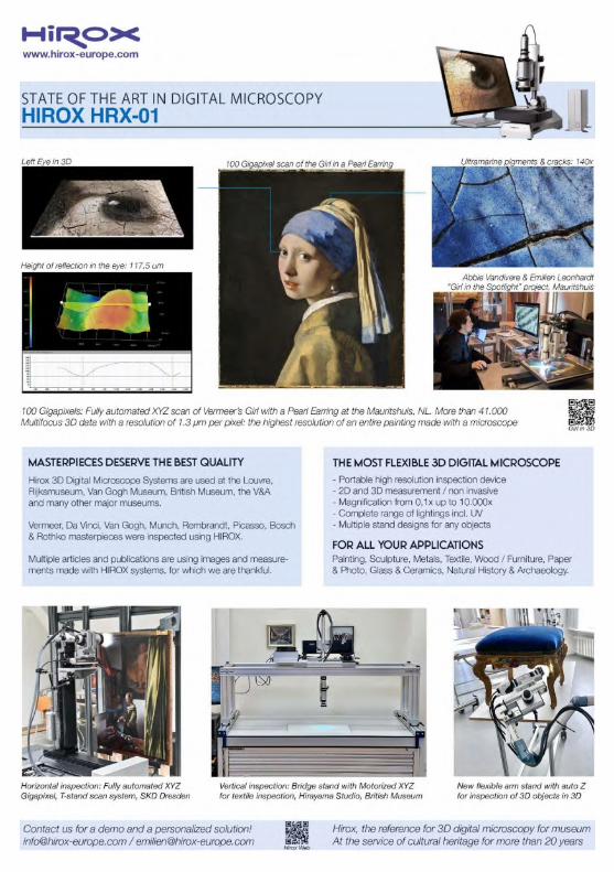
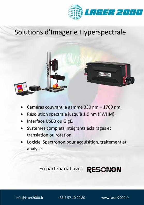
Solutions d’Imagerie Hyperspectrale
Caméras couvrant la gamme 330 nm – 1700 nm. Résolution spectrale jusqu’à 1.9 nm (FWHM). Interface USB3 ou GigE. Systèmes complets intégrants éclairages et
translation ou rotation. Logiciel Spectronon pour acquisition, traitement et
analyse.
En partenariat avec
[email protected] +33 5 57 10 92 80 www.laser2000.fr
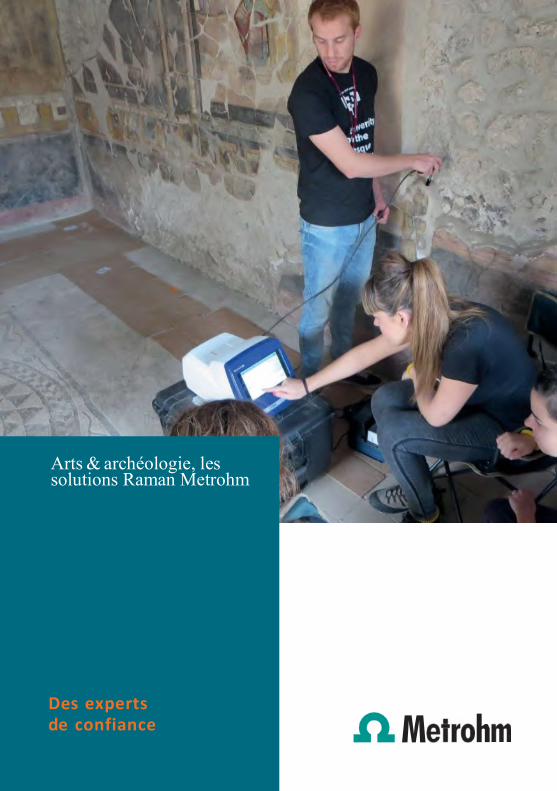
Arts & archéologie, les solutions Raman Metrohm
Des experts de confiance
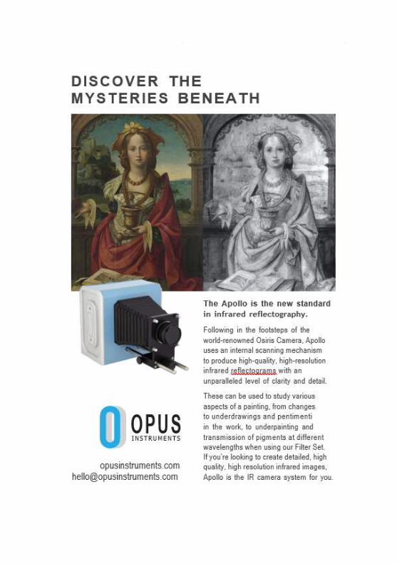

Become an author with Springer,
we welcome new book proposals.
XpectralTEK et XpeCAM
We propose the first Conservator’s and Art Researchers Cloud Base platform.
For Cultural Heritage needs, we have XpeCAM. Professionals need to know more about the surfaces they study and act to preserve. Because there is much more information hidden to our eyes!
Using XpeCAM X02 automatic multispectral camera, we can acquire millions of reflectance samples at one capture, in less than 15 minutes. To light up our artwork, we use LAMPA, a 3-in-1 UV+VIS+IR single body equipment that operates in parallel to camera needs. Upload this data to XpeCAM Platform, the Conservator’s professional workspace, cloud based A.I. plus machine learning structured.
Born in the world of imaging diagnostics, Xpectraltek is the result of several years of work, experience, and ambition for constant improvement. Adjusting the knowledge in spectroscopy, imaging and computers to the flaws and needs of the market, we were able to create several tools that help professionals in their daily work. Since 2014, we occupy a privileged position in the spectral imaging and computer vision market. We are offering complete and quality proven solutions to any user who wants to certify and document his work using imaging.
For more information, please contact us:
[email protected] or our representant in France [email protected]

dŚĞ�ŝŶ�ƌƚ�ϮϬϮϮ�ĐŽŶĨĞƌĞŶĐĞ�ƚŚĂŶŬƐ�ƚŚĞ�ƐƵƉƉŽƌƚ�ŽĨ͗
tĞ�ĂůƐŽ�ƚŚĂŶŬ�ŽƵƌ�ƐƉŽŶƐŽƌƐ�ĂŶĚ�ĞdžŚŝďŝƚŽƌƐ͗
�ƌƵŬĞƌ�KƉƟĐƐ �ƌƵŬĞƌ�EĂŶŽ
dƌŝŽƉƟĐƐ
Related Documents