51 Cystic Lung Disease on Computed Tomography

51 cystic lung disease on computed tomography
Aug 14, 2015
Welcome message from author
This document is posted to help you gain knowledge. Please leave a comment to let me know what you think about it! Share it to your friends and learn new things together.
Transcript
51 Cystic Lung Disease on Computed Tomography

CLINICAL IMAGAGINGAN ATLAS OF DIFFERENTIAL DAIGNOSIS
EISENBERG
DR. Muhammad Bin Zulfiqar PGR-FCPS III SIMS/SHL

• Fig C 51-1 Emphysema. There are scattered low-density cysts without clearly definable walls. Many appear to be aligned adjacent to peripheral vessels corresponding to lobular anatomy (straight arrows). Note that residual lobular vessels can still be identified within the center of some of these cysts (curved arrows).98
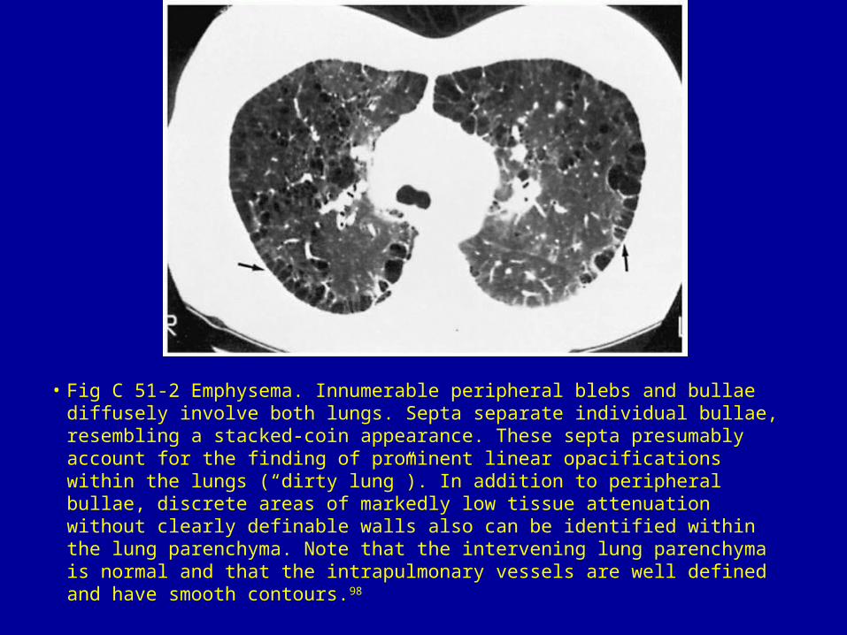
• Fig C 51-2 Emphysema. Innumerable peripheral blebs and bullae diffusely involve both lungs. Septa separate individual bullae, resembling a stacked-coin appearance. These septa presumably account for the finding of prominent linear opacifications within the lungs (“dirty lung”). In addition to peripheral bullae, discrete areas of markedly low tissue attenuation without clearly definable walls also can be identified within the lung parenchyma. Note that the intervening lung parenchyma is normal and that the intrapulmonary vessels are well defined and have smooth contours.98
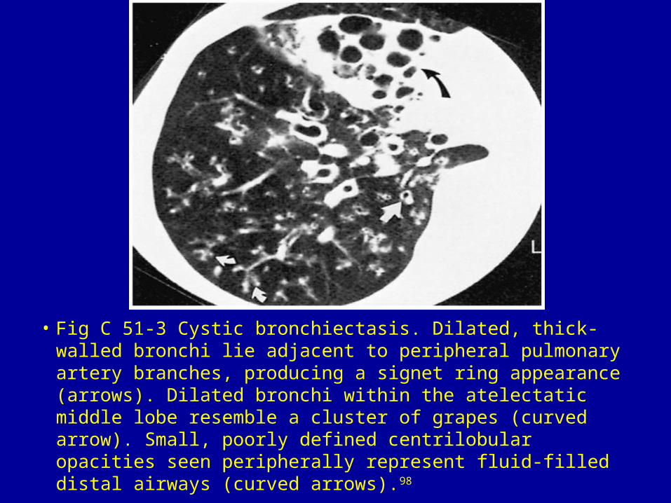
• Fig C 51-3 Cystic bronchiectasis. Dilated, thick-walled bronchi lie adjacent to peripheral pulmonary artery branches, producing a signet ring appearance (arrows). Dilated bronchi within the atelectatic middle lobe resemble a cluster of grapes (curved arrow). Small, poorly defined centrilobular opacities seen peripherally represent fluid-filled distal airways (curved arrows).98
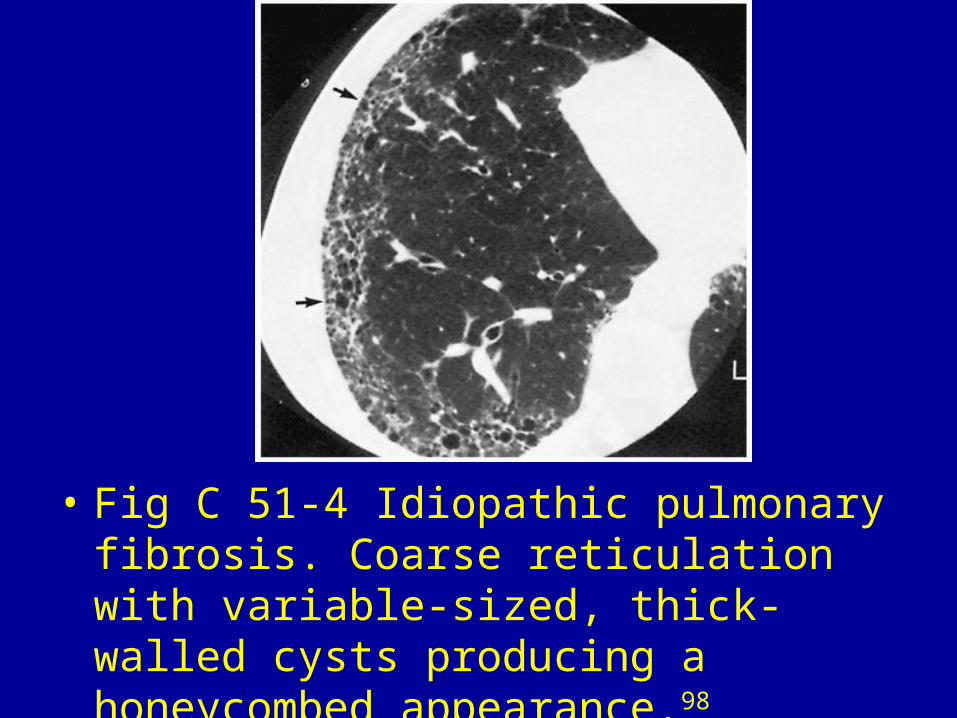
• Fig C 51-4 Idiopathic pulmonary fibrosis. Coarse reticulation with variable-sized, thick-walled cysts producing a honeycombed appearance.98
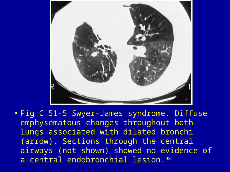
• Fig C 51-5 Swyer-James syndrome. Diffuse emphysematous changes throughout both lungs associated with dilated bronchi (arrow). Sections through the central airways (not shown) showed no evidence of a central endobronchial lesion.98
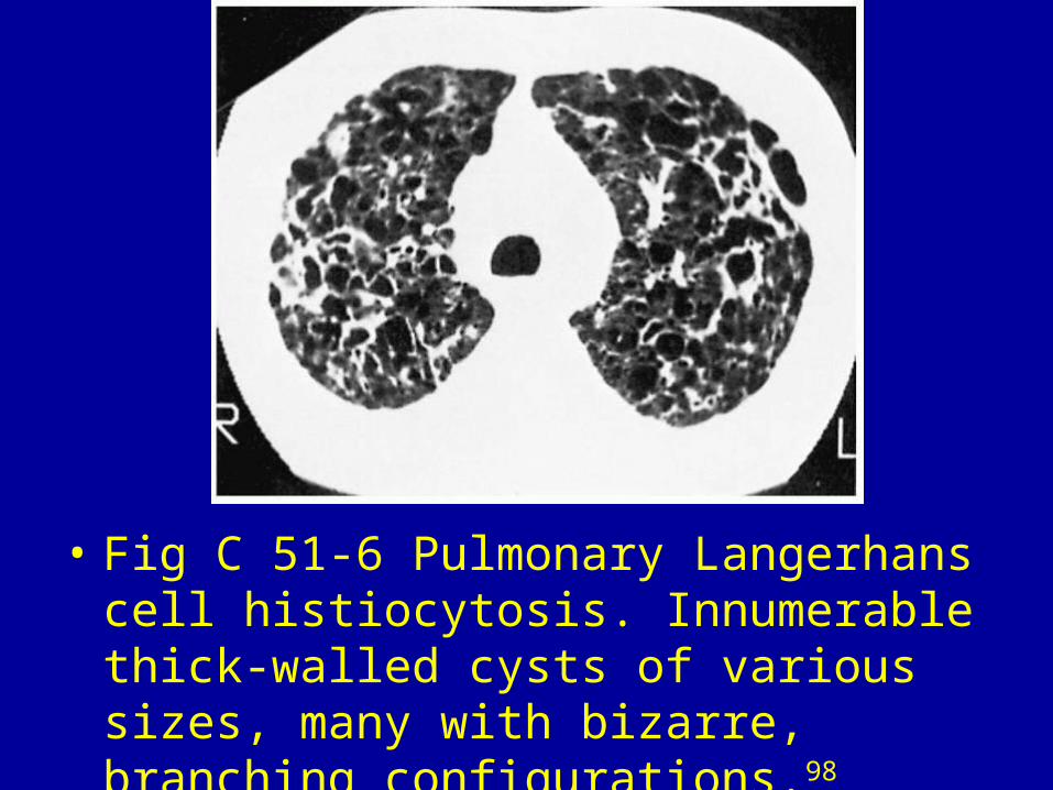
• Fig C 51-6 Pulmonary Langerhans cell histiocytosis. Innumerable thick-walled cysts of various sizes, many with bizarre, branching configurations.98
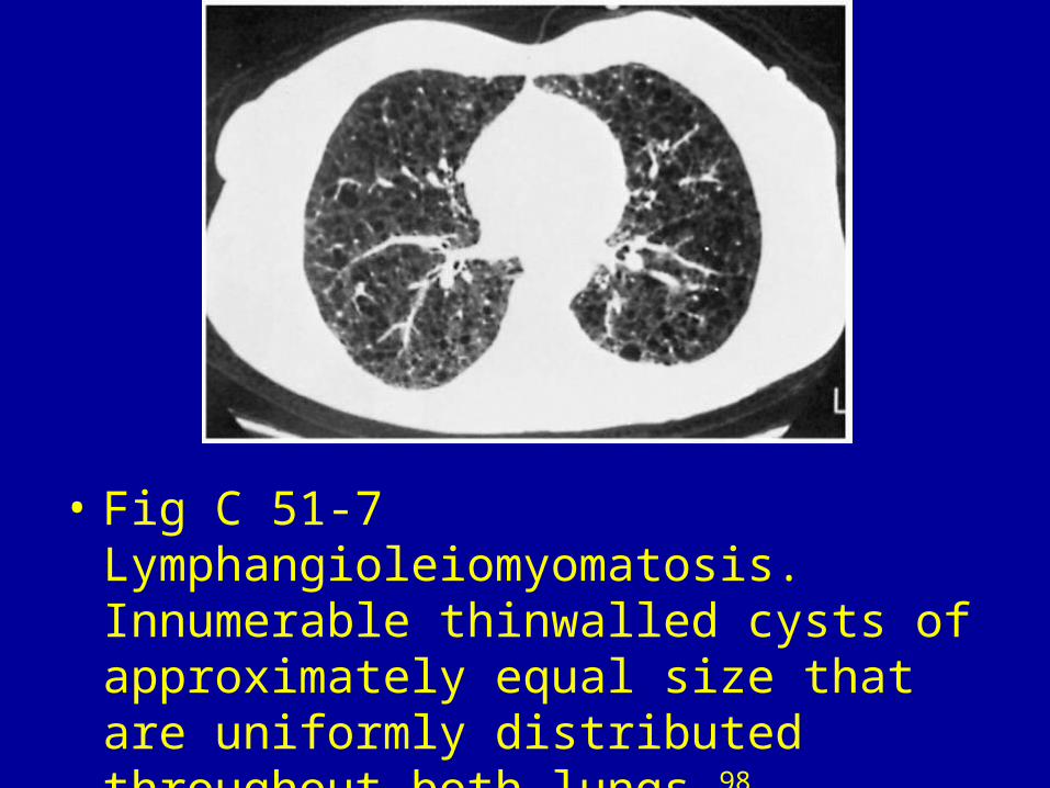
• Fig C 51-7 Lymphangioleiomyomatosis. Innumerable thinwalled cysts of approximately equal size that are uniformly distributed throughout both lungs.98

• Fig C 51-8 Pneumocystis carinii pneumonia. Discrete thin- and thickwalled cysts occurring in association with consolidated lung. Coalescence of cysts results in the formation of a few bizarreshaped cysts (arrows). Note that the intervening parenchyma appears grossly normal.98
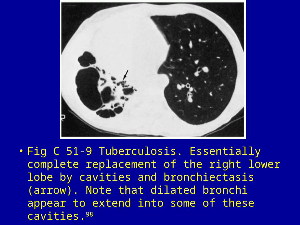
• Fig C 51-9 Tuberculosis. Essentially complete replacement of the right lower lobe by cavities and bronchiectasis (arrow). Note that dilated bronchi appear to extend into some of these cavities.98

• Fig C 51-10 Septic emboli. Scattered nodules in varying stages of cavitation in a patient with staphylococcal endocarditis. Many of the cavities are clearly related to adjacent vessels (arrows).98
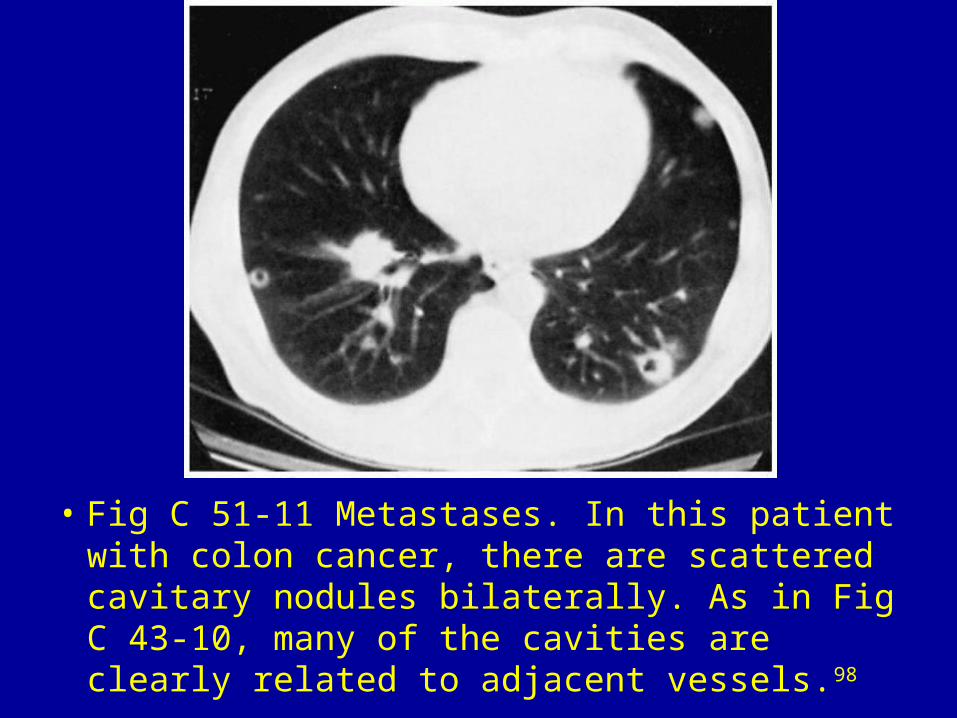
• Fig C 51-11 Metastases. In this patient with colon cancer, there are scattered cavitary nodules bilaterally. As in Fig C 43-10, many of the cavities are clearly related to adjacent vessels.98
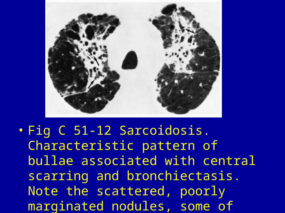
• Fig C 51-12 Sarcoidosis. Characteristic pattern of bullae associated with central scarring and bronchiectasis. Note the scattered, poorly marginated nodules, some of which appear to have a perivascular distribution.98


Related Documents