MIT OpenCourseWare http://ocw.mit.edu 5.069 Crystal Structure Analysis Spring 2008 For information about citing these materials or our Terms of Use, visit: http://ocw.mit.edu/terms.

Welcome message from author
This document is posted to help you gain knowledge. Please leave a comment to let me know what you think about it! Share it to your friends and learn new things together.
Transcript

MIT OpenCourseWare http://ocw.mit.edu
5.069 Crystal Structure AnalysisSpring 2008
For information about citing these materials or our Terms of Use, visit: http://ocw.mit.edu/terms.
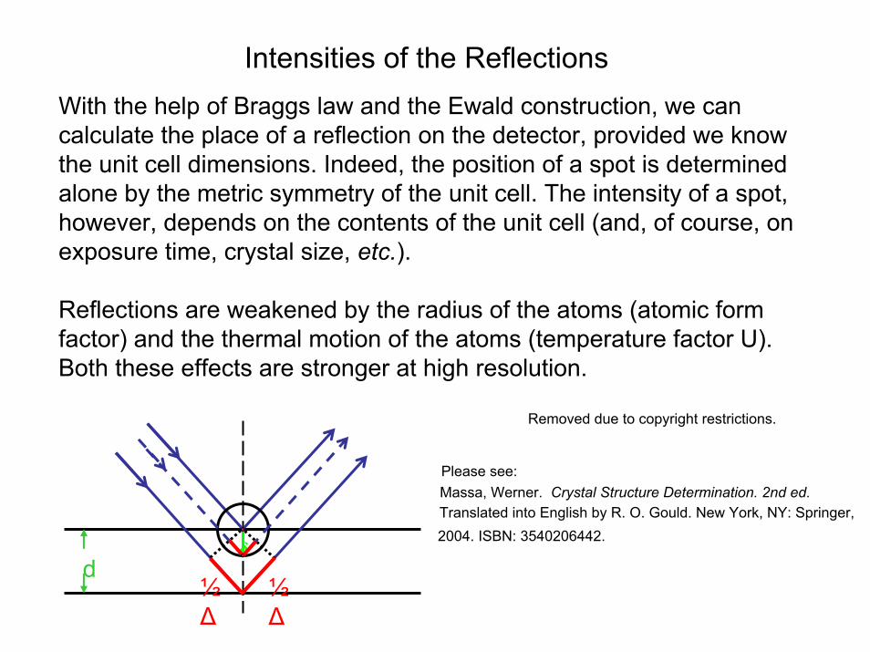
Intensities of the ReflectionsWith the help of Braggs law and the Ewald construction, we can calculate the place of a reflection on the detector, provided we know the unit cell dimensions. Indeed, the position of a spot is determined alone by the metric symmetry of the unit cell. The intensity of a spot, however, depends on the contents of the unit cell (and, of course, on exposure time, crystal size, etc.).
Reflections are weakened by the radius of the atoms (atomic formfactor) and the thermal motion of the atoms (temperature factor U). Both these effects are stronger at high resolution.
d½∆
½∆
δ
Removed due to copyright restrictions.
Please see:Massa, Werner. Crystal Structure Determination. 2nd ed.
Translated into English by R. O. Gould. New York, NY: Springer,2004. ISBN: 3540206442.
Structure Factors
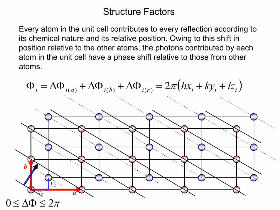
Every atom in the unit cell contributes to every reflection according to its chemical nature and its relative position. Owing to this shift in position relative to the other atoms, the photons contributed by each atom in the unit cell have a phase shift relative to those from other atoms.
a
b
y2x2
π20 ≤∆Φ≤
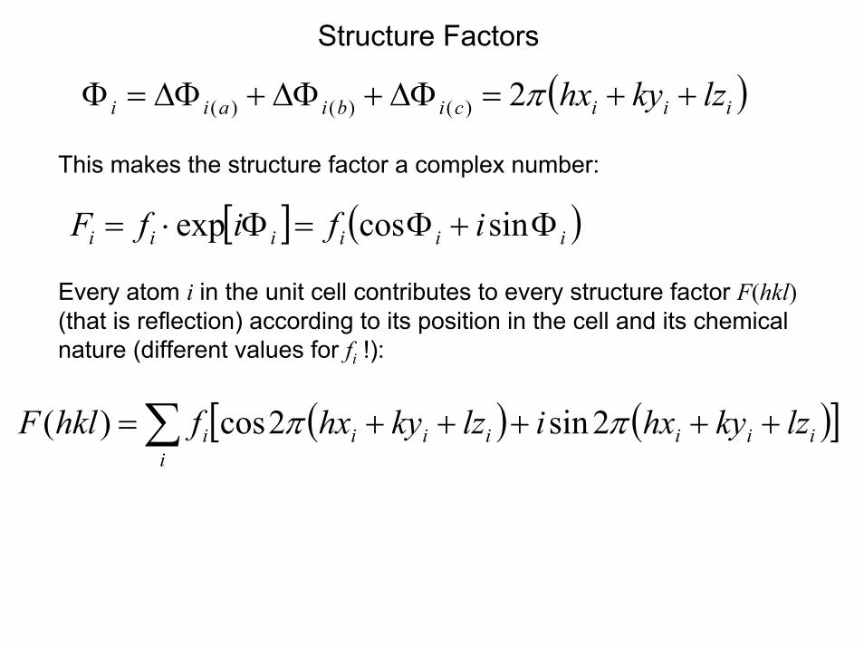
( )iiicibiaii lzkyhx ++=∆Φ+∆Φ+∆Φ=Φ π2)()()(
Structure Factors
( )iiicibiaii lzkyhx ++=∆Φ+∆Φ+∆Φ=Φ π2)()()(
This makes the structure factor a complex number:
[ ] ( )iiiiii ififF Φ+Φ=Φ⋅= sincosexp
Every atom i in the unit cell contributes to every structure factor F(hkl)(that is reflection) according to its position in the cell and its chemical nature (different values for fi !):
( ) ( )[ ]∑ +++++=i
iiiiiii lzkyhxilzkyhxfhklF ππ 2sin2cos)(
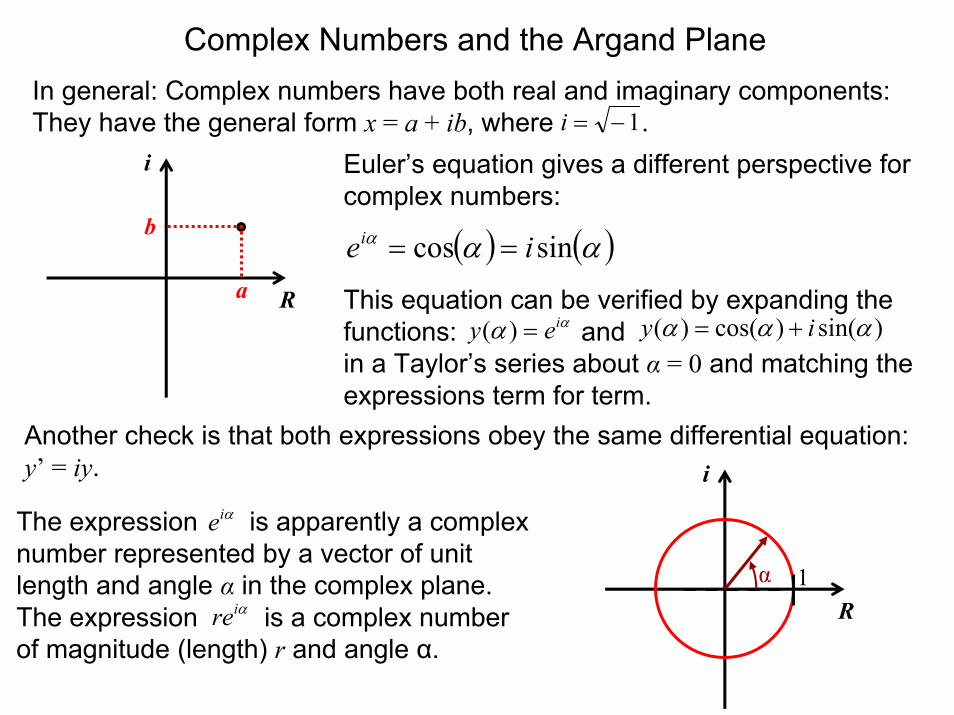
Complex Numbers and the Argand PlaneIn general: Complex numbers have both real and imaginary components:They have the general form x = a + ib, where .1−=i
i
b
a
Euler’s equation gives a different perspective for complex numbers:
( ) ( )ααα sincos iei ==
functions: andin a Taylor’s series about α = 0 and matching the expressions term for term.
αi ααα +=
Another check is that both expressions obey the same differential equation: y’ = iy.
R
i
α 1
The expression is apparently a complex number represented by a vector of unit length and angle α in the complex plane.The expression is a complex number of magnitude (length) r and angle α.
αie
αire
R This equation can be verified by expanding the α = )sin()cos()( iyey )(
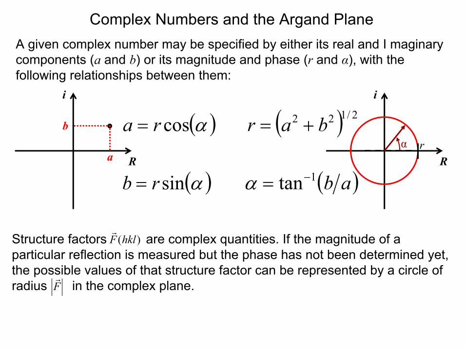
Complex Numbers and the Argand PlaneA given complex number may be specified by either its real and I maginarycomponents (a and b) or its magnitude and phase (r and α), with the following relationships between them:
R R
i
b
a
( )αcosra =
( )αsinrb =
i
α r( ) 2/122 bar +=
( )ab1tan−=α
Structure factors are complex quantities. If the magnitude of a particular reflection is measured but the phase has not been determined yet, the possible values of that structure factor can be represented by a circle of radius in the complex plane.
)(hklFr
Fr
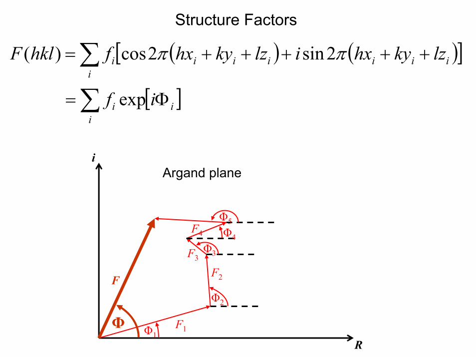
Structure Factors
( ) ( )[ ]∑ +++++=i
iiiiiii lzkyhxilzkyhxfhklF ππ 2sin2cos)(
[ ]∑ Φ=i
ii if exp
iArgand plane
RΦ1
Φ2
Φ3
Φ4
Φ5
F1
F2
F3
F4
F
Φ
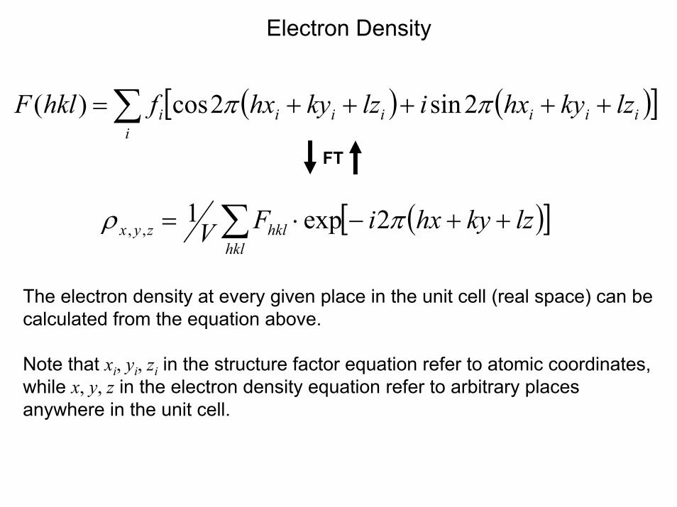
Electron Density
( ) ( )[ ]∑ +++++=i
iiiiiii lzkyhxilzkyhxfhklF ππ 2sin2cos)(
FT
( )[ ]∑ ++−⋅=hkl
hklzyx lzkyhxiFV πρ 2exp1,,
The electron density at every given place in the unit cell (real space) can be calculated from the equation above.
Note that xi, yi, zi in the structure factor equation refer to atomic coordinates, while x, y, z in the electron density equation refer to arbitrary places anywhere in the unit cell.
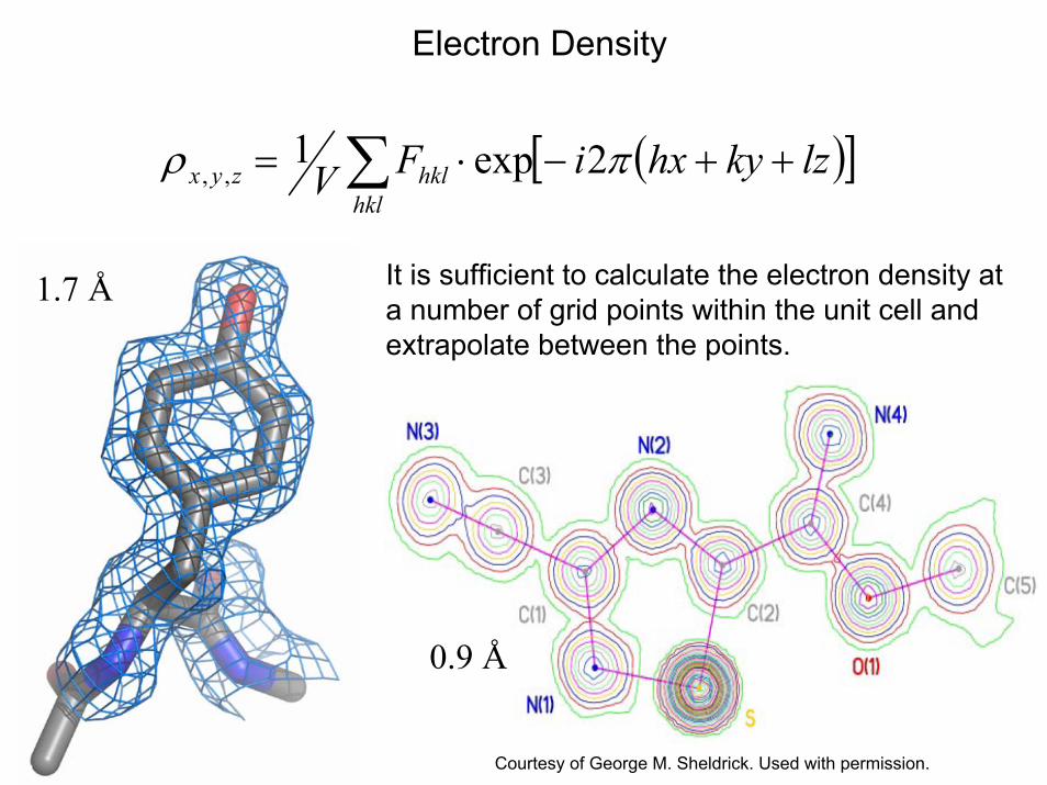
Electron Density
( )[ ]∑ ++−⋅=hkl
hklzyx lzkyhxiFV πρ 2exp1,,
It is sufficient to calculate the electron density at a number of grid points within the unit cell and extrapolate between the points.
0.9 Å
1.7 Å
Courtesy of George M. Sheldrick. Used with permission.
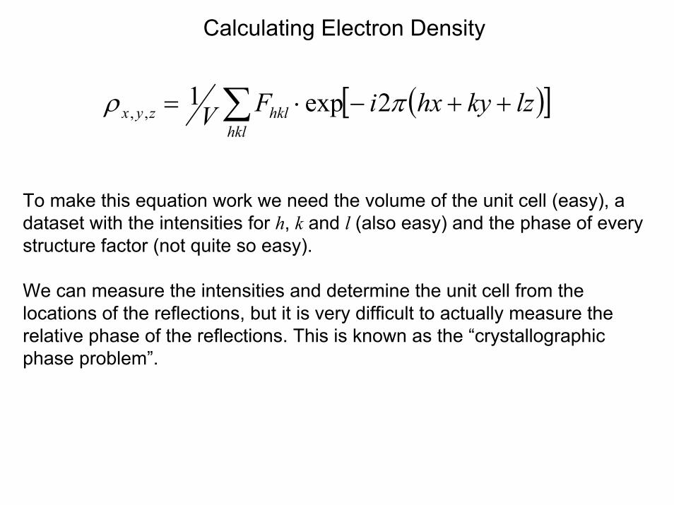
Calculating Electron Density
( )[ ]∑ ++−⋅=hkl
hklzyx lzkyhxiFV πρ 2exp1,,
To make this equation work we need the volume of the unit cell (easy), a dataset with the intensities for h, k and l (also easy) and the phase of every structure factor (not quite so easy).
We can measure the intensities and determine the unit cell from the locations of the reflections, but it is very difficult to actually measure the relative phase of the reflections. This is known as the “crystallographic phase problem”.
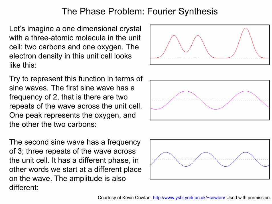
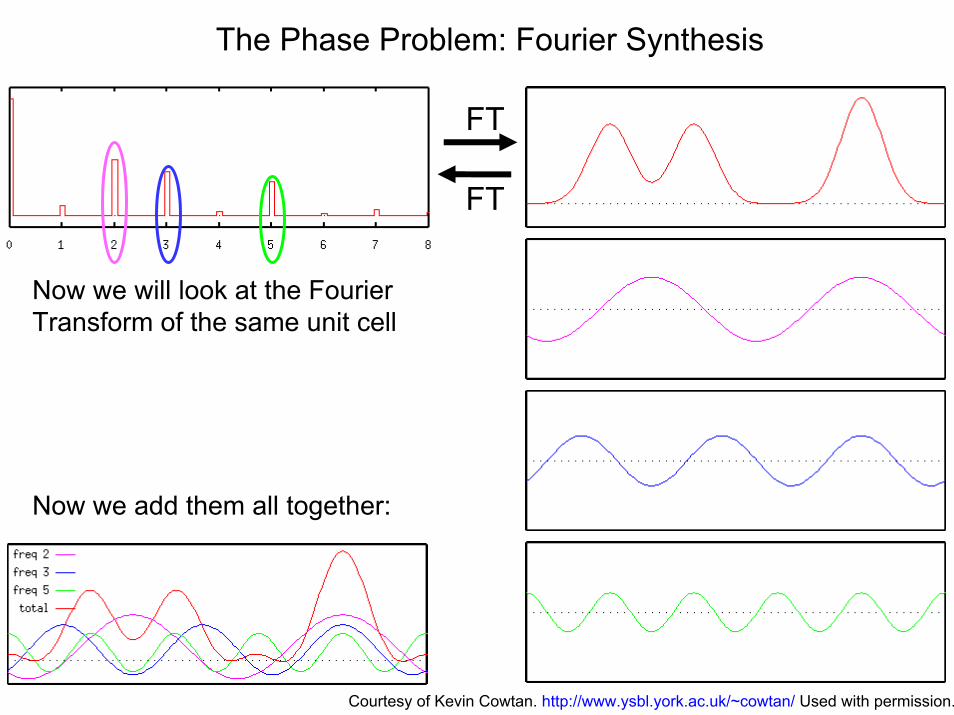
The Phase Problem: Fourier Synthesis
Let’s imagine a one dimensional crystal with a three-atomic molecule in the unit cell: two carbons and one oxygen. The electron density in this unit cell looks like this:
Try to represent this function in terms of sine waves. The first sine wave has a frequency of 2, that is there are two repeats of the wave across the unit cell. One peak represents the oxygen, and the other the two carbons:
The second sine wave has a frequency of 3; three repeats of the wave across the unit cell. It has a different phase, in other words we start at a different place on the wave. The amplitude is also different:
Courtesy of Kevin Cowtan. http://www.ysbl.york.ac.uk/~cowtan/ Used with permission.
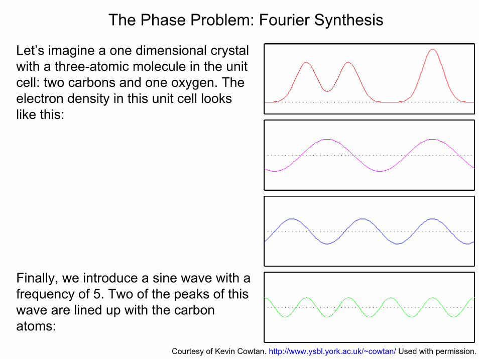
The Phase Problem: Fourier Synthesis
Let’s imagine a one dimensional crystal with a three-atomic molecule in the unit cell: two carbons and one oxygen. The electron density in this unit cell looks like this:
Finally, we introduce a sine wave with a frequency of 5. Two of the peaks of this wave are lined up with the carbon atoms:
Courtesy of Kevin Cowtan. http://www.ysbl.york.ac.uk/~cowtan/ Used with permission.
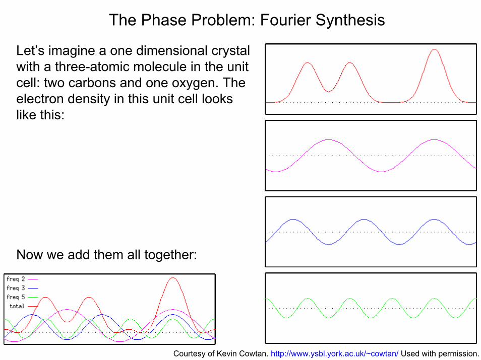
The Phase Problem: Fourier Synthesis
Let’s imagine a one dimensional crystal with a three-atomic molecule in the unit cell: two carbons and one oxygen. The electron density in this unit cell looks like this:
Now we add them all together:
Courtesy of Kevin Cowtan. http://www.ysbl.york.ac.uk/~cowtan/ Used with permission.
The Phase Problem: Fourier Synthesis
FT
FT
Now we will look at the Fourier Transform of the same unit cell
Now we add them all together:
Courtesy of Kevin Cowtan. http://www.ysbl.york.ac.uk/~cowtan/ Used with permission.
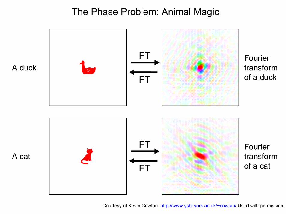
The Phase Problem: Animal Magic
FT
FT
Fourier transform of a duck
A duck
FT
FT
Fourier transform of a cat
A cat
Courtesy of Kevin Cowtan. http://www.ysbl.york.ac.uk/~cowtan/ Used with permission.
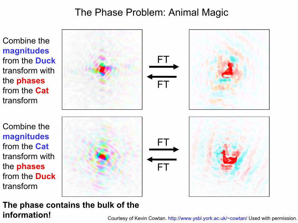
The Phase Problem: Animal Magic
FT
FT
Combine the magnitudesfrom the Ducktransform with the phasesfrom the Cattransform
Combine the magnitudesfrom the Cattransform with the phasesfrom the Ducktransform
FT
FT
The phase contains the bulk of the information! Courtesy of Kevin Cowtan. http://www.ysbl.york.ac.uk/~cowtan/ Used with permission.
Symmetry in Reciprocal Space
Before we can talk about how to solve the phase problem, we need to talk about symmetry in reciprocal space.
Symmetry in the crystal (real space) influences the symmetry in reciprocal space. A twofold in real space causes a twofold in reciprocal space, a mirror, causes a mirror, a fourfold a fourfold, etc.
The reciprocal space, however, is always centrosymmetric, and translational symmetry has no effect on the symmetry in reciprocal space. That means a P2, P21, P2/m, P21/m, P21/c, etc. all correspond to the same symmetry group in reciprocal space.
The symmetry group in reciprocal space is called Laue group. There are eleven of them.
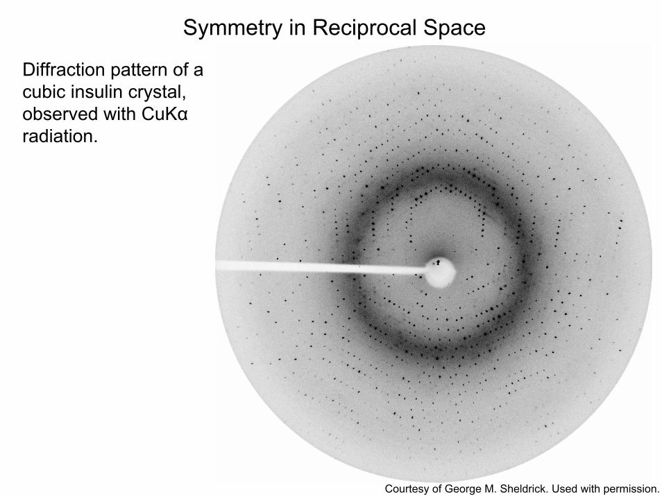
Symmetry in Reciprocal Space
Diffraction pattern of a cubic insulin crystal, observed with CuKαradiation.
Courtesy of George M. Sheldrick. Used with permission.
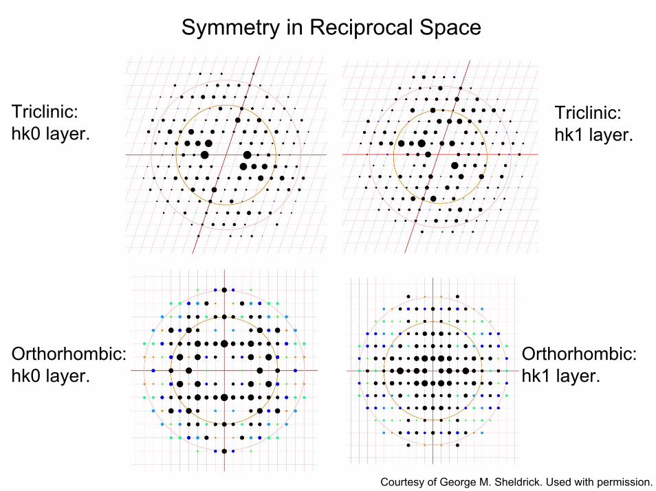
Symmetry in Reciprocal Space
Triclinic:hk0 layer.
Triclinic:hk1 layer.
Orthorhombic:hk1 layer.
Orthorhombic:hk0 layer.
Courtesy of George M. Sheldrick. Used with permission.
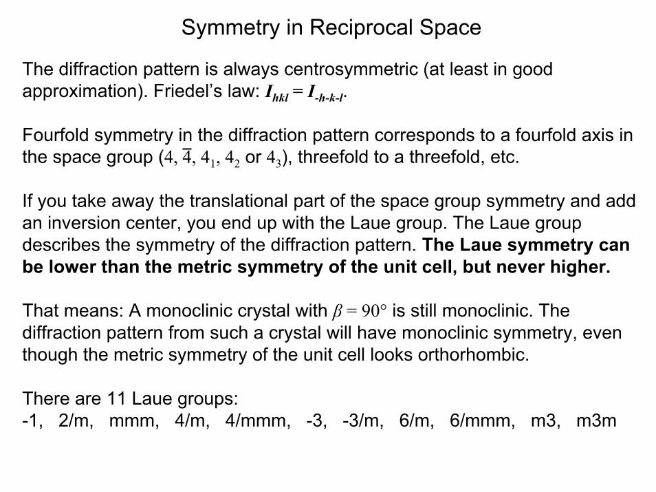
Symmetry in Reciprocal Space
The diffraction pattern is always centrosymmetric (at least in good approximation). Friedel’s law: Ihkl = I-h-k-l.
Fourfold symmetry in the diffraction pattern corresponds to a fourfold axis in the space group (4, 4, 41, 42 or 43), threefold to a threefold, etc.
If you take away the translational part of the space group symmetry and add an inversion center, you end up with the Laue group. The Laue group describes the symmetry of the diffraction pattern. The Laue symmetry can be lower than the metric symmetry of the unit cell, but never higher.
That means: A monoclinic crystal with β = 90° is still monoclinic. The diffraction pattern from such a crystal will have monoclinic symmetry, even though the metric symmetry of the unit cell looks orthorhombic.
There are 11 Laue groups: -1, 2/m, mmm, 4/m, 4/mmm, -3, -3/m, 6/m, 6/mmm, m3, m3m
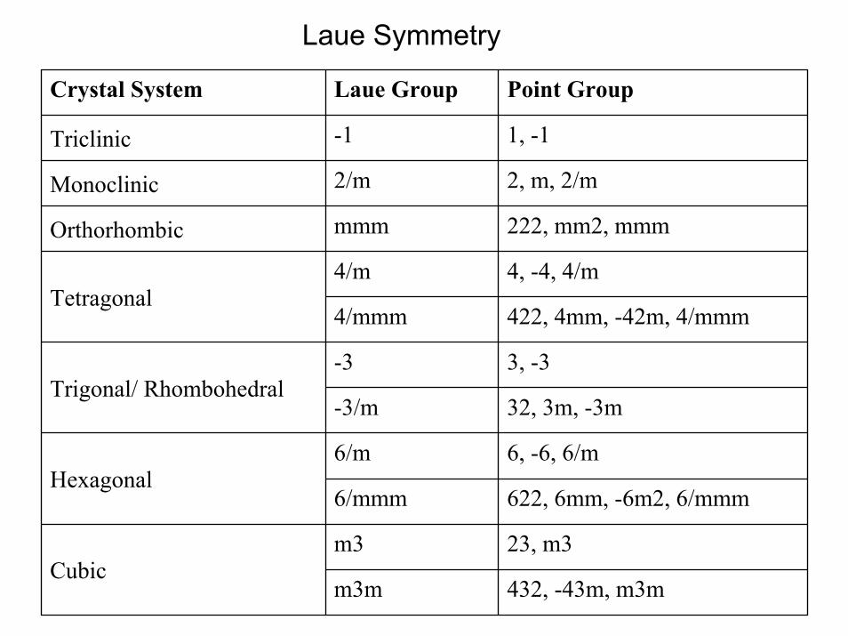
Laue Symmetry
432, -43m, m3mm3m
23, m3m3Cubic
622, 6mm, -6m2, 6/mmm6/mmm
6, -6, 6/m6/mHexagonal
32, 3m, -3m-3/m
3, -3-3Trigonal/ Rhombohedral
422, 4mm, -42m, 4/mmm4/mmm
4, -4, 4/m4/mTetragonal
222, mm2, mmmmmmOrthorhombic
2, m, 2/m2/mMonoclinic
1, -1-1Triclinic
Point GroupLaue GroupCrystal System
Space Group Determination
The first step in the determination of a crystal structure is the determination of the unit cell from the diffraction pattern.
Second step: Space group determination.
From the symmetry of the diffraction pattern, we can determine the Lauegroup, which narrows down the choice quite considerably. Usually the Lauegroup and the metric symmetry of the unit cell match.
The <| E2-1 |> statistics, can give us an idea, whether the space group is centrosymmetric or acentric. Even thought the diffraction pattern is always centrosymmetric, the intensity distribution across the reciprocal space is much more even for a centrosymmetric space group.
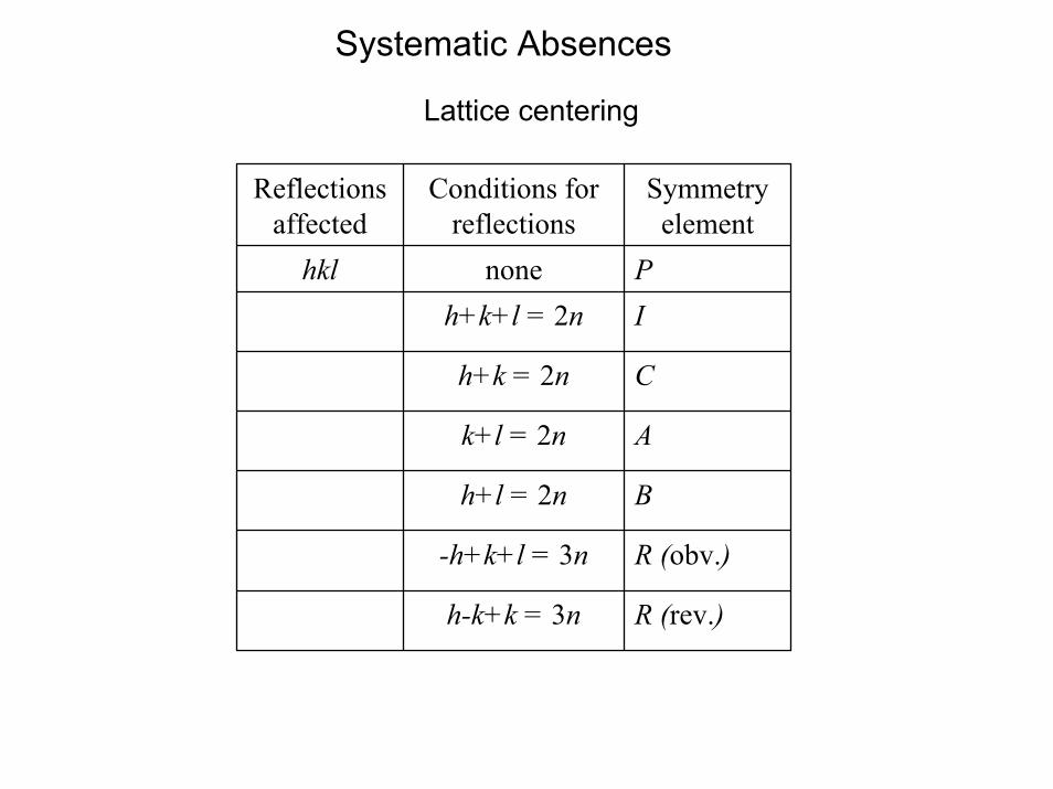
From systematic absences, we can determine the lattice type as well as screw axes and glide planes.
This is usually enough to narrow down the choice to a very short list.
E2-1 Statistics
We measure intensities II F2 F: structure factors
Normalized structure factors E:E2 = F2/<F2> <F2>: mean value for reflections at same resolution
<E2> = 1
< | E2-1 | > = 0.736 for non-centrosymmetric structures0.968 for centrosymmetric structures
Heavy atoms on special positions and twinning tend to lower this value. Pseudo translational symmetry tend to increase this value.
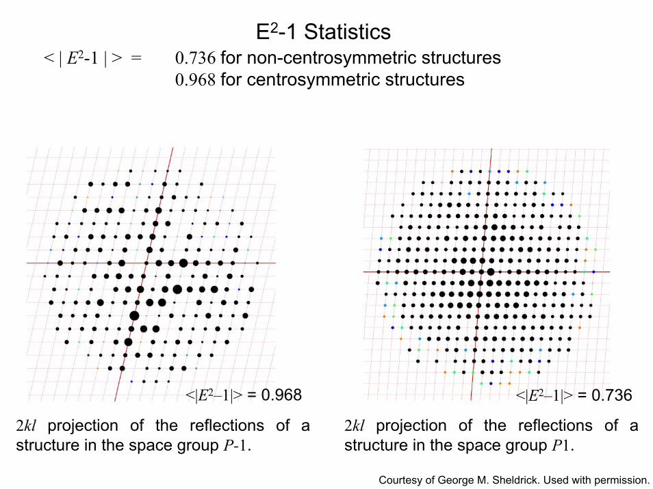
E2-1 Statistics< | E2-1 | > = 0.736 for non-centrosymmetric structures
0.968 for centrosymmetric structures
<|E2–1|> = 0.968 <|E2–1|> = 0.736
2kl projection of the reflections of a structure in the space group P-1.
2kl projection of the reflections of a structure in the space group P1.
Courtesy of George M. Sheldrick. Used with permission.
Systematic Absences
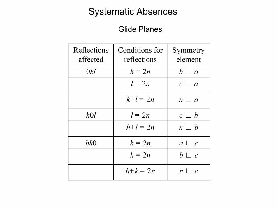
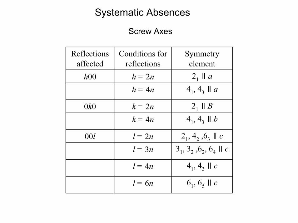
Lattice centering and symmetry elements with translation (glide planes and screw axes) cause certain reflections to have zero intensity in the diffraction pattern. If, e.g., all reflections 0, k, 0 with odd values for k are absent, we know that we have a 21 axis along b.Other example: if all reflections h, 0, l with odd values for l are absent, we have a c glide plane perpendicular to b.
How come?
Systematic Absences
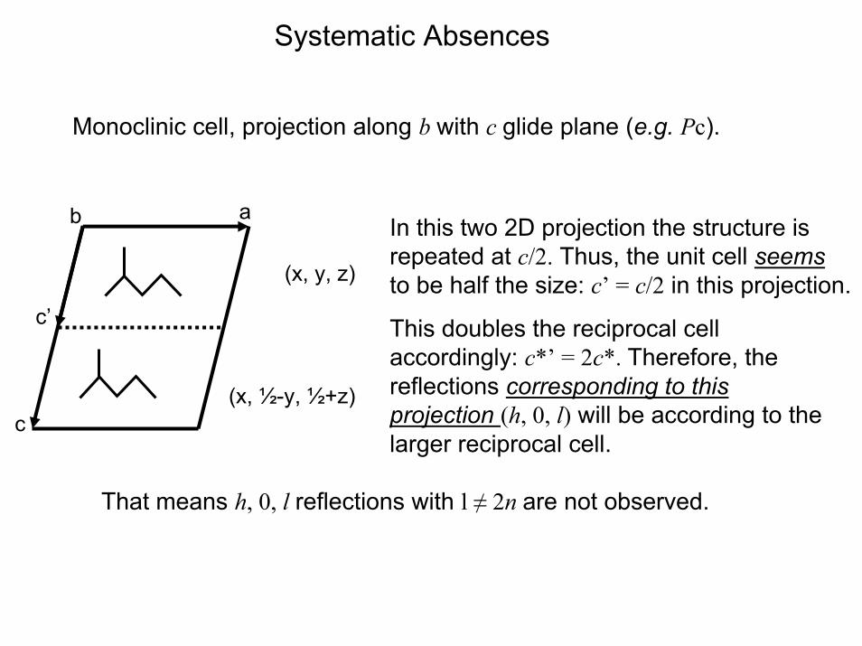
Monoclinic cell, projection along b with c glide plane (e.g. Pc).
b
(x, ½-y, ½+z)
c’
aIn this two 2D projection the structure is repeated at c/2. Thus, the unit cell seemsto be half the size: c’ = c/2 in this projection. (x, y, z)
This doubles the reciprocal cell accordingly: c*’ = 2c*. Therefore, the reflections corresponding to this projection (h, 0, l) will be according to the larger reciprocal cell.
c
That means h, 0, l reflections with l ≠ 2n are not observed.
Systematic Absences
Lattice centering
R (rev.)h-k+k = 3n
R (obv.)-h+k+l = 3n
Bh+l = 2n
Ak+l = 2n
Ch+k = 2n
Ih+k+l = 2nPnonehkl
Symmetryelement
Conditions for reflections
Reflectionsaffected
Systematic Absences
Glide Planes
n ∟ ch+k = 2n
b ∟ ck = 2na ∟ ch = 2nhk0
n ∟ bh+l = 2nc ∟ bl = 2nh0l
n ∟ ak+l = 2n
c ∟ al = 2nb ∟ ak = 2n0kl
Symmetryelement
Conditions for reflections
Reflectionsaffected
Systematic Absences
Screw Axes
61, 65 ॥ cl = 6n
41, 43 ॥ cl = 4n
31, 32 ,62, 64 ॥ cl = 3n21, 42 ,63 ॥ cl = 2n00l
41, 43 ॥ bk = 4n21 ॥ Bk = 2n0k0
41, 43 ॥ ah = 4n21 ॥ ah = 2nh00
Symmetryelement
Conditions for reflections
Reflectionsaffected
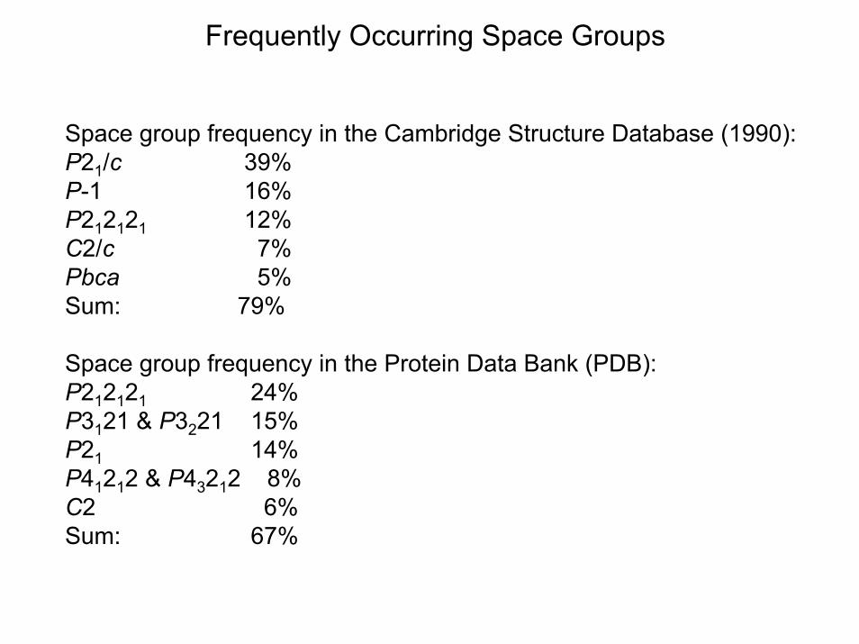
Frequently Occurring Space Groups
Space group frequency in the Cambridge Structure Database (1990):P21/c 39% P-1 16% P212121 12% C2/c 7%Pbca 5%Sum: 79%
Space group frequency in the Protein Data Bank (PDB):P212121 24%P3121 & P3221 15%P21 14%P41212 & P43212 8%C2 6%Sum: 67%
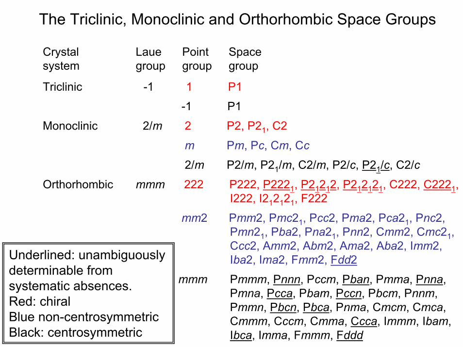
The Triclinic, Monoclinic and Orthorhombic Space Groups
Crystal Laue Point Spacesystem group group group
Triclinic -1 1 P1
-1 P1
Monoclinic 2/m 2 P2, P21, C2
2/m P2/m, P21/m, C2/m, P2/c, P21/c, C2/c
Orthorhombic mmm 222 P222, P2221, P21212, P212121, C222, C2221, I222, I212121, F222
mm2 Pmm2, Pmc21, Pcc2, Pma2, Pca21, Pnc2,Pmn21, Pba2, Pna21, Pnn2, Cmm2, Cmc21,Ccc2, Amm2, Abm2, Ama2, Aba2, Imm2,Iba2, Ima2, Fmm2, Fdd2
mmm Pmmm, Pnnn, Pccm, Pban, Pmma, Pnna,Pmna, Pcca, Pbam, Pccn, Pbcm, Pnnm,Pmmn, Pbcn, Pbca, Pnma, Cmcm, Cmca,Cmmm, Cccm, Cmma, Ccca, Immm, Ibam,Ibca, Imma, Fmmm, Fddd
Underlined: unambiguously determinable from systematic absences. Red: chiralBlue non-centrosymmetricBlack: centrosymmetric
m Pm, Pc, Cm, Cc
Related Documents