227 5. Molecularbiology and In-situ-Hybridization 5.1. Fluorescence-In-situ-Hybridization (FISH) FISH is a molecularbiological method to detect chromosomal abberrations, such as deletions, translocations, inversions, insertions, amplifications and aneuploidies. Therefore, it is a major tool in cytogenetic, tumor diagnosis and human chromosomal research. Artificially produced, fluorescence labeled DNA probes bind by complementary base pairing to specific target DNA sequences in situ. This is called hybridization. ProTaqs ® jellyFISH probes are used to visualize genomic DNA in formalin-fixed paraffin-embedded sections, in living cells, in metaphase chromosome spreads and interphase nuclei by fluorescence microscopy. ▶ A: Fluorescence labeling of DNA probe ▶ B: Target DNA sequence ▶ C: Denaturation of DNA double strands (probe DNA and target DNA) ▶ D: In-situ-Hybidization ▶ E: Fluorescence signal in sample seen in fluorescence microscopy A C B D E E Scheme of FISH: ▶ Most proximal marker covered by DNA probe ▶ Size and color of fluorescence signal of DNA probe ▶ Target gene detected by DNA probe ▶ Most distal marker covered by DNA probe ▶ Chromosom - Centromere ▶ Chromosom - Telomere ▶ Chromosom - Long arm ▶ Chromosom - Short arm ▶ Chromosom - Telomere Scheme of probe map:

Welcome message from author
This document is posted to help you gain knowledge. Please leave a comment to let me know what you think about it! Share it to your friends and learn new things together.
Transcript
227
5. Molecularbiology and In-situ-Hybridization
5.1. Fluorescence-In-situ-Hybridization (FISH)FISH is a molecularbiological method to detect chromosomal abberrations, such as deletions, translocations, inversions, insertions, amplifications and aneuploidies. Therefore, it is a major tool in cytogenetic, tumor diagnosis and human chromosomal research.
Artificially produced, fluorescence labeled DNA probes bind by complementary base pairing to specific target DNA sequences in situ. This is called hybridization. ProTaqs® jellyFISH probes are used to visualize genomic DNA in formalin-fixed paraffin-embedded sections, in living cells, in metaphase chromosome spreads and interphase nuclei by fluorescence microscopy.
▶ A: Fluorescence labeling of DNA probe ▶ B: Target DNA sequence ▶ C: Denaturation of DNA double strands
(probe DNA and target DNA) ▶ D: In-situ-Hybidization ▶ E: Fluorescence signal in sample seen in
fluorescence microscopy
A
C
B
D E
E
Scheme of FISH:
▶ Most proximal marker covered by DNA probe
▶ Size and color of fluorescence signal of DNA probe
▶ Target gene detected by DNA probe
▶ Most distal marker covered by DNA probe
▶ Chromosom - Centromere
▶ Chromosom - Telomere
▶ Chromosom - Long arm
▶ Chromosom - Short arm
▶ Chromosom - Telomere
Scheme of probe map:

228
5.1.1. ProTaqs® jellyFISH ProbesThe detection of genetic abberrations with ProTaqs® jellyFISH technology is a powerful aid to guide more confident diagnoses and treatment decisions. quartett‘s expanding product portfolio includes a variety of direct labeled DNA probes for oncology to detect solid tumors and hematologic disorders:
▶ Bi-Color probes (Orange & green signal) ▶ Tri-Color probes (Orange, green and blue signal)
The following fluorophores are used:
Cat.-No. Product Name Color Signals Spezification Indication Probe Map Application Quantity
FISH011-37FISH011-38
ProTaqs® jellyFISH ALK Probe
Bi-Color Break-apart probe, orange labeled probe hybridizes to the proximal region within ALK in 2p23, green labeled probe hybridizes distal to ALK.
Lung carcinoma (NSCLC), B cell lymphoma, Neuroblastoma, Myofibroblastic tumors
FISH 80 µl160 µl
FISH012-37FISH012-38
ProTaqs® jellyFISH ROS1 Probe
Bi-Color Break apart probe, orange labeled probe hybridizes proximal to the ROS1 gene at 6q22, green labeled probe hybridizes to the distal region of ROS1; also suitable to analyze the FIG-ROS1 fusion caused by an interstitial 240 kb deletion.
Lung carcinoma (NSCLC)
FISH 80 µl160 µl
FISH013-37FISH013-38
ProTaqs® jellyFISH RET Probe
Bi-Color Break apart probe, orange labeled part hybridizes proximal to the RET gene at 10q11, green labeled probe hybridizes to the distal region of RET.
Lung carcinoma (NSCLC), Thyroid carcinoma
FISH 80 µl160 µl
Fluorophor Absorption max. [nm] Emission max. [nm]
Blue 426 480Green 505 530
Orange 552 576
Troubleshooting
Problem Potential Cause(s) Recommended Solution
No FISH signals can be detected in the microscope.
• Reflected light shutter closed / stop slider in light path.
• Fluorescent lamp is off. • Wrong fluorescence filter in light path. • Objective out of position. • Phototube in camera position.
• Open shutter / move stop slider out of the light path.
• Switch on fluorescent lamp. • Move correct filter into light path. • Swing objective into light path. • Direct light path to eyepieces.
Hybridization signals become weak after a while.
• Immersion oil soaked in-between slide and coverslip.
• Replace coverslip and DAPI/antifade. Use 24 x 32 mm² coverslip even if only a small region is hybridized.
Diffuse signals. • Preparation not adequately illuminated.
• Focus plane cannot be adjusted properly.
• Antifade layer is too thick for focusing.
• Check optical pathway of microscope. Adjust the UV light properly. Check the lifetime of the UV lamp.
• Use enough immersion oil. Do not mix different immersion oils. Use immersion oil suitable for fluorescence.
• Do not use too much DAPI/antifade. 10 µl per slide (24 mm x 32 mm coverslip) are sufficient.
Weak signals. • Chromosome slide preparation too old. Denaturation of chromosomes not adequate.
• Slides should not be older than two weeks. • Aging, baking or further fixation may inhibit the
hybridization and is not recommended.
High diffuse background in green color channel.
• pH value of washing solutions to low. DAPI intensity is too high resulting in crosstalk to FITC filter.
• Ensure that pH value is between 7.0 and 7.5 of solutions. FITC fluorophores are sensitive to pH below 7.
• Reduce DAPI/antifade concentration. If the recommended measures do not solve the problem, or your problem is not listed, please contact MetaSystems.
Customer Support
Please contact MetaSystems GmbH in Germany or MetaSystems Group, Inc. in the USA by telephone or e-mail (contact details, see below). MetaSystems disclaims any proprietary interest in the marks and names of others.
M MetaSystems GmbH Representative USA MetaSystems Group, Inc.
Robert-Bosch-Str. 6 70 Bridge Street, Suite 100
D-68804 Altlussheim 02458 Newton, MA
Germany United States
Tel.: +49 (0)6205 39610 Tel.: (888)489-9959 (toll free)
Fax: +49 (0)6205 32270 Fax: +1 617 924-9954
email: [email protected] email: [email protected]
URL: www.metasystems.de URL: www.metasystems.org
Revision: Rev H 130814
Symbols Used
Symbol Symbol Description
ASR Analyte -specific reagent
Analytical and performance characteristics are not established.
h Reference no
s Maximum storage temperature
M Manufacturer g Lot number
X No of tests
Y All warnings are marked by warning triangle with exclamation mark. Depending on their character they are supplemented with the words ATTENTION or CAUTION
H Expiry date
The XT ROS1-GOPC probe is designed as a break apart probe. Its green labeled part hybridizes proximal to the ROS1 gene at 6q22, the orange labeled probe hybridizes to the distal region of ROS1. This probe is also suited to analyze the FIG-ROS1 fusion, caused by an interstitial deletion of 240 kb in size. This probe is intended for Tissue-FISH applications and has been optimized for combined use with MetaSystems‘ pretreatment kit. Analytical and performance characteristics are not established. Probe Diagram:
V 140307 Chromosome 6
Meta
Sys
tem
s G
mb
H
Ro
bert
-Bo
sch-S
tr.
6
D-6
88
04
Altl
uss
heim
T
el.:
+4
9-6
20
5-3
96
10
to
xic
toxi
que
conta
ins:
Form
am
ide
R 6
1:
May
cause
harm
to t
he
unb
orn
child
. S
45:
In c
ase
of acc
iden
t or
if yo
u fee
l unw
ell,
se
ek
med
ical a
dvi
ce im
med
iatly
(sh
ow
the la
bel
where
poss
ible
).
S 5
3:
Avo
id e
xposu
re -
-- o
bta
in s
peci
al i
nst
ructio
n
befo
re u
se.
Only
for
indust
ry c
onsu
mers
. contie
nt:
Form
amid
e R
61:
Ris
que p
endant la
gro
ssess
e d
'effe
ts
néfa
stes
pour
l'enfa
nt.
S 4
5:
En c
as
d'a
ccid
ent
ou d
e m
ala
ise, consu
lter
imm
édia
tem
ent un m
éd
ecin
(si
poss
ible
, lu
i montr
er
l'étiq
uett
e).
S 5
3:
Évi
ter
l'exp
osi
tion -
-- s
e p
rocu
rer
des
inst
ructio
ns
spécia
les
ava
nt
l'util
isatio
n.
Uniq
uem
ent
pour
les
util
isate
ur s
pro
fess
ionnels
.
AS
R
s-1
8°C
g X
XX
XX
H X
XX
X-X
X
XT R
OS
1-G
OP
C B
A B
reak Ap
art Pro
be
100µl ( X
10
)
h D
-602
9-1
00
-OG
M
For Professional Use Only
Further information available at www.metasystems.de
Product Label Order No. Pack Size
XT ROS1-GOPC BA orange/green D-6029-100-OG 100µl jel
ly FISHby ProTaqs®

229
Cat.-No. Product Name Color Signals Spezification Indication Probe Map Application Quantity
FISH014-37FISH014-38
ProTaqs® jellyFISH EGFR Probe
Bi-Color Detects amplifications in the short arm of chromosome 7, orange labeled probe hybridizes to the EGFR locus at 7p11, green labeled probe hybridizes to the 7cen region.
Carcinoma in head and neck, brain, bladder, stomach, breast, lung, endometri-um, cervix, ovar, esophagus
FISH 80 µl160 µl
FISH015-37FISH015-38
ProTaqs® jellyFISH ERG-TMPRSS2 Probe
Tri-Color Detects deletions by building a fusion product between ERG and TMPRSS2, detects translocations involving the ERG locus in 21q22, green labeled probe detects a region proximal to the ERG locus, orange labeled probe hybridizes specifically to the distal region adjacent to the ERG gene, blue labeled probe detects a region distal to the TMPRSS2 region which can be fused to ERG by a deletion of sequences between ERG and TMPRSS2.
Prostata carcinoma
FISH 80 µl160 µl
FISH016-37FISH016-38
ProTaqs® jellyFISH PTEN/GRID1 Probe
Tri-Color Locus-specific probe detects deletions in 10q23, PTEN specific probe is labeled in orange, green labeled probe functions as a reference hybridizing to the GRID1 locus, centromere of chromosome 10 is detected by a blue labeled probe.
Glioma, carcinoma of prostate and breast
FISH 80 µl160 µl
FISH017-37FISH017-38
ProTaqs® jellyFISH MYC Probe
Bi-Color Detects amplifications in the long arm of chromosome 8, orange labeled probe hybridizes to the MYC locus at 8q24, green labeled probe hybridizes to the 8cen region and functions as a control probe.
Carcinoma of breast, lung, esophagus, bladder, cervix; leukemia (CLL)
FISH 80 µl160 µl
FISH018-37FISH018-38
ProTaqs® jellyFISH Her2/Neu Probe
Bi-Color Detects amplifications in the long arm of chromosome 17, orange labeled probe hybridizes to the Her2/Neu (ERBB2) locus at 17q12, green labeled probe hybridizes to the 17cen region and functions as a control probe.
Carcinoma of breast, prostate, lung, colon, ovar
FISH 80 µl160 µl
Troubleshooting
Problem Potential Cause(s) Recommended Solution
No FISH signals can be detected in the microscope.
• Reflected light shutter closed / stop slider in light path.
• Fluorescent lamp is off. • Wrong fluorescence filter in light path. • Objective out of position. • Phototube in camera position.
• Open shutter / move stop slider out of the light path.
• Switch on fluorescent lamp. • Move correct filter into light path. • Swing objective into light path. • Direct light path to eyepieces.
Hybridization signals become weak after a while.
• Immersion oil soaked in-between slide and coverslip.
• Replace coverslip and DAPI/antifade. Use 24 x 32 mm² coverslip even if only a small region is hybridized.
Diffuse signals. • Preparation not adequately illuminated.
• Focus plane cannot be adjusted properly.
• Antifade layer is too thick for focusing.
• Check optical pathway of microscope. Adjust the UV light properly. Check the lifetime of the UV lamp.
• Use enough immersion oil. Do not mix different immersion oils. Use immersion oil suitable for fluorescence.
• Do not use too much DAPI/antifade. 10 µl per slide (24 mm x 32 mm coverslip) are sufficient.
Weak signals. • Chromosome slide preparation too old. Denaturation of chromosomes not adequate.
• Slides should not be older than two weeks. • Aging, baking or further fixation may inhibit the
hybridization and is not recommended.
High diffuse background in green color channel.
• pH value of washing solutions to low. DAPI intensity is too high resulting in crosstalk to FITC filter.
• Ensure that pH value is between 7.0 and 7.5 of solutions. FITC fluorophores are sensitive to pH below 7.
• Reduce DAPI/antifade concentration. If the recommended measures do not solve the problem, or your problem is not listed, please contact MetaSystems.
Customer Support
Please contact MetaSystems GmbH in Germany or MetaSystems Group, Inc. in the USA by telephone or e-mail (contact details, see below). MetaSystems disclaims any proprietary interest in the marks and names of others.
M MetaSystems GmbH RepresentativeRepresentativeRepresentativeRepresentative USAUSAUSAUSA MetaSystems Group, Inc.
Robert-Bosch-Str. 6 70 Bridge Street, Suite 100
D-68804 Altlussheim 02458 Newton, MA
Germany United States
Tel.: +49 (0)6205 39610 Tel.: (888)489-9959 (toll free)
Fax: +49 (0)6205 32270 Fax: +1 617 924-9954
email: [email protected] email: [email protected]
URL: www.metasystems.de URL: www.metasystems.org
RevisionRevisionRevisionRevision: Rev : Rev : Rev : Rev HHHH 130130130130814814814814
Symbols Used
Symbol Symbol Description
ASRASRASRASR Analyte -specific reagent
Analytical and performance characteristics are not established.
hhhh Reference no
ssss Maximum storage temperature
MMMM Manufacturer gggg Lot number
XXXX No of tests
YYYY All warnings are marked by warning triangle with exclamation mark. Depending on their character they are supplemented with the words ATTENTION or CAUTION
HHHH Expiry date
The XT ERG-TMPRSS2 probe detects deletions and translocations involving the ERG locus in 21q22. The green labeled probe detects a region proximal to the ERG locus, an orange labeled probe hybridizes specifically to the distal region adjacent to the ERG gene. A blue (aqua) labeled probe detects a region distal to the TMPRSS2 region which can be fused to ERG by a deletion of sequences between ERG and TMPRSS2. This probe is intended for Tissue-FISH applications and has been optimized for combined use with MetaSystems‘ pretreatment kit. Analytical and performance characteristics are not established. Probe Diagram:
D21S1917D21S1917D21S1917D21S1917
ERGERGERGERG
400 kb
21q22
DSCR4DSCR4DSCR4DSCR4
TMPRSS2TMPRSS2TMPRSS2TMPRSS2
Gap ~2 Mb
360 kb
340 kb
D21S228D21S228D21S228D21S228
D21S1820D21S1820D21S1820D21S1820 not not not not totototo scalescalescalescale
V 130110 Chromosome 21 M
eta
Syste
ms G
mb
H
Ro
bert
-Bo
sch-S
tr.
6
D-6
88
04
Altlu
ssheim
T
el.:
+4
9-6
20
5-3
96
10
to
xic
to
xiq
ue
conta
ins: Form
am
ide
R 6
1:
May c
ause h
arm
to t
he u
nb
orn
child
. S
45:
In c
ase o
f accid
ent
or
if y
ou feel unw
ell,
seek m
ed
ical ad
vic
e im
med
iatly (show
the label
where
possib
le).
S 5
3:
Avoid
exp
osure
---
ob
tain
sp
ecia
l in
str
uction
befo
re u
se.
Only
for
industr
y c
onsum
ers
. contient:
Form
am
ide
R 6
1:
Ris
que p
endant la
gro
ssesse d
'effets
néfa
ste
s p
our
l'enfa
nt.
S 4
5:
En c
as d
'accid
ent
ou d
e m
ala
ise, consulter
imm
édia
tem
ent un m
éd
ecin
(si p
ossib
le, lu
i m
ontr
er
l'étiquett
e).
S 5
3:
Éviter
l'exp
ositio
n -
-- s
e p
rocure
r d
es
instr
uctions s
pécia
les a
vant
l'utilis
ation.
Uniq
uem
ent
pour
les u
tilis
ate
ur s
pro
fessio
nnels
.
AS
R
ss ss-1
8°C
g
g
g
g xxxxx
H
H
H
H xxxx-x
x
XT E
RG
-TM
PR
SS
2
BA
,del
Dele
tion P
rob
e/T
ranslo
catio
n
100µl ( X
10)
hh hh D
-600
6-1
00
-TC
M
For Professional Use OnlyFor Professional Use OnlyFor Professional Use OnlyFor Professional Use Only
Further information available at www.metasystems.Further information available at www.metasystems.Further information available at www.metasystems.Further information available at www.metasystems.dededede
Product Label Order No. Pack Size
XT ERG-TMPRSS2 BA,del orange/green/blue D-6006-100-TC 100µl
Troubleshooting
Problem Potential Cause(s) Recommended Solution
No FISH signals can be detected in the microscope.
• Reflected light shutter closed / stop slider in light path.
• Fluorescent lamp is off. • Wrong fluorescence filter in light path. • Objective out of position. • Phototube in camera position.
• Open shutter / move stop slider out of the light path.
• Switch on fluorescent lamp. • Move correct filter into light path. • Swing objective into light path. • Direct light path to eyepieces.
Hybridization signals become weak after a while.
• Immersion oil soaked in-between slide and coverslip.
• Replace coverslip and DAPI/antifade. Use 24 x 32 mm² coverslip even if only a small region is hybridized.
Diffuse signals. • Preparation not adequately illuminated.
• Focus plane cannot be adjusted properly.
• Antifade layer is too thick for focusing.
• Check optical pathway of microscope. Adjust the UV light properly. Check the lifetime of the UV lamp.
• Use enough immersion oil. Do not mix different immersion oils. Use immersion oil suitable for fluorescence.
• Do not use too much DAPI/antifade. 10 µl per slide (24 mm x 32 mm coverslip) are sufficient.
Weak signals. • Chromosome slide preparation too old. Denaturation of chromosomes not adequate.
• Slides should not be older than two weeks. • Aging, baking or further fixation may inhibit the
hybridization and is not recommended.
High diffuse background in green color channel.
• pH value of washing solutions to low. DAPI intensity is too high resulting in crosstalk to FITC filter.
• Ensure that pH value is between 7.0 and 7.5 of solutions. FITC fluorophores are sensitive to pH below 7.
• Reduce DAPI/antifade concentration. If the recommended measures do not solve the problem, or your problem is not listed, please contact MetaSystems.
Customer Support
Please contact MetaSystems GmbH in Germany or MetaSystems Group, Inc. in the USA by telephone or e-mail (contact details, see below). MetaSystems disclaims any proprietary interest in the marks and names of others.
M MetaSystems GmbH RepresentativeRepresentativeRepresentativeRepresentative USAUSAUSAUSA MetaSystems Group, Inc.
Robert-Bosch-Str. 6 70 Bridge Street, Suite 100
D-68804 Altlussheim 02458 Newton, MA
Germany United States
Tel.: +49 (0)6205 39610 Tel.: (888)489-9959 (toll free)
Fax: +49 (0)6205 32270 Fax: +1 617 924-9954
email: [email protected] email: [email protected]
URL: www.metasystems.de URL: www.metasystems.org
RevisionRevisionRevisionRevision: Rev : Rev : Rev : Rev HHHH 130130130130814814814814
Symbols Used
Symbol Symbol Description
ASRASRASRASR Analyte -specific reagent
Analytical and performance characteristics are not established.
hhhh Reference no
ssss Maximum storage temperature
MMMM Manufacturer gggg Lot number
XXXX No of tests
YYYY All warnings are marked by warning triangle with exclamation mark. Depending on their character they are supplemented with the words ATTENTION or CAUTION
HHHH Expiry date
The XT EGFR probe detects amplifications in the short arm of chromosome 7. The orange labeled probe hybridizes to the EGFR locus at 7p11. A green labeled probe hybridizes to the 7cen region. This probe is intended for Tissue-FISH applications and has been optimized for combined use with MetaSystems‘ pretreatment kit. Analytical and performance characteristics are not established. Probe Diagram:
D7S2276
EGFREGFREGFREGFR
7p11.2
not not not not totototo scalescalescalescale
325 kb
7p11-q11
RH122798
V 130225 Chromosome 7
Meta
Syste
ms G
mb
H
Ro
bert
-Bo
sch-S
tr.
6
D-6
88
04
Altlu
ssheim
T
el.:
+4
9-6
20
5-3
96
10
to
xic
to
xiq
ue
conta
ins: Form
am
ide
R 6
1:
May c
ause h
arm
to t
he u
nb
orn
child
. S
45:
In c
ase o
f accid
ent
or
if y
ou feel unw
ell,
seek m
ed
ical ad
vic
e im
med
iatly (show
the label
where
possib
le).
S 5
3:
Avoid
exp
osure
---
ob
tain
sp
ecia
l in
str
uction
befo
re u
se.
Only
for
industr
y c
onsum
ers
. contient:
Form
am
ide
R 6
1:
Ris
que p
endant la
gro
ssesse d
'effets
néfa
ste
s p
our
l'enfa
nt.
S 4
5:
En c
as d
'accid
ent
ou d
e m
ala
ise, consulter
imm
édia
tem
ent un m
éd
ecin
(si p
ossib
le, lu
i m
ontr
er
l'étiquett
e).
S 5
3:
Éviter
l'exp
ositio
n -
-- s
e p
rocure
r d
es
instr
uctions s
pécia
les a
vant
l'utilis
ation.
Uniq
uem
ent
pour
les u
tilis
ate
ur s
pro
fessio
nnels
.
AS
R
ss ss-1
8°C
g
g
g
g xxxxx
H
H
H
H xxxx-x
x
XT E
GFR
am
p D
ele
tion/A
mp
lificatio
n
100µl ( X
10)
hh hh D
-600
5-1
00
-OG
M
For Professional Use OnlyFor Professional Use OnlyFor Professional Use OnlyFor Professional Use Only
Further information available at www.metasystems.Further information available at www.metasystems.Further information available at www.metasystems.Further information available at www.metasystems.dededede
Product Label Order No. Pack Size
XT EGFR amp orange/green D-6005-100-OG 100µl
230
Cat.-No. Product Name Color Signals Spezification Indication Probe Map Application Quantity
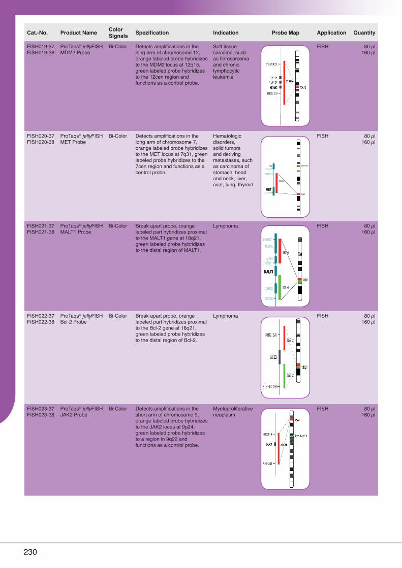
FISH019-37FISH019-38
ProTaqs® jellyFISH MDM2 Probe
Bi-Color Detects amplifications in the long arm of chromosome 12, orange labeled probe hybridizes to the MDM2 locus at 12q15, green labeled probe hybridizes to the 12cen region and functions as a control probe.
Soft tissue sarcoma, such as fibrosarcoma and chronic lymphocytic leukemia
FISH 80 µl160 µl
FISH020-37FISH020-38
ProTaqs® jellyFISH MET Probe
Bi-Color Detects amplifications in the long arm of chromosome 7, orange labeled probe hybridizes to the MET locus at 7q31, green labeled probe hybridizes to the 7cen region and functions as a control probe.
Hematologic disorders, solid tumors and deriving metastases, such as carcinoma of stomach, head and neck, liver, ovar, lung, thyroid
FISH 80 µl160 µl
FISH021-37FISH021-38
ProTaqs® jellyFISH MALT1 Probe
Bi-Color Break apart probe, orange labeled part hybridizes proximal to the MALT1 gene at 18q21, green labeled probe hybridizes to the distal region of MALT1.
Lymphoma FISH 80 µl160 µl
FISH022-37FISH022-38
ProTaqs® jellyFISH Bcl-2 Probe
Bi-Color Break apart probe, orange labeled part hybridizes proximal to the Bcl-2 gene at 18q21, green labeled probe hybridizes to the distal region of Bcl-2.
Lymphoma FISH 80 µl160 µl
FISH023-37FISH023-38
ProTaqs® jellyFISH JAK2 Probe
Bi-Color Detects amplifications in the short arm of chromosome 9, orange labeled probe hybridizes to the JAK2 locus at 9p24, green labeled probe hybridizes to a region in 9q22 and functions as a control probe.
Myeloproliferative neoplasm
FISH 80 µl160 µl
•
• • • •
•
• • • •
• •
•
•
•
•
•
•
•
•
•
•
•
•
•
•
M
V Y
M h
X g
H s
C V
s g
H
X
h
M
.
.
.
.
.
.
.
.
.
.
.
.
.
.
.
.
.
.
.
.
.
.
Troubleshooting
Problem Potential Cause(s) Recommended Solution
No FISH signals can be detected in the microscope.
• Reflected light shutter closed / stop slider in light path.
• Fluorescent lamp is off. • Wrong fluorescence filter in light path. • Objective out of position. • Phototube in camera position.
• Open shutter / move stop slider out of the light path.
• Switch on fluorescent lamp. • Move correct filter into light path. • Swing objective into light path. • Direct light path to eyepieces.
Hybridization signals become weak after a while.
• Immersion oil soaked in-between slide and coverslip.
• Replace coverslip and DAPI/antifade. Use 24 x 32 mm² coverslip even if only a small region is hybridized.
Diffuse signals. • Preparation not adequately illuminated.
• Focus plane cannot be adjusted properly.
• Antifade layer is too thick for focusing.
• Check optical pathway of microscope. Adjust the UV light properly. Check the lifetime of the UV lamp.
• Use enough immersion oil. Do not mix different immersion oils. Use immersion oil suitable for fluorescence.
• Do not use too much DAPI/antifade. 10 µl per slide (24 mm x 32 mm coverslip) are sufficient.
Weak signals. • Chromosome slide preparation too old. Denaturation of chromosomes not adequate.
• Slides should not be older than two weeks. • Aging, baking or further fixation may inhibit the
hybridization and is not recommended.
High diffuse background in green color channel.
• pH value of washing solutions to low. DAPI intensity is too high resulting in crosstalk to FITC filter.
• Ensure that pH value is between 7.0 and 7.5 of solutions. FITC fluorophores are sensitive to pH below 7.
• Reduce DAPI/antifade concentration. If the recommended measures do not solve the problem, or your problem is not listed, please contact MetaSystems.
Customer Support
Please contact MetaSystems GmbH in Germany or MetaSystems Group, Inc. in the USA by telephone or e-mail (contact details, see below). MetaSystems disclaims any proprietary interest in the marks and names of others.
M MetaSystems GmbH RepresentativeRepresentativeRepresentativeRepresentative USAUSAUSAUSA MetaSystems Group, Inc.
Robert-Bosch-Str. 6 70 Bridge Street, Suite 100
D-68804 Altlussheim 02458 Newton, MA
Germany United States
Tel.: +49 (0)6205 39610 Tel.: (888)489-9959 (toll free)
Fax: +49 (0)6205 32270 Fax: +1 617 924-9954
email: [email protected] email: [email protected]
URL: www.metasystems.de URL: www.metasystems.org
RevisionRevisionRevisionRevision: Rev : Rev : Rev : Rev HHHH 130130130130814814814814
Symbols Used
Symbol Symbol Description
ASRASRASRASR Analyte -specific reagent
Analytical and performance characteristics are not established.
hhhh Reference no
ssss Maximum storage temperature
MMMM Manufacturer gggg Lot number
XXXX No of tests
YYYY All warnings are marked by warning triangle with exclamation mark. Depending on their character they are supplemented with the words ATTENTION or CAUTION
HHHH Expiry date
The XT MET probe detects amplifications in the long arm of chromosome 7. The orange labeled probe is designed to hybridize to the MET locus at 7q31. A green labeled probe hybridizes to the 7cen region and functions as a control probe. This probe is intended for Tissue-FISH applications and has been optimized for combined use with MetaSystems‘ pretreatment kit. Analytical and performance characteristics are not established. Probe Diagram:
395 kb
D7S522
TESTESTESTES
D7S486
7q31
METMETMETMET
7p11-q11
D7S2460
V 130110 Chromosome 7
Meta
Sys
tem
s G
mb
H
Ro
bert
-Bo
sch-S
tr.
6
D-6
88
04
Altlu
ssheim
T
el.:
+4
9-6
20
5-3
96
10
to
xic
to
xiq
ue
conta
ins: Form
am
ide
R 6
1:
May
cause
harm
to t
he u
nb
orn
child
. S
45:
In c
ase
of accid
ent
or
if yo
u feel u
nw
ell,
seek m
ed
ical a
dvi
ce im
med
iatly
(show
the la
bel
where
possib
le).
S 5
3:
Avo
id e
xp
osu
re -
-- o
bta
in s
pecia
l instr
uction
befo
re u
se.
Only
for
industr
y consum
ers
. contient:
Form
am
ide
R 6
1:
Ris
que p
endant la
gro
ssesse
d'e
ffets
néfa
ste
s p
our
l'enfa
nt.
S 4
5:
En c
as d
'accid
ent
ou d
e m
ala
ise, consulter
imm
édia
tem
ent un m
éd
ecin
(si p
ossib
le, lu
i montr
er
l'étiquett
e).
S 5
3:
Évi
ter
l'exp
ositio
n -
-- s
e p
rocure
r d
es
instr
uctions s
pécia
les
ava
nt
l'utilis
ation.
Uniq
uem
ent
pour
les
util
isate
ur s
pro
fessi
onnels
.
AS
R
ss ss-1
8°C
g
g
g
g xxxxx
H
H
H
H xxxx-x
x
XT M
ET a
mp
Dele
tion/A
mp
lificatio
n
100µl ( X
10)
hh hh D
-601
3-1
00
-OG
M
For Professional Use OnlyFor Professional Use OnlyFor Professional Use OnlyFor Professional Use Only
Further information available at www.metasystems.Further information available at www.metasystems.Further information available at www.metasystems.Further information available at www.metasystems.dededede
Product Label Order No. Pack Size
XT MET amp orange/green D-6013-100-OG 100µl
Troubleshooting
Problem Potential Cause(s) Recommended Solution
No FISH signals can be detected in the microscope.
• Reflected light shutter closed / stop slider in light path.
• Fluorescent lamp is off. • Wrong fluorescence filter in light path. • Objective out of position. • Phototube in camera position.
• Open shutter / move stop slider out of the light path.
• Switch on fluorescent lamp. • Move correct filter into light path. • Swing objective into light path. • Direct light path to eyepieces.
Hybridization signals become weak after a while.
• Immersion oil soaked in-between slide and coverslip.
• Replace coverslip and DAPI/antifade. Use 24 x 32 mm² coverslip even if only a small region is hybridized.
Diffuse signals. • Preparation not adequately illuminated.
• Focus plane cannot be adjusted properly.
• Antifade layer is too thick for focusing.
• Check optical pathway of microscope. Adjust the UV light properly. Check the lifetime of the UV lamp.
• Use enough immersion oil. Do not mix different immersion oils. Use immersion oil suitable for fluorescence.
• Do not use too much DAPI/antifade. 10 µl per slide (24 mm x 32 mm coverslip) are sufficient.
Weak signals. • Chromosome slide preparation too old. Denaturation of chromosomes not adequate.
• Slides should not be older than two weeks. • Aging, baking or further fixation may inhibit the
hybridization and is not recommended.
High diffuse background in green color channel.
• pH value of washing solutions to low. DAPI intensity is too high resulting in crosstalk to FITC filter.
• Ensure that pH value is between 7.0 and 7.5 of solutions. FITC fluorophores are sensitive to pH below 7.
• Reduce DAPI/antifade concentration. If the recommended measures do not solve the problem, or your problem is not listed, please contact MetaSystems.
Customer Support
Please contact MetaSystems GmbH in Germany or MetaSystems Group, Inc. in the USA by telephone or e-mail (contact details, see below). MetaSystems disclaims any proprietary interest in the marks and names of others.
M MetaSystems GmbH RepresentativeRepresentativeRepresentativeRepresentative USAUSAUSAUSA MetaSystems Group, Inc.
Robert-Bosch-Str. 6 70 Bridge Street, Suite 100
D-68804 Altlussheim 02458 Newton, MA
Germany United States
Tel.: +49 (0)6205 39610 Tel.: (888)489-9959 (toll free)
Fax: +49 (0)6205 32270 Fax: +1 617 924-9954
email: [email protected] email: [email protected]
URL: www.metasystems.de URL: www.metasystems.org
RevisionRevisionRevisionRevision: Rev : Rev : Rev : Rev HHHH 130130130130814814814814
Symbols Used
Symbol Symbol Description
ASRASRASRASR Analyte -specific reagent
Analytical and performance characteristics are not established.
hhhh Reference no
ssss Maximum storage temperature
MMMM Manufacturer gggg Lot number
XXXX No of tests
YYYY All warnings are marked by warning triangle with exclamation mark. Depending on their character they are supplemented with the words ATTENTION or CAUTION
HHHH Expiry date
The XT MALT1 probe is designed as a break apart probe. Its orange labeled part hybridizes proximal to the MALT1 gene at 18q21, the green labeled probe hybridizes to the distal region of MALT1. This probe is intended for Tissue-FISH applications and has been optimized for combined use with MetaSystems‘ pretreatment kit. Analytical and performance characteristics are not established. Probe Diagram:
18q21
ALPK2
D18S887
not not not not totototo scalescalescalescale
NEDD4L
290 kb
D18S881
MALT1MALT1MALT1MALT1
330 kb
D18S529
ZNF532
V 130715 Chromosome 18
Meta
Syste
ms G
mb
H
Ro
bert
-B
osch-S
tr.
6
D-6
88
04
Altlu
ssheim
T
el.:
+4
9-62
05
-3
96
10
to
xic
to
xiq
ue
conta
ins: Form
am
ide
R 6
1:
May c
ause h
arm
to t
he u
nb
orn
child.
S 4
5:
In c
ase o
f accid
ent
or
if y
ou feel unw
ell,
seek m
ed
ical ad
vic
e im
med
iatly (show
the label
where
possib
le).
S 5
3:
Avoid
exp
osure
--- o
bta
in s
pecia
l in
str
uction
befo
re u
se.
Only
for
industr
y c
onsum
ers
. contient:
Form
am
ide
R 6
1:
Ris
que p
endant la
gro
ssesse d
'effets
néfa
ste
s p
our
l'enfa
nt.
S 4
5:
En c
as d
'accid
ent
ou d
e m
ala
ise, consulter
imm
édia
tem
ent un m
éd
ecin
(si p
ossib
le, lu
i m
ontr
er
l'étiquett
e).
S 5
3:
Éviter
l'exp
ositio
n -
-- s
e p
rocure
r d
es
instr
uctions s
pécia
les a
vant
l'utilisation.
Uniq
uem
ent
pour
les u
tilisate
ur s
pro
fessio
nnels
.
AS
R
ss ss-18°C
g
g
g
g xxxxx
H
H
H
H xxxx-xx
XT M
ALT
1
Bre
ak A
part P
rob
e
100µl ( X
10)
hh hh D
-6
01
5-1
00
-O
G
M
For Professional Use OnlyFor Professional Use OnlyFor Professional Use OnlyFor Professional Use Only
Further information available at www.metasystems.Further information available at www.metasystems.Further information available at www.metasystems.Further information available at www.metasystems.dededede
Product Label Order No. Pack Size
XT MALT1 orange/green D-6015-100-OG 100µl
Troubleshooting
Problem Potential Cause(s) Recommended Solution
No FISH signals can be detected in the microscope.
• Reflected light shutter closed / stop slider in light path.
• Fluorescent lamp is off. • Wrong fluorescence filter in light path. • Objective out of position. • Phototube in camera position.
• Open shutter / move stop slider out of the light path.
• Switch on fluorescent lamp. • Move correct filter into light path. • Swing objective into light path. • Direct light path to eyepieces.
Hybridization signals become weak after a while.
• Immersion oil soaked in-between slide and coverslip.
• Replace coverslip and DAPI/antifade. Use 24 x 32 mm² coverslip even if only a small region is hybridized.
Diffuse signals. • Preparation not adequately illuminated.
• Focus plane cannot be adjusted properly.
• Antifade layer is too thick for focusing.
• Check optical pathway of microscope. Adjust the UV light properly. Check the lifetime of the UV lamp.
• Use enough immersion oil. Do not mix different immersion oils. Use immersion oil suitable for fluorescence.
• Do not use too much DAPI/antifade. 10 µl per slide (24 mm x 32 mm coverslip) are sufficient.
Weak signals. • Chromosome slide preparation too old. Denaturation of chromosomes not adequate.
• Slides should not be older than two weeks. • Aging, baking or further fixation may inhibit the
hybridization and is not recommended.
High diffuse background in green color channel.
• pH value of washing solutions to low. DAPI intensity is too high resulting in crosstalk to FITC filter.
• Ensure that pH value is between 7.0 and 7.5 of solutions. FITC fluorophores are sensitive to pH below 7.
• Reduce DAPI/antifade concentration. If the recommended measures do not solve the problem, or your problem is not listed, please contact MetaSystems.
Customer Support
Please contact MetaSystems GmbH in Germany or MetaSystems Group, Inc. in the USA by telephone or e-mail (contact details, see below). MetaSystems disclaims any proprietary interest in the marks and names of others.
M MetaSystems GmbH RepresentativeRepresentativeRepresentativeRepresentative USAUSAUSAUSA MetaSystems Group, Inc.
Robert-Bosch-Str. 6 70 Bridge Street, Suite 100
D-68804 Altlussheim 02458 Newton, MA
Germany United States
Tel.: +49 (0)6205 39610 Tel.: (888)489-9959 (toll free)
Fax: +49 (0)6205 32270 Fax: +1 617 924-9954
email: [email protected] email: [email protected]
URL: www.metasystems.de URL: www.metasystems.org
RevisionRevisionRevisionRevision: Rev : Rev : Rev : Rev HHHH 130130130130814814814814
Symbols Used
Symbol Symbol Description
ASRASRASRASR Analyte -specific reagent
Analytical and performance characteristics are not established.
hhhh Reference no
ssss Maximum storage temperature
MMMM Manufacturer gggg Lot number
XXXX No of tests
YYYY All warnings are marked by warning triangle with exclamation mark. Depending on their character they are supplemented with the words ATTENTION or CAUTION
HHHH Expiry date
The XT JAK2 probe detects amplifications in the short arm of chromosome 9. The orange labeled probe is designed to hybridize to the JAK2 locus at 9p24. A green labeled probe hybridizes to the 9cen region and functions as a control probe. This probe is intended for Tissue-FISH applications and has been optimized for combined use with MetaSystems‘ pretreatment kit. Analytical and performance characteristics are not established. Probe Diagram:
V 130311 Chromosome 9
Meta
Syste
ms G
mb
H
Ro
bert
-B
osch-S
tr.
6
D-6
88
04
Altlu
ssheim
T
el.:
+4
9-62
05
-3
96
10
to
xic
to
xiq
ue
conta
ins: Form
am
ide
R 6
1:
May c
ause h
arm
to t
he u
nb
orn
child.
S 4
5:
In c
ase o
f accid
ent
or
if y
ou feel unw
ell,
seek m
ed
ical ad
vic
e im
med
iatly (show
the label
where
possib
le).
S 5
3:
Avoid
exp
osure
--- o
bta
in s
pecia
l in
str
uction
befo
re u
se.
Only
for
industr
y c
onsum
ers
. contient:
Form
am
ide
R 6
1:
Ris
que p
endant la
gro
ssesse d
'effets
néfa
ste
s p
our
l'enfa
nt.
S 4
5:
En c
as d
'accid
ent
ou d
e m
ala
ise, consulter
imm
édia
tem
ent un m
éd
ecin
(si p
ossib
le, lu
i m
ontr
er
l'étiquett
e).
S 5
3:
Éviter
l'exp
ositio
n -
-- s
e p
rocure
r d
es
instr
uctions s
pécia
les a
vant
l'utilisation.
Uniq
uem
ent
pour
les u
tilisate
ur s
pro
fessio
nnels
.
AS
R
ss ss-18°C
g
g
g
g xxxxx
H
H
H
H xxxx-xx
XT J
AK
2 a
mp
Dele
tion/A
mp
lificatio
n
100µl ( X
10)
hh hh D
-6
02
6-1
00
-O
G
M
For Professional Use OnlyFor Professional Use OnlyFor Professional Use OnlyFor Professional Use Only
Further information available at www.metasystems.Further information available at www.metasystems.Further information available at www.metasystems.Further information available at www.metasystems.dededede
Product Label Order No. Pack Size
XT JAK2 amp orange/green D-6026-100-OG 100µl

231
Cat-No. Product Name Synonym Delivered/Stock Solution
Working Solution Format pH Application
400900492 Post Hybridization Wash Solution
6 x 20 ml (20x) 6 x 400 ml Concentrate ISH
400900192 SSC Buffer Concentrate Saline Sodium Citrate Buffer 6 x 20 ml (20x) 6 x 400 ml Concentrate 7.0 Southern Blot, ISH, DNA Microarray,
Northern Blot
Description: SSC buffer is used as a hybridization buffer.
402001592 TdT Buffer (TUNEL) Terminal Deoxynucleotidyl Transferase Buffer
6 x 20 ml (5x) 6 x 100 ml Concentrate 7.2 Molecularbiology
402001692 TB Buffer (TUNEL) Termination Buffer 6 x 20 ml (5x) 6 x 100 ml Concentrate Molecularbiology
Description: TUNEL technology (Terminal deoxynucleotidyl Transferase Biotin-dUTP Nick End Labeling) is used for detection and quantification of apoptosis at single cell level, based on labeling of DNA strand breaks.
400900292 TES Washbuffer Concentrate
N-[TRIS(hydroxymethyl)methyl]-2-aminoethanesulfonic Acid
6 x 20 ml (25x) 6 x 500 ml Concentrate 7.5 Molecularbiology
Description: It is one of the Good's buffers and can be used to make buffer solutions in the pH range 6.8 – 8.2.
5.2. Wash Solutions and Buffer
5.1.2. Preatreatment and FISH on Paraffin TissuesProTaqs® jellyFISH Pretreatment Kit is recommended for pretreatment of FFPE tissue sections. The kit is optimized for ProTaqs® jellyFISH probes. The kit and additional reagents are performed to increase the permeabilization of the cell membranes to facilitate penetration of the ProTaqs® jellyFISH probes.
Cat.-No. Product Name Format Application QuantityFISH001 ProTaqs® jellyFISH Pretreatment Kit Concentrate FISH Pretreatment of FFPE tissues 20 - 30
Tests
ProTaqs® jellyFISH Pretreatment Kit contains Pretreatment Buffer, Protease and Protease Buffer. Please find below all additionally required reagents.
Cat.-No. Product Name Synonym Format pH Application Quantity400801301 ProTaqs® Alcohol 100 % Ethanol Ready-to-use FISH: Deparaffinization 1 l
400801201 ProTaqs® Xylene 100 % Ready-to-use FISH: Deparaffinization 1 l
400301105 ProTaqs® Clear Xylene Alternative Ready-to-use FISH: Deparaffinization 5 l
400900501 SSC Buffer (2x) Saline Sodium Citrate Buffer Ready-to-use 7.0 FISH: Pretreatment, Post-Hybridization 1 l
400900601 SSC Buffer (0.4x) Saline Sodium Citrate Buffer Ready-to-use 7.0 FISH: Post-Hybridization 1 l
400900701 SSC Buffer (2x) w. 0.05 % Tween20
Saline Sodium Citrate Buffer Ready-to-use 7.0 FISH: Post-Hybridization 1 l
401603392 ProTaqs® Mount Fluor Anti Fading
Ready-to-use FISH: Counterstain 15 ml
Description: ProTaqs® Mount Fluor Anti Fading contains 500 ng/ml DAPI for counterstain step.
Related Documents