5 High-Attenuation Masses in a Cerebral Hemisphere on Computed Tomography

5 high attenuation masses in a cerebral hemisphere on
Aug 18, 2015
Welcome message from author
This document is posted to help you gain knowledge. Please leave a comment to let me know what you think about it! Share it to your friends and learn new things together.
Transcript
5 High-Attenuation Masses in a Cerebral Hemisphere on Computed Tomography

CLINICAL IMAGAGINGAN ATLAS OF DIFFERENTIAL DAIGNOSIS
EISENBERG
DR. Muhammad Bin Zulfiqar PGR-FCPS III SIMS/SHL
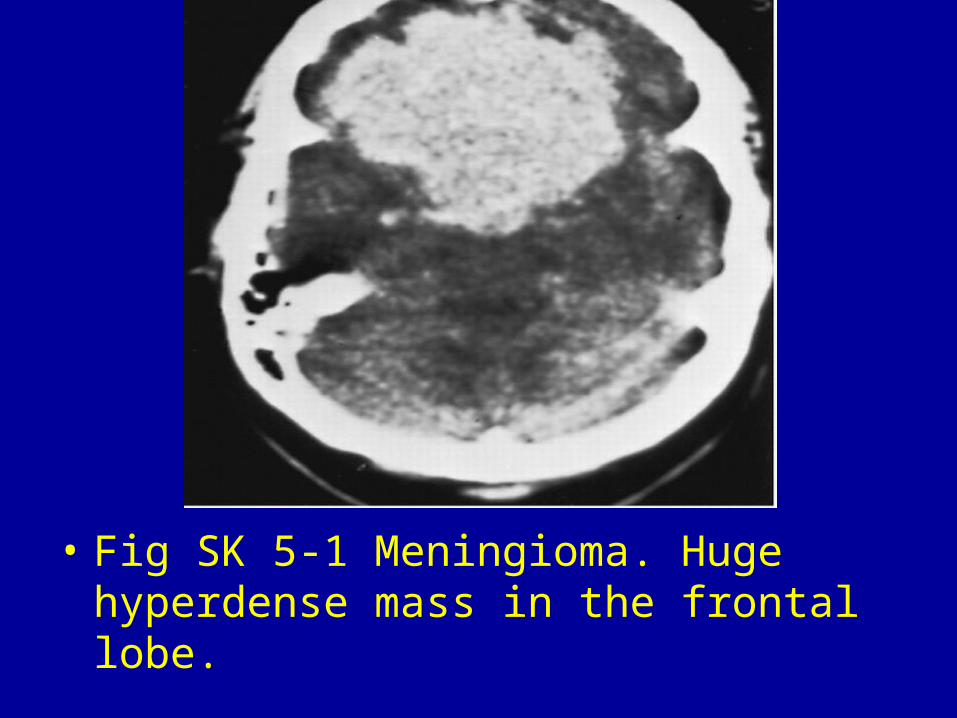
• Fig SK 5-1 Meningioma. Huge hyperdense mass in the frontal lobe.
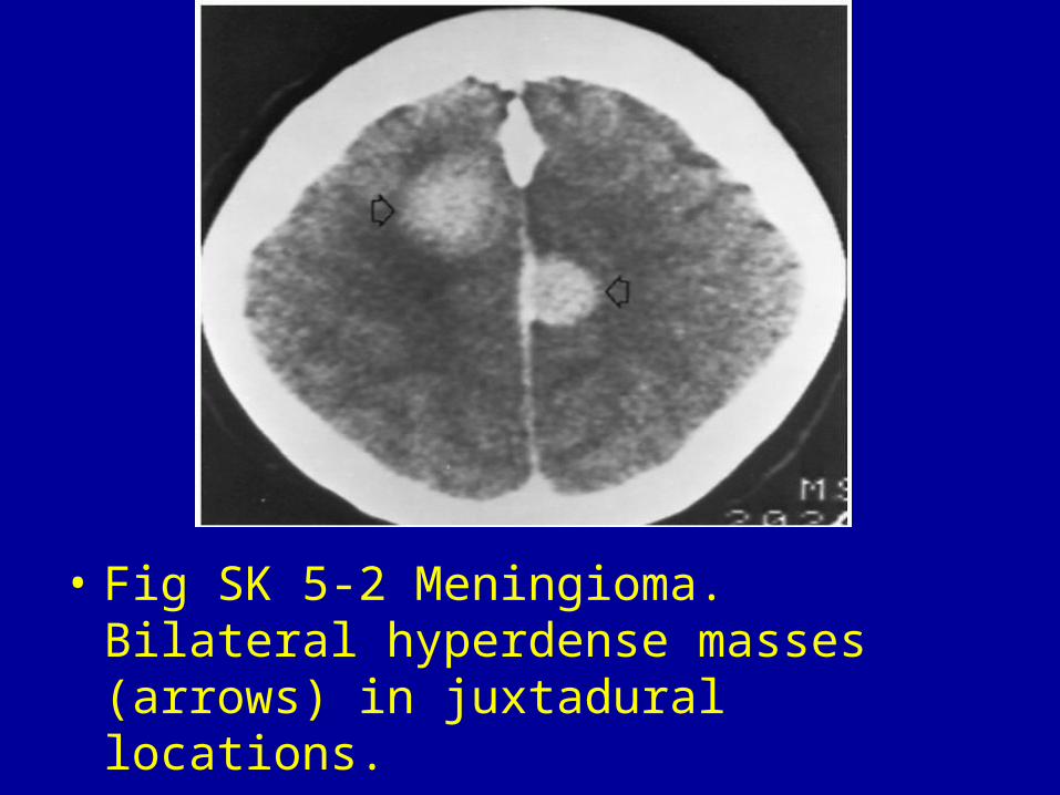
• Fig SK 5-2 Meningioma. Bilateral hyperdense masses (arrows) in juxtadural locations.

• Fig SK 5-3 Metastasis. Nonenhanced scan shows a hyperdense mass (arrow) in the right frontal region representing a metastasis from carcinoma of the lung.
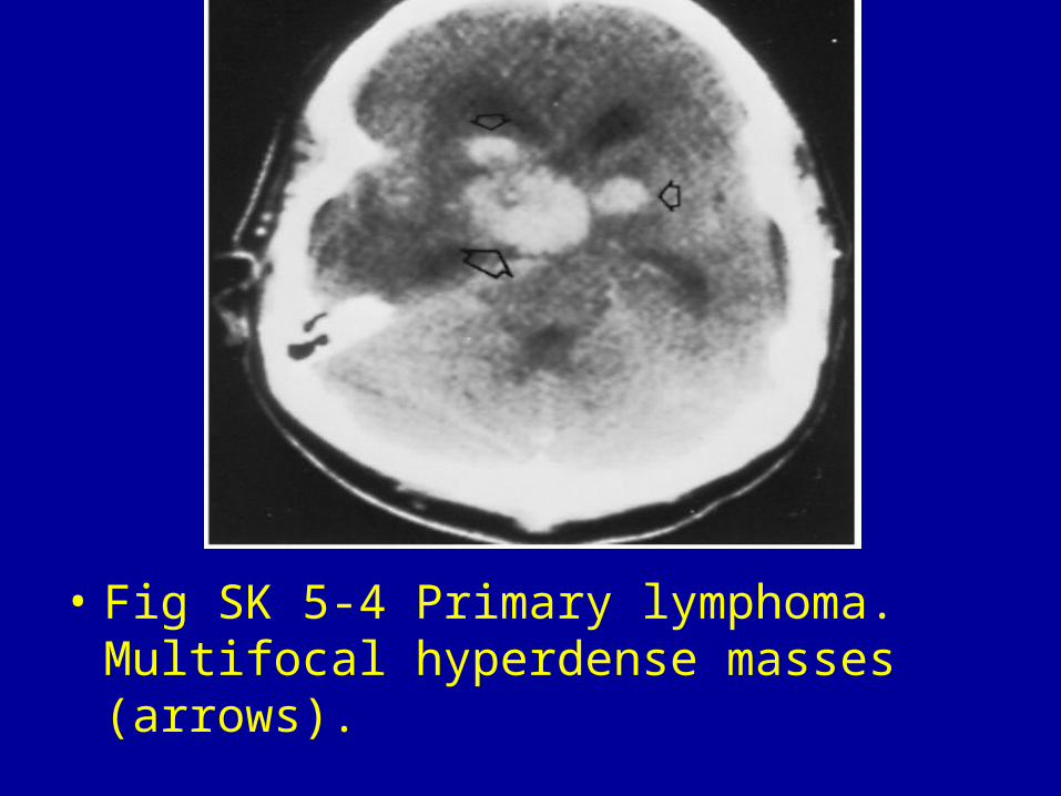
• Fig SK 5-4 Primary lymphoma. Multifocal hyperdense masses (arrows).

• Fig SK 5-5 Intracerebral hematoma. Large, homogeneous high-density area with extensive acute bleeding into the lateral ventricles.
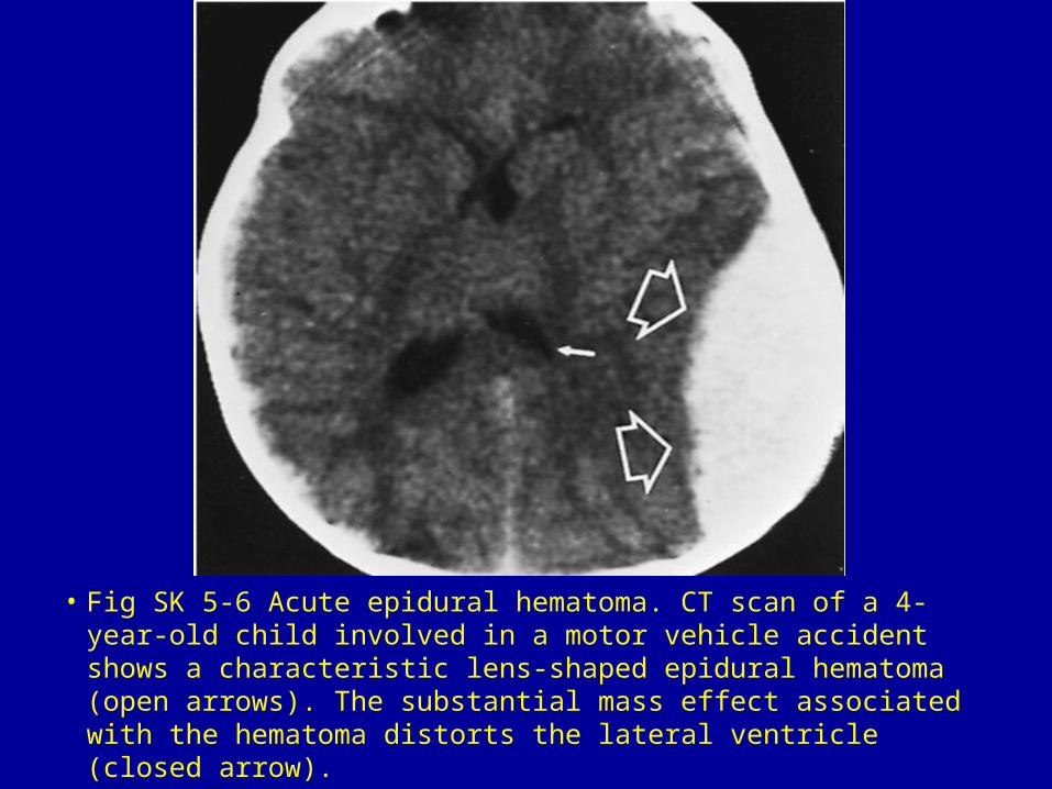
• Fig SK 5-6 Acute epidural hematoma. CT scan of a 4-year-old child involved in a motor vehicle accident shows a characteristic lens-shaped epidural hematoma (open arrows). The substantial mass effect associated with the hematoma distorts the lateral ventricle (closed arrow).

Fig SK 5-7 Epidural hematoma. Bilaterally symmetric posterior high-density areas (arrows) with lens-shaped configurations.
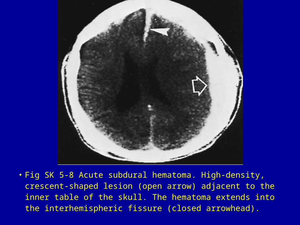
• Fig SK 5-8 Acute subdural hematoma. High-density, crescent-shaped lesion (open arrow) adjacent to the inner table of the skull. The hematoma extends into the interhemispheric fissure (closed arrowhead).


Related Documents