5-Chloro-N-(4,5-dihydro-1H-imidazol-2- yl)-2,1,3-benzothiadiazol-4-amine (tizanidine) Peter John, a Islam Ullah Khan, a Mehmet Akkurt, b * Muhammad Shahid Ramzan a and Shahzad Sharif a a Materials Chemistry Laboratory, Department of Chemistry, GC University, Lahore 54000, Pakistan, and b Department of Physics, Faculty of Sciences, Erciyes University, 38039 Kayseri, Turkey Correspondence e-mail: [email protected], [email protected] Received 17 February 2011; accepted 4 March 2011 Key indicators: single-crystal X-ray study; T = 296 K; mean (C–C) = 0.003 A ˚ ; R factor = 0.042; wR factor = 0.120; data-to-parameter ratio = 16.9. There are two independent molecules (A and B) with similar conformations in the asymmetric unit of the title compound, C 9 H 8 ClN 5 S. The benzothiadiazole ring systems of both molecules are essentially planar [maximum deviation = 0.021 (2) A ˚ in molecule A and 0.022 (1) A ˚ in molecule B] and make dihedral angles of 68.78 (9) and 54.39 (8) , respectively, with the mean planes of their 4,5-dihydro-1H- imidazole rings. An intramolecular N—HCl hydrogen bond occurs in molecule B. In the crystal, both molecules form centrosymmetric dimers through -stacking of their benzothiadiazole rings, with interplanar distances of 3.3174 (7) and 3.2943 (6) A ˚ . These dimers are further linked via pairs of N—HN hydrogen bonds with the dihydro- imidazole rings as the hydrogen-bonding donors and one of the benzothiadiazole N atoms as the acceptors, generating R 2 2 (16) ring motifs. The A 2 and B 2 dimers in turn form additional N—HN hydrogen bonds with the secondary amine as the H-atom donor and the dihydroimidazole N atom as the acceptor. These R 2 2 (8)-type interactions connect the A 2 and B 2 dimers with each other, forming infinite chains along [1 11]. Related literature For the medicinal importance of tizanidine, see: Koch et al. (1989); Shellenberger et al. (1999); Tse et al. (1987). For hydrogen-bond motifs, see: Bernstein et al. (1995). Experimental Crystal data C 9 H 8 ClN 5 S M r = 253.72 Triclinic, P 1 a = 7.6927 (3) A ˚ b = 10.8558 (4) A ˚ c = 12.9969 (5) A ˚ = 95.790 (1) = 101.126 (1) = 92.192 (1) V = 1057.69 (7) A ˚ 3 Z =4 Mo K radiation = 0.54 mm 1 T = 296 K 0.29 0.18 0.08 mm Data collection Bruker APEXII CCD diffractometer 17897 measured reflections 5104 independent reflections 4449 reflections with I >2(I) R int = 0.024 Refinement R[F 2 >2(F 2 )] = 0.042 wR(F 2 ) = 0.120 S = 1.03 5104 reflections 302 parameters 5 restraints H atoms treated by a mixture of independent and constrained refinement max = 0.65 e A ˚ 3 min = 0.43 e A ˚ 3 Table 1 Hydrogen-bond geometry (A ˚ , ). D—HA D—H HA DA D—HA N3A—HN3AN5B 0.92 (2) 2.10 (2) 3.003 (2) 168 (3) N4A—HN4AN1A i 0.86 (3) 2.38 (3) 3.205 (3) 160 (2) N3B—HN3BN5A 0.88 (2) 1.98 (2) 2.864 (2) 177 (2) N4B—HN4BCl1B 0.84 (2) 2.75 (2) 3.1927 (15) 114 (2) N4B—HN4BN1B ii 0.84 (2) 2.48 (2) 3.227 (2) 150 (2) Symmetry codes: (i) x þ 1; y þ 1; z þ 1; (ii) x þ 3; y; z þ 2. Data collection: APEX2 (Bruker, 2007); cell refinement: SAINT (Bruker, 2007); data reduction: SAINT; program(s) used to solve structure: SHELXS97 (Sheldrick, 2008); program(s) used to refine structure: SHELXL97 (Sheldrick, 2008); molecular graphics: ORTEP-3 for Windows (Farrugia, 1997); software used to prepare material for publication: WinGX (Farrugia, 1999) and PLATON (Spek, 2009). The authors are grateful to the Higher Education Commission (HEC), Pakistan, for providing funds for the single-crystal XRD facilities at GC University, Lahore. Supplementary data and figures for this paper are available from the IUCr electronic archives (Reference: ZL2351). organic compounds o838 John et al. doi:10.1107/S1600536811008348 Acta Cryst. (2011). E67, o838–o839 Acta Crystallographica Section E Structure Reports Online ISSN 1600-5368

Welcome message from author
This document is posted to help you gain knowledge. Please leave a comment to let me know what you think about it! Share it to your friends and learn new things together.
Transcript
5-Chloro-N-(4,5-dihydro-1H-imidazol-2-yl)-2,1,3-benzothiadiazol-4-amine(tizanidine)
Peter John,a Islam Ullah Khan,a Mehmet Akkurt,b*
Muhammad Shahid Ramzana and Shahzad Sharifa
aMaterials Chemistry Laboratory, Department of Chemistry, GC University, Lahore
54000, Pakistan, and bDepartment of Physics, Faculty of Sciences, Erciyes
University, 38039 Kayseri, Turkey
Correspondence e-mail: [email protected], [email protected]
Received 17 February 2011; accepted 4 March 2011
Key indicators: single-crystal X-ray study; T = 296 K; mean �(C–C) = 0.003 A;
R factor = 0.042; wR factor = 0.120; data-to-parameter ratio = 16.9.
There are two independent molecules (A and B) with similar
conformations in the asymmetric unit of the title compound,
C9H8ClN5S. The benzothiadiazole ring systems of both
molecules are essentially planar [maximum deviation =
0.021 (2) A in molecule A and 0.022 (1) A in molecule B]
and make dihedral angles of 68.78 (9) and 54.39 (8)�,
respectively, with the mean planes of their 4,5-dihydro-1H-
imidazole rings. An intramolecular N—H� � �Cl hydrogen bond
occurs in molecule B. In the crystal, both molecules form
centrosymmetric dimers through �-stacking of their
benzothiadiazole rings, with interplanar distances of
3.3174 (7) and 3.2943 (6) A. These dimers are further linked
via pairs of N—H� � �N hydrogen bonds with the dihydro-
imidazole rings as the hydrogen-bonding donors and one of
the benzothiadiazole N atoms as the acceptors, generating
R22(16) ring motifs. The A2 and B2 dimers in turn form
additional N—H� � �N hydrogen bonds with the secondary
amine as the H-atom donor and the dihydroimidazole N atom
as the acceptor. These R22(8)-type interactions connect the A2
and B2 dimers with each other, forming infinite chains along
[111].
Related literature
For the medicinal importance of tizanidine, see: Koch et al.
(1989); Shellenberger et al. (1999); Tse et al. (1987). For
hydrogen-bond motifs, see: Bernstein et al. (1995).
Experimental
Crystal data
C9H8ClN5SMr = 253.72Triclinic, P1a = 7.6927 (3) Ab = 10.8558 (4) Ac = 12.9969 (5) A� = 95.790 (1)�
� = 101.126 (1)�
� = 92.192 (1)�
V = 1057.69 (7) A3
Z = 4Mo K� radiation� = 0.54 mm�1
T = 296 K0.29 � 0.18 � 0.08 mm
Data collection
Bruker APEXII CCDdiffractometer
17897 measured reflections
5104 independent reflections4449 reflections with I > 2�(I)Rint = 0.024
Refinement
R[F 2 > 2�(F 2)] = 0.042wR(F 2) = 0.120S = 1.035104 reflections302 parameters5 restraints
H atoms treated by a mixture ofindependent and constrainedrefinement
��max = 0.65 e A�3
��min = �0.43 e A�3
Table 1Hydrogen-bond geometry (A, �).
D—H� � �A D—H H� � �A D� � �A D—H� � �A
N3A—HN3A� � �N5B 0.92 (2) 2.10 (2) 3.003 (2) 168 (3)N4A—HN4A� � �N1Ai 0.86 (3) 2.38 (3) 3.205 (3) 160 (2)N3B—HN3B� � �N5A 0.88 (2) 1.98 (2) 2.864 (2) 177 (2)N4B—HN4B� � �Cl1B 0.84 (2) 2.75 (2) 3.1927 (15) 114 (2)N4B—HN4B� � �N1Bii 0.84 (2) 2.48 (2) 3.227 (2) 150 (2)
Symmetry codes: (i) �xþ 1;�yþ 1;�zþ 1; (ii) �xþ 3;�y;�zþ 2.
Data collection: APEX2 (Bruker, 2007); cell refinement: SAINT
(Bruker, 2007); data reduction: SAINT; program(s) used to solve
structure: SHELXS97 (Sheldrick, 2008); program(s) used to refine
structure: SHELXL97 (Sheldrick, 2008); molecular graphics:
ORTEP-3 for Windows (Farrugia, 1997); software used to prepare
material for publication: WinGX (Farrugia, 1999) and PLATON
(Spek, 2009).
The authors are grateful to the Higher Education
Commission (HEC), Pakistan, for providing funds for the
single-crystal XRD facilities at GC University, Lahore.
Supplementary data and figures for this paper are available from theIUCr electronic archives (Reference: ZL2351).
organic compounds
o838 John et al. doi:10.1107/S1600536811008348 Acta Cryst. (2011). E67, o838–o839
Acta Crystallographica Section E
Structure ReportsOnline
ISSN 1600-5368

References
Bernstein, J., Davis, R. E., Shimoni, L. & Chang, N.-L. (1995). Angew. Chem.Int. Ed. Engl. 34, 1555–1573.
Bruker (2007). APEX2 and SAINT. Bruker AXS Inc., Madison, Wisconsin,USA.
Farrugia, L. J. (1997). J. Appl. Cryst. 30, 565.Farrugia, L. J. (1999). J. Appl. Cryst. 32, 837–838.
Koch, P., Hirst, D. R. & Wartburg Von, B. R. (1989). Xenobiotica, 19, 1255–1260.
Sheldrick, G. M. (2008). Acta Cryst. A64, 112–122.Shellenberger, M. K., Groves, L., Shah, J. & Novak, G. D. (1999). Drug Metab.
Dispos. 27, 201–205.Spek, A. L. (2009). Acta Cryst. D65, 148–155.Tse, F. L. S., Jaffe, J. M. & Bhuta, S. (1987). Fundam. Clin. Pharmacol. 1, 479–
485.
organic compounds
Acta Cryst. (2011). E67, o838–o839 John et al. � C9H8ClN5S o839
supplementary materials
supplementary materials
sup-1
Acta Cryst. (2011). E67, o838-o839 [ doi:10.1107/S1600536811008348 ]
5-Chloro-N-(4,5-dihydro-1H-imidazol-2-yl)-2,1,3-benzothiadiazol-4-amine (tizanidine)
P. John, I. U. Khan, M. Akkurt, M. S. Ramzan and S. Sharif
Comment
Tizanidine {or (5-chloro-N-(4,5-dihydro-1H-imidazol-2-yl)-2,1,3-benzothiadiazol-4-amine)} is an adrenergic agonist andproved to be an active myotonolytic skeletal muscle relaxant with a chemical structure different from other muscle relaxants(Koch et al., 1989; Tse et al., 1987). It also reduces increased muscle tone associated with spasticity in patients with multiplesclerosis or spinal cord injury (Shellenberger et al., 1999). Herein, we report the crystal structure of Tizanidine.
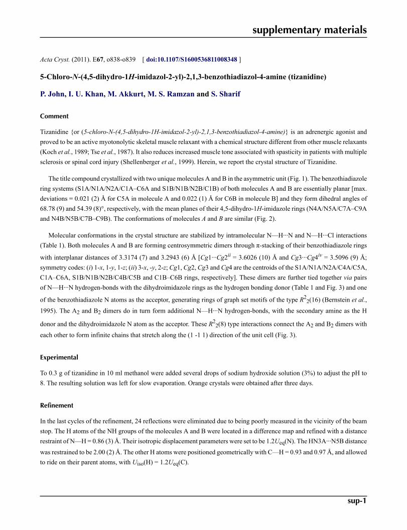
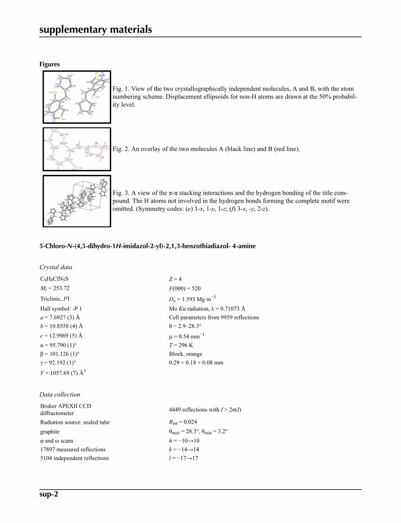
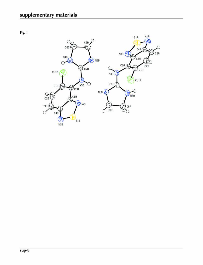
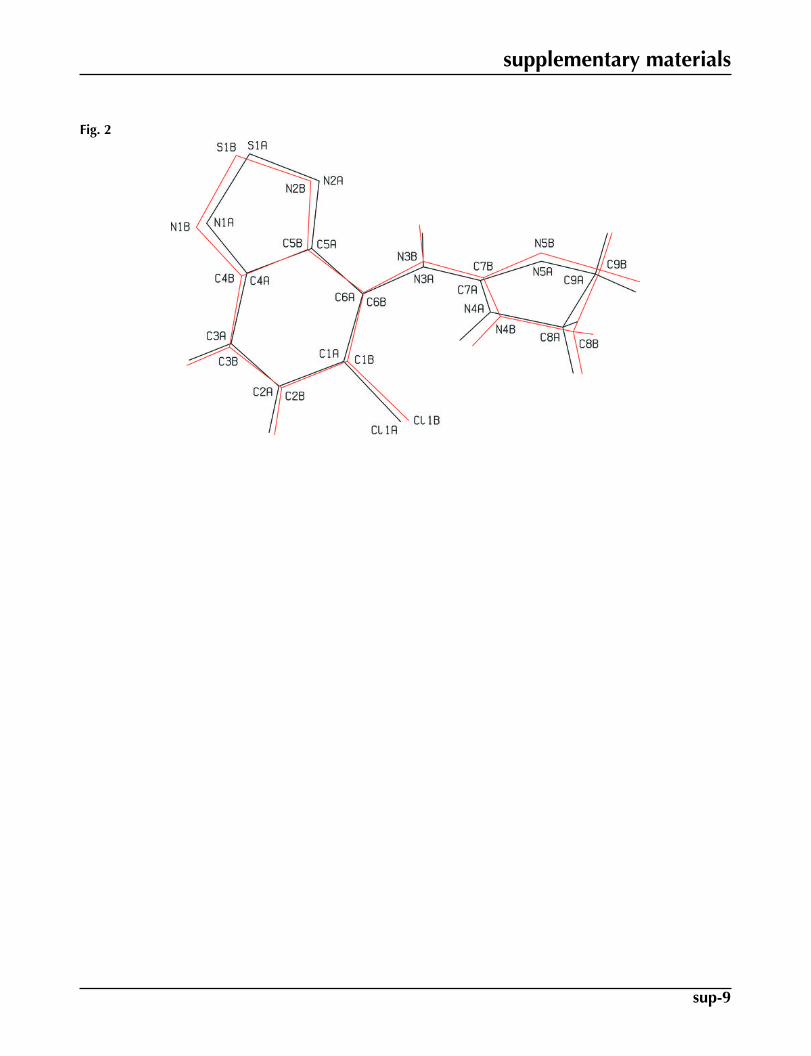
The title compound crystallized with two unique molecules A and B in the asymmetric unit (Fig. 1). The benzothiadiazolering systems (S1A/N1A/N2A/C1A–C6A and S1B/N1B/N2B/C1B) of both molecules A and B are essentially planar [max.deviations = 0.021 (2) Å for C5A in molecule A and 0.022 (1) Å for C6B in molecule B] and they form dihedral angles of68.78 (9) and 54.39 (8)°, respectively, with the mean planes of their 4,5-dihydro-1H-imidazole rings (N4A/N5A/C7A–C9Aand N4B/N5B/C7B–C9B). The conformations of molecules A and B are similar (Fig. 2).
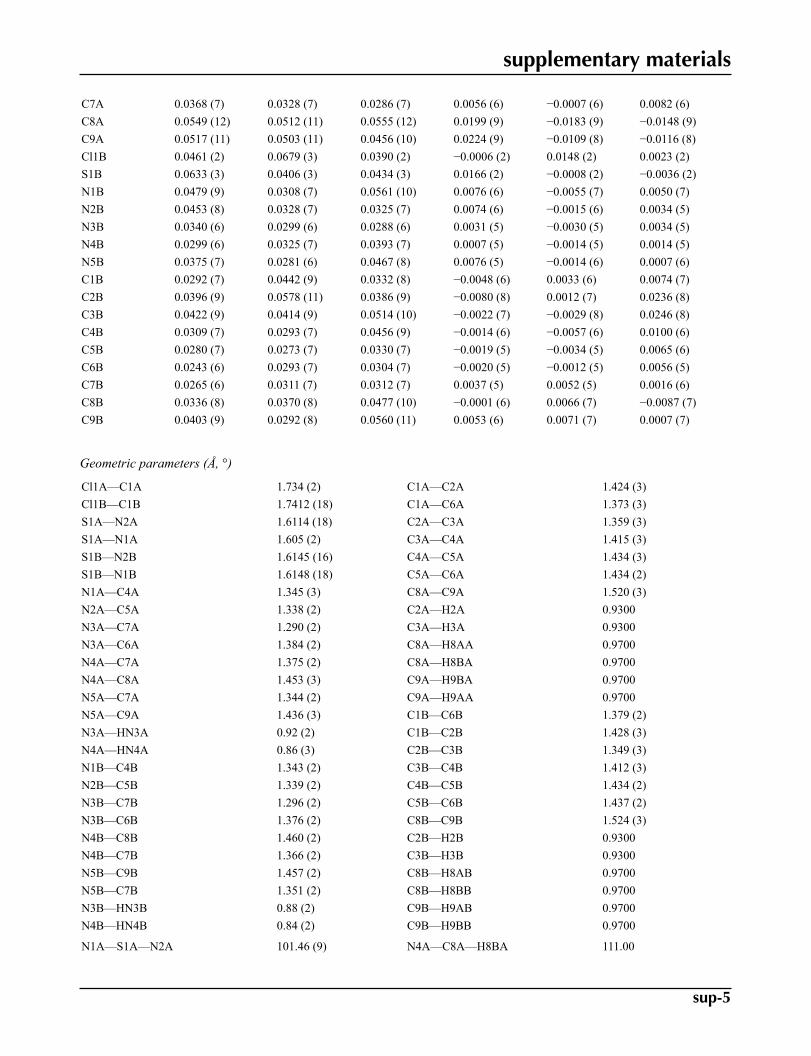
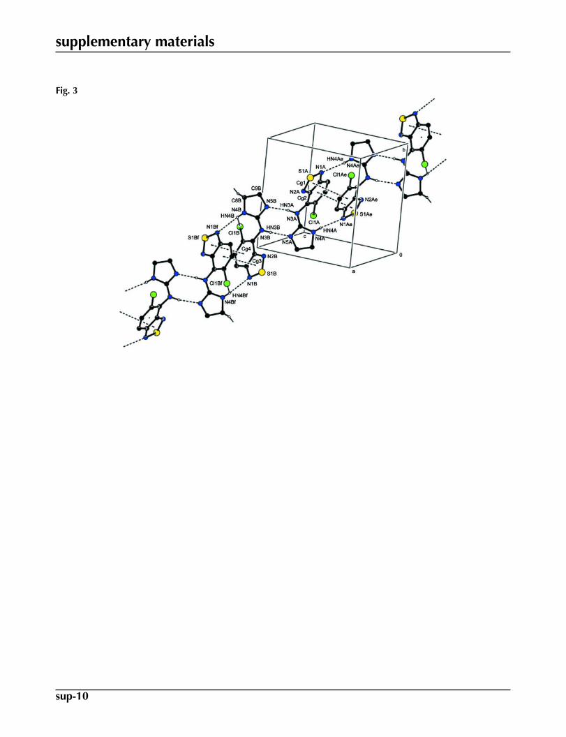
Molecular conformations in the crystal structure are stabilized by intramolecular N—H···N and N—H···Cl interactions(Table 1). Both molecules A and B are forming centrosymmetric dimers through π-stacking of their benzothiadiazole rings
with interplanar distances of 3.3174 (7) and 3.2943 (6) Å [Cg1···Cg2ii = 3.6026 (10) Å and Cg3···Cg4iv = 3.5096 (9) Å;symmetry codes: (i) 1-x, 1-y, 1-z; (ii) 3-x, -y, 2-z; Cg1, Cg2, Cg3 and Cg4 are the centroids of the S1A/N1A/N2A/C4A/C5A,C1A–C6A, S1B/N1B/N2B/C4B/C5B and C1B–C6B rings, respectively]. These dimers are further tied together via pairsof N—H···N hydrogen-bonds with the dihydroimidazole rings as the hydrogen bonding donor (Table 1 and Fig. 3) and one
of the benzothiadiazole N atoms as the acceptor, generating rings of graph set motifs of the type R22(16) (Bernstein et al.,
1995). The A2 and B2 dimers do in turn form additional N—H···N hydrogen-bonds, with the secondary amine as the H
donor and the dihydroimidazole N atom as the acceptor. These R22(8) type interactions connect the A2 and B2 dimers with
each other to form infinite chains that stretch along the (1 -1 1) direction of the unit cell (Fig. 3).
Experimental
To 0.3 g of tizanidine in 10 ml methanol were added several drops of sodium hydroxide solution (3%) to adjust the pH to8. The resulting solution was left for slow evaporation. Orange crystals were obtained after three days.
Refinement
In the last cycles of the refinement, 24 reflections were eliminated due to being poorly measured in the vicinity of the beamstop. The H atoms of the NH groups of the molecules A and B were located in a difference map and refined with a distancerestraint of N—H = 0.86 (3) Å. Their isotropic displacement parameters were set to be 1.2Ueq(N). The HN3A···N5B distance
was restrained to be 2.00 (2) Å. The other H atoms were positioned geometrically with C—H = 0.93 and 0.97 Å, and allowedto ride on their parent atoms, with Uiso(H) = 1.2Ueq(C).
supplementary materials
sup-2
Figures
Fig. 1. View of the two crystallographically independent molecules, A and B, with the atomnumbering scheme. Displacement ellipsoids for non-H atoms are drawn at the 50% probabil-ity level.
Fig. 2. An overlay of the two molecules A (black line) and B (red line).
Fig. 3. A view of the π-π stacking interactions and the hydrogen bonding of the title com-pound. The H atoms not involved in the hydrogen bonds forming the complete motif wereomitted. (Symmetry codes: (e) 1-x, 1-y, 1-z; (f) 3-x, -y, 2-z).
5-Chloro-N-(4,5-dihydro-1H-imidazol-2-yl)-2,1,3-benzothiadiazol- 4-amine
Crystal data
C9H8ClN5S Z = 4Mr = 253.72 F(000) = 520
Triclinic, P1 Dx = 1.593 Mg m−3
Hall symbol: -P 1 Mo Kα radiation, λ = 0.71073 Åa = 7.6927 (3) Å Cell parameters from 9959 reflectionsb = 10.8558 (4) Å θ = 2.9–28.3°c = 12.9969 (5) Å µ = 0.54 mm−1
α = 95.790 (1)° T = 296 Kβ = 101.126 (1)° Block, orangeγ = 92.192 (1)° 0.29 × 0.18 × 0.08 mm
V = 1057.69 (7) Å3
Data collection
Bruker APEXII CCDdiffractometer 4449 reflections with I > 2σ(I)
Radiation source: sealed tube Rint = 0.024
graphite θmax = 28.3°, θmin = 3.2°φ and ω scans h = −10→1017897 measured reflections k = −14→145104 independent reflections l = −17→17

supplementary materials
sup-3
Refinement
Refinement on F2 Primary atom site location: structure-invariant directmethods
Least-squares matrix: full Secondary atom site location: difference Fourier map
R[F2 > 2σ(F2)] = 0.042Hydrogen site location: inferred from neighbouringsites
wR(F2) = 0.120H atoms treated by a mixture of independent andconstrained refinement
S = 1.03w = 1/[σ2(Fo
2) + (0.0635P)2 + 0.4931P]where P = (Fo
2 + 2Fc2)/3
5104 reflections (Δ/σ)max = 0.001
302 parameters Δρmax = 0.65 e Å−3
5 restraints Δρmin = −0.43 e Å−3
Special details
Geometry. Bond distances, angles etc. have been calculated using the rounded fractional coordinates. All su's are estimated from thevariances of the (full) variance-covariance matrix. The cell e.s.d.'s are taken into account in the estimation of distances, angles and tor-sion angles
Refinement. Refinement on F2 for ALL reflections except those flagged by the user for potential systematic errors. Weighted R-
factors wR and all goodnesses of fit S are based on F2, conventional R-factors R are based on F, with F set to zero for negative F2. The
observed criterion of F2 > σ(F2) is used only for calculating -R-factor-obs etc. and is not relevant to the choice of reflections for refine-
ment. R-factors based on F2 are statistically about twice as large as those based on F, and R-factors based on ALL data will be evenlarger.
Fractional atomic coordinates and isotropic or equivalent isotropic displacement parameters (Å2)
x y z Uiso*/Ueq
Cl1A 0.40681 (8) 0.26166 (5) 0.72358 (5) 0.0597 (2)S1A 0.85136 (8) 0.69598 (5) 0.58255 (5) 0.0609 (2)N1A 0.6430 (3) 0.71482 (15) 0.57232 (14) 0.0518 (5)N2A 0.8692 (2) 0.56543 (15) 0.63157 (15) 0.0504 (5)N3A 0.7911 (2) 0.33724 (14) 0.71346 (12) 0.0441 (5)N4A 0.6996 (2) 0.18129 (15) 0.56630 (12) 0.0426 (4)N5A 0.9113 (2) 0.14570 (13) 0.69909 (11) 0.0393 (4)C1A 0.4819 (3) 0.39656 (17) 0.68103 (13) 0.0410 (5)C2A 0.3519 (3) 0.48262 (19) 0.65002 (14) 0.0449 (6)C3A 0.3954 (3) 0.58968 (17) 0.61257 (15) 0.0452 (5)C4A 0.5753 (3) 0.61502 (15) 0.60785 (13) 0.0407 (5)C5A 0.7058 (2) 0.52907 (15) 0.64166 (13) 0.0378 (5)C6A 0.6584 (2) 0.41428 (15) 0.67789 (13) 0.0375 (5)C7A 0.7965 (2) 0.22922 (15) 0.66323 (12) 0.0333 (4)C8A 0.7270 (3) 0.0496 (2) 0.55046 (19) 0.0588 (7)C9A 0.9066 (3) 0.0408 (2) 0.62174 (16) 0.0526 (6)Cl1B 1.12566 (6) 0.18138 (5) 1.13956 (4) 0.0505 (1)

supplementary materials
sup-4
S1B 1.35483 (8) −0.13008 (5) 0.75206 (4) 0.0509 (2)N1B 1.3845 (2) −0.19353 (14) 0.86079 (14) 0.0469 (5)N2B 1.2747 (2) −0.00089 (13) 0.78769 (11) 0.0380 (4)N3B 1.13963 (18) 0.19625 (12) 0.90223 (11) 0.0321 (3)N4B 1.32552 (18) 0.36020 (13) 1.01366 (12) 0.0352 (4)N5B 1.09790 (19) 0.40512 (13) 0.89434 (12) 0.0388 (4)C1B 1.2069 (2) 0.07385 (17) 1.05470 (13) 0.0359 (5)C2B 1.2668 (2) −0.03721 (19) 1.09644 (15) 0.0452 (6)C3B 1.3290 (2) −0.12898 (18) 1.03814 (16) 0.0454 (5)C4B 1.3311 (2) −0.11275 (15) 0.93194 (14) 0.0367 (4)C5B 1.26880 (19) −0.00179 (14) 0.88987 (12) 0.0306 (4)C6B 1.20705 (18) 0.09766 (14) 0.95246 (12) 0.0288 (4)C7B 1.18549 (19) 0.31124 (14) 0.93720 (12) 0.0297 (4)C8B 1.3016 (2) 0.49159 (16) 1.03987 (15) 0.0406 (5)C9B 1.1930 (2) 0.52377 (16) 0.93606 (16) 0.0424 (5)H8AA 0.73030 0.02310 0.47730 0.0710*H8BA 0.63520 0.00040 0.57190 0.0710*H2A 0.23490 0.46520 0.65550 0.0540*H9AA 0.91290 −0.03620 0.65370 0.0630*H3A 0.30950 0.64460 0.59070 0.0540*H9BA 1.00250 0.04780 0.58350 0.0630*HN3A 0.887 (3) 0.368 (3) 0.7644 (17) 0.0720*HN4A 0.597 (3) 0.209 (3) 0.544 (2) 0.0720*H2B 1.26270 −0.04660 1.16630 0.0540*H3B 1.36890 −0.20030 1.06680 0.0550*H8AB 1.41440 0.53940 1.05880 0.0490*H8BB 1.23730 0.50460 1.09690 0.0490*H9AB 1.11220 0.58780 0.94760 0.0510*H9BB 1.26870 0.55020 0.88970 0.0510*HN3B 1.072 (3) 0.179 (2) 0.8391 (15) 0.0610*HN4B 1.370 (3) 0.317 (2) 1.0608 (18) 0.0610*
Atomic displacement parameters (Å2)
U11 U22 U33 U12 U13 U23
Cl1A 0.0697 (3) 0.0577 (3) 0.0579 (3) 0.0072 (2) 0.0179 (3) 0.0245 (2)S1A 0.0653 (3) 0.0384 (3) 0.0738 (4) −0.0043 (2) 0.0007 (3) 0.0089 (2)N1A 0.0690 (11) 0.0306 (7) 0.0500 (9) 0.0073 (7) −0.0036 (8) 0.0047 (7)N2A 0.0487 (9) 0.0364 (8) 0.0591 (10) 0.0036 (6) −0.0049 (7) 0.0019 (7)N3A 0.0512 (9) 0.0320 (7) 0.0401 (8) 0.0131 (6) −0.0144 (6) 0.0016 (6)N4A 0.0459 (8) 0.0443 (8) 0.0320 (7) 0.0146 (6) −0.0074 (6) 0.0016 (6)N5A 0.0450 (8) 0.0329 (7) 0.0348 (7) 0.0129 (6) −0.0057 (6) 0.0016 (6)C1A 0.0548 (10) 0.0375 (9) 0.0300 (8) 0.0107 (7) 0.0041 (7) 0.0054 (6)C2A 0.0492 (10) 0.0483 (10) 0.0367 (9) 0.0146 (8) 0.0070 (7) 0.0002 (7)C3A 0.0545 (10) 0.0386 (9) 0.0392 (9) 0.0227 (8) −0.0003 (8) −0.0008 (7)C4A 0.0570 (10) 0.0274 (7) 0.0320 (8) 0.0130 (7) −0.0048 (7) −0.0017 (6)C5A 0.0466 (9) 0.0286 (7) 0.0324 (8) 0.0089 (6) −0.0053 (6) −0.0021 (6)C6A 0.0496 (9) 0.0305 (8) 0.0275 (7) 0.0119 (7) −0.0054 (6) 0.0008 (6)
supplementary materials
sup-5
C7A 0.0368 (7) 0.0328 (7) 0.0286 (7) 0.0056 (6) −0.0007 (6) 0.0082 (6)C8A 0.0549 (12) 0.0512 (11) 0.0555 (12) 0.0199 (9) −0.0183 (9) −0.0148 (9)C9A 0.0517 (11) 0.0503 (11) 0.0456 (10) 0.0224 (9) −0.0109 (8) −0.0116 (8)Cl1B 0.0461 (2) 0.0679 (3) 0.0390 (2) −0.0006 (2) 0.0148 (2) 0.0023 (2)S1B 0.0633 (3) 0.0406 (3) 0.0434 (3) 0.0166 (2) −0.0008 (2) −0.0036 (2)N1B 0.0479 (9) 0.0308 (7) 0.0561 (10) 0.0076 (6) −0.0055 (7) 0.0050 (7)N2B 0.0453 (8) 0.0328 (7) 0.0325 (7) 0.0074 (6) −0.0015 (6) 0.0034 (5)N3B 0.0340 (6) 0.0299 (6) 0.0288 (6) 0.0031 (5) −0.0030 (5) 0.0034 (5)N4B 0.0299 (6) 0.0325 (7) 0.0393 (7) 0.0007 (5) −0.0014 (5) 0.0014 (5)N5B 0.0375 (7) 0.0281 (6) 0.0467 (8) 0.0076 (5) −0.0014 (6) 0.0007 (6)C1B 0.0292 (7) 0.0442 (9) 0.0332 (8) −0.0048 (6) 0.0033 (6) 0.0074 (7)C2B 0.0396 (9) 0.0578 (11) 0.0386 (9) −0.0080 (8) 0.0012 (7) 0.0236 (8)C3B 0.0422 (9) 0.0414 (9) 0.0514 (10) −0.0022 (7) −0.0029 (8) 0.0246 (8)C4B 0.0309 (7) 0.0293 (7) 0.0456 (9) −0.0014 (6) −0.0057 (6) 0.0100 (6)C5B 0.0280 (7) 0.0273 (7) 0.0330 (7) −0.0019 (5) −0.0034 (5) 0.0065 (6)C6B 0.0243 (6) 0.0293 (7) 0.0304 (7) −0.0020 (5) −0.0012 (5) 0.0056 (5)C7B 0.0265 (6) 0.0311 (7) 0.0312 (7) 0.0037 (5) 0.0052 (5) 0.0016 (6)C8B 0.0336 (8) 0.0370 (8) 0.0477 (10) −0.0001 (6) 0.0066 (7) −0.0087 (7)C9B 0.0403 (9) 0.0292 (8) 0.0560 (11) 0.0053 (6) 0.0071 (7) 0.0007 (7)
Geometric parameters (Å, °)
Cl1A—C1A 1.734 (2) C1A—C2A 1.424 (3)Cl1B—C1B 1.7412 (18) C1A—C6A 1.373 (3)S1A—N2A 1.6114 (18) C2A—C3A 1.359 (3)S1A—N1A 1.605 (2) C3A—C4A 1.415 (3)S1B—N2B 1.6145 (16) C4A—C5A 1.434 (3)S1B—N1B 1.6148 (18) C5A—C6A 1.434 (2)N1A—C4A 1.345 (3) C8A—C9A 1.520 (3)N2A—C5A 1.338 (2) C2A—H2A 0.9300N3A—C7A 1.290 (2) C3A—H3A 0.9300N3A—C6A 1.384 (2) C8A—H8AA 0.9700N4A—C7A 1.375 (2) C8A—H8BA 0.9700N4A—C8A 1.453 (3) C9A—H9BA 0.9700N5A—C7A 1.344 (2) C9A—H9AA 0.9700N5A—C9A 1.436 (3) C1B—C6B 1.379 (2)N3A—HN3A 0.92 (2) C1B—C2B 1.428 (3)N4A—HN4A 0.86 (3) C2B—C3B 1.349 (3)N1B—C4B 1.343 (2) C3B—C4B 1.412 (3)N2B—C5B 1.339 (2) C4B—C5B 1.434 (2)N3B—C7B 1.296 (2) C5B—C6B 1.437 (2)N3B—C6B 1.376 (2) C8B—C9B 1.524 (3)N4B—C8B 1.460 (2) C2B—H2B 0.9300N4B—C7B 1.366 (2) C3B—H3B 0.9300N5B—C9B 1.457 (2) C8B—H8AB 0.9700N5B—C7B 1.351 (2) C8B—H8BB 0.9700N3B—HN3B 0.88 (2) C9B—H9AB 0.9700N4B—HN4B 0.84 (2) C9B—H9BB 0.9700
N1A—S1A—N2A 101.46 (9) N4A—C8A—H8BA 111.00

supplementary materials
sup-6
N1B—S1B—N2B 101.08 (8) C9A—C8A—H8AA 111.00S1A—N1A—C4A 105.99 (15) C9A—C8A—H8BA 111.00S1A—N2A—C5A 106.15 (13) N4A—C8A—H8AA 111.00C6A—N3A—C7A 120.01 (15) H8AA—C8A—H8BA 109.00C7A—N4A—C8A 108.65 (16) H9AA—C9A—H9BA 109.00C7A—N5A—C9A 111.44 (15) C8A—C9A—H9BA 111.00C6A—N3A—HN3A 120.0 (19) C8A—C9A—H9AA 111.00C7A—N3A—HN3A 118.9 (18) N5A—C9A—H9AA 111.00C8A—N4A—HN4A 121 (2) N5A—C9A—H9BA 111.00C7A—N4A—HN4A 119.2 (19) Cl1B—C1B—C6B 119.74 (13)S1B—N1B—C4B 106.09 (12) C2B—C1B—C6B 123.76 (16)S1B—N2B—C5B 106.29 (11) Cl1B—C1B—C2B 116.49 (13)C6B—N3B—C7B 123.72 (14) C1B—C2B—C3B 122.33 (17)C7B—N4B—C8B 108.92 (13) C2B—C3B—C4B 117.52 (17)C7B—N5B—C9B 110.47 (14) C3B—C4B—C5B 120.03 (15)C6B—N3B—HN3B 117.1 (14) N1B—C4B—C3B 126.69 (16)C7B—N3B—HN3B 118.8 (14) N1B—C4B—C5B 113.27 (15)C7B—N4B—HN4B 119.4 (15) N2B—C5B—C6B 124.10 (14)C8B—N4B—HN4B 120.4 (15) C4B—C5B—C6B 122.63 (14)C2A—C1A—C6A 124.01 (18) N2B—C5B—C4B 113.27 (14)Cl1A—C1A—C6A 119.56 (15) N3B—C6B—C1B 128.30 (15)Cl1A—C1A—C2A 116.43 (17) N3B—C6B—C5B 117.62 (14)C1A—C2A—C3A 121.3 (2) C1B—C6B—C5B 113.69 (14)C2A—C3A—C4A 117.99 (19) N3B—C7B—N4B 129.45 (15)N1A—C4A—C3A 126.55 (19) N4B—C7B—N5B 108.75 (14)N1A—C4A—C5A 113.2 (2) N3B—C7B—N5B 121.71 (14)C3A—C4A—C5A 120.22 (16) N4B—C8B—C9B 101.13 (14)C4A—C5A—C6A 121.60 (15) N5B—C9B—C8B 101.10 (14)N2A—C5A—C6A 125.17 (15) C1B—C2B—H2B 119.00N2A—C5A—C4A 113.17 (16) C3B—C2B—H2B 119.00N3A—C6A—C1A 125.98 (16) C2B—C3B—H3B 121.00N3A—C6A—C5A 118.99 (14) C4B—C3B—H3B 121.00C1A—C6A—C5A 114.83 (15) N4B—C8B—H8AB 112.00N3A—C7A—N5A 122.83 (15) N4B—C8B—H8BB 112.00N3A—C7A—N4A 128.44 (16) C9B—C8B—H8AB 112.00N4A—C7A—N5A 108.66 (14) C9B—C8B—H8BB 112.00N4A—C8A—C9A 102.08 (17) H8AB—C8B—H8BB 109.00N5A—C9A—C8A 101.67 (17) N5B—C9B—H9AB 112.00C1A—C2A—H2A 119.00 N5B—C9B—H9BB 112.00C3A—C2A—H2A 119.00 C8B—C9B—H9AB 112.00C2A—C3A—H3A 121.00 C8B—C9B—H9BB 112.00C4A—C3A—H3A 121.00 H9AB—C9B—H9BB 109.00
N2A—S1A—N1A—C4A −0.35 (15) C2A—C1A—C6A—C5A 0.8 (2)N1A—S1A—N2A—C5A 0.49 (16) Cl1A—C1A—C2A—C3A −178.11 (15)N2B—S1B—N1B—C4B −0.13 (13) C6A—C1A—C2A—C3A 1.2 (3)N1B—S1B—N2B—C5B 0.42 (13) Cl1A—C1A—C6A—C5A −179.89 (12)S1A—N1A—C4A—C5A 0.10 (18) C1A—C2A—C3A—C4A −1.7 (3)S1A—N1A—C4A—C3A −179.03 (15) C2A—C3A—C4A—C5A 0.2 (3)S1A—N2A—C5A—C6A 176.96 (14) C2A—C3A—C4A—N1A 179.23 (18)
supplementary materials
sup-7
S1A—N2A—C5A—C4A −0.48 (19) C3A—C4A—C5A—N2A 179.45 (17)C6A—N3A—C7A—N4A 10.0 (3) C3A—C4A—C5A—C6A 1.9 (3)C7A—N3A—C6A—C5A −115.36 (18) N1A—C4A—C5A—N2A 0.3 (2)C7A—N3A—C6A—C1A 70.1 (2) N1A—C4A—C5A—C6A −177.28 (16)C6A—N3A—C7A—N5A −173.51 (15) C4A—C5A—C6A—N3A −177.48 (15)C8A—N4A—C7A—N3A −169.83 (18) N2A—C5A—C6A—C1A −179.54 (17)C7A—N4A—C8A—C9A −24.8 (2) C4A—C5A—C6A—C1A −2.3 (2)C8A—N4A—C7A—N5A 13.3 (2) N2A—C5A—C6A—N3A 5.3 (3)C7A—N5A—C9A—C8A −20.2 (2) N4A—C8A—C9A—N5A 26.1 (2)C9A—N5A—C7A—N3A −171.84 (17) Cl1B—C1B—C6B—N3B −4.3 (2)C9A—N5A—C7A—N4A 5.3 (2) C2B—C1B—C6B—C5B 1.7 (2)S1B—N1B—C4B—C5B −0.18 (17) C6B—C1B—C2B—C3B −0.1 (3)S1B—N1B—C4B—C3B 178.69 (15) Cl1B—C1B—C6B—C5B −176.84 (11)S1B—N2B—C5B—C6B 179.30 (13) C2B—C1B—C6B—N3B 174.28 (16)S1B—N2B—C5B—C4B −0.57 (17) Cl1B—C1B—C2B—C3B 178.50 (14)C7B—N3B—C6B—C1B 55.9 (2) C1B—C2B—C3B—C4B −0.9 (3)C7B—N3B—C6B—C5B −131.72 (16) C2B—C3B—C4B—N1B −178.73 (17)C6B—N3B—C7B—N5B −170.11 (15) C2B—C3B—C4B—C5B 0.1 (2)C6B—N3B—C7B—N4B 13.9 (3) N1B—C4B—C5B—N2B 0.5 (2)C8B—N4B—C7B—N5B 14.92 (18) N1B—C4B—C5B—C6B −179.36 (14)C7B—N4B—C8B—C9B −28.11 (16) C3B—C4B—C5B—N2B −178.44 (15)C8B—N4B—C7B—N3B −168.70 (16) C3B—C4B—C5B—C6B 1.7 (2)C7B—N5B—C9B—C8B −22.90 (17) C4B—C5B—C6B—C1B −2.5 (2)C9B—N5B—C7B—N3B −170.68 (15) N2B—C5B—C6B—C1B 177.67 (15)C9B—N5B—C7B—N4B 6.04 (19) C4B—C5B—C6B—N3B −175.91 (14)Cl1A—C1A—C6A—N3A −5.1 (2) N2B—C5B—C6B—N3B 4.2 (2)C2A—C1A—C6A—N3A 175.59 (17) N4B—C8B—C9B—N5B 29.36 (15)
Hydrogen-bond geometry (Å, °)
D—H···A D—H H···A D···A D—H···AN3A—HN3A···N5B 0.92 (2) 2.10 (2) 3.003 (2) 168 (3)
N4A—HN4A···N1Ai 0.86 (3) 2.38 (3) 3.205 (3) 160 (2)N3B—HN3B···N5A 0.88 (2) 1.98 (2) 2.864 (2) 177 (2)N4B—HN4B···Cl1B 0.84 (2) 2.75 (2) 3.1927 (15) 114 (2)
N4B—HN4B···N1Bii 0.84 (2) 2.48 (2) 3.227 (2) 150 (2)Symmetry codes: (i) −x+1, −y+1, −z+1; (ii) −x+3, −y, −z+2.
supplementary materials
sup-8
Fig. 1
supplementary materials
sup-9
Fig. 2
supplementary materials
sup-10
Fig. 3
Related Documents
![Igualização Turbo em Sistemas de Comunicações Óticas · 2017-08-28 · Organização do documento ... Figura 35 - Treliça do codificador sistemático recursivo (2,1,3) [19]](https://static.cupdf.com/doc/110x72/5f0f017e7e708231d44204ce/igualizao-turbo-em-sistemas-de-comunicaes-ticas-2017-08-28-organizao.jpg)