THE JOURNAL OF BIOLOGICAL CHEMISTRY Vol. 266, No. 16, Issue of June 5, pp. 10319-10323,1991 Printed in U.S.A. Down-regulation of Cystic Fibrosis Gene mRNA Transcript Levels and Induction of the Cystic Fibrosis Chloride Secretory Phenotype in Epithelial Cells by Phorbol Ester* (Received for publication, February 8, 1991) Bruce C. TrapnellSQ, Pamela L. Zeitlinll, Chin-Shyan ChuS, Kunihiko YoshimuraS, Hidenori NakamuraS, William B. GugginoQ, Joachim BargonS, Tyrone C. Banks$, Wilfried DalemansII, Andrea PaviraniII , Jean-Pierre Lecocqll, and Ronald G. Crystal4 From the $Pulmonary Branch, National Heart, Lung, and Blood Institute, National Institutes of Health, Bethesda, Maryland 20892, the VDepartments of Pediatrics and Physiology, The JohnsHopkins School of Medicine, Baltimore, Maryland 21205, and )I Transgene SA 67082 Strasbourg Ceder, France To evaluate the hypothesis that phorbol myristate acetate (PMA) might modulate the expression of the cystic fibrosis (CF) gene in epithelial cells, we exam- ined the effect of PMA on CF mRNA levels and regu- lation of C1- secretion. Strikingly, PMA down-regu- lated CF mRNA transcript numbers in a dose- and time-dependent manner. Importantly, in parallel with the reduction of CF mRNA levels, PMA-treated cells were unable to up-regulate C1- secretion in a normal fashion in response to forskolin, an effect which was also dose- and time-dependent. Thus, PMA is capable of modulating expression of the CF gene and induces T84 cells toadopt the "CF phenotype" in regard to regulation of C1- ion transport. ~~~~ ~ ~ ~ ~ ~ ~ ~~ Cysticfibrosis (CF),' the most common fatal hereditary disorder of Caucasians in North America, resultsinearly death primarily due to clinical manifestations within the lung (1, 2). Several lines of evidence suggest that the pathophysi- ology of CF is associated with an inability of epithelial cells of affected organs to increase C1- secretion in response to activation of specific pathways regulated by increased intra- cellular cAMP concentration or protein kinases (3-16). Re- cent identification of the normal CF gene and its mutations, together with studies demonstrating that defective CAMP- regulated C1- secretion in epithelial cells derived from indi- viduals with CF can be restored by in vitro transfer of a normal functional CF gene cDNA, have linked this "CF phenotype" to the cystic fibrosistransmembrane conductance regulator (CFTR), the putative 1480-amino acid CF gene polypeptide product (17-29). Analysis of sequences 5' to exon I of the CF gene has demonstrated multiple potential binding sites for the AP-l/c-Jun nuclear transcription factor, a factor known to be induced by phorbol myristate acetate (PMA) (30).Consistent with the presence of AP-l/c-Jun binding * This work was supported in part by The American Cystic Fibrosis Foundation, 1'Association Frangaise de Lutte contre la Mucovisci- dose, and National Institutes of Health Grants K08HL02188 (to P. Z.) and ROlHL40178 (to W. B. G.). The costs of publication of this article were defrayed in part by the payment of page charges. This article must therefore be hereby marked "aduertisement" in accord- ance with 18 U.S.C. Section 1734 solely to indicate this fact. To whom reprint requests should be addressed: Bldg. 10, Rm. 6D03, National Institutes of Health, Bethesda, MD 20892. Tel.: 301- I The abbreviations used are: CF, cystic fibrosis; CFTR, cystic fibrosis transmembrane conductance regulator; kb, kilobase(s); PMA, phorbol myristate acetate. 496-1597. sitesinthepromoter,theaddition of PMAtothe colon carcinoma cell line T84 caused a suppression in the rate of transcription of the CFgene (31). The T84 cell line, originally derived from a human colon carcinoma, has typical features of epithelial cells and is known to express the CF gene and to regulate apical membrane C1- secretion in a normal fashion in response to agents which increase intracellular cAMP (e.g. forskolin (32-35)). In this regard, we hypothesized that the PMA suppression of tran- scription of the CF gene in T84 cells might down-regulate expression of the CFgene at the mRNA level and ultimately at the functional level in terms of regulation of C1- secretion. MATERIALS AND METHODS T84 colon carcinoma cells (AmericanTypeCulture Collection, CCL 248) were grown to confluence in Dulbecco's modified Eagle's medium supplemented with 10 units/ml penicillin, 10 pg/ml strepto- mycin, 5% fetal bovine serum, and then incubated with 40 nM PMA for 12 h. Total RNA was then purified and evaluated by Northern analysis (IO pg RNA/lane) using a "P-labeled (36) CF cDNA probe (pTG4976; a 4.5-kb cDNA encompassing the entire CFTR protein coding sequence, constructed by standard methods from oligo(dT)- primed cDNA and PCR-amplified fragments of cDNA derived from human lung poly(A)* RNA). As a control, a "'P-labeled y-actin cDNA probe (pHyA-F1, provided by L. Kedes and P. Gunning, Stanford University) (37) was used. For PMA dose dependence and time course studies, following incubation with PMA, total RNA was extracted and mRNA transcript levels quantified by slot blot hybridization analysis and densitometry (36) using the CF and y-actin probes. To evaluate the C1- secretory phenotypeof T84 cells before and at various times after PMA exposure, confluent monolayers of T84 cells were incubated alone or with PMA for various times, washed twice in bicarbonate-free Ringer's solution, and loaded with 36Cl- (3 pCi/ ml, 2-4 h, 37 "C). Bicarbonate-free ringerssolution alone or with 13 pM forskolin was added and 36CI- efflux was quantified at 25 "C by sequentially removing and replacing aliquots of fresh solution. The amount of radioactivity in the aliquots was quantified by liquid scintillation counting, and the data were expressed as the percentage of forskolin-induced "Cl- secrected from cells as a function of time. Because the unstimulated base-line "(21- efflux was similar in controls and PMA-exposed cells at the various times evaluated (p > 0.3, all comparisons of base-line efflux in PMA-exposed to corresponding groups of unexposed cells), the base-line "Cl- efflux was subtracted from the forskolin-stimulated efflux. To correlate forskolin-stimulated "Cl- efflux with CF mRNA levels, cells were exposed to PMA for various times at either 40 or 100 nM, CF mRNA levels were determined as described above, and in parallel, "Cl- efflux was measured alone or in response to forskolin stimulation. efflux was determined as described above and expressed as the percentage of radioactivity remaining within cells. Rate constants for "Cl- efflux were determined by fitting to the equation Y = Ae"kt' + B, where Y is the amount of radioactivity remaining within cells at time t, k is the rate constant, A is the 10319

Welcome message from author
This document is posted to help you gain knowledge. Please leave a comment to let me know what you think about it! Share it to your friends and learn new things together.
Transcript
THE JOURNAL OF BIOLOGICAL CHEMISTRY Vol. 266, No. 16, Issue of June 5, pp. 10319-10323,1991 Printed in U.S.A.
Down-regulation of Cystic Fibrosis Gene mRNA Transcript Levels and Induction of the Cystic Fibrosis Chloride Secretory Phenotype in Epithelial Cells by Phorbol Ester*
(Received for publication, February 8, 1991)
Bruce C. TrapnellSQ, Pamela L. Zeitlinll, Chin-Shyan ChuS, Kunihiko YoshimuraS, Hidenori NakamuraS, William B. GugginoQ, Joachim BargonS, Tyrone C. Banks$, Wilfried DalemansII, Andrea PaviraniII , Jean-Pierre Lecocqll, and Ronald G . Crystal4 From the $Pulmonary Branch, National Heart, Lung, and Blood Institute, National Institutes of Health, Bethesda, Maryland 20892, the VDepartments of Pediatrics and Physiology, The Johns Hopkins School of Medicine, Baltimore, Maryland 21205, and )I Transgene SA 67082 Strasbourg Ceder, France
To evaluate the hypothesis that phorbol myristate acetate (PMA) might modulate the expression of the cystic fibrosis (CF) gene in epithelial cells, we exam- ined the effect of PMA on CF mRNA levels and regu- lation of C1- secretion. Strikingly, PMA down-regu- lated CF mRNA transcript numbers in a dose- and time-dependent manner. Importantly, in parallel with the reduction of CF mRNA levels, PMA-treated cells were unable to up-regulate C1- secretion in a normal fashion in response to forskolin, an effect which was also dose- and time-dependent. Thus, PMA is capable of modulating expression of the CF gene and induces T84 cells to adopt the "CF phenotype" in regard to regulation of C1- ion transport.
~~~~ ~ ~ ~ ~ ~ ~ ~~
Cystic fibrosis (CF),' the most common fatal hereditary disorder of Caucasians in North America, results in early death primarily due to clinical manifestations within the lung (1, 2). Several lines of evidence suggest that the pathophysi- ology of CF is associated with an inability of epithelial cells of affected organs to increase C1- secretion in response to activation of specific pathways regulated by increased intra- cellular cAMP concentration or protein kinases (3-16). Re- cent identification of the normal CF gene and its mutations, together with studies demonstrating that defective CAMP- regulated C1- secretion in epithelial cells derived from indi- viduals with CF can be restored by in vitro transfer of a normal functional CF gene cDNA, have linked this "CF phenotype" to the cystic fibrosis transmembrane conductance regulator (CFTR), the putative 1480-amino acid CF gene polypeptide product (17-29). Analysis of sequences 5' to exon I of the CF gene has demonstrated multiple potential binding sites for the AP-l/c-Jun nuclear transcription factor, a factor known to be induced by phorbol myristate acetate (PMA) (30). Consistent with the presence of AP-l/c-Jun binding
* This work was supported in part by The American Cystic Fibrosis Foundation, 1'Association Frangaise de Lutte contre la Mucovisci- dose, and National Institutes of Health Grants K08HL02188 (to P. Z.) and ROlHL40178 (to W. B. G.). The costs of publication of this article were defrayed in part by the payment of page charges. This article must therefore be hereby marked "aduertisement" in accord- ance with 18 U.S.C. Section 1734 solely to indicate this fact.
To whom reprint requests should be addressed: Bldg. 10, Rm. 6D03, National Institutes of Health, Bethesda, MD 20892. Tel.: 301-
I The abbreviations used are: CF, cystic fibrosis; CFTR, cystic fibrosis transmembrane conductance regulator; kb, kilobase(s); PMA, phorbol myristate acetate.
496-1597.
sites in the promoter, the addition of PMA to the colon carcinoma cell line T84 caused a suppression in the rate of transcription of the CF gene (31).
The T84 cell line, originally derived from a human colon carcinoma, has typical features of epithelial cells and is known to express the CF gene and to regulate apical membrane C1- secretion in a normal fashion in response to agents which increase intracellular cAMP (e.g. forskolin (32-35)). In this regard, we hypothesized that the PMA suppression of tran- scription of the CF gene in T84 cells might down-regulate expression of the CF gene at the mRNA level and ultimately at the functional level in terms of regulation of C1- secretion.
MATERIALS AND METHODS
T84 colon carcinoma cells (American Type Culture Collection, CCL 248) were grown to confluence in Dulbecco's modified Eagle's medium supplemented with 10 units/ml penicillin, 10 pg/ml strepto- mycin, 5% fetal bovine serum, and then incubated with 40 nM PMA for 12 h. Total RNA was then purified and evaluated by Northern analysis (IO pg RNA/lane) using a "P-labeled (36) CF cDNA probe (pTG4976; a 4.5-kb cDNA encompassing the entire CFTR protein coding sequence, constructed by standard methods from oligo(dT)- primed cDNA and PCR-amplified fragments of cDNA derived from human lung poly(A)* RNA). As a control, a "'P-labeled y-actin cDNA probe (pHyA-F1, provided by L. Kedes and P. Gunning, Stanford University) (37) was used. For PMA dose dependence and time course studies, following incubation with PMA, total RNA was extracted and mRNA transcript levels quantified by slot blot hybridization analysis and densitometry (36) using the CF and y-actin probes.
To evaluate the C1- secretory phenotype of T84 cells before and at various times after PMA exposure, confluent monolayers of T84 cells were incubated alone or with PMA for various times, washed twice in bicarbonate-free Ringer's solution, and loaded with 36Cl- (3 pCi/ ml, 2-4 h, 37 "C). Bicarbonate-free ringers solution alone or with 13 p M forskolin was added and 36CI- efflux was quantified at 25 "C by sequentially removing and replacing aliquots of fresh solution. The amount of radioactivity in the aliquots was quantified by liquid scintillation counting, and the data were expressed as the percentage of forskolin-induced "Cl- secrected from cells as a function of time. Because the unstimulated base-line "(21- efflux was similar in controls and PMA-exposed cells at the various times evaluated (p > 0.3, all comparisons of base-line efflux in PMA-exposed to corresponding groups of unexposed cells), the base-line "Cl- efflux was subtracted from the forskolin-stimulated efflux.
To correlate forskolin-stimulated "Cl- efflux with CF mRNA levels, cells were exposed to PMA for various times a t either 40 or 100 nM, CF mRNA levels were determined as described above, and in parallel, "Cl- efflux was measured alone or in response to forskolin stimulation. efflux was determined as described above and expressed as the percentage of radioactivity remaining within cells. Rate constants for "Cl- efflux were determined by fitting to the equation Y = Ae"kt' + B, where Y is the amount of radioactivity remaining within cells a t time t, k is the rate constant, A is the
10319
10320 Modulation of Cystic Fibrosis Gene Expression
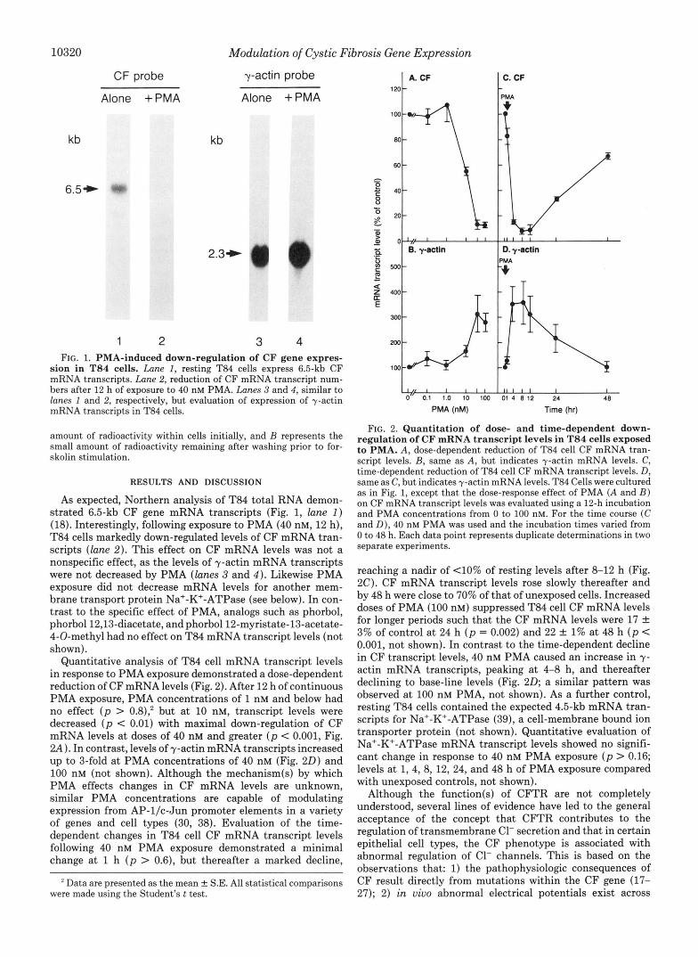
CF probe y-actin probe
Alone +PMA Alone +PMA
kb kb
2.3+
1 2 3 4 FIG. 1. PMA-induced down-regulation of CF gene expres-
sion in T84 cells. Lane 1, resting T84 cells express 6.5-kb CF mRNA transcripts. Lane 2, reduction of CF mRNA transcript num- bers after 12 h of exposure to 40 nM PMA. Lanes 3 and 4, similar to lanes 1 and 2, respectively, but evaluation of expression of y-actin mRNA transcripts in T84 cells.
amount of radioactivity within cells initially, and B represents the small amount of radioactivity remaining after washing prior to for- skolin stimulation.
RESULTS AND DISCUSSION
As expected, Northern analysis of T84 total RNA demon- strated 6.5-kb CF gene mRNA transcripts (Fig. 1, lune 2 ) (18). Interestingly, following exposure to PMA (40 nM, 12 h), T84 cells markedly down-regulated levels of CF mRNA tran- scripts (lane 2) . This effect on CF mRNA levels was not a nonspecific effect, as the levels of y-actin mRNA transcripts were not decreased by PMA (lanes 3 and 4 ) . Likewise PMA exposure did not decrease mRNA levels for another mem- brane transport protein Na'-K'-ATPase (see below). In con- trast to the specific effect of PMA, analogs such as phorbol, phorbol12,13-diacetate, andphorbol12-myristate-13-acetate- 4-0-methyl had no effect on T84 mRNA transcript levels (not shown).
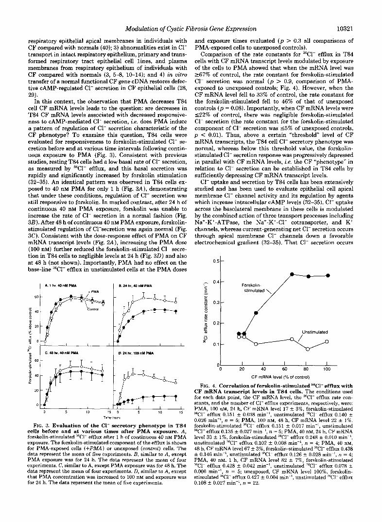
Quantitative analysis of T84 cell mRNA transcript levels in response to PMA exposure demonstrated a dose-dependent reduction of CF mRNA levels (Fig. 2). After 12 h of continuous PMA exposure, PMA concentrations of 1 nM and below had no effect ( p > 0.8); but at 10 nM, transcript levels were decreased ( p < 0.01) with maximal down-regulation of CF mRNA levels a t doses of 40 nM and greater ( p < 0.001, Fig. 2 A ) . In contrast, levels of y-actin mRNA transcripts increased up to %fold a t PMA concentrations of 40 nM (Fig. 20) and 100 nM (not shown). Although the mechanism(s) by which PMA effects changes in CF mRNA levels are unknown, similar PMA concentrations are capable of modulating expression from AP-l/c-Jun promoter elements in a variety of genes and cell types (30, 38). Evaluation of the time- dependent changes in T84 cell CF mRNA transcript levels following 40 nM PMA exposure demonstrated a minimal change at 1 h ( p > 0.6), but thereafter a marked decline,
Data are presented as the mean k S.E. All statistical comparisons were made using the Student's t test.
120
I -
I
48
PMA (nM) Time (hr)
FIG. 2. Quantitation of dose- and time-dependent down- regulation of CF mRNA transcript levels in T84 cells exposed to PMA. A , dose-dependent reduction of T84 cell CF mRNA tran- script levels. B, same as A , but indicates y-actin mRNA levels. C, time-dependent reduction of T84 cell CF mRNA transcript levels. D, same as C, but indicates y-actin mRNA levels. T84 Cells were cultured as in Fig. 1, except that the dose-response effect of PMA ( A and B ) on CF mRNA transcript levels was evaluated using a 12-h incubation and PMA concentrations from 0 to 100 nM. For the time course (C and D), 40 nM PMA was used and the incubation times varied from 0 to 48 h. Each data point represents duplicate determinations in two separate experiments.
reaching a nadir of (10% of resting levels after 8-12 h (Fig. 2C). CF mRNA transcript levels rose slowly thereafter and by 48 h were close to 70% of that of unexposed cells. Increased doses of PMA (100 nM) suppressed T84 cell CF mRNA levels for longer periods such that the CF mRNA levels were 17 f 3% of control a t 24 h ( p = 0.002) and 22 & 1% at 48 h ( p < 0.001, not shown). In contrast to the time-dependent decline in CF transcript levels, 40 nM PMA caused an increase in y- actin mRNA transcripts, peaking at 4-8 h, and thereafter declining to base-line levels (Fig. 20; a similar pattern was observed a t 100 nM PMA, not shown). As a further control, resting T84 cells contained the expected 4.5-kb mRNA tran- scripts for Na'-K+-ATPase (39), a cell-membrane bound ion transporter protein (not shown). Quantitative evaluation of Na+-K'-ATPase mRNA transcript levels showed no signifi- cant change in response to 40 nM PMA exposure ( p > 0.16; levels at 1,4,8, 12, 24, and 48 h of PMA exposure compared with unexposed controls, not shown).
Although the function(s) of CFTR are not completely understood, several lines of evidence have led to the general acceptance of the concept that CFTR contributes to the regulation of transmembrane C1- secretion and that in certain epithelial cell types, the CF phenotype is associated with abnormal regulation of C1- channels. This is based on the observations that: 1) the pathophysiologic consequences of CF result directly from mutations within the CF gene (17- 27); 2) in uiuo abnormal electrical potentials exist across
Modulation of Cystic Fibrosis Gene Expression 10321
respiratory epithelial apical membranes in individuals with CF compared with normals (40); 3) abnormalities exist in C1- transport in intact respiratory epithelium, primary and trans- formed respiratory tract epithelial cell lines, and plasma membranes from respiratory epithelium of individuals with CF compared with normals (3, 5-8, 10-14); and 4) in vitro transfer of a normal functional CF gene cDNA restores defec- tive CAMP-regulated C1- secretion in CF epithelial cells (28, 29).
In this context, the observation that PMA decreases T84 cell CF mRNA levels leads to the question: are decreases in T84 CF mRNA levels associated with decreased responsive- ness to CAMP-mediated C1- secretion, i.e. does PMA induce a pattern of regulation of C1- secretion characteristic of the CF phenotype? To examine this question, T84 cells were evaluated for responsiveness to forskolin-stimulated C1- se- cretion before and at various time intervals following contin- uous exposure to PMA (Fig. 3). Consistent with previous studies, resting T84 cells had a low basal rate of C1- secretion, as measured by 36Cl- efflux, and this basal secretion was rapidly and significantly increased by forskolin stimulation (32-35). An identical pattern was observed in T84 cells ex- posed to 40 nM PMA for only 1 h (Fig. 3A), demonstrating that under these conditions, regulation of C1- secretion was still responsive to forskolin. In marked contrast, after 24 h of continuous 40 nM PMA exposure, forskolin was unable to increase the rate of C1- secretion in a normal fashion (Fig. 3B). After 48 h of continuous 40 nM PMA exposure, forskolin- stimulated regulation of C1-secretion was again normal (Fig. 3C). Consistent with the dose-response effect of PMA on CF mRNA transcript levels (Fig. ZA), increasing the PMA dose (100 nM) further reduced the forskolin-stimulated C1- secre- tion in T84 cells to negligible levels at 24 h (Fig. 3 0 ) and also at 48 h (not shown). Importantly, PMA had no effect on the base-line 36Cl- efflux in unstimulated cells at the PMA doses
1 6.24 hr, 40 nM PMA
+ PMA - 60
- - g 40
8 a3
5 E O 3
5 0.24 hr, 100 nM PMA
60
t.J
0 5 10 15 0 5 10 15
Tlme Imln)
FIG. 3. Evaluation of the C1- secretory phenotype in T84 cells before and at various times after PMA exposure. A , forskolin-stimulated 36Cl- efflux after 1 h of continuous 40 nM PMA exposure. The forskolin-stimulated component of the efflux is shown for PMA-exposed cells (+PIMA) or unexposed (control) cells. The data represent the mean of five experiments. E , similar to A , except PMA exposure was for 24 h. The data represent the mean of four experiments. C, similar to A, except PMA exposure was for 48 h. The data represent the mean of four experiments. D, similar to A, except that PMA concentration was increased to 100 nM and exposure was for 24 h. The data represent the mean of five experiments.
and exposure times evaluated ( p > 0.3 all comparisons of PMA-exposed cells to unexposed controls).
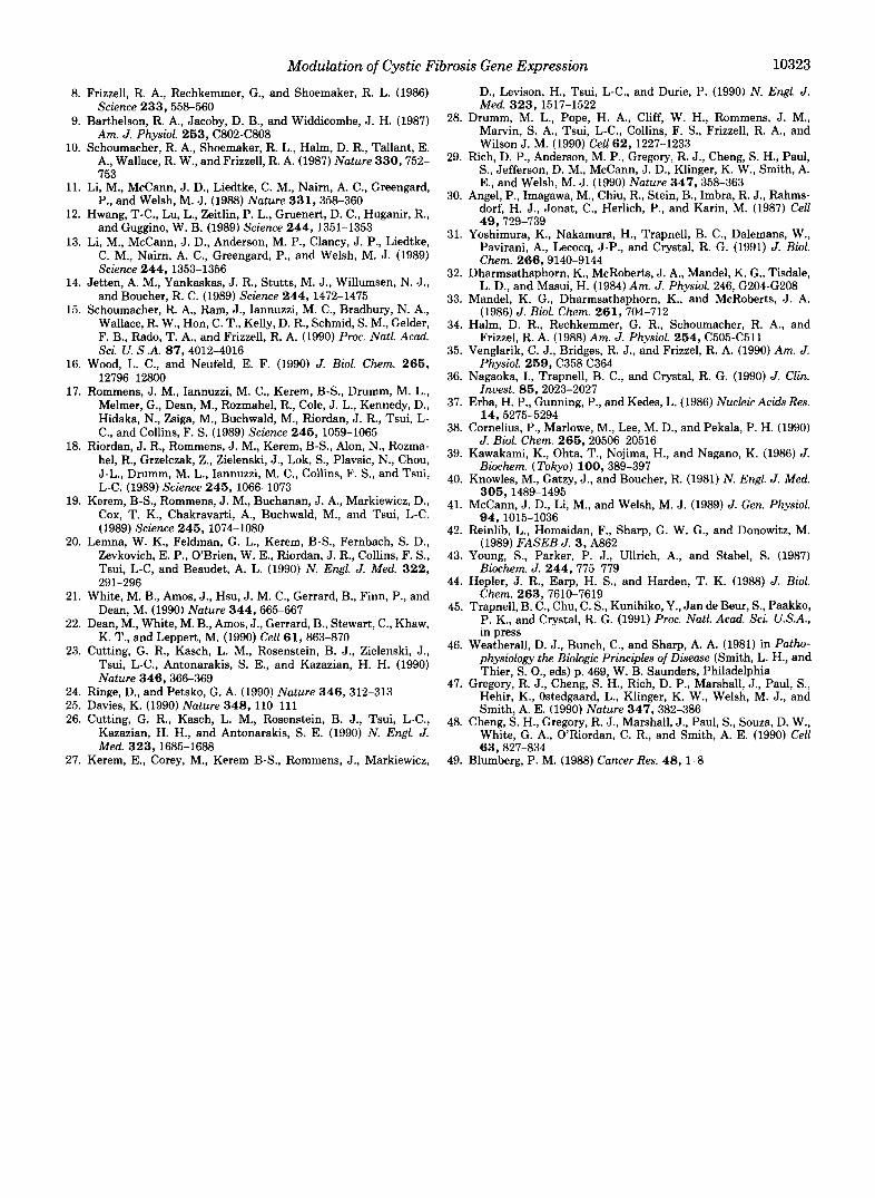
Comparison of the rate constants for 36Cl- efflux in T84 cells with CF mRNA transcript levels modulated by exposure of the cells to PMA showed that when the mRNA level was 267% of control, the rate constant for forskolin-stimulated C1- secretion was normal ( p > 0.9, comparison of PMA- exposed to unexposed controls; Fig. 4). However, when the CF mRNA level fell to 33% of control, the rate constant for the forskolin-stimulated fell to 46% of that of unexposed controls ( p = 0.08). Importantly, when CF mRNA levels were 522% of control, there was negligible forskolin-stimulated C1- secretion (the rate constant for the forskolin-stimulated component of C1- secretion was 15% of unexposed controls, p < 0.01). Thus, above a certain "threshold level of CF mRNA transcripts, the T84 cell C1- secretory phenotype was normal, whereas below this threshold value, the forskolin- stimulated C1- secretion response was progressively depressed in parallel with CF mRNA levels, i.e. the CF "phenotype" in relation to C1- secretion can be established in T84 cells by sufficiently depressing CF mRNA transcript levels.
C1- uptake and secretion by T84 cells has been extensively studied and has been used to evaluate epithelial cell apical membrane C1- channel activity and its regulation by agents which increase intracellular CAMP levels (32-35). C1- uptake across the basolateral membrane in these cells is modulated by the combined action of three transport processes including Na'-K'-ATPase, the Na'-K+-Cl- cotransporter, and K' channels, whereas current-generating net C1- secretion occurs through apical membrane C1- channels down a favorable electrochemical gradient (32-35). That C1- secretion occurs
0.5 t 0.4 -
0.3 -
0.2 -
0.1 -
0 I I I I I 0 20 40 60 80 100
CF mRNA level (% of control)
FIG. 4. Correlation of forskolin-stimulated 36Cl- efflux with CF mRNA transcript levels in T84 cells. The conditions used for each data point, the CF mRNA level, the 36Cl- efflux rate con- stants, and the number of C1- efflux experiments, respectively, were: PMA, 100 nM, 24 h, CF mRNA level 17 f 3%, forskolin-stimulated 3fiCl- efflux 0.151 f 0.038 min-', unstimulated 3GCl- efflux 0.140 f 0.026 min-', n = 5; PMA, 100 nM, 48 h, CF mRNA level 22 + 1%, forskolin-stimulated 36Cl- efflux 0.151 f 0.017 min-', unstimulated 36Cl- efflux 0.135 f 0.027 min", n = 5; PMA, 40 nM, 24 h, CF mRNA level 33 f 1%, forskolin-stimulated 36Cl- efflux 0.248 f 0.010 min-l, unstimulated 36Cl- efflux 0.102 f 0.008 rnin", n = 4; PMA, 40 nM, 48 h, CF mRNA level 67 f 3%, forskolin-stimulated 36Cl- efflux 0.438 f 0.146 rnin", unstimulated "Cl- efflux 0.126 -t 0.028 rnin", n = 4; PMA, 40 nM, 1 h, CF mRNA level 82 f 7%, forskolin-stimulated 36Cl- efflux 0.428 f 0.042 rnin", unstimulated "C1- efflux 0.078 C 0.006 rnin", n = 5; unexposed, CF mRNA level loo%, forskolin- stimulated 36Cl- efflux 0.427 f 0.004 min", unstimulated 36Cl- efflux 0.108 f 0.027 min-', n = 22.
10322 Modulation of Cystic Fibrosis Gene Expression
through apical membrane C1- channels is supported by Ussing chamber studies demonstrating vectoral C1- transport and studies with C1- channel blocking agents such as diphenyl carboxylate and 5-nitro-2-(3-phenylpropylamino)benzoic acid which block membrane C1- channels in T84 and other cells (33, 35, 41) and which block the CAMP-mediated C1- efflux in T84 cells (33,35) and apical membrane patches from these cells (34).
In the context of these considerations, if CFTR must be present in sufficient amounts and functioning normally to permit activation of the C1- channel by a CAMP-dependent process like phosphorylation, then decreased levels of CFTR would interfere with forskolin-stimulated C1- secretion. Thus, one explanation for our observations is that PMA, by reducing CF mRNA transcript levels, and presumably CFTR as well, interferes in this activation pathway by producing a physio- logic state with severely reduced levels of otherwise functional CFTR. It should be possible to test this hypothesis when a reliable quantitative CFTR-specific assay is available and its activity can be directly measured. An alternative explanation is that protein kinase C-mediated phosphorylation normally activates C1- channels independent of CFTR and that PMA induces the observed pattern of altered regulation of C1- secretion by down-regulating protein kinase C levels. This mechanism is unlikely, because it is now well established that CAMP-mediated C1- secretion is clearly dependent on CFTR, and because PMA does not alter adenylate cyclase activity nor the activity of CAMP-dependent protein kinase A (or its sensitivity to CAMP (9)) which is fully capable of activating the C1- channel in the absence of protein kinase C (11, 12). Furthermore, although there is evidence (for short periods at high intracellular Ca2+ levels), that protein kinase C can inhibit the outwardly rectifying C1- channel (13), it is also known that the phorbol ester phorbol dibutyrate rapidly down-regulates protein kinase C activity in T84 and other cells (42-45), and thus, after 24 h of PMA treatment when forskolin-activated C1- efflux is reduced, protein kinase C activity may be very low. Therefore, the effect of 24-h PMA exposure on T84 cell forskolin-stimulated C1- efflux is un- likely to be a direct effect of protein kinase C on membrane C1- channels but is likely a result of the reduced CFTR levels. It is also unlikely that PMA has a direct effect on the C1- channel itself, because after 1 h of PMA exposure, T84 cells were able to respond to forskolin stimulation by increasing ‘“C1- efflux normally (Fig. 3A). Theoretically, it is also pos- sible that PMA might alter the capacity of T84 cells to secrete C1- by affecting one of the transport processes that regulate C1- uptake. For example, PMA might affect Na+-K+-ATPase which might then affect the cell membrane potential and/or C1- uptake and accumulation within the cells. Although this might occur at the protein level, it does not at the level of gene expression, since Na+-K+-ATPase mRNA levels were unchanged by PMA exposure. Regardless of the exact mech- anism, by definition, because PMA blocks the forskolin-stim- ulated (CAMP-induced) C1- secretion in T84 cells, PMA in- duces the “CF C1- secretory phenotype.”
These observations have a number of interesting implica- tions for the control of expression of the CF gene and the relationship of the putative CF gene product to modulation of C1- secretion. First, the observation that a decrease in CF mRNA levels by PMA is associated with induction of the CF C1- secretory phenotype supports the concept of a direct link between abnormalities of the function and/or amount of the CF gene product and the anion secretory abnormalities ob- served in cells expressing two abnormal CF genes.
Second, in the context that in T84 cells there appears to be
a threshold level of CF mRNA required for normalcy, and because of the similarities between T84 cells and other human epithelial cells in regard to the regulation of C1- secretion, it seems reasonable to suspect that a threshold of expression of normal CF mRNA (and presumably also CFTR protein) exists in uiuo as well. This is consistent with the knowledge that individuals heterozygous for normal and AFb08 CF alleles have equal levels of normal and abnormal CF mRNA transcripts, yet have no clinically apparent disease (45). Furthermore, the concept of a threshold level of CF gene expression is also consistent with other biologic systems such as the human blood complement cascade where factor deficiencies cause coagulopathies when factor levels fall below certain critical values (e.g. factor IX has a threshold level of 10% (46)). Interestingly, recent findings indicate that CF gene mutations other than AFsos can result in a less severe clinical course (26, 27). This suggests there may be individuals in which decreased expression of a structurally normal (with respect to CFTR) CF gene or normal expression of a CFTR molecule with partial function results in mild disease because of partially dimin- ished, but not absent, CAMP-mediated regulation of C1- se- cretion. This expectation is also consistent with the analogy to the coagulation system in that for some factors (e.g. factor VIII), the severity of the coagulopathy correlates with the level of depression of the factor.
Third, although CFTR protein has not been directly char- acterized, the fact that the transient decrease in CF mRNA levels induced by 40 nM PMA exposure was paralleled by a similar transient decrease in forskolin-stimulated C1- efflux suggests that the CFTR protein may have a half-life in the range of hours. This indirect estimate is consistent with recent data (47, 48) demonstrating, with pulse-chase studies, that the intracellular product of the normal CF cDNA in COS7 cells has a lifespan of hours.
Fourth, in the context that PMA is a potent “inflammatory” stimulus (49), it is conceivable that other endogenous inflam- matory stimuli might also down-regulate CF gene expression in normal epithelium in a milieu of local inflammation, e.g. by down-regulating CF mRNA transcript levels, inflamma- tory stimuli might induce a “localized cystic fibrosis” in the affected region of an organ. Thus, it will be interesting to evaluate CF mRNA transcript levels in respiratory epithelial cells of individuals with inflammatory airway disorders such as chronic bronchitis.
Finally, this model of down-regulation of CF gene expres- sion and induction of the CF epithelial cell C1- secretory phenotype induced by PMA in cells with a normal CF gene and non-CF physiology may be useful to study the regulation of the CF gene expression and the precise function(s) of its gene product in health and disease.
Acknowledgments-We thank C. Layton and H. Lu for help with the 36Cl- efflux assays and D. Markakis for help with the statistical analysis.
REFERENCES 1. Boat, T. F., Welsh, M. J., and Beaudet, A. L. (1989) in The
Metabolic Basis of Inherited Disease (Scriver, C. R., Beaudet, A. L., Sly, W. S., and Valle, D., eds) 6th Ed., pp. 2649-2680, McGraw-Hill, New York
2. Welsh, M. J., and Fick, R. B. (1987) J. Clin. Invest. 80, 1523- 1526
3. Knowles, M. R., Stutts, M. J., Spock, A., Fischer, N., Gatzy, J. T., and Boucher, R. C. (1983) Science 221,1067-1070
4. Sato, K., and Sato, F. (1984) J. Clin. Invest. 7 3 , 1763-1771 5. Welsh, M. J., and Liedtke, C. M. (1986) Nature 322,467-470 6. Widdicombe, J. H. (1986) Am. J. Physiol. 261 , R818-FU22 7. Welsh, M. J. (1986) Science 232,164&1650
8.
9.
10.
11.
12.
13.
14.
15.
16.
17.
18.
19.
20.
21.
22.
23.
24. 25. 26.
Modulation of Cystic Fibros
Frizzell, R. A., Rechkemmer, G., and Shoemaker, R. L. (1986)
Barthelson, R. A., Jacoby, D. B., and Widdicombe, J. H. (1987) 28.
Schoumacher, R. A., Shoemaker, R. L., Halm, D. R., Tallant, E.
Science 233,558-560
Am. J. Physiol. 253, C802-C808
A., Wallace, R. W., and Frizzell, R. A. (1987) Nature 330, 752- 29. 753
Li, M., McCann, J. D., Liedtke, C. M., Nairn, A. C., Greengard, P., and Welsh, M. J. (1988) Nature 331,358-360
Hwang, T-C., Lu, L., Zeitlin, P. L., Gruenert, D. C., Huganir, R., and Guggino, W. B. (1989) Science 244, 1351-1353
Li, M., McCann, J. D., Anderson, M. P., Clancy, J. P., Liedtke, C. M., Nairn, A. C., Greengard, P., and Welsh, M. J. (1989) Science 244, 1353-1356
Jetten, A. M., Yankaskas, J. R., Stutts, M. J., Willumsen, N. J., 32.
and Boucher, R. C. (1989) Science 244, 1472-1475 Schoumacher, R. A., Ram, J., Iannuzzi, M. C., Bradbury, N. A.,
33.
Wallace, R. W., Hon, C. T., Kelly, D. R., Schmid, S. M., Gelder, 34. F. B., Rado, T. A,, and Frizzell, R. A. (1990) Proc. Natl. Acud. Sci. U. S .A. 8 7 , 4012-4016 35.
Wood, L. C., and Neufeld, E. F. (1990) J. Biol. Chem. 265, 12796-12800 36.
Rommens, J. M., Iannuzzi, M. C., Kerem, B-S., Drumm, M. L., Melmer, G., Dean, M., Rozmahel, R., Cole, J . L., Kennedy, D., 37. Hidaka, N., Zsiga, M., Buchwald, M., Riordan, J. R., Tsui, L- C., and Collins, F. S . (1989) Science 245, 1059-1065 38.
Riordan, J. R., Rommens, J . M., Kerem, B-S., Alon, N., Rozma- hel, R., Grzelczak, Z., Zielenski, J., Lok, S., Plavsic, N., Chou, 39' J-L., Drumm, M. L., Iannuzzi, M. C., Collins, F. S., and Tsui, 40. L-C. (1989) Science 245, 1066-1073
Kerem, B-S., Rommens, J. M., Buchanan, J. A., Markiewicz, D., 41. Cox, T. K., Chakravarti, A,, Buchwald, M., and Tsui, L-C. (1989) Science 245, 1074-1080 42.
Lemna, W. K., Feldman, G. L., Kerem, B-S., Fernbach, S. D., Zevkovich, E. P., O'Brien, W. E., Riordan, J. R., Collins, F. S., 43. Tsui, L-C, and Beaudet, A. L. (1990) N. Engl. J. Med. 322,
White, M. B., Amos, J., Hsu, J . M. C., Gerrard, B., Finn, P., and
Dean, M., White, M. B., Amos, J., Gerrard, B., Stewart, C., Khaw,
Cutting, G. R., Kasch, L. M., Rosenstein, B. J., Zielenski, J., 46'
47.
30.
31.
291-296 44.
Dean, M. (1990) Nature 344,665-667 45.
K. T., and Leppert, M. (1990) Cell 61,863-870
Tsui, L-C., Antonarakis, S. E., and Kazazian, H. H. (1990) Nature 346,366-369
Ringe, D., and Petsko, G. A. (1990) Nature 346,312-313 Davies, K. (1990) Nature 348, 110-111 Cutting, G. R., Kasch, L. M., Rosenstein, B. J., Tsui, L-C., 48.
Kazazian, H. H., and Antonarakis, S. E. (1990) N. Engl. J. Med. 323,1685-1688
, i s Gene Expression 10323
D., Levison, H., Tsui, L-C., and Durie, P. (1990) N. Engl. J. Med. 323,1517-1522
Drumm, M. L., Pope, H. A., Cliff, W. H., Rommens, J. M., Marvin, S. A., Tsui, L-C., Collins, F. S., Frizzell, R. A., and Wilson J. M. (1990) Cell 62, 1227-1233
Rich, D. P., Anderson, M. P., Gregory, R. J., Cheng, S. H., Paul, S., Jefferson, D. M., McCann, J. D., Klinger, K. W., Smith, A. E., and Welsh, M. J. (1990) Nature 347,358-363
Angel, P., Imagawa, M., Chiu, R., Stein, B., Imbra, R. J., Rahms- dorf. H. J.. Jonat. C.. Herlich. P.. and Karin. M. (1987) Cell , ,
49,729-739 , , , . .
Yoshimura. K.. Nakamura. H.. Tramell. B. C.. Dalemans. W.. Pavirani,' A.,' Lecocq, J-P., and Cbstal, R. G. (1991) J. 'Biol: Chem. 266,9140-9144
Dharmsathaphorn, K., McRoberts, J. A., Mandel, K. G., Tisdale, L. D., and Masui, H. (1984) Am. J . Physiol. 246, G204-G208
Mandel, K. G., Dharmsathaphorn, K., and McRoberts, J. A. (1986) J. Biol. Chem. 261, 704-712
Halm, D. R., Rechkemmer, G. R., Schoumacher, R. A., and Frizzel, R. A. (1988) Am. J . Physiol. 254, C505-C511
Venglarik, C. J., Bridges, R. J., and Frizzel, R. A. (1990) Am. J. Physiol. 259, C358 C364
Nagaoka. I., Tramell. B. C., and Cnrstal, R. G. (1990) J. Clin. I~uest.'85,20i3-2027
- . . .
Erba, H. P.. Gunning. P.. and Kedes. L. (1986) Nucleic Acids Res. -. I , . . 14; 527515294
J. Biol. Chem. 265, 20506-20516
Biochem. (Tokyo) 100, 389-397
Cornelius, P., Marlowe, M., Lee, M. D., and Pekala, P. H. (1990)
Kawakami, K., Ohta, T., Nojima, H., and Nagano, K. (1986) J.
Knowles, M., Gatzy, J., and Boucher, R. (1981) N. Engl. J. Med.
McCann, J. D., Li, M., and Welsh, M. J. (1989) J. Gen. Physiol.
Reinlib, L., Homaidan, F., Sharp, G. W. G., and Donowitz, M. (1989) FASEB J. 3, A862
Young, S., Parker, P. J., Ullrich, A., and Stabel, S. (1987) Biochem. J . 244, 775-779
Hepler, J. R., Earp, H. S., and Harden, T. K. (1988) J. Biol. Chem. 263, 7610-7619
Trapnell, B. C., Chu, C. S., Kunihiko, Y., Jan de Beur, S., Paakko, P. K., and Crystal, R. G. (1991) Proc. Natl. A d . Sci. U.S.A., in press
Weatherall, D. J., Bunch, C., and Sharp, A. A. (1981) in Patho- physiology the Biologic Principles of Diseuse (Smith, L. H., and Thier, S. O., eds) p. 469, W. B. Saunders, Philadelphia
Gregory, R. J., Cheng, S. H., Rich, D. P., Marshall, J., Paul, S., Hehir, K., Ostedgaard, L., Klinger, K. W., Welsh, M. J., and Smith, A. E. (1990) Nature 347,382-386
Cheng, S. H., Gregory, R. J., Marshall, J., Paul, S., Souza, D. W., White, G. A., O'Riordan, C. R., and Smith, A. E. (1990) Cell 63.827-834
305,1489-1495
94,1015-1036
27. Kerem, E., Corey, M., Kerem B-S., Rommens, J., Markiewicz, 49. Blumberg, P. M. (1988) Cancer Res. 48, 1-8
Related Documents