Biochem. J. (2013) 453, 155–166 (Printed in Great Britain) doi:10.1042/BJ20130316 155 REVIEW ARTICLE CRISPR interference: a structural perspective Judith REEKS, James H. NAISMITH 1 and Malcolm F. WHITE 1 Biomedical Sciences Research Complex, University of St Andrews, St Andrews, Fife KY16 9ST, U.K. CRISPR (cluster of regularly interspaced palindromic repeats) is a prokaryotic adaptive defence system, providing immunity against mobile genetic elements such as viruses. Genomically encoded crRNA (CRISPR RNA) is used by Cas (CRISPR-associated) proteins to target and subsequently degrade nucleic acids of invading entities in a sequence-dependent manner. The process is known as ‘interference’. In the present review we cover recent progress on the structural biology of the CRISPR/Cas system, focusing on the Cas proteins and complexes that catalyse crRNA biogenesis and interference. Structural studies have helped in the elucidation of key mechanisms, including the recognition and cleavage of crRNA by the Cas6 and Cas5 proteins, where remarkable diversity at the level of both substrate recognition and catalysis has become apparent. The RNA-binding RAMP (repeat-associated mysterious protein) domain is present in the Cas5, Cas6, Cas7 and Cmr3 protein families and RAMP-like domains are found in Cas2 and Cas10. Structural analysis has also revealed an evolutionary link between the small subunits of the type I and type III-B interference complexes. Future studies of the interference complexes and their constituent components will transform our understanding of the system. Key words: antiviral defence, cluster of regularly interspaced palindromic repeats (CRISPR), crystallography, evolution, protein structure, repeat-associated mysterious protein (RAMP). INTRODUCTION CRISPRs (cluster of regularly interspaced palindromic repeats) are a prokaryotic defence mechanism against viral infection and horizontal gene transfer. CRISPRs are the largest family of prokaryotic repeats [1] and have been found in 48 % of bacterial and 84 % of archaeal sequenced genomes to date [2]. A CRISPR array consists of a series of short identical repeat sequences separated by similarly short variable sequences known as spacers [3]. Located adjacent to the CRISPR array are clusters of cas (CRISPR-associated) genes [4] that encode for the proteins responsible for mediating the CRISPR response to foreign nucleic acids. The spacers are derived from foreign nucleic acids, such as viruses and conjugative plasmids, and provide the host with a ‘genetic memory’ of threats previously encountered [1,5,6]. New spacers are captured in a poorly understood process known as ‘adaptation’ and incorporated into the CRISPR locus [7]. The spacers are used to target foreign nucleic acids containing sequences complementary to the spacer, termed protospacers, for degradation [8]; the process is termed ‘interference’. The first step in the interference pathway is the transcription of the CRISPR array from a promoter located in the ‘leader’ sequence, an AT-rich region located upstream of the CRISPR array [4,9]. The array transcript {pre-crRNA [precursor crRNA (CRISPR RNA)]} is then processed into short crRNAs containing a spacer and flanking repeat fragments (Figure 1) [10]. These crRNAs are subsequently bound by complexes of Cas proteins and used to target homologous foreign dsDNA (double- stranded DNA) or ssRNA (single-stranded RNA) for nucleolytic degradation during CRISPR interference (Figure 1) [8,11]. The CRISPR/Cas systems are divided into three main types (I, II and III) on the basis of the identity and organisation of genes within a cas locus [12]. These types are further divided into a total of ten subtypes (I-A, I-B and so on), each of which expresses a different protein complex responsible for interference (Figures 1 and 2). The Cascade (CRISPR-associated complex for antiviral defence) is the effector complex for type I systems [8,13–15]. This name was originally used solely for the type I-E complex [8], which we here call eCascade, but increasingly Cascade is used more as a general term for all type I complexes. Type II systems use a single protein for interference (Cas9) [16], whereas the III-B subtype uses the CMR complex [11]. The interference complex of the III-A subtype has yet to be characterized biochemically, but the similarity of the III-A and III-B operons suggests that interference is indeed mediated by an effector complex rather than a single protein. As a result the putative complex has been termed the CSM complex [12]. Every CRISPR/Cas system apart from the III-B subtype is thought to target dsDNA by forming an R-loop structure, consisting of a heteroduplex between crRNA and the complementary protospacer strand and a ssDNA (single-stranded DNA) non-complementary strand, followed by degradation by the interference nuclease (Figure 1) [8,17–19]. The CMR complex targets ssRNA by forming an RNA duplex, which is subsequently cleaved [11,20]. The mechanisms of adaptation and CRISPR interference have been extensively reviewed (see references [21–26]). In the present review we will focus on the structural biology of the CRISPR system. Crystal structures are available for eight of the ‘core’ Cas proteins (those found in multiple subtypes) as well as a number of subtype-specific proteins (Figure 2 and Supplementary Table Abbreviations used: BhCas5c, Bacillus halodurans Cas5c; CRISPR, cluster of regularly interspaced palindromic repeats; Cas, CRISPR-associated; Cascade, CRISPR-associated complex for antiviral defence; crRNA, CRISPR RNA; dsDNA, double-stranded DNA; EcoCas3, Escherichia coli Cas3; EM, electron microscopy; HD, histidine–aspartate; MjaCas3 , Methanocaldococcus jannaschii Cas3 ; PaCas6f, Pseudomonas aeruginosa Cas6f; PAM, protospacer adjacent motif; PfuCas, Pyrococcus furiosus Cas; pre-crRNA, precursor crRNA; RAMP, repeat-associated mysterious protein; RRM, RNA recognition motif; ssDNA, single-stranded DNA; SsoCas, Sulfolobus solfataricus Cas; ssRNA, single-stranded RNA; SthCas3, Streptococcus thermophilus Cas3; tracrRNA, trans-activating crRNA; TtCas, Thermus thermophilus Cas. 1 Correspondence may be addressed to either of these authors (email [email protected] or [email protected]). c The Authors Journal compilation c 2013 Biochemical Society Biochemical Journal www.biochemj.org © 2013 The Author(s) The author(s) has paid for this article to be freely available under the terms of the Creative Commons Attribution Licence (CC-BY) (http://creativecommons.org/licenses/by/3.0/) which permits unrestricted use, distribution and reproduction in any medium, provided the original work is properly cited.

Welcome message from author
This document is posted to help you gain knowledge. Please leave a comment to let me know what you think about it! Share it to your friends and learn new things together.
Transcript
Biochem. J. (2013) 453, 155–166 (Printed in Great Britain) doi:10.1042/BJ20130316 155
REVIEW ARTICLECRISPR interference: a structural perspectiveJudith REEKS, James H. NAISMITH1 and Malcolm F. WHITE1
Biomedical Sciences Research Complex, University of St Andrews, St Andrews, Fife KY16 9ST, U.K.
CRISPR (cluster of regularly interspaced palindromic repeats) is aprokaryotic adaptive defence system, providing immunity againstmobile genetic elements such as viruses. Genomically encodedcrRNA (CRISPR RNA) is used by Cas (CRISPR-associated)proteins to target and subsequently degrade nucleic acids ofinvading entities in a sequence-dependent manner. The processis known as ‘interference’. In the present review we cover recentprogress on the structural biology of the CRISPR/Cas system,focusing on the Cas proteins and complexes that catalyse crRNAbiogenesis and interference. Structural studies have helped inthe elucidation of key mechanisms, including the recognitionand cleavage of crRNA by the Cas6 and Cas5 proteins, whereremarkable diversity at the level of both substrate recognition
and catalysis has become apparent. The RNA-binding RAMP(repeat-associated mysterious protein) domain is present in theCas5, Cas6, Cas7 and Cmr3 protein families and RAMP-likedomains are found in Cas2 and Cas10. Structural analysis hasalso revealed an evolutionary link between the small subunits ofthe type I and type III-B interference complexes. Future studiesof the interference complexes and their constituent componentswill transform our understanding of the system.
Key words: antiviral defence, cluster of regularly interspacedpalindromic repeats (CRISPR), crystallography, evolution,protein structure, repeat-associated mysterious protein (RAMP).
INTRODUCTION
CRISPRs (cluster of regularly interspaced palindromic repeats)are a prokaryotic defence mechanism against viral infectionand horizontal gene transfer. CRISPRs are the largest familyof prokaryotic repeats [1] and have been found in 48 % ofbacterial and 84% of archaeal sequenced genomes to date [2].A CRISPR array consists of a series of short identical repeatsequences separated by similarly short variable sequences knownas spacers [3]. Located adjacent to the CRISPR array are clustersof cas (CRISPR-associated) genes [4] that encode for the proteinsresponsible for mediating the CRISPR response to foreign nucleicacids. The spacers are derived from foreign nucleic acids, suchas viruses and conjugative plasmids, and provide the host witha ‘genetic memory’ of threats previously encountered [1,5,6].New spacers are captured in a poorly understood process knownas ‘adaptation’ and incorporated into the CRISPR locus [7].The spacers are used to target foreign nucleic acids containingsequences complementary to the spacer, termed protospacers, fordegradation [8]; the process is termed ‘interference’.
The first step in the interference pathway is the transcriptionof the CRISPR array from a promoter located in the ‘leader’sequence, an AT-rich region located upstream of the CRISPRarray [4,9]. The array transcript {pre-crRNA [precursor crRNA(CRISPR RNA)]} is then processed into short crRNAs containinga spacer and flanking repeat fragments (Figure 1) [10].These crRNAs are subsequently bound by complexes of Casproteins and used to target homologous foreign dsDNA (double-stranded DNA) or ssRNA (single-stranded RNA) for nucleolyticdegradation during CRISPR interference (Figure 1) [8,11].
The CRISPR/Cas systems are divided into three main types (I,II and III) on the basis of the identity and organisation of geneswithin a cas locus [12]. These types are further divided into a totalof ten subtypes (I-A, I-B and so on), each of which expresses adifferent protein complex responsible for interference (Figures 1and 2). The Cascade (CRISPR-associated complex for antiviraldefence) is the effector complex for type I systems [8,13–15].This name was originally used solely for the type I-E complex[8], which we here call eCascade, but increasingly Cascade is usedmore as a general term for all type I complexes. Type II systemsuse a single protein for interference (Cas9) [16], whereas the III-Bsubtype uses the CMR complex [11]. The interference complexof the III-A subtype has yet to be characterized biochemically,but the similarity of the III-A and III-B operons suggests thatinterference is indeed mediated by an effector complex rather thana single protein. As a result the putative complex has been termedthe CSM complex [12]. Every CRISPR/Cas system apart from theIII-B subtype is thought to target dsDNA by forming an R-loopstructure, consisting of a heteroduplex between crRNA and thecomplementary protospacer strand and a ssDNA (single-strandedDNA) non-complementary strand, followed by degradation by theinterference nuclease (Figure 1) [8,17–19]. The CMR complextargets ssRNA by forming an RNA duplex, which is subsequentlycleaved [11,20].
The mechanisms of adaptation and CRISPR interference havebeen extensively reviewed (see references [21–26]). In the presentreview we will focus on the structural biology of the CRISPRsystem. Crystal structures are available for eight of the ‘core’ Casproteins (those found in multiple subtypes) as well as a numberof subtype-specific proteins (Figure 2 and Supplementary Table
Abbreviations used: BhCas5c, Bacillus halodurans Cas5c; CRISPR, cluster of regularly interspaced palindromic repeats; Cas, CRISPR-associated;Cascade, CRISPR-associated complex for antiviral defence; crRNA, CRISPR RNA; dsDNA, double-stranded DNA; EcoCas3, Escherichia coli Cas3;EM, electron microscopy; HD, histidine–aspartate; MjaCas3′ ′, Methanocaldococcus jannaschii Cas3′ ′; PaCas6f, Pseudomonas aeruginosa Cas6f; PAM,protospacer adjacent motif; PfuCas, Pyrococcus furiosus Cas; pre-crRNA, precursor crRNA; RAMP, repeat-associated mysterious protein; RRM, RNArecognition motif; ssDNA, single-stranded DNA; SsoCas, Sulfolobus solfataricus Cas; ssRNA, single-stranded RNA; SthCas3, Streptococcus thermophilusCas3; tracrRNA, trans-activating crRNA; TtCas, Thermus thermophilus Cas.
1 Correspondence may be addressed to either of these authors (email [email protected] or [email protected]).
c© The Authors Journal compilation c© 2013 Biochemical Society
Bio
chem
ical
Jo
urn
al
ww
w.b
ioch
emj.o
rg
© 2013 The Author(s)
The author(s) has paid for this article to be freely available under the terms of the Creative Commons Attribution Licence (CC-BY) (http://creativecommons.org/licenses/by/3.0/)which permits unrestricted use, distribution and reproduction in any medium, provided the original work is properly cited.
156 J. Reeks, J. H. Naismith and M. F. White
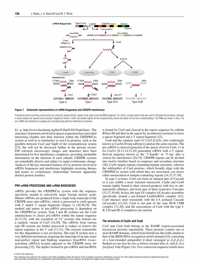
Figure 1 Schematic representation of crRNA biogenesis and CRISPR interference
Processing events involving nucleic acids are coloured; repeats (black), spacers (red–green) and tracrRNA (magenta). For clarity, a single spacer (red) was used to illustrate the processes, althoughin actual systems all spacers are processed. Targets are shown in other red shades (lighter for the complementary strand and darker for the non-complementary). The PAMs are shown in blue. Thepre-crRNA and interference nucleases are indicated along with the interference complexes.
S1 at http://www.biochemj.org/bj/453/bj4530155add.htm). Thestructures of proteins involved in spacer acquisition have providedinteresting insights into their function within the CRISPR/Cassystem as well as to similarities to non-Cas proteins, such as theparallels between Cas2 and VapD of the toxin/antitoxin system[27], but will not be discussed further in the present review.EM (electron microscopy) images and structures have beendetermined for five interference complexes, providing invaluableinformation on the function of each subunit. CRISPR systemsare remarkably diverse and subject to rapid evolutionary change.Analysis of the key structural features of Cas proteins involved incrRNA biogenesis and interference highlights recurring themesand points to evolutionary relationships between apparentlydistinct protein families.
PRE-crRNA PROCESSING AND crRNA BIOGENESIS
crRNA provides the CRISPR/Cas system with the sequencespecificity needed to selectively target foreign nucleic acids.Mature crRNAs are produced from a single long transcript of theCRISPR array (pre-crRNA), which is processed to yield spacerswith 5′ and/or 3′ repeat fragments (Figure 1) [10,28,29]. Themethod and nature of pre-crRNA processing is dependent onthe CRISPR/Cas system. Type I and III systems use the Cas6endonuclease to cleave pre-crRNA within the repeat sequence[8,13,15], with the exception of I-C systems that instead usea catalytic variant of Cas5 [14,30]. The crRNAs from varioustype III systems are further processed to reduce or remove therepeat sequence at the 3′ end [11,31]. The enzyme responsiblefor this degradation is not yet known. The type II system uses avery different mechanism, requiring the transcript of an anti-sensenear-perfect repeat and flanking sequences [tracrRNA (trans-activating crRNA)] located adjacent to the CRISPR array forprocessing [32]. The duplex formed by pre-crRNA and tracrRNA
is bound by Cas9 and cleaved in the repeat sequence by cellularRNase III and then in the spacer by an unknown nuclease to leavea spacer fragment and a 3′ repeat fragment [32].
Cas6 and the catalytic type I-C Cas5 [Cas5c, also confusinglyknown as Cas5d (Dvulg subtype)] catalyse the same reaction. Thepre-crRNA is cleaved upstream of the spacer (8 nt for Cas6, 11 ntfor Cas5c) [8,13,14,33,34] generating crRNA with a 5′ repeat-derived sequence known as the ‘5′-handle’ or ‘5′-tag’ that iscritical for interference [20,35]. CRISPR repeats can be dividedinto twelve families based on sequence and secondary structure[36]. Cas5c targets repeats containing hairpin structures, whereasthe subfamilies of Cas6 proteins, which broadly align with theCRISPR/Cas system with which they are associated, can cleaveeither unstructured or hairpin-containing repeats [14,33,37,38].
In type I systems, Cas6 can form an integral part of Cascadeor it can exhibit a more transient interaction. Cas6e and Cas6fremain tightly bound to their cleaved products with low or sub-nanomolar affinities, and form part of their respective Cascades[15,37,39,40]. In fact, the type I-F complex (f Cascade) assemblesspecifically around a pre-formed Cas6f/crRNA complex [41].Cas6 interacts more transiently with the I-A archaeal Cascade(aCascade) [13,42]. Cas6 is not part of the type III-B CMRcomplex [11,20], and the associations of Cas6 with the type I-B, I-D and III-A complexes are unclear.
The structures of Cas5c and Cas6
Cas5 and Cas6 both belong to the RAMP (repeat-associatedmysterious protein) superfamily. These proteins contain one ormore RAMP domains, which form ferredoxin-like folds similar tothat of the RRM (RNA recognition motif) domain [43], consistingof a four-stranded antiparallel β-sheet (arranged as β4β1β3β2)flanked on one face by two α-helices located after β1 and β3 in aβαββαβ fold (Figure 3A). Five conserved sequence motifs have
c© The Authors Journal compilation c© 2013 Biochemical Society© 2013 The Author(s)
The author(s) has paid for this article to be freely available under the terms of the Creative Commons Attribution Licence (CC-BY) (http://creativecommons.org/licenses/by/3.0/)which permits unrestricted use, distribution and reproduction in any medium, provided the original work is properly cited.
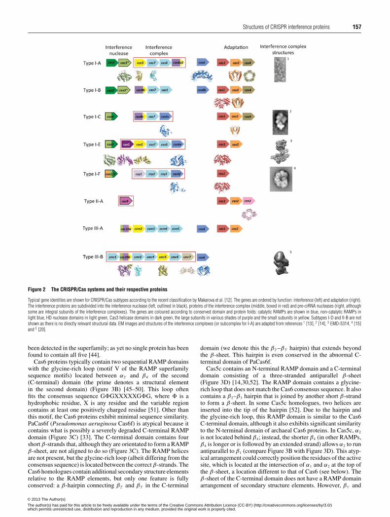
Structures of CRISPR interference proteins 157
Figure 2 The CRISPR/Cas systems and their respective proteins
Typical gene identities are shown for CRISPR/Cas subtypes according to the recent classification by Makarova et al. [12]. The genes are ordered by function: interference (left) and adaptation (right).The interference proteins are subdivided into the interference nuclease (left, outlined in black), proteins of the interference complex (middle, boxed in red) and pre-crRNA nucleases (right, althoughsome are integral subunits of the interference complexes). The genes are coloured according to conserved domain and protein folds: catalytic RAMPs are shown in blue, non-catalytic RAMPs inlight blue, HD nuclease domains in light green, Cas3 helicase domains in dark green, the large subunits in various shades of purple and the small subunits in yellow. Subtypes I-D and II-B are notshown as there is no directly relevant structural data. EM images and structures of the interference complexes (or subcomplex for I-A) are adapted from references 1 [13], 2 [14], 3 EMD-5314, 4 [15]and 5 [20].
been detected in the superfamily; as yet no single protein has beenfound to contain all five [44].
Cas6 proteins typically contain two sequential RAMP domainswith the glycine-rich loop (motif V of the RAMP superfamilysequence motifs) located between α2′ and β4′ of the second(C-terminal) domain (the prime denotes a structural elementin the second domain) (Figure 3B) [45–50]. This loop oftenfits the consensus sequence G�GXXXXXG�G, where � is ahydrophobic residue, X is any residue and the variable regioncontains at least one positively charged residue [51]. Other thanthis motif, the Cas6 proteins exhibit minimal sequence similarity.PaCas6f (Pseudomonas aeruginosa Cas6f) is atypical because itcontains what is possibly a severely degraded C-terminal RAMPdomain (Figure 3C) [33]. The C-terminal domain contains fourshort β-strands that, although they are orientated to form a RAMPβ-sheet, are not aligned to do so (Figure 3C). The RAMP helicesare not present, but the glycine-rich loop (albeit differing from theconsensus sequence) is located between the correct β-strands. TheCas6 homologues contain additional secondary structure elementsrelative to the RAMP elements, but only one feature is fullyconserved: a β-hairpin connecting β2′ and β3′ in the C-terminal
domain (we denote this the β2′ –β3′ hairpin) that extends beyondthe β-sheet. This hairpin is even conserved in the abnormal C-terminal domain of PaCas6f.
Cas5c contains an N-terminal RAMP domain and a C-terminaldomain consisting of a three-stranded antiparallel β-sheet(Figure 3D) [14,30,52]. The RAMP domain contains a glycine-rich loop that does not match the Cas6 consensus sequence. It alsocontains a β2–β3 hairpin that is joined by another short β-strandto form a β-sheet. In some Cas5c homologues, two helices areinserted into the tip of the hairpin [52]. Due to the hairpin andthe glycine-rich loop, this RAMP domain is similar to the Cas6C-terminal domain, although it also exhibits significant similarityto the N-terminal domain of archaeal Cas6 proteins. In Cas5c, α2
is not located behind β4; instead, the shorter β4 (in other RAMPs,β4 is longer or is followed by an extended strand) allows α2 to runantiparallel to β1 (compare Figure 3B with Figure 3D). This atyp-ical arrangement could correctly position the residues of the activesite, which is located at the intersection of α1 and α2 at the top ofthe β-sheet, a location different to that of Cas6 (see below). Theβ-sheet of the C-terminal domain does not have a RAMP domainarrangement of secondary structure elements. However, β1′ and
c© The Authors Journal compilation c© 2013 Biochemical Society© 2013 The Author(s)
The author(s) has paid for this article to be freely available under the terms of the Creative Commons Attribution Licence (CC-BY) (http://creativecommons.org/licenses/by/3.0/)which permits unrestricted use, distribution and reproduction in any medium, provided the original work is properly cited.

158 J. Reeks, J. H. Naismith and M. F. White
Figure 3 The structures of catalytic RAMP proteins
(A) Topology diagram of a RAMP domain. The β-strands are shown in blue and the α-helices in cyan. The glycine-rich loop found in many RAMPs is shown in yellow and the β2–β3 hairpin observedin some RAMPs is shown in green. The N- and C-termini are shown as blue and red spheres respectively. (B) The structure of TtCas6e (PDB code 1WJ9) highlighting the two RAMP domains thatmay have arisen from a pseudo-duplication event. Secondary structural elements are labelled as described in the text. Conserved RAMP elements are coloured as in (A) and non-conserved elementsin grey. Disordered regions are shown as broken black lines. (C) The atypical C-terminal domain of PaCas6f (PDB code 2XLK) that probably diverged from the standard RAMP fold. The recognizablefeatures are labelled. (D) The structure of BhCas5c (PDB code 4F3M), a catalytic variant of the typically non-catalytic Cas5 family. The short β4 strand and parallel α2 helix are boxed in black. Thepossible β2′ –β3′ hairpin in the C-terminal domain is shown in black.
β2′ form an extended β-hairpin reminiscent of the β2′ –β3′ hairpinof Cas6, although this is the only feature that is potentially RAMP-like. Thus it is not possible to say with certainty whether theC-terminal domain of Cas5c is a highly divergent RAMP domain.
RNA binding and cleavage
Cas5c and Cas6 are both metal-independent ribonucleases thatform products with 5′-hydroxyls and 2′,3′-cyclic phosphates[30,33,46,53], indicative of a general acid/base mechanisminvolving nucleophilic attack by the deprotonated 2′-hydroxyl onthe scissile phosphate. The active site of Cas6 is located betweenα1 and the glycine-rich loop, although the exact position of the sitevaries amongst the subfamilies (Figures 4A–4D). Remarkably, thecatalytic residues also vary between the proteins and none ofthe residues are conserved in all of the Cas6 subfamilies. Cas6enzymes from Pyrococcus furiosus (PfuCas6) and Thermusthermophilus (TtCas6) possess a catalytic triad of histidine,tyrosine and lysine residues similar to the RNA-splicingendonuclease [37,46,54,55]. The tyrosine residue has beenassigned as the general base and the histidine residue as the generalacid, with the lysine residue stabilizing the pentacoordinatephosphate intermediate. PaCas6f, however, uses a catalytic dyadof histidine and serine residues, with the histidine residue acting
as the general base and the serine residue holding the ribose ring inthe correct conformation [41]. Two active Cas6 paralogues fromSulfolobus solfataricus contain neither a general acid nor a generalbase, instead using conserved positively charged residues tocorrectly orientate the substrate and stabilize the pentacoordinatephosphate intermediate [49,50]. The presence of a catalytichistidine residue in the N-terminal domain had previously beenhighlighted as a characteristic feature of Cas6s [56], but it is nowclear that this is not necessarily the case.
The location of the Cas5c active site is different to that ofCas6, suggesting that the active sites evolved independently ofeach other. The catalytic triad of BhCas5c (Bacillus haloduransCas5c) consists of a tyrosine residue located in α1 and histidineand lysine residues in α2, similar to the PfuCas6 and TtCas6eactive sites [14]. The lysine is the only residue of the triadthat is invariant across the family; the tyrosine residue canbe exchanged for histidine (as in the active Cas5c nucleasesfrom Mannheimia succiniciproducens and Xanthomonas oryzae[30,52]), phenylalanine or leucine, whereas the catalytic histidineresidue can be replaced by other aromatic residues (phenylalan-ine/tyrosine) (Supplementary Figure S1 at http://www.biochemj.org/bj/453/bj4530155add.htm), but the roles of the residues arenot yet understood. None of these supposed catalytic residuesare conserved in other Cas5 proteins, perhaps unsurprisingly sinceonly Cas5c is catalytically active.
c© The Authors Journal compilation c© 2013 Biochemical Society© 2013 The Author(s)
The author(s) has paid for this article to be freely available under the terms of the Creative Commons Attribution Licence (CC-BY) (http://creativecommons.org/licenses/by/3.0/)which permits unrestricted use, distribution and reproduction in any medium, provided the original work is properly cited.
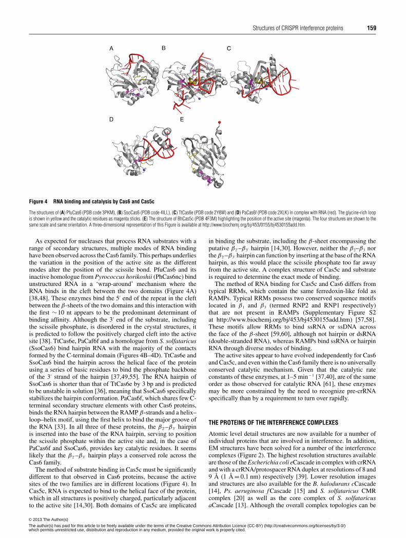
Structures of CRISPR interference proteins 159
Figure 4 RNA binding and catalysis by Cas6 and Cas5c
The structures of (A) PfuCas6 (PDB code 3PKM), (B) SsoCas6 (PDB code 4ILL), (C) TtCas6e (PDB code 2Y8W) and (D) PaCas6f (PDB code 2XLK) in complex with RNA (red). The glycine-rich loopis shown in yellow and the catalytic residues as magenta sticks. (E) The structure of BhCas5c (PDB 4F3M) highlighting the position of the active site (magenta). The four structures are shown to thesame scale and same orientation. A three-dimensional representation of this Figure is available at http://www.biochemj.org/bj/453/0155/bj4530155add.htm.
As expected for nucleases that process RNA substrates with arange of secondary structures, multiple modes of RNA bindinghave been observed across the Cas6 family. This perhaps underliesthe variation in the position of the active site as the differentmodes alter the position of the scissile bond. PfuCas6 and itsinactive homologue from Pyrococcus horikoshii (PhCas6nc) bindunstructured RNA in a ‘wrap-around’ mechanism where theRNA binds in the cleft between the two domains (Figure 4A)[38,48]. These enzymes bind the 5′ end of the repeat in the cleftbetween the β-sheets of the two domains and this interaction withthe first ∼10 nt appears to be the predominant determinant ofbinding affinity. Although the 3′ end of the substrate, includingthe scissile phosphate, is disordered in the crystal structures, itis predicted to follow the positively charged cleft into the activesite [38]. TtCas6e, PaCaf6f and a homologue from S. solfataricus(SsoCas6) bind hairpin RNA with the majority of the contactsformed by the C-terminal domain (Figures 4B–4D). TtCas6e andSsoCas6 bind the hairpin across the helical face of the proteinusing a series of basic residues to bind the phosphate backboneof the 3′ strand of the hairpin [37,49,55]. The RNA hairpin ofSsoCas6 is shorter than that of TtCas6e by 3 bp and is predictedto be unstable in solution [36], meaning that SsoCas6 specificallystabilizes the hairpin conformation. PaCas6f, which shares few C-terminal secondary structure elements with other Cas6 proteins,binds the RNA hairpin between the RAMP β-strands and a helix–loop–helix motif, using the first helix to bind the major groove ofthe RNA [33]. In all three of these proteins, the β2′ –β3′ hairpinis inserted into the base of the RNA hairpin, serving to positionthe scissile phosphate within the active site and, in the case ofPaCas6f and SsoCas6, provides key catalytic residues. It seemslikely that the β2′ –β3′ hairpin plays a conserved role across theCas6 family.
The method of substrate binding in Cas5c must be significantlydifferent to that observed in Cas6 proteins, because the activesites of the two families are in different locations (Figure 4). InCas5c, RNA is expected to bind to the helical face of the protein,which in all structures is positively charged, particularly adjacentto the active site [14,30]. Both domains of Cas5c are implicated
in binding the substrate, including the β-sheet encompassing theputative β2′ –β3′ hairpin [14,30]. However, neither the β2–β3 northe β2′ –β3′ hairpin can function by inserting at the base of the RNAhairpin, as this would place the scissile phosphate too far awayfrom the active site. A complex structure of Cas5c and substrateis required to determine the exact mode of binding.
The method of RNA binding for Cas5c and Cas6 differs fromtypical RRMs, which contain the same ferredoxin-like fold asRAMPs. Typical RRMs possess two conserved sequence motifslocated in β1 and β3 (termed RNP2 and RNP1 respectively)that are not present in RAMPs (Supplementary Figure S2at http://www.biochemj.org/bj/453/bj4530155add.htm) [57,58].These motifs allow RRMs to bind ssRNA or ssDNA acrossthe face of the β-sheet [59,60], although not hairpin or dsRNA(double-stranded RNA), whereas RAMPs bind ssRNA or hairpinRNA through diverse modes of binding.
The active sites appear to have evolved independently for Cas6and Cas5c, and even within the Cas6 family there is no universallyconserved catalytic mechanism. Given that the catalytic rateconstants of these enzymes, at 1–5 min− 1 [37,40], are of the sameorder as those observed for catalytic RNA [61], these enzymesmay be more constrained by the need to recognize pre-crRNAspecifically than by a requirement to turn over rapidly.
THE PROTEINS OF THE INTERFERENCE COMPLEXES
Atomic level detail structures are now available for a number ofindividual proteins that are involved in interference. In addition,EM structures have been solved for a number of the interferencecomplexes (Figure 2). The highest resolution structures availableare those of the Escherichia coli eCascade in complex with crRNAand with a crRNA/protospacer RNA duplex at resolutions of 8 and9 Å (1 Å = 0.1 nm) respectively [39]. Lower resolution imagesand structures are also available for the B. halodurans cCascade[14], Ps. aeruginosa f Cascade [15] and S. solfataricus CMRcomplex [20] as well as the core complex of S. solfataricusaCascade [13]. Although the overall complex topologies can be
c© The Authors Journal compilation c© 2013 Biochemical Society© 2013 The Author(s)
The author(s) has paid for this article to be freely available under the terms of the Creative Commons Attribution Licence (CC-BY) (http://creativecommons.org/licenses/by/3.0/)which permits unrestricted use, distribution and reproduction in any medium, provided the original work is properly cited.

160 J. Reeks, J. H. Naismith and M. F. White
Figure 5 The structure of Cas7, the core subunit of Cascade
(A) The structure of SsoCas7 (PDB code 3PS0) where the central RAMP domain is extended by an αβα motif (orange) and flanked by two unique domains (grey). The proposed crRNA-binding cleftlocated across the face of the β-sheet is indicated. (B) Topology diagram of SsoCas7 showing the connectivity of the RAMP fold relative to the other domains.
discerned, the resolution of these structures has precluded reliableplacement of individual proteins within the complex.
Cas7, the backbone of the type I complex
The structural backbone of Cascade is composed of multiplemonomers of Cas7 [13,14,39]. In eCascade, Cas7 assembles intoa helical hexameric structure with crRNA binding in a grooveformed along the outer face of the oligomer [39]. This helicalarrangement is conserved in the core complex of the S. solfataricusaCascade, although this complex of Cas5 and Cas7 forms oli-gomers of variable length [13]. It is possible that further factorsare needed to produce a complex of defined length or perhapsaCascade exhibits greater structural plasticity than eCascade. Asimilar helical arrangement to eCascade was observed in EMimages of cCascade [14], and, although it was not possible tounambiguously define the quaternary structure of the complex,it is probable that the six Cas7 subunits of the complex formthe same backbone. f Cascade contains six Csy3 subunits witha similar twisted topology to both cCascade and eCascade [15].This, combined with secondary structure predictions and MS frag-mentation analysis, has recently led to the hypothesis that Csy3actually belongs in an expanded Cas7 family [56,62]. Similarstructure predictions place Csc2 of dCascade in the Cas7 family[56], suggesting that the Cas7 helical backbone is a conservedand perhaps characteristic feature of all Cascade complexes.
The structure of Cas7 from one of the S. solfataricus aCascadecomplexes [13] (termed SsoCas7) contains a central RAMP foldmodified with an additional αβα motif located immediately afterβ4 (Figure 5A). This motif adds a fifth strand to the β-sheet(β5β4β1β3β2) with the two helices on either side of β5. The loopbetween α2 and β4 is disordered in the structure and is not glycine-rich, a conserved feature of the Cas7 family [56]. Significantinsertions are located between each of the four β-strands; theseform two distinct regions above and below the β-sheet to forma crescent-shaped molecule (Figure 5B). Residues located in thecleft of SsoCas7 have been implicated in binding crRNA [13].The structure of eCascade shows that the E. coli Cas7 adopts asimilar topology to SsoCas7 and that the cleft forms the extended
groove along the helical assembly of Cas7 [39]. Given the likelyubiquitous nature of the Cas7 backbone, it is probable that allCascade complexes bind crRNA in the same manner.
Non-catalytic variants of Cas5
Although Cas5c possesses catalytic activity, the other membersof the Cas5 family are non-catalytic and are limited to structuralroles. In both aCascade and eCascade, Cas5 interacts stably withCas7 [13,39]. Cas5e also interacts with Cse1 and Cse2 in eCascadeand appears to help stabilize the protospacer-bound conformationof the complex [39]. cCascade contains two copies of Cas5c,which appear to occupy the positions of Cas5 and Cas6e ineCascade [14,39]. Cas5c from Streptococcus pyogenes and X.oryzae bind dsDNA, which could be mimicking target dsDNA orthe heteroduplex of the interference R-loop [52]. Therefore Cas5cseems to be able to function as both a catalytic Cas6 equivalentand a structural Cas5 equivalent.
Of the Cascade complexes, only dCascade and f Cascade do notcontain Cas5 [12]. On the basis of secondary structure predictions,Makarova et al. [56] predicted that Csc1 (I-D) and Csy2 (I-F) belong to the Cas5 family. EM images and the small-angleX-ray scattering (SAXS) structure of f Cascade place Csy2 in asimilar position to the structural Cas5s of cCascade and eCascade[14,15,39]. However, the fragmentation patterns of eCascade andf Cascade suggest that Csy2 does not interact with Csy3 (probableCas7 equivalent) in the same manner as Cas5 and Cas7 fromeCascade, leading van Duijn et al. [62] to conclude that f Cascadedoes not contain a Cas5 equivalent. Further data are required tosettle the relationships between the complexes.
The small subunits of the interference complexes
Several of the interference complexes contain so-called ‘small’subunits, which are typically <200 residues. These proteins areCsa5 (I-A), Cse2 (I-E), Csm2 (III-A) and Cmr5 (III-B) and ithas been hypothesized that these proteins belong to a singlefamily (Cas11) [56]. Analysis of the structures of Csa5 [63], Cse2[64,65] and Cmr5 [66] (PDB codes 2OEB and 4GKF) shows that,
c© The Authors Journal compilation c© 2013 Biochemical Society© 2013 The Author(s)
The author(s) has paid for this article to be freely available under the terms of the Creative Commons Attribution Licence (CC-BY) (http://creativecommons.org/licenses/by/3.0/)which permits unrestricted use, distribution and reproduction in any medium, provided the original work is properly cited.

Structures of CRISPR interference proteins 161
Figure 6 The small subunits of interference complexes
Comparison of T. thermophilus Cmr5 (PDB code 2ZOP, left), T. thermophilus Cse2 (PDB code 2ZCA, middle) and S. solfataricus Csa5 (PDB code 3ZC4, right). The N-terminal domain of Cse2 (lightorange) is superimposed on Cmr5 (blue) and the C-terminal domain of Cse2 (yellow) is superimposed on Csa5 (green).
although structural homology can be detected, the evolutionarylinks between the proteins are complex. Cse2 contains N- andC-terminal domains that consist of four and five α-helicesrespectively. The N-terminal domain is homologous with the corestructure of Cmr5, whereas the C-terminal domain is homologouswith one of the domains of Csa5 (Figure 6). Csa5 consists of anα-helical domain (homologous with the Cse2 C-terminal domain)and a β-sheet domain that is not homologous with Cse2 or Cmr5.In fact, this domain is very poorly conserved across the Csa5family and is likely to vary significantly between homologues.
Possible evolutionary scenarios for the homology includefusion of csa5 and cmr5 genes to form cse2 or the evolutionof the three proteins from a single cse2-like gene with domainloss to form Csa5 and Cmr5 [63]. Csm2, the remaining smallsubunit for which there is no structure available, may be critical fordetermining the likely scenario, although it is certainly possiblethat Csm2 may not possess any homology with the other smallsubunits. Makarova et al. [56] suggested that the Cas8 C-terminaldomain, which is predicted to be helical, might be homologouswith the small subunits, although no experimental structure existsto confirm this.
The Cse2 dimer is an integral part of eCascade [39] and isresponsible for stabilizing the R-loop, increasing the affinity ofeCascade for dsDNA approximately 10-fold [67]. Cse2 alonebinds non-specifically to dsDNA and ssRNA [65]. Conversely,the S. solfataricus Csa5 does not stably interact with Cas5/Cas7 inthe presence of crRNA or with nucleic acids alone [63]. Cmr5,in contrast with both Csa5 and Cse2, appears to be non-essentialto the function of the CMR complex [11]. Thus we concludethat the similarity of the small subunits is structural rather thanfunctional.
The large subunits of the interference complexes
Similarly to the small subunits, each of the type I andIII interference complexes contains a ‘large’ (>500 residues)subunit: Cas8 (I-A, I-B, I-C), Cse1 (I-E), Csy1 (I-F) and Cas10(I-D, III-A and III-B). Cas10 was originally predicted to be apolymerase (hence the name polymerase cassette for the III-Bsubtype) on the basis of sequence features typical of a palmdomain commonly found in polymerases and cyclases [44].Subsequently it was proposed that all of the large subunits werehomologous and part of a Cas10 superfamily [56]. However,recent structures of a type III-B Cas10 [68,69], denoted Cas10b,show that, although the prediction of the palm domain was correct(albeit more akin to cyclases), no significant structural homologyexists with Cse1 [70,71] (PDB codes 4H3T and 4EJ3). This arguesagainst a single common ancestor for all of the large subunits.
Cas10, the large subunit of type III systems
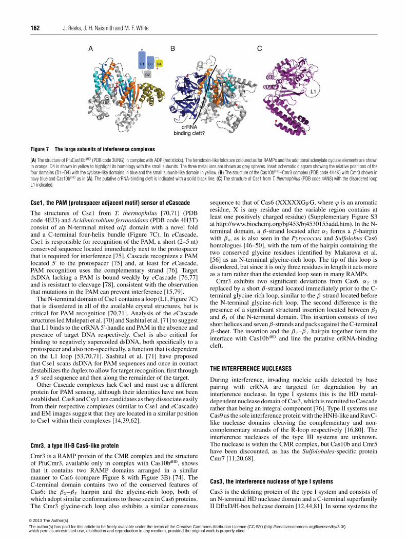
Cas10 is the defining protein of the type III system and consistsof an N-terminal HD (histidine–aspartate) phosphohydrolasedomain (for which there is no structure) and a C-terminalregion (Cas10dHD) that contains the palm domain [56]. Cas10bdHD
from P. furiosus consists of two adenylate cyclase-like domains(denoted D1 and D3) and two α-helical domains (D2 and D4)(Figures 7A and 7B) [68,69]. D2 is not significantly homologouswith known structures, but D4 is structurally homologous withCmr5 and the N-terminal domain of Cse2, although sequenceconservation is minimal and the biological implications of thehomology are unclear. A typical adenylate cyclase domainconsists of a ferredoxin-like fold with a C-terminal α3β5α4β6β7
modification, which creates a seven-stranded β-sheet with thetwo additional helices located on either side of the sheet [72].D1 and D3 lack some of these key structural elements: D3 lacksα4 and β6, whereas D1 lacks every additional element bar α3.Individually, D1 and D3 are most similar to the type III adenylatecyclase from Mycobacterium tuberculosis [72]. However, thesebacterial cyclases are typically homodimers, whereas D1 andD3 of Cas10bdHD exist as a pseudoheterodimer more similar tothe arrangement of mammalian cyclases [73]. The orientationbetween D1 and D3 is markedly different to that of typical cyclaseswhich, combined with the loss of key structural and sequencefeatures, is consistent with PfuCas10bdHD lacking a cyclase-likecatalytic activity, although D3 retains the ability to bind ADP[68].
In the CMR complex Cas10b interacts with Cmr3, aninteraction observed in both S. solfataricus and P. furiosus [20,74].The structure of the P. furiosus Cas10bdHD–Cmr3 complex showsthat the two proteins form a heterodimer with the interface formedby D1 of Cas10bdHD and one face of Cmr3 (see below) [74]. At theinterface between the two proteins is a highly positively chargedcleft ∼50 Å in length, which is suggestive of a role in crRNAbinding. The nucleotide bound by D3 in both the Cas10bdHD andCas10bdHD–Cmr3 structures lies at the centre of this cleft and socould be mimicking crRNA binding by the complex rather thansubstrate binding by the ‘cyclase’ domains of Cas10bdHD. This isconsistent with the nucleotide binding in a different orientation tothat observed in cyclases.
If the Cas10b–Cmr3 complex does bind to part of the crRNA,the remainder of the crRNA must be bound by other subunits of theCMR complex. Three subunits of the complex (Cmr1, Cmr4 andCmr6) are RAMPs and thus are plausible candidates. Makarova etal. [56] have predicted Cmr4 and Cmr6 to be Cas7 homologues.However, EM structures of the CMR complex (which targetsssRNA and not dsDNA) show that it is more compact than Cascadeand lacks a central helical structure [20].
c© The Authors Journal compilation c© 2013 Biochemical Society© 2013 The Author(s)
The author(s) has paid for this article to be freely available under the terms of the Creative Commons Attribution Licence (CC-BY) (http://creativecommons.org/licenses/by/3.0/)which permits unrestricted use, distribution and reproduction in any medium, provided the original work is properly cited.
162 J. Reeks, J. H. Naismith and M. F. White
Figure 7 The large subunits of interference complexes
(A) The structure of PfuCas10bdHD (PDB code 3UNG) in complex with ADP (red sticks). The ferredoxin-like folds are coloured as for RAMPs and the additional adenylate cyclase elements are shownin orange. D4 is shown in yellow to highlight its homology with the small subunits. The three metal ions are shown as grey spheres. Inset: schematic diagram showing the relative positions of thefour domains (D1–D4) with the cyclase-like domains in blue and the small subunit-like domain in yellow. (B) The structure of the Cas10bdHD–Cmr3 complex (PDB code 4H4K) with Cmr3 shown innavy blue and Cas10bdHD as in (A). The putative crRNA-binding cleft is indicated with a solid black line. (C) The structure of Cse1 from T. thermophilus (PDB code 4AN8) with the disordered loopL1 indicated.
Cse1, the PAM (protospacer adjacent motif) sensor of eCascade
The structures of Cse1 from T. thermophilus [70,71] (PDBcode 4EJ3) and Acidimicrobium ferrooxidans (PDB code 4H3T)consist of an N-terminal mixed α/β domain with a novel foldand a C-terminal four-helix bundle (Figure 7C). In eCascade,Cse1 is responsible for recognition of the PAM, a short (2–5 nt)conserved sequence located immediately next to the protospacerthat is required for interference [75]. Cascade recognizes a PAMlocated 5′ to the protospacer [75] and, at least for eCascade,PAM recognition uses the complementary strand [76]. TargetdsDNA lacking a PAM is bound weakly by eCascade [76,77]and is resistant to cleavage [78], consistent with the observationthat mutations in the PAM can prevent interference [15,79].
The N-terminal domain of Cse1 contains a loop (L1, Figure 7C)that is disordered in all of the available crystal structures, but iscritical for PAM recognition [70,71]. Analysis of the eCascadestructures led Mulepati et al. [70] and Sashital et al. [71] to suggestthat L1 binds to the crRNA 5′-handle and PAM in the absence andpresence of target DNA respectively. Cse1 is also critical forbinding to negatively supercoiled dsDNA, both specifically to aprotospacer and also non-specifically, a function that is dependenton the L1 loop [53,70,71]. Sashital et al. [71] have proposedthat Cse1 scans dsDNA for PAM sequences and once in contactdestabilizes the duplex to allow for target recognition, first througha 5′ seed sequence and then along the remainder of the target.
Other Cascade complexes lack Cse1 and must use a differentprotein for PAM sensing, although their identities have not beenestablished. Cas8 and Csy1 are candidates as they dissociate easilyfrom their respective complexes (similar to Cse1 and eCascade)and EM images suggest that they are located in a similar positionto Cse1 within their complexes [14,39,62].
Cmr3, a type III-B Cas6-like protein
Cmr3 is a RAMP protein of the CMR complex and the structureof PfuCmr3, available only in complex with Cas10bdHD, showsthat it contains two RAMP domains arranged in a similarmanner to Cas6 (compare Figure 8 with Figure 3B) [74]. TheC-terminal domain contains two of the conserved features ofCas6: the β2′ –β3′ hairpin and the glycine-rich loop, both ofwhich adopt similar conformations to those seen in Cas6 proteins.The Cmr3 glycine-rich loop also exhibits a similar consensus
sequence to that of Cas6 (XXXXXGϕG, where ϕ is an aromaticresidue, X is any residue and the variable region contains atleast one positively charged residue) (Supplementary Figure S3at http://www.biochemj.org/bj/453/bj4530155add.htm). In the N-terminal domain, a β-strand located after α2 forms a β-hairpinwith β4, as is also seen in the Pyrococcus and Sulfolobus Cas6homologues [46–50], with the turn of the hairpin containing thetwo conserved glycine residues identified by Makarova et al.[56] as an N-terminal glycine-rich loop. The tip of this loop isdisordered, but since it is only three residues in length it acts moreas a turn rather than the extended loop seen in many RAMPs.
Cmr3 exhibits two significant deviations from Cas6. α2′ isreplaced by a short β-strand located immediately prior to the C-terminal glycine-rich loop, similar to the β-strand located beforethe N-terminal glycine-rich loop. The second difference is thepresence of a significant structural insertion located between β2
and β3 of the N-terminal domain. This insertion consists of twoshort helices and seven β-strands and packs against the C-terminalβ-sheet. The insertion and the β2′ –β3′ hairpin together form theinterface with Cas10bdHD and line the putative crRNA-bindingcleft.
THE INTERFERENCE NUCLEASES
During interference, invading nucleic acids detected by basepairing with crRNA are targeted for degradation by aninterference nuclease. In type I systems this is the HD metal-dependent nuclease domain of Cas3, which is recruited to Cascaderather than being an integral component [76]. Type II systems useCas9 as the sole interference protein with the HNH-like and RuvC-like nuclease domains cleaving the complementary and non-complementary strands of the R-loop respectively [16,80]. Theinterference nucleases of the type III systems are unknown.The nuclease is within the CMR complex, but Cas10b and Cmr5have been discounted, as has the Sulfolobales-specific proteinCmr7 [11,20,68].
Cas3, the interference nuclease of type I systems
Cas3 is the defining protein of the type I system and consists ofan N-terminal HD nuclease domain and a C-terminal superfamilyII DExD/H-box helicase domain [12,44,81]. In some systems the
c© The Authors Journal compilation c© 2013 Biochemical Society© 2013 The Author(s)
The author(s) has paid for this article to be freely available under the terms of the Creative Commons Attribution Licence (CC-BY) (http://creativecommons.org/licenses/by/3.0/)which permits unrestricted use, distribution and reproduction in any medium, provided the original work is properly cited.

Structures of CRISPR interference proteins 163
Figure 8 The structure of Cmr3
(A) The structure of PfuCmr3 (PDB code 4H4K) showing the RAMP elements and the structural insertion in the N-terminal domain (orange). (B) Topology diagram of PfuCmr3 highlighting theconserved RAMP features and the connectivity of the insertion domain.
Figure 9 The structures of Cas protein HD domains
(A) The structure of TtCas3HD (PDB code 3SKD) with the conserved HD superfamily helices in green and numbered. The Ni2 + ion is shown as a dark grey sphere. Residues 222–260 are not shownas they are predicted to belong to the helicase domain. (B) A homology model of the HD domain of Cas10a from S. thermophilus created using PHYRE2 and consisting of residues 4–79. The fourHD domain helices are coloured in green and labelled. (C–E) Views of the active sites of (C) TtCas3HD, (D) MjaCas3′ ′ (PDB code 3S4L) and (E) SthCas10aHD. The HD superfamily motifs are shownas sticks with motif numbers in parentheses and the metal ions as grey spheres with site numbers in white.
two domains are expressed as separate proteins (Cas3′ ′ and Cas3′
respectively); other variations are also known, such as domainfusion to other Cas proteins (for example, Cas3–Cas2 in the I-F subtype and Cas3–Cse1 in some I-E systems) and inversionof the domain order (Figure 2) [12,44,76]. Cas3 is recruited byCascade after R-loop formation where it catalyses the unwindingand degradation of the invading DNA [76,78].
Cas3 proteins contain all five HD superfamily sequence motifs(H-HD-H-H-D) and the structures of TtCas3HD (HD domain ofTtCas3) and MjaCas3′ ′ (Methanocaldococcus jannaschii Cas3′ ′)revealed eight conserved helices, five of which are characteristicof the HD superfamily (Figure 9A) [82,83]. In the TtCas3HD
structure a single Ni2 + ion is bound by motifs I, II and V (site1), whereas site 2 (a binding site formed by motifs II, III andIV) remains unoccupied (Figure 9C). Metal binding at site 2 hasbeen observed in a number of HD domains (for example, see PDBcodes 2OGI, 2O08, 2PQ7, 3CCG and 3HC1) and its absence in theTtCas3HD structure is likely to be a crystal artefact. The MjaCas3′ ′
structure shows a Ca2 + ion bound at site 2 as well as a second ionbound by the histidine of motif II (site 3) (Figure 9D). However,
the binding at site 3 and the lack of binding at site 1 are likelyto be artefacts resulting from the protein engineering required forcrystallization.
Characterization of type I-E Cas3 nuclease domains from T.thermophilus, Streptococcus thermophilus, and E. coli and thetype I-A Cas3′ ′ proteins from M. jannaschii and P. furiosusshowed that they are all metal-dependent nucleases specific forssDNA, although the Cas3′ ′ proteins also cleave ssRNA in vitro[82–84]. These proteins are both endo- and exo-nucleases, withthe latter activity proceeding in the 3′→5′ direction. MjaCas3′ ′,SthCas3 (Streptococcus thermophilus Cas3) and EcoCas3 (E.coli Cas3) cleave R-loops, the biological substrate of Cas3 andMjaCas3′ ′ and SthCas3 have been shown to target the non-complementary ssDNA strand specifically [76,78,82]. Structuraldata is not available for the helicase domain of Cas3, but the type I-E helicase domains of SthCas3 and EcoCas3 catalyse the 3′→5′
Mg2 + - and ATP-dependent unwinding of dsDNA and DNA/RNAduplexes [84,85]. Nicking of the non-complementary strand by theHD domain followed by the unwinding of the DNA duplex bythe helicase domain would allow for progressive degradation
c© The Authors Journal compilation c© 2013 Biochemical Society© 2013 The Author(s)
The author(s) has paid for this article to be freely available under the terms of the Creative Commons Attribution Licence (CC-BY) (http://creativecommons.org/licenses/by/3.0/)which permits unrestricted use, distribution and reproduction in any medium, provided the original work is properly cited.
164 J. Reeks, J. H. Naismith and M. F. White
of the non-complementary strand (Supplementary Figure S4 athttp://www.biochemj.org/bj/453/bj4530155add.htm). The com-plementary strand is also targeted by Cas3 [78] and would occurafter dissociation of DNA from the R-loop.
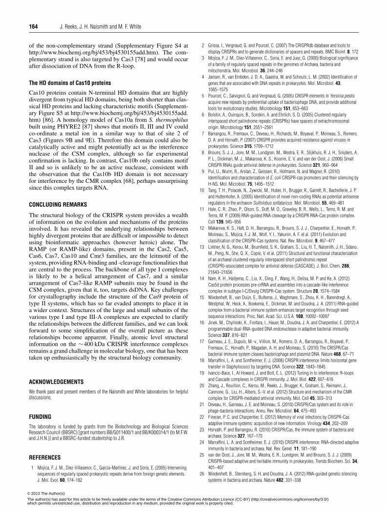
The HD domains of Cas10 proteins
Cas10 proteins contain N-terminal HD domains that are highlydivergent from typical HD domains, being both shorter than clas-sical HD proteins and lacking characteristic motifs (Supplement-ary Figure S5 at http://www.biochemj.org/bj/453/bj4530155add.htm) [86]. A homology model of Cas10a from S. thermophilusbuilt using PHYRE2 [87] shows that motifs II, III and IV couldco-ordinate a metal ion in a similar way to that of site 2 ofCas3 (Figures 9B and 9E). Therefore this domain could also becatalytically active and might potentially act as the interferencenuclease of the CSM complex, although so far experimentalconfirmation is lacking. In contrast, Cas10b only contains motifII and so is unlikely to be an active nuclease, consistent withthe observation that the Cas10b HD domain is not necessaryfor interference by the CMR complex [68], perhaps unsurprisingsince this complex targets RNA.
CONCLUDING REMARKS
The structural biology of the CRISPR system provides a wealthof information on the evolution and mechanisms of the proteinsinvolved. It has revealed the underlying relationships betweenhighly divergent proteins that are difficult or impossible to detectusing bioinformatic approaches (however heroic) alone. TheRAMP (or RAMP-like) domains, present in the Cas2, Cas5,Cas6, Cas7, Cas10 and Cmr3 families, are the leitmotif of thesystem, providing RNA-binding and -cleavage functionalities thatare central to the process. The backbone of all type I complexesis likely to be a helical arrangement of Cas7, and a similararrangement of Cas7-like RAMP subunits may be found in theCSM complex, given that it, too, targets dsDNA. Key challengesfor crystallography include the structure of the Cas9 protein oftype II systems, which has so far evaded attempts to place it ina wider context. Structures of the large and small subunits of thevarious type I and type III-A complexes are expected to clarifythe relationships between the different families, and we can lookforward to some simplification of the overall picture as theserelationships become apparent. Finally, atomic level structuralinformation on the ∼400 kDa CRISPR interference complexesremains a grand challenge in molecular biology, one that has beentaken up enthusiastically by the structural biology community.
ACKNOWLEDGEMENTS
We thank past and present members of the Naismith and White laboratories for helpfuldiscussions.
FUNDING
The laboratory is funded by grants from the Biotechnology and Biological SciencesResearch Council (BBSRC) [grant numbers BB/G011400/1 and BB/K000314/1 (to M.F.W.and J.H.N.)] and a BBSRC-funded studentship to J.R.
REFERENCES
1 Mojica, F. J. M., Diez-Villasenor, C., Garcia-Martinez, J. and Soria, E. (2005) Interveningsequences of regularly spaced prokaryotic repeats derive from foreign genetic elements.J. Mol. Evol. 60, 174–182
2 Grissa, I., Vergnaud, G. and Pourcel, C. (2007) The CRISPRdb database and tools todisplay CRISPRs and to generate dictionaries of spacers and repeats. BMC Bioinf. 8, 172
3 Mojica, F. J. M., Diez-Villasenor, C., Soria, E. and Juez, G. (2000) Biological significanceof a family of regularly spaced repeats in the genomes of Archaea, bacteria andmitochondria. Mol. Microbiol. 36, 244–246
4 Jansen, R., van Embden, J. D. A., Gaastra, W. and Schouls, L. M. (2002) Identification ofgenes that are associated with DNA repeats in prokaryotes. Mol. Microbiol. 43,1565–1575
5 Pourcel, C., Salvignol, G. and Vergnaud, G. (2005) CRISPR elements in Yersinia pestisacquire new repeats by preferential uptake of bacteriophage DNA, and provide additionaltools for evolutionary studies. Microbiology 151, 653–663
6 Bolotin, A., Ouinquis, B., Sorokin, A. and Ehrlich, S. D. (2005) Clustered regularlyinterspaced short palindrome repeats (CRISPRs) have spacers of extrachromosomalorigin. Microbiology 151, 2551–2561
7 Barrangou, R., Fremaux, C., Deveau, H., Richards, M., Boyaval, P., Moineau, S., Romero,D. A. and Horvath, P. (2007) CRISPR provides acquired resistance against viruses inprokaryotes. Science 315, 1709–1712
8 Brouns, S. J. J., Jore, M. M., Lundgren, M., Westra, E. R., Slijkhuis, R. J. H., Snijders, A.P. L., Dickman, M. J., Makarova, K. S., Koonin, E. V. and van der Oost, J. (2008) SmallCRISPR RNAs guide antiviral defense in prokaryotes. Science 321, 960–964
9 Pul, U., Wurm, R., Arslan, Z., Geissen, R., Hofmann, N. and Wagner, R. (2010)Identification and characterization of E. coli CRISPR-cas promoters and their silencing byH-NS. Mol. Microbiol. 75, 1495–1512
10 Tang, T. H., Polacek, N., Zywicki, M., Huber, H., Brugger, K., Garrett, R., Bachellerie, J. P.and Huttenhofer, A. (2005) Identification of novel non-coding RNAs as potential antisenseregulators in the archaeon Sulfolobus solfataricus. Mol. Microbiol. 55, 469–481
11 Hale, C. R., Zhao, P., Olson, S., Duff, M. O., Graveley, B. R., Wells, L., Terns, R. M. andTerns, M. P. (2009) RNA-guided RNA cleavage by a CRISPR RNA-Cas protein complex.Cell 139, 945–956
12 Makarova, K. S., Haft, D. H., Barrangou, R., Brouns, S. J. J., Charpentier, E., Horvath, P.,Moineau, S., Mojica, F. J. M., Wolf, Y. I., Yakunin, A. F. et al. (2011) Evolution andclassification of the CRISPR-Cas systems. Nat. Rev. Microbiol. 9, 467–477
13 Lintner, N. G., Kerou, M., Brumfield, S. K., Graham, S., Liu, H. T., Naismith, J. H., Sdano,M., Peng, N., She, Q. X., Copie, V. et al. (2011) Structural and functional characterizationof an archaeal clustered regularly interspaced short palindromic repeat(CRISPR)-associated complex for antiviral defense (CASCADE). J. Biol. Chem. 286,21643–21656
14 Nam, K. H., Haitjema, C., Liu, X., Ding, F., Wang, H., Delisa, M. P. and Ke, A. (2012)Cas5d protein processes pre-crRNA and assembles into a cascade-like interferencecomplex in subtype I-C/Dvulg CRISPR-Cas system. Structure 20, 1574–1584
15 Wiedenheft, B., van Duijn, E., Bultema, J., Waghmare, S., Zhou, K. H., Barendregt, A.,Westphal, W., Heck, A., Boekema, E., Dickman, M. and Doudna, J. A. (2011) RNA-guidedcomplex from a bacterial immune system enhances target recognition through seedsequence interactions. Proc. Natl. Acad. Sci. U.S.A. 108, 10092–10097
16 Jinek, M., Chylinski, K., Fonfara, I., Hauer, M., Doudna, J. A. and Charpentier, E. (2012) Aprogrammable dual-RNA-guided DNA endonuclease in adaptive bacterial immunity.Science 337, 816–821
17 Garneau, J. E., Dupuis, M.-v., Villion, M., Romero, D. A., Barrangou, R., Boyaval, P.,Fremaux, C., Horvath, P., Magadan, A. H. and Moineau, S. (2010) The CRISPR/Casbacterial immune system cleaves bacteriophage and plasmid DNA. Nature 468, 67–71
18 Marraffini, L. A. and Sontheimer, E. J. (2008) CRISPR interference limits horizontal genetransfer in Staphylococci by targeting DNA. Science 322, 1843–1845
19 Ivancic-Bace, I., Al Howard, J. and Bolt, E. L. (2012) Tuning in to interference: R-loopsand Cascade complexes in CRISPR immunity. J. Mol. Biol. 422, 607–616
20 Zhang, J., Rouillon, C., Kerou, M., Reeks, J., Brugger, K., Graham, S., Reimann, J.,Cannone, G., Liu, H., Albers, S.-V. et al. (2012) Structure and mechanism of the CMRcomplex for CRISPR-mediated antiviral immunity. Mol. Cell 45, 303–313
21 Deveau, H., Garneau, J. E. and Moineau, S. (2010) CRISPR/Cas system and its role inphage-bacteria interactions. Annu. Rev. Microbiol. 64, 475–493
22 Fineran, P. C. and Charpentier, E. (2012) Memory of viral infections by CRISPR-Casadaptive immune systems: acquisition of new information. Virology 434, 202–209
23 Horvath, P. and Barrangou, R. (2010) CRISPR/Cas, the immune system of bacteria andarchaea. Science 327, 167–170
24 Marraffini, L. A. and Sontheimer, E. J. (2010) CRISPR interference: RNA-directed adaptiveimmunity in bacteria and archaea. Nat. Rev. Genet. 11, 181–190
25 van der Oost, J., Jore, M. M., Westra, E. R., Lundgren, M. and Brouns, S. J. J. (2009)CRISPR-based adaptive and heritable immunity in prokaryotes. Trends Biochem. Sci. 34,401–407
26 Wiedenheft, B., Sternberg, S. H. and Doudna, J. A. (2012) RNA-guided genetic silencingsystems in bacteria and archaea. Nature 482, 331–338
c© The Authors Journal compilation c© 2013 Biochemical Society© 2013 The Author(s)
The author(s) has paid for this article to be freely available under the terms of the Creative Commons Attribution Licence (CC-BY) (http://creativecommons.org/licenses/by/3.0/)which permits unrestricted use, distribution and reproduction in any medium, provided the original work is properly cited.
Structures of CRISPR interference proteins 165
27 Kwon, A.-R., Kim, J.-H., Park, S. J., Lee, K.-Y., Min, Y.-H., Im, H., Lee, I., Lee, K.-Y. andLee, B.-J. (2012) Structural and biochemical characterization of HP0315 fromHelicobacter pylori as a VapD protein with an endoribonuclease activity. Nucleic AcidsRes. 40, 4216–4228
28 Tang, T. H., Bachellerie, J. P., Rozhdestvensky, T., Bortolin, M. L., Huber, H., Drungowski,M., Elge, T., Brosius, J. and Huttenhofer, A. (2002) Identification of 86 candidates forsmall non-messenger RNAs from the archaeon Archaeoglobus fulgidus. Proc. Natl. Acad.Sci. U.S.A. 99, 7536–7541
29 Lillestol, R. K., Shah, S. A., Brugger, K., Redder, P., Phan, H., Christiansen, J. and Garrett,R. A. (2009) CRISPR families of the crenarchaeal genus Sulfolobus: bidirectionaltranscription and dynamic properties. Mol. Microbiol. 72, 259–272
30 Garside, E. L., Schellenberg, M. J., Gesner, E. M., Bonanno, J. B., Sauder, J. M., Burley,S. K., Almo, S. C., Mehta, G. and Macmillan, A. M. (2012) Cas5d processes pre-crRNAand is a member of a larger family of CRISPR RNA endonucleases. RNA 18, 2020–2028
31 Hatoum-Aslan, A., Maniv, I. and Marraffini, L. A. (2011) Mature clustered, regularlyinterspaced, short palindromic repeats RNA (crRNA) length is measured by a rulermechanism anchored at the precursor processing site. Proc. Natl. Acad. Sci. U.S.A. 108,21218–21222
32 Deltcheva, E., Chylinski, K., Sharma, C. M., Gonzales, K., Chao, Y. J., Pirzada, Z. A.,Eckert, M. R., Vogel, J. and Charpentier, E. (2011) CRISPR RNA maturation bytrans-encoded small RNA and host factor RNase III. Nature 471, 602–607
33 Haurwitz, R. E., Jinek, M., Wiedenheft, B., Zhou, K. H. and Doudna, J. A. (2010)Sequence- and structure-specific RNA processing by a CRISPR endonuclease. Science329, 1355–1358
34 Richter, H., Zoephel, J., Schermuly, J., Maticzka, D., Backofen, R. and Randau, L. (2012)Characterization of CRISPR RNA processing in Clostridium thermocellum andMethanococcus maripaludis. Nucleic Acids Res. 40, 9887–9896
35 Marraffini, L. A. and Sontheimer, E. J. (2010) Self versus non-self discrimination duringCRISPR RNA-directed immunity. Nature 463, 568–571
36 Kunin, V., Sorek, R. and Hugenholtz, P. (2007) Evolutionary conservation of sequence andsecondary structures in CRISPR repeats. Genome Biol. 8, R61
37 Sashital, D. G., Jinek, M. and Doudna, J. A. (2011) An RNA-induced conformationalchange required for CRISPR RNA cleavage by the endoribonuclease Cse3. Nat. Struct.Mol. Biol. 18, 680–687
38 Wang, R. Y., Preamplume, G., Terns, M. P., Terns, R. M. and Li, H. (2011) Interaction ofthe Cas6 riboendonuclease with CRISPR RNAs: recognition and cleavage. Structure 19,257–264
39 Wiedenheft, B., Lander, G. C., Zhou, K. H., Jore, M. M., Brouns, S. J. J., van der Oost, J.,Doudna, J. A. and Nogales, E. (2011) Structures of the RNA-guided surveillance complexfrom a bacterial immune system. Nature 477, 486–489
40 Sternberg, S. H., Haurwitz, R. E. and Doudna, J. A. (2012) Mechanism of substrateselection by a highly specific CRISPR endoribonuclease. RNA 18, 661–672
41 Haurwitz, R. E., Sternberg, S. H. and Doudna, J. A. (2012) Csy4 relies on an unusualcatalytic dyad to position and cleave CRISPR RNA. EMBO J. 31, 2824–2832
42 Plagens, A., Tjaden, B., Hagemann, A., Randau, L. and Hensel, R. (2012) Characterizationof the CRISPR/Cas subtype I-A system of the hyperthermophilic crenarchaeonThermoproteus tenax. J. Bacteriol. 194, 2491–2500
43 Wang, R. Y. and Li, H. (2012) The mysterious RAMP proteins and their roles in smallRNA-based immunity. Protein Sci. 21, 463–470
44 Makarova, K. S., Grishin, N. V., Shabalina, S. A., Wolf, Y. I. and Koonin, E. V. (2006) Aputative RNA-interference-based immune system in prokaryotes: computational analysisof the predicted enzymatic machinery, functional analogies with eukaryotic RNAi, andhypothetical mechanisms of action. Biol. Direct 1, 7
45 Ebihara, A., Yao, M., Masui, R., Tanaka, I., Yokoyama, S. and Kuramitsu, S. (2006) Crystalstructure of hypothetical protein TTHB192 from Thermus thermophilus HB8 reveals a newprotein family with an RNA recognition motif-like domain. Protein Sci. 15, 1494–1499
46 Carte, J., Wang, R. Y., Li, H., Terns, R. M. and Terns, M. P. (2008) Cas6 is anendoribonuclease that generates guide RNAs for invader defense in prokaryotes. GenesDev. 22, 3489–3496
47 Park, H.-M., Shin, M., Sun, J., Kim, G. S., Lee, Y. C., Park, J.-H., Kim, B. Y. and Kim, J.-S.(2012) Crystal structure of a Cas6 paralogous protein from Pyrococcus furiosus.Proteins: Struct., Funct., Bioinf. 80, 1895–1900
48 Wang, R., Zheng, H., Preamplume, G., Shao, Y. and Li, H. (2012) The impact of CRISPRrepeat sequence on structures of a Cas6 protein-RNA complex. Protein Sci. 21, 405–417
49 Shao, Y. and Li, H. (2013) Recognition and cleavage of a nonstructured CRISPR RNA byits processing endoribonuclease Cas6. Structure 21, 385–393
50 Reeks, J., Sokolowski, R. D., Graham, S., Liu, H., Naismith, J. H. and White, M. F. (2013)Structure of a dimeric crenarchaeal Cas6 enzyme with an atypical active site for CRISPRRNA processing. Biochem. J. 452, 223–230
51 Makarova, K. S., Aravind, L., Grishin, N. V., Rogozin, I. B. and Koonin, E. V. (2002) A DNArepair system specific for thermophilic Archaea and bacteria predicted by genomic contextanalysis. Nucleic Acids Res. 30, 482–496
52 Koo, Y., Ka, D., Kim, E.-J., Suh, N. and Bae, E. (2013) Conservation and variability in thestructure and function of the Cas5d endoribonuclease in the CRISPR-mediated microbialimmune system. J. Mol. Biol., doi:10.1016/j.jmb.2013.02.032
53 Jore, M. M., Lundgren, M., van Duijn, E., Bultema, J. B., Westra, E. R., Waghmare, S. P.,Wiedenheft, B., Pul, U., Wurm, R., Wagner, R. et al. (2011) Structural basis for CRISPRRNA-guided DNA recognition by Cascade. Nat. Struct. Mol. Biol. 18, 529–536
54 Calvin, K. and Li, H. (2008) RNA-splicing endonuclease structure and function. Cell. Mol.Life Sci. 65, 1176–1185
55 Gesner, E. M., Schellenberg, M. J., Garside, E. L., George, M. M. and MacMillan, A. M.(2011) Recognition and maturation of effector RNAs in a CRISPR interference pathway.Nat. Struct. Mol. Biol. 18, 688–692
56 Makarova, K. S., Aravind, L., Wolf, Y. I. and Koonin, E. V. (2011) Unification of Cas proteinfamilies and a simple scenario for the origin and evolution of CRISPR-Cas systems. Biol.Direct 6, 38
57 Maris, C., Dominguez, C. and Allain, F. H. T. (2005) The RNA recognition motif, a plasticRNA-binding platform to regulate post-transcriptional gene expression. FEBS J. 272,2118–2131
58 Clery, A., Blatter, M. and Allain, F. H. T. (2008) RNA recognition motifs: boring? Not quite.Curr. Opin. Struct. Biol. 18, 290–298
59 Oubridge, C., Ito, N., Evans, P. R., Teo, C. H. and Nagai, K. (1994) Crystal-structure at1.92 angstrom resolution of the RNA-binding domain of the U1A spliceosomal proteincomplexed with an RNA hairpin. Nature 372, 432–438
60 Ding, J. Z., Hayashi, M. K., Zhang, Y., Manche, L., Krainer, A. R. and Xu, R. M. (1999)Crystal structure of the two-RRM domain of hnRNP A1 (UP1) complexed withsingle-stranded telomeric DNA. Genes Dev. 13, 1102–1115
61 Lilley, D. M. J. (2003) The origins of RNA catalysis in ribozymes. Trends Biochem. Sci.28, 495–501
62 van Duijn, E., Barbu, I. M., Barendregt, A., Jore, M. M., Wiedenheft, B., Lundgren, M.,Westra, E. R., Brouns, S. J. J., Doudna, J. A., van der Oost, J. and Heck, A. J. R. (2012)Native tandem and ion mobility mass spectrometry highlight structural and modularsimilarities in clustered-regularly-interspaced shot-palindromic-repeats(CRISPR)-associated protein complexes from Escherichia coli and Pseudomonasaeruginosa. Mol. Cell. Proteomics 11, 1430–1441
63 Reeks, J., Graham, S., Anderson, L., Liu, H., White, M. F. and Naismith, J. H. (2013)Structure of the archaeal Cascade subunit Csa5: relating the small subunits of CRISPReffector complexes. RNA Biol. 10, doi:10.4161/rna.23854
64 Agari, Y., Yokoyama, S., Kuramitsu, S. and Shinkai, A. (2008) X-ray crystal structure of aCRISPR-associated protein, Cse2, from Thermus thermaphilus HB8. Proteins: Struct.,Funct., Bioinf. 73, 1063–1067
65 Nam, K. H., Huang, Q. and Ke, A. (2012) Nucleic acid binding surface and dimer interfacerevealed by CRISPR-associated CasB protein structures. FEBS Lett. 586, 3956–3961
66 Sakamoto, K., Agari, Y., Agari, K., Yokoyama, S., Kuramitsu, S. and Shinkai, A. (2009)X-ray crystal structure of a CRISPR-associated RAMP superfamily protein, Cmr5, fromThermus thermophilus HB8. Proteins: Struct., Funct., Bioinf. 75, 528–532
67 Westra, E. R., Nilges, B., van Erp, P. B. G., van der Oost, J., Dame, R. T. and Brouns, S. J.J. (2012) Cascade-mediated binding and bending of negatively supercoiled DNA. RNABiol. 9, 1134–1138
68 Cocozaki, A. I., Ramia, N. F., Shao, Y., Hale, C. R., Terns, R. M., Terns, M. P. and Li, H.(2012) Structure of the Cmr2 subunit of the CRISPR-Cas RNA silencing complex.Structure 20, 545–553
69 Zhu, X. and Ye, K. (2012) Crystal structure of Cmr2 suggests a nucleotide cyclase-relatedenzyme in type III CRISPR-Cas systems. FEBS Lett. 586, 939–945
70 Mulepati, S., Orr, A. and Bailey, S. (2012) Crystal structure of the largest subunit of abacterial RNA-guided immune complex and its role in DNA target binding. J. Biol. Chem.287, 22445–22449
71 Sashital, D. G., Wiedenheft, B. and Doudna, J. A. (2012) Mechanism of foreign DNAselection in a bacterial adaptive immune system. Mol. Cell 46, 606–615
72 Sinha, S. C., Wetterer, M., Sprang, S. R., Schultz, J. E. and Linder, J. U. (2005) Origin ofasymmetry in adenylyl cyclases: structures of Mycobacterium tuberculosis Rv1900c.EMBO J. 24, 663–673
73 Linder, J. U. and Schultz, J. E. (2003) The class III adenylyl cyclases: multi-purposesignalling modules. Cell. Signalling 15, 1081–1089
74 Shao, Y., Cocozaki, A. I., Ramia, N. F., Terns, R. M., Terns, M. P. and Li, H. (2013) Structureof the cmr2-cmr3 subcomplex of the cmr RNA silencing complex. Structure 21, 376–384
75 Mojica, F. J. M., Diez-Villasenor, C., Garcia-Martinez, J. and Almendros, C. (2009) Shortmotif sequences determine the targets of the prokaryotic CRISPR defence system.Microbiology 155, 733–740
76 Westra, E. R., van Erp, P. B. G., Kunne, T., Wong, S. P., Staals, R. H. J., Seegers, C. L. C.,Bollen, S., Jore, M. M., Semenova, E., Severinov, K. et al. (2012) CRISPR immunity relieson the consecutive binding and degradation of negatively supercoiled invader DNA byCascade and Cas3. Mol. Cell 46, 595–605
c© The Authors Journal compilation c© 2013 Biochemical Society© 2013 The Author(s)
The author(s) has paid for this article to be freely available under the terms of the Creative Commons Attribution Licence (CC-BY) (http://creativecommons.org/licenses/by/3.0/)which permits unrestricted use, distribution and reproduction in any medium, provided the original work is properly cited.

166 J. Reeks, J. H. Naismith and M. F. White
77 Semenova, E., Jore, M. M., Datsenko, K. A., Semenova, A., Westra, E. R., Wanner, B., vander Oost, J., Brouns, S. J. J. and Severinov, K. (2011) Interference by clustered regularlyinterspaced short palindromic repeat (CRISPR) RNA is governed by a seed sequence.Proc. Natl. Acad. Sci. U.S.A. 108, 10098–10103
78 Sinkunas, T., Gasiunas, G., Waghmare, S. P., Dickman, M. J., Barrangou, R., Horvath, P.and Siksnys, V. (2013) In vitro reconstitution of Cascade-mediated CRISPR immunity inStreptococcus thermophilus. EMBO J. 32, 385–394
79 Deveau, H., Barrangou, R., Garneau, J. E., Labonte, J., Fremaux, C., Boyaval, P., Romero,D. A., Horvath, P. and Moineau, S. (2008) Phage response to CRISPR-encoded resistancein Streptococcus thermophilus. J. Bacteriol. 190, 1390–1400
80 Gasiunas, G., Barrangou, R., Horvath, P. and Siksnys, V. (2012) Cas9-crRNAribonucleoprotein complex mediates specific DNA cleavage for adaptive immunity inbacteria. Proc. Natl. Acad. Sci. U.S.A. 109, E2579–E2586
81 Haft, D. H., Selengut, J., Mongodin, E. F. and Nelson, K. E. (2005) A guild of 45CRISPR-associated (Cas) protein families and multiple CRISPR/Cas subtypes exist inprokaryotic genomes. PLoS Comput. Biol. 1, 474–483
82 Beloglazova, N., Petit, P., Flick, R., Brown, G., Savchenko, A. and Yakunin, A. F. (2011)Structure and activity of the Cas3 HD nuclease MJ0384, an effector enzyme of the CRISPRinterference. EMBO J. 30, 4616–4627
83 Mulepati, S. and Bailey, S. (2011) Structural and biochemical analysis of nucleasedomain of clustered regularly interspaced short palindromic repeat (CRISPR)-associatedprotein 3 (Cas3). J. Biol. Chem. 286, 31896–31903
84 Sinkunas, T., Gasiunas, G., Fremaux, C., Barrangou, R., Horvath, P. and Siksnys, V.(2011) Cas3 is a single-stranded DNA nuclease and ATP-dependent helicase in theCRISPR/Cas immune system. EMBO J. 30, 1335–1342
85 Howard, J. A. L., Delmas, S., Ivancic-Bace, I. and Bolt, E. L. (2011) Helicase dissociationand annealing of RNA-DNA hybrids by Escherichia coli Cas3 protein. Biochem. J. 439,85–95
86 Galperin, M. Y. and Koonin, E. V. (2012) Divergence and convergence in enzymeevolution. J. Biol. Chem. 287, 21–28
87 Kelley, L. A. and Sternberg, M. J. E. (2009) Protein structure prediction on the Web: acase study using the Phyre server. Nat. Protoc. 4, 363–371
Received 4 March 2013/9 April 2013; accepted 17 April 2013Published on the Internet 28 June 2013, doi:10.1042/BJ20130316
c© The Authors Journal compilation c© 2013 Biochemical Society© 2013 The Author(s)
The author(s) has paid for this article to be freely available under the terms of the Creative Commons Attribution Licence (CC-BY) (http://creativecommons.org/licenses/by/3.0/)which permits unrestricted use, distribution and reproduction in any medium, provided the original work is properly cited.
Biochem. J. (2013) 453, 155–166 (Printed in Great Britain) doi:10.1042/BJ20130316
SUPPLEMENTARY ONLINE DATACRISPR interference: a structural perspectiveJudith REEKS, James H. NAISMITH1 and Malcolm F. WHITE1
Biomedical Sciences Research Complex, University of St Andrews, St Andrews, Fife KY16 9ST, U.K.
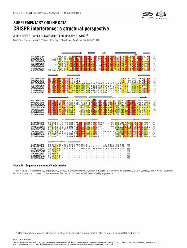
Figure S1 Sequence alignments of Cas5c proteins
Sequence similarity is shaded from red (highest) to green (lowest). The secondary structure elements of BhCas5c are shown above the alignments and are coloured according to Figure 3 of the maintext. Gaps in the elements represent disordered residues. The catalytic residues of BhCas5c are indicated by magenta stars.
1 Correspondence may be addressed to either of these authors (email [email protected] or [email protected]).
c© The Authors Journal compilation c© 2013 Biochemical Society© 2013 The Author(s)
The author(s) has paid for this article to be freely available under the terms of the Creative Commons Attribution Licence (CC-BY) (http://creativecommons.org/licenses/by/3.0/)which permits unrestricted use, distribution and reproduction in any medium, provided the original work is properly cited.
J. Reeks, J. H. Naismith and M. F. White
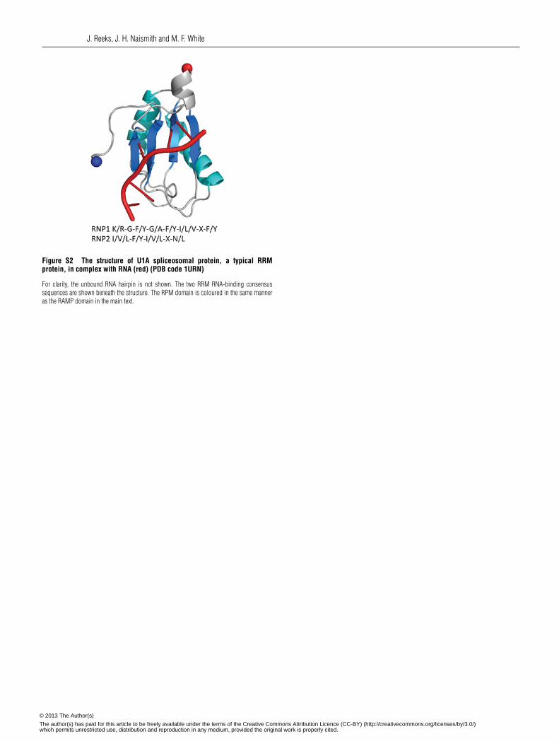
Figure S2 The structure of U1A spliceosomal protein, a typical RRMprotein, in complex with RNA (red) (PDB code 1URN)
For clarity, the unbound RNA hairpin is not shown. The two RRM RNA-binding consensussequences are shown beneath the structure. The RPM domain is coloured in the same manneras the RAMP domain in the main text.
c© The Authors Journal compilation c© 2013 Biochemical Society© 2013 The Author(s)
The author(s) has paid for this article to be freely available under the terms of the Creative Commons Attribution Licence (CC-BY) (http://creativecommons.org/licenses/by/3.0/)which permits unrestricted use, distribution and reproduction in any medium, provided the original work is properly cited.
Structures of CRISPR interference proteins
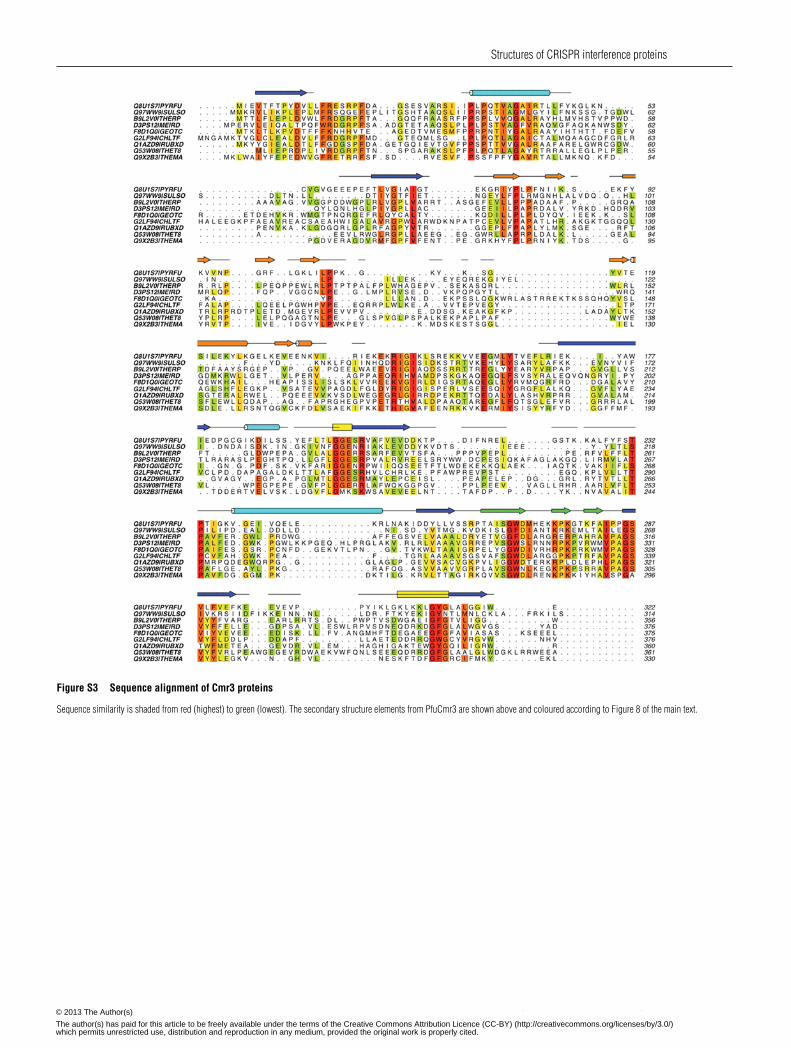
Figure S3 Sequence alignment of Cmr3 proteins
Sequence similarity is shaded from red (highest) to green (lowest). The secondary structure elements from PfuCmr3 are shown above and coloured according to Figure 8 of the main text.
c© The Authors Journal compilation c© 2013 Biochemical Society© 2013 The Author(s)
The author(s) has paid for this article to be freely available under the terms of the Creative Commons Attribution Licence (CC-BY) (http://creativecommons.org/licenses/by/3.0/)which permits unrestricted use, distribution and reproduction in any medium, provided the original work is properly cited.

J. Reeks, J. H. Naismith and M. F. White
Figure S4 Schematic diagram of dsDNA degradation by Cas3 in the type I-E system
Repeats are shown in black, protospacers and spacers in red, PAMs in blue and DNA in grey. The Cas3 HD domain is represented by a light green dotted line, the Cas3 helicase domain in dark greenand eCascade by a grey line.
Figure S5 Sequence alignments of the HD domains of Cas10a
The putative HD superfamily sequence motifs are highlighted with magenta stars and the motif number is indicated.
c© The Authors Journal compilation c© 2013 Biochemical Society© 2013 The Author(s)
The author(s) has paid for this article to be freely available under the terms of the Creative Commons Attribution Licence (CC-BY) (http://creativecommons.org/licenses/by/3.0/)which permits unrestricted use, distribution and reproduction in any medium, provided the original work is properly cited.
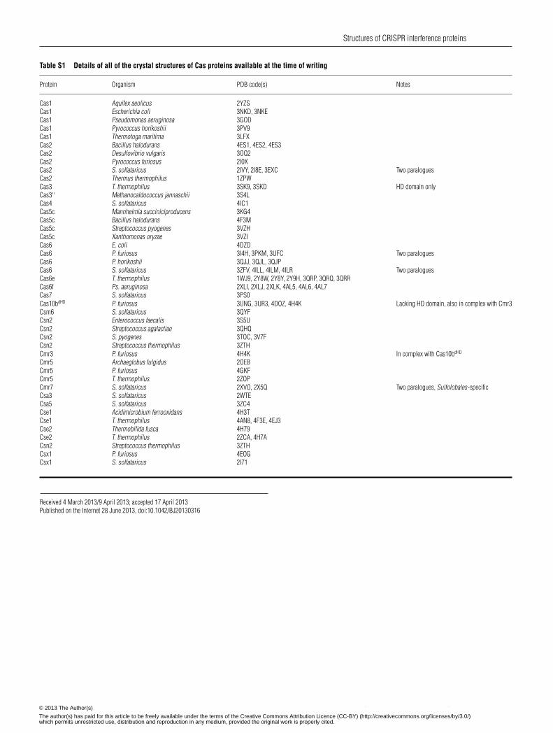
Structures of CRISPR interference proteins
Table S1 Details of all of the crystal structures of Cas proteins available at the time of writing
Protein Organism PDB code(s) Notes
Cas1 Aquifex aeolicus 2YZSCas1 Escherichia coli 3NKD, 3NKECas1 Pseudomonas aeruginosa 3GODCas1 Pyrococcus horikoshii 3PV9Cas1 Thermotoga maritima 3LFXCas2 Bacillus halodurans 4ES1, 4ES2, 4ES3Cas2 Desulfovibrio vulgaris 3OQ2Cas2 Pyrococcus furiosus 2I0XCas2 S. solfataricus 2IVY, 2I8E, 3EXC Two paraloguesCas2 Thermus thermophilus 1ZPWCas3 T. thermophilus 3SK9, 3SKD HD domain onlyCas3′ ′ Methanocaldococcus jannaschii 3S4LCas4 S. solfataricus 4IC1Cas5c Mannheimia succiniciproducens 3KG4Cas5c Bacillus halodurans 4F3MCas5c Streptococcus pyogenes 3VZHCas5c Xanthomonas oryzae 3VZICas6 E. coli 4DZDCas6 P. furiosus 3I4H, 3PKM, 3UFC Two paraloguesCas6 P. horikoshii 3QJJ, 3QJL, 3QJPCas6 S. solfataricus 3ZFV, 4ILL, 4ILM, 4ILR Two paraloguesCas6e T. thermophilus 1WJ9, 2Y8W, 2Y8Y, 2Y9H, 3QRP, 3QRQ, 3QRRCas6f Ps. aeruginosa 2XLI, 2XLJ, 2XLK, 4AL5, 4AL6, 4AL7Cas7 S. solfataricus 3PS0Cas10bdHD P. furiosus 3UNG, 3UR3, 4DOZ, 4H4K Lacking HD domain, also in complex with Cmr3Csm6 S. solfataricus 3QYFCsn2 Enterococcus faecalis 3S5UCsn2 Streptococcus agalactiae 3QHQCsn2 S. pyogenes 3TOC, 3V7FCsn2 Streptococcus thermophilus 3ZTHCmr3 P. furiosus 4H4K In complex with Cas10bdHD
Cmr5 Archaeglobus fulgidus 2OEBCmr5 P. furiosus 4GKFCmr5 T. thermophilus 2ZOPCmr7 S. solfataricus 2XVO, 2X5Q Two paralogues, Sulfolobales-specificCsa3 S. solfataricus 2WTECsa5 S. solfataricus 3ZC4Cse1 Acidimicrobium ferrooxidans 4H3TCse1 T. thermophilus 4AN8, 4F3E, 4EJ3Cse2 Thermobifida fusca 4H79Cse2 T. thermophilus 2ZCA, 4H7ACsn2 Streptococcus thermophilus 3ZTHCsx1 P. furiosus 4EOGCsx1 S. solfataricus 2I71
Received 4 March 2013/9 April 2013; accepted 17 April 2013Published on the Internet 28 June 2013, doi:10.1042/BJ20130316
c© The Authors Journal compilation c© 2013 Biochemical Society© 2013 The Author(s)
The author(s) has paid for this article to be freely available under the terms of the Creative Commons Attribution Licence (CC-BY) (http://creativecommons.org/licenses/by/3.0/)which permits unrestricted use, distribution and reproduction in any medium, provided the original work is properly cited.
Related Documents