Chapter 8 Marsupialization of Keratocystic Odontogenic Tumors of the Mandible: Longitudinal Image Analysis of Tumor Size via 3D Visualized CT Scans Hajime Shudou, Masanori Sasaki, Takahiro Yamashiro, Shizuo Tsunomachi, Yasuharu Takenoshita, Yasutaka Kubota, Tomohiro Ninomiya, Toshiyuki Kawazu and Yoshihide Mori Additional information is available at the end of the chapter http://dx.doi.org/10.5772/52432 1. Introduction The odontogenic keratocyst (OKC) was designated by the World Health Organization (WHO) as a keratocystic odontogenic tumor (KCOT) in 2005. KCOT has been defined as a benign uni- or multicystic, intraosseous tumor of odontogenic origin, with a characteristic lining of parakeratinized stratified squamous epithelium and potential for aggressive, infiltrative behavior. Additionally, these tumors have been characterized by a high recurrence rate [1, 2]. Because the recurrence rate of KCOTs ranges from 13.1% [2] to 62.5% [3, 4, 5, 6], many attempts have been made to reduce the high recurrence rate with improved surgical techniques. Recommended techniques have included tanning the cystic cavity with Carnoy’s solution before enucleation [7, 8], or using a combination of enucleation and liquid nitrogen cryotherapy [9], whereas others recommend techniques such as marsupialization or decompression of the cysts followed by secondary enucleation [10, 11, 12]. Specifically, Bramley [13] recommended the use of radical surgery with resection and bone transplantation, whereas Ephros and Lee [14] advocated the removal of the lateral cortical plate and enucleation of the cyst. Bataineh and al Qudah [15] advocated resection without continuity defects as a standard treatment for preoperatively diagnosed KCOTs. To reduce the high recurrence rate of KCOTs, it is essential to completely eradicate the epithelial components of the cyst [16]. However, radical treatment © 2013 Shudou et al.; licensee InTech. This is an open access article distributed under the terms of the Creative Commons Attribution License (http://creativecommons.org/licenses/by/3.0), which permits unrestricted use, distribution, and reproduction in any medium, provided the original work is properly cited.

Welcome message from author
This document is posted to help you gain knowledge. Please leave a comment to let me know what you think about it! Share it to your friends and learn new things together.
Transcript
Chapter 8
Marsupialization of Keratocystic Odontogenic Tumorsof the Mandible: Longitudinal Image Analysis of TumorSize via 3D Visualized CT Scans
Hajime Shudou, Masanori Sasaki,Takahiro Yamashiro, Shizuo Tsunomachi,Yasuharu Takenoshita, Yasutaka Kubota,Tomohiro Ninomiya, Toshiyuki Kawazu andYoshihide Mori
Additional information is available at the end of the chapter
http://dx.doi.org/10.5772/52432
1. Introduction
The odontogenic keratocyst (OKC) was designated by the World Health Organization (WHO)as a keratocystic odontogenic tumor (KCOT) in 2005. KCOT has been defined as a benign uni-or multicystic, intraosseous tumor of odontogenic origin, with a characteristic lining ofparakeratinized stratified squamous epithelium and potential for aggressive, infiltrativebehavior. Additionally, these tumors have been characterized by a high recurrence rate [1, 2].Because the recurrence rate of KCOTs ranges from 13.1% [2] to 62.5% [3, 4, 5, 6], many attemptshave been made to reduce the high recurrence rate with improved surgical techniques.Recommended techniques have included tanning the cystic cavity with Carnoy’s solutionbefore enucleation [7, 8], or using a combination of enucleation and liquid nitrogen cryotherapy[9], whereas others recommend techniques such as marsupialization or decompression of thecysts followed by secondary enucleation [10, 11, 12]. Specifically, Bramley [13] recommendedthe use of radical surgery with resection and bone transplantation, whereas Ephros and Lee[14] advocated the removal of the lateral cortical plate and enucleation of the cyst. Batainehand al Qudah [15] advocated resection without continuity defects as a standard treatment forpreoperatively diagnosed KCOTs. To reduce the high recurrence rate of KCOTs, it is essentialto completely eradicate the epithelial components of the cyst [16]. However, radical treatment
© 2013 Shudou et al.; licensee InTech. This is an open access article distributed under the terms of theCreative Commons Attribution License (http://creativecommons.org/licenses/by/3.0), which permitsunrestricted use, distribution, and reproduction in any medium, provided the original work is properly cited.
has been associated with numerous complications, including facial deformity, missing teeth,infection of transplanted bone, and / or permanent numbness of the region innervated by themental nerve when the KCOTs involved the inferior alveolar nerve. Therefore, consideringthe benign characteristics of KCOTs, the first priority of the treatment method should bediscussed from the perspective of morbidity and the quality of life of the patients; the recur‐rence rate should not always be the primary factor.
1.1. Marsupialization
Marsupialization or decompression has been used in the past as a conservative treatmentmodality for large KCOTs, minimizing the tumor size and limiting the extent of surgery [10,11, 12, 17-19, 20]. Nakamura et al. [19] reported that marsupialization did not affect therecurrence rate of KCOTs. Marker et al. [11] reported long-term results after decompressionfor 23 KCOTs, and they concluded that these cysts could be treated successfully by marsupi‐alization and secondary enucleation. However, there were some disadvantages in marsupial‐ization, and one of the disadvantages was that, when considering other treatment methods,the time necessary for this treatment was comparatively long [21].
This chapter determines how KCOTs in the mandible are reduced in size by marsupializationand predicts the best time for secondary enucleation by means of analyzing computerizedtomography (CT) images. Fifteen patients with KCOTs were treated with marsupializationsurgery, and 42 series of CT data taken during the marsupialization process were analyzed.CT data were reconstructed in 3-dimensional (3D) images. The 3D images were used tomeasure the diameter and volume, and to analyze the changes that occurred after marsupial‐ization. Marsupialized KCOTs tended to be equally reduced towards the window in the tumor.The amount of volume reduction per day (Vr) was reduced in proportion to the volume (V)with the formula:
Vr = -0.0029 × V. The formula manipulation for V was V = V1 × e-0.0029 t (t = duration aftermarsupialization in days). The volume of marsupialized KCOTs was reduced by half over a239 day period. These results demonstrate that the future shape of marsupialized mandibularKCOTs, under good control, can be predicted with significant accuracy using CT data. Thisprediction can decrease the prolonged marsupialization period in patients with KCOTs.Herein we clarify how KCOTs are reduced in size during the marsupialization and to predictthe best time for secondary enucleation by means of analyzing computerized tomography (CT)images.
1.2. Treatment
Our series of 15 patients with histologically proven KCOTs of the mandible were treated bymarsupialization surgery from 2000 to 2010. Of the 15 patients, 9 were male (60.0%) and 6 werefemale (40.0%). The mean age was 35.9 years (range: 16 to 57). The tumors were located in theposterior molar to mandibular ramus in 5, in the angle to mandibular ramus in 7, and in theanterior molar region in 3 patients. Using X-ray images, the tumors were classified; 10 wereunilocular lesions and 5 were multilocular lesions (Table1).
A Textbook of Advanced Oral and Maxillofacial Surgery242

Postoperative follow-up consisted of clinical and radiographic examinations. Cases ofrecurrent tumors or cases associated with basal cell nevus syndrome were excluded from thisstudy. Clinical information and CT images were obtained from the records of the Section ofOral and Maxillofacial Surgery, Kyushu University Hospital.
Table 1. Distribution of patients treated by marsupialisation for KCOT.
1.3. Marsupialization and secondary enucleation technique
Marsupialization has been used to relieve the pressure within the cystic cavity and allow newbone to fill the defect [22] (Fig. 1). This surgical technique usually involves making a bonewindow or opening in the wall of the tumor, partially debriding with an excision on the topportion of the tumor, and suturing the edges of the remaining cyst to the surrounding softtissue. At the beginning of each treatment, marsupialization was performed simultaneouslywith a biopsy. In multilocular lesions, the intracystic partitions were removed to make a singlecavity. After marsupialization, an obturator, made of acrylic resin, was used to keep thewindow open. The purpose of the obturator was to maintain continuity between the marsu‐pialized tumor and the oral environment during the treatment process. The marsupializedwindow was kept open until the patients could withstand less aggressive treatment, such asenucleation and curettage. The inner volume of the tumor was estimated by filling theintracavity with saline [23] and measuring the distance between the inferior alveolar nerveand the tumor as well as the thickness of the peritumoral bone. Thus, the duration of marsu‐pialization was different for each case.
The secondary surgery after marsupialization, was enucleation and curettage in which theKCOTs were enucleated, and the overlying mucosa was excised and subsequently curetted toadjacent healthy bone. Using a large round bur, curettage usually extended 1 to 2 mm in depth.This procedure was performed to ensure the removal of the epithelial remnants.
Marsupialization of Keratocystic Odontogenic Tumors of the Mandible: Longitudinal Image Analysis of Tumor Size ...http://dx.doi.org/10.5772/52432
243
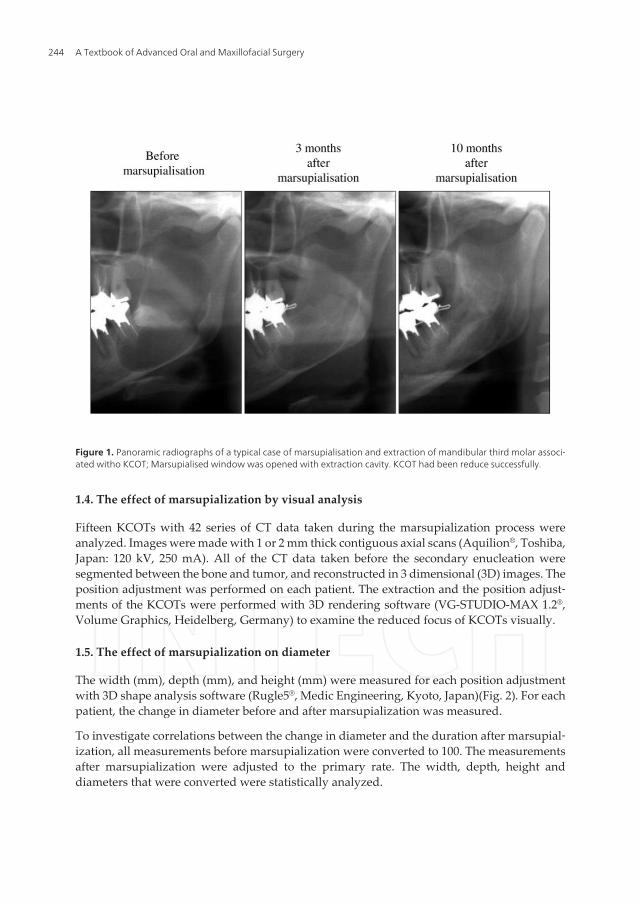
Figure 1. Panoramic radiographs of a typical case of marsupialisation and extraction of mandibular third molar associ‐ated witho KCOT; Marsupialised window was opened with extraction cavity. KCOT had been reduce successfully.
1.4. The effect of marsupialization by visual analysis
Fifteen KCOTs with 42 series of CT data taken during the marsupialization process wereanalyzed. Images were made with 1 or 2 mm thick contiguous axial scans (Aquilion®, Toshiba,Japan: 120 kV, 250 mA). All of the CT data taken before the secondary enucleation weresegmented between the bone and tumor, and reconstructed in 3 dimensional (3D) images. Theposition adjustment was performed on each patient. The extraction and the position adjust‐ments of the KCOTs were performed with 3D rendering software (VG-STUDIO-MAX 1.2®,Volume Graphics, Heidelberg, Germany) to examine the reduced focus of KCOTs visually.
1.5. The effect of marsupialization on diameter
The width (mm), depth (mm), and height (mm) were measured for each position adjustmentwith 3D shape analysis software (Rugle5®, Medic Engineering, Kyoto, Japan)(Fig. 2). For eachpatient, the change in diameter before and after marsupialization was measured.
To investigate correlations between the change in diameter and the duration after marsupial‐ization, all measurements before marsupialization were converted to 100. The measurementsafter marsupialization were adjusted to the primary rate. The width, depth, height anddiameters that were converted were statistically analyzed.
A Textbook of Advanced Oral and Maxillofacial Surgery244

* 3-D visualised CT showed that all of the marsupialised KCOTs were visually reduced towards the window.** (A) The width (B) The depth (C) The height
Figure 2. Overlaid 3-D images in the marsupialised period
1.6. The effect of marsupialization on the volume
The 3D data were used to measure the volume (mm3) with Rugle5® and to determine the changein volume before and after marsupialization, similar to the observation on diameter.
The amount of volume reduction (mm3) per day (Vr) was calculated using the formula (Fig.3). Vr was defined as the quotient of the difference of the volume divided by the duration aftermarsupialization. To investigate correlations between the volume and Vr, these data werestatistically analyzed. The regression formula obtained from former analysis of the volumewas calculated using differential equations.
Marsupialization of Keratocystic Odontogenic Tumors of the Mandible: Longitudinal Image Analysis of Tumor Size ...http://dx.doi.org/10.5772/52432
245
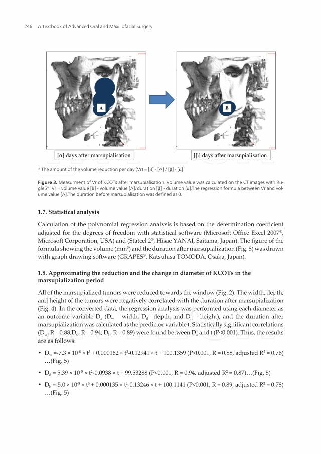
* The amount of the volume reduction per day (Vr) = [B] - [A] / [β] - [α]
Figure 3. Measurment of Vr of KCOTs after marsupialisation. Volume value was calculated on the CT images with Ru‐gle5®. Vr = volume value [B] - volume value [A]/duration [β] - duration [α].The regression formula between Vr and vol‐ume value [A].The duration before marsupialisation was defined as 0.
1.7. Statistical analysis
Calculation of the polynomial regression analysis is based on the determination coefficientadjusted for the degrees of freedom with statistical software (Microsoft Office Excel 2007®,Microsoft Corporation, USA) and (Statcel 2®, Hisae YANAI, Saitama, Japan). The figure of theformula showing the volume (mm3) and the duration after marsupialization (Fig. 8) was drawnwith graph drawing software (GRAPES®, Katsuhisa TOMODA, Osaka, Japan).
1.8. Approximating the reduction and the change in diameter of KCOTs in themarsupialization period
All of the marsupialized tumors were reduced towards the window (Fig. 2). The width, depth,and height of the tumors were negatively correlated with the duration after marsupialization(Fig. 4). In the converted data, the regression analysis was performed using each diameter asan outcome variable Dx (Dw = width, Dd= depth, and Dh = height), and the duration aftermarsupialization was calculated as the predictor variable t. Statistically significant correlations(Dw, R = 0.88;Dd, R = 0.94; Dh, R = 0.89) were found between Dx and t (P<0.001). Thus, the resultsare as follows:
• Dw =-7.3 × 10⁻⁸ × t3 + 0.000162 × t2-0.12941 × t + 100.1359 (P<0.001, R = 0.88, adjusted R2 = 0.76)…(Fig. 5)
• Dd = 5.39 × 10⁻⁵ × t2-0.0938 × t + 99.53288 (P<0.001, R = 0.94, adjusted R2 = 0.87)…(Fig. 5)
• Dh =-5.0 × 10⁻⁸ × t3 + 0.000135 × t2-0.13246 × t + 100.1141 (P<0.001, R = 0.89, adjusted R2 = 0.78)…(Fig. 5)
A Textbook of Advanced Oral and Maxillofacial Surgery246

As shown in these results, each diameter had similar curves (Figs. 4 and5). The regressionanalysis was performed in the same way, using all diameters as outcome variable Da andduration after marsupialization as the predictor variable t.
• Da =-4.9 × 10⁻⁸ × t3 + 0.00013 × t2-0.12273 × t + 100.0652 (P<0.001, R = 0.90, adjusted R2 = 0.80)…(Fig. 5)
Strong correlations were found between Da and t.
Therefore, based on these results, marsupialized KCOTs tended to be reduced towards thewindow equally.
Figure 4. The width (mm), the depth (mm), and the height (mm) of KCOTs were negatively-correlated to the durationafter marsupialisation.
Marsupialization of Keratocystic Odontogenic Tumors of the Mandible: Longitudinal Image Analysis of Tumor Size ...http://dx.doi.org/10.5772/52432
247
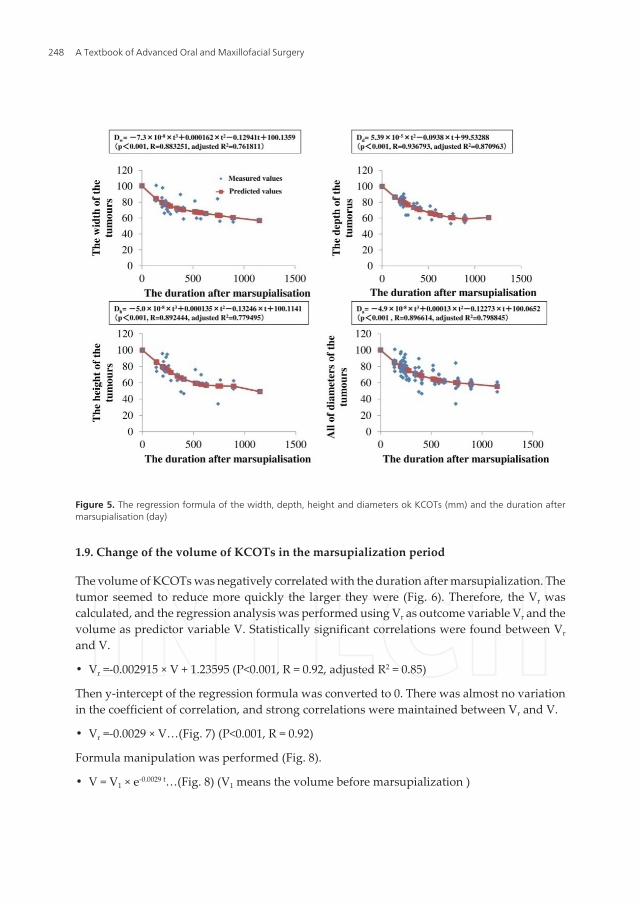
Figure 5. The regression formula of the width, depth, height and diameters ok KCOTs (mm) and the duration aftermarsupialisation (day)
1.9. Change of the volume of KCOTs in the marsupialization period
The volume of KCOTs was negatively correlated with the duration after marsupialization. Thetumor seemed to reduce more quickly the larger they were (Fig. 6). Therefore, the Vr wascalculated, and the regression analysis was performed using Vr as outcome variable Vr and thevolume as predictor variable V. Statistically significant correlations were found between Vr
and V.
• Vr =-0.002915 × V + 1.23595 (P<0.001, R = 0.92, adjusted R2 = 0.85)
Then y-intercept of the regression formula was converted to 0. There was almost no variationin the coefficient of correlation, and strong correlations were maintained between Vr and V.
• Vr =-0.0029 × V…(Fig. 7) (P<0.001, R = 0.92)
Formula manipulation was performed (Fig. 8).
• V = V1 × e-0.0029 t…(Fig. 8) (V1 means the volume before marsupialization )
A Textbook of Advanced Oral and Maxillofacial Surgery248
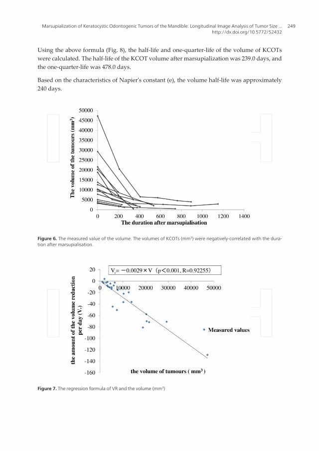
Using the above formula (Fig. 8), the half-life and one-quarter-life of the volume of KCOTswere calculated. The half-life of the KCOT volume after marsupialization was 239.0 days, andthe one-quarter-life was 478.0 days.
Based on the characteristics of Napier's constant (e), the volume half-life was approximately240 days.
Figure 6. The measured value of the volume. The volumes of KCOTs (mm3) were negatively-correlated with the dura‐tion after marsupialisation.
Figure 7. The regression formula of VR and the volume (mm3)
Marsupialization of Keratocystic Odontogenic Tumors of the Mandible: Longitudinal Image Analysis of Tumor Size ...http://dx.doi.org/10.5772/52432
249
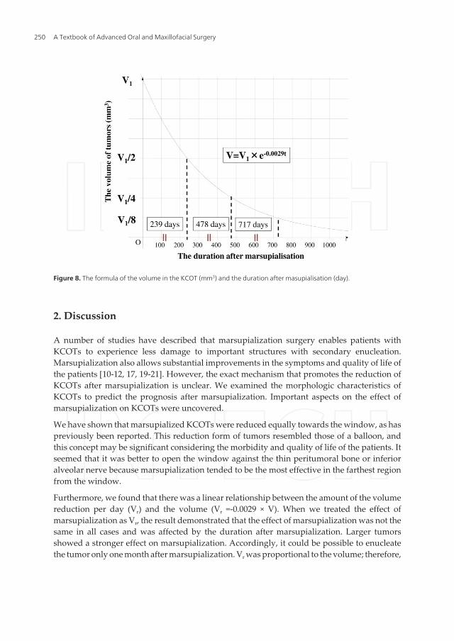
Figure 8. The formula of the volume in the KCOT (mm3) and the duration after masupialisation (day).
2. Discussion
A number of studies have described that marsupialization surgery enables patients withKCOTs to experience less damage to important structures with secondary enucleation.Marsupialization also allows substantial improvements in the symptoms and quality of life ofthe patients [10-12, 17, 19-21]. However, the exact mechanism that promotes the reduction ofKCOTs after marsupialization is unclear. We examined the morphologic characteristics ofKCOTs to predict the prognosis after marsupialization. Important aspects on the effect ofmarsupialization on KCOTs were uncovered.
We have shown that marsupialized KCOTs were reduced equally towards the window, as haspreviously been reported. This reduction form of tumors resembled those of a balloon, andthis concept may be significant considering the morbidity and quality of life of the patients. Itseemed that it was better to open the window against the thin peritumoral bone or inferioralveolar nerve because marsupialization tended to be the most effective in the farthest regionfrom the window.
Furthermore, we found that there was a linear relationship between the amount of the volumereduction per day (Vr) and the volume (Vr =-0.0029 × V). When we treated the effect ofmarsupialization as Vr, the result demonstrated that the effect of marsupialization was not thesame in all cases and was affected by the duration after marsupialization. Larger tumorsshowed a stronger effect on marsupialization. Accordingly, it could be possible to enucleatethe tumor only one month after marsupialization. Vr was proportional to the volume; therefore,
A Textbook of Advanced Oral and Maxillofacial Surgery250
the differential equation and the formula between the volume and the duration after marsu‐pialization (V = V1 × e-0.0029 t) was applied. The volume showed the exponential decay in theduration after marsupialization, and the V-t formula indicated that the characteristics or thespeed of the reduction in postmarsupialized KCOTs was dependent on the premarsupializedvolume (V1). We felt that the marsupialized tumor was not significantly reduced when it wasof small size.The marsupialized tumor was reduced in the half life of 240 days, according toNapier's constant e and the exponential function. When we focused on the cyclic nature andthe regression coefficient of Vr-V formula (- 0.0029), we recognized that the reduction percent‐age in the volume tended to be unchanged for the same duration. This result was expectedbased on the hypothesis that the number of cells per unit volume was not based on thesurrounding tumor, and that there was no significant change in pressure in the cavity and atthe molecular level after marsupialization. However, the regulatory mechanisms of howtumors are reduced or the repair mechanisms of the normal bone are unclear. Many factorsmust be considered, including the elasticity of the wall, and a comparison between the maxillaand mandible, with or without impacted teeth, must be made. Further studies will be requiredto clarify these factors.
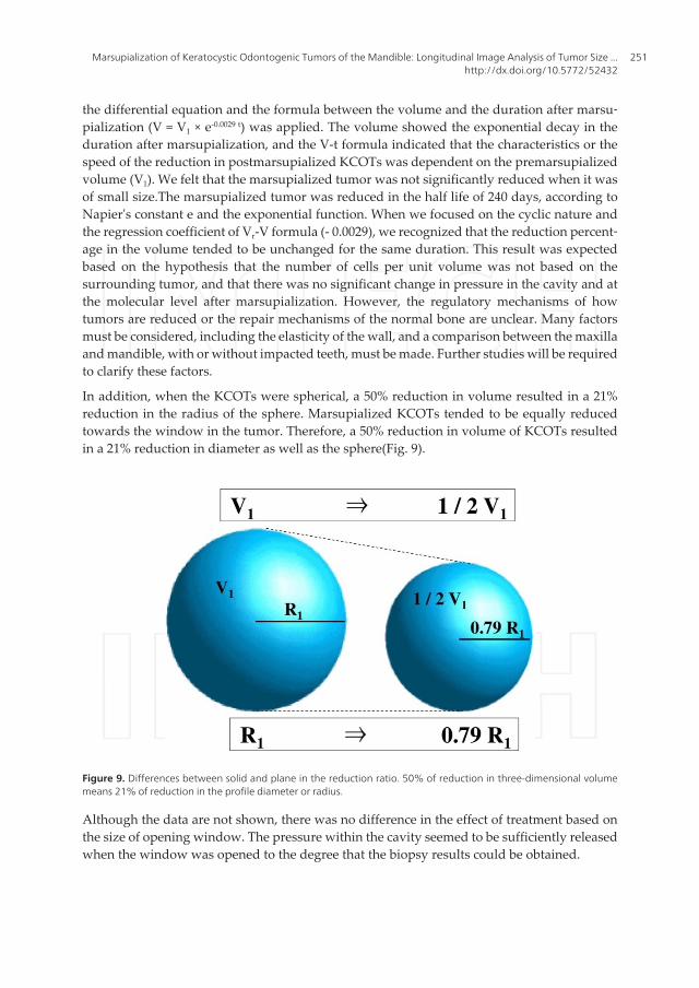
In addition, when the KCOTs were spherical, a 50% reduction in volume resulted in a 21%reduction in the radius of the sphere. Marsupialized KCOTs tended to be equally reducedtowards the window in the tumor. Therefore, a 50% reduction in volume of KCOTs resultedin a 21% reduction in diameter as well as the sphere(Fig. 9).
Figure 9. Differences between solid and plane in the reduction ratio. 50% of reduction in three-dimensional volumemeans 21% of reduction in the profile diameter or radius.
Although the data are not shown, there was no difference in the effect of treatment based onthe size of opening window. The pressure within the cavity seemed to be sufficiently releasedwhen the window was opened to the degree that the biopsy results could be obtained.
Marsupialization of Keratocystic Odontogenic Tumors of the Mandible: Longitudinal Image Analysis of Tumor Size ...http://dx.doi.org/10.5772/52432
251
We only referred to the evaluation of CT images, but we also needed follow-up with 2D images,such as a panoramic radiograph. Panoramic radiographs were easy to measure the tumor, andsuperior for grasping a whole image. Panoramic radiographs and intracystic cavities withsterile physiological saline were important to use as simple diagnostic methods for estimatingthe size of the cyst because there was a good correlation between the volume and the radio‐lucent area [23].
The well-controlled cases of marsupialized mandibular KCOTs could predict the future shapeof the tumor with significant accuracy. Considering these features, the primary location of themental nerve, and the thickness of the peritumoral bone, secondary operative planning beforemarsupialization could be carried out using CT,which would therefore decrease the mentalburden on patients. Finally, there have been various types of treatments for KCOTs. Therefore,the choice of therapy was very important because marsupialization required a long period fortreatment, and the patients may not have had a medical examination before treatment wascompleted.
Author details
Hajime Shudou1*, Masanori Sasaki2, Takahiro Yamashiro1, Shizuo Tsunomachi1,Yasuharu Takenoshita3, Yasutaka Kubota4, Tomohiro Ninomiya2, Toshiyuki Kawazu5 andYoshihide Mori6
*Address all correspondence to: [email protected]
1 Clinical Fellow, Section of Oral and Maxillofacial Surgery, Division of Maxillofacial Diag‐nostic and Surgical Sciences, Graduate school of Dental Science, Kyushu University, Fukuo‐ka, Japan
2 Assistant Professor, Section of Oral and Maxillofacial Surgery, Division of MaxillofacialDiagnostic and Surgical Sciences, Graduate school of Dental Science, Kyushu University,Fukuoka, Japan
3 Associate Professor, Section of Oral and Maxillofacial Surgery, Division of MaxillofacialDiagnostic and Surgical Sciences, Graduate school of Dental Science, Kyushu University,Fukuoka, Japan
4 Lecturer, Section of Oral and Maxillofacial Surgery, Division of Maxillofacial Diagnostic andSurgical Sciences, Graduate school of Dental Science, Kyushu University, Fukuoka, Japan
5 Assistant Professor, Department of Oral and Maxillofacial Radiology, Graduate School ofDental Science, Kyushu University, Fukuoka, Japan
6 Professor, Section of Oral and Maxillofacial Surgery, Division of Maxillofacial Diagnostic andSurgical Sciences, Graduate school of Dental Science, Kyushu University, Fukuoka, Japan
A Textbook of Advanced Oral and Maxillofacial Surgery252
References
[1] Brannon RB: The odontogenic keratocystA clinicopathologic study of 312 cases. partI. clinical features. Oral Surg Oral Med and Oral Pathol(1976). , 42, 54-72.
[2] Gonzalez-alva, P, Tanaka, A, Oku, Y, Yoshizawa, D, Itoh, S, Sakashita, H, Ide, F, &Tajima, Y. Kusama K: Keratocysticodontogenic tumor: A retrospective study of 183cases. J Oral Sci(2008). , 50, 205-212.
[3] Browne RM: The odontogenickeratocystClinical aspects. Br Dent J(1970). , 128,225-231.
[4] Payne TF: An analysis of the clinical and histopathologic parameters of the odonto‐genickeratocystOral Surg Oral Med and Oral Pathol(1972). , 33, 538-546.
[5] Pindborg, J. J. Hansen J: Studies on odontogenic cyst epithelium. 2. clinical and roent‐genologic aspects of odontogenickeratocysts.ActaPatholMicrobiolScand (1963). , 58,283-294.
[6] Toller P: Origin and growth of cysts of the jawsAnn R CollSurgEngl(1967). , 40,306-336.
[7] Voorsmit, R. A, & Stoelinga, P. J. van Haelst UJ: The management of keratocysts. JMaxillofacSurg (1981). , 9, 228-236.
[8] Williams, T. P. Connor FA Jr: Surgical management of the odontogenickeratocyst:Aggressive approach. J Oral and MaxillofacSurg (1994). , 52, 964-966.
[9] Schmidt, B. L. Pogrel MA: The use of enucleation and liquid nitrogen cryotherapy inthe management of odontogenickeratocysts. J Oral and MaxillofacSurg (2001). , 59,720-725.
[10] Brondum, N. Jensen VJ: Recurrence of keratocysts and decompression treatment. Along-term follow-up of forty-four cases. Oral Surg Oral Med and Oral Pathol (1991). ,72, 265-269.
[11] Marker, P, Brondum, N, & Clausen, P. P. Bastian HL: Treatment of large odontoge‐nickeratocysts by decompression and later cystectomy: A long-term follow-up and ahistologic study of 23 cases. Oral Surg Oral Med Oral Pathol Oral RadiolEn‐dod(1996). , 82, 122-131.
[12] Tucker, W. M, & Pleasants, J. E. MacComb WS: Decompression and secondary enu‐cleation of a mandibular cyst: Report of case. J Oral Surg (1972). , 30, 669-673.
[13] Bramley P: The odontogenickeratocyst--an approach to treatmentInt J OralSurg(1974). , 3, 337-341.
[14] Ephros, H. Lee HY: Treatment of a large odontogenickeratocyst using the brosch pro‐cedure. J Oral and MaxillofacSurg(1991). , 49, 871-874.
Marsupialization of Keratocystic Odontogenic Tumors of the Mandible: Longitudinal Image Analysis of Tumor Size ...http://dx.doi.org/10.5772/52432
253
[15] Bataineh, A. B. al Qudah M: Treatment of mandibular odontogenickeratocysts. OralSurg Oral Med Oral Pathol Oral RadiolEndod (1998). , 86, 42-47.
[16] Forssell, K, & Forssell, H. Kahnberg KE: Recurrence of keratocysts. A long-term fol‐low-up study.Int J Oral and MaxillofacSurg (1988). , 17, 25-28.
[17] Cranin, A. N, & Madan, S. Fayans E: Novel method of treating large cysts of jaws inchildren. N Y State Dent J (1994). , 60, 41-44.
[18] Maurette, P. E, & Jorge, J. de Moraes M: Conservative treatment protocol of odonto‐genickeratocyst: A preliminary study. J Oral and MaxillofacSurg (2006). , 64, 379-383.
[19] Nakamura, N, Mitsuyasu, T, Mitsuyasu, Y, Taketomi, T, & Higuchi, Y. Ohishi M:Marsupialization for odontogenickeratocysts: Long-term follow-up analysis of the ef‐fects and changes in growth characteristics. Oral Surg Oral Med Oral Pathol Oral Ra‐diolEndod (2002). , 94, 543-553.
[20] Pogrel MA: Treatment of keratocysts: The case for decompression and marsupializa‐tionJ Oral and MaxillofacSurg (2005). , 63, 1667-1673.
[21] Zhao, Y. F, & Wei, J. X. Wang SP: Treatment of odontogenickeratocysts: A follow-upof 255 chinese patients. Oral Surg Oral Med Oral Pathol Oral RadiolEndod (2002). ,94, 151-156.
[22] Giuliani, M, Grossi, G. B, Lajolo, C, & Bisceglia, M. Herb KE: Conservative manage‐ment of a large odontogenickeratocyst: Report of a case and review of the literature. JOral and MaxillofacSurg (2006). , 64, 308-316.
[23] Kubota, Y, Yamashiro, T, Oka, S, Ninomiya, T, & Ogata, S. Shirasuna K: Relation be‐tween size of odontogenic jaw cysts and the pressure of fluid within. Br J Oral Maxil‐lofacSurg (2004). , 42, 391-395.
A Textbook of Advanced Oral and Maxillofacial Surgery254
Related Documents