44 y/o female with dizziness and dull headache Edward Gillis, DO Mark Kane, MD Leo Wolansky, MD

Welcome message from author
This document is posted to help you gain knowledge. Please leave a comment to let me know what you think about it! Share it to your friends and learn new things together.
Transcript

44 y/o female with dizziness
and dull headache
Edward Gillis, DO
Mark Kane, MD
Leo Wolansky, MD

Axial T1W

Axial 3D-T2W
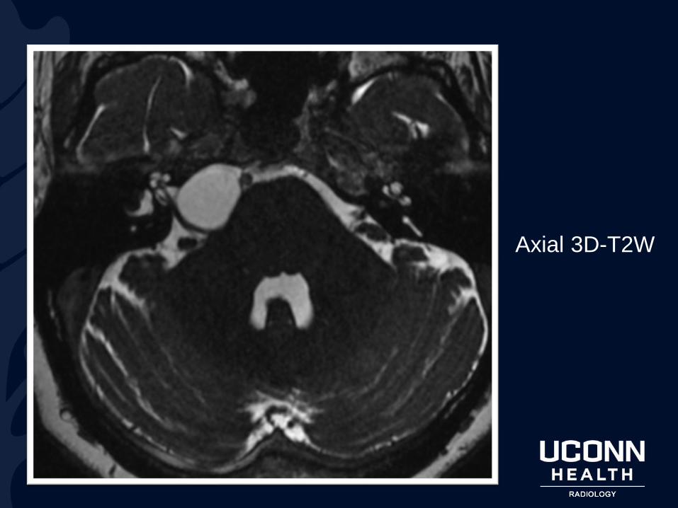
Axial 3D-T2W
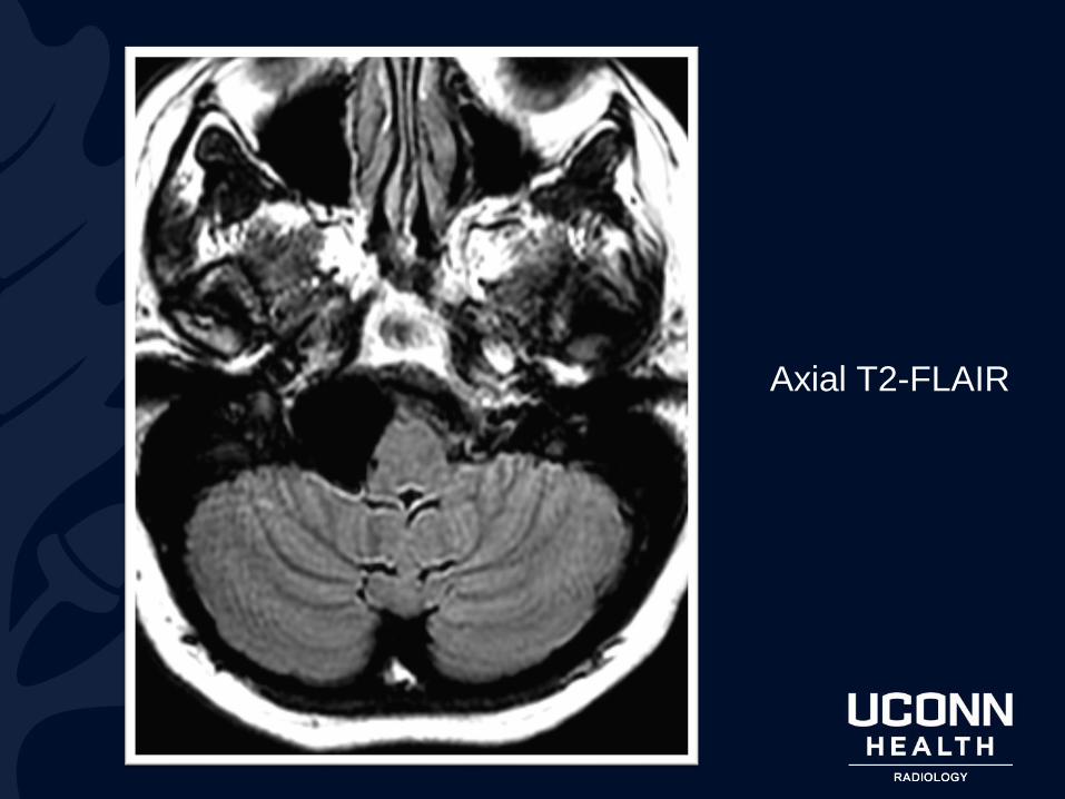
Axial T2-FLAIR

Axial T1W

?

Arachnoid Cyst

Axial T1W:
Right CP angle
mass isointense
to CSF, with
leftward mass
effect on the
medulla

Axial MRI T2W:
Right CP angle
mass
isointense to
CSF, with
leftward mass
effect on the
medulla
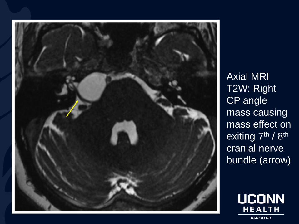
Axial MRI
T2W: Right
CP angle
mass causing
mass effect on
exiting 7th / 8th
cranial nerve
bundle (arrow)
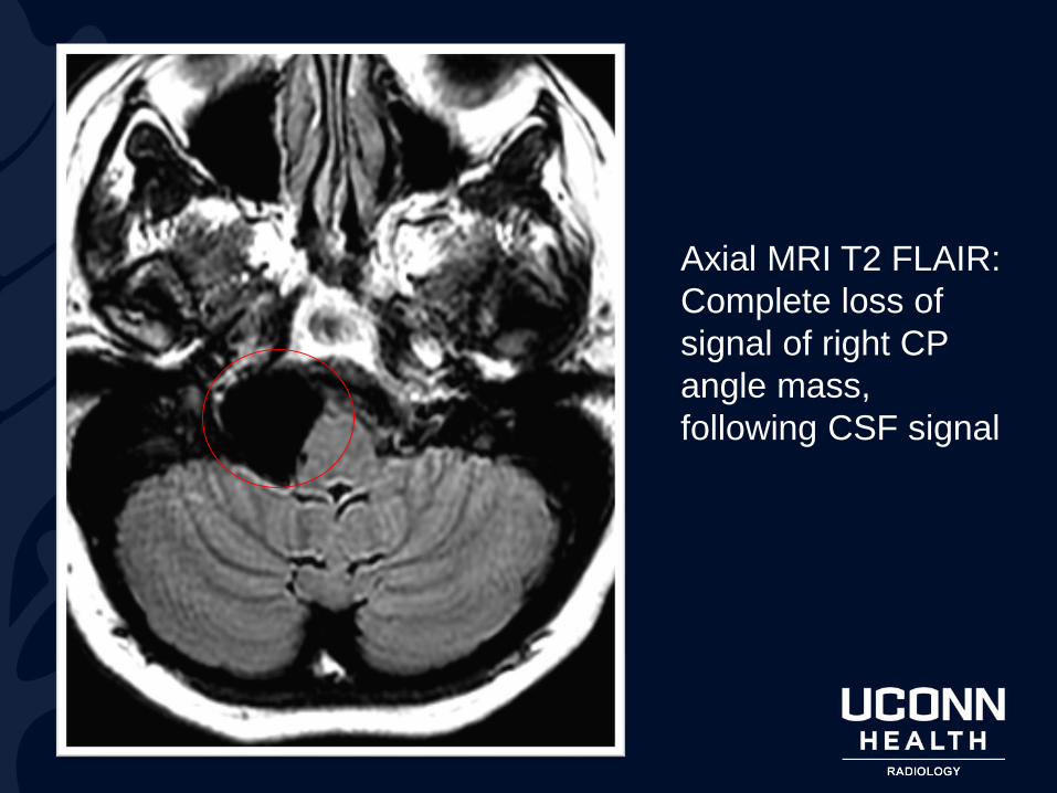
Axial MRI T2 FLAIR:
Complete loss of
signal of right CP
angle mass,
following CSF signal
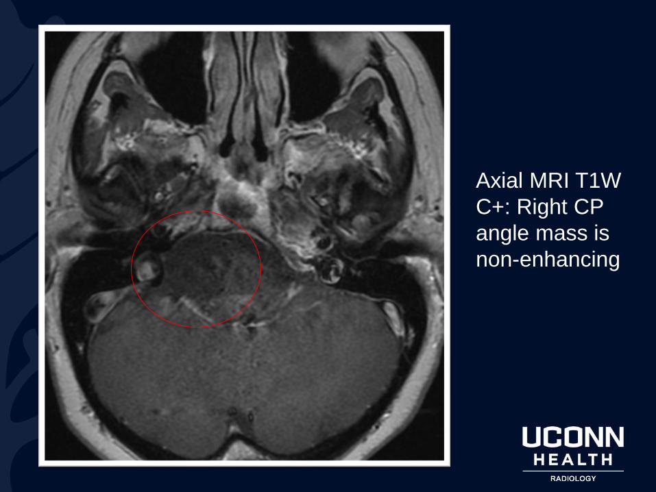
Axial MRI T1W
C+: Right CP
angle mass is
non-enhancing
Arachnoid Cyst
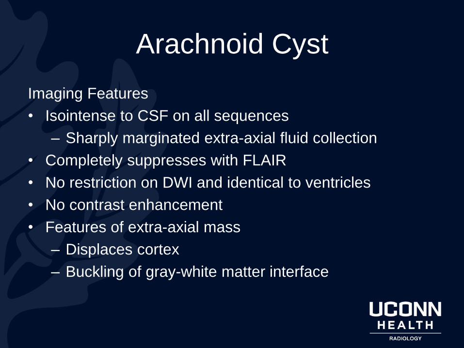
Imaging Features
• Isointense to CSF on all sequences
– Sharply marginated extra-axial fluid collection
• Completely suppresses with FLAIR
• No restriction on DWI and identical to ventricles
• No contrast enhancement
• Features of extra-axial mass
– Displaces cortex
– Buckling of gray-white matter interface
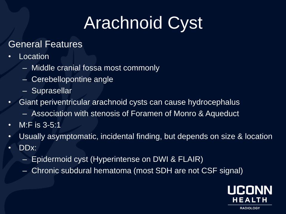
Arachnoid CystGeneral Features
• Location
– Middle cranial fossa most commonly
– Cerebellopontine angle
– Suprasellar
• Giant periventricular arachnoid cysts can cause hydrocephalus
– Association with stenosis of Foramen of Monro & Aqueduct
• M:F is 3-5:1
• Usually asymptomatic, incidental finding, but depends on size & location
• DDx:
– Epidermoid cyst (Hyperintense on DWI & FLAIR)
– Chronic subdural hematoma (most SDH are not CSF signal)

References
1. Brant, W. E., & Helms, C. A. (2012). Fundamentals of diagnostic
radiology. Philadelphia: Wolters Kluwer Health/Lippincott Williams &
Wilkins
2. Diagnostic Imaging for Radiology. (n.d.). Retrieved October 17, 2017, from
http://www.statdx.com/
Related Documents


![RESEARCH ARTICLE Open Access Head-Eye movement control ... Casa_Head-eye... · impairment are dizziness, headache, light-headedness and visual disorders [3]. Movements requiring the](https://static.cupdf.com/doc/110x72/5d2d87a488c99309368c2886/research-article-open-access-head-eye-movement-control-casahead-eye.jpg)