Glencoe Science Chapter Resources Cells Includes: Reproducible Student Pages ASSESSMENT ✔ Chapter Tests ✔ Chapter Review HANDS-ON ACTIVITIES ✔ Lab Worksheets for each Student Edition Activity ✔ Laboratory Activities ✔ Foldables–Reading and Study Skills activity sheet MEETING INDIVIDUAL NEEDS ✔ Directed Reading for Content Mastery ✔ Directed Reading for Content Mastery in Spanish ✔ Reinforcement ✔ Enrichment ✔ Note-taking Worksheets TRANSPARENCY ACTIVITIES ✔ Section Focus Transparency Activities ✔ Teaching Transparency Activity ✔ Assessment Transparency Activity Teacher Support and Planning ✔ Content Outline for Teaching ✔ Spanish Resources ✔ Teacher Guide and Answers

Welcome message from author
This document is posted to help you gain knowledge. Please leave a comment to let me know what you think about it! Share it to your friends and learn new things together.
Transcript

Glencoe Science
Chapter Resources
Cells
Includes:
Reproducible Student Pages
ASSESSMENT
✔ Chapter Tests
✔ Chapter Review
HANDS-ON ACTIVITIES
✔ Lab Worksheets for each Student Edition Activity
✔ Laboratory Activities
✔ Foldables–Reading and Study Skills activity sheet
MEETING INDIVIDUAL NEEDS
✔ Directed Reading for Content Mastery
✔ Directed Reading for Content Mastery in Spanish
✔ Reinforcement
✔ Enrichment
✔ Note-taking Worksheets
TRANSPARENCY ACTIVITIES
✔ Section Focus Transparency Activities
✔ Teaching Transparency Activity
✔ Assessment Transparency Activity
Teacher Support and Planning
✔ Content Outline for Teaching
✔ Spanish Resources
✔ Teacher Guide and Answers
429-i-vi-MSS05-000000_CR 15.04.2004 15:32 Page i tammyb 301:goscanc:scanc429:layouts:
Glencoe Science
Photo CreditsSection Focus Transparency 2: (l) Index Stock/ASAP Ltd., (r,inset) Wanner/Eye of Science/Photo Researchers;Section Focus Transparency 3: Lester V. Bergman/CORBIS
Copyright © by The McGraw-Hill Companies, Inc. All rights reserved.Permission is granted to reproduce the material contained herein on the conditionthat such material be reproduced only for classroom use; be provided to students,teachers, and families without charge; and be used solely in conjunction with theCells program. Any other reproduction, for use or sale, is prohibited without priorwritten permission of the publisher.
Send all inquiries to:Glencoe/McGraw-Hill8787 Orion Place Columbus, OH 43240-4027
ISBN 0-07-867092-6
Printed in the United States of America.
1 2 3 4 5 6 7 8 9 10 024 09 08 07 06 05 04
429-i-vi-MSS05-000000_CR 3/24/04 3:59PM Pageiiimpos03301:goscanc:scanc429:layouts:

Cells 1
ReproducibleStudent Pages
Reproducible Student Pages■ Hands-On Activities
MiniLAB: Modeling Cytoplasm. . . . . . . . . . . . . . . . . . . . . . . . . . . . . . . 3MiniLAB: Try at Home Observing Magnified Objects . . . . . . . . . . . . . 4Lab: Comparing Cells . . . . . . . . . . . . . . . . . . . . . . . . . . . . . . . . . . . . . . 5Lab: Design Your Own Comparing Light Microscopes. . . . . . . . . . . . . . . 7Laboratory Activity 1: The Compound Light Microscope . . . . . . . . . . . 9Laboratory Activity 2: Observing Cells . . . . . . . . . . . . . . . . . . . . . . . . 13Foldables: Reading and Study Skills. . . . . . . . . . . . . . . . . . . . . . . . . . 17
■ Meeting Individual NeedsExtension and Intervention
Directed Reading for Content Mastery . . . . . . . . . . . . . . . . . . . . . . . 19Directed Reading for Content Mastery in Spanish . . . . . . . . . . . . . . 23Reinforcement . . . . . . . . . . . . . . . . . . . . . . . . . . . . . . . . . . . . . . . . . . 27Enrichment. . . . . . . . . . . . . . . . . . . . . . . . . . . . . . . . . . . . . . . . . . . . . 30Note-taking Worksheet . . . . . . . . . . . . . . . . . . . . . . . . . . . . . . . . . . . 33
■ AssessmentChapter Review . . . . . . . . . . . . . . . . . . . . . . . . . . . . . . . . . . . . . . . . . 37Chapter Test . . . . . . . . . . . . . . . . . . . . . . . . . . . . . . . . . . . . . . . . . . . . 39
■ Transparency ActivitiesSection Focus Transparency Activities . . . . . . . . . . . . . . . . . . . . . . . . 44Teaching Transparency Activity . . . . . . . . . . . . . . . . . . . . . . . . . . . . . 47Assessment Transparency Activity . . . . . . . . . . . . . . . . . . . . . . . . . . . 49
429-1-50-MSS05-000000_CR 3/24/04 4:02 PM Page 1 impos03 301:goscanc:scanc429:layouts:
2 Cells
Hands-OnActivities
Hands-On Activities
429-1-50-MSS05-000000_CR 3/24/04 4:02 PM Page 2 impos03 301:goscanc:scanc429:layouts:

Cop
yrig
ht ©
Gle
ncoe
/McG
raw
-Hill
,a d
ivis
ion
of t
he M
cGra
w-H
ill C
ompa
nies
,Inc
.
Cells 3
Name Date Class
Modeling CytoplasmProcedure 1. Add 100 mL of water to a clear container.
2. Add unflavored gelatin and stir.
3. Shine a flashlight through the solution.
Analysis1. Describe what you see.
2. How does a model help you understand what cytoplasm might be like?
Hand
s-On
Act
iviti
es
429-1-50-MSS05-000000_CR 3/24/04 4:02 PM Page 3 impos03 301:goscanc:scanc429:layouts:

4 Cells
Name Date Class
Observing Magnified Objects
Hands-On Activities
Cop
yrig
ht ©
Gle
ncoe
/McG
raw
-Hill
,a d
ivis
ion
of t
he M
cGra
w-H
ill C
ompa
nies
,Inc
.
Procedure 1. Look at a newspaper through the curved side and through the flat
bottom of an empty, clear glass.
2. Look at the newspaper through a clear glass bowl filled with waterand then with a magnifying lens. Record your observations in Table 1 below.
Data and Observations
Table 1
AnalysisOn the following lines, compare how well you can see the newspaper through each of the objects.
Tools Observations
Flat bottom of glass
Curved side of glass
Bowl filled with water
Magnifying glass
1.
2.
3.
4.
429-1-50-MSS05-000000_CR 3/24/04 4:02 PM Page 4 impos03 301:goscanc:scanc429:layouts:
Cop
yrig
ht ©
Gle
ncoe
/McG
raw
-Hill
,a d
ivis
ion
of t
he M
cGra
w-H
ill C
ompa
nies
,Inc
.
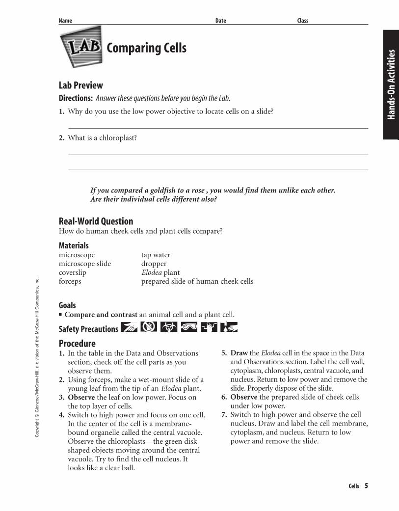
Cells 5
Name Date Class
Lab PreviewDirections: Answer these questions before you begin the Lab.
1. Why do you use the low power objective to locate cells on a slide?
2. What is a chloroplast?
If you compared a goldfish to a rose , you would find them unlike each other.Are their individual cells different also?
Comparing Cells
Hand
s-On
Act
iviti
es
Real-World QuestionHow do human cheek cells and plant cells compare?
Materialsmicroscope tap watermicroscope slide droppercoverslip Elodea plantforceps prepared slide of human cheek cells
Procedure1. In the table in the Data and Observations
section, check off the cell parts as youobserve them.
2. Using forceps, make a wet-mount slide of ayoung leaf from the tip of an Elodea plant.
3. Observe the leaf on low power. Focus onthe top layer of cells.
4. Switch to high power and focus on one cell.In the center of the cell is a membrane-bound organelle called the central vacuole.Observe the chloroplasts—the green disk-shaped objects moving around the centralvacuole. Try to find the cell nucleus. Itlooks like a clear ball.
5. Draw the Elodea cell in the space in the Dataand Observations section. Label the cell wall,cytoplasm, chloroplasts, central vacuole, andnucleus. Return to low power and remove theslide. Properly dispose of the slide.
6. Observe the prepared slide of cheek cellsunder low power.
7. Switch to high power and observe the cellnucleus. Draw and label the cell membrane,cytoplasm, and nucleus. Return to lowpower and remove the slide.
Goals■ Compare and contrast an animal cell and a plant cell.
Safety Precautions
429-1-50-MSS05-000000_CR 3/24/04 4:02 PM Page 5 impos03 301:goscanc:scanc429:layouts:
6 Cells
Cop
yrig
ht ©
Gle
ncoe
/McG
raw
-Hill
,a d
ivis
ion
of t
he M
cGra
w-H
ill C
ompa
nies
,Inc
.
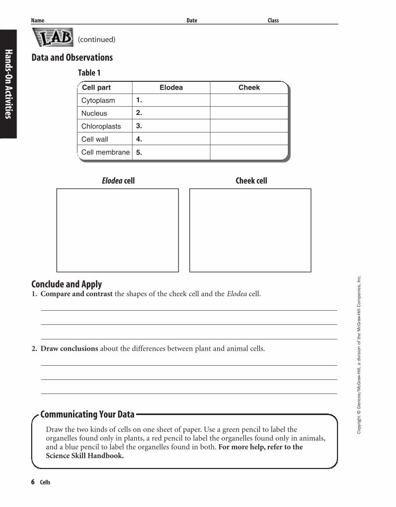
Name Date Class
Data and Observations
Table 1
Conclude and Apply1. Compare and contrast the shapes of the cheek cell and the Elodea cell.
2. Draw conclusions about the differences between plant and animal cells.
Hands-On Activities
Communicating Your Data
Draw the two kinds of cells on one sheet of paper. Use a green pencil to label theorganelles found only in plants, a red pencil to label the organelles found only in animals,and a blue pencil to label the organelles found in both. For more help, refer to the Science Skill Handbook.
Elodea cell Cheek cell
Cell part Elodea Cheek
Cytoplasm
Nucleus
Chloroplasts
Cell wall
Cell membrane
1.
2.
3.
4.
5.
(continued)
429-1-50-MSS05-000000_CR 3/24/04 4:02 PM Page 6 impos03 301:goscanc:scanc429:layouts:

Cop
yrig
ht ©
Gle
ncoe
/McG
raw
-Hill
,a d
ivis
ion
of t
he M
cGra
w-H
ill C
ompa
nies
,Inc
.
Cells 7
Name Date Class
Lab PreviewDirections: Answer these questions before you begin the Lab.
1. Why should you wear gloves during this experiment?
2. Describe a stereomicroscope.
You’re a technician in a police forensic laboratory. You use a stereomicroscopeand a compound light microscope in the laboratory. A detective just returnedfrom a crime scene with bags of evidence. You must examine each piece ofevidence under a microscope. How do you decide which microscope is the besttool to use?
Real-World QuestionWill all of the evidence that you’ve collectedbe viewable through both microscopes?
Form a HypothesisCompare the items to be examined under themicroscopes. Form a hypothesis to predictwhich microscope will be used for each itemand explain why.
Possible Materialscompound light microscopestereomicroscopeitems from the classroom—include some living or
once-living items (8)microscope slides and coverslipsplastic petri dishesdistilled waterdropper
Goals■ Learn how to correctly use a stereomicro-
scope and a compound light microscope.■ Compare the uses of the stereomicroscope
and compound light microscope.
Safety Precautions
Test Your Hypothesis
Make a Plan1. As a group, decide how you will test your
hypothesis.2. Describe how you will carry out this
experiment using a series of specific steps.Make sure the steps are in a logical order.Remember that you must place an item in the bottom of a plastic petri dish toexamine it under the stereomicroscopeand you must make a wet mount of anyitem to be examined under the compoundlight microscope. For more help, see theReference Handbook.
3. If you need a data table or an observationtable, design one on a separate sheet ofpaper.
Follow Your Plan1. Make sure your teacher approves the
objects you’ll examine, your plan, and yourdata table before you start.
2. Carry out the experiment.3. While doing the experiment, record your
observations and complete the data table.
Design Your Own
Comparing Light Microscopes
Hand
s-On
Act
iviti
es
429-1-50-MSS05-000000_CR 3/24/04 4:02 PM Page 7 impos03 301:goscanc:scanc429:layouts:
8 Cells
Cop
yrig
ht ©
Gle
ncoe
/McG
raw
-Hill
,a d
ivis
ion
of t
he M
cGra
w-H
ill C
ompa
nies
,Inc
.
Name Date Class
Hands-On Activities
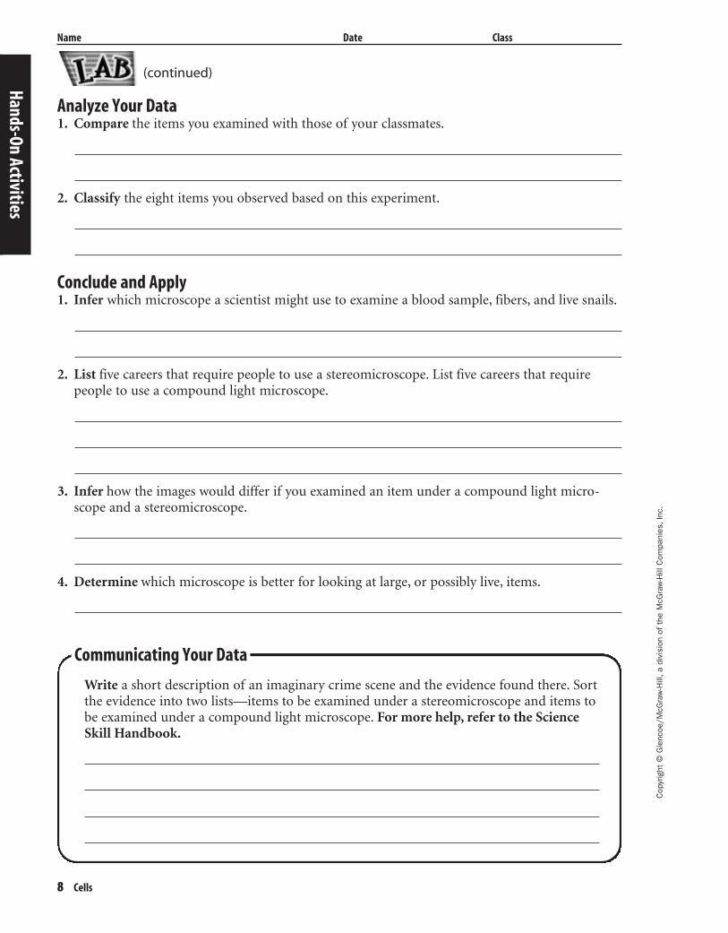
Analyze Your Data1. Compare the items you examined with those of your classmates.
2. Classify the eight items you observed based on this experiment.
Conclude and Apply1. Infer which microscope a scientist might use to examine a blood sample, fibers, and live snails.
2. List five careers that require people to use a stereomicroscope. List five careers that require people to use a compound light microscope.
3. Infer how the images would differ if you examined an item under a compound light micro-scope and a stereomicroscope.
4. Determine which microscope is better for looking at large, or possibly live, items.
Communicating Your Data
Write a short description of an imaginary crime scene and the evidence found there. Sortthe evidence into two lists—items to be examined under a stereomicroscope and items tobe examined under a compound light microscope. For more help, refer to the ScienceSkill Handbook.
(continued)
429-1-50-MSS05-000000_CR 3/24/04 4:02 PM Page 8 impos03 301:goscanc:scanc429:layouts:
Cop
yrig
ht ©
Gle
ncoe
/McG
raw
-Hill
,a d
ivis
ion
of t
he M
cGra
w-H
ill C
ompa
nies
,Inc
.
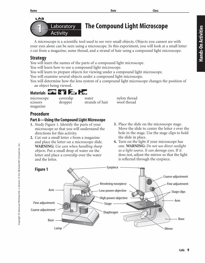
Cells 9
Name Date Class
The Compound Light Microscope
A microscope is a scientific tool used to see very small objects. Objects you cannot see withyour eyes alone can be seen using a microscope. In this experiment, you will look at a small lettere cut from a magazine, some thread, and a strand of hair using a compound light microscope.
StrategyYou will learn the names of the parts of a compound light microscope.You will learn how to use a compound light microscope.You will learn to prepare objects for viewing under a compound light microscope.You will examine several objects under a compound light microscope.You will determine how the lens system of a compound light microscope changes the position of
an object being viewed.
Materials microscope coverslip water nylon threadscissors dropper strands of hair wool threadmagazine
Hand
s-On
Act
iviti
es
ProcedurePart A—Using the Compound Light Microscope1. Study Figure 1. Identify the parts of your
microscope so that you will understand thedirections for this activity.
2. Cut out a small letter e from a magazineand place the letter on a microscope slide.WARNING: Use care when handling sharpobjects. Put a small drop of water on theletter and place a coverslip over the waterand the letter.
3. Place the slide on the microscope stage.Move the slide to center the letter e over thehole in the stage. Use the stage clips to holdthe slide in place.
4. Turn on the light if your microscope hasone. WARNING: Do not use direct sunlightas a light source. It can damage eyes. If itdoes not, adjust the mirror so that the lightis reflected through the eyepiece.
Arm
ArmFine adjustment
Fine adjustment
Coarse adjustment
Coarse adjustment
Base Base
Mirror
Eyepiece
Revolving nosepiece
Low power objective
High power objective
Stage
Stage clips
Diaphragm
Lamp
Figure 1
LaboratoryActivity11
429-1-50-MSS05-000000_CR 3/24/04 4:02 PM Page 9 impos03 301:goscanc:scanc429:layouts:

10 Cells
Cop
yrig
ht ©
Gle
ncoe
/McG
raw
-Hill
,a d
ivis
ion
of t
he M
cGra
w-H
ill C
ompa
nies
,Inc
.
Laboratory Activity 1 (continued)
Name Date Class
5. Look to see how the letter e is positionedon the slide before looking through theeyepiece. In the space for Figure 2a in Dataand Observations, draw the letter as yousee it without the aid of the microscope.
6. Click the low power objective lens (short-est, if more than one lens is present) intoposition. The lens should be directly overthe hole in the stage. Bring the lens close tothe slide using the coarse adjustment knob.NOTE: Be careful not to touch the slidewith the lens. This might break the lens andthe slide.
7. Look through the eyepiece of the micro-scope. Carefully bring the letter into focusby slowly turning the coarse adjustmentknob. If you cannot see the letter, movethe slide a little bit to be sure the letter isunder the lens. If your microscope hasonly one objective lens, proceed directly tostep 9; skip step 8.
8. Click the high power objective lens intoplace. If your microscope has a highpower objective, it will also have a fineadjustment knob. Look through the eye-piece again. Carefully bring the letter einto focus by slowly turning the fineadjustment knob. NOTE: Never turn the coarse adjustment knob when thehigh power objective lens is in place.
Click the low power objective lens backinto place before going on to step 9.
9. When the letter e is clearly visible, draw inFigure 2b the position of the letter as yousee it through the microscope. Next, movethe slide to the left as you look throughthe eyepiece. Note which way the letterappears to move. Move the slide forward.Note which way it appears to move now.
10. Remove the slide and clean it.
Part B—Preparing Microscope Slides1. Place a drop of water on a clean glass slide.
Put a strand of hair from your head and ahair from your forearm on the water drop.Place a coverslip over the drop of water andthe two different strands of hair.
2. Observe the hair using the procedure youused in Part A to observe the letter e.
3. In the space for Figure 3a in Data andObservations, draw the two hair strands asthey appear through the microscope. Labelthe hairs “head” and “arm.” Notice whichstrand appears thicker and show this differ-ence in your sketch.
4. Repeat Part B using a strand of nylonthread and a strand of wool thread. Drawand label the threads in Figure 3b in Dataand Observations.
Hands-On Activities
Data and ObservationsIn the spaces below, draw what you observed.
Letter e withoutmicroscope
Letter e throughmicroscope
baArm and
head hairsthrough
microscope
Wool andnylon threads
throughmicroscope
a b
Figure 2 Figure 3
429-1-50-MSS05-000000_CR 3/24/04 4:02 PM Page 10 impos03 301:goscanc:scanc429:layouts:

Cop
yrig
ht ©
Gle
ncoe
/McG
raw
-Hill
,a d
ivis
ion
of t
he M
cGra
w-H
ill C
ompa
nies
,Inc
.
Cells 11
Name Date Class
Questions and Conclusions1. Compare your drawing of the letter e without the microscope to your drawing of the letter
seen through the microscope. Describe how the microscope changes the position of the letter.
2. In what direction does the slide under the microscope appear to move when you move it to the left?
3. Describe the differences in thickness you observed between arm hair and head hair.
4. Describe the differences you observed between wool thread and nylon thread.
5. What is the total magnification of your microscope? (Multiply the magnification of the eye-piece lens by the magnification of the objective lens. These numbers are printed on the lenses.)
6. Describe how you would correctly prepare a microscope slide of an insect wing for viewingunder the microscope.
7. What precautions must be taken when using the high power lens?Ha
nds-
On A
ctiv
ities
Laboratory Activity 1 (continued)
429-1-50-MSS05-000000_CR 3/24/04 4:02 PM Page 11 impos03 301:goscanc:scanc429:layouts:
12 Cells
Cop
yrig
ht ©
Gle
ncoe
/McG
raw
-Hill
,a d
ivis
ion
of t
he M
cGra
w-H
ill C
ompa
nies
,Inc
.
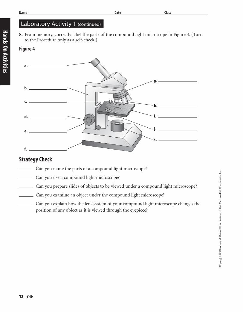
Laboratory Activity 1 (continued)
Name Date Class
8. From memory, correctly label the parts of the compound light microscope in Figure 4. (Turnto the Procedure only as a self-check.)
Figure 4
Strategy Check
Can you name the parts of a compound light microscope?
Can you use a compound light microscope?
Can you prepare slides of objects to be viewed under a compound light microscope?
Can you examine an object under the compound light microscope?
Can you explain how the lens system of your compound light microscope changes the
position of any object as it is viewed through the eyepiece?
Hands-On Activities
a.
b.
c.
d.
e.
f.
g.
h.
i.
j.
k.
429-1-50-MSS05-000000_CR 3/24/04 4:02 PM Page 12 impos03 301:goscanc:scanc429:layouts:

Cop
yrig
ht ©
Gle
ncoe
/McG
raw
-Hill
,a d
ivis
ion
of t
he M
cGra
w-H
ill C
ompa
nies
,Inc
.
Cells 13
Name Date Class
Observing Cells
Hand
s-On
Act
iviti
es
If you were asked how a tree, a fly, and you are alike, you might answer, “We are all alive.” If youcould examine each under a microscope, you might answer, “We all contain cells.” One veryimportant similarity among all living things is that each is made of very small units called cells.
StrategyYou will prepare living things for microscopic viewing.You will see that each living thing is made of cells and be able to name the parts of a cell.You will compare plant cells to animal cells.
Materials
Procedure
Part A—Observing Cork Cells1. Add a drop of water to a clean microscope
slide. Use forceps to add a small piece ofcork. Cover with a coverslip.
2. View the cork under low power magnifica-tion. Change to high power if your micro-scope has a high power lens.
3. Draw what you observe under Data andObservations, Part A. Label what you see.
Part B—Observing Frog Blood Cells1. View the prepared slide of frog blood
under low power magnification. Change tohigh power if your microscope has a highpower lens.
2. Draw what you observe under Data andObservations, Part B. Label the cell mem-brane, cytoplasm, and nucleus.
Part C—Observing Lettuce Leaf Cells1. Add a drop of water to a clean microscope
slide.2. Remove a small piece of lettuce leaf and
place it in the drop of water. Cover with acoverslip. Identify as many cell parts as youcan.
3. Under Data and Observation, Part C, drawwhat you observe. Label the cell wall,chloroplast, cytoplasm, nucleus, and vacuoles. (The nucleus may be difficult toobserve.)
water forceps microscope prepared slide ofdropper cork shavings lettuce leaf frog bloodmicroscope slide coverslip
LaboratoryActivity22
Part A Part B Part C
Data and Observations
Figure 1
429-1-50-MSS05-000000_CR 3/24/04 4:02 PM Page 13 impos03 301:goscanc:scanc429:layouts:
14 Cells
Cop
yrig
ht ©
Gle
ncoe
/McG
raw
-Hill
,a d
ivis
ion
of t
he M
cGra
w-H
ill C
ompa
nies
,Inc
.
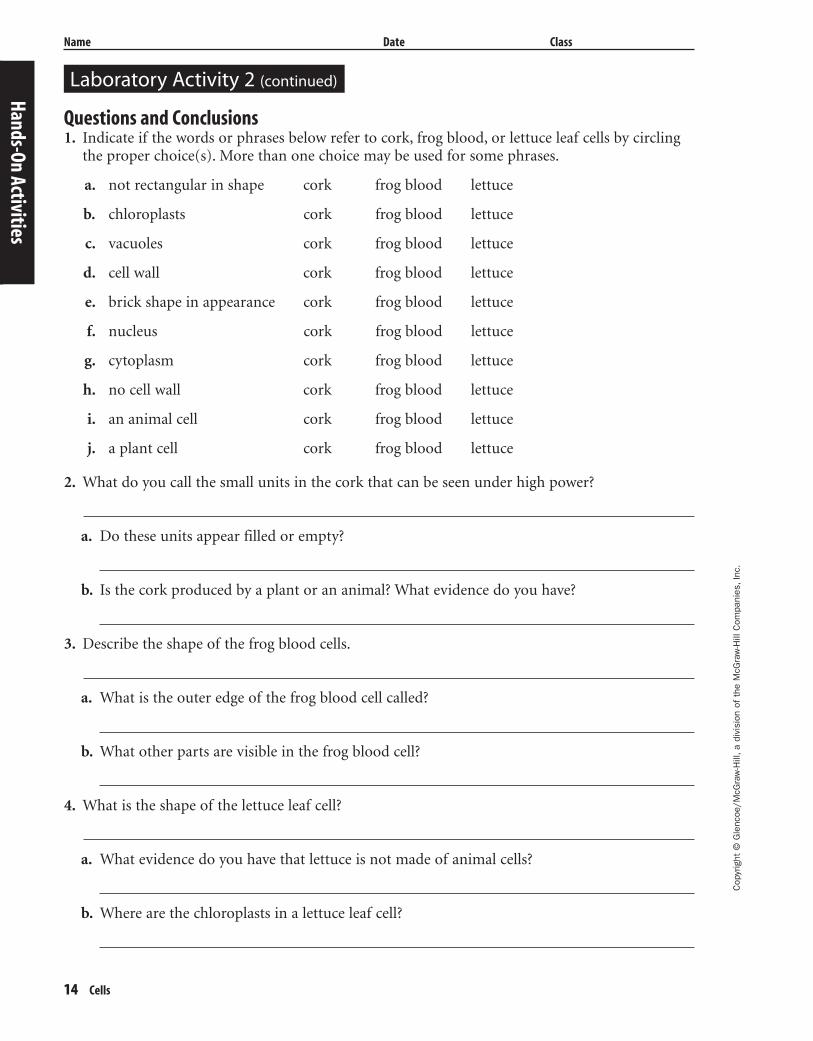
Laboratory Activity 2 (continued)
Name Date Class
Questions and Conclusions1. Indicate if the words or phrases below refer to cork, frog blood, or lettuce leaf cells by circling
the proper choice(s). More than one choice may be used for some phrases.
a. not rectangular in shape cork frog blood lettuce
b. chloroplasts cork frog blood lettuce
c. vacuoles cork frog blood lettuce
d. cell wall cork frog blood lettuce
e. brick shape in appearance cork frog blood lettuce
f. nucleus cork frog blood lettuce
g. cytoplasm cork frog blood lettuce
h. no cell wall cork frog blood lettuce
i. an animal cell cork frog blood lettuce
j. a plant cell cork frog blood lettuce
2. What do you call the small units in the cork that can be seen under high power?
a. Do these units appear filled or empty?
b. Is the cork produced by a plant or an animal? What evidence do you have?
3. Describe the shape of the frog blood cells.
a. What is the outer edge of the frog blood cell called?
b. What other parts are visible in the frog blood cell?
4. What is the shape of the lettuce leaf cell?
a. What evidence do you have that lettuce is not made of animal cells?
b. Where are the chloroplasts in a lettuce leaf cell?
Hands-On Activities
429-1-50-MSS05-000000_CR 3/24/04 4:02 PM Page 14 impos03 301:goscanc:scanc429:layouts:
Cop
yrig
ht ©
Gle
ncoe
/McG
raw
-Hill
,a d
ivis
ion
of t
he M
cGra
w-H
ill C
ompa
nies
,Inc
.
Cells 15
Name Date Class
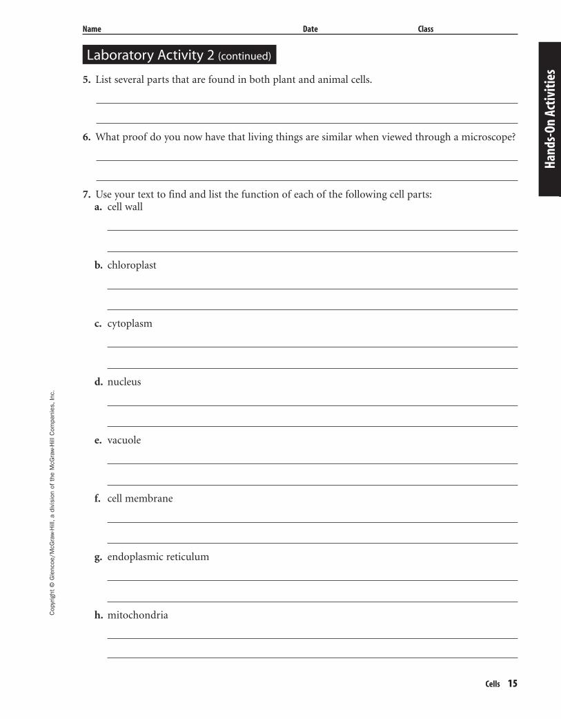
5. List several parts that are found in both plant and animal cells.
6. What proof do you now have that living things are similar when viewed through a microscope?
7. Use your text to find and list the function of each of the following cell parts:a. cell wall
b. chloroplast
c. cytoplasm
d. nucleus
e. vacuole
f. cell membrane
g. endoplasmic reticulum
h. mitochondria
Hand
s-On
Act
iviti
es
Laboratory Activity 2 (continued)
429-1-50-MSS05-000000_CR 3/24/04 4:02 PM Page 15 impos03 301:goscanc:scanc429:layouts:
16 Cells
Cop
yrig
ht ©
Gle
ncoe
/McG
raw
-Hill
,a d
ivis
ion
of t
he M
cGra
w-H
ill C
ompa
nies
,Inc
.
Laboratory Activity 2 (continued)
Name Date Class
Hands-On Activities
a. corridors
b. boiler room
c. cafeteria
d. principal’s office
e. bricks of building
8. Compare parts of a cell with parts of your school building. Match the cell part function withthe function of the corresponding school area.
Strategy Check
Can you prepare living things for microscopic viewing?
Do you agree that all living things are made of cells?
Can you correctly label those cell parts observed?
Can you compare plant cells with animal cells?
cell wall
nucleus
endoplasmic reticulum
chloroplast
mitochondria
429-1-50-MSS05-000000_CR 3/24/04 4:02 PM Page 16 impos03 301:goscanc:scanc429:layouts:

Cop
yrig
ht ©
Gle
ncoe
/McG
raw
-Hill
,a d
ivis
ion
of t
he M
cGra
w-H
ill C
ompa
nies
,Inc
.
Cells 17
Cells
Directions: Use this page to label your Foldable at the beginning of the chapter.
Cells
Plant Cell
Animal Cell
Name Date Class
Hand
s-On
Act
iviti
es
429-1-50-MSS05-000000_CR 3/24/04 4:02 PM Page 17 impos03 301:goscanc:scanc429:layouts:

18 Cells
Meeting IndividualNeeds
Meeting Individual Needs
429-1-50-MSS05-000000_CR 3/24/04 4:02 PM Page 18 impos03 301:goscanc:scanc429:layouts:

Cop
yrig
ht ©
Gle
ncoe
/McG
raw
-Hill
,a d
ivis
ion
of t
he M
cGra
w-H
ill C
ompa
nies
,Inc
.
Name Date Class
Cells 19
OverviewCells
Directions: Complete the concept map using the terms listed below.
lysosome chloroplast mitochondria cell wall
Directions: Complete the following sentences using the terms listed below.
virus cytoplasm microscope vaccine cell theory
5. A ____________________ is often used to prevent viral diseases.
6. We need to use a ____________________ to see most cells.
7. Cells are filled with a gelatinlike mixture called ____________________.
8. A ____________________ is a disease-causing strand of hereditary material surrounded by a protein coating.
9. The ___________________ was developed from the observations and conclusionsof several scientists.
Mee
ting
Indi
vidu
al N
eeds
1.
ribosome
2.
3.
nucleus
4.
has
has
has
has
has
has
has
has
Directed Reading for
Content Mastery
An animal cell
has
A plant cell
429-1-50-MSS05-000000_CR 3/24/04 4:02 PM Page 19 impos03 301:goscanc:scanc429:layouts:
20 Cells
Cop
yrig
ht ©
Gle
ncoe
/McG
raw
-Hill
,a d
ivis
ion
of t
he M
cGra
w-H
ill C
ompa
nies
,Inc
.
Name Date Class
Section 1 ■ Cell StructureSection 2 ■ Viewing Cells
Directions: Write T if the statement is true; write F if the statement is false.
1. Fungi have prokaryotic cells.
2. Most one-celled organisms are prokaryotic.
3. Plants and animals have eukaryotic cells.
4. Animal cells are enclosed in a cell wall.
5. The cell membrane is the protective layer around all cells.
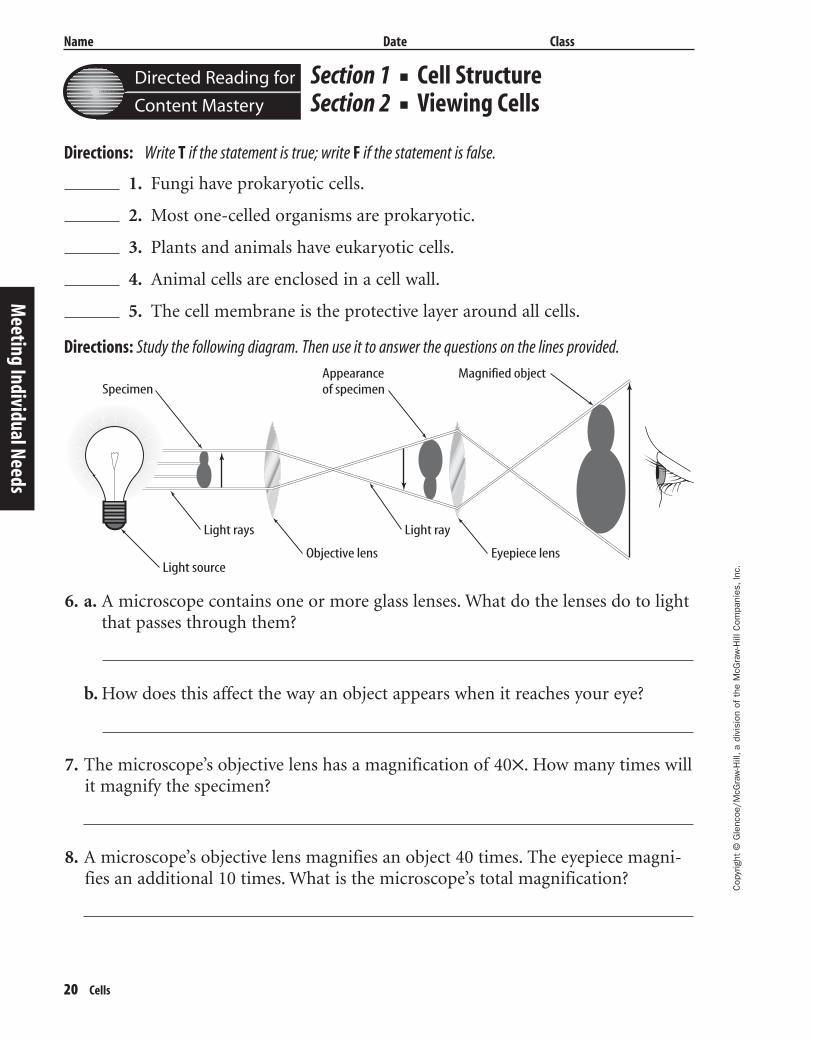
Directions: Study the following diagram. Then use it to answer the questions on the lines provided.
6. a. A microscope contains one or more glass lenses. What do the lenses do to light that passes through them?
b. How does this affect the way an object appears when it reaches your eye?
7. The microscope’s objective lens has a magnification of 40✕. How many times willit magnify the specimen?
8. A microscope’s objective lens magnifies an object 40 times. The eyepiece magni-fies an additional 10 times. What is the microscope’s total magnification?
Meeting Individual Needs
Directed Reading for
Content Mastery
SpecimenAppearanceof specimen
Magnified object
Light source
Light rays Light ray
Eyepiece lensObjective lens
429-1-50-MSS05-000000_CR 3/24/04 4:02 PM Page 20 impos03 301:goscanc:scanc429:layouts:
Cop
yrig
ht ©
Gle
ncoe
/McG
raw
-Hill
,a d
ivis
ion
of t
he M
cGra
w-H
ill C
ompa
nies
,Inc
.
Name Date Class
Cells 21
Section 3 ■ Viruses
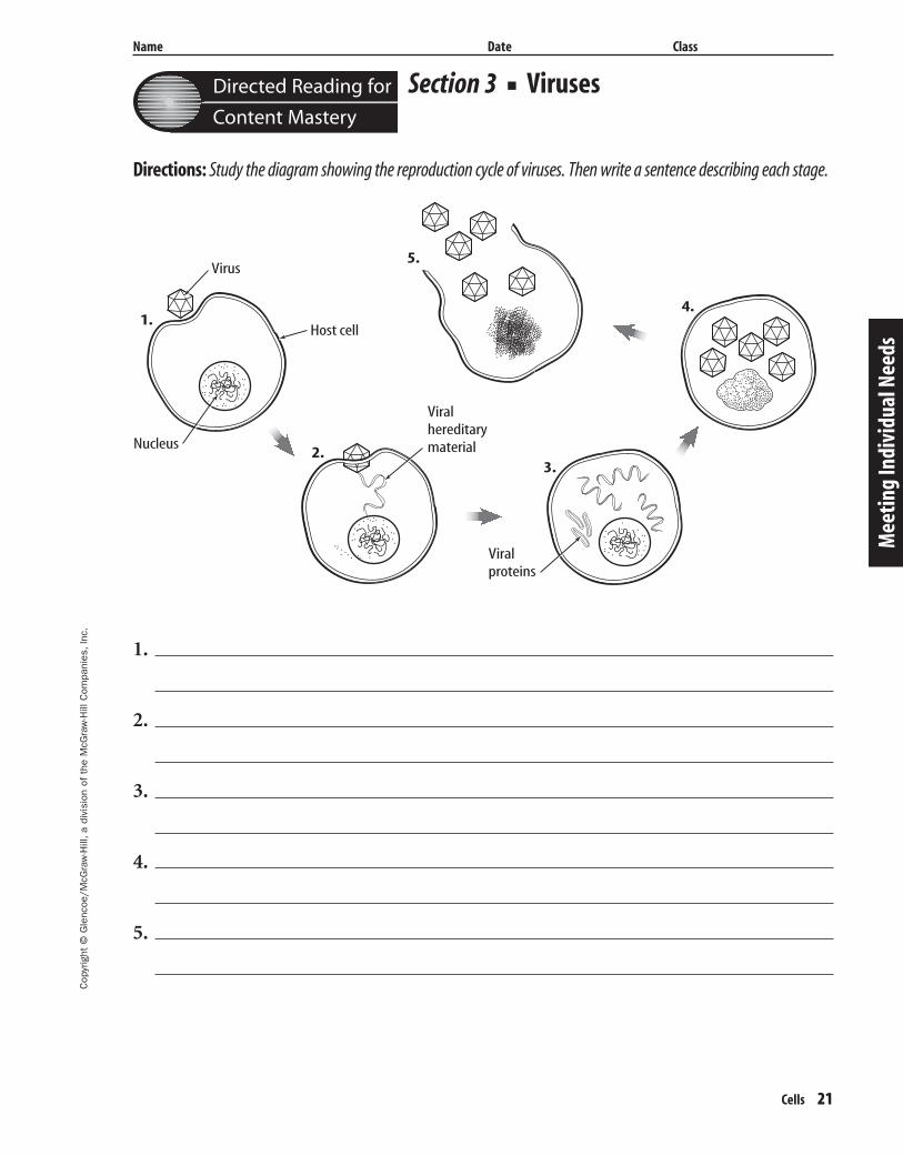
Directions: Study the diagram showing the reproduction cycle of viruses. Then write a sentence describing each stage.
1.
2.
3.
4.
5.
Mee
ting
Indi
vidu
al N
eeds
Directed Reading for
Content Mastery
Virus
Host cell
Nucleus
Viralhereditarymaterial
Viralproteins
1.
2.3.
4.
5.
429-1-50-MSS05-000000_CR 3/24/04 4:02 PM Page 21 impos03 301:goscanc:scanc429:layouts:
22 Cells
Cop
yrig
ht ©
Gle
ncoe
/McG
raw
-Hill
,a d
ivis
ion
of t
he M
cGra
w-H
ill C
ompa
nies
,Inc
.
Name Date Class
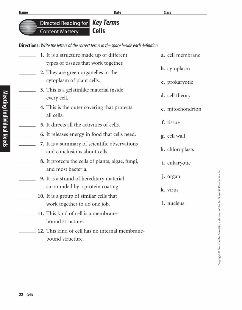
Key TermsCells
Directions: Write the letters of the correct terms in the space beside each definition.
1. It is a structure made up of different
types of tissues that work together.
2. They are green organelles in the
cytoplasm of plant cells.
3. This is a gelatinlike material inside
every cell.
4. This is the outer covering that protects
all cells.
5. It directs all the activities of cells.
6. It releases energy in food that cells need.
7. It is a summary of scientific observations
and conclusions about cells.
8. It protects the cells of plants, algae, fungi,
and most bacteria.
9. It is a strand of hereditary material
surrounded by a protein coating.
10. It is a group of similar cells that
work together to do one job.
11. This kind of cell is a membrane-
bound structure.
12. This kind of cell has no internal membrane-
bound structure.M
eeting Individual Needs
a. cell membrane
b. cytoplasm
c. prokaryotic
d. cell theory
e. mitochondrion
f. tissue
g. cell wall
h. chloroplasts
i. eukaryotic
j. organ
k. virus
l. nucleus
Directed Reading for
Content Mastery
429-1-50-MSS05-000000_CR 3/24/04 4:02 PM Page 22 impos03 301:goscanc:scanc429:layouts:
Cop
yrig
ht ©
Gle
ncoe
/McG
raw
-Hill
,a d
ivis
ion
of t
he M
cGra
w-H
ill C
ompa
nies
,Inc
.
Nombre Fecha Clase
Estructura y funciones de la vida 23
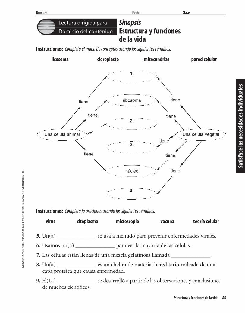
SinopsisEstructura y funciones de la vida
Instrucciones: Completa el mapa de conceptos usando los siguientes términos.
lisosoma cloroplasto mitocondrias pared celular
Instrucciones: Completa la oraciones usando los siguientes términos.
virus citoplasma microscopio vacuna teoría celular
5. Un(a) ______________ se usa a menudo para prevenir enfermedades virales.
6. Usamos un(a) ______________ para ver la mayoría de las células.
7. Las células están llenas de una mezcla gelatinosa llamada ______________.
8. Un(a) ______________ es una hebra de material hereditario rodeada de unacapa proteíca que causa enfermedad.
9. El(La) ______________ se desarrolló a partir de las observaciones y conclusionesde muchos científicos.
Lectura dirigida para
Dominio del contenido
Satis
face
las n
eces
idad
es in
divi
dual
es
1.
ribosoma
2.
3.
núcleo
4.
tiene
tiene
tiene
tiene
tiene
tiene
tiene
Una célula animal
tiene
Una célula vegetal
429-1-50-MSS05-000000_CR 3/24/04 4:02 PM Page 23 impos03 301:goscanc:scanc429:layouts:
24 Estructura y funciones de la vida
Cop
yrig
ht ©
Gle
ncoe
/McG
raw
-Hill
,a d
ivis
ion
of t
he M
cGra
w-H
ill C
ompa
nies
,Inc
.
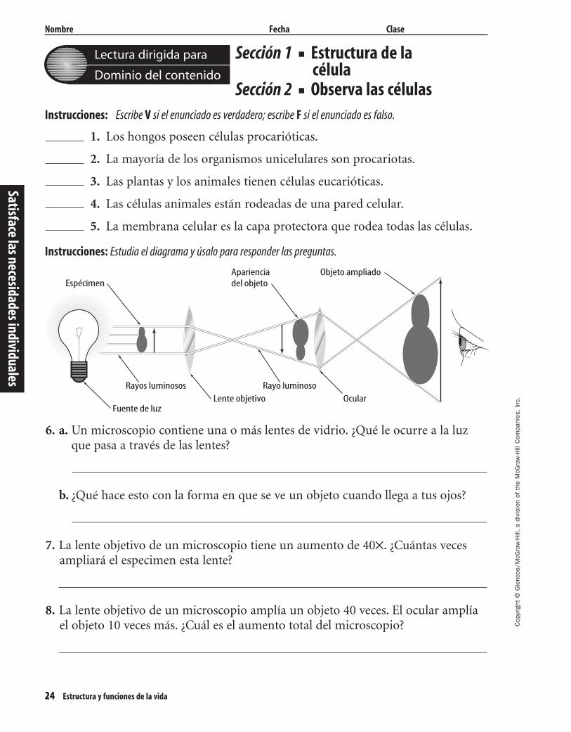
Nombre Fecha Clase
Sección 1 ■ Estructura de lacélula
Sección 2 ■ Observa las célulasInstrucciones: Escribe V si el enunciado es verdadero; escribe F si el enunciado es falso.
1. Los hongos poseen células procarióticas.
2. La mayoría de los organismos unicelulares son procariotas.
3. Las plantas y los animales tienen células eucarióticas.
4. Las células animales están rodeadas de una pared celular.
5. La membrana celular es la capa protectora que rodea todas las células.
Instrucciones: Estudia el diagrama y úsalo para responder las preguntas.
\
6. a. Un microscopio contiene una o más lentes de vidrio. ¿Qué le ocurre a la luz que pasa a través de las lentes?
b. ¿Qué hace esto con la forma en que se ve un objeto cuando llega a tus ojos?
7. La lente objetivo de un microscopio tiene un aumento de 40✕. ¿Cuántas vecesampliará el especimen esta lente?
8. La lente objetivo de un microscopio amplía un objeto 40 veces. El ocular amplíael objeto 10 veces más. ¿Cuál es el aumento total del microscopio?
Lectura dirigida para
Dominio del contenido
EspécimenApariencia del objeto
Objeto ampliado
Fuente de luz
Rayos luminosos Rayo luminosoOcularLente objetivo
Satisface las necesidades individuales
429-1-50-MSS05-000000_CR 3/24/04 4:02 PM Page 24 impos03 301:goscanc:scanc429:layouts:

Cop
yrig
ht ©
Gle
ncoe
/McG
raw
-Hill
,a d
ivis
ion
of t
he M
cGra
w-H
ill C
ompa
nies
,Inc
.
Nombre Fecha Clase
Estructura y funciones de la vida 25
Sección 3 ■ Los virus
Instrucciones: Estudia el diagrama que muestra el ciclo reproductor de los virus y escribe una oración describiendocada etapa.
1.
2.
3.
4.
5.
Lectura dirigida para
Dominio del contenido
Virus
Célula huésped
Núcleo
Material hereditario viral
Proteínas virales
1.
2.3.
4.
5.
Satis
face
las n
eces
idad
es in
divi
dual
es
429-1-50-MSS05-000000_CR 3/24/04 4:02 PM Page 25 impos03 301:goscanc:scanc429:layouts:

26 Estructura y funciones de la vida
Cop
yrig
ht ©
Gle
ncoe
/McG
raw
-Hill
,a d
ivis
ion
of t
he M
cGra
w-H
ill C
ompa
nies
,Inc
.
Nombre Fecha Clase
Términos clavesEstructura y funciones de la vida
Instrucciones: Escribe la letra del término correcto en el espacio dado al lado de cada definición.
1. Es una estructura compuesta de diferentes
tipos de tejidos que trabajan juntos.
2. Son organelos verdes en el citoplasma de las
células vegetales.
3. Este es el material gelatinoso dentro de cada
célula.
4. Esta es la cubierta que protege todas las
células.
5. Dirige todas las actividades de las células.
6. Libera la energía de los alimentos, la cual
necesitan las células.
7. Es el resumen de las observaciones y
conclusiones científicas sobre las células.
8. Protege las células vegetales, las algas, los
hongos y la mayoría de las bacterias.
9. Es una hebra de material hereditario rodeada
de una capa proteica.
10. Es un grupo de células similares que trabajan
en conjunto para llevar a cabo una función.
11. Este tipo de célula es una estructura rodeada
por una membrana.
12. Este tipo de célula no tiene una estructura
rodeada por una membrana.
a. membranacelular
b. citoplasma
c. procariótico(a)
d. teoría celular
e. mitocondria
f. tejido
g. pared celular
h. cloroplastos
i. eucariótico(a)
j. órgano
k. virus
l. núcleo
Lectura dirigida para
Dominio del contenido
Satisface las necesidades individuales
429-1-50-MSS05-000000_CR 3/24/04 4:02 PM Page 26 impos03 301:goscanc:scanc429:layouts:
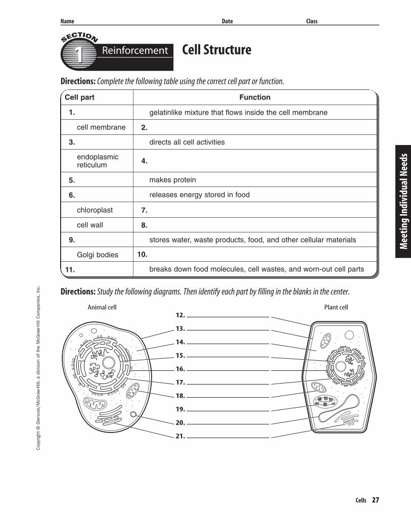
1.
cell membrane
3.
endoplasmicreticulum
5.
6.
chloroplast
cell wall
9.
Golgi bodies
11.
FunctionCell part
2.
4.
7.
8.
10.
makes protein
releases energy stored in food
directs all cell activities
gelatinlike mixture that flows inside the cell membrane
stores water, waste products, food, and other cellular materials
breaks down food molecules, cell wastes, and worn-out cell parts
Cop
yrig
ht ©
Gle
ncoe
/McG
raw
-Hill
,a d
ivis
ion
of t
he M
cGra
w-H
ill C
ompa
nies
,Inc
.
Name Date Class
Cells 27
Directions: Complete the following table using the correct cell part or function.
Directions: Study the following diagrams. Then identify each part by filling in the blanks in the center.
Cell Structure
Mee
ting
Indi
vidu
al N
eeds
Reinforcement11
12.
13.
14.
15.
16.
17.
18.
19.
20.
21.
Animal cell Plant cell
429-1-50-MSS05-000000_CR 3/24/04 4:02 PM Page 27 impos03 301:goscanc:scanc429:layouts:
28 Cells
Cop
yrig
ht ©
Gle
ncoe
/McG
raw
-Hill
,a d
ivis
ion
of t
he M
cGra
w-H
ill C
ompa
nies
,Inc
.
Name Date Class
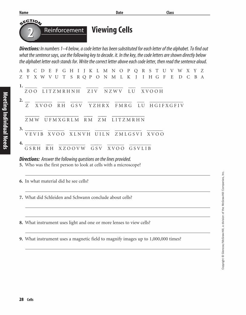
Viewing Cells
Directions: In numbers 1–4 below, a code letter has been substituted for each letter of the alphabet. To find outwhat the sentence says, use the following key to decode. it. In the key, the code letters are shown directly belowthe alphabet letter each stands for. Write the correct letter above each code letter, then read the sentence aloud.
Reinforcement22
Meeting Individual Needs
Directions: Answer the following questions on the lines provided.5. Who was the first person to look at cells with a microscope?
6. In what material did he see cells?
7. What did Schleiden and Schwann conclude about cells?
8. What instrument uses light and one or more lenses to view cells?
9. What instrument uses a magnetic field to magnify images up to 1,000,000 times?
A B C D E F G H I J K L M N O P Q R S T U V W X Y ZZ Y X W V U T S R Q P O N M L K J I H G F E D C B A
1. _____ ________________ _____ ________ ___ _________Z O O L I T Z M R H N H Z I V N Z W V L U X V O O H
2. __ _______ ____ _____ _________ _______ ___ ______________Z X V O O R H G S V Y Z H R X F M R G L U H G I F X G F I V
______ ______________ _____ ____ ______________Z M W U F M X G R L M R M Z M L I T Z M R H N
3. ________ _______ _________ ______ ___________ _______V E V I B X V O O X L N V H U I L N Z M L G S V I X V O O
4. _______ ____ ___________ _____ _______ _________G S R H R H X Z O O V W G S V X V O O G S V L I B
429-1-50-MSS05-000000_CR 3/24/04 4:02 PM Page 28 impos03 301:goscanc:scanc429:layouts:
Cop
yrig
ht ©
Gle
ncoe
/McG
raw
-Hill
,a d
ivis
ion
of t
he M
cGra
w-H
ill C
ompa
nies
,Inc
.
Name Date Class
Cells 29
1. Listed below are the steps by which an active virus copies itself and destroys a cell. Number thesteps in the correct order in the blanks provided at the left.
a. The cell bursts open and hundreds of new virus particles are released. These new virus particles go on to infect other cells.
b. A specific virus attaches to the surface of a specific host cell.
c. The viral hereditary material takes control of the host cell and the cell begins to make new virus particles.
d. The hereditary material of the virus entering the host cell.
Directions: Answer the following questions using complete sentences.2. Explain what a latent virus does when it enters a cell.
3. Discuss several ways to prevent viral infections.
4. What are vaccines made from?
5. How does gene therapy work?
Viruses
Mee
ting
Indi
vidu
al N
eeds
Reinforcement33
429-1-50-MSS05-000000_CR 3/24/04 4:02 PM Page 29 impos03 301:goscanc:scanc429:layouts:
30 Cells
Cop
yrig
ht ©
Gle
ncoe
/McG
raw
-Hill
,a d
ivis
ion
of t
he M
cGra
w-H
ill C
ompa
nies
,Inc
.
Name Date Class
The Early Cell Explorers
It’s hard to believe, but there was a time when we didn’t know anythingabout cell structure. In fact, the word cell (from the Latin word for chamber,cello) wasn’t used as a biological term until 1665. That’s when Robert Hooke,an English-born scientist, looked at a thin slice of a cork plant under a com-pound microscope he had built himself. Hooke noticed small holes sur-rounded by walls and named these tiny pores cells. After that, scientistsbelieved cells were found only in plants. But in 1839, Matthias Schleiden andTheodor Schwann, both German scientists, shared their scientific findings withone another. Schleiden had been studying plant cells and Schwann had beenstudying animal structures. Together, they compared plant and animal struc-tures and found that the structures were very similar—too similar to be acci-dental. They concluded that cells are the basic building blocks for both plantsand animals. In 1858, Rudolf Virchow took Schleiden’s and Schwann’s theoryand stated it simply: all cells come from other cells. This remains known as thecell theory.
Throughout the mid-1800s and into the 1900s, scientists continued to dis-cover more and more about cells thanks in part to Gregor Mendel’s study ofgenetics, Friedrich Miescher’s discovery of nuclein (which later became knownas DNA), and James Watson’s findings about DNA’s structure. Although manyamazing discoveries have happened in recent years, including genetic engineer-ing and gene therapy, all of it is because of the work of those early cell explorers.
Enrichment11
Meeting Individual Needs
1. How important was Hooke’s homemade microscope to the discovery of the plant cell? Explain.
2. Restate the cell theory in your own words.
3. Why do you think it took almost 200 years for scientists to formulate the cell theory?
429-1-50-MSS05-000000_CR 3/24/04 4:02 PM Page 30 impos03 301:goscanc:scanc429:layouts:
Cop
yrig
ht ©
Gle
ncoe
/McG
raw
-Hill
,a d
ivis
ion
of t
he M
cGra
w-H
ill C
ompa
nies
,Inc
.
Name Date Class
Cells 31
Using the MicroscopeEnrichment22
Mee
ting
Indi
vidu
al N
eeds
There are many different kinds of micro-scopes. A magnifying glass is a simple micro-scope. The term microscope commonly refers to acompound light microscope. These microscopesare called compound because they are made oftwo sets of glass lenses in a tube or tubes.
The total magnifying power of a compoundlight microscope is the product of the magni-fying power of the lens in the eyepiece and themagnifying power of the lens in the objective.Most compound light microscopes can mag-nify a specimen up to 1,000 times its real size.
Microscopes allow you to see fine details.Spaces between objects that are closertogether than 0.1 mm can be seen. The abilityof a microscope to separate very small dis-tances is called resolving power. If the resolv-ing power of the lens is not good, the imagewill appear blurred.
When you look into the eyepiece of amicroscope, the circular area you see is thefield of view. When a ruler is placed across theopening on the stage, the field of view can bemeasured in millimeters.
Directions: Using the information above, complete the table by filling in the blanks.
Directions: Answer the following questions on the lines provided.
6. How do you find the total magnifying power of a microscope?
7. What would cause an image to appear blurred?
Figure 1
8. What is the width of the field of view shown above? In centimeters? ______
In millimeters? ______
Eyepiece lens Objective lens Total
Microscope 1 Low 5x High 40x Low 1. High 400x
Microscope 2 8x 10x 60x
Microscope 3 10x 50x 300x4.
10x
5.
2. 3.
1 cm
1 2 3 4 5 6 7
429-1-50-MSS05-000000_CR 3/24/04 4:02 PM Page 31 impos03 301:goscanc:scanc429:layouts:
32 Cells
Cop
yrig
ht ©
Gle
ncoe
/McG
raw
-Hill
,a d
ivis
ion
of t
he M
cGra
w-H
ill C
ompa
nies
,Inc
.
Name Date Class
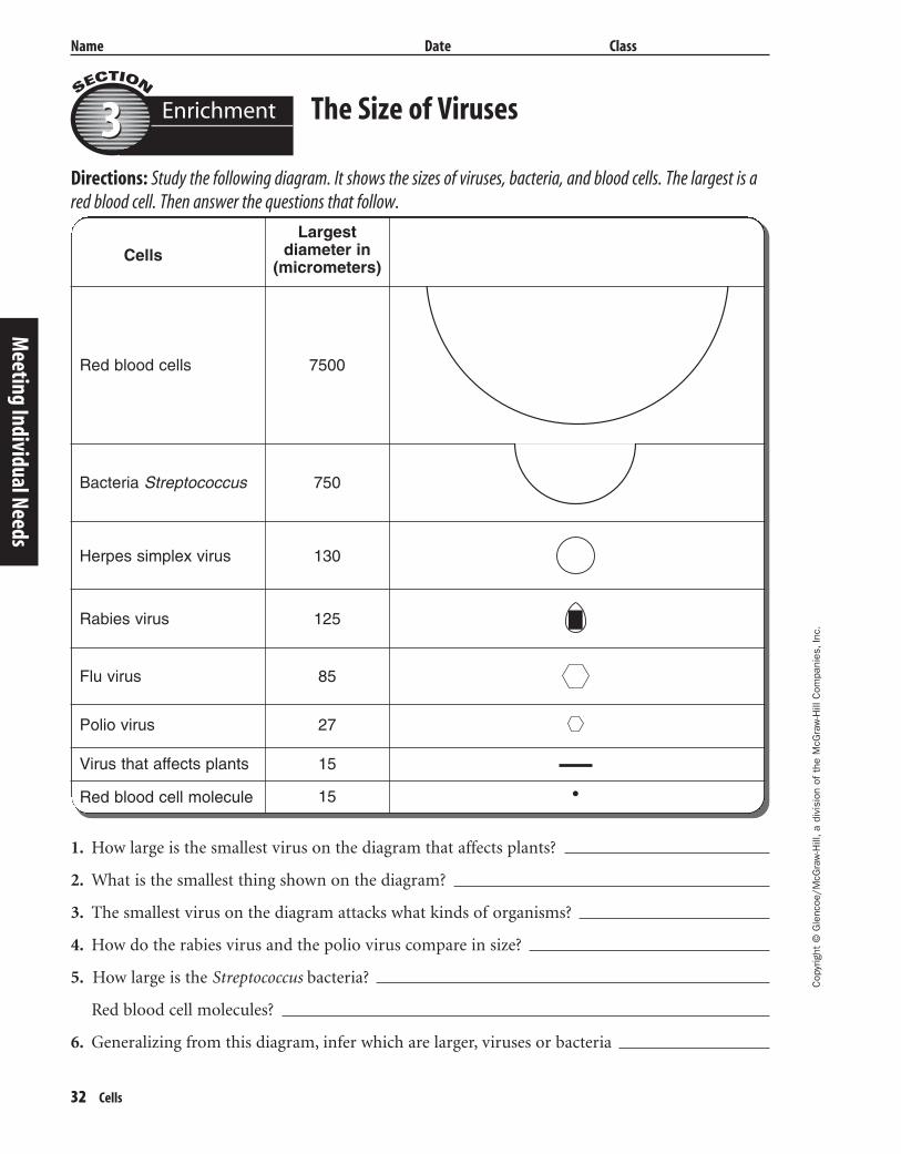
The Size of Viruses
Directions: Study the following diagram. It shows the sizes of viruses, bacteria, and blood cells. The largest is ared blood cell. Then answer the questions that follow.
Enrichment33
Meeting Individual Needs
1. How large is the smallest virus on the diagram that affects plants?
2. What is the smallest thing shown on the diagram?
3. The smallest virus on the diagram attacks what kinds of organisms?
4. How do the rabies virus and the polio virus compare in size?
5. How large is the Streptococcus bacteria?
Red blood cell molecules?
6. Generalizing from this diagram, infer which are larger, viruses or bacteria
CellsLargest
diameter in (micrometers)
Red blood cells
Bacteria Streptococcus
Herpes simplex virus
Flu virus
Polio virus
Virus that affects plants
Red blood cell molecule
Rabies virus
750
130
7500
125
85
27
15
15
429-1-50-MSS05-000000_CR 3/24/04 4:02 PM Page 32 impos03 301:goscanc:scanc429:layouts:
Cop
yrig
ht ©
Gle
ncoe
/McG
raw
-Hill
,a d
ivis
ion
of t
he M
cGra
w-H
ill C
ompa
nies
,Inc
.
Name Date Class
Cells 33
Cells
Section 1 Cell Structure
A. Common cell structures—outer covering called ________________ and internal gelatinlike
________________
1. Comparing cells—size and shape relate to ________________
2. Two cell types
a. ________________ cells lack membrane-bound internal structures.
b. ________________ cells contain membrane-bound internal structures.
B. Cell organization
1. Composed of cellulose, a cell wall grows, gives shape to, and protects the cells of
________________, algae, fungi, and most bacteria.
2. Cell ________________—protective layer around all cells
a. For cells with cell ________________, the cell membrane is inside the cell wall
b. A cell ________________ allows food and oxygen into the cell and waste products outof the cell.
3. Cytoplasm—gelatinlike substance inside cell membrane
a. ________________—scaffolding-like structure in cytoplasm which helps cell keep its shape
b. In the cytoplasm, eukaryotic cells have ________________ which help with cell life
processes.
4. Nucleus—contains instructions for everything cell does; includes DNA
5. Energy-processing organelles—help cells do their________________
a. Green organelles in plant cells contain ________________ to make food.
b. Organelles which release ________________ from food are called mitochondria.
6. Manufacturing organelles
a. Ribosomes make ________________ for cell activities.
b. Some ribosomes attach to the rough part of the endoplasmic reticulum, a series of
smooth or rough ________________ that move materials around in a cell.
Mee
ting
Indi
vidu
al N
eeds
Note-takingWorksheet
429-1-50-MSS05-000000_CR 3/24/04 4:02 PM Page 33 impos03 301:goscanc:scanc429:layouts:

34 Cells
Cop
yrig
ht ©
Gle
ncoe
/McG
raw
-Hill
,a d
ivis
ion
of t
he M
cGra
w-H
ill C
ompa
nies
,Inc
.
Name Date Class
7. Transporting and storing organelles
a. ________________ move substances out of a cell or to other parts of a cell.
b. ________________–membrane-bound temporary storage spaces
8. Recycling organelles–________________ break down food molecules and cell wastes.
C. From cell to organism
1. ________________–group of similar cells working together on one job
2. Different types of tissues working together make up an ________________.
3. A group of organs working together on a particular function form a(n) ________________
________________.
Section 2 Viewing Cells
A. Magnifying cells
1. Early microscopes–lenses made images ________________ but not always clear.
2. Modern microscopes that use lenses to bend ________________
a. A simple microscope has one lens while a ________________ microscope has two setsof lenses.
b. A stereomicroscope, which has two eyepieces, creates a ________________ image.
c. Powers of the eyepiece multiplied by objective lenses determine total ________________.
3. Electron microscopes–more powerful than other microscopes
a. Use a ________________ in a vacuum to bend electronic beams
b. ________________ must be photographed or produced electronically.
B. Development of the cell theory
1. The ________________ resulted from many scientists’ observations and conclusions.
2. The basic ________________ of organization is the cell.
3. All ________________ are composed of one or more cells.
4. New cells come from old cells through cell ________________.
Meeting Individual Needs
Note-taking Worksheet (continued)
429-1-50-MSS05-000000_CR 3/24/04 4:02 PM Page 34 impos03 301:goscanc:scanc429:layouts:
Cop
yrig
ht ©
Gle
ncoe
/McG
raw
-Hill
,a d
ivis
ion
of t
he M
cGra
w-H
ill C
ompa
nies
,Inc
.
Name Date Class
Cells 35
Section 3 Viruses
A. Virus–a nonliving strand of hereditary material surrounded by a ________________ coating
B. Virus multiplication–viruses can make copies of themselves only inside a living
________________ cell.
1. ________________ viruses–make the host cell produce new viruses, which kills the host cell
2. ________________ viruses–hide in the host cell without destroying it
a. Virus hereditary material becomes part of the ________________ cell’s hereditary material.
b. Latent viruses can become ________________ and then destroy the host cells.
C. Virus effects on organisms
1. Most viruses infect only specific kinds of cells.
2. Viruses are often carried to the host through the ________________.
3. The ________________ and host cell must fit together exactly to begin a viral infection.
4. ________________ attach to bacteria and inject their hereditary material.
D. Fighting viruses
1. Vaccines–weakened ________________ which allow the host to fight some diseases
2. Treating viral diseases
a. ________________ are not effective treatments for viral infections.
b. Infected cells sometimes produce ________________ which are proteins that can protect noninfected cells.
c. Antiviral drugs often have adverse ________________, limiting their use.
d. Public health measures can ________________ or slow disease spread.
E. Research with viruses–________________ uses viruses to replace defective cell hereditarymaterial with normal cell hereditary material.
Mee
ting
Indi
vidu
al N
eeds
Note-taking Worksheet (continued)
429-1-50-MSS05-000000_CR 3/24/04 4:02 PM Page 35 impos03 301:goscanc:scanc429:layouts:
36 Cells
Assessment
Assessment
429-1-50-MSS05-000000_CR 3/24/04 4:02 PM Page 36 impos03 301:goscanc:scanc429:layouts:
Cop
yrig
ht ©
Gle
ncoe
/McG
raw
-Hill
,a d
ivis
ion
of t
he M
cGra
w-H
ill C
ompa
nies
,Inc
.
Cells 37
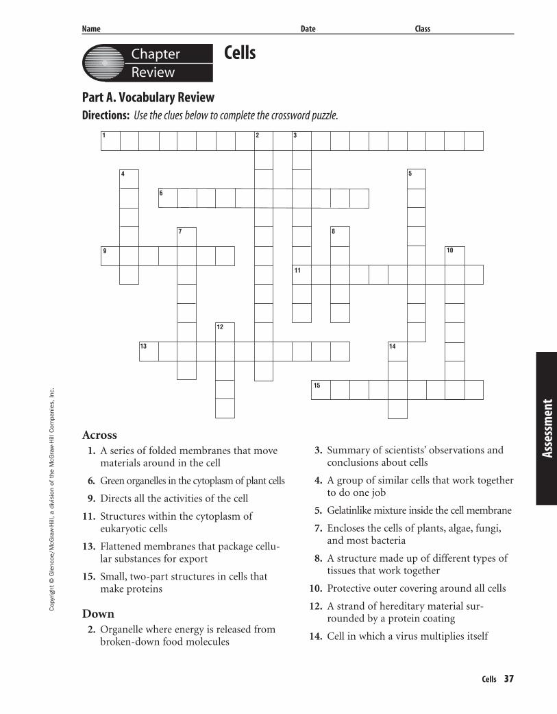
Chapter Review
Name Date Class
Cells
Part A. Vocabulary ReviewDirections: Use the clues below to complete the crossword puzzle.
Asse
ssm
ent
Across1. A series of folded membranes that move
materials around in the cell
6. Green organelles in the cytoplasm of plant cells
9. Directs all the activities of the cell
11. Structures within the cytoplasm ofeukaryotic cells
13. Flattened membranes that package cellu-lar substances for export
15. Small, two-part structures in cells thatmake proteins
Down2. Organelle where energy is released from
broken-down food molecules
3. Summary of scientists’ observations andconclusions about cells
4. A group of similar cells that work togetherto do one job
5. Gelatinlike mixture inside the cell membrane
7. Encloses the cells of plants, algae, fungi,and most bacteria
8. A structure made up of different types oftissues that work together
10. Protective outer covering around all cells
12. A strand of hereditary material sur-rounded by a protein coating
14. Cell in which a virus multiplies itself
13
6
7
4
8
12
14
15
11
5
10
2 31
9
429-1-50-MSS05-000000_CR 3/24/04 4:02 PM Page 37 impos03 301:goscanc:scanc429:layouts:
Cop
yrig
ht ©
Gle
ncoe
/McG
raw
-Hill
,a d
ivis
ion
of t
he M
cGra
w-H
ill C
ompa
nies
,Inc
.
Name Date Class
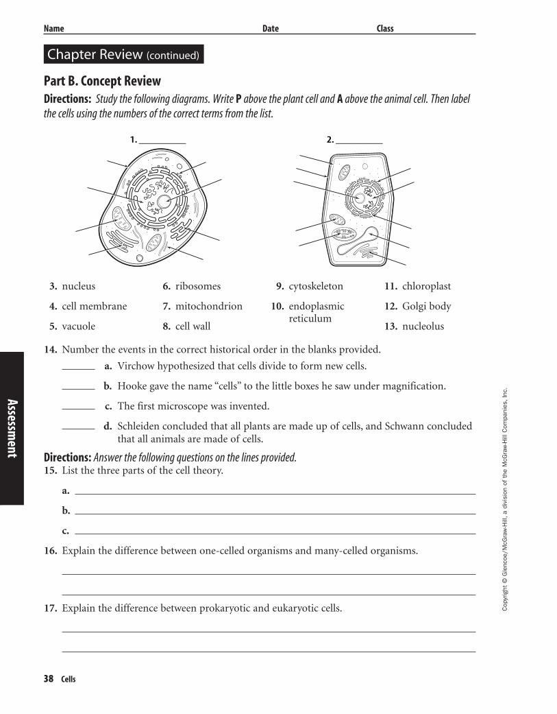
Chapter Review (continued)
38 Cells
Part B. Concept ReviewDirections: Study the following diagrams. Write P above the plant cell and A above the animal cell. Then labelthe cells using the numbers of the correct terms from the list.
Assessment
3. nucleus
4. cell membrane
5. vacuole
6. ribosomes
7. mitochondrion
8. cell wall
9. cytoskeleton
10. endoplasmicreticulum
11. chloroplast
12. Golgi body
13. nucleolus
14. Number the events in the correct historical order in the blanks provided.
1. ________ 2. ________
Directions: Answer the following questions on the lines provided.15. List the three parts of the cell theory.
a.
b.
c.
16. Explain the difference between one-celled organisms and many-celled organisms.
17. Explain the difference between prokaryotic and eukaryotic cells.
a. Virchow hypothesized that cells divide to form new cells.
b. Hooke gave the name “cells” to the little boxes he saw under magnification.
c. The first microscope was invented.
d. Schleiden concluded that all plants are made up of cells, and Schwann concludedthat all animals are made of cells.
429-1-50-MSS05-000000_CR 3/24/04 4:02 PM Page 38 impos03 301:goscanc:scanc429:layouts:

Transparency Activities
Cells 43
Tran
spar
ency
Act
iviti
es
429-1-50-MSS05-000000_CR 3/24/04 4:02 PM Page 43 impos03 301:goscanc:scanc429:layouts:

Cop
yrig
ht ©
Gle
ncoe
/McG
raw
-Hill
,a d
ivis
ion
of t
he M
cGra
w-H
ill C
ompa
nies
,Inc
.
Name Date Class
44 Cells
If this factory were a cell, it would run 24 hours a day and 7 days aweek. Just like a factory, cells use raw materials to produce what’sneeded. Like a factory, they have a control center, a source of power, anda way to move products and waste.
A Factory Analogy
Control center
Electricgenerator
Factory wall
Storage barrel
Section FocusTransparency Activity11
Transparency Activities
1. What part of the drawing directs the activities in the factory?
2. Identify the part of the drawing that provides energy to the factory.
3. What function do the storage barrels have?
429-1-50-MSS05-000000_CR 3/24/04 4:02 PM Page 44 impos03 301:goscanc:scanc429:layouts:

Cop
yrig
ht ©
Gle
ncoe
/McG
raw
-Hill
,a d
ivis
ion
of t
he M
cGra
w-H
ill C
ompa
nies
,Inc
.
Cells 45
Name Date Class
The Dead Sea has very high salt concentrations, and people haveused it as a salt resource since ancient times. But is the Dead Seareally dead? The concentration of salt is too high for most livingthings, but bacteria like the ones below are able to live in its waters.
At Home in the SaltSection FocusTransparency Activity22
Tran
spar
ency
Act
iviti
es1. Why might ancient people have thought the Dead Sea was totallywithout life?
2. What tool would you use to show there really is life in the DeadSea?
3. Do you think the living thing pictured is simple or complex?Defend your answer.
429-1-50-MSS05-000000_CR 3/24/04 4:02 PM Page 45 impos03 301:goscanc:scanc429:layouts:
Cop
yrig
ht ©
Gle
ncoe
/McG
raw
-Hill
,a d
ivis
ion
of t
he M
cGra
w-H
ill C
ompa
nies
,Inc
.
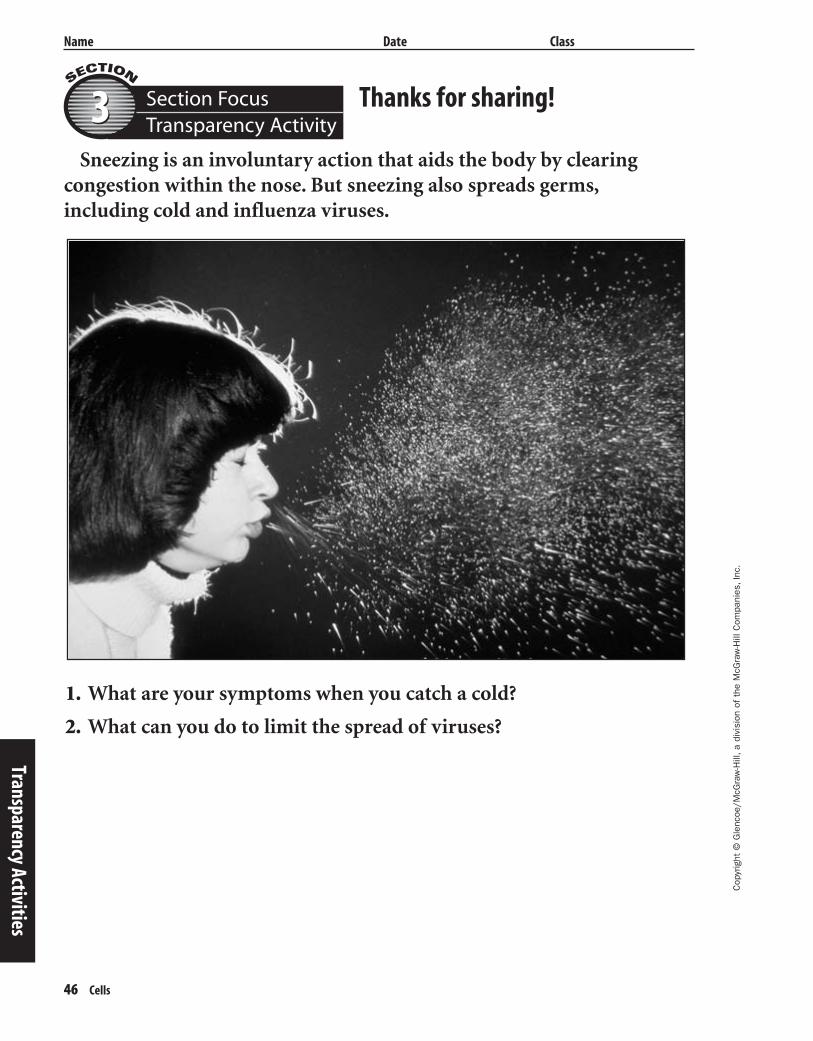
Name Date Class
46 Cells
Sneezing is an involuntary action that aids the body by clearing congestion within the nose. But sneezing also spreads germs,including cold and influenza viruses.
Thanks for sharing!Section FocusTransparency Activity33
Transparency Activities
1. What are your symptoms when you catch a cold?
2. What can you do to limit the spread of viruses?
429-1-50-MSS05-000000_CR 3/24/04 4:02 PM Page 46 impos03 301:goscanc:scanc429:layouts:
Cop
yrig
ht ©
Gle
ncoe
/McG
raw
-Hill
,a d
ivis
ion
of t
he M
cGra
w-H
ill C
ompa
nies
,Inc
.
Name Date Class
Cells 47
Nucleus
Nucleolus
Ribosome
Ribosome
Smooth endoplasmicreticulum (SER)
Smooth endoplasmicreticulum (SER)
Cell membrane Cytoskeleton
Centrioles
Mitochondrion
Nucleus
Nucleolus
Mitochondrion
Rough endoplasmicreticulum (RER)
Rough endoplasmicreticulum (RER)
LysosomeLysosome
Golgi complex
Cell membraneCell wall
Cell wall of adjacent cell
Golgi complex
Central vacuoleChloroplast
Free ribosome
Free ribosome
Animal and Plant Cells
Tran
spar
ency
Act
iviti
es
Teaching TransparencyActivity11
429-1-50-MSS05-000000_CR 3/24/04 4:02 PM Page 47 impos03 301:goscanc:scanc429:layouts:

48 Cells
Cop
yrig
ht ©
Gle
ncoe
/McG
raw
-Hill
,a d
ivis
ion
of t
he M
cGra
w-H
ill C
ompa
nies
,Inc
.
Name Date Class
Teaching Transparency Activity (continued)
1. Which organelles are common to both plant and animal cells?
2. Why are plant and animal cells classified as eukaryotic cells?
3. Which organelles are found in plant cells, but not in animal cells?
4. What is the major physical difference between vacuoles in a plant cell and vacuoles in an ani-mal cell?
5. What is the function of a plant cell that contains many chloroplasts?
6. What is the cell membrane made up of?
7. Which organelles are needed in cells that make protein?
8. Why might a cell that moves by means of cilia or flagella contain many mitochondria?
Transparency Activities
429-1-50-MSS05-000000_CR 3/24/04 4:02 PM Page 48 impos03 301:goscanc:scanc429:layouts:
Cop
yrig
ht ©
Gle
ncoe
/McG
raw
-Hill
,a d
ivis
ion
of t
he M
cGra
w-H
ill C
ompa
nies
,Inc
.
Name Date Class
Cells 49
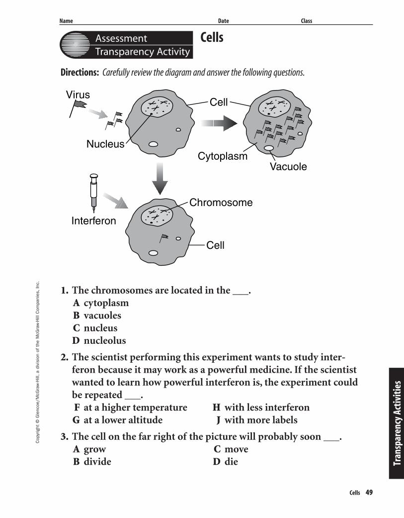
AssessmentTransparency Activity
Tran
spar
ency
Act
iviti
es
Directions: Carefully review the diagram and answer the following questions.
Cells
1. The chromosomes are located in the ___.A cytoplasmB vacuolesC nucleusD nucleolus
2. The scientist performing this experiment wants to study inter-feron because it may work as a powerful medicine. If the scientistwanted to learn how powerful interferon is, the experiment couldbe repeated ___.F at a higher temperature H with less interferonG at a lower altitude J with more labels
3. The cell on the far right of the picture will probably soon ___.A grow C moveB divide D die
Virus
Interferon
Chromosome
Cell
Cell
VacuoleCytoplasm
Nucleus
429-1-50-MSS05-000000_CR 3/24/04 4:02 PM Page 49 impos03 301:goscanc:scanc429:layouts:
Related Documents