3D printing of composite calcium phosphate and collagen scaffolds for bone regeneration Jason A. Inzana a, b , Diana Olvera a, b , Seth M. Fuller d , James P. Kelly d, e , Olivia A. Graeve d, e , Edward M. Schwarz a, b, c , Stephen L. Kates a, b, c , Hani A. Awad a, b, c, * a Center for Musculoskeletal Research, University of Rochester Medical Center, 601 Elmwood Avenue, Box 665, Rochester, NY 14642, United States b Department of Biomedical Engineering, University of Rochester, 207 Robert B. Goergen Hall, Rochester, NY 14642, United States c Department of Orthopaedics, University of Rochester Medical Center, 601 Elmwood Avenue, Rochester, NY 14642, United States d Kazuo Inamori School of Engineering, Alfred University, 1 Saxon Drive, Alfred, NY 14802, United States e Department of Mechanical and Aerospace Engineering, University of California, San Diego, 9500 Gilman Drive e MC 0411, La Jolla, CA 92093-0411, United States article info Article history: Received 27 November 2013 Accepted 24 January 2014 Available online 14 February 2014 Keywords: Three dimensional printing Calcium phosphate scaffold Collagen Bone regeneration Tissue engineering abstract Low temperature 3D printing of calcium phosphate scaffolds holds great promise for fabricating syn- thetic bone graft substitutes with enhanced performance over traditional techniques. Many design pa- rameters, such as the binder solution properties, have yet to be optimized to ensure maximal biocompatibility and osteoconductivity with sufficient mechanical properties. This study tailored the phosphoric acid-based binder solution concentration to 8.75 wt% to maximize cytocompatibility and mechanical strength, with a supplementation of Tween 80 to improve printing. To further enhance the formulation, collagen was dissolved into the binder solution to fabricate collagen-calcium phosphate composites. Reducing the viscosity and surface tension through a physiologic heat treatment and Tween 80, respectively, enabled reliable thermal inkjet printing of the collagen solutions. Supplementing the binder solution with 1e2 wt% collagen significantly improved maximum flexural strength and cell viability. To assess the bone healing performance, we implanted 3D printed scaffolds into a critically sized murine femoral defect for 9 weeks. The implants were confirmed to be osteoconductive, with new bone growth incorporating the degrading scaffold materials. In conclusion, this study demonstrates optimization of material parameters for 3D printed calcium phosphate scaffolds and enhancement of material properties by volumetric collagen incorporation via inkjet printing. Ó 2014 Elsevier Ltd. All rights reserved. 1. Introduction Major bone reconstruction procedures often use autografts or allografts to improve bone healing in a critically sized defect or non-union; however, these bone grafts suffer from multiple limi- tations that make synthetic alternatives an attractive option [1,2]. Calcium phosphates are a primary focus for synthetic bone graft substitutes because they are osteoconductive and provide sufficient mechanical strength. Inkjet-based 3D printing has been employed to fabricate calcium phosphate scaffolds (CPS), where the calcium phosphate powder is temporarily bound by an adhesive polymer and then permanently bound by sintering of the printed green body [3,4]. This technique, however, requires high temperatures that preclude the incorporation of bioactive molecules and drugs during the 3D printing process that could stimulate bone regeneration or combat infection. The feasibility of low temperature 3D printing of CPS has also been demonstrated, where calcium phosphate powder is bound by aqueous (often acidic) binder solutions delivered from the inkjets through a dissolutioneprecipitation reaction [5e7]. In addition to enabling drug incorporation [8], low temperature 3D printing provides the potential to create composites with synthetic or bio- logical polymers such as collagen. Type I collagen is the most abundant structural protein in the human body and is a critical component of bone extracellular matrix, where it plays important roles in this mineralized tissue’s strength and toughness. It has been shown that incorporating collagen into mineralized bone * Corresponding author. Center for Musculoskeletal Research, University of Rochester Medical Center, 601 Elmwood Avenue, Box 665, Rochester, NY 14642, United States. Tel.: þ1 585 273 5268; fax: þ1 585 276 2177. E-mail addresses: [email protected] (J.A. Inzana), diana_olvera@ urmc.rochester.edu (D. Olvera), [email protected] (S.M. Fuller), [email protected] (J.P. Kelly), [email protected] (O.A. Graeve), [email protected] (E.M. Schwarz), [email protected] (S.L. Kates), hani_awad@urmc. rochester.edu (H.A. Awad). Contents lists available at ScienceDirect Biomaterials journal homepage: www.elsevier.com/locate/biomaterials 0142-9612/$ e see front matter Ó 2014 Elsevier Ltd. All rights reserved. http://dx.doi.org/10.1016/j.biomaterials.2014.01.064 Biomaterials 35 (2014) 4026e4034

Welcome message from author
This document is posted to help you gain knowledge. Please leave a comment to let me know what you think about it! Share it to your friends and learn new things together.
Transcript
lable at ScienceDirect
Biomaterials 35 (2014) 4026e4034
Contents lists avai
Biomaterials
journal homepage: www.elsevier .com/locate/biomateria ls
3D printing of composite calcium phosphate and collagen scaffolds forbone regeneration
Jason A. Inzana a,b, Diana Olvera a,b, Seth M. Fuller d, James P. Kelly d,e, Olivia A. Graeve d,e,Edward M. Schwarz a,b,c, Stephen L. Kates a,b,c, Hani A. Awad a,b,c,*
aCenter for Musculoskeletal Research, University of Rochester Medical Center, 601 Elmwood Avenue, Box 665, Rochester, NY 14642, United StatesbDepartment of Biomedical Engineering, University of Rochester, 207 Robert B. Goergen Hall, Rochester, NY 14642, United StatescDepartment of Orthopaedics, University of Rochester Medical Center, 601 Elmwood Avenue, Rochester, NY 14642, United StatesdKazuo Inamori School of Engineering, Alfred University, 1 Saxon Drive, Alfred, NY 14802, United StateseDepartment of Mechanical and Aerospace Engineering, University of California, San Diego, 9500 Gilman Drive e MC 0411, La Jolla, CA 92093-0411,United States
a r t i c l e i n f o
Article history:Received 27 November 2013Accepted 24 January 2014Available online 14 February 2014
Keywords:Three dimensional printingCalcium phosphate scaffoldCollagenBone regenerationTissue engineering
* Corresponding author. Center for MusculoskelRochester Medical Center, 601 Elmwood Avenue, BoUnited States. Tel.: þ1 585 273 5268; fax: þ1 585 276
E-mail addresses: [email protected] (D. Olvera), [email protected] (S.M(J.P. Kelly), [email protected] (O.A. Graeve), edward_(E.M. Schwarz), [email protected] (Srochester.edu (H.A. Awad).
0142-9612/$ e see front matter � 2014 Elsevier Ltd.http://dx.doi.org/10.1016/j.biomaterials.2014.01.064
a b s t r a c t
Low temperature 3D printing of calcium phosphate scaffolds holds great promise for fabricating syn-thetic bone graft substitutes with enhanced performance over traditional techniques. Many design pa-rameters, such as the binder solution properties, have yet to be optimized to ensure maximalbiocompatibility and osteoconductivity with sufficient mechanical properties. This study tailored thephosphoric acid-based binder solution concentration to 8.75 wt% to maximize cytocompatibility andmechanical strength, with a supplementation of Tween 80 to improve printing. To further enhance theformulation, collagen was dissolved into the binder solution to fabricate collagen-calcium phosphatecomposites. Reducing the viscosity and surface tension through a physiologic heat treatment and Tween80, respectively, enabled reliable thermal inkjet printing of the collagen solutions. Supplementing thebinder solution with 1e2 wt% collagen significantly improved maximum flexural strength and cellviability. To assess the bone healing performance, we implanted 3D printed scaffolds into a criticallysized murine femoral defect for 9 weeks. The implants were confirmed to be osteoconductive, with newbone growth incorporating the degrading scaffold materials. In conclusion, this study demonstratesoptimization of material parameters for 3D printed calcium phosphate scaffolds and enhancement ofmaterial properties by volumetric collagen incorporation via inkjet printing.
� 2014 Elsevier Ltd. All rights reserved.
1. Introduction
Major bone reconstruction procedures often use autografts orallografts to improve bone healing in a critically sized defect ornon-union; however, these bone grafts suffer from multiple limi-tations that make synthetic alternatives an attractive option [1,2].Calcium phosphates are a primary focus for synthetic bone graftsubstitutes because they are osteoconductive and provide sufficientmechanical strength. Inkjet-based 3D printing has been employed
etal Research, University ofx 665, Rochester, NY 14642,2177.
u (J.A. Inzana), diana_olvera@. Fuller), [email protected]@urmc.rochester.edu.L. Kates), hani_awad@urmc.
All rights reserved.
to fabricate calcium phosphate scaffolds (CPS), where the calciumphosphate powder is temporarily bound by an adhesive polymerand then permanently bound by sintering of the printed green body[3,4]. This technique, however, requires high temperatures thatpreclude the incorporation of bioactive molecules and drugs duringthe 3D printing process that could stimulate bone regeneration orcombat infection.
The feasibility of low temperature 3D printing of CPS has alsobeen demonstrated, where calcium phosphate powder is bound byaqueous (often acidic) binder solutions delivered from the inkjetsthrough a dissolutioneprecipitation reaction [5e7]. In addition toenabling drug incorporation [8], low temperature 3D printingprovides the potential to create composites with synthetic or bio-logical polymers such as collagen. Type I collagen is the mostabundant structural protein in the human body and is a criticalcomponent of bone extracellular matrix, where it plays importantroles in this mineralized tissue’s strength and toughness. It hasbeen shown that incorporating collagen into mineralized bone
J.A. Inzana et al. / Biomaterials 35 (2014) 4026e4034 4027
cements could enhance their biomechanical properties, as well astheir osteoconductive and osteoinductive characteristics. Forexample, collagen incorporation into hand-mixed calcium phos-phate cements has been shown to improve cellular attachment,viability, proliferation, and activity as well as mechanical properties[9e12]; but the feasibility of 3D printed collagen-calcium phos-phate (col-CaP) composites has not been demonstrated to ourknowledge. Advantages of 3D printing over molding or paste in-jection include patient specific geometries [13] and controlledspatial patterning of drugs or polymers within the scaffold [8].Furthermore, few studies have assessed the in vivo performance oflow temperature 3D printed CPS, and these studies have focused onectopic models of intramuscular implantation in rats [6,14] andgoats [15] and orthotopic models of cranial healing in rabbits [16]and dogs [5]. While these models demonstrated promising re-sults, long bone healing might be more challenging based on dif-ferences in matrix composition and in vivo loading [17], anddifferences between osteochondral and intramebraneous boneformation processes that contribute differently to the healing oflong and cranial bones, respectively [18,19].
To address these points, this study focused first on optimizingthe formulation of the acidic binder solution used in low temper-ature 3D printing of CPS for optimized accuracy, mechanicalstrength, and biocompatibility. Next, we evaluated the in vivoregenerative potential of these 3D printed CPS in a critically sizedmurine femoral defect. We hypothesized that col-CaP compositescould be fabricated through low temperature 3D printing by dis-solving collagen into the phosphoric acid binder solution; and thatthese composites will improve the scaffold’s mechanical strength,cytocompatibility and bone regeneration in a critically sizedfemoral defect.
2. Materials and methods
2.1. Powder and binder solution formulations
Dilutions of phosphoric acid (5e20wt%)wereused as thebase binder solution for3D printing the CPS. Tween 80 (SigmaeAldrich, St. Louis, MO), a non-cytotoxic sur-factant, was added to some of the binder formulations at a concentration of 0.25 wt%based on the approximate critical micelle concentration in phosphate buffer [20].
Lyophilized bovine dermal type I collagen was kindly provided by Kensey Nash(Exton, PA). Collagen solutions were prepared by dissolving the collagen intophosphoric acid at 4 �C until completely dissolved and then stored at 4 �C until use.Concentrations of each solution were confirmed by optical absorbance at 280 nmand compared with a linear standard curve using a spectrophotometer (Synergy MxMicroplate Reader, BioTek Instruments Inc.; Winooski, VT).
The calcium phosphate powder was prepared by solution combustion synthesisfollowing similar procedures as described previously [21e23]. As an initial step, amixture of 12.1058 g of Ca(NO3)3$4H2O (Alfa Aesar #A16645), 3.9433 (NH4)2HPO4
(Alfa Aesar #11597), and 6.9270 g of carbohydrazide (Alfa Aesar #A11145) wasmixedin a Pyrex crystallization dish with water as the solvent. The mixture was thenplaced in a muffle furnace at 500 �C and allowed to combust, taking approximately15 min for the reaction to complete. After synthesis, the powders were crushed in amortar and pestle and subsequently calcined at 1300 �C for 2 h. The resultingpowder was a composite of hydroxyapatite and a-tricalcium phosphate (Fig. S1).
2.2. Adaptation of a commercial 3D printer and preparation of 3D printed scaffolds
A ZPrinter� 450 (3D Systems; Andover, MA) was adapted to print with calciumphosphate powder and acidic binder solutions using bench scale volumes (�50 g ofpowder). The binder solution fluid lines were replaced with polypropylene or pol-ytetrafluoroethylene (PTFE) tubing and an inert glass reservoir was added to avoiddegradation by the acidic solutions. A custom overhead powder distribution systemand build chamber (60 � 60 � 30 mm) were employed to isolate the biomaterialsduring the 3D printing process and allow formation of a packed powder bed in asmaller area (Fig. S2). The new powder feed systemwas linked via a belt mechanismto themotor of the original equipment manufacturer’s (OEM) powder feed system toexploit the automated software control of the 3D printing process. For all experi-ments, the powder layer thickness was set to 89 mm and the binder liquid/powderratio was set as 0.46 (homogenous) in the ZPrint� software. The phosphoric acid-based binder solutions were delivered by thermal inkjets (HP11, HewlettePack-ard; Palo Alto, CA) to selectively bind the powder, which produces dicalciumphosphate dihydrate (DCPD or brushite; Fig. S1). After printing, the samples werepost-processed by flash dipping in 0.1 wt% phosphoric acid and then washing in
deionized water (3 � 120 s) to improve surface binding and remove residual acidity.Some samples were coated with collagen by dipping them into a 0.5 wt% collagen in0.1 wt% phosphoric acid solution, neutralized to pH 7with NaOH, and allowing to airdry. For in vivo experiments, the scaffolds were sterilized by supercritical CO2
(NovaSterilis, Lansing, NY).
2.3. Characterization of binder solutions and printability
Considering that fluid properties dictate the printability of a solution via inkjets,the density, viscosity, surface tension, and printing saturation of the binder solutionswere measured. The viscosities were measured using a 40 mm cone-plate rheometer(Discovery HR-2, TA Instruments; New Castle, DE) at 25 �C and a shear rate of 100 s�1,except for shear rate-dependent experiments where data was collected from 25 to630 s�1. The fluid surface tensionsweremeasured on aModel 500Goniometer (Ramé-Hart Instrument Co., Succasunna,NJ) at 20 �C andwere averagedover at least 40 pointson multiple droplets. Printing saturation was quantified by measuring the mass ofbinderfluid printed intoweighing boats, converting to volume, and normalizing to thetheoretical powder volume. Four measurements were made per print job, averagingover 5 layers for each measurement, with 2 different ink cartridges per binderformulation. To investigate the accuracy of the HP 11 print heads over a range of vis-cosities, a series of 1 mm lines were printed onto blue litmus paper to visualize theacidic solutions. The accuracy was quantified with a custom MATLAB algorithm(R2013a;MathWorks Inc.;Natick,MA) byoverlaying the ideal line set atop the imageofthe printed lines and calculating the area of erroneous printing (Fig. S3a,b).
2.4. Optimization of calcium phosphate powder particle size
The powder particle size distribution is critical for enabling a well-packed,smooth powder bed for printing and also for dictating the intrinsic micro-porosityand resolution of the printed material [7]. Therefore, the calcium phosphate pow-der was sieved and collected from between two stainless steel meshes (ranging 30e150 mm) to remove particulate that was too large for accurate printing and too smallto avoid agglomeration. Solid elliptical cylinders that represent the outer di-mensions of a mouse femoral diaphysis (1.2 � 2 � 3 mm) were printed with eachparticle size range and imaged by high-resolution micro-computed tomography(micro-CT; VivaCT 40, Scanco Medical; Bassersdorf, Switzerland) with a 10.5 mmisotropic voxel size using an integration time of 300 ms, energy of 55 kV, and in-tensity of 145 mA. Three-dimensional renderings of the micro-CT images were co-registered with the computer-aided design (CAD) image that was used to guidethe printing process and the error was quantified by volumetric differences betweenthe overlaid images (Fig. 2a). 3D image reconstruction and quantitative analysis wasperformed using Amira software (Amira 5.4.5, FEI Visualization Sciences Group;Burlington, MA).
2.5. Material characterization of 3D printed scaffolds
The flexural properties of the 3D printed CPS were assessed by 3 point bendingsince cements tend to be most susceptible to shear and tensile failure. Samples were3D printed as 4 � 1 � 14 mm bars and tested on a 10 mm support span using anInstron 8841 DynaMight� Axial Testing System (Instron Corp.; Canton, MA) with a50 N load cell. Flexural tests were performed at a displacement rate of 1 mm/minuntil failure.
The volumetric porosity and pore size distributions were estimated frommicro-CT images using Amira. The pore size distributions were measured using Amira’ssurface thickness algorithm, which calculates the distance of the normal projectionfrom each facet on the triangulated surface to the intersectionwith the next surface.
Scanning electron microscopy (SEM; LEO 982 FE-SEM, Carl Zeiss SMT; Thorn-wood, NY) of gold-sputtered samples was used for qualitative assessment of thescaffold surfaces and porosity. X-ray diffraction (XRD; X’Pert PRO MRD; PANalytical,Westborough, MA) spectra were acquired to qualitatively assess phase compositionof the calcium phosphate powder and 3D printed samples. Spectra were acquiredfrom 2q ¼ 20e40� with a step size of 0.02� .
2.6. In vitro cytocompatibility of 3D printed CPS materials
2.6.1. Cytotoxicity analysis based on ISO 10993 for preliminary screeningThe cytocompatibility of the scaffold materials was assessed by XTT assay with
C3H/10T1/2 cells (ATCC; Manassas, VA) exposed to media extracts as described byISO 10993-5. Extraction was performed according to the mass-based ratios of ISO10993-12 in sealed tubes within a static incubator at 37 �C and 95% humidity for24 h. PTFE and PVC were used as negative and positive controls for toxicity,respectively. Viability measurements were normalized to media-only controls.
2.6.2. Viability of cells cultured on 3D printed CPS materialsSamples were 3D printed as 1.2 mm thick discs with a 6.3 mm diameter to
completely cover the well bottoms in a 96 well plate. C3H/10T1/2 cells were seededonto the discs in the wells at 10,000 cells/well. After 24 or 72 h, the cell viability wasmeasured by a live-dead fluorescence assay (MultiTox-Fluor Cytotoxicity Assay,Promega Corp.; Madison,WI). The relative cell viability is reported as the ratio of livesignal intensity to dead signal intensity, after subtracting intensities of acellularcontrols and normalizing to cells on tissue culture plastic.
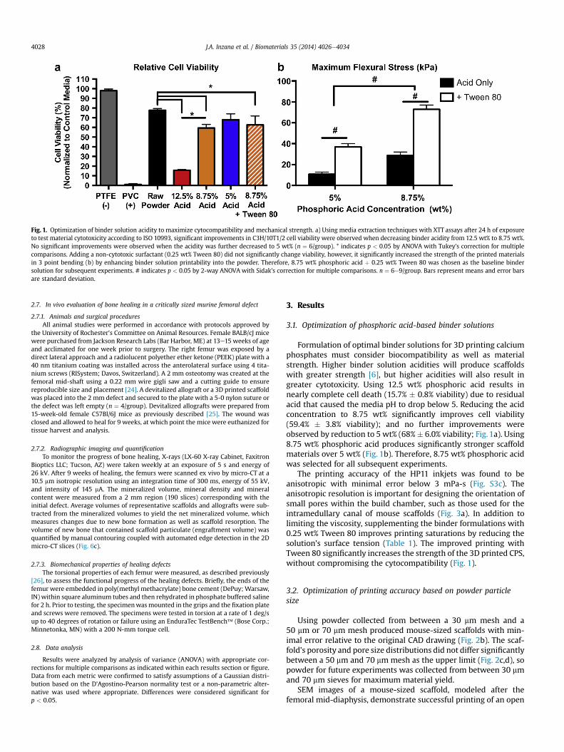
Fig. 1. Optimization of binder solution acidity to maximize cytocompatibility and mechanical strength. a) Using media extraction techniques with XTT assays after 24 h of exposureto test material cytotoxicity according to ISO 10993, significant improvements in C3H/10T1/2 cell viability were observed when decreasing binder acidity from 12.5 wt% to 8.75 wt%.No significant improvements were observed when the acidity was further decreased to 5 wt% (n ¼ 6/group). * indicates p < 0.05 by ANOVA with Tukey’s correction for multiplecomparisons. Adding a non-cytotoxic surfactant (0.25 wt% Tween 80) did not significantly change viability, however, it significantly increased the strength of the printed materialsin 3 point bending (b) by enhancing binder solution printability into the powder. Therefore, 8.75 wt% phosphoric acid þ 0.25 wt% Tween 80 was chosen as the baseline bindersolution for subsequent experiments. # indicates p < 0.05 by 2-way ANOVA with Sidak’s correction for multiple comparisons. n ¼ 6e9/group. Bars represent means and error barsare standard deviation.
J.A. Inzana et al. / Biomaterials 35 (2014) 4026e40344028
2.7. In vivo evaluation of bone healing in a critically sized murine femoral defect
2.7.1. Animals and surgical proceduresAll animal studies were performed in accordance with protocols approved by
the University of Rochester’s Committee on Animal Resources. Female BALB/cJ micewere purchased from Jackson Research Labs (Bar Harbor, ME) at 13e15 weeks of ageand acclimated for one week prior to surgery. The right femur was exposed by adirect lateral approach and a radiolucent polyether ether ketone (PEEK) plate with a40 nm titanium coating was installed across the anterolateral surface using 4 tita-nium screws (RISystem; Davos, Switzerland). A 2 mm osteotomy was created at thefemoral mid-shaft using a 0.22 mm wire gigli saw and a cutting guide to ensurereproducible size and placement [24]. A devitalized allograft or a 3D printed scaffoldwas placed into the 2 mm defect and secured to the plate with a 5-0 nylon suture orthe defect was left empty (n ¼ 4/group). Devitalized allografts were prepared from15-week-old female C57Bl/6J mice as previously described [25]. The wound wasclosed and allowed to heal for 9 weeks, at which point the mice were euthanized fortissue harvest and analysis.
2.7.2. Radiographic imaging and quantificationTo monitor the progress of bone healing, X-rays (LX-60 X-ray Cabinet, Faxitron
Bioptics LLC; Tucson, AZ) were taken weekly at an exposure of 5 s and energy of26 kV. After 9 weeks of healing, the femurs were scanned ex vivo by micro-CT at a10.5 mm isotropic resolution using an integration time of 300 ms, energy of 55 kV,and intensity of 145 mA. The mineralized volume, mineral density and mineralcontent were measured from a 2 mm region (190 slices) corresponding with theinitial defect. Average volumes of representative scaffolds and allografts were sub-tracted from the mineralized volumes to yield the net mineralized volume, whichmeasures changes due to new bone formation as well as scaffold resorption. Thevolume of new bone that contained scaffold particulate (engraftment volume) wasquantified by manual contouring coupled with automated edge detection in the 2Dmicro-CT slices (Fig. 6c).
2.7.3. Biomechanical properties of healing defectsThe torsional properties of each femur were measured, as described previously
[26], to assess the functional progress of the healing defects. Briefly, the ends of thefemur were embedded in poly(methyl methacrylate) bone cement (DePuy;Warsaw,IN) within square aluminum tubes and then rehydrated in phosphate buffered salinefor 2 h. Prior to testing, the specimenwasmounted in the grips and the fixation plateand screws were removed. The specimens were tested in torsion at a rate of 1 deg/sup to 40 degrees of rotation or failure using an EnduraTec TestBench� (Bose Corp.;Minnetonka, MN) with a 200 N-mm torque cell.
2.8. Data analysis
Results were analyzed by analysis of variance (ANOVA) with appropriate cor-rections for multiple comparisons as indicated within each results section or figure.Data from each metric were confirmed to satisfy assumptions of a Gaussian distri-bution based on the D’Agostino-Pearson normality test or a non-parametric alter-native was used where appropriate. Differences were considered significant forp < 0.05.
3. Results
3.1. Optimization of phosphoric acid-based binder solutions
Formulation of optimal binder solutions for 3D printing calciumphosphates must consider biocompatibility as well as materialstrength. Higher binder solution acidities will produce scaffoldswith greater strength [6], but higher acidities will also result ingreater cytotoxicity. Using 12.5 wt% phosphoric acid results innearly complete cell death (15.7% � 0.8% viability) due to residualacid that caused the media pH to drop below 5. Reducing the acidconcentration to 8.75 wt% significantly improves cell viability(59.4% � 3.8% viability); and no further improvements wereobserved by reduction to 5 wt% (68%� 6.0% viability; Fig. 1a). Using8.75 wt% phosphoric acid produces significantly stronger scaffoldmaterials over 5 wt% (Fig. 1b). Therefore, 8.75 wt% phosphoric acidwas selected for all subsequent experiments.
The printing accuracy of the HP11 inkjets was found to beanisotropic with minimal error below 3 mPa-s (Fig. S3c). Theanisotropic resolution is important for designing the orientation ofsmall pores within the build chamber, such as those used for theintramedullary canal of mouse scaffolds (Fig. 3a). In addition tolimiting the viscosity, supplementing the binder formulations with0.25 wt% Tween 80 improves printing saturations by reducing thesolution’s surface tension (Table 1). The improved printing withTween 80 significantly increases the strength of the 3D printed CPS,without compromising the cytocompatibility (Fig. 1).
3.2. Optimization of printing accuracy based on powder particlesize
Using powder collected from between a 30 mm mesh and a50 mm or 70 mm mesh produced mouse-sized scaffolds with min-imal error relative to the original CAD drawing (Fig. 2b). The scaf-fold’s porosity and pore size distributions did not differ significantlybetween a 50 mm and 70 mm mesh as the upper limit (Fig. 2c,d), sopowder for future experiments was collected from between 30 mmand 70 mm sieves for maximum material yield.
SEM images of a mouse-sized scaffold, modeled after thefemoral mid-diaphysis, demonstrate successful printing of an open
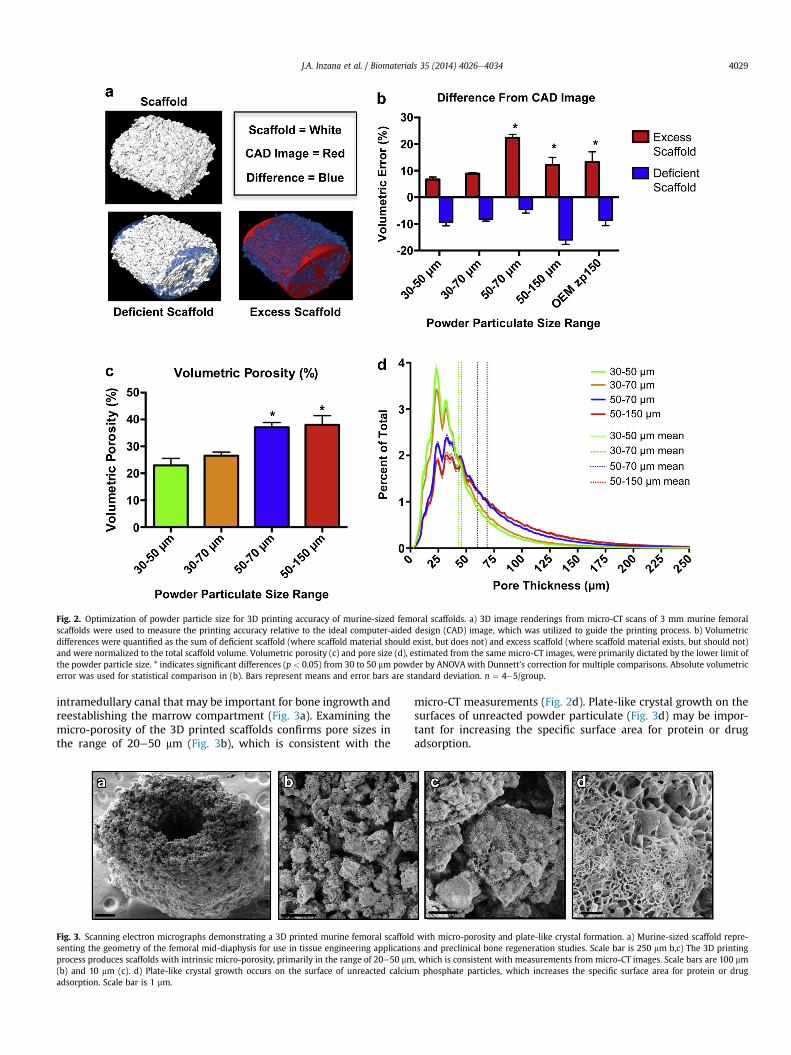
Fig. 2. Optimization of powder particle size for 3D printing accuracy of murine-sized femoral scaffolds. a) 3D image renderings from micro-CT scans of 3 mm murine femoralscaffolds were used to measure the printing accuracy relative to the ideal computer-aided design (CAD) image, which was utilized to guide the printing process. b) Volumetricdifferences were quantified as the sum of deficient scaffold (where scaffold material should exist, but does not) and excess scaffold (where scaffold material exists, but should not)and were normalized to the total scaffold volume. Volumetric porosity (c) and pore size (d), estimated from the same micro-CT images, were primarily dictated by the lower limit ofthe powder particle size. * indicates significant differences (p < 0.05) from 30 to 50 mm powder by ANOVAwith Dunnett’s correction for multiple comparisons. Absolute volumetricerror was used for statistical comparison in (b). Bars represent means and error bars are standard deviation. n ¼ 4e5/group.
J.A. Inzana et al. / Biomaterials 35 (2014) 4026e4034 4029
intramedullary canal that may be important for bone ingrowth andreestablishing the marrow compartment (Fig. 3a). Examining themicro-porosity of the 3D printed scaffolds confirms pore sizes inthe range of 20e50 mm (Fig. 3b), which is consistent with the
Fig. 3. Scanning electron micrographs demonstrating a 3D printed murine femoral scaffoldsenting the geometry of the femoral mid-diaphysis for use in tissue engineering applicatioprocess produces scaffolds with intrinsic micro-porosity, primarily in the range of 20e50 mm(b) and 10 mm (c). d) Plate-like crystal growth occurs on the surface of unreacted calciuadsorption. Scale bar is 1 mm.
micro-CT measurements (Fig. 2d). Plate-like crystal growth on thesurfaces of unreacted powder particulate (Fig. 3d) may be impor-tant for increasing the specific surface area for protein or drugadsorption.
with micro-porosity and plate-like crystal formation. a) Murine-sized scaffold repre-ns and preclinical bone regeneration studies. Scale bar is 250 mm b,c) The 3D printing, which is consistent with measurements from micro-CT images. Scale bars are 100 mmm phosphate particles, which increases the specific surface area for protein or drug
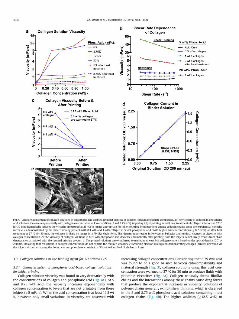
Fig. 4. Viscosity adjustment of collagen solutions in phosphoric acid enables 3D inkjet printing of collagen-calcium phosphate composites. a) The viscosity of collagen in phosphoricacid solutions increases exponentially with collagen concentration at lower acidities (5 and 8.75 wt%), impeding inkjet printing. A brief heat treatment of collagen solutions at 37 �Cfor 30 min dramatically reduces the viscosity (measured at 25 �C) to ranges appropriate for inkjet printing. b) Interactions among collagen chains cause the exponential viscosityincrease, as demonstrated by the shear thinning present with 0.3 wt% and 1 wt% collagen in 5 wt% phosphoric acid. With higher acid concentrations (�12.5 wt%), or after heattreatment at 37 �C for 30 min, the collagen is likely no longer in a fibrillar chain form. This denaturation results in Newtonian behavior and minimal changes in viscosity withcollagen concentration. c) The viscosity of collagen solutions in 8.75 wt% phosphoric acid decreases dramatically after printing from the inkjets, which likely results from chaindenaturation associated with the thermal printing process. d) The printed solutions were confirmed to maintain at least 94% collagen content based on the optical density (OD) at280 nm, indicating that reductions in collagen concentration do not explain the reduced viscosity. e) Scanning electron micrograph demonstrating collagen (arrow), delivered viathe inkjets, dispersed among the bound calcium phosphate crystals in a 3D printed scaffold. Scale bar is 5 mm.
J.A. Inzana et al. / Biomaterials 35 (2014) 4026e40344030
3.3. Collagen solutions as the binding agent for 3D printed CPS
3.3.1. Characterization of phosphoric acid-based collagen solutionsfor inkjet printing
Collagen solution viscosity was found to vary dramatically withthe concentrations of collagen and phosphoric acid (Fig. 4a). At 5and 8.75 wt% acid, the viscosity increases exponentially withcollagen concentration to levels that are not printable from theseinkjets (>5 mPa-s). When the acid concentration is at least 12.5 wt%, however, only small variations in viscosity are observed with
increasing collagen concentrations. Considering that 8.75 wt% acidwas found to be a good balance between cytocompatibility andmaterial strength (Fig. 3), collagen solutions using this acid con-centration were warmed to 37 �C for 30 min to produce fluids withprintable viscosities (Fig. 4a). Collagen naturally forms fibrillarchains and the interactions among these chains cause drag forcesthat produce the exponential increases in viscosity. Solutions ofpolymer chains generally exhibit shear thinning, which is observedin the 5 and 8.75 wt% phosphoric acid solutions containing intactcollagen chains (Fig. 4b). The higher acidities (�12.5 wt%) or
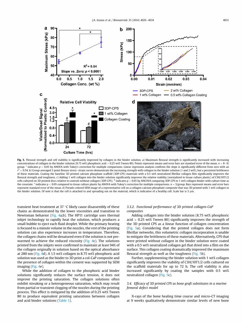
Fig. 5. Flexural strength and cell viability is significantly improved by collagen in the binder solution. a) Maximum flexural strength is significantly increased with increasingconcentrations of collagen in the binder solution (8.75 wt% phosphoric acid þ 0.25 wt% Tween 80). Points represent means and error bars are standard error of the mean. n ¼ 8e9/group. * indicates p < 0.05 by ANOVA with Tukey’s correction for multiple comparisons. Linear regression analysis confirms the slope is significantly different from zero with anr2 ¼ 0.54. b) Group-averaged 3 point flexure stressestrain curves demonstrate the increasing strength with collagen in the binder solution (1 and 2 wt%), but a persistent brittlenessof these materials. Coating the baseline 3D printed calcium phosphate scaffold (3DP-CPS) materials with a 0.5 wt% neutralized fibrillar collagen film significantly improves theflexural strength and toughness. c) Adding 1 wt% collagen into the binder solution significantly improves the relative viability (normalized to tissue culture plastic) of C3H/10T1/2cells cultured on 3D printed discs relative to controls without collagen (3DP-CPS). # indicates p < 0.05 by ANCOVA comparing 3DP-CPS to 1 wt% collagen binder with culture time asthe covariate. * indicates p < 0.05 compared to tissue culture plastic by ANOVA with Tukey’s correction for multiple comparisons. n ¼ 5/group. Bars represent means and error barsrepresent standard error of the mean. d) Pseudo-colored SEM image of a representative cell on a collagen-calcium phosphate composite that was 3D printed with 1 wt% collagen inthe binder solution. Of note is that the cell is attached to and spreading out on the material, which is indicative of a healthy cell. Scale bar is 5 mm.
J.A. Inzana et al. / Biomaterials 35 (2014) 4026e4034 4031
transient heat treatment at 37 �C likely cause disassembly of thesechains as demonstrated by the lower viscosities and transition toNewtonian behavior (Fig. 4a,b). The HP11 cartridge uses thermalinkjet technology to rapidly heat the solution, which produces asmall bubble to eject each fluid droplet. While the primary heatingis focused to aminute volume in the nozzles, the rest of the printingsolution can also experience increases in temperature. Therefore,the collagen chains will be denatured even if the solution is not pre-warmed to achieve the reduced viscosity (Fig. 4c). The solutionsprinted from the inkjets were confirmed to maintain at least 94% ofthe collagen originally in solution based on the optical absorbanceat 280 nm (Fig. 4d). A 1.5 wt% collagen in 8.75 wt% phosphoric acidsolutionwas used as the binder to 3D print a col-CaP composite andthe presence of collagen within the cement was supported by SEMimaging (Fig. 4e).
While the addition of collagen to the phosphoric acid bindersolutions significantly reduces the surface tension, it does notimprove the printing saturation. The collagen solutions oftenexhibit streaking or a heterogeneous saturation, which may resultfrom partial or transient clogging of the nozzles during the printingprocess. This effect is mitigated by the addition of 0.25 wt% Tween80 to produce equivalent printing saturations between collagenand acid binder solutions (Table 1).
3.3.2. Functional performance of 3D printed collagen-CaPcomposites
Adding collagen into the binder solution (8.75 wt% phosphoricacid þ 0.25 wt% Tween 80) significantly improves the strength ofthe 3D printed CPS as a linear function of collagen concentration(Fig. 5a). Considering that the printed collagen does not formfibrillar networks, this volumetric collagen incorporation is unableto mitigate the brittleness of these materials. Alternatively, CPS thatwere printed without collagen in the binder solution were coatedwith a 0.5 wt% neutralized collagen gel that dried into a film on thesurface. This collagen coating dramatically improved the maximumflexural strength as well as the toughness (Fig. 5b).
Further, supplementing the binder solution with 1 wt% collagensignificantly improves the viability of C3H/10T1/2 cells cultured onthe scaffold materials for up to 72 h. The cell viability is alsoincreased significantly by coating the samples with 0.5 wt%neutralized collagen (Fig. 5c).
3.4. Efficacy of 3D printed CPS as bone graft substitutes in a murinefemoral defect model
X-rays of the bone healing time course and micro-CT imagingat 9 weeks qualitatively demonstrate similar levels of new bone
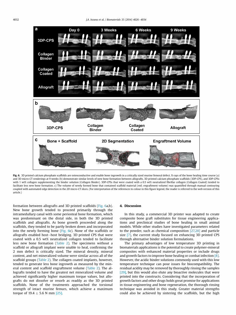
Fig. 6. 3D printed calcium phosphate scaffolds are osteoconductive and enable bone ingrowth in a critically sized murine femoral defect. X-rays of the bone healing time course (a)and 3D micro-CT renderings at 9 weeks (b) demonstrate similar levels of new bone formation between allografts, 3D printed calcium phosphate scaffolds (3DP-CPS), and 3DP-CPSswith 1 wt% collagen supplementing the binder solution (Collagen Binder). 3DP-CPSs that were coated with a 0.5 wt% neutralized fibrillar collagen (Collagen Coated) tended tofacilitate less new bone formation. c) The volume of newly formed bone that contained scaffold material (red; engraftment volume) was quantified through manual contouringcoupled with automated edge detection in the 2D micro-CT slices. (For interpretation of the references to colour in this figure legend, the reader is referred to the web version of thisarticle.)
J.A. Inzana et al. / Biomaterials 35 (2014) 4026e40344032
formation between allografts and 3D printed scaffolds (Fig. 6a,b).New bone growth tended to proceed primarily through theintramedullary canal with some periosteal bone formation, whichwas predominant on the distal side, in both the 3D printedscaffolds and allografts. As bone growth proceeded along thescaffolds, they tended to be partly broken down and incorporatedinto the newly forming bone (Fig. 6c). None of the scaffolds orallografts enabled hostehost bridging. 3D printed CPS that werecoated with a 0.5 wt% neutralized collagen tended to facilitateless new bone formation (Table 2). The specimens without ascaffold or allograft implant were unable to heal, confirming the2 mm defect is critically sized. The mineral density, mineralcontent, and net mineralized volume were similar across all of thescaffold groups (Table 2). The collagen coated implants, however,tended to generate less bone ingrowth as measured by the min-eral content and scaffold engraftment volume (Table 2). The al-lografts tended to have the greatest net mineralized volume andachieved significantly higher maximum torque values, but allo-grafts do not dissolve or resorb as readily as the 3D printedscaffolds. None of the treatments approached the torsionalstrength of intact murine femurs, which achieve a maximumtorque of 19.4 � 5.6 N mm [25].
4. Discussion
In this study, a commercial 3D printer was adapted to createcomposite bone graft substitutes for tissue engineering applica-tions and preclinical studies of bone healing in small animalmodels. While other studies have investigated parameters relatedto the powder, such as chemical composition [27,28] and particlesize [7], the current study focused on enhancing 3D printed CPSthrough alternative binder solution formulations.
The primary advantages of low temperature 3D printing inbiomaterials applications is the potential to create polymer-mineralcomposites with enhanced material properties or include drugsand growth factors to improve bone healing or combat infection [8].However, the acidic binder solutions commonly used with this lowtemperature technique can pose issues for biocompatibility. Theresidual acidity may be removed by thoroughly rinsing the samples[29], but this would also elute any bioactive molecules that wereprinted into the constructs. Considering that the incorporation ofgrowth factors and other drugs holds great promise for applicationsin tissue engineering and bone regeneration, the thorough rinsingtechnique was avoided in this study. Greater material strengthscould also be achieved by sintering the scaffolds, but the high
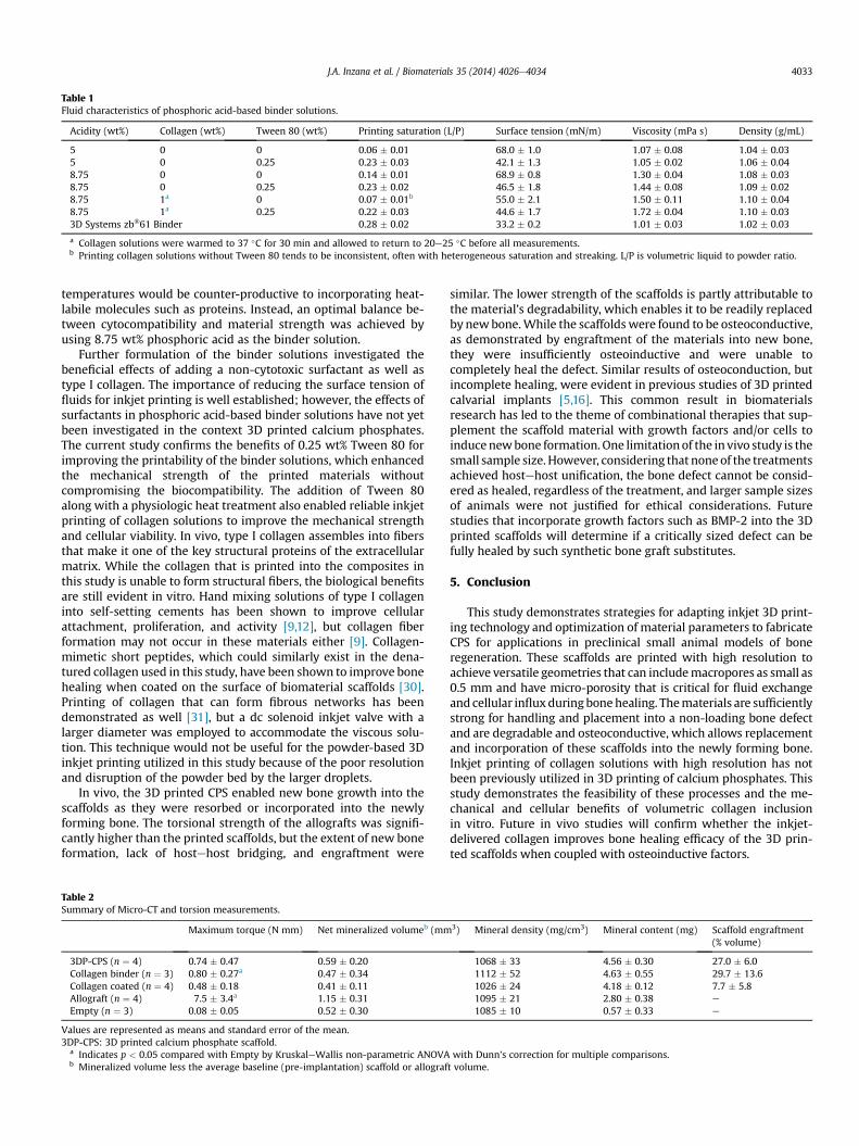
Table 1Fluid characteristics of phosphoric acid-based binder solutions.
Acidity (wt%) Collagen (wt%) Tween 80 (wt%) Printing saturation (L/P) Surface tension (mN/m) Viscosity (mPa s) Density (g/mL)
5 0 0 0.06 � 0.01 68.0 � 1.0 1.07 � 0.08 1.04 � 0.035 0 0.25 0.23 � 0.03 42.1 � 1.3 1.05 � 0.02 1.06 � 0.048.75 0 0 0.14 � 0.01 68.9 � 0.8 1.30 � 0.04 1.08 � 0.038.75 0 0.25 0.23 � 0.02 46.5 � 1.8 1.44 � 0.08 1.09 � 0.028.75 1a 0 0.07 � 0.01b 55.0 � 2.1 1.50 � 0.11 1.10 � 0.048.75 1a 0.25 0.22 � 0.03 44.6 � 1.7 1.72 � 0.04 1.10 � 0.033D Systems zb�61 Binder 0.28 � 0.02 33.2 � 0.2 1.01 � 0.03 1.02 � 0.03
a Collagen solutions were warmed to 37 �C for 30 min and allowed to return to 20e25 �C before all measurements.b Printing collagen solutions without Tween 80 tends to be inconsistent, often with heterogeneous saturation and streaking. L/P is volumetric liquid to powder ratio.
J.A. Inzana et al. / Biomaterials 35 (2014) 4026e4034 4033
temperatures would be counter-productive to incorporating heat-labile molecules such as proteins. Instead, an optimal balance be-tween cytocompatibility and material strength was achieved byusing 8.75 wt% phosphoric acid as the binder solution.
Further formulation of the binder solutions investigated thebeneficial effects of adding a non-cytotoxic surfactant as well astype I collagen. The importance of reducing the surface tension offluids for inkjet printing is well established; however, the effects ofsurfactants in phosphoric acid-based binder solutions have not yetbeen investigated in the context 3D printed calcium phosphates.The current study confirms the benefits of 0.25 wt% Tween 80 forimproving the printability of the binder solutions, which enhancedthe mechanical strength of the printed materials withoutcompromising the biocompatibility. The addition of Tween 80along with a physiologic heat treatment also enabled reliable inkjetprinting of collagen solutions to improve the mechanical strengthand cellular viability. In vivo, type I collagen assembles into fibersthat make it one of the key structural proteins of the extracellularmatrix. While the collagen that is printed into the composites inthis study is unable to form structural fibers, the biological benefitsare still evident in vitro. Hand mixing solutions of type I collageninto self-setting cements has been shown to improve cellularattachment, proliferation, and activity [9,12], but collagen fiberformation may not occur in these materials either [9]. Collagen-mimetic short peptides, which could similarly exist in the dena-tured collagen used in this study, have been shown to improve bonehealing when coated on the surface of biomaterial scaffolds [30].Printing of collagen that can form fibrous networks has beendemonstrated as well [31], but a dc solenoid inkjet valve with alarger diameter was employed to accommodate the viscous solu-tion. This technique would not be useful for the powder-based 3Dinkjet printing utilized in this study because of the poor resolutionand disruption of the powder bed by the larger droplets.
In vivo, the 3D printed CPS enabled new bone growth into thescaffolds as they were resorbed or incorporated into the newlyforming bone. The torsional strength of the allografts was signifi-cantly higher than the printed scaffolds, but the extent of new boneformation, lack of hostehost bridging, and engraftment were
Table 2Summary of Micro-CT and torsion measurements.
Maximum torque (N mm) Net mineralized volumeb (mm
3DP-CPS (n ¼ 4) 0.74 � 0.47 0.59 � 0.20Collagen binder (n ¼ 3) 0.80 � 0.27a 0.47 � 0.34Collagen coated (n ¼ 4) 0.48 � 0.18 0.41 � 0.11Allograft (n ¼ 4) 7.5 � 3.4a 1.15 � 0.31Empty (n ¼ 3) 0.08 � 0.05 0.52 � 0.30
Values are represented as means and standard error of the mean.3DP-CPS: 3D printed calcium phosphate scaffold.
a Indicates p < 0.05 compared with Empty by KruskaleWallis non-parametric ANOVAb Mineralized volume less the average baseline (pre-implantation) scaffold or allograf
similar. The lower strength of the scaffolds is partly attributable tothe material’s degradability, which enables it to be readily replacedby newbone.While the scaffoldswere found to be osteoconductive,as demonstrated by engraftment of the materials into new bone,they were insufficiently osteoinductive and were unable tocompletely heal the defect. Similar results of osteoconduction, butincomplete healing, were evident in previous studies of 3D printedcalvarial implants [5,16]. This common result in biomaterialsresearch has led to the theme of combinational therapies that sup-plement the scaffold material with growth factors and/or cells toinducenewbone formation. One limitationof the in vivo study is thesmall sample size. However, considering that noneof the treatmentsachieved hostehost unification, the bone defect cannot be consid-ered as healed, regardless of the treatment, and larger sample sizesof animals were not justified for ethical considerations. Futurestudies that incorporate growth factors such as BMP-2 into the 3Dprinted scaffolds will determine if a critically sized defect can befully healed by such synthetic bone graft substitutes.
5. Conclusion
This study demonstrates strategies for adapting inkjet 3D print-ing technology and optimization of material parameters to fabricateCPS for applications in preclinical small animal models of boneregeneration. These scaffolds are printed with high resolution toachieve versatile geometries that can includemacropores as small as0.5 mm and have micro-porosity that is critical for fluid exchangeand cellular influxduring bonehealing. Thematerials are sufficientlystrong for handling and placement into a non-loading bone defectand are degradable and osteoconductive, which allows replacementand incorporation of these scaffolds into the newly forming bone.Inkjet printing of collagen solutions with high resolution has notbeen previously utilized in 3D printing of calcium phosphates. Thisstudy demonstrates the feasibility of these processes and the me-chanical and cellular benefits of volumetric collagen inclusionin vitro. Future in vivo studies will confirm whether the inkjet-delivered collagen improves bone healing efficacy of the 3D prin-ted scaffolds when coupled with osteoinductive factors.
3) Mineral density (mg/cm3) Mineral content (mg) Scaffold engraftment(% volume)
1068 � 33 4.56 � 0.30 27.0 � 6.01112 � 52 4.63 � 0.55 29.7 � 13.61026 � 24 4.18 � 0.12 7.7 � 5.81095 � 21 2.80 � 0.38 e
1085 � 10 0.57 � 0.33 e
with Dunn’s correction for multiple comparisons.t volume.
J.A. Inzana et al. / Biomaterials 35 (2014) 4026e40344034
Disclosure
The authors have no conflicts of interest and nothing to disclose.
Acknowledgments
The Authors would like to thank Michael Thullen (University ofRochester) for his assistance with micro-CT, Christine Pratt (Uni-versity of Rochester) for her assistance with XRD, Teresa Porri(Cornell) for her assistance in measuring the fluid surface tensions,and Gino Bradica (Kensey Nash) for the kind gift of the collagenused in these experiments. This study was supported by the AOTrauma Research Fund and NIAMS/NIH grant P30AR061307. JasonInzana is supported by an NSF graduate research fellowship(2012116002). The content is solely the responsibility of the authorsand does not necessarily represent the official views of the NationalScience Foundation, National Institutes of Health, or AOFoundation.
Appendix A. Supplementary data
Supplementary data related to this article can be found at http://dx.doi.org/10.1016/j.biomaterials.2014.01.064.
References
[1] De Long Jr WG, Einhorn TA, Koval K, McKee M, Smith W, Sanders R, et al. Bonegrafts and bone graft substitutes in orthopaedic trauma surgery. A criticalanalysis. J Bone Jt Surg Am 2007;89(3):649e58.
[2] Kurien T, Pearson RG, Scammell BE. Bone graft substitutes currently availablein orthopaedic practice: the evidence for their use. Bone Jt J 2013;95-B(5):583e97.
[3] Seitz H, Rieder W, Irsen S, Leukers B, Tille C. Three-dimensional printing ofporous ceramic scaffolds for bone tissue engineering. J Biomed Mater Res BAppl Biomater 2005;74(2):782e8.
[4] Fierz FC, Beckmann F, Huser M, Irsen SH, Leukers B, Witte F, et al. Themorphology of anisotropic 3D-printed hydroxyapatite scaffolds. Biomaterials2008;29(28):3799e806.
[5] Igawa K, Mochizuki M, Sugimori O, Shimizu K, Yamazawa K, Kawaguchi H,et al. Tailor-made tricalcium phosphate bone implant directly fabricated by athree-dimensional ink-jet printer. J Artif Organs 2006;9(4):234e40.
[6] Gbureck U, Hölzel T, Klammert U, Würzler K, Müller FA, Barralet JE. Resorb-able dicalcium phosphate bone substitutes prepared by 3D powder printing.Adv Funct Mater 2007;17(18):3940e5.
[7] Butscher A, Bohner M, Roth C, Ernstberger A, Heuberger R, Doebelin N, et al.Printability of calcium phosphate powders for three-dimensional printing oftissue engineering scaffolds. Acta Biomater 2012;8(1):373e85.
[8] Vorndran E, Klammert U, Ewald A, Barralet JE, Gbureck U. Simultaneousimmobilization of bioactives during 3D powder printing of bioceramic drug-release matrices. Adv Funct Mater 2010;20(10):1585e91.
[9] Tamimi F, Kumarasami B, Doillon C, Gbureck U, Le Nihouannen D, Cabarcos EL,et al. Brushite-collagen composites for bone regeneration. Acta Biomater2008;4(5):1315e21.
[10] Moreau JL, Weir MD, Xu HH. Self-setting collagen-calcium phosphate bonecement: mechanical and cellular properties. J Biomed Mater Res A2009;91(2):605e13.
[11] Thein-Han W, Xu HH. Collagen-calcium phosphate cement scaffolds seededwith umbilical cord stem cells for bone tissue engineering. Tissue Eng Pt A2011;17(23e24):2943e54.
[12] Perez RA, Ginebra MP. Injectable collagen/alpha-tricalcium phosphatecement: collagen-mineral phase interactions and cell response. J Mater SciMater Med 2013;24(2):381e93.
[13] Klammert U, Gbureck U, Vorndran E, Rodiger J, Meyer-Marcotty P, Kubler AC.3D powder printed calcium phosphate implants for reconstruction of cranialand maxillofacial defects. J Cranio Maxill Surg 2010;38(8):565e70.
[14] Klammert U, Ignatius A, Wolfram U, Reuther T, Gbureck U. In vivo degradationof low temperature calcium and magnesium phosphate ceramics in a het-erotopic model. Acta Biomater 2011;7(9):3469e75.
[15] Habibovic P, Gbureck U, Doillon CJ, Bassett DC, van Blitterswijk CA, Barralet JE.Osteoconduction and osteoinduction of low-temperature 3D printed bio-ceramic implants. Biomaterials 2008;29(7):944e53.
[16] Tamimi F, Torres J, Gbureck U, Lopez-Cabarcos E, Bassett DC, Alkhraisat MH,et al. Craniofacial vertical bone augmentation: a comparison between 3Dprinted monolithic monetite blocks and autologous onlay grafts in the rabbit.Biomaterials 2009;30(31):6318e26.
[17] van den Bos T, Speijer D, Bank RA, Bromme D, Everts V. Differences in matrixcomposition between calvaria and long bone in mice suggest differences inbiomechanical properties and resorption: special emphasis on collagen. Bone2008;43(3):459e68.
[18] Everts V, Korper W, Hoeben KA, Jansen ID, Bromme D, Cleutjens KB, et al.Osteoclastic bone degradation and the role of different cysteine proteinasesand matrix metalloproteinases: differences between calvaria and long bone.J Bone Miner Res 2006;21(9):1399e408.
[19] Himeno-Ando A, Izumi Y, Yamaguchi A, Iimura T. Structural differences in theosteocyte network between the calvaria and long bone revealed by three-dimensional fluorescence morphometry, possibly reflecting distinctmechano-adaptations and sensitivities. Biochem Biophys Res Commun2012;417(2):765e70.
[20] Feng J, Zeng Y, Ma C, Cai X, Zhang Q, Tong M, et al. The surfactant tween 80enhances biodesulfurization. Appl Environ Microbiol 2006;72(11):7390e3.
[21] Graeve OA, Kanakala R, Madadi A, Williams BC, Glass KC. Luminescence var-iations in hydroxyapatites doped with Eu2þ and Eu3þ ions. Biomaterials2010;31(15):4259e67.
[22] Sinha K, Pearson B, Casolco SR, Garay JE, Graeve OA. Synthesis and consoli-dation of BaAl2Si2O8:Eu: development of an integrated process for lumi-nescent smart ceramic materials. J Am Ceram Soc 2009;92(11):2504e11.
[23] Graeve OA, Varma S, Rojas-George G, Brown DR, Lopez EA. Synthesis andcharacterization of luminescent yttrium oxide doped with Tm and Yb. J AmCeram Soc 2006;89(3):926e31.
[24] Manassero M, Viateau V, Matthys R, Deschepper M, Vallefuoco R,Bensidhoum M, et al. A novel murine femoral segmental critical-sized defectmodel stabilized by plate osteosynthesis for bone tissue engineering pur-poses. Tissue Eng Pt C Meth 2013;19(4):271e80.
[25] Xie C, Reynolds D, Awad H, Rubery PT, Pelled G, Gazit D, et al. Structural boneallograft combined with genetically engineered mesenchymal stem cells as anovel platform for bone tissue engineering. Tissue Eng 2007;13(3):435e45.
[26] Reynolds DG, Hock C, Shaikh S, Jacobson J, Zhang X, Rubery PT, et al. Micro-computed tomography prediction of biomechanical strength in murinestructural bone grafts. J Biomech 2007;40(14):3178e86.
[27] Vorndran E, Klarner M, Klammert U, Grover LM, Patel S, Barralet JE, et al. 3Dpowder printing of beta-tricalcium phosphate ceramics using different stra-tegies. Adv Eng Mater 2008;10(12):B67e71.
[28] Klammert U, Vorndran E, Reuther T, Muller FA, Zorn K, Gbureck U. Lowtemperature fabrication of magnesium phosphate cement scaffolds by 3Dpowder printing. J Mater Sci Mater Med 2010;21(11):2947e53.
[29] Klammert U, Reuther T, Jahn C, Kraski B, Kubler AC, Gbureck U. Cyto-compatibility of brushite and monetite cell culture scaffolds made by three-dimensional powder printing. Acta Biomater 2009;5(2):727e34.
[30] Wojtowicz AM, Shekaran A, Oest ME, Dupont KM, Templeman KL,Hutmacher DW, et al. Coating of biomaterial scaffolds with the collagen-mimetic peptide GFOGER for bone defect repair. Biomaterials 2010;31(9):2574e82.
[31] Xu T, Binder KW, Albanna MZ, Dice D, Zhao W, Yoo JJ, et al. Hybrid printing ofmechanically and biologically improved constructs for cartilage tissue engi-neering applications. Biofabrication 2012;5(1):015001.
Related Documents